fosmid-based structure-function analysis reveals
TRANSCRIPT

INVESTIGATION
Fosmid-Based Structure-Function Analysis RevealsFunctionally Distinct Domains in the CytoplasmicDomain of Drosophila CrumbsSven Klose,1 David Flores-Benitez, Falko Riedel,2 and Elisabeth Knust3
Max-Planck-Institute of Molecular Cell Biology and Genetics, 01307-Dresden, Germany
ABSTRACT The evolutionarily conserved transmembrane protein Crumbs is required for epithelial polarityand morphogenesis in the embryo, control of tissue size in imaginal discs and morphogenesis ofphotoreceptor cells, and prevents light-dependent retinal degeneration. The small cytoplasmic domaincontains two highly conserved regions, a FERM (i.e., protein 4.1/ezrin/radixin/moesin)-binding and a PDZ(i.e., postsynaptic density/discs large/ZO-1)-binding domain. Using a fosmid-based transgenomic approach,we analyzed the role of the two domains during invagination of the tracheae and the salivary glands in theDrosophila embryo. We provide data to show that the PDZ-binding domain is essential for the maintenanceof cell polarity in both tissues. In contrast, in embryos expressing a Crumbs protein with an exchange ofa conserved Tyrosine residue in the FERM-binding domain to an Alanine, both tissues are internalized,despite some initial defects in apical constriction, phospho-Moesin recruitment, and coordinated invagina-tion movements. However, at later stages these embryos fail to undergo dorsal closure, germ band re-traction, and head involution. In addition, frequent defects in tracheal fusion were observed. These resultssuggest stage and/or tissue specific binding partners. We discuss the power of this fosmid-based system fordetailed structure-function analyses in comparison to the UAS/Gal4 system.
KEYWORDSmorphogenesispolarityPDZ-bindingdomain
FERM-bindingdomain
Many internal organs of multicellular organisms develop from epithe-lial tubes, which further differentiate to serve a variety of functions.They can be specialized for secretion, as salivary glands or the pan-creas, or for respiration, forming branched networks of interconnectedtubes, e.g., in the lung of vertebrates and the tracheal system of ar-thropods (Affolter et al. 2009; Kerman et al. 2006; Warburton et al.2010). Others are specialized for absorption and filtration, for exam-ple, the kidney or the Malpighian tubules, the excretory organs ofvertebrates and arthropods, respectively (Denholm and Skaer 2009;Little et al. 2010). Several mechanisms are used to form tubular or-gans, including budding and invagination from an existing epithelium,
oriented cell division, cavitation of a solid epithelial rod, or formationof a lumen by fusion of intracellular vesicles (reviewed in Andrewand Ewald 2010; Rodriguez-Fraticelli et al. 2011). Strikingly, manymolecules and pathways involved in tube formation are conservedbetween invertebrates and vertebrates. This finding and the relativelysimple organization of the fly embryo, its accessibility to high resolu-tion in vivo imaging, and the availability of a large genetic toolbox hasmade the Drosophila embryo an ideal system to study the cellbiological and genetic basis of tubulogenesis. In particular, studies ofa simple tube, the salivary gland, and a branched tubular system, thetracheae, have provided detailed insight into the different steps oftubulogenesis and their regulation (Affolter et al. 2009; Baer et al.2009; Maruyama and Andrew 2012; Pirraglia and Myat 2010;Schottenfeld et al. 2010).
Most processes during salivary gland and tracheal developmenttake place in the absence of any cell division. This means that the finalorganization of the organ depends on changes in cell shape and cellsize, on remodeling of junctions, and modification of apical andbasolateral surface areas (reviewed in Andrew and Ewald 2010;St Johnston and Sanson 2011). After allocation of ectodermal cellsto either salivary gland or tracheal cell fate, the initial morphogeneticprocesses common to both organs can be subdivided into three
Copyright © 2013 Klose et al.doi: 10.1534/g3.112.005074Manuscript received August 28, 2012; accepted for publication November 27, 2012This is an open-access article distributed under the terms of the CreativeCommons Attribution Unported License (http://creativecommons.org/licenses/by/3.0/), which permits unrestricted use, distribution, and reproduction in anymedium, provided the original work is properly cited.1Present address: Institute of Genetics, Heinrich-Heine University of Düsseldorf,Universitätsstr. 1, 40225 Düsseldorf, Germany.
2Present address: MRC Laboratory of Molecular Biology, Hills Road, CambridgeCB20QH, United Kingdom.
3Corresponding author: Max-Planck-Institute of Molecular Cell Biology and Genetics,Pfotenhauerstr. 108, D-01307 Dresden, Germany. E-mail: [email protected]
Volume 3 | February 2013 | 153

different steps: apical constriction, internalization, and elongation.Apical constriction depends on the coordinated activity of signalingmolecules and components of the actin cytoskeleton. This leads toa shrinking of the actino-myosin belt and a reduction of the apicalsurface (reviewed in Sawyer et al. 2010). Internalization of cellsoccurs by coordinated and often patterned invagination, resulting ina small sac or pit. Once internalized, the sac expands to form a tube.Directed migration of the tube is under genetic control, whichensures the stereotypic localization and patterning of the organ.While the salivary glands stay as simple tubes, the tracheal sacs startto branch in a very precise and stereotypic pattern. Individualbranches further elongate and eventually fuse at later stages (Affolterand Caussinus 2008).
The Drosophila ectoderm, from which salivary glands and tracheaeoriginate, is a single-layered epithelial sheet, with a pronounced apico-basal polarity. A hallmark of epithelial cell polarity is the apical zonulaadherens (ZA), a belt-like structure, characterized by accumulation ofthe homophilic adhesion molecule Drosophila E-cadherin, which islinked to the actin cytoskeleton via Armadillo, the Drosophila orthologof b-catenin. Maintenance of epithelial cell polarity is crucial forproper embryonic development, and its loss results in severe morpho-genetic defects and embryonic lethality. One of the key regulators ofepithelial cell polarity is the evolutionarily conserved Crumbs com-plex. It is composed of the transmembrane protein Crumbs (Crb),which recruits the scaffolding proteins Stardust (Sdt), DPATJ, andDLin-7. Embryos lacking Crb or Sdt fail to maintain the integrity ofmost of their epithelia. Cells of the epidermis, for example, are unableto establish a proper ZA, lose contacts to their neighbors, and undergoextensive apoptosis. The same happens to cells of the salivary glands,whereas the tracheae invaginate, but fall apart later, forming smallvesicles with proper apicobasal polarity (Grawe et al. 1996; Tepass1996; Tepass and Knust 1990).
Crb, a type I transmembrane protein, contains a large extracellulardomain, composed of an array of repeats with similarity to theepidermal growth factor (EGF-like repeats), interspersed by fourdomains with similarity to the globular domain of laminin A. Its smallcytoplasmic portion of only 37 amino acids contains two highlyconserved motifs, a C-terminal, PDZ (postsynaptic density/discs large/ZO-1)-binding motif, -ERLI, which can bind the PDZ-domain of Sdtand Drosophila Par-6, and a FERM-binding motif, which can directlyinteract with the FERM (protein 4.1/ezrin/radixin/moesin)-domain ofYurt and Expanded (Ex) (Laprise et al. 2006; Ling et al. 2010) and isrequired to recruit Moesin to the apical membrane (Medina et al.2002). Besides a role in epithelial cell polarity, Drosophila crb controlstissue size in imaginal discs by acting upstream of the Hippo pathway(reviewed in Boggiano and Fehon 2012; Genevet and Tapon 2011),regulates the morphogenesis of photoreceptor cells, and preventslight-dependent retinal degeneration (reviewed in Bazellieres et al.2009; Bulgakova and Knust 2009). The modular organization ofthe Crb protein raises the question of whether individual domainsserve particular functions. This idea is supported by the observationthat one of the three mammalian Crb orthologs, Crb3, contains onlythe conserved cytoplasmic domain but lacks the typical extracellulardomain with EGF- and laminin A-like repeats (Lemmers et al. 2004;Makarova et al. 2003).
Using the Gal4/UAS system (reviewed in Elliott and Brand2008), we elucidated the function of the short cytoplasmic domainof Crb. We found that ubiquitous expression of a membrane-boundcytoplasmic domain, called Crbintra, suppressed the crb mutant em-bryonic phenotype to the same extend as the full-length protein.Removing the PDZ-binding motif (CrbintraDERLI) completely abolished
this activity (Klebes and Knust 2000; Wodarz et al. 1995). This resultwas in agreement with the observation that crb8F105, an allele encod-ing a mutant protein that lacks the C-terminal 23 amino acids,develops only a slightly weaker embryonic phenotype than the pro-tein null allele crb11A22 (Wodarz et al. 1993). Expression of UAS-crbintraY10A or UAS-crbintraY10AE16A, which carry mutations inconserved amino acid residues within the FERM-binding domain,also failed to suppress the crb mutant phenotype (numbering ofamino acids according to Klebes and Knust 2000), starting withthe Arginine residue C-terminal to the transmembrane domain (Fig-ure 1). However, unlike UAS-crbintraDERLI, these constructs inducedpolarity defects when overexpressed in wild-type embryos, similar tothose observed with the full-length cytoplasmic domain (Klebes andKnust 2000). Using the same UAS-constructs, it was found that theFERM-binding domain is required to rescue invagination defects ofthe anlagen of the tracheae in crb mutant embryos (Letizia et al.2011) and to activate the Salvador/Warts/Hippo pathway in other-wise wild-type wing imaginal discs (Robinson et al. 2010). On theother hand, a UAS-construct encoding the membrane-bound extra-cellular domain, which does not rescue the embryonic phenotype,could rescue the overgrowth phenotype in heads and eyes associatedwith loss of crb (Richardson and Pichaud 2010), but not the embry-onic crb phenotype.
Despite the tremendous power of the Gal4/UAS system, there areseveral disadvantages. In most cases, a heterologous promoter is used,which does not reflect the endogenous expression pattern of the geneand often results in ectopic and/or strong overexpression. In addition,only one isoform of the gene of interest is expressed. Recentlydeveloped methods using large genomic fragments, such as bacterialartificial chromosomes (BACs) or fosmids, which cover whole genes,including all splice variants and regulatory elements, overcome mostof these problems (reviewed in Ejsmont et al. 2011; Venken andBellen 2012). In combination with recombineering, which allowsthe introduction of mutations into the transgenes by homologousrecombination in bacteria before insertion into the genome (reviewedin Ciotta et al. 2011), this technology now opens the possibility forstructure-function analysis under optimized in vivo conditions.
Here, we used fosmid-based transgenesis to analyze the role of thePDZ- and FERM-binding domains of the cytoplasmic tail of Crbduring early stages of salivary gland and tracheal development. Weshow that the PDZ-binding motif is essential for proper invaginationof both salivary glands and tracheae. Surprisingly, however, and incontrast to previous results obtained with UAS-constructs, a Crbprotein with a mutated FERM-binding domain (fosCrbY10A) rescuedapical constriction, invagination, and elongation of salivary glands andtracheae despite some defects during apical constriction observed dur-ing tracheal morphogenesis. Embryos expressing fosCrbY10A showedlater defects, such as incomplete tracheal fusion, defective dorsal clo-sure, and germ band retraction.
MATERIALS AND METHODS
Fly stocksFlies were kept at 25�. The following stocks/mutant alleles were used:OregonR as wild-type control, crb11A22 (Jürgens et al. 1984), crbGX24
(Huang et al. 2009), Gal4daG32, Gal4385.3, UAS-crbintramyc2b (Wodarzet al. 1995), UAS-crb8xMycintra16.1 (S. Özüyaman and E. Knust, unpub-lished data), w; foscrb;crbGX24, w; foscrbEGFP; crbGX24, w; foscrbY10F;crbGX24, w; foscrbY10A;crbGX24, w; foscrbDERLI; crbGX24, and w;foscrbY10A,DERLI; crbGX24 (this work). Mutant stocks were balancedover TM3, twist-GAL4, UAS-EGFP (Bloomington Stock Center).
154 | S. Klose et al.
Recombineering protocol to generate foscrb variantsThe foscrb variants are based on the fosmid library clone pFlyFos NoP52 G02 obtained from Pavel Tomancak [MPI-CBG, Dresden(Ejsmont et al. 2009); named foscrb throughout the text]. The containedgenomic region of crumbs was modified by recombineering in Escher-ichia coli in vivo by use of the Red/ET Recombination technologyaccording to the technical protocol for the “Counter-Selection BACModification Kit by Red/ET Recombination” (version 3, 2007; GeneBridges) with following major changes: The recombineering as well asthe amplification of the vector foscrb were performed in the E. colistrain TOP10 (Invitrogen). Whenever foscrb was kept in liquid cul-ture, 0.01% L-arabinose and 20 mg/mL chloramphenicol are added(Ejsmont et al. 2009). The concentration of streptomycin in thecounter-selection step was increased from 50 mg/mL to 8000 mg/mLto enhance the efficiency of the counter-selection. In addition, thecounter-selection by streptomycin was performed overnight in liquidculture, and then 1 mL of this liquid culture was plated onto LB-Agarplates containing chloramphenicol and streptomycin for a finalcounter-selection step. Cotransformed recombineering source servedthe plasmid pRed4Flp (Sarov et al. 2006), whereas 0.35% L-rhamnosewas used to induce Red expression and was selected by adding100 mg/mL hygromycin in low-salt LB medium, pH 8, cultured at 30�due to the temperature-sensitive origin of replication. However, after weadded L-rhamnose and before the addition of the recombineeringcassette, the temperature was shifted to 37� to obtain an optimalRed expression. The plasmid pR6K-rpsL-neo (Wang et al. 2006)was used as template to amplify the respective counter-selection cas-sette in the first recombineering step. After the first as well as secondrecombineering step a medium-scale plasmid DNA isolation (QIAGEN),followed by retransformation into E. coli TOP10 cells, was per-formed to identify the clone containing the correct integration ofthe counter-selection/modification cassette by colony-polymerasechain reaction (PCR). A detailed description of the protocol canbe obtained from the authors.
Generation of foscrb variantsfoscrb contains the nonmodified wild-type crb locus. For the generationof foscrbEGFP the oligonucleotides 59-GGGTCAGGTGGTTCTGGCATGGTGAGCAAGGGCGAGGAGC-3 and 59-CCCGGATCCTCCCGAGCCCTTGTACAGCTCGTCCATGCCGA-39 were used to amplifythe sequence encoding the EGFP tag from the plasmid pEGFP-C1(Clontech), without STOP codon and to flank the tag with a stretchof glycines and serines (GSGGSG). The linker shall enhance a properfolding and reduce effects onto the Crb protein. The PCR productserved as template in a second PCR to add the homology arms forrecombineering using the oligonucleotides 59-TCGAATTTTGCCAACACGTTACATGTCCGGGACAGAGCTTGTGCCAAAATGGGTCAGGTGGTTCTGGCATGGTGAGCAAGGGCGAGGAGC-39 and 59- TCCTGCCCAGTAAATGTGGTGTTCGTAACACACTCATAGCCATCGTCCAGCCCGGATCCTCCCGAGCCCTTGTACAGCTCGTCCATGCCGA-39 (homology arms, linker GSGGSG). The counter-selectioncassette was amplified from the template pR6K-rpsL-neo (Wang et al.2006) by using the oligonucleotides 59-TCGAATTTTGCCAACACGTTACATGTCCGGGACAGAGCTTGTGCCAAAATGGCCTGGTGATGATGGCGGGATCG-39 and 59- TCCTGCCCAGTAAATGTGGTGTTCGTAACACACTCATAGCCATCGTCCAGTCAGAAGAACTCGTCAAGAAGG-39 (homology arms). For all foscrb variants mutated in thecytoplasmic domain, the same counter-selection cassette was ampli-fied from the template pR6K-rpsL-neo (Wang et al. 2006) by usingthe oligonucleotides 59-GACATTGCCATCATTGTAATACCCGTAGTGGTGGTGCTGCTGCTGATCGCGGCCTGGTGATGATGGCGG
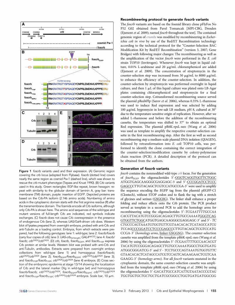
Figure 1 foscrb variants used and their expression. (A) Genomic regioncovering the crb locus (adopted from Flybase). foscrb (dotted line) coversnearly the same region as cosmid Pw-1 (dashed line), which was shown torescue the crbmutant phenotype (Tepass and Knust 1990). (B) Crb variantsused in this study. Green rectangles: EGF-like repeat, brown hexagon: re-peat with similarity to the globular domain of laminin A, gray bar: trans-membrane (TM) domain, purple: insertion of EGFP. Depicted proteins arebased on the Crb-PA isoform (2.146 amino acids). Numbering of aminoacids in the cytoplasmic domain starts with the first arginine residue (R) afterthe transmembrane domain. The fosmids encode all Crb isoforms, althoughonly Crb-PA is shown here. The amino acid sequences of the wild-type andmutant versions of full-length Crb are indicated, red symbols indicateexchanges. (C) foscrb does not cause Crb overexpression in the presenceof endogenous Crb (lane 2), whereas UAS/Gal4-driven crb does. Westernblot of lysates prepared from overnight embryos, probed with anti-Crb andanti-Tubulin as a loading control. Embryos, from which extracts were pre-pared, had the following genotypes: lane 1: wild-type; lane 2: foscrb/foscrb(carry four copies of crb); lane 3: UAS-crbfull-length;GAL4daG32; lane 4: foscrb/foscrb; crbGX24/crbGX24. (D) crb, foscrb, foscrbEGFP, and foscrbY10F expressCrb protein at similar levels. Western blot was probed with anti-Crb andanti-Tubulin, antibodies. Extracts were prepared from overnight embryocollections from wild-type (lane 1) and homozygous foscrb/foscrb;crbGX24/crbGX24 (lane 2); foscrbEGFP/foscrbEGFP; crbGX24/crbGX24 (lane 3);and foscrbY10F/foscrbY10F; crbGX24/crbGX24 (lane 4) embryos. (E) Cross sec-tion of the embryonic epidermis at stage 13/14 showing the localizationof Crb and the lateral marker Dlg in wild-type (wt) and homozygousfoscrb/foscrb; crbGX24/crbGX24, foscrbEGFP/foscrbEGFP; crbGX24/crbGX24
and foscrbY10F/foscrbY10F; crbGX24/crbGX24 embryos. Scale bar, 10 mm.
Volume 3 February 2013 | Crb and Epithelial Morphogenesis | 155
GATCG-39 and 59-TGTAAACCATAACTAGGGGCCAACTTAGTACAAAACATTGAGTTACTCCTTCAGAAGAACTCGTCAAGAAGG-39 (homology arms). The oligonucleotides 59-GACATTGCCATCATTGTAATA-3 and 59-TGTAAACCATAACTAGGGGCCA-39 wereused to amplify the mutated cytoplasmic domains of crumbs fromthe following pre-existing plasmids: foscrbY10F from pBS-8xMyc-crbintra-Y10F (C. Clemens and E. Knust, unpublished data); foscrbY10A frompUAST-8xMyc-crbintra-Y10A (S. Özüyaman and E. Knust, unpublisheddata); foscrbDERLI from pBS-8xMyc-crbintra-DERLI (S. Özüyaman andE. Knust, unpublished data); foscrbY10A, DERLI from pUAST-8xMyc-crbintra-Y10A,DERLI (C. Clemens and E. Knust, unpublished data). Theobtained modification cassettes exchanged the counter-selection cas-settes in the second recombineering step in the different foscrb var-iants, respectively. All foscrb variants were verified by sequencingbefore injection into Drosophila melanogaster.
Generation of transgenic fliesTransgenic flies were generated via the phiC31 integrase mediatedsite-specific integration into attP landing-sites (reviewed in Venkenand Bellen 2007). For the injection and establishment of transgeniclines standard protocols were followed (Bachmann et al. 2008). Allfoscrb variants were integrated into the landing site attP40, of thestock y, v, P(nos-phiC31\int.NLS)X ; P(CaryP)attP40 (Bloomington#25709).
Embryo collection, antibody staining,and cuticle preparationEmbryos were collected on apple juice plates for 2 hr at 25� and thenincubated for 6 hr at 25� or 12 hr at 19�, dechorionated in 50% bleachfor 3 min, and fixed for 20 min in 4% formaldehyde in phosphate-buffered saline/heptane. For heat fixation, dechorionated embryoswere sunk into boiling TTS solution (68 mM NaCl, 0.03% TritonX-100) and then transferred immediately to ice. Devitellinizationwas done in heptane/methanol. Embryos were blocked for 2 hr atroom temperature in PBT (phosphate-buffered saline + 0.1% TritonX-100) + 5% normal horse serum. Embryos were incubated for 2 hr atroom temperature with primary antibodies: rat anti-Crb 2.8, 1:500,(Richard et al. 2006a), mouse anti-Crb-Cq4, 1:300 (Tepass et al. 1990),mouse anti-Discs large (Dlg) 4F3, 1:50 [Developmental StudiesHybridoma Bank (DSHB), 1:50], mouse anti-Armadillo N2 7A2,1:50 (DSHB) (Riggleman et al. 1990), rabbit anti-Stranded at second(Sas, 1:500; kindly provided by E. Organ and D. Cavener), rabbit anti-Canoe (Cno), 1:1.000 (Matsuo et al. 1999, kindly provided byK. Takahashi), rabbit anti-Pyd, 1:5.000 (Djiane et al. 2011, kindlyprovided by Sarah Bray), guinea pig anti-Eyegone (1:1000) (Aldaz et al.2003, kindly provided by Natalia Azpiazu), rabbit anti-phosphomoesin (Cell Signaling Technology, cat. no. 3150, 1:100), mouseanti-alpha-spectrin SA9, 1:25 (DSHB), rabbit anti-GFP (Invitrogen,cat. no. A11122, 1:500). Incubations with the appropriate secondaryantibodies were performed for 1 hr at room temperature: 1:500 forAlexa Fluor 488-, 568-, and 647-conjugated antibodies (Invitrogen).Stained embryos were mounted in glycerin propyl gallate (75% glyc-erol, 50 mg/mL propyl gallate) and visualized using a Zeiss LSM 780NLO confocal microscope with a C-Apochromat 40x/1.2W Corr ob-jective with the correction collar at 0.18 (at this position the brightnessand contrast was enhanced). All images were taken under the samesettings for laser power, PMT gain and offset. Maximal projectionsand merging was performed using Fiji and Adobe Photoshop CS4.Cuticle preparations were performed according to standard proce-dures (Wieschaus and Nüsslein-Volhard 1986).
Viability testAdult flies of the desired genotype were kept on apple juice agar at 25�and removed after 2 hr. The number of embryos on the plate wascounted and the plate was further incubated at 25�. After approxi-mately 48 hr, the number of empty eggshells was determined anddivided by the total number of embryos to determine the viability.This experiment was done three times for each genotype. In summary201, 296, and 351 embryos were collected after 2 hr for WT; 322, 246,and 331 for foscrb/foscrb; crbGX24/crbGX24, 114, 187, and 273 for foscr-bEGFP/foscrbEGFP; crbGX24/crbGX24; and 187, 175, and 169 for foscrbY10F/foscrbY10F; crbGX24/crbGX24.
Preparation of embryonic extracts and western blotFor embryo collection, adult flies of the respective genotype were keptovernight at 25� on apple juice agar plates. Embryos were dechorio-nated for approximately 3 min in 3% bleach and homogenized withBiovortexer (Biospec products) in lysis buffer (50 mM Tris, pH 8.0;150 mM NaCl; 0.5% Triton X-100; 1 mM MgCl2) supplemented withcomplete protease inhibitor cocktail (Roche). The lysate was centri-fuged for 2 min at 4� at 1.900 g and protein concentration was de-termined by standard Bradford biochemistry using Roti-Quant(Roth). The supernatant was analyzed by western blotting using stan-dard procedures. Primary antibodies used were: rat anti-Crb 2.8(1:1.000) (Richard et al. 2006a) and mouse anti-a-Tubulin (1:3.000,Sigma-Aldrich).
RESULTS
foscrb, foscrbEGFP, and foscrbY10F completely rescuecrb-induced embryonic lethalityDrosophila Crb is involved in several processes, such as maintenanceof epithelial cell polarity, regulation of the Hippo pathway, morpho-genesis of photoreceptor cells, and prevention of light-dependent ret-inal degeneration. To better understand which region(s) of thismultidomain protein are required for these different functions, trans-genes encoding the whole genomic region of crb have been designed.It has previously been shown that the cosmid clone Pw-1, containinga genomic region of approximately 32 kb, completely rescues the crbmutant embryonic and adult eye phenotypes [(Tepass et al. 1990);M. Richard and E. Knust, unpublished data]. Beside the ~19-kb tran-scribed region, this clone contains ~9 kb upstream genomic sequenceof the crb locus and ~4 kb downstream (Figure 1A). The fly fosmidclone pFlyfoscrb (no.P52GD2) used here (called foscrb) contains thecomplete transcribed region of the crb locus plus ~7 kb upstream and~5 kb downstream genomic sequence, thus spanning a similar geno-mic region to that contained in cosmid Pw-1 (Figure 1A). A secondvariant, foscrbEGFP, was generated by recombineering, which containsan EGFP tag N-terminal to the fourth laminin A G domain-like repeat(Figure 1B), thus allowing to distinguish the transgene-encoded fromthe endogenous Crb protein. foscrb was the template for several mu-tant variants, which are summarized in Figure 1B. The design of thesevariants was based on previous constructs, which used the GAL4/UASsystem (Klebes and Knust 2000; Wodarz et al. 1995). The deletion ofthe C-terminal amino acids in foscrbDERLI removes the PDZ-bindingmotif, ERLI, which links Crb with Sdt (Bachmann et al. 2001; Honget al. 2001) and DmPar-6 (Kempkens et al. 2006). Two variants,foscrbY10F and foscrbY10A, carry mutations in Tyrosine10 of the cyto-plasmic domain, which is conserved in all Crb variants described sofar (Richard et al. 2006b). This residue is part of a conserved FERM-binding domain (Klebes and Knust 2000), which has been shown tobind the FERM protein Yurt, a negative regulator of Crb (Laprise et al.
156 | S. Klose et al.

2006), and the FERM domain of Ex, an upstream regulator of theHippo pathway (Ling et al. 2010). In addition, a version carrying bothmutations, foscrbY10A,DERLI, was generated (Figure 1B).
Both foscrb and foscrbEGFP rescued crb the loss-of-function muta-tion crbGX24 (Huang et al. 2009) or crb11A22: 87% and 75% of theembryos homozygous for crbGX24 and carrying two copies of thefosmid, i.e., foscrb; crbGX24 or foscrbEGFP; crbGX24, respectively, hatched,which is slightly less than wild-type embryos (95%). Flies with eithergenotype were fertile, did not show any obvious mutant phenotype,and could be kept as homozygous stocks. In contrast, a UAS-transgeneencoding the full-length Crb protein was unable to rescue em-bryonic lethality and could only suppress some aspects of the crbmutant embryonic phenotype (Wodarz et al. 1995). Surprisingly,82% of the embryos with the genotype foscrbY10F; crbGX24 hatchedand gave rise to adult viable and fertile flies. This result is strikingbecause expression of UAS-crbintraY10F, which encodes a proteinconsisting of the transmembrane and the cytoplasmic domain ofCrb, in which Tyr10 was mutated to Phenylalanine, did not rescueembryonic lethality and showed only minor suppression of thecrb mutant embryonic cuticle phenotype upon ubiquitous expres-sion, compared to that of UAS-crbintra (C. Clemens and E. Knust,unpublished data).
It is well established that the amount of Crb protein expressed ina cell is crucial for the maintenance of apicobasal polarity and proper
size of the apical domain (Hamaratoglu et al. 2009; Klebes and Knust2000; Muschalik and Knust 2011; Wodarz et al. 1995). Therefore, weanalyzed the levels of Crb protein in flies carrying different copynumbers of the endogenous and/or fosmid-encoded crb gene. Surpris-ingly, the presence of four copies of crb did not increase the overallCrb protein levels. In the absence of endogenous crb, each of the threefosmids that rescued embryonic lethality expressed comparableamounts of Crb protein when present in two copies (Figure 1, Cand D).
foscrb and foscrbEGFP show wild-type expressionpattern and subcellular localization of the Crb proteinTo analyze the expression pattern of foscrb- and foscrbEGFP-encodedCrb protein in embryos in the absence of endogenous crb, we stainedfoscrb; crbGX24, foscrbEGFP; crbGX24 and foscrbY10F; crbGX24 embryoswith different markers at different developmental stages. At all de-velopmental stages, fosCrb protein in these embryos was expressed inepithelia of ectodermal origin as in wild-type, i.e., in the epidermis, theamnioserosa, the tracheae, the salivary gland, the hindgut and theMalpighian tubules (Figure 2 and data not shown). As revealed bythe apical marker Sas, epithelia in embryos carrying one of thesefosmids maintain proper apicobasal polarity (Figure 2, C92E9). Inembryos with these genotypes, fosCrb is localized apical to the baso-lateral marker Dlg (Figure 1E and data not shown).
Figure 2 foscrb variants completely rescuing crbmutant embryos. Lateral view of stage 13/14whole-mount embryos stained for Crb and Sas(right). (A) Wild-type. (B) crbGX24. (C) foscrb;crbGX24. (D) foscrbEGFP; crbGX24. (E) foscrbY10F;crbGX24. Arrows in A9, C9, D9, and E9 indicatesalivary glands, and arrowheads in A, C, D, Ethe tracheae. In B, the embryo negative for Crbstaining is outlined with the dotted line. Inset inB9 is a maximal projection of a stack through theremnant tracheal system. The arrowheads in theinset in B9 indicate discontinuities in the trans-verse connective (TC) and dorsal trunk (DT)branches. Anterior is to the left, dorsal up. Scalebar, 100 mm.
Volume 3 February 2013 | Crb and Epithelial Morphogenesis | 157

foscrbY10A, foscrbDERLI, and foscrbY10A, DERLI do notrescue crb-induced embryonic lethalitycrb mutant embryos lack a continuous cuticle, and only grains ofcuticle can be detected (compare Figure 3, A and B) (Jürgens et al.1984; Tepass and Knust 1990). crb mutant embryos with transgenesthat either carried a mutated FERM-binding domain (foscrbY10A;crbGX24), lacked the PDZ-binding motif (foscrbDERLI; crbGX24), or car-ried both mutations (foscrbY10A,DERLI; crbGX24) did not hatch. Thedifferent transgenes suppressed the crb mutant phenotype to differentdegrees. In comparison with foscrb, which completely rescued thecuticle phenotype (Figure 3C), crb mutant embryos carrying twocopies of foscrbY10A form continuous anterior and ventral cuticle withrather fully developed denticle belts (Figure 3D). This result is strikingin view of previous observations showing that Gal4-mediated expres-sion of UAS-crbintraY10A completely failed to suppress the crb mutantcuticle phenotype (Klebes and Knust 2000). The phenotype offoscrbY10A; crbGX24 embryos resembles that of wild-type embryos over-expressing UAS-crbintra with a ubiquitously expressed GAL4 line(compare Figure 3, D and E) (Wodarz et al. 1995) and is characteristicfor embryos with impaired germ band retraction and dorsal closure.crbmutant embryos carrying foscrbDERLI developed only small patchesof continuous cuticle (Figure 3F) reminiscent to the phenotype of crbmutant embryos with a strong intermediate phenotype (Tepass andKnust 1990) but more severe than a crbmutant embryo expressing themembrane-bound intracellular domain (UAS-crbintra) using an ubiq-uitously expressed GAL4 line (Figure 3G) (Klebes and Knust 2000).The phenotype of crb mutant embryos with one copy of foscrbY10Aand one copy of foscrbDERLI was the same as that of crb mutantembryos with two copies of foscrbY10A (compare Figure 3, D andH). crb mutant embryos carrying foscrbY10A,DERLI did not developany continuous cuticle and resembled crb embryos with a strong lossof function allele (data not shown).
Loss of crb differentially affects various ectodermally derived em-bryonic epithelia. Although some epithelia, such as the epidermis,almost completely die, others, such as the tracheal system, partiallysurvive and their cells form vesicles that maintain epithelial cell po-larity. Some organs, such as the hindgut, are nearly unaffected (Tepass
and Knust 1990). To analyze in more detail the effects of the expres-sion of the different foscrb variants during embryonic development,we stained these embryos for Crb and the apical marker Sas at dif-ferent developmental stages.
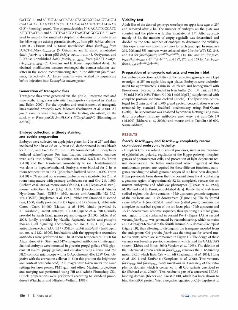
Epithelial polarity and integrity in foscrbY10A; crbGX24 was normalin the epidermis (compare Figure 4, A and B), the fore- and hindgut,the salivary glands (compare Figure 4, A9 and B9, arrows), and theMalpighian tubules until the end of embryogenesis. In contrast, theamnioserosa exhibited defects from stage 13 onwards (not shown) andwas completely lost at stage 15. This defect was accompanied byimpaired germ band retraction, head involution (arrowhead in Figure4C9), and dorsal closure. In fact, the leading edge of the epidermisappears uneven and wiggling, instead of straight as observed duringdorsal closure in wild-type embryos (highlighted by a dotted line inFigure 4C). In addition, segments exhibited variable widths. For ex-ample, the width of segments T3, A1, and A2 is largely unequal,contrary to what is observed in wild-type embryos (Figure 4C9). Thisfinding suggests defects in the tension of the actin cable in the leadingedge cells. In contrast, the hindgut (Figure 4C, asterisk) and the Mal-pighian tubules (Figure 4C9, inset) were maintained. Interruption ofCrb staining in some transverse connective branches manifesteddefects in the tracheal system already at stage 13 (Figure 4B, arrow-head in inset; see below). These discontinuities were also observedalong the dorsal trunk at later stages (not shown). In foscrbDERLI;crbGX24 embryos at stage 13, nearly no Crb protein was detected(Figure 4D). Sas expression indicated the presence of remnants ofthe dorsal epidermis at this stage, whereas the tracheal tubes werefragmented (Figure 4D9, inset). At the end of embryogenesis, onlythe hindgut (Figure 4E, asterisk), the Malpighian tubules (Figure4E9, inset) and vesicular structures, probably remnants of the tracheae,were visible (Figure 4E9). The phenotype of foscrbY10A,DERLI; crbGX24
embryos was similar to that of crb mutant embryos carrying thefoscrbDERLI transgene (Figure 4, F2G9).
As previously shown, cells of the developing epidermis of crbmutant embryos fail to form a proper zonula adherens (ZA) (Figure5, A and A9) (Grawe et al. 1996; Tepass 1996). To analyze the effectsof the different transgenes on the development of the ZA we stained
Figure 3 foscrb variants and their ability torescue the crb mutant phenotype. Cuticle ofDrosophila embryos (lateral views) with thefollowing genotypes: (A) wild-type (WT), (B)crbGX24, (C) foscrb; crbGX24, (D) foscrbY10A;crbGX24, (E) Wild-type embryo overexpressingUAS-crb8xMyc-intra under the control ofa strong, ubiquitously expressed Gal4 line,GAL4daG32, (F) foscrbΔERLI; crbGX24, (G)crb11A22 mutant embryo overexpressingUAS-crbMyc-intra under the control of a ubiqui-tously expressed Gal4 line, GAL4385.3, and (H)foscrbY10A/foscrbΔERLI; crbGX24. Anterior is tothe left, dorsal up.
158 | S. Klose et al.
stage 11 embryos with antibodies against Armadillo, the Drosophilaortholog of b-catenin. Both foscrb and foscrbY10A restored wild-typeArmadillo staining, suggesting a normal formation of the ZA (Figure5, B, B9, C, and C9), whereas foscrbDERLI completely failed to restorea continuous adhesion belt (Figure 5D9).
Taken together, a Crb protein lacking the PDZ-binding motif isunable to maintain epithelial polarity from early stages onwards. Incontrast, the Y10A mutation in the FERM-binding domain had noeffect on epithelial polarity. Embryos expressing this mutant Crbprotein showed defects in fusion of tracheal tubes and in the
development of the amnioserosa, resulting in defective dorsal closure,head involution and germ band retraction.
Role of the PDZ-binding and the FERM-binding domainduring tracheal morphogenesisThe Drosophila tracheal system originates from groups of about 40ectodermal cells each, called tracheal placodes. They form on thelateral side of the embryo from the second thoracic segment to theeighth abdominal segment. Invagination of epithelial cells of the pla-code is initiated by localized apical cell constriction, which is preceded
Figure 4 foscrb variants and their ability to res-cue the crb mutant phenotype. Lateral view ofwhole embryos stained for Crb (left column)and the apical marker Sas (right column). (A, A9)Wild-type embryo at stage 13/14. All ectoder-mally derived epithelia show apical localizationof Crb and Sas. (B2C9) foscrbY10A; crbGX24 em-bryos at stage 13 (B, B9) and 15/16 (C, C9). Thearrowhead in the inset in (B) indicates a disconti-nuity in Crb staining along the transverse connec-tive branch. The dotted line in (C) delineates theleading edge of the epidermis. Arrowhead in (C9)indicates the head that fails to undergo involu-tion. In (C9), the third thoracic segment (T3) andthe first two abdominal segments (A1, A2) aremarked to highlight their variable width. (D-E9)foscrbΔERLI; crbGX24 embryo at stage 13 (D, D9)and stage 15/16 (E, E9). (F-G9) foscrbY10A,ΔERLI;crbGX24 embryo at stage 13 (F, F9) and stage15/16 embryo (G, G9). Arrowheads in A, B, D9,and F9 indicate the tracheae, insets in B, D9, andF9 show tracheal hemisegments. Insets in C9, E9,and G9 show a Malpighian tubule. � in C, E, andG point to the hindgut. Arrows in A9 and B9 pointto the salivary gland. Anterior is to the left, dorsalup. Scale bar, 100 mm.
Volume 3 February 2013 | Crb and Epithelial Morphogenesis | 159

by apical enrichment of Crb and actinomyosin (Brodu and Casanova2006; Letizia et al. 2011; Llimargas and Casanova 1999). At early stage11, tracheal cells are internalized and undergo their last round ofpostblastodermal division. Together, apical constriction, cell rear-rangements, and oriented cell divisions are important for organizedinvagination of tracheal cells (Brodu and Casanova 2006; Nishimuraet al. 2007). This results in the formation of the segmentally arrangedtracheal sacs or pits at stage 11, each composed of about 80 cells,which are organized in a polarized epithelial monolayer. At stage12, expression of the chemoattractant fibroblast growth factor inducesthe formation of primary branches in the tracheal sac in a stereotypicpattern. These branches elongate by cell intercalation, a process thatinvolves changing neighbors and remodeling of adhesive contacts, toform long, thin tubes. At stage 15, fusion of branches is initiated,which finally leads to an elaborate network of interconnected trachealtubes (reviewed in Affolter and Caussinus 2008).
Tracheal development was completely normal in foscrb;crbGX24,foscrbEGFP;crbGX24 and foscrbY10F;crbGX24 embryos (Figure 2, C2E9,Figure 6, A and B9, and data not shown). crb mutant embryos, whichdo not express any Crb protein (Figure 2B, Figure 6, C and D),showed aberrations already in the initial invagination process at earlystage 11, as revealed by the mislocalization of the ZA marker Cno, theDrosophila ortholog of mammalian Afadin (Figure 6, C9 and D9).Nevertheless, some internalization occurred, resulting in the forma-tion of an irregular tracheal sac (Figure 6D9). At stage 13/14, thetracheae showed abnormal branching, discontinuities in differentbranches, or lack of a discernible single lumen later on (Figure 2B9,inset). At the end of embryogenesis, a complete breakdown of thetracheal system was observed (data not shown) (Tepass and Knust1990).
Strikingly, crb mutant embryos expressing a Crb variant witha mutated FERM-binding domain (foscrbY10A; crbGX24) showed nearlynormal accumulation of Crb in the placode and normal apical con-striction and invagination (Figure 6, E and F9). However, at stage 13,some transverse connective tubes showed discontinuities in Crb stain-ing as in crb mutants (Figure 4B, inset). In later stages, gaps also wereobserved in the dorsal trunk (data not shown). In contrast, fosmidsencoding a Crb protein, which lacked the PDZ-binding motif, com-pletely failed to rescue the crb mutant phenotype in the tracheae(Figure 4, D9 and F9, Figure 6, G2H9). Similar to crbmutant embryos,development of the trachea in foscrbDERLI; crbGX24 and foscrbY10A,DERLI;crbGX24 mutant embryos proceeded to stage 13, at which the dorsal andvisceral branches as well as the posterior lateral trunk could be
identified, (Figure 4D9 and F9, insets). Later on, the complete trachealsystem disintegrated (Figure 4, E9 and G9).
To further dissect the early steps in the different mutant back-grounds, we used additional markers to follow the invaginationprocess. One of the earliest events during invagination is the apicalaccumulation of the phosphorylated form of Moesin, phospho-Moesin, or pMoe (Letizia et al. 2011). Moesin is the single Drosophilamember of the ERM (ezrin-, radixin-, moesin) protein family(McCartney and Fehon 1996). ERM proteins act as linkers betweenmembrane proteins and the actin cytoskeleton and are crucial inorganizing distinct membrane domains (Fehon et al. 2010). ApicalpMoe, together with some apical enrichment of a-spectrin and baso-lateral localization of Dlg, highlights the apicobasal polarization ofcells in the tracheal placode, both in wild-type and in crb embryoscarrying foscrb, foscrbEGFP, or foscrbY10F (Figure 7, A2C, and data notshown). Loss of polarized pMoe, a-spectrin and Dlg expression in-dicated loss of cell polarity in the placodes of crb mutant embryos(Figure 7, D2F) (Letizia et al. 2011). foscrbY10A;crbGX24 mutant em-bryos showed reduced levels of pMoe, diffuse staining of a-spectrin,and mostly baso-lateral, but also diffuse Dlg staining (Figure 7, G2I).These data indicate that the polarity defects of crb mutants were notcompletely rescued by the CrbY10A variant. Nevertheless, the placodesunderwent normal invagination and elongation. In contrast,foscrbDERLI; crbGX24 mutant embryos failed to accumulate pMoe anddid not show polarized distribution of a-spectrin and Dlg, yet someuncoordinated internalization of cells occurred, resulting in a tiny andirregular lumen (Figure 7, J2L).
Taken together, expression of CrbY10A with a mutated FERM-binding domain allowed proper invagination of the tracheal sac andnormal elongation of the branches, but was not sufficient for properfusion of the tracheal branches at later stages. In contrast, in theabsence of the PDZ-binding motif, Crb is not stabilized by Sdt, result-ing in defects in apical constriction, apico-basal polarity, invaginationand outgrowth of the tracheal system.
Role of the PDZ-binding and the FERM-binding domainduring salivary gland morphogenesisDevelopment of the salivary glands is initiated by the formation of twoplacodes of approximately 100 cells each on both sides of the ventralpart of parasegment two. Invagination is initiated in an orchestratedmanner at stage 11, with dorsal2posterior cells constricting theirapical surfaces first, followed by dorsal2anterior, ventral2 anterior,and ventral2posterior cells (reviewed in Maruyama and Andrew
Figure 5 foscrb variants and their abilityto rescue the ZA defects of crb mutantembryos. Optical sections taken from theepidermis of stage 11 embryos of the fol-lowing genotypes: crbGX24 (A, A9), foscrb;crbGX24 (B, B9), foscrbY10A; crbGX24 (C, C9),and forscbDERLI; crbGX24 (D, D9). The em-bryos were stained with Crb (A, B, C, D)and Armadillo (Arm; A9, B9, C9, D9), a com-ponent of the zonula adherens. Scale bar,10 mm.
160 | S. Klose et al.

2012). In wild-type embryos, Crb accumulates in the salivary glandsplacode before invagination (Myat and Andrew 2002). A similar ac-cumulation of Crb and the ZA markers Cno and Polychaetoid (Junget al. 2006) as well as normal invagination of cells of the placode wasobserved in foscrb;crbGX24, foscrbEGFP;crbGX24 and foscrbY10F;crbGX24
embryos (Figure 8, A and B, data not shown). In all cases, properlyelongated salivary glands developed (Figure 2, C92E9 arrows, data notshown). In crb mutant embryos the expression of eyegone, a markerfor the anlage of the salivary glands (Jones et al. 1998), is normal,demonstrating that the salivary gland placodes are properly specified.However, cells fail to undergo apical constriction (Figure 8, C and D)and invagination does not occur. Interestingly, although foscrbY10Apartially restored accumulation of Crb in the placode (Figure 8E),apical constriction was less well organized as revealed by Cno staining(Figure 8E9). Nevertheless, invagination and elongation proceeded ina similar way as in wild-type, and CrbY10A protein was properlylocalized apically (Figure 8F and Figure 4B). In contrast, foscrbDERLI;crbGX24 (Figure 8, G2H) as well as foscrbY10A,DERLI; crbGX24 (data notshown) mutant embryos did not show any apical constriction nor
Figure 6 foscrb variants and their ability to rescue tracheal develop-ment of crb mutant embryos. Tracheal sacs of the second and thirdhemisegments of stage 11/12 embryos, stained for Crb and Cno, withthe following genotypes: (A, A9, B, B9) foscrb; crbGX24, (C, C9, D, D9)crbGX24, (E, E9, F, F9) foscrbY10A; crbGX24, (G, G9) foscrbΔERLI; crbGX24,and (H, H9) foscrbY10A,ΔERLI; crbGX24. The images shown in (A), (A9), (C),(C9), (E), and (E9) are single planes through the spiracular branch, thetube that connects the sac to the exterior. This plane of section allowsone to detect whether a normal tube is formed and whether the tubedevelops normal apical-basal polarity. In (C9), the branch in the thirdsegment is encircled by a dotted line. Scale bar, 10 mm.
Figure 7 foscrb variants and their ability to rescue the early stages oftracheal development of crb mutant embryos. Spiracular branch of theembryonic tracheae at stage 11, stained for Crb, pMoe, a-spectrin(a-Spec), and Dlg in the following genotypes: (A, A9, B, C) foscrb;crbGX24, (D, D9, E, F) crbGX24, (G, G9, H, I) foscrbY10A; crbGX24 , and(J, J9, K, L) foscrbΔERLI; crbGX24. The arrowheads point to the lumenobserved in the reconstructed stacks of the spiracular branch. Scalebar: 10 mm.
Volume 3 February 2013 | Crb and Epithelial Morphogenesis | 161

invagination. Similarly, and in contrast to recently published results(Roper 2012), we observed that salivary glands of crb8F105 mutantembryos, which carry a premature stop codon that removes the last23 amino acids of Crb, including the -ERLI motif (Wodarz et al.1993), do not accumulate Crb and fail to invaginate (data not shown).
To summarize, foscrbY10A allowed normal invagination and mor-phogenesis of the salivary gland in the absence of endogenous Crb,despite uncoordinated apical constriction at early stages. In contrast,the PDZ-binding motif of Crb is essential for all steps of salivary glanddevelopment that require Crb.
DISCUSSIONTwo functional domains have been characterized in the cytoplasmictail of Crb: the C-terminal PDZ-binding motif -ERLI and thejuxtamembrane FERM-binding domain. Using fosmid-based trans-genes, we could confirm previous results showing that the PDZ-binding domain is essential for the maintenance of epithelial polarityin the early embryo and for proper invagination of cells of the trachealand salivary placodes. The PDZ binding domain is required forrecruiting Crb to the apical membrane by binding to Sdt, and embryos
expressing this truncated version of Crb develop the same phenotypeas those lacking sdt function (Bachmann et al. 2001; Hong et al. 2001;Krahn et al. 2010). Similarly, the crb allele crb8F105, which lacks the-ERLI motif, behaves like a null allele in the embryo (Wodarz et al.1993). These results are in line with the observation that UAS-crbintraDERLI, when ubiquitously expressed in a crb mutant embryo,did not suppress the polarity phenotype in crb mutant embryosdespite the presence of the FERM domain (Klebes and Knust 2000;Letizia et al. 2011). This finding supports the view that the PDZ-binding domain is essential for maintenance of apicobasal polarityby stabilizing the Crb-associated complex members, including Crbitself, at the plasma membrane. However, since apical localizationof Crb depends on its binding to Sdt via its PDZ-domain, no con-clusions can be drawn for possible function(s) of the residual part ofthe cytoplasmic domain under these experimental conditions.
As suggested by S2 cell culture capping assays, the truncatedcytoplasmic domain encoded by crb8F105 still carries an intact FERM-binding domain, which could recruit Moesin and bH-spectrin, butfailed to do so after introducing a Tyr10 or Arg7 mutation(Medina et al. 2002). Therefore, we assume that the Y10A mutationin foscrbY10A abolishes the function of the FERM-binding domain ofCrb also in vivo and conclude that the FERM-binding domain of Crbis of little importance for epithelial polarity at early embryonic stages.However, foscrbY10A ; crbGX24 embryos showed later defects in dorsalclosure and germ band retraction. Structural analyses have revealedthat Tyr10 in the nonpolar region of the intercellular adhesion mole-cule (ICAM)-2 is crucial for the interaction with the FERM domain ofRadixin. An exchange of Tyr10 to Ala in the cytoplasmic tail of ICAM-2 resulted in a 16-fold reduction in its binding affinity to Radixin(Hamada et al. 2003). Therefore, we speculate that the proteinencoded by foscrbY10A fails to interact with its FERM-domain contain-ing partner(s). One of its binding partners is Ex, a regulator of theHippo pathway. Results on the effect of a similarly engineered crbgene, which carried several mutations in the FERM-binding domain(crbY10AP12AE16A), are controversial. Although Ling et al. showed baso-lateral mislocalisation of Ex, Robinson et al. showed elevated level ofEx in mutant wing disc clones (Ling et al. 2010; Robinson et al. 2010).Because the Hippo pathway has not been shown to act in the embryo,a more likely partner of the FERM-binding domain of Crb in theembryo is Yurt (Laprise et al. 2006). In fact, phenotypes observed infoscrbY10A;crb embryos, e.g., defects in germ band retraction and dorsalclosure, are similar to those of embryos lacking zygotic yurt activity(Hoover and Bryant 2002). Yurt binds to the FERM-binding domainof Crb and shows apical colocalization with Crb from stage 13onwards. Complete removal of Yurt results in apical enrichment ofCrb and an expansion of the apical surface (Laprise et al. 2006). Weobserved slightly lower levels of Crb and higher levels of Sas infoscrbY10A; crbGX24 embryos at later stages (Figure 4C and C9). How-ever, more detailed analysis is required to find out whether the latephenotypes of foscrbY10A; crbGX24 and yurt mutant embryos have thesame cell biological basis. Interestingly, some aspects of the mutantphenotype of foscrbY10A;crbGX24 embryos, such as un-coordinated api-cal constriction, resemble those described for human colon cancerepithelial DLD-1 cells upon RNAi-mediated knock-down of Lulu,the mammalian ortholog of Yurt. These cells fail to organize the apicalcircumferential actinomyosin belt and exhibited discontinuities inb-catenin staining (Nakajima and Tanoue 2011), comparable withour observations of foscrbY10A;crbGX24 embryos stained with the junc-tional marker Canoe (see Figure 8E9).
Despite a defective FERM-binding domain, Crb proteins expressedin foscrbY10A; crbGX24 embryos promote tracheal and salivary gland
Figure 8 foscrb variants and their ability to rescue the early stages ofsalivary gland development of crbmutant embryos. Embryonic salivarygland placodes stained for Crb (A, B, C, D, E, F, G, H), Cno (A9, C9, E9,G9) Polychaetoid (Pyd; B, D, F, H, magenta) and Eyegone (Eyg; B, D, H,blue). (A), (A9), and (B) foscrb; crbGX24 early-stage 11 embryo. (C), (C9),and (D) crbGX24 stage 11 embryo. In (C9), the placode is highlighted bythe dotted line. (E), (E9), and (F) foscrbY10A; crbGX24 early-stage 11 (E,E9) and stage 12 (F) embryos. (G), (G9), and (H) foscrbΔERLI; crbGX24
early-stage 11 embryo. Anti-Eyg was used in every staining to identifythe salivary gland placode but only shown in B, D, F, H. Scale bar in(A),10 mm, applies to all images except for F (5 mm).
162 | S. Klose et al.

invagination, although these processes occur in a less-coordinatedmanner, possibly due to reduced pMoe recruitment. In contrast, over-expression of UAS-crbintraY10A in crb mutant embryos did not rescuethe tracheal invagination defect of crb mutant embryos, which wastraced back to a failure in pMoe recruitment (Letizia et al. 2011). Thisdiscrepancy could be the result of a dominant-negative activity of theUAS-encoded protein due to overexpression, by which the normalbinding partner of the FERM-binding domain of Crb is outcompeted,leading to a delayed and uncoordinated invagination. Currently wecannot explain the discontinuities in Crb staining observed in sometracheal branches in foscrbY10A; crbGX24 embryos. Fusion of trachealbranches is a complex process, which requires, among others, forma-tion of filopodia, DE-cadherin-mediated cell-cell contact and regula-tion of associated F-actin structures (Lee et al. 2003; Lee and Kolodziej2002; Tanaka-Matakatsu et al. 1996). Although not studied yet, any ofthese processes could require ERM-protein(s), the function of whichmay depend on an intact FERM-binding domain of Crb.
Strikingly, foscrbY10F rescues lethality of crb mutant embryos, sug-gesting that the phosphorylation of Tyr10 (as predicted by NetPhos) isnot important for viability, but we cannot exclude subtle defects, suchas modified dynamics of internalization. Nevertheless, this finding issurprising, given the observation that UAS-crbintraY10F, when ubiqui-tously expressed in a crb mutant embryo, suppressed the mutantphenotype much less than UAS-crbintra, which encodes the wild-typecytoplasmic domain (C. Clemens and E. Knust, unpublished data).
Our results also revealed differential requirement of Crb duringtracheal and salivary gland invagination. Although cells of the trachealanlage invaginate to some extent in crb mutant embryos, Crb is ab-solutely essential for polarity, apical constriction, invagination, andsurvival of cells of the salivary gland placode (Tepass and Knust1990; Xu et al. 2008). In the salivary gland, the FERM-binding domainis necessary for coordinated apical constriction, organized localizationof the junctional markers Cno and Pyd, and invagination movementsbut dispensable for internalization of cells and correct elongation ofthe tube. Our data do not support the function of a putative negativeregulator acting via the FERM-binding because we did not observegreater Crb levels at later stages nor an expanded apical domainattributed to overexpression of UAS-Crb (Myat and Andrew 2002;Wodarz et al. 1995).
The presence of both a FERM binding- and a PDZ binding-motif isnot uncommon in transmembrane proteins and has been described,among others, for ICAM-2, syndecans (Bass and Humphries 2002;Kwon et al. 2012), and the immunoglobulin-like, Ca2+-independentcell2cell adhesion molecule nectin (Dudak et al. 2011; Ishiuchi andTakeichi 2012). These proteins have short cytoplasmic domains, inwhich a nonpolar region, flanked on both sides by basic regions, con-tains a conserved tyrosine residue at position 10. The Tyr residue in theputative FERM binding domains of syndecan-3 can be phosphorylatedin vitro (Asundi and Carey 1997). In syndecan-2, Tyr10 and another,more C-terminal tyrosine residue is phosphorylated by the EphB2 re-ceptor tyrosine kinase, and this phosphorylation is essential for cluster-ing of syndecan-2 and spine formation in hippocampal neurons (Ethellet al. 2001). In contrast, exchange of Tyr10 in syndecan-1 by phenylal-anine had no effect on the association of syndecan-1 with the actincytoskeleton (Carey et al. 1996), suggesting that the functional impor-tance of phosphorylation may be syndecan and/or cell-type specific.
Taken together, although the Gal4/UAS system provides aninvaluable tool to analyze gene functions, fosmid- or BAC-basedtransgenes combined with the recombineering technology will be ourpreferred approach for in depth structure-function analyses ofproteins in an in vivo system.
ACKNOWLEDGMENTSWe acknowledge Pavel Tomancak and Radoslaw Ejsmont for pro-viding pFlyFos vector foscrb, the TransgeneOmics facility for pro-viding tools and advice for the recombineering, Sven Ssykor forinjection of Drosophila embryos, and Catrin Hälsig for technical sup-port. We thank the Bloomington Drosophila Stock Center for flystocks, and Kuniaki Takahashi, Sarah Bray, Natalia Azpiazu, andthe Developmental Studies Hybridoma Bank for antibodies. We thankNadine Muschalik for many discussions throughout the work andShirin Pocha for critical reading of the manuscript. This work wassupported by the Max-Planck Society (MPG).
LITERATURE CITEDAffolter, M., and E. Caussinus, 2008 Tracheal branching morphogenesis in
Drosophila: new insights into cell behaviour and organ architecture.Development 135: 2055–2064.
Affolter, M., R. Zeller, and E. Caussinus, 2009 Tissue remodelling throughbranching morphogenesis. Nat Rev Cell and Mol Biol 10: 831–842.
Aldaz, S., G. Morata, and N. Azpiazu, 2003 The Pax-homeobox gene eye-gone is involved in the subdivision of the thorax of Drosophila. Devel-opment 130: 4473–4482.
Andrew, D. J., and A. J. Ewald, 2010 Morphogenesis of epithelial tubes:Insights into tube formation, elongation, and elaboration. Dev. Biol. 341:34–55.
Asundi, V. K., and D. J. Carey, 1997 Phosphorylation of recombinant N-syndecan (syndecan 3) core protein. Biochem. Biophys. Res. Commun.240: 502–506.
Bachmann, A., M. Schneider, F. Grawe, E. Theilenberg, and E. Knust,2001 Drosophila Stardust is a partner of Crumbs in the control ofepithelial cell polarity. Nature 414: 638–643.
Bachmann, A., F. Grawe, K. Johnson, and E. Knust, 2008 Drosophila Lin-7 isa component of the Crumbs complex in epithelia and photoreceptor cells andprevents light-induced retinal degeneration. Eur. J. Cell Biol. 87: 123–136.
Baer, M. M., H. Chanut-Delalande, and M. Affolter, 2009 Cellular andmolecular mechanisms underlying the formation of biological tubes.Curr. Top. Dev. Biol. 89: 137–162.
Bass, M. D., and M. J. Humphries, 2002 Cytoplasmic interactions of syndecan-4orchestrate adhesion receptor and growth factor receptor signalling. Biochem.J. 368: 1–15.
Bazellieres, E., E. Assemat, J. P. Arsanto, A. Le Bivic, and D. Massey-Harroche,2009 Crumbs proteins in epithelial morphogenesis. Front. Biosci. 14:2149–2169.
Boggiano, J. C., and R. G. Fehon, 2012 Growth control by committee: in-tercellular junctions, cell polarity, and the cytoskeleton regulate Hipposignaling. Dev. Cell 22: 695–702.
Brodu, V., and J. Casanova, 2006 The RhoGAP crossveinless-c links trachealessand EGFR signaling to cell shape remodeling in Drosophila tracheal invagi-nation. Genes Dev. 20: 1817–1828.
Bulgakova, N. A., and E. Knust, 2009 The Crumbs complex. J. Cell Sci. 122:2587–2596.
Carey, D. J., K. M. Bendt, and R. C. Stahl, 1996 The cytoplasmic domain ofsyndecan-1 is required for cytoskeleton association but not detergentinsolubility. Identification of essential cytoplasmic domain residues. J.Biol. Chem. 271: 15253–15260.
Ciotta, G., H. Hofemeister, M. Maresca, J. Fu, M. Sarov et al.,2011 Recombineering BAC transgenes for protein tagging. Methods 53:113–119.
Denholm, B., and H. Skaer, 2009 Bringing together components of the flyrenal system. Curr. Opin. Genet. Dev. 19: 526–532.
Djiane, A., H. Shimizu, M. Wilkin, S. Mazleyrat, M. D. Jennings et al.,2011 Su(dx) E3 ubiquitin ligase-dependent and -independent functions ofpolychaetoid, the Drosophila ZO-1 homologue. J. Cell Biol. 192: 189–200.
Dudak, A., J. Kim, B. Cheong, H. J. Federoff, and S. T. Lim,2011 Membrane palmitoylated proteins regulate trafficking and pro-cessing of nectins. Eur. J. Cell Biol. 90: 365–375.
Volume 3 February 2013 | Crb and Epithelial Morphogenesis | 163

Ejsmont, R. K., M. Sarov, S. Winkler, K. A. Lipinski, and P. Tomancak,2009 A toolkit for high-throughput, cross-species gene engineering inDrosophila. Nat. Methods 6: 435–437.
Ejsmont, R. K., P. Ahlfeld, A. Pozniakovsky, A. F. Stewart, P. Tomancak et al.,2011 Recombination-mediated genetic engineering of large genomicDNA transgenes. Methods Mol. Biol. 772: 445–458.
Elliott, D. A., and A. H. Brand, 2008 The GAL4 system: a versatile systemfor the expression of genes. Methods Mol. Biol. 420: 79–95.
Ethell, I. M., F. Irie, M. S. Kalo, J. R. Couchman, E. B. Pasquale et al.,2001 EphB/syndecan-2 signaling in dendritic spine morphogenesis.Neuron 31: 1001–1013.
Fehon, R. G., A. I. McClatchey, and A. Bretscher, 2010 Organizing the cellcortex: the role of ERM proteins. Nat Rev Cell and Mol Biol 11: 276–287.
Genevet, A., and N. Tapon, 2011 The Hippo pathway and apico-basal cellpolarity. Biochem. J. 436: 213–224.
Grawe, F., A. Wodarz, B. Lee, E. Knust, and H. Skaer, 1996 The Drosophilagenes crumbs and stardust are involved in the biogenesis of adherensjunctions. Development 122: 951–959.
Hamada, K., T. Shimizu, S. Yonemura, S. Tsukita, and T. Hakoshima,2003 Structural basis of adhesion-molecule recognition by ERM pro-teins revealed by the crystal structure of the radixin-ICAM-2 complex.EMBO J. 22: 502–514.
Hamaratoglu, F., K. Gajewski, L. Sansores-Garcia, C. Morrison, C. Tao et al.,2009 The Hippo tumor-suppressor pathway regulates apical-domainsize in parallel to tissue growth. J. Cell Sci. 122: 2351–2359.
Hong, Y., B. Stronach, N. Perrimon, L. Y. Jan, and Y. N. Jan,2001 Drosophila Stardust interacts with Crumbs to control polarity ofepithelia but not neuroblasts. Nature 414: 634–638.
Hoover, K. B., and P. J. Bryant, 2002 Drosophila Yurt is a new protein-4.1-like protein required for epithelial morphogenesis. Dev. Genes Evol. 212:230–238.
Huang, J., W. Zhou, W. Dong, A. M. Watson, and Y. Hong, 2009 Directed,efficient, and versatile modifications of the Drosophila genome by geno-mic engineering. Proc. Natl. Acad. Sci. USA 106: 8284–8289.
Ishiuchi, T., and M. Takeichi, 2012 Nectins localize Willin to cell-celljunctions. Genes Cells 17: 387–397.
Jones, N. A., Y. M. Kuo, Y. H. Sun, and S. K. Beckendorf, 1998 The Dro-sophila Pax gene eye gone is required for embryonic salivary duct de-velopment. Development 125: 4163–4174.
Jung, A. C., C. Ribeiro, L. Michaut, U. Certa, and M. Affolter,2006 Polychaetoid/ZO-1 is required for cell specification and rear-rangement during Drosophila tracheal morphogenesis. Curr. Biol. 16:1224–1231.
Jürgens, G., E. Wieschaus, C. Nüsslein-Volhard, and H. Kluding,1984 Mutations affecting the pattern of the larval cuticle of Drosophilamelanogaster. II. Zygotic loci on the third chromosome. Rouxs Arch. Dev.Biol. 193: 283–295.
Kempkens, Ö., E. Médina, G. Fernandez-Ballester, S. Özüyaman, A. Le Bivicet al., 2006 Computer modelling in combination with in vitro studiesreveals similar binding affinities of Drosophila Crumbs for the PDZ do-mains of Stardust and DmPar-6. Eur. J. Cell Biol. 85: 753–767.
Kerman, B. E., A. M. Cheshire, and D. J. Andrew, 2006 From fate tofunction: the Drosophila trachea and salivary gland as models for tubu-logenesis. Differentiation 74: 326–348.
Klebes, A., and E. Knust, 2000 A conserved motif in Crumbs is required forE-cadherin localisation and zonula adherens formation in Drosophila.Curr. Biol. 10: 76–85.
Krahn, M. P., J. Buckers, L. Kastrup, and A. Wodarz, 2010 Formation ofa Bazooka-Stardust complex is essential for plasma membrane polarity inepithelia. J. Cell Biol. 190: 751–760.
Kwon, M. J., B. Jang, J. Y. Yi, I. O. Han, and E. S. Oh, 2012 Syndecans playdual roles as cell adhesion receptors and docking receptors. FEBS Lett.586: 2207–2211.
Laprise, P., S. Beronja, N. F. Silva-Gagliardi, M. Pellikka, A. M. Jensen et al.,2006 The FERM protein Yurt is a negative regulatory component of theCrumbs complex that controls epithelial polarity and apical membranesize. Dev. Cell 11: 363–374.
Lee, S., and P. A. Kolodziej, 2002 The plakin Short Stop and the RhoAGTPase are required for E-cadherin-dependent apical surface remodelingduring tracheal tube fusion. Development 129: 1509–1520.
Lee, M., S. Lee, A. D. Zadeh, and P. A. Kolodziej, 2003 Distinct sites in E-cadherin regulate different steps in Drosophila tracheal tube fusion. De-velopment 130: 5889–5899.
Lemmers, C., D. Michel, L. Lane-Guermonprez, M.-H. Delgrossi, E. Médinaet al., 2004 CRB3 binds directly to Par6 and regulates the morpho-genesis of the tight junctions in mammalian epithelial cells. Mol. Biol.Cell 15: 1324–1333.
Letizia, A., S. Sotillos, S. Campuzano, and M. Llimargas, 2011 RegulatedCrb accumulation controls apical constriction and invagination in Dro-sophila tracheal cells. J. Cell Sci. 124: 240–251.
Ling, C., Y. Zheng, F. Yin, J. Yu, J. Huang et al., 2010 The apical trans-membrane protein Crumbs functions as a tumor suppressor that regu-lates Hippo signaling by binding to Expanded. Proc. Natl. Acad. Sci. USA107: 10532–10537.
Little, M., K. Georgas, D. Pennisi, and L. Wilkinson, 2010 Kidney devel-opment: two tales of tubulogenesis. Curr. Top. Dev. Biol. 90: 193–229.
Llimargas, M., and J. Casanova, 1999 EGF signalling regulates cell invagi-nation as well as cell migration during formation of tracheal system inDrosophila. Dev. Genes Evol. 209: 174–179.
Makarova, O., M. H. Roh, C.-J. Liu, S. Laurinec, and B. Margolis,2003 Mammalian Crumbs3 is a small transmembrane protein linked toprotein associated with Lin-7 (Pals1). Gene 302: 21–29.
Maruyama, R., and D. J. Andrew, 2012 Drosophila as a model for epithelialtube formation. Dev. Dyn. 241: 119–135.
Matsuo, T., K. Takahashi, E. Suzuki, and D. Yamamoto, 1999 The Canoeprotein is necessary in adherens junctions for development of ommatidialarchitecture in the Drosophila compound eye. Cell Tissue Res. 298: 397–404.
McCartney, B. M., and R. G. Fehon, 1996 Distinct cellular and subcellularpatterns of expression imply distinct functions for the Drosophila ho-mologues of moesin and the neurofibromatosis 2 tumor suppressor,merlin. J. Cell Biol. 133: 843–852.
Medina, E., J. Williams, E. Klipfell, D. Zarnescu, G. Thomas et al.,2002 Crumbs interacts with moesin and bHeavy-spectrin in the apicalmembrane skeleton of Drosophila. J. Cell Biol. 158: 941–951.
Muschalik, N., and E. Knust, 2011 Increased levels of the cytoplasmic do-main of Crumbs repolarise developing Drosophila photoreceptors. J. CellSci. 124: 3715–3725.
Myat, M. M., and D. J. Andrew, 2002 Epithelial tube morphology is de-termined by the polarized growth and delivery of apical membrane. Cell111: 879–891.
Nakajima, H., and T. Tanoue, 2011 Lulu2 regulates the circumferentialactomyosin tensile system in epithelial cells through p114RhoGEF. J. CellBiol. 195: 245–261.
Nishimura, M., Y. Inoue, and S. Hayashi, 2007 A wave of EGFR signalingdetermines cell alignment and intercalation in the Drosophila trachealplacode. Development 134: 4273–4282.
Pirraglia, C., and M. M. Myat, 2010 Genetic regulation of salivary glanddevelopment in Drosophila melanogaster. Frontiers Oral Biol 14: 32–47.
Richard, M., F. Grawe, and E. Knust, 2006a DPATJ plays a role in retinalmorphogenesis and protects against light-dependent degeneration ofphotoreceptor cells in the Drosophila eye. Dev. Dyn. 235: 895–907.
Richard, M., R. Roepman, W. M. Aartsen, A. G. van Rossum, A. I. denHollander et al., 2006b Towards understanding CRUMBS function inretinal dystrophies. Hum. Mol. Genet. 15: R235–R243.
Richardson, E. C., and F. Pichaud, 2010 Crumbs is required to achieveproper organ size control during Drosophila head development. Devel-opment 137: 641–650.
Riggleman, B., P. Schedl, and E. Wieschaus, 1990 Spatial expression of theDrosophila segment polarity gene armadillo is posttranscriptionally reg-ulated by wingless. Cell 63: 549–560.
Robinson, B. S., J. Huang, Y. Hong, and K. H. Moberg, 2010 Crumbsregulates Salvador/Warts/Hippo signaling in Drosophila via the FERM-domain protein expanded. Curr. Biol. 20: 582–590.
164 | S. Klose et al.

Rodriguez-Fraticelli, A. E., M. Galvez-Santisteban, and F. Martin-Belmonte,2011 Divide and polarize: recent advances in the molecular mechanismregulating epithelial tubulogenesis. Curr. Opin. Cell Biol. 23: 638–646.
Roper, K., 2012 Anisotropy of Crumbs and aPKC drives myosin cableassembly during tube formation. Dev. Cell 23: 939–953.
Sarov, M., S. Schneider, A. Pozniakovski, A. Roguev, S. Ernst et al., 2006 Arecombineering pipeline for functional genomics applied to Caenorhab-ditis elegans. Nat. Methods 3: 839–844.
Sawyer, J. M., J. R. Harrell, G. Shemer, J. Sullivan-Brown, M. Roh-Johnsonet al., 2010 Apical constriction: a cell shape change that can drivemorphogenesis. Dev. Biol. 341: 5–19.
Schottenfeld, J., Y. Song, and A. S. Ghabrial, 2010 Tube continued: mor-phogenesis of the Drosophila tracheal system. Curr. Opin. Cell Biol. 22:633–639.
St Johnston, D., and B. Sanson, 2011 Epithelial polarity and morphogenesis.Curr. Opin. Cell Biol. 23: 540–546.
Tanaka-Matakatsu, M., T. Uemura, H. Oda, M. Takeichi, and S. Hayashi,1996 Cadherin-mediated cell adhesion and cell motility in Drosophilatrachea regulated by the transcription factor Escargot. Development 122:3697–3705.
Tepass, U., 1996 Crumbs, a component of the apical membrane, is requiredfor zonula adherens formation in primary epithelia of Drosophila. Dev.Biol. 177: 217–225.
Tepass, U., and E. Knust, 1990 Phenotypic and developmental analysis ofmutations at the crumbs locus, a gene required for the development ofepithelia in Drosophila melanogaster. Rouxs Arch. Dev. Biol. 199: 189–206.
Tepass, U., C. Theres, and E. Knust, 1990 crumbs encodes an EGF-likeprotein expressed on apical membranes of Drosophila epithelial cells andrequired for organization of epithelia. Cell 61: 787–799.
Venken, K. J., and H. J. Bellen, 2007 Transgenesis upgrades for Drosophilamelanogaster. Development 134: 3571–3584.
Venken, K. J., and H. J. Bellen, 2012 Genome-wide manipulations ofDrosophila melanogaster with transposons, Flp recombinase, and PhiC31integrase. Methods Mol. Biol. 859: 203–228.
Wang, J., M. Sarov, J. Rientjes, J. Fu, H. Hollak et al., 2006 An improvedrecombineering approach by adding RecA to lambda Red recombination.Mol. Biotechnol. 32: 43–53.
Warburton, D., A. El-Hashash, G. Carraro, C. Tiozzo, F. Sala et al., 2010 Lungorganogenesis. Curr. Top. Dev. Biol. 90: 73–158.
Wieschaus, E., and C. Nüsslein-Volhard, 1986 Looking at embryos, pp.199–227 in Drosophila, A Practical Approach, edited by D. B. Roberts. IRLPress, Oxford.
Wodarz, A., F. Grawe, and E. Knust, 1993 Crumbs is involved in thecontrol of apical protein targeting during Drosophila epithelial develop-ment. Mech. Dev. 44: 175–187.
Wodarz, A., U. Hinz, M. Engelbert, and E. Knust, 1995 Expression ofCrumbs confers apical character on plasma membrane domains of ec-todermal epithelia of Drosophila. Cell 82: 67–76.
Xu, N., B. Keung, and M. M. Myat, 2008 Rho GTPase controls invaginationand cohesive migration of the Drosophila salivary gland through Crumbsand Rho-kinase. Dev. Biol. 321: 88–100.
Communicating editor: B. J. Andrews
Volume 3 February 2013 | Crb and Epithelial Morphogenesis | 165