four common neurological problems: an update for 2009 · four common neurological problems: an...
TRANSCRIPT
Four Common Neurological Problems:
An Update for 2009
Vanja Douglas, M.D.Clinical Instructor
UCSF Department of Neurology, Neurohospitalist Program

Disclosures
None
Objectives
• Learn an approach to the neurologic evaluation of a patient with a gait disorder and falls.
• Understand the initial workup and management of Parkinson Disease.
• Understand the initial workup and management of peripheral neuropathy.
• Review recent developments in the management of back pain.
Clinical Scenario
• A 67 year‐old man comes to your office for routine yearly follow‐up. You notice that his walking has gotten slower and he takes a lot more time in getting up from his chair in the waiting room. He is a stoic man and has never mentioned a problem with walking. When you ask him about it he says:
• “You know, maybe I have gotten a little slower. I always assumed that’s just what happens when you get older. Now that you mention it ‐ I did lose my balance and fall the other day when I turned around to answer the phone. Why do you ask, doc?”
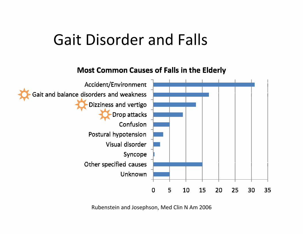
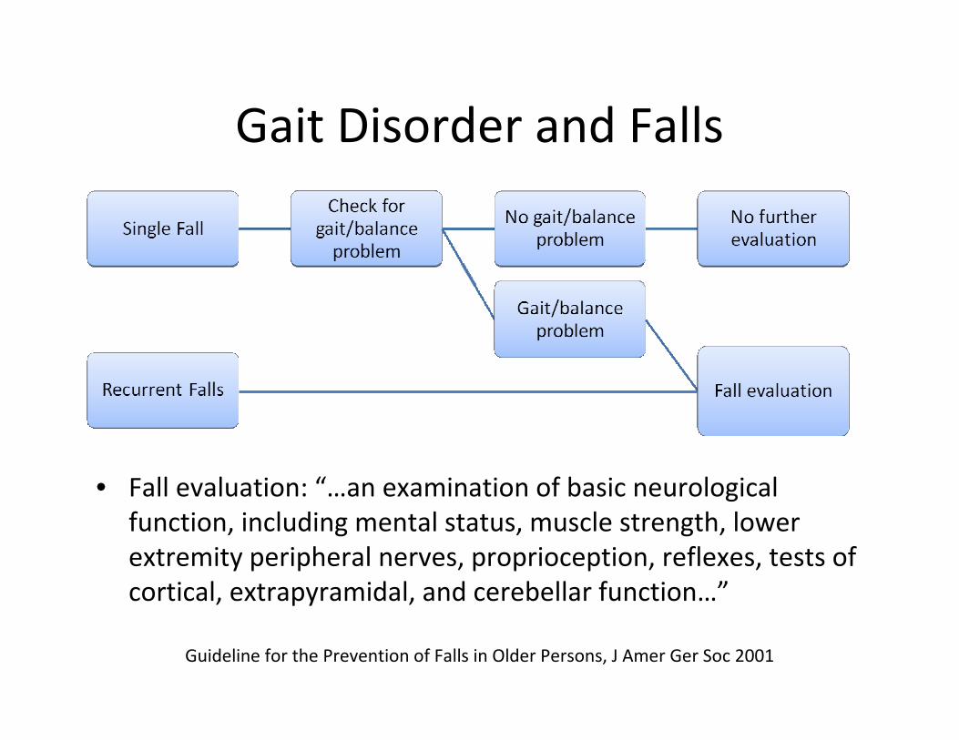
Gait Disorder and Falls
Rubenstein and Josephson, Med Clin N Am 2006

Gait Disorder and Falls
Rubenstein and Josephson, Med Clin N Am 2006
Gait Disorder and Falls
• Fall evaluation: “…an examination of basic neurological function, including mental status, muscle strength, lower extremity peripheral nerves, proprioception, reflexes, tests of cortical, extrapyramidal, and cerebellar function…”
Guideline for the Prevention of Falls in Older Persons, J Amer Ger Soc 2001
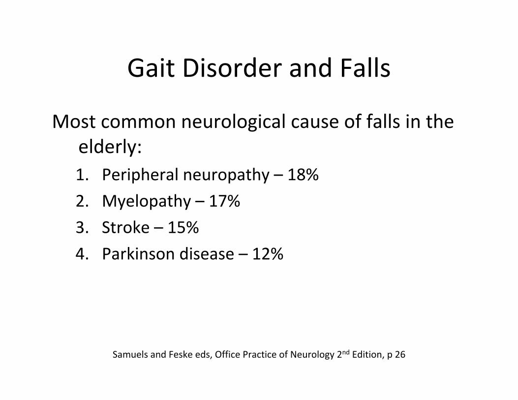
Gait Disorder and Falls
Most common neurological cause of falls in the elderly:1. Peripheral neuropathy – 18%
2. Myelopathy – 17%
3. Stroke – 15%
4. Parkinson disease – 12%
Samuels and Feske eds, Office Practice of Neurology 2nd Edition, p 26
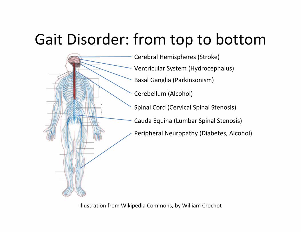
Gait Disorder: from top to bottom
Illustration from Wikipedia Commons, by William Crochot
Cerebral Hemispheres (Stroke)
Ventricular System (Hydrocephalus)
Basal Ganglia (Parkinsonism)
Cerebellum (Alcohol)
Spinal Cord (Cervical Spinal Stenosis)
Peripheral Neuropathy (Diabetes, Alcohol)
Cauda Equina (Lumbar Spinal Stenosis)
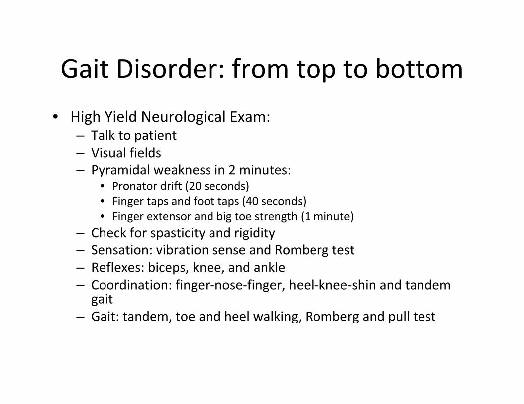
Gait Disorder: from top to bottom
• High Yield Neurological Exam:– Talk to patient– Visual fields– Pyramidal weakness in 2 minutes:
• Pronator drift (20 seconds)• Finger taps and foot taps (40 seconds)• Finger extensor and big toe strength (1 minute)
– Check for spasticity and rigidity– Sensation: vibration sense and Romberg test– Reflexes: biceps, knee, and ankle– Coordination: finger‐nose‐finger, heel‐knee‐shin and tandem
gait– Gait: tandem, toe and heel walking, Romberg and pull test
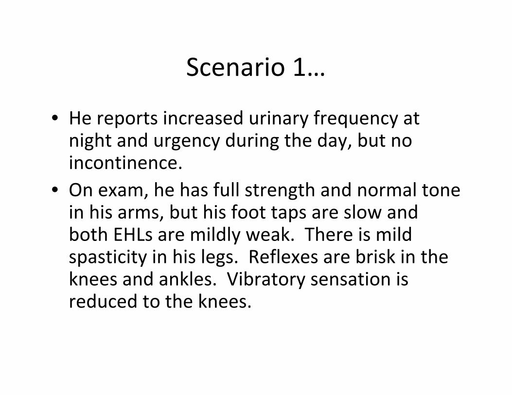
Scenario 1…
• He reports increased urinary frequency at night and urgency during the day, but no incontinence.
• On exam, he has full strength and normal tone in his arms, but his foot taps are slow and both EHLs are mildly weak. There is mild spasticity in his legs. Reflexes are brisk in the knees and ankles. Vibratory sensation is reduced to the knees.
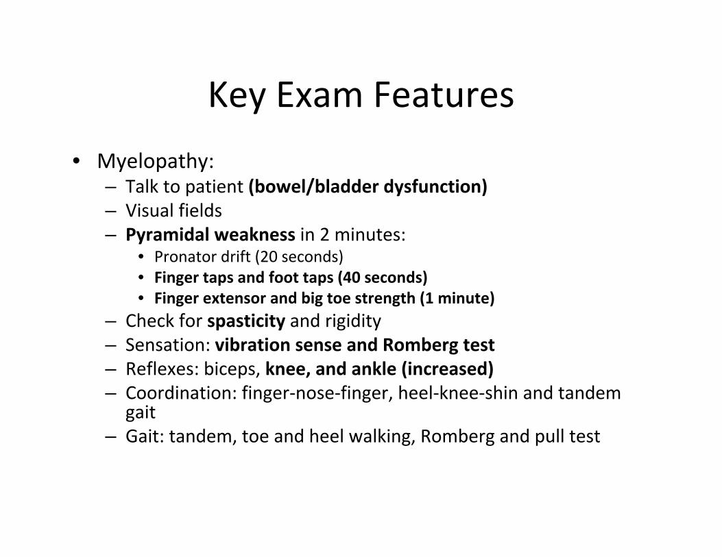
Key Exam Features
• Myelopathy:– Talk to patient (bowel/bladder dysfunction)– Visual fields– Pyramidal weakness in 2 minutes:
• Pronator drift (20 seconds)• Finger taps and foot taps (40 seconds)• Finger extensor and big toe strength (1 minute)
– Check for spasticity and rigidity– Sensation: vibration sense and Romberg test– Reflexes: biceps, knee, and ankle (increased)– Coordination: finger‐nose‐finger, heel‐knee‐shin and tandem
gait– Gait: tandem, toe and heel walking, Romberg and pull test
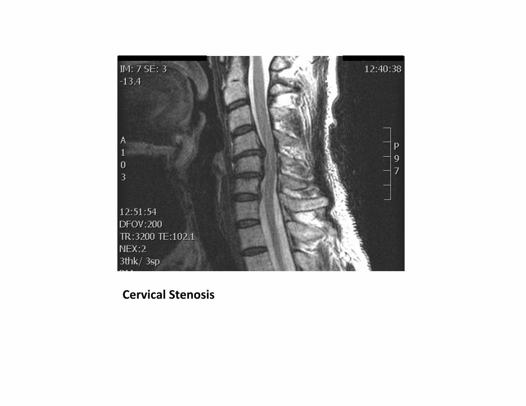
Cervical Stenosis
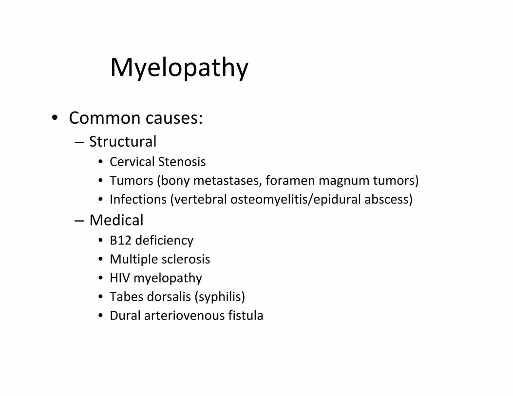
Myelopathy
• Common causes:– Structural
• Cervical Stenosis• Tumors (bony metastases, foramen magnum tumors)• Infections (vertebral osteomyelitis/epidural abscess)
– Medical• B12 deficiency• Multiple sclerosis• HIV myelopathy• Tabes dorsalis (syphilis)• Dural arteriovenous fistula
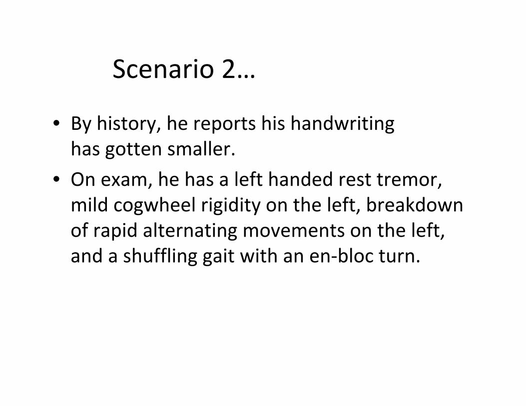
Scenario 2…
• By history, he reports his handwriting has gotten smaller.
• On exam, he has a left handed rest tremor, mild cogwheel rigidity on the left, breakdown of rapid alternating movements on the left, and a shuffling gait with an en‐bloc turn.
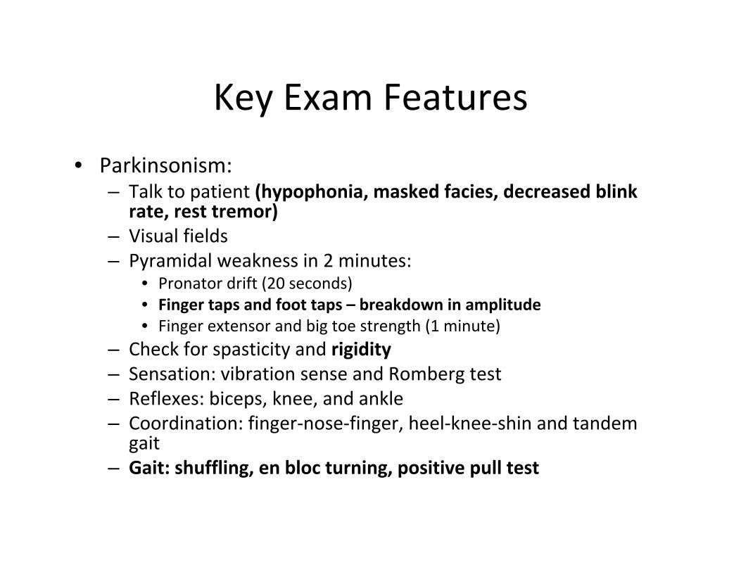
Key Exam Features
• Parkinsonism:– Talk to patient (hypophonia, masked facies, decreased blink
rate, rest tremor)– Visual fields– Pyramidal weakness in 2 minutes:
• Pronator drift (20 seconds)• Finger taps and foot taps – breakdown in amplitude• Finger extensor and big toe strength (1 minute)
– Check for spasticity and rigidity– Sensation: vibration sense and Romberg test– Reflexes: biceps, knee, and ankle– Coordination: finger‐nose‐finger, heel‐knee‐shin and tandem
gait– Gait: shuffling, en bloc turning, positive pull test
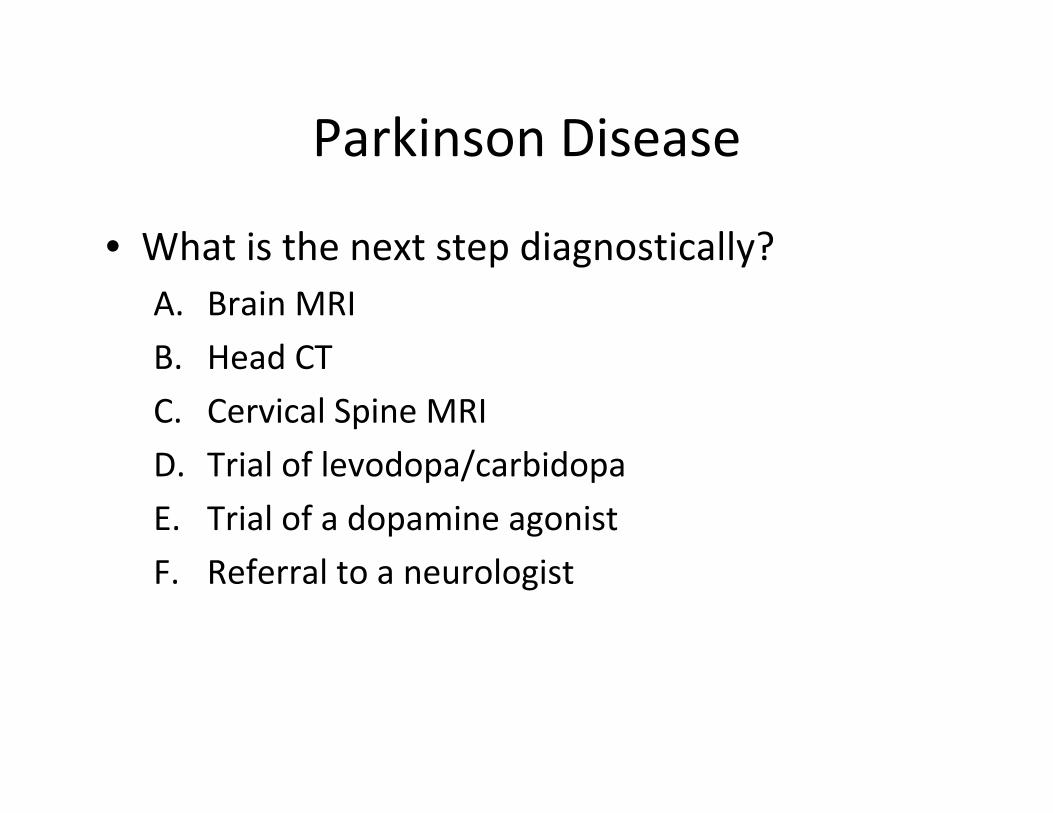
Parkinson Disease
• What is the next step diagnostically?A. Brain MRI
B. Head CT
C. Cervical Spine MRI
D. Trial of levodopa/carbidopa
E. Trial of a dopamine agonist
F. Referral to a neurologist
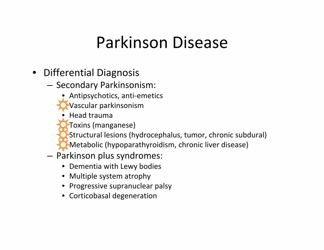
Parkinson Disease
• Differential Diagnosis– Secondary Parkinsonism:
• Antipsychotics, anti‐emetics• Vascular parkinsonism• Head trauma• Toxins (manganese)• Structural lesions (hydrocephalus, tumor, chronic subdural)• Metabolic (hypoparathyroidism, chronic liver disease)
– Parkinson plus syndromes:• Dementia with Lewy bodies• Multiple system atrophy• Progressive supranuclear palsy• Corticobasal degeneration
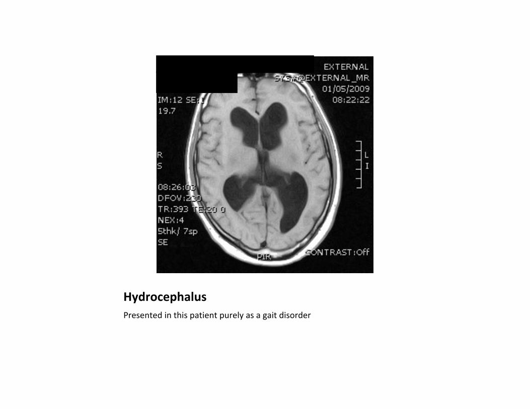
HydrocephalusPresented in this patient purely as a gait disorder
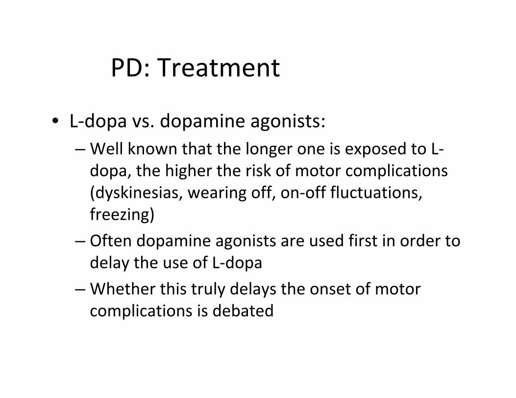
PD: Treatment
• L‐dopa vs. dopamine agonists:– Well known that the longer one is exposed to L‐dopa, the higher the risk of motor complications (dyskinesias, wearing off, on‐off fluctuations, freezing)
– Often dopamine agonists are used first in order to delay the use of L‐dopa
– Whether this truly delays the onset of motor complications is debated
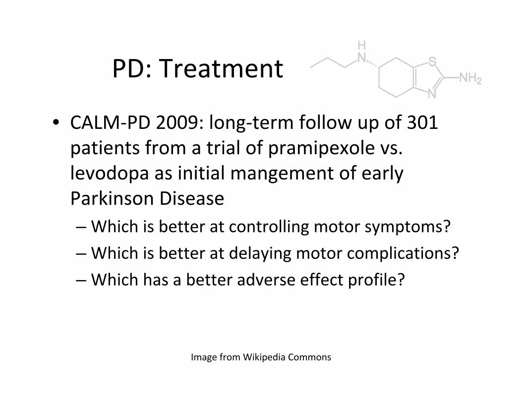
PD: Treatment
• CALM‐PD 2009: long‐term follow up of 301 patients from a trial of pramipexole vs. levodopa as initial mangement of early Parkinson Disease– Which is better at controlling motor symptoms?
– Which is better at delaying motor complications?
– Which has a better adverse effect profile?
Image from Wikipedia Commons
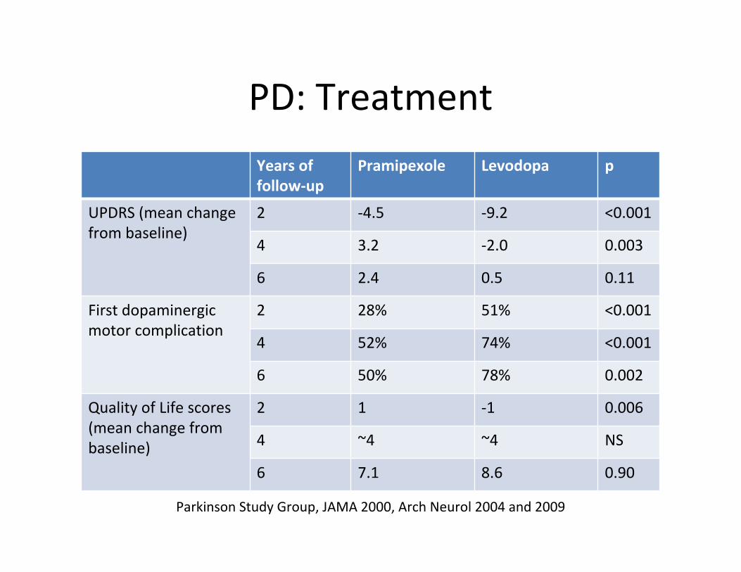
PD: Treatment
Years of follow‐up
Pramipexole Levodopa p
UPDRS (mean changefrom baseline)
2 ‐4.5 ‐9.2 <0.001
4 3.2 ‐2.0 0.003
6 2.4 0.5 0.11
First dopaminergic motor complication
2 28% 51% <0.001
4 52% 74% <0.001
6 50% 78% 0.002
Quality of Life scores (mean change from baseline)
2 1 ‐1 0.006
4 ~4 ~4 NS
6 7.1 8.6 0.90
Parkinson Study Group, JAMA 2000, Arch Neurol 2004 and 2009
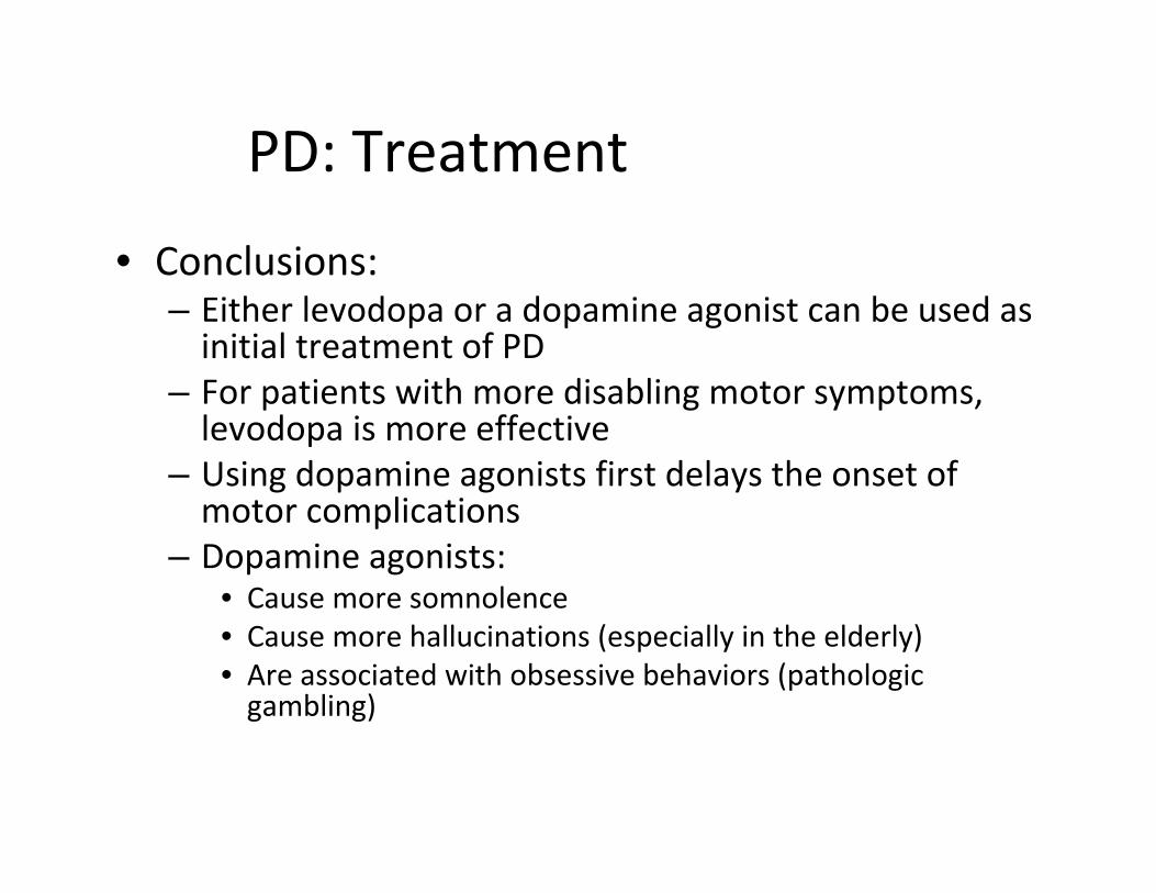
PD: Treatment
• Conclusions:– Either levodopa or a dopamine agonist can be used as initial treatment of PD
– For patients with more disabling motor symptoms, levodopa is more effective
– Using dopamine agonists first delays the onset of motor complications
– Dopamine agonists:• Cause more somnolence• Cause more hallucinations (especially in the elderly)• Are associated with obsessive behaviors (pathologic gambling)
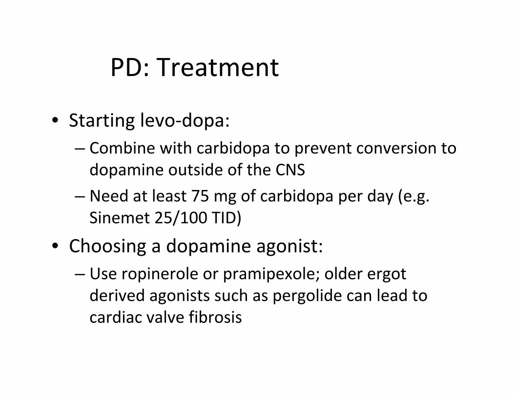
PD: Treatment
• Starting levo‐dopa:– Combine with carbidopa to prevent conversion to dopamine outside of the CNS
– Need at least 75 mg of carbidopa per day (e.g. Sinemet 25/100 TID)
• Choosing a dopamine agonist:– Use ropinerole or pramipexole; older ergot derived agonists such as pergolide can lead to cardiac valve fibrosis
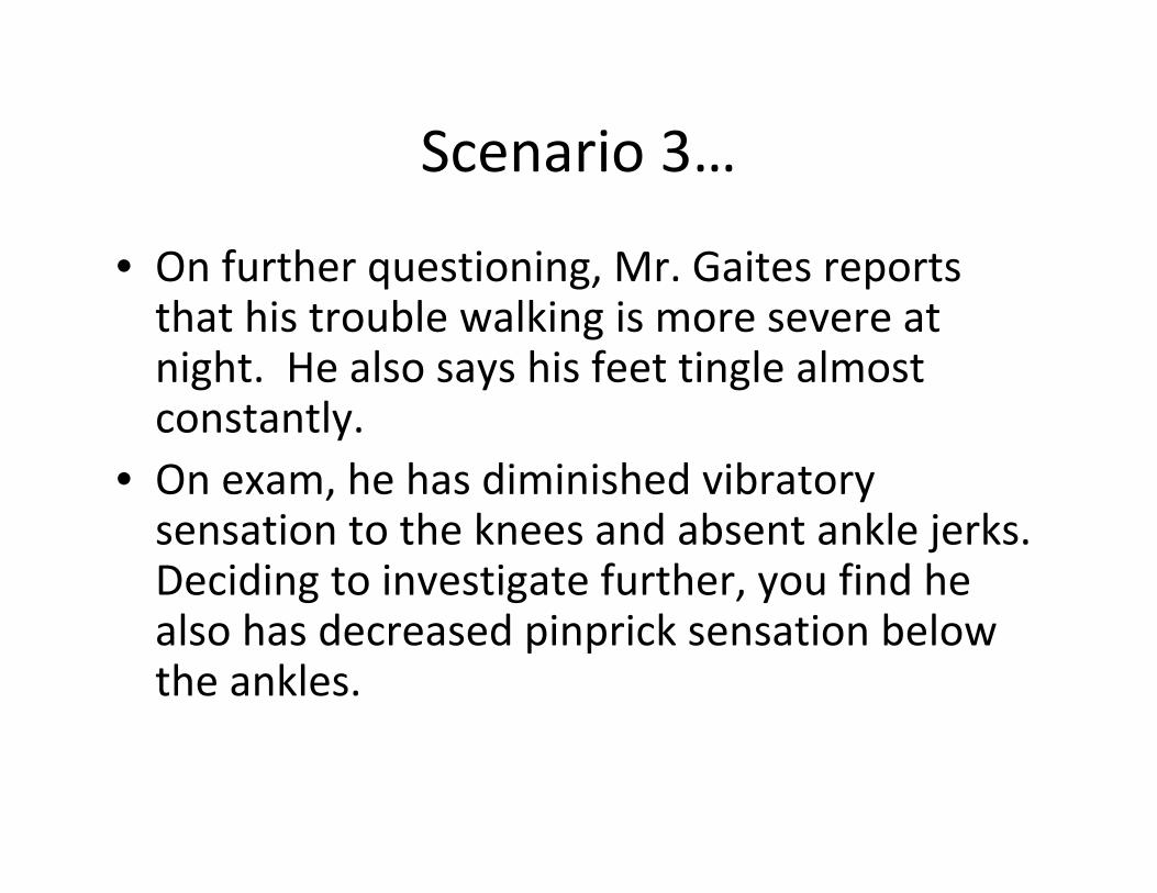
Scenario 3…
• On further questioning, Mr. Gaites reports that his trouble walking is more severe at night. He also says his feet tingle almost constantly.
• On exam, he has diminished vibratory sensation to the knees and absent ankle jerks. Deciding to investigate further, you find he also has decreased pinprick sensation below the ankles.
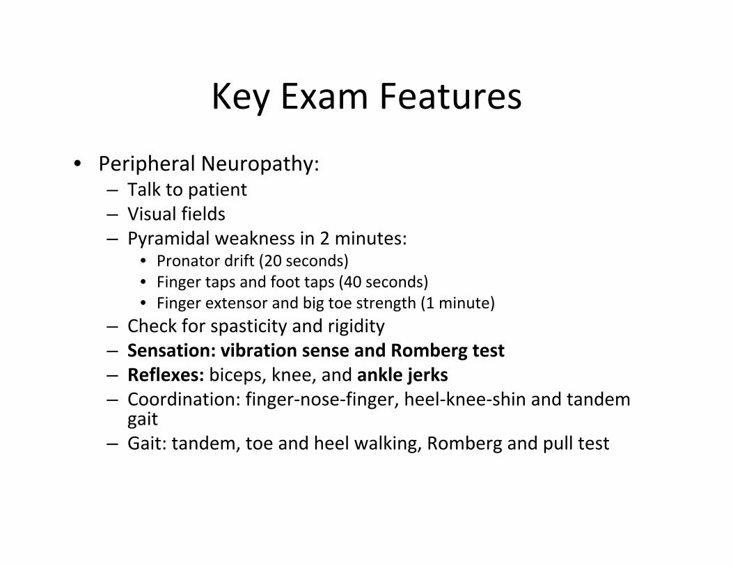
Key Exam Features
• Peripheral Neuropathy:– Talk to patient– Visual fields– Pyramidal weakness in 2 minutes:
• Pronator drift (20 seconds)• Finger taps and foot taps (40 seconds)• Finger extensor and big toe strength (1 minute)
– Check for spasticity and rigidity– Sensation: vibration sense and Romberg test– Reflexes: biceps, knee, and ankle jerks– Coordination: finger‐nose‐finger, heel‐knee‐shin and tandem
gait– Gait: tandem, toe and heel walking, Romberg and pull test
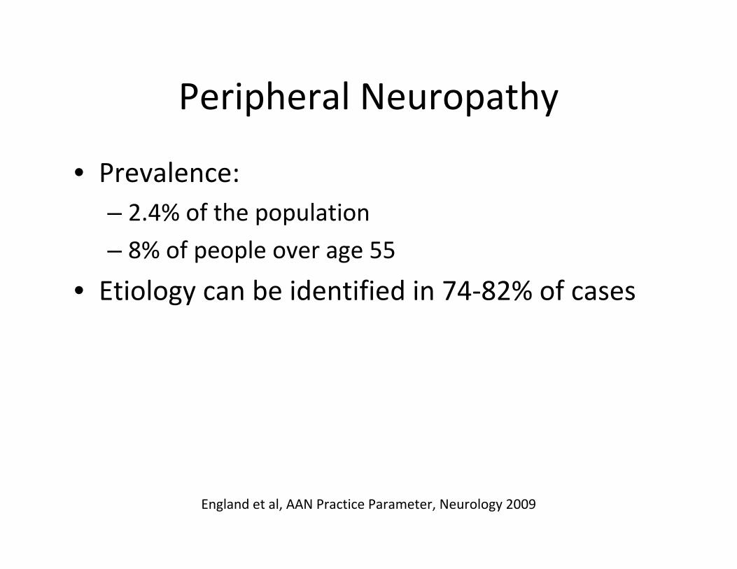
Peripheral Neuropathy
• Prevalence:– 2.4% of the population
– 8% of people over age 55
• Etiology can be identified in 74‐82% of cases
England et al, AAN Practice Parameter, Neurology 2009
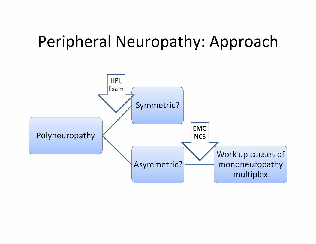
Peripheral Neuropathy: Approach

Mononeuropathy MultiplexDifferential Diagnosis Workup
Inflammatory Vasculitis Aggressive vasculitis workup
Sarcoid CXR, Chest CT, serum ACE
CIDP (Chronic Inflammatory Demyelinating Polyneuropathy)
EMG/NCS
Infectious Leprosy
HIV (especially with CMV) HIV Ab
Lyme Lyme Ab
Neoplastic Lymphoma Directed workup
Waldenstrom’s Macroglobulinemia SPEP/UPEP
Compressive/ Ischemic
Diabetes Fasting glucose, OGTT
Hypothyroidism TSH, free T4
HNPP (Hereditary Neuropathy with susceptibility to Pressure Palsies)
Family history, Genetic testing
Image from Wikipedia Commons

Peripheral Neuropathy: Approach
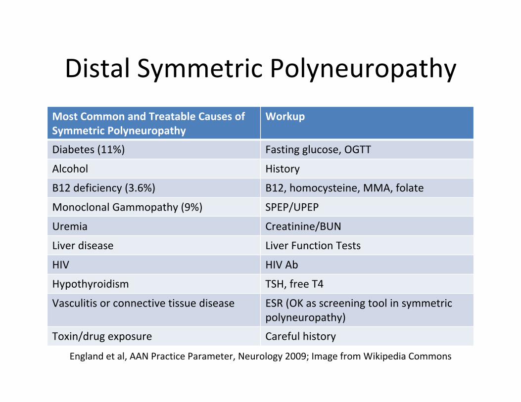
Distal Symmetric Polyneuropathy
Most Common and Treatable Causes of Symmetric Polyneuropathy
Workup
Diabetes (11%) Fasting glucose, OGTT
Alcohol History
B12 deficiency (3.6%) B12, homocysteine, MMA, folate
Monoclonal Gammopathy (9%) SPEP/UPEP
Uremia Creatinine/BUN
Liver disease Liver Function Tests
HIV HIV Ab
Hypothyroidism TSH, free T4
Vasculitis or connective tissue disease ESR (OK as screening tool in symmetric polyneuropathy)
Toxin/drug exposure Careful history
England et al, AAN Practice Parameter, Neurology 2009; Image from Wikipedia Commons
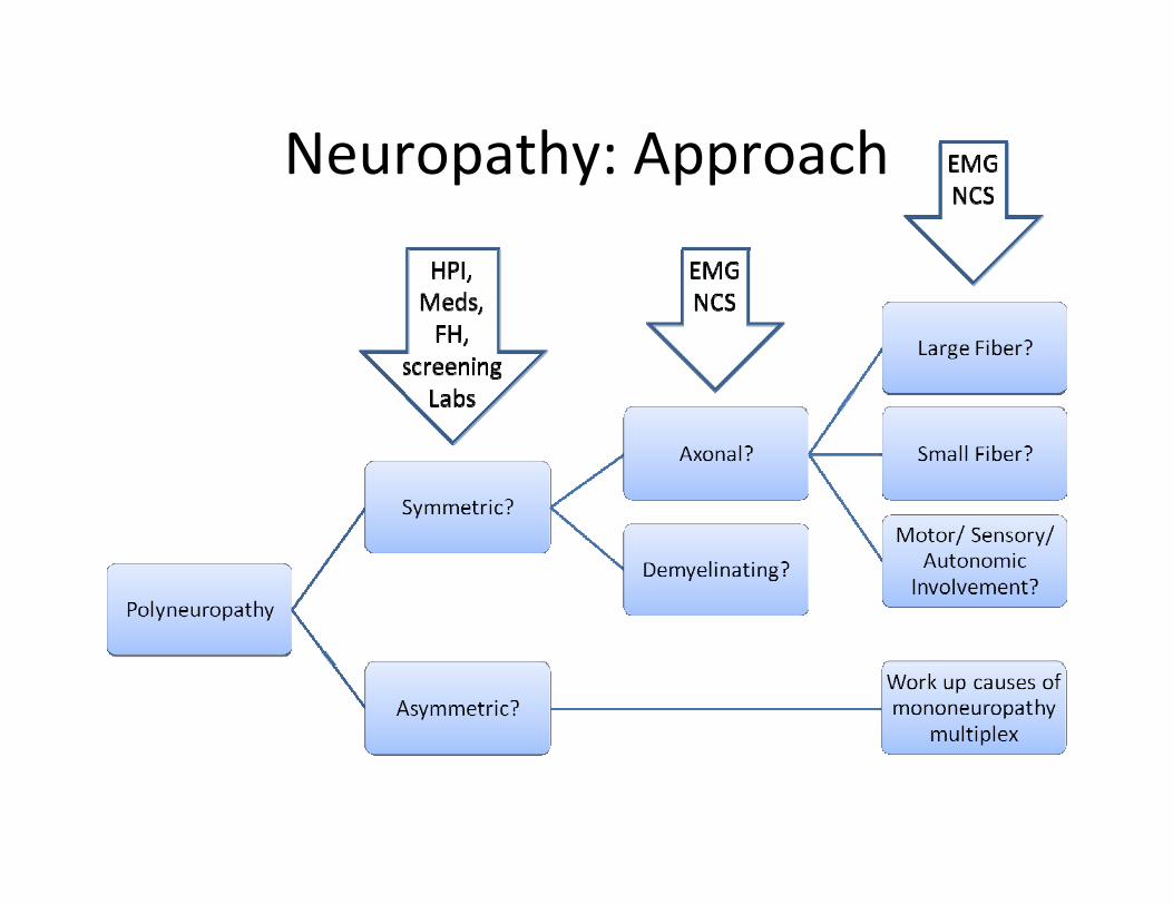
Neuropathy: Approach
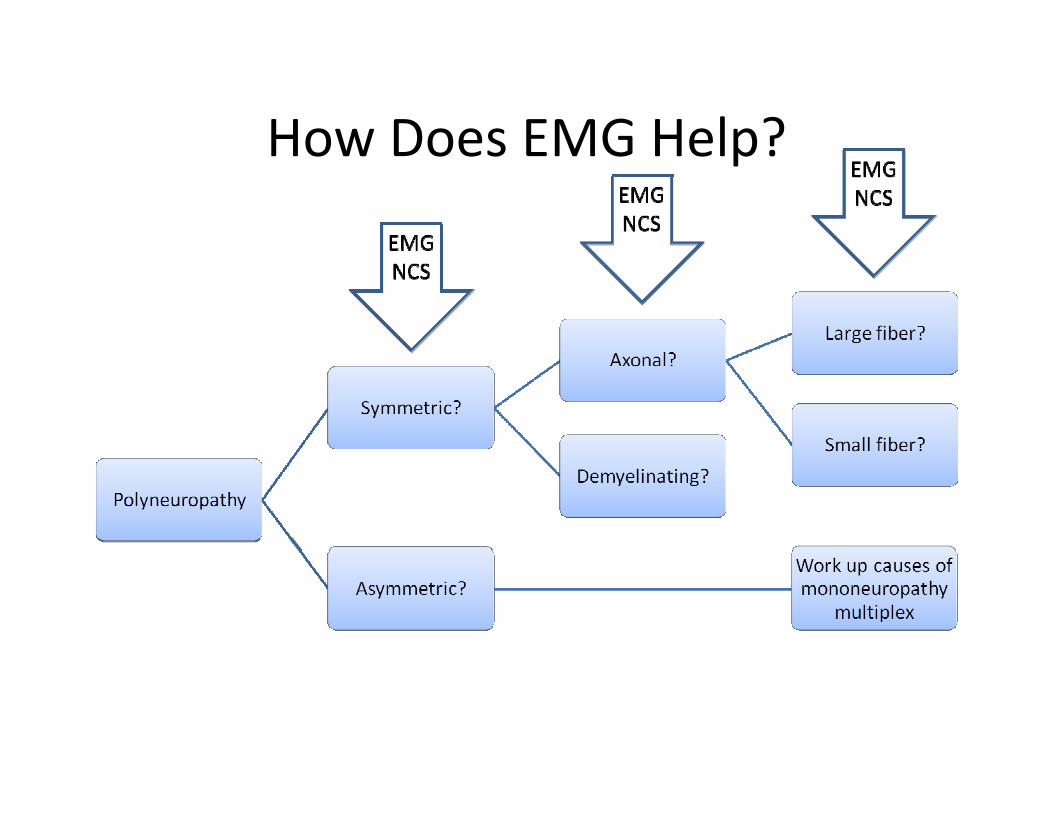
How Does EMG Help?
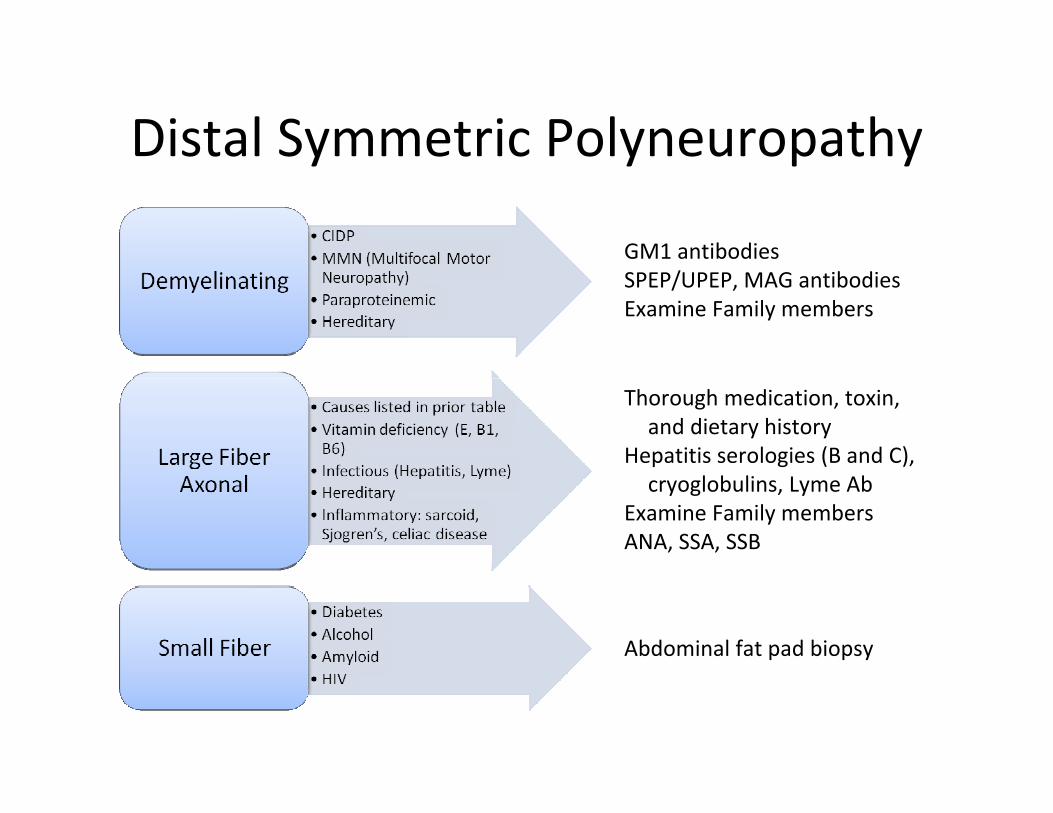
Distal Symmetric Polyneuropathy
GM1 antibodiesSPEP/UPEP, MAG antibodiesExamine Family members
Thorough medication, toxin, and dietary history
Hepatitis serologies (B and C), cryoglobulins, Lyme Ab
Examine Family membersANA, SSA, SSB
Abdominal fat pad biopsy
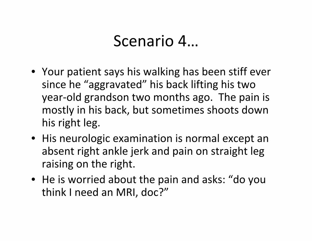
Scenario 4…
• Your patient says his walking has been stiff ever since he “aggravated” his back lifting his two year‐old grandson two months ago. The pain is mostly in his back, but sometimes shoots down his right leg.
• His neurologic examination is normal except an absent right ankle jerk and pain on straight leg raising on the right.
• He is worried about the pain and asks: “do you think I need an MRI, doc?”
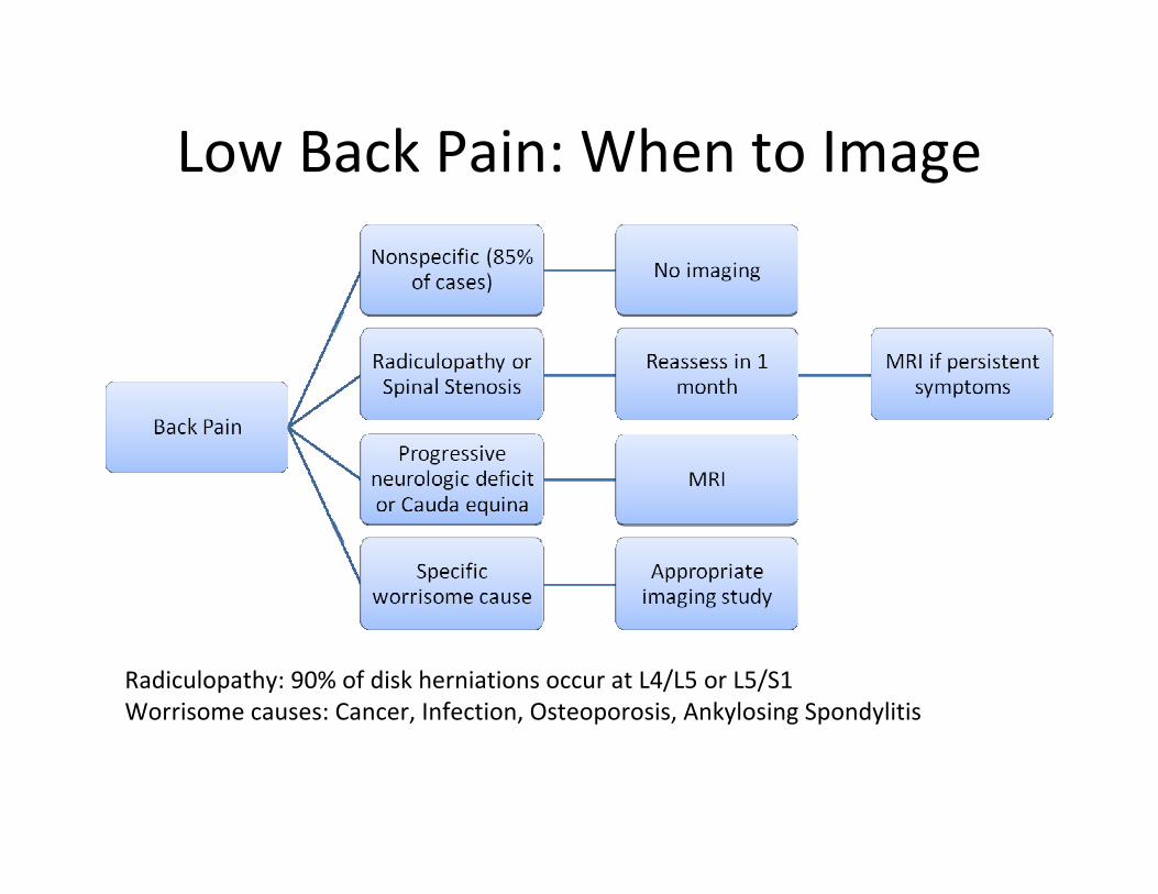
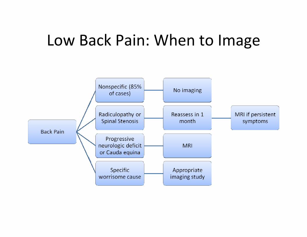
Low Back Pain: When to Image
Radiculopathy: 90% of disk herniations occur at L4/L5 or L5/S1Worrisome causes: Cancer, Infection, Osteoporosis, Ankylosing Spondylitis
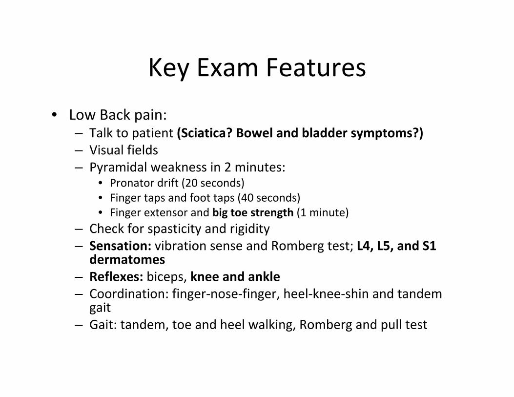
Key Exam Features
• Low Back pain:– Talk to patient (Sciatica? Bowel and bladder symptoms?)– Visual fields– Pyramidal weakness in 2 minutes:
• Pronator drift (20 seconds)• Finger taps and foot taps (40 seconds)• Finger extensor and big toe strength (1 minute)
– Check for spasticity and rigidity– Sensation: vibration sense and Romberg test; L4, L5, and S1
dermatomes– Reflexes: biceps, knee and ankle– Coordination: finger‐nose‐finger, heel‐knee‐shin and tandem
gait– Gait: tandem, toe and heel walking, Romberg and pull test
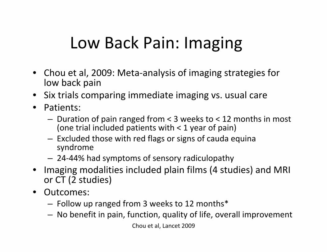
Low Back Pain: Imaging
• Chou et al, 2009: Meta‐analysis of imaging strategies for low back pain
• Six trials comparing immediate imaging vs. usual care• Patients:
– Duration of pain ranged from < 3 weeks to < 12 months in most (one trial included patients with < 1 year of pain)
– Excluded those with red flags or signs of cauda equina syndrome
– 24‐44% had symptoms of sensory radiculopathy• Imaging modalities included plain films (4 studies) and MRI
or CT (2 studies)• Outcomes:
– Follow up ranged from 3 weeks to 12 months*– No benefit in pain, function, quality of life, overall improvement
Chou et al, Lancet 2009
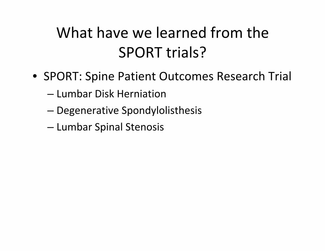
What have we learned from the SPORT trials?
• SPORT: Spine Patient Outcomes Research Trial– Lumbar Disk Herniation
– Degenerative Spondylolisthesis
– Lumbar Spinal Stenosis
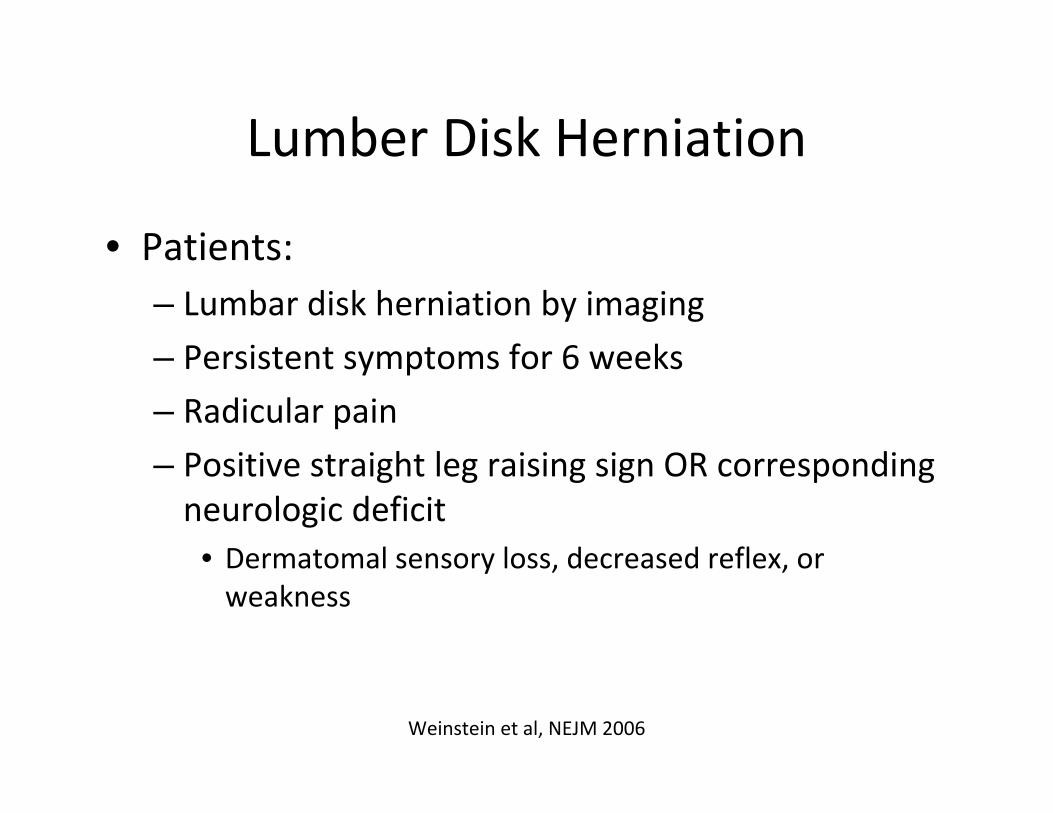
Lumber Disk Herniation
• Patients:– Lumbar disk herniation by imaging
– Persistent symptoms for 6 weeks
– Radicular pain
– Positive straight leg raising sign OR corresponding neurologic deficit
• Dermatomal sensory loss, decreased reflex, or weakness
Weinstein et al, NEJM 2006
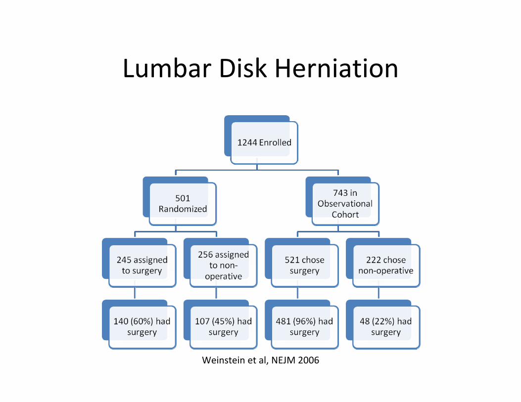
Lumbar Disk Herniation
Weinstein et al, NEJM 2006
Lumbar Disk Herniation
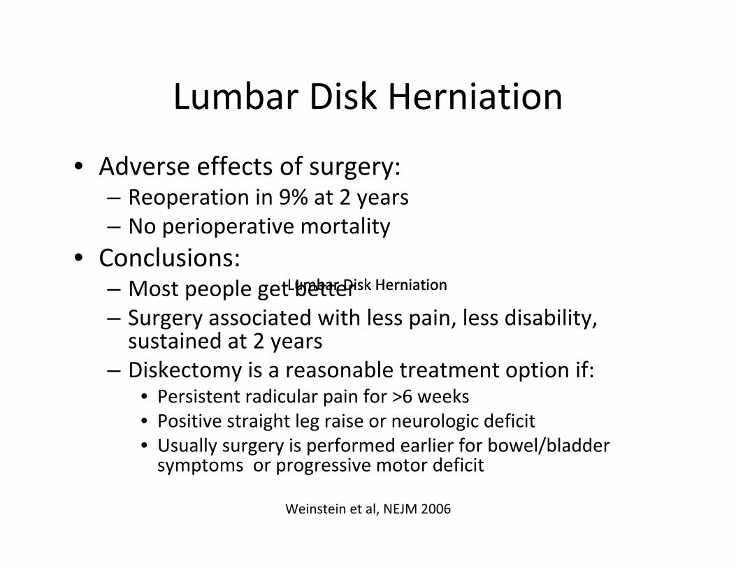
• Adverse effects of surgery:– Reoperation in 9% at 2 years– No perioperative mortality
• Conclusions:– Most people get better– Surgery associated with less pain, less disability, sustained at 2 years
– Diskectomy is a reasonable treatment option if:• Persistent radicular pain for >6 weeks• Positive straight leg raise or neurologic deficit• Usually surgery is performed earlier for bowel/bladder symptoms or progressive motor deficit
Weinstein et al, NEJM 2006
Lumbar Disk HerniationLumbar Disk Herniation
Lumbar Disk Herniation
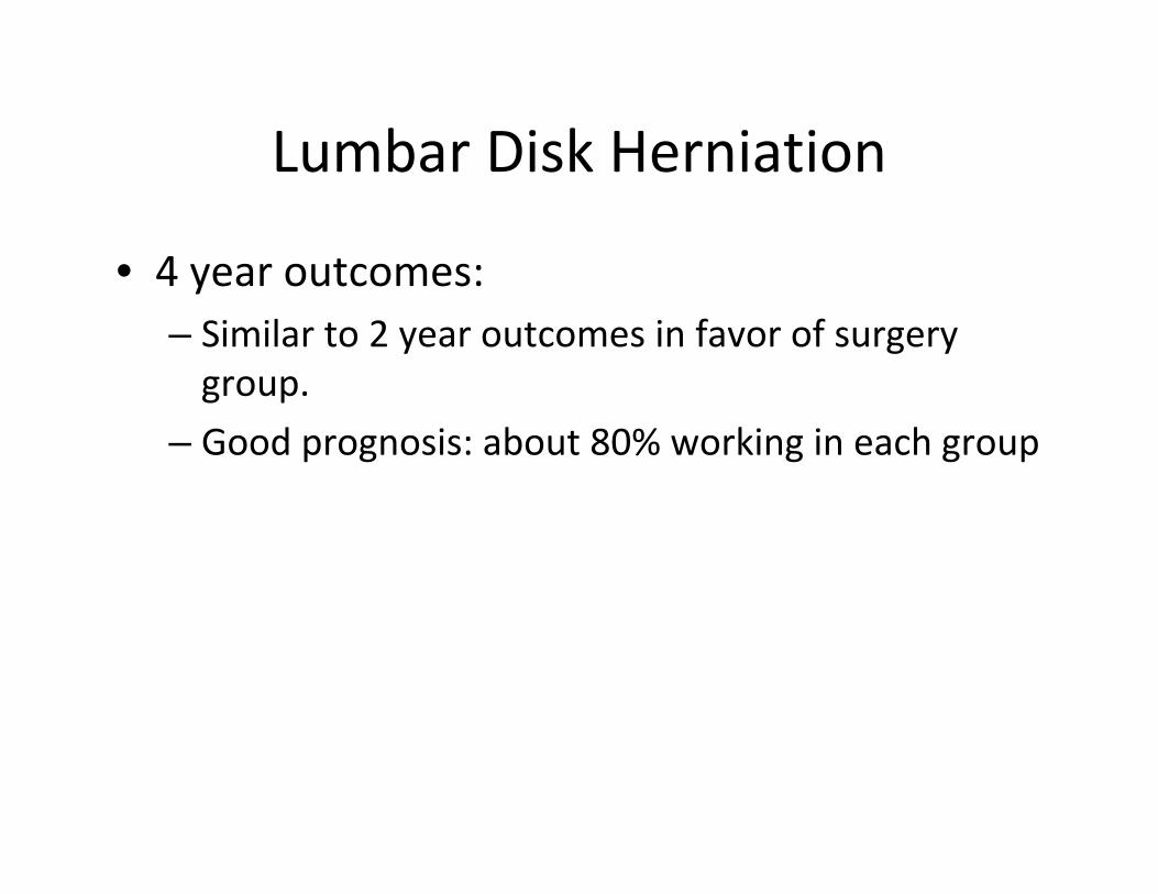
• 4 year outcomes:– Similar to 2 year outcomes in favor of surgery group.
– Good prognosis: about 80% working in each group
Spondylolisthesis
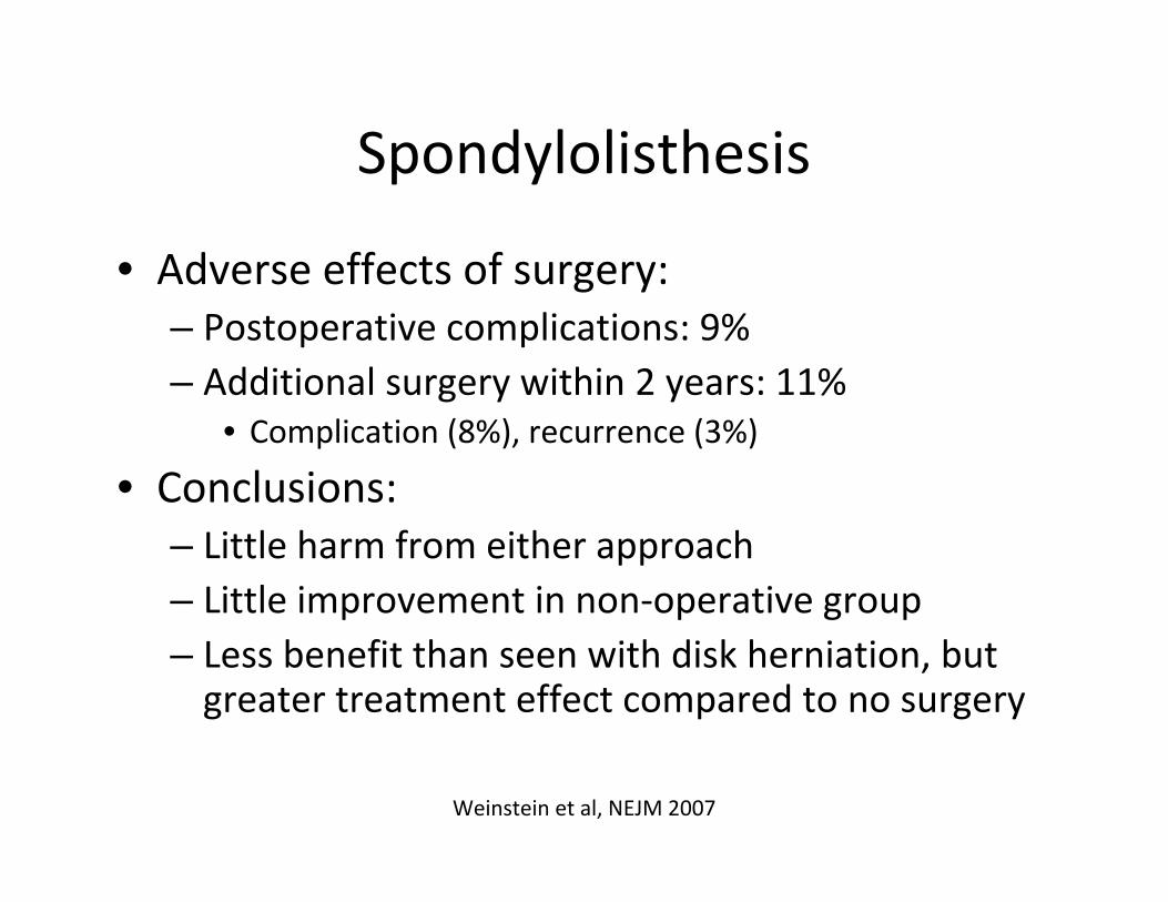
• Adverse effects of surgery:– Postoperative complications: 9%– Additional surgery within 2 years: 11%
• Complication (8%), recurrence (3%)
• Conclusions:– Little harm from either approach– Little improvement in non‐operative group– Less benefit than seen with disk herniation, but greater treatment effect compared to no surgery
Weinstein et al, NEJM 2007

Spinal Stenosis
• Adverse effects of surgery:– Postoperative complication: 8%– Reoperation within 2 years: 8%
• Complication (4%)• Recurrence or progressive spondylolisthesis (3%)
• Conclusions:– Little harm from either approach – Little improvement in non‐operative group– For persistent spinal stenosis lasting more than 12 weeks, surgery is a reasonable option
Weinstein et al, NEJM 2008
Low Back Pain: When to Image
References• Miyasaki, J.M., et al., Practice parameter: initiation of treatment for Parkinson's disease: an
evidence‐based review: report of the Quality Standards Subcommittee of the American Academy of Neurology. Neurology, 2002. 58(1): p. 11‐7.
• England, J.D., et al., Practice Parameter: evaluation of distal symmetric polyneuropathy: role of laboratory and genetic testing (an evidence‐based review). Report of the American Academy of Neurology, American Association of Neuromuscular and Electrodiagnostic Medicine, and American Academy of Physical Medicine and Rehabilitation. Neurology, 2009. 72(2): p. 185‐92.
• Chou, R., et al., Imaging strategies for low‐back pain: systematic review and meta‐analysis. Lancet, 2009. 373(9662): p. 463‐72.
• Weinstein, J.N., et al., Surgical vs nonoperative treatment for lumbar disk herniation: the Spine Patient Outcomes Research Trial (SPORT): a randomized trial. JAMA, 2006. 296(20): p. 2441‐50.
• Weinstein, J.N., et al., Surgical vs nonoperative treatment for lumbar disk herniation: the Spine Patient Outcomes Research Trial (SPORT) observational cohort. JAMA, 2006. 296(20): p. 2451‐9.
• Weinstein, J.N., et al., Surgical versus nonsurgical treatment for lumbar degenerative spondylolisthesis. N Engl J Med, 2007. 356(22): p. 2257‐70.
• Weinstein, J.N., et al., Surgical versus nonsurgical therapy for lumbar spinal stenosis. N Engl J Med, 2008. 358(8): p. 794‐810.