four quadrants: anatomy of midsagittal plane ... salivary gland tongue – function? mainly skeletal...
TRANSCRIPT

4/14/2009
1
ANATOMY OF THE G.I PART 1UPPER GIUPPER GI
D.HAMMOUDI.MD
Four Quadrants: •Midsagittal Plane: Vertical line going through the middle of the abdomen. •Transumbilical Plane: Horizontal line going through the umbilicus. •Four Quadrants based on those planes:
•Right Upper Quadrant: RUQ •Right Lower Quadrant: RLQ •Left Upper Quadrant: LUQ •Left Lower Quadrant: LLQ
Nine Regions: •Vertical lines of division: Left and Right Mid-Clavicular Lines •Horizontal lines of division:•Horizontal lines of division:
•Transpyloric Plane: Sometimes used. It is halfway between the jugular notch and the pubic bone. •Subcostal Plane: Upper plane, passing through the inferior-most margin of the ribs. •Transtubercular Plane: The line transversing the pubic tubercle.
•Divisions: •Upper: Right Hypochondriac, Epigastric, Left Hypochondriac•Middle: Right Lumbar, Umbilical, Left Lumbar•Lower: Right Inguinal, Hypogastric (Suprapubic), Left Inguinal
Abdominal quadrants
Right upper quadrantRight upper quadrant Left upper quadrantLeft upper quadrantLiver right lobeLiver right lobeGallbladder, stomach, pylorus, doudenum, Gallbladder, stomach, pylorus, doudenum, Pancreas head R suprarenal gland R kidneyPancreas head R suprarenal gland R kidney
Liver left lobeLiver left lobeSpleen, stomach, jejunum, prox ileum, Spleen, stomach, jejunum, prox ileum, pancreas body and tail left kidney Lpancreas body and tail left kidney LPancreas head, R suprarenal gland, R kidney, Pancreas head, R suprarenal gland, R kidney,
R colic flexure, Ascending colon superior part, R colic flexure, Ascending colon superior part, Transvrse colon R half. Transvrse colon R half.
pancreas body and tail, left kidney, L pancreas body and tail, left kidney, L suprarenal, left colic flexure, Transverse colon suprarenal, left colic flexure, Transverse colon left part, descending colon superior part.left part, descending colon superior part.
Right lower quadrantRight lower quadrant Left lower quadrantLeft lower quadrantCecum, Appendix, Ileum, Asc. Colon, R ovary, Cecum, Appendix, Ileum, Asc. Colon, R ovary, R uterine tube, R ureter, R spermatic cord, R uterine tube, R ureter, R spermatic cord, Uterus, Urinary bladder (full)Uterus, Urinary bladder (full)
Sigmoid colon, Desc. Colon, L ovary, L uterine Sigmoid colon, Desc. Colon, L ovary, L uterine tube, L ureter, L spermatic cord, Uterus tube, L ureter, L spermatic cord, Uterus enlarge, Urinary bladder ( full).enlarge, Urinary bladder ( full).

4/14/2009
2
8
Anatomy of the Mouth and Throat
Mouth: lips non-keratinized therefore evaporation occurs, must lick lips
Tongue: frenulum (bridle) ties downtaste buds:
fungiform, circumvallate,filiform
Oral Cavity (mouth)Entrance to the GI tract.� Initial site of mechanical digestion (via mastication) and chemical digestion (via enzymes in saliva).� Bounded anteriorly by the teeth and lips and posteriorly by the oropharynx.� Superior boundary is formed by the hard and soft palates.p y y p� Floor, or inferior surface, of the oral cavity contains the tongue as well as the mylohyoidmuscle covered with mucosa.� Vestibule is the space between the cheeks or lips and the gums.� Oral cavity proper.� The lateral walls are formed by the cheeks.� Lips (labia).� Gingivae, or gums.� Labial frenulum.
11
Human Deciduous and Permanent Teeth
4/14/2009
3
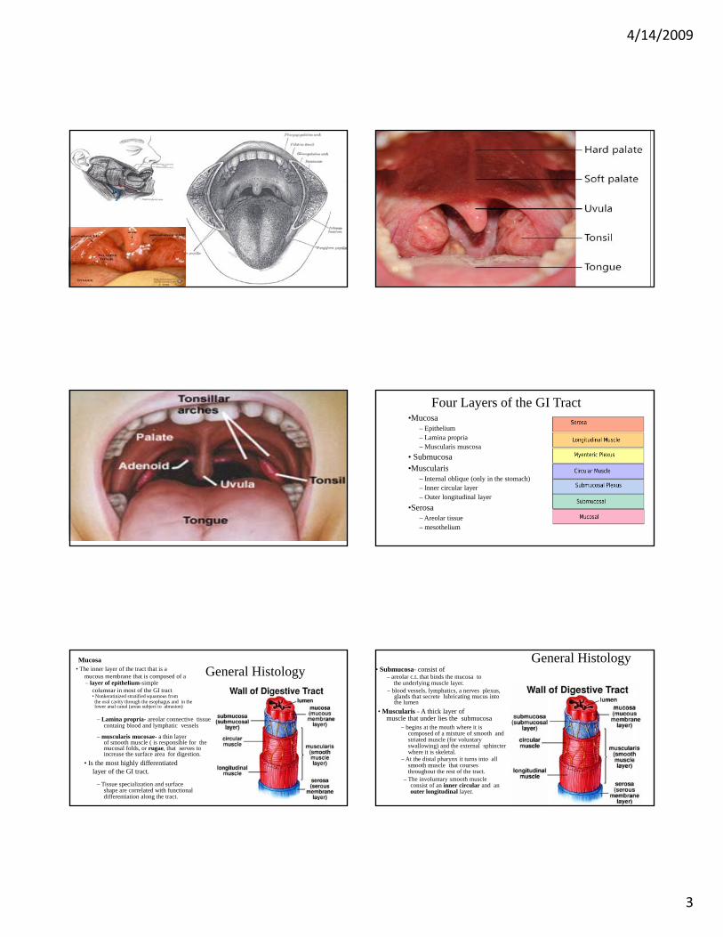
Four Layers of the GI Tract •Mucosa
– Epithelium – Lamina propria– Muscularis muscosa
• Submucosa•Muscularis•Muscularis
– Internal oblique (only in the stomach) – Inner circular layer – Outer longitudinal layer
•Serosa– Areolar tissue – mesothelium
General Histology Mucosa
• The inner layer of the tract that is a mucous membrane that is composed of a – layer of epithelium-simple
columnar in most of the GI tract • Nonkeratinized stratified squamous from the oral cavity through the esophagus and in the lower anal canal (areas subject to abrasion)
– Lamina propria- areolar connective tissue containg blood and lymphatic vessels g y p
– muscularis mucosae- a thin layer of smooth muscle ( is responsible for the mucosal folds, or rugae, that serves to increase the surface area for digestion.
• Is the most highly differentiated layer of the GI tract.
– Tissue specialization and surface shape are correlated with functional differentiation along the tract.
General Histology • Submucosa- consist of
– areolar c.t. that binds the mucosa to the underlying muscle layer.
– blood vessels, lymphatics, a nerves plexus, glands that secrete lubricating mucus into the lumen
• Muscularis - A thick layer of muscle that under lies the submucosa
– begins at the mouth where it is– begins at the mouth where it is composed of a mixture of smooth and striated muscle (for voluntary swallowing) and the external sphincter where it is skeletal.
– At the distal pharynx it turns into all smooth muscle that courses throughout the rest of the tract.
– The involuntary smooth muscle consist of an inner circular and an outer longitudinal layer.
4/14/2009
4
General Histology Serosa- The outermost layer
of the GI tract. • Composed of a thin layer
of areolar tissue topped by a serous membrane (mesothelium)
• Begins in the lower 3 to 4 cm of the esophagus and ends with the sigmoid colonends with the sigmoid colon
• When the outer fibrous c.t. layer is attached to surrounding tissue it is called adventitia –
– See this at the oral cavity, pharynx, most of the esophagus, and the rectum It secretes fluid that allows the tract structures to glide over each other without friction. It is also referred to as visceral peritoneum.
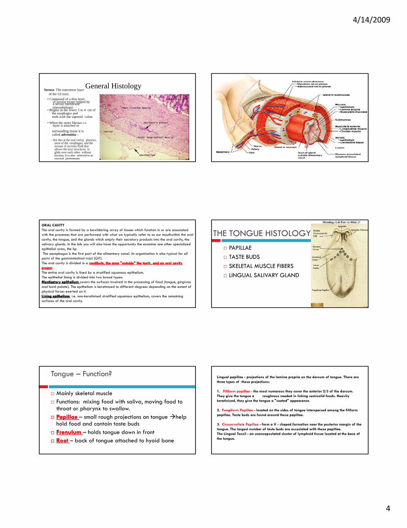
ORAL CAVITYThe oral cavity is formed by a bewildering array of tissues which function in or are associated with the processes that are performed with what we typically refer to as our mouthwithin the oral cavity, the tongue, and the glands which empty their secretory products into the oral cavity, the salivary glands. In the lab you will also have the opportunity the examine one other specialized epithelial area, the lip.The oesophagus is the first part of the alimentary canal. Its organisation is also typical for all parts of the gastrointestinal tract (GIT).The oral cavity is divided in a vestibule, the area "outside" the teeth, and an oral cavity vestibule, the area "outside" the teeth, and an oral cavity The oral cavity is divided in a vestibule, the area outside the teeth, and an oral cavity vestibule, the area outside the teeth, and an oral cavity properproper. The entire oral cavity is lined by a stratified squamous epithelium. The epithelial lining is divided into two broad types:MasticatoryMasticatory epithelium epithelium covers the surfaces involved in the processing of food (tongue, gingivaeand hard palate). The epithelium is keratinized to different degrees depending on the extent of physical forces exerted on it.Lining epitheliumLining epithelium, i.e. non-keratinised stratified squamous epithelium, covers the remaining surfaces of the oral cavity.
THE TONGUE HISTOLOGYPAPILLAETASTE BUDSS A M SC SSKELETAL MUSCLE FIBERSLINGUAL SALIVARY GLAND
Tongue – Function?
Mainly skeletal muscle Functions: mixing food with saliva, moving food to throat or pharynx to swallow. throat or pharynx to swallow. Papillae Papillae – small rough projections on tongue help hold food and contain taste buds FrenulumFrenulum – holds tongue down in front Root Root – back of tongue attached to hyoid bone
Lingual papillae - projections of the lamina propria on the dorsum of tongue. There are three types of these projections:
1. Filiform papillae - the most numerous they cover the anterior 2/3 of the dorsum. They give the tongue a roughness needed in licking semisolid foods. Heavily keratinized, they give the tongue a "coated'' appearance.
2 Fungiform Papillae located on the sides of tongue interspersed among the filiform2. Fungiform Papillae - located on the sides of tongue interspersed among the filiformpapillae. Taste buds are found around these papillae.
3. Circumvallate Papillae - form a V - shaped formation near the posterior margin of the tongue. The largest number of taste buds are associated with these papillae.The Lingual Tonsil - an unencapsulated cluster of lymphoid tissue located at the base of the tongue.
4/14/2009
5
26
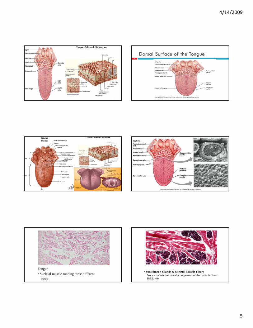
Dorsal Surface of the Tongue
Tongue • Skeletal muscle running three different
ways
• von Ebner's Glands & Skeletal Muscle Fibers Notice the tri-directional arrangement of the muscle fibers. H&E, 40x

4/14/2009
6
Detail of circumvallate papilla, showing pale taste buds opening into the lumen of the furrow that surrounds the papilla.
LEAF SHAPED PAPILLAE OF THE TONGUEStained with haematoxylin and eosin 1 - epithelium
covering papilla(stratified squamous nonkeratinizing)
2 - core of the papilla (lamina propriaof the mucosa of dorsal surface of the tongue)
3 - taste bud
FILIFORM PAPILLAE OF THE TONGUEStained with haematoxylin and eosin
1 - epithelium covering papilla (stratified squamous keratinizing)2 - keratinized layer of the epithelium3 - core of the papilla (lamina propria of the mucosa of dorsal surface of the tongue)2 - tongue muscles
CIRCUMVALLATE PAPILLAE OF THE TONGUE
Stained with haematoxylin and eosin
h l ll1 - epithelium covering papilla(stratified squamous
nonkeratinizing)2 - core of the papilla (lamina propria
of the mucosa of dorsal surface of the tongue)3 - taste buds
Detail of skeletal muscle and secretory glands of the body of the tongue.
Mucous cells are to the left, with their flattened, basal nuclei, while serous cells are in the center and to the right, with their round nuclei.
View of foliate papillae, typical of rabbit and some other animals.
These have a characteristic 3-pronged connective tissue pattern extending up into the papilla, and there are taste buds on the outside walls.
Notice the bundles of skeletal muscle down below.
Higher magnification of tongue surface, showing two filiform papillae. They are obviously extensions of stratified stratified squamoussquamousepithelium.epithelium.
Cut-away ysection of tongue to show three-dimensional view of papillae and underlying c.t. and muscle.
4/14/2009
7
Section of surface of tongue, showing one rather tangentially cut fungiformpapilla at the left and some filiform papillae with sharp, semicornified tips at the semicornified tips at the right.
Cornification is less extensive in human tongue than in cats, dogs, etc.
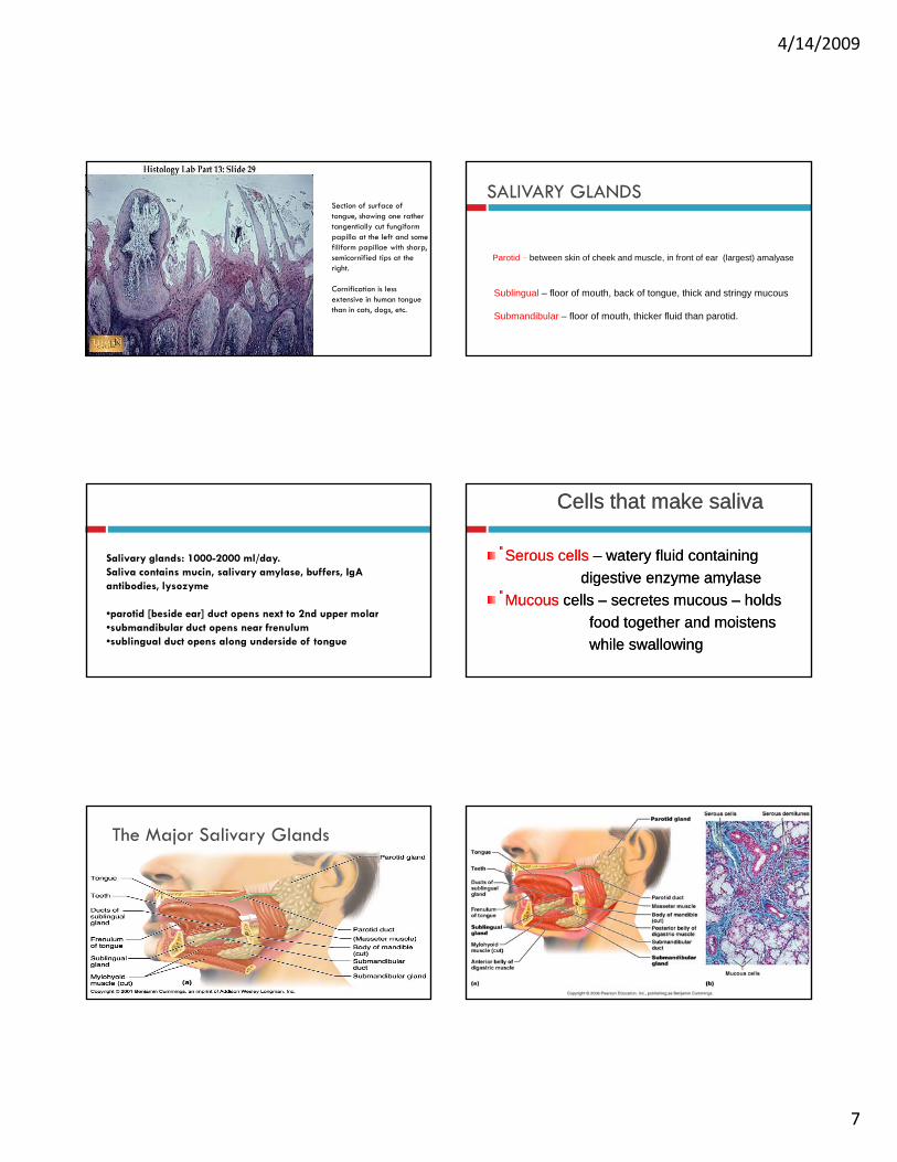
SALIVARY GLANDS
Parotid – between skin of cheek and muscle in front of ear (largest) amalyaseParotid between skin of cheek and muscle, in front of ear (largest) amalyase
Submandibular – floor of mouth, thicker fluid than parotid.
Sublingual – floor of mouth, back of tongue, thick and stringy mucous
Salivary glands: 1000-2000 ml/day. Saliva contains mucin, salivary amylase, buffers, IgAantibodies, lysozyme
•parotid [beside ear] duct opens next to 2nd upper molar•submandibular duct opens near frenulum•sublingual duct opens along underside of tongue
Cells that make salivaCells that make saliva
Serous cellsSerous cells –– watery fluid containing watery fluid containing digestive enzyme amylasedigestive enzyme amylasedigestive enzyme amylase digestive enzyme amylase
Mucous Mucous cells cells –– secretes mucous secretes mucous –– holds holds food together and moistens food together and moistens while swallowingwhile swallowing
41
The Major Salivary Glands

4/14/2009
8
The Salivary Glands - Ducted exocrine glands producing saliva. Two types of secretory cells are found in the glandular tissue:
1. Serous cells producing a watery secretion containing amylase.2. Mucous cells producing a viscous liquid containing the glycoprotein mucin.
Submandibular Glands - are bilaterally located at the median aspect of the mandibular angle. Their ducts bring saliva to the oral cavity at the base of the frenulum. They are mixed glands, containing approximately equal numbers of serous and mucous cells.
Sublingual Glands - are anterior to the submandibular glands under the tongue. Cells of these glands are mostly mucous producing. Very little amylase is found in this saliva.
Parotid Glands - are anterior and inferior to the external ears lying in a connective tissue capsule. Parotid ducts bring saliva into the vestibule along side of the second upper molar. The glandular cells are mostly serous.
The The FaucesFauces - are the passageway from the mouth to the pharynx. This short corridor is guarded by four pillars; the two palatoglossal arches are more anterior followed by the two palatopharyngeal arches. In between the two sets of arches on either side are the palatine tonsils. During swallowing, contraction of the muscles in these arches constricts the pillars preventing food from reentering the mouth.
Salivary GlandsCollectively produce and secrete saliva.
• a fluid that assists in the initial activities of digestion� Volume of saliva secreted daily ranges between 1.0 and 1.5 L.� Most is produced during mealtime, but� Smaller amounts are produced continuously to ensure that the oral cavity remains moist� Smaller amounts are produced continuously to ensure that the oral cavity remains moist.� Water makes up 99% of the volume of saliva.� Also contains a mixture of other components.� Three pairs of large, multicellular salivary glands:
•parotid glands•submandibular glands•sublingual glands
1. SialorrhoeaSialorrhoea: : Hypersecretion of saliva.S i ki iSeen in pregnancy, parkinsonism2. XerostomiaXerostomia: : Suppression of salivary secretionSeen in anxiety, stress, duct blockage, irradiation therapy.
The Parotid Glands
Largest salivary glands.� Each parotid gland is located anterior and inferior to the ear, partially overlying the masseter muscle.� P d b 25 30% f h li hi h i d d h h h id � Produce about 25–30% of the saliva, which is conducted through the parotid duct to the oral cavity.
The Submandibular GlandsInferior to the body of the mandible.Inferior to the body of the mandible.� Produce most of the saliva (about 60–70%).� A duct opens from each gland through a papilla in the floor of the mouth on the lateral sides of the lingual frenulum.Inferior to the tongue and internal to the oral cavity mucosa.Inferior to the tongue and internal to the oral cavity mucosa.� Each gland extends multiple tiny sublingual ducts that open onto the inferior surface of � Each gland extends multiple tiny sublingual ducts that open onto the inferior surface of the oral cavity, posterior to the submandibular duct papilla.� Contribute only about 3–5% of the total saliva.

4/14/2009
9
PAROTID SALIVARY GLANDStained with haematoxylin
and eosin1 - serous secretory units (acini)2 - intercalated excretory duct3 - striated excretory duct4 i t l b l t 4 - interlobular excretory duct5 - interlobular connective tissue septa
PAROTID SALIVARY GLANDStained with haematoxylin and eosin
1 - serous secretory units2 - striated excretory duct3 - interlobular excretory duct
PAROTID SALIVARY GLANDStained with haematoxylin and eosin 1 -
serous secretory units2 - intercalated excretory duct3 - striated excretory duct
PAROTID SALIVARY GLANDStained with haematoxylin
and eosin1 - serous secretory units2 myoepithelial cells2 - myoepithelial cells4 - interlobular excretory duct5 - interlobular connective tissue septa
PAROTID SALIVARY GLANDinterlobular excretory duct
Stained with haematoxylin and eosin1 - interlobular excretory duct2 - interlobular connective tissue septa
SUBLINGUAL SALIVARY GLANDStained with haematoxylin and eosin
1 - lobules of the glandg2 - interlobular connective tissue septa3 - interlobular excretory duct

4/14/2009
10
SUBLINGUAL SALIVARY GLANDStained with haematoxylin and eosin 1 -
mucous part of mixed secretory unit2 - serous part (serous demilune) of
mixed secretory unit3 - serous secretory unity4 - mucous secretory unit5 - intercalated excretory duct6 - striated excretory duct7 - interlobular excretory duct8 - interlobular connective tissue septa
SUBLINGUAL SALIVARY GLANDStained with haematoxylin and eosin
1 - mucous part of mixed secretory unit2 - serous part (serous demilune) of
mixed secretory unit3 - serous secretory unit4 - mucous secretory unit5 i h li l ll5 - myoepithelial cells
SUBLINGUAL SALIVARY GLANDStained with haematoxylin and eosin
1 - mucous part of mixed secretory unit2 - serous part (serous demilune) of
mixed secretory unit3 - serous secretory unit5 - myoepithelial cells6 - intercalated excretory duct7 - interlobular excretory duct8 - interlobular connective tissue septa
SUBMANDIBULAR SALIVARY GLANDStained with haematoxylin and eosin 1 -
serous secretory unit2 - mixed secretory unit3 - intercalated excretory duct4 - striated excretory duct5 - interlobular excretory duct6 - interlobular connective tissue septa7 - mucous part of mixed secretory unit8 - serous part (serous demilune) of
mixed secretory unit
SUBMANDIBULAR SALIVARY GLANDStained with haematoxylin and eosin
1 - serous secretory unit2 - mixed secretory unit3 - intercalated excretory duct4 - striated excretory ducty