from elements to perception: local and global processing ...pages.uoregon.edu/prd/tilt/spillmann,...
TRANSCRIPT

1 IntroductionJung, Baumgartner, Creutzfeldt, Gru« sseröthe founding fathers of the Freiburg schoolof neurophysiologyöthey are all gone now within a span of only eight years (1986 ^1993). It is an appropriate moment to pause and look back. Based on my memories,I would like to present this personal account of the development of visual neurophysiol-ogy and perception during the last forty years, with an emphasis on research conductedin Germany. I am, of course, well aware that ground-breaking work in the same fieldwas performed in several European countries, as well as on the other side of the Atlantic,in the United States.While these developments have been the subject of much discussion,many of the early experiments in single-cell visual neurophysiology were publishedin German journals and are therefore less known. This article claims to be neithersystematic nor comprehensive. It totally ignores, for example, what has been done formany decades in EEG recording, and only briefly mentions the new imaging methodsused to study functional brain topography in human observers. Rather, it attempts tobalance the scale and promote a trend in vision research that has been dear to myheart: linking visual psychophysics and neurophysiology.
I will use selected examples to show how during the period under considerationthe field of vision research has progressed from the study of elements to the study ofperceptions. By elements I mean dots, lines, and edges; by perceptionsöillusory contours,
From elements to perception: Local and global processingin visual neuronsÀ
Perception, 1999, volume 28, pages 1461 ^ 1492
Lothar SpillmannInstitute of Biophysics and Radiation Biology, Brain Research Unit, University of Freiburg,Hansastrasse 9, 79104 Freiburg, Germany; e-mail: [email protected] 24 February 1998, in revised form 4 January 1999
Abstract. Gestalt psychologists in the early part of the century challenged psychophysical notionsthat perceptual phenomena can be understood from a punctate (atomistic) analysis of the elementspresent in the stimulus. Their ideas slowed later attempts to explain vision in terms of single-cell recordings from individual neurons. A rapprochement between Gestalt phenomenology andneurophysiology seemed unlikely when the first ECVP was held in Marburg, Germany, in 1978.Since that time, response properties of neurons have been discovered that invite an interpretation ofvisual phenomena (including illusions) in terms of neuronal processing by long-range interactions,as first proposed by Mach and Hering in the last century.
This article traces a personal journey into the early days of neurophysiological visionresearch to illustrate the progress that has taken place from the first attempts to correlate single-cell responses with visual perceptions. Whereas initially the receptive-field properties of individualclasses of cellsöeg contrast, wavelength, orientation, motion, disparity, and spatial-frequencydetectorsöwere used to account for relatively simple visual phenomena, nowadays complex percep-tions are interpreted in terms of long-range interactions, involving many neurons. This changein paradigm from local to global processing was made possible by recent findings, in the cortex,on horizontal interactions and backward propagation (feedback loops) in addition to classicalfeedforward processing. These mechanisms are exemplified by studies of the tilt effect and tiltaftereffect, direction-specific motion adaptation, illusory contours, filling-in and fading, figure ^ground segregation by orientation and motion contrast, and pop-out in dynamic visual-noisepatterns. Major questions for future research and a discussion of their epistemological implica-tions conclude the article.
À Based on an invited lecture at the 20th European Conference on Visual Perception, Helsinki-Espoo, Finland, 27 August 1997.
DOI:10.1068/p2763
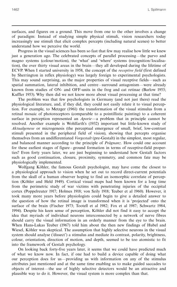
surfaces, and figures on a ground. This move from one to the other involves a changeof paradigm: Instead of studying simple physical stimuli, vision researchers todayincreasingly use stimuli that elicit complex percepts (including natural scenes) to betterunderstand how we perceive the world.
Progress in the visual sciences has been so fast that few may realise how little we knewjust a generation ago. The celebrated concepts of parallel processingöthe parvo andmagno systems (colour/motion), the `what' and `where' systems (recognition/localisa-tion), the over thirty visual areas in the brainöthey all developed during the lifetime ofECVP. When I started university in 1958, the concept of the receptive field (first definedby Sherrington in reflex physiology) was largely foreign to experimental psychologists.This may sound surprising, as the major properties of visual receptive fieldsösuch asspatial summation, lateral inhibition, and centre ^ surround antagonismöwere alreadyknown from studies of ON- and OFF-units in the frog and cat retinae (Barlow 1953;Kuffler 1953). Why then did we not know more about visual processing at that time?
The problem was that few psychologists in Germany (and not just there) read thephysiological literature, and, if they did, they could not easily relate it to visual percep-tion. For example, to Metzger (1961) the transformation of the visual stimulus from aretinal mosaic of photoreceptors (comparable to a pointillistic painting) to a coherentsurface in perception represented an Aporieöa problem that in principle cannot beresolved. Another example is Wohlfarth's (1932) important but little-known study ofAktualgenese or microgenesis (the perceptual emergence of small, brief, low-contraststimuli presented in the peripheral field of vision), showing that percepts organisethemselves from an undifferentiated Vorgestalt (pre-Gestalt) in the simplest, most regular,and balanced manner according to the principle of Pra« gnanz. How could one accountfor these earliest stages of figure ^ ground formation in terms of receptive-field proper-ties? Even forty years later, we are just beginning to understand how Gestalt factorssuch as good continuation, closure, proximity, symmetry, and common fate may bephysiologically implemented.
Wolfgang Ko« hler, the famous Gestalt psychologist, may have come the closest toa physiological approach to vision when he set out to record direct-current potentialsfrom the skull of a human observer hoping to find an isomorphic correlate of percep-tion (Ko« hler and Held 1949). Cortical visual maps had been known for a long timefrom the perimetric study of war victims with penetrating injuries of the occipitalcortex (Poppelreuter 1917; Holmes 1918; von Szily 1918; Teuber et al 1960). However, ittook many more years before physiologists could begin to give a detailed answer tothe question of how the retinal image is transformed when it is `projected' onto thesurface of the brain (Fischer 1973; Tootell et al 1982; Fox et al 1987; Schwartz 1984,1994). Despite his keen sense of perception, Ko« hler did not find it easy to accept theidea that myriads of individual neurons interconnected by a network of nerve fibresshould carry the visual information in an orderly manner from the eye to the brain.When Hans-Lukas Teuber (1967) told him about the then new findings of Hubel andWiesel, Ko« hler was skeptical. The assumption that highly selective neurons in the visualsystem should analyse (`dissect') a stimulus and mediate its contrast, polarity, brightness,colour, orientation, direction of motion, and depth, seemed to be too atomistic to fitinto the framework of Gestalt psychology.
On looking back forty-five years later, it seems that we could have predicted muchof what we know now. In fact, if one had to build a device capable of doing whatour perception does for usöproviding us with information on any of the stimulusattributes just mentioned and at the same time enabling us to make guided saccades toobjects of interestöthe use of highly selective detectors would be an attractive andplausible way to do it. However, the visual system is more complex than that.
1462 L Spillmann

1.1 The pioneersIn comparison to psychologists, physiologists had a much easier task unravelling thesecrets of vision and putting it, step by step, on a firm basis. They had been exposedto the neuron doctrine of Ramon y Cajal (1893) for more than fifty years. Three papersprepared the ground for scientific study. Adrian and Matthews (1927, 1928) determineda visual receptive field for the first time by recording responses to two or more smalllight spots from the optic nerve of the conger eel as a function of stimulusseparation. They found that the response latency for a given quantity of light remainedthe same regardless of whether it was concentrated into a crowded patch or spread outover several small areas, and that it was shorter than for each of the stimuli presentedindividually (spatial summation). Two years later, Hartline (1938, 1940) published twopapers on the receptive fields of optic nerve fibres in the frog. Therefore, the questionwhether a receptive-field organisation also existed in the mammalian brain seemed notonly timely but perfectly legitimate to physiologists. The first to lower an electrode intothe visual cortex of the cat and record action potentials from a single neuron were Rudolfvon Baumgarten and Gu« nter Baumgartner in Richard Jung's laboratory in Freiburg.
Figure 1 is taken from the classic article by Jung et al (1952). The upper trace showsan original recording from a neuron in area 17. The thin horizontal line below marks thelight stimulus. It can be seen that 20 ms after switching the light on, the neuron respondedwith an initial burst, followed by a few scattered spikes as the stimulus continued. Thesecond trace refers to the local EEG, recorded with a longer time constant. Jung termedthis kind of light-activated neuron, B-neuron (B for brighter). Its counterpart was calledD-neuron (D for darker).
After this pioneering work it took seven more years until the next large step wastaken by two research groups at the Massachusetts Institute of Technology (MIT) andthe Harvard Medical School. The authors of the first paper were Jerome Lettvin,Humberto Maturana, Warren McCulloch, and Walter Pitts, and the title of their 1959paper was ` What the frog's eye tells the frog's brain''. In this paper, the authors laidthe groundwork for the question of how the anatomy and physiology of the frog retinaand optic tectum might be related to the frog's vision (for a review see Gru« sser andGru« sser-Cornehls 1976). This is when the name `bug detector' came up among psychol-ogists. The term is derived from Barlow's (1953) paper where he writes (page 86):` The receptive field of an `on ^ off' unit would be nicely filled by the image of a flyat 2 in. distance and it is difficult to avoid the conclusion that the `on ^ off ' units arematched to this stimulus and act as `fly detectors' ''.
The second paper was by David Hubel and TorstenWiesel (1959) on ` Receptive fieldsof single neurones in the cat's striate cortex''. This was the first in a long and systematicseries of pioneering studies that eventually would earn their authors the Nobel prizein physiology and medicine. In the same year,Walter Rosenblith held a meeting at MITon Sensory Communication (proceedings published in 1961) to which he invited a great
light stimulus100 mV
100 ms
Figure 1. Action potentials of a cortical neuron in the cat in response to a continuous light stim-ulus (thin long horizontal line). Upper trace recorded with a short time constant, middle tracewith a long time constant. Bottom line 50 Hz-time mark. (From Jung et al 1952.)
Local and global processing in visual neurons 1463
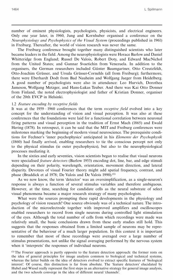
number of eminent physiologists, psychologists, physicists, and electrical engineers.Only one year later, in 1960, Jung and Kornhuber organised a conference on theNeurophysiology and Psychophysics of the Visual System (proceedings published in 1961)in Freiburg. Thereafter, the world of vision research was never the same.
The Freiburg conference brought together many distinguished scientists who laterbecame leaders in the field. Among the neurophysiologists were Horace Barlow and DanielWhitteridge from England; Russel De Valois, Robert Doty, and Edward MacNicholfrom the United States; and Gunnar Svaetichin from Venezuela. In addition to theorganisers, the German researchers included Gu« nter Baumgartner, Otto Creutzfeldt,Otto-Joachim Gru« sser, and Ursula Gru« sser-Cornehls (all from Freiburg); furthermore,there were Eberhardt Dodt from Bad Nauheim and Wolfgang Jaeger from Heidelberg.A good number of psychologists were also in attendance: Leo Hurvich, DorotheaJameson, Wolfgang Metzger, and Hans-Lukas Teuber. And there was Kai Otto Donnerfrom Finland, the noted electrophysiologist and father of Kristian Donner, organiserof the 20th EVCP in Helsinki.
1.2 Feature encoding by receptive fieldsIt was at the 1959 ^ 1960 conferences that the term receptive field evolved into a keyconcept for the understanding of vision and visual perception. It was also at theseconferences that the foundations were laid for a functional correlation between neuronalfiring patterns and visual perception in the tradition of Ernst Mach (1865) and EwaldHering (1878). In retrospect, it can be said that the MIT and Freiburg conferences weremilestones marking the beginning of modern visual neuroscience. The prerequisite condi-tions for Fechner's `inner psychophysics' anticipated in his Elemente der Psychophysik(1860) had finally arrived, enabling researchers to tie the conscious percept not onlyto the physical stimulus (ie outer psychophysics), but also to the neurophysiologicalprocesses mediating it.
In the sixties and early seventies, vision scientists began to realise that visual neuronswere specialised feature detectors (Barlow 1953) encoding dot, line, bar, and edge stimulidepending on their polarity, wavelength, orientation, movement direction, and lateraldisparity. Devotees of visual Fourier theory might add spatial frequency, contrast, andphase (Braddick et al 1978; De Valois and De Valois 1990).(1)
As we now know, the term `detector' was an oversimplification, as a single-neuron'sresponse is always a function of several stimulus variables and therefore ambiguous.However, at the time, searching for candidate cells as the neural substrate of selectvisual phenomena became a major research strategy of many psychologists.
What were the sources prompting these rapid developments in the physiology andpsychology of vision research? One source obviously was of a technical nature. The intro-duction of the microelectrode together with improved amplifiers and oscilloscopesenabled researchers to record from single neurons during controlled light stimulationof the eyes. Although the total number of cells from which recordings were made wasrelatively small, the basic conclusions drawn from these early studies still hold. Thissuggests that the responses obtained from a limited sample of neurons may be repre-sentative of the behaviour of a much larger population. In this context it is importantto remember that most of these recordings were averaged over a large number ofstimulus presentations, not unlike the signal averaging performed by the nervous systemwhen it `interprets' the responses of individual neurons.(1) The Fourier approach is partly opposite to the feature detection approach: the former rests onthe idea of general principles for image analysis common to biological and technical systems,whereas the latter builds on the idea of detectors evolved to extract specific features of `biologicalinterest'. Of course, this distinction is far from absolute. The `feature detectors' elucidated byHubel and Wiesel really represent the first steps in an alternative strategy for general image analysis,and the two schools converge in the idea of different neural channels'.
1464 L Spillmann

In recent years, the development of multiple-electrode techniques (Eckhorn et al1988; Kru« ger 1990) and powerful computers, as well as their availability and ease ofuse, has enlarged the number of cells that can be studied simultaneously. Of course,just because one records from a dozen or so neurons at once, this does not by itselfmean that one understands the nerve network (Kru« ger and Becker 1991). Also, withoutknowing what the nature of the sampling process is and how representative thosenumbers are, the real advantage of using multiple electrodes is difficult to assess. Thekey to modeling neuronal response patterns is the development of analytical tools forexamining interactions and temporal synchronies among those neuronsönot just themere collection of data.
2 Neuronal correlates of visual perceptionIn addition to the technical advances, the other source which contributed greatly totoday's understanding of visual function was conceptual. It implied that single-cellresponses are neuronal correlates of visual phenomena and, vice versa, visual phenomenaare perceptual correlates of neuronal mechanisms. This concept was most strongly advo-cated by Richard Jung, a clinical neurologist and neurophysiologist, and his twofriends, Donald MacKay, a communications engineer from Keele, England, and Hans-Lukas Teuber, the MIT neuropsychologist (figure 2).
Together they would later edit the Handbook of Sensory Physiology (1971 ^ 1981) whichremains a landmark achievement to this day. One continues to marvel how so manyof the phenomena that had been demonstrated and described by sensory physiologistsin the last century had been accounted for within a span of only twenty years after thefirst single-unit recordings.
The Freiburg school of neurophysiology contributed greatly to these early successes.By investigating the microphysiology of cortical neurons and their significance forvision and visual perception (Jung 1959), these researchers aimed to demonstrate that
Figure 2. Left: Richard Jung (1911 ^ 1986). Right: Donald MacKay (1922 ^ 1987) in the middle,Hans-Lukas Teuber (1916 ^ 1976) on the right, and Sir John Eccles (1903 ^ 1997) in the back-ground during the conference on ` Brain Mechanisms and Conscious Experience'' at the VaticanScientific Academy in 1966. (From Creutzfeldt 1990.)
Local and global processing in visual neurons 1465

the neuronal response (ie firing rate) presumably underlying a given perceptual quality(eg brightness) varied in parallel with a given stimulus parameter (luminance). In quicksuccession, the visual systems of fish, rabbit, and cat were studied, not only to elucidatethe processes underlying vision at the cellular level, but more specifically to find neuronalcorrelates of human perception.
What an exciting time it was for a young student to watch the neurologists at theend of a clinical day go to the crammed basement of the hospital and begin theirneurophysiological experiments which often lasted until the early morning (Dichgans1998). Encephale isole preparations were the rule then (not to my pleasure), glassmicropipettes were being used for recording, spike trains were recorded on film (notcommon then) and counted by hand to arrive at quantitative relationships. None ofthis research could have been done without the close cooperation of the ingeniousJan Friedrich To« nnies (formerly at the Rockefeller Institute of Medical Research andinventor of the differential amplifier) who developed many of the instruments; andthe technical assistance, as well as active participation, of the ever-present engineerHermann Kapp who had his workshop next door. Neurons could be heard cracklingfrom late at night until the early morning hours. It was the time of fast discoveries,something new emerging almost every month. I remember illustrious visitors andguests: Denise Albe-Fessard, Ragnar Granit, Giuseppe Moruzzi, Henri Hecain, JohnSzentagothai, John Eccles, Donald MacKay, David Hubel. Many more signed theomnipresent guest book.
Gru« sser was the first to recognise and defendöon philosophical groundsöthepotential of a combined psychophysical and neurophysiological approach (Gru« sser1956; Gru« sser and Gru« sser-Cornehls 1973; for a review see Przybyszewski 1997). In hisdissertation (1956) he clearly stated that although there can be no proof of a causalconnection between objective neuronal responses and subjective observations, we mayassume a mutual correlation (`wechselseitige Entsprechung') between one and the other,in the sense of Wundt's psychophysical parallelism. He quotes Wundt (1911, page 746):` Wherever systematic relationships exist between psychical and physical phenomena,they are neither identical nor can they be transformed into each other, as they are notcomparable; however, they are referred to each other ( einander zugeordnet') in such away that certain psychical processes lawfully correspond to certain physical processes,or as one may express it in a picture, both go parallel with each other'' (translationand italics by the author).
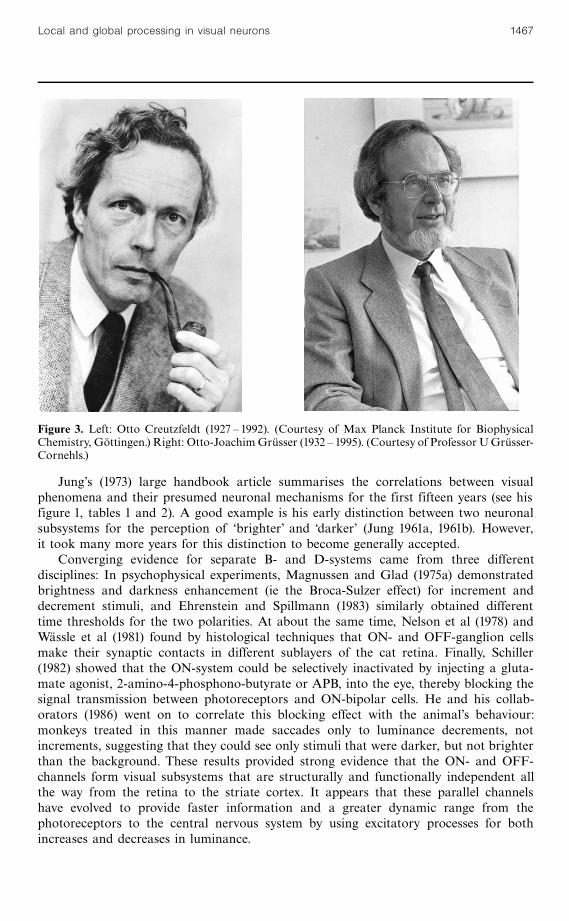
In 1961, Gru« sser and Carmen Rabelo, a doctoral student from Venezuela, showedthat the brightness enhancement of a light flickering at medium temporal frequencies(Bru« cke ^ Bartley effect) correlated with the peak discharge rates of retinal and cortexneurons in the cat (Gru« sser and Creutzfeldt 1957). Figure 3 portrays Otto Creutzfeldtand Otto-Joachim Gru« sser, after they had left Freiburg and became directors of theirrespective institutes in Go« ttingen and Berlin.
Further research on the possible neuronal correlates of perceptual phenomenadeveloped quickly in the late fifties and sixties. For example, the logarithmic increaseof the spike rate in retinal and geniculate neurons with physical light intensity (Fechner'slaw); the periodical change of B- and D-discharges corresponding to the Charpentier,Purkinje, and Hess afterimages following a bright flash (Gru« sser and Gru« tzner 1958);and the enhancement of neuronal activity near an edge, ie border contrast (Baumgartner1961), were demonstrated at that time. Sinusoidal flicker and moving gratingsöthepreferred stimuli of the next three decadesöwere introduced in Freiburg as early asthe late fifties. There might have been more. In a memorable retrospective, Jung (1975)muses why the orientation specificity of the cortical neurons that could have beeneasily found by a single qualitative experiment was missed by the Freiburg researchersin 1958.
1466 L Spillmann
Jung's (1973) large handbook article summarises the correlations between visualphenomena and their presumed neuronal mechanisms for the first fifteen years (see hisfigure 1, tables 1 and 2). A good example is his early distinction between two neuronalsubsystems for the perception of `brighter' and `darker' (Jung 1961a, 1961b). However,it took many more years for this distinction to become generally accepted.
Converging evidence for separate B- and D-systems came from three differentdisciplines: In psychophysical experiments, Magnussen and Glad (1975a) demonstratedbrightness and darkness enhancement (ie the Broca-Sulzer effect) for increment anddecrement stimuli, and Ehrenstein and Spillmann (1983) similarly obtained differenttime thresholds for the two polarities. At about the same time, Nelson et al (1978) andWa« ssle et al (1981) found by histological techniques that ON- and OFF-ganglion cellsmake their synaptic contacts in different sublayers of the cat retina. Finally, Schiller(1982) showed that the ON-system could be selectively inactivated by injecting a gluta-mate agonist, 2-amino-4-phosphono-butyrate or APB, into the eye, thereby blocking thesignal transmission between photoreceptors and ON-bipolar cells. He and his collab-orators (1986) went on to correlate this blocking effect with the animal's behaviour:monkeys treated in this manner made saccades only to luminance decrements, notincrements, suggesting that they could see only stimuli that were darker, but not brighterthan the background. These results provided strong evidence that the ON- and OFF-channels form visual subsystems that are structurally and functionally independent allthe way from the retina to the striate cortex. It appears that these parallel channelshave evolved to provide faster information and a greater dynamic range from thephotoreceptors to the central nervous system by using excitatory processes for bothincreases and decreases in luminance.
Figure 3. Left: Otto Creutzfeldt (1927 ^ 1992). (Courtesy of Max Planck Institute for BiophysicalChemistry, Go« ttingen.) Right: Otto-JoachimGru« sser (1932 ^ 1995). (Courtesy of Professor UGru« sser-Cornehls.)
Local and global processing in visual neurons 1467
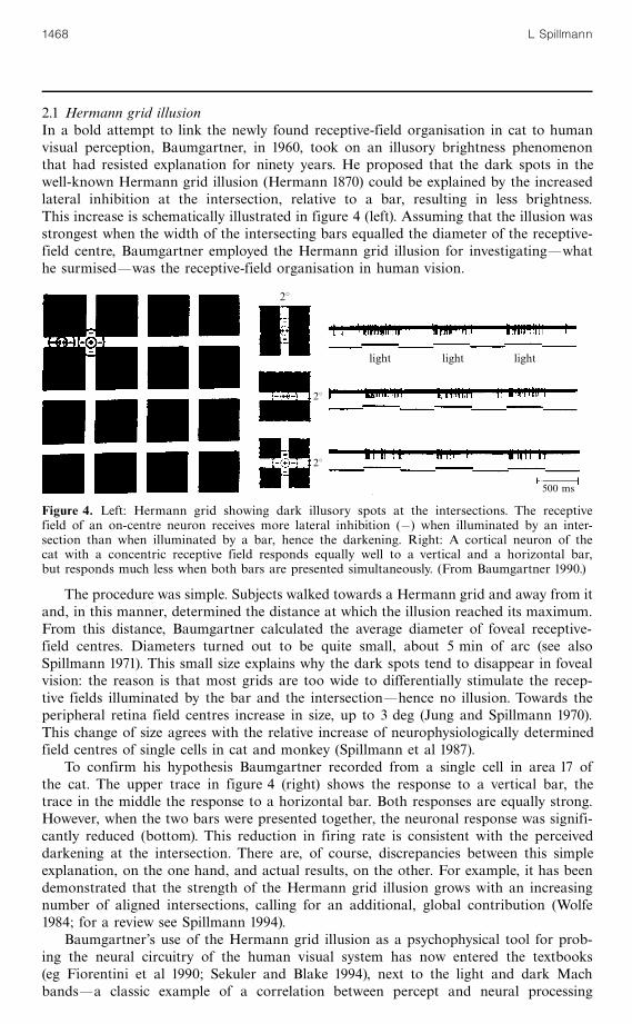
2.1 Hermann grid illusionIn a bold attempt to link the newly found receptive-field organisation in cat to humanvisual perception, Baumgartner, in 1960, took on an illusory brightness phenomenonthat had resisted explanation for ninety years. He proposed that the dark spots in thewell-known Hermann grid illusion (Hermann 1870) could be explained by the increasedlateral inhibition at the intersection, relative to a bar, resulting in less brightness.This increase is schematically illustrated in figure 4 (left). Assuming that the illusion wasstrongest when the width of the intersecting bars equalled the diameter of the receptive-field centre, Baumgartner employed the Hermann grid illusion for investigatingöwhathe surmisedöwas the receptive-field organisation in human vision.
The procedure was simple. Subjects walked towards a Hermann grid and away from itand, in this manner, determined the distance at which the illusion reached its maximum.From this distance, Baumgartner calculated the average diameter of foveal receptive-field centres. Diameters turned out to be quite small, about 5 min of arc (see alsoSpillmann 1971). This small size explains why the dark spots tend to disappear in fovealvision: the reason is that most grids are too wide to differentially stimulate the recep-tive fields illuminated by the bar and the intersectionöhence no illusion. Towards theperipheral retina field centres increase in size, up to 3 deg (Jung and Spillmann 1970).This change of size agrees with the relative increase of neurophysiologically determinedfield centres of single cells in cat and monkey (Spillmann et al 1987).
To confirm his hypothesis Baumgartner recorded from a single cell in area 17 ofthe cat. The upper trace in figure 4 (right) shows the response to a vertical bar, thetrace in the middle the response to a horizontal bar. Both responses are equally strong.However, when the two bars were presented together, the neuronal response was signifi-cantly reduced (bottom). This reduction in firing rate is consistent with the perceiveddarkening at the intersection. There are, of course, discrepancies between this simpleexplanation, on the one hand, and actual results, on the other. For example, it has beendemonstrated that the strength of the Hermann grid illusion grows with an increasingnumber of aligned intersections, calling for an additional, global contribution (Wolfe1984; for a review see Spillmann 1994).
Baumgartner's use of the Hermann grid illusion as a psychophysical tool for prob-ing the neural circuitry of the human visual system has now entered the textbooks(eg Fiorentini et al 1990; Sekuler and Blake 1994), next to the light and dark Machbandsöa classic example of a correlation between percept and neural processing
28
28
light light light
500 ms
28
Figure 4. Left: Hermann grid showing dark illusory spots at the intersections. The receptivefield of an on-centre neuron receives more lateral inhibition (ÿ) when illuminated by an inter-section than when illuminated by a bar, hence the darkening. Right: A cortical neuron of thecat with a concentric receptive field responds equally well to a vertical and a horizontal bar,but responds much less when both bars are presented simultaneously. (From Baumgartner 1990.)
1468 L Spillmann
(Ratliff 1965). The study of these contrast phenomena opened the door for using otherillusions as well. An example is Wertheimer's phi phenomenon. By measuring the largestdistance between two alternating stimuli across which apparent motion could be perceived,I attempted in my dissertation to determine the size of receptive fields of human movement-sensitive neurons. It turned out to be about 20 times larger than the receptive-field sizeobtained with the Hermann grid illusion (Kornhuber and Spillmann 1964).
To distinguish a neuronal receptive fieldöas defined by the response of a single cell toa moving or flashing light spotöfrom its correlate in human perception, Jung andSpillmann (1970) introduced the term perceptive field. In analogy to the receptive field,the perceptive field refers to an area on the retina (and in visual space) within which achange of stimulation changes the resultant percept (or threshold) in monkey and humanobservers. Its size and response properties can be inferred from psychophysics, such asthe Westheimer function (Westheimer 1965; Ransom-Hogg and Spillmann 1980). Like thereceptive field (Kuffler 1953; Rodieck 1965), the perceptive field is thought to possessan antagonistic centre ^ surround organisation. However, unlike the neurophysiologicallydefined receptive field, it reflects the simultaneous activity of a large number of neuronscontributing to a given percept (Spillmann et al 1987). Depending on the stimulus, theseneurons may reside at different levels within the visual system (retina, LGN, visual cortex).The assumption that many neurons interact synergistically (ie like one) to produce agiven percept is analogous to Barlow's (1972) concept of the most sensitive neuron.
When I presented these ideas to David Hubel and Torsten Wiesel in 1964, there wasa mixed response. While the latter took a cautious interest, the former was skeptical[about 25 years later, Livingstone and Hubel (1987, 1988) would publish their pivotalpapers on the functional correlates of the parvocellular and magnocellular streams].His advice then was: Never mind percepts, do straightforward physiology. This wassurprising because the Hubel and Wiesel detectors as they came to be known, appearedto lend themselves so readily to an explanation of a number of well-known perceptualeffects and aftereffects that could be used to noninvasively probe the human visualsystem. Let me give two examples.
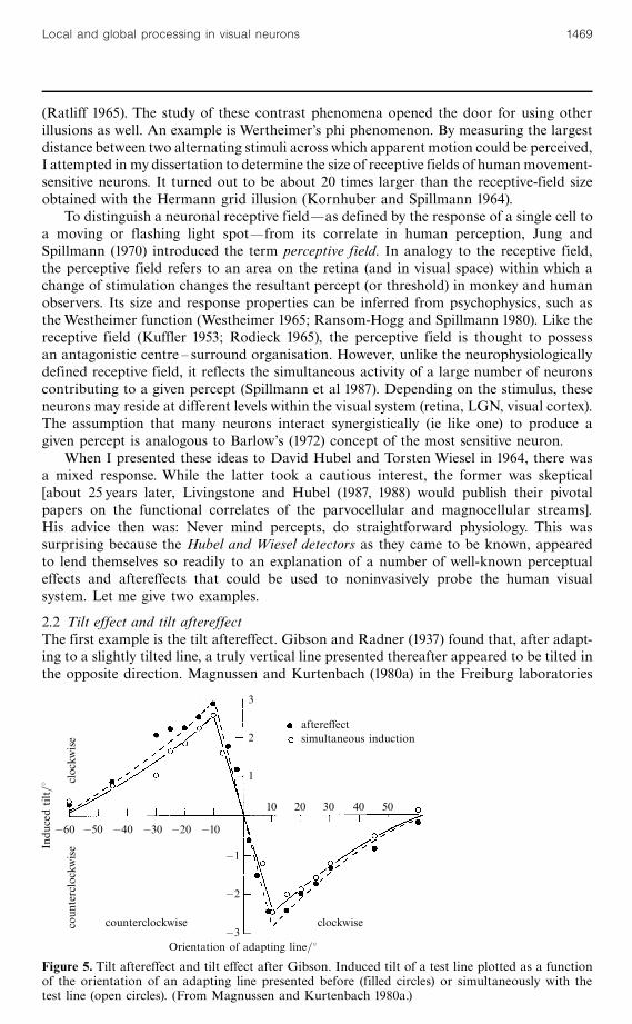
2.2 Tilt effect and tilt aftereffectThe first example is the tilt aftereffect. Gibson and Radner (1937) found that, after adapt-ing to a slightly tilted line, a truly vertical line presented thereafter appeared to be tilted inthe opposite direction. Magnussen and Kurtenbach (1980a) in the Freiburg laboratories
ÿ60 ÿ50 ÿ40 ÿ30 ÿ20 ÿ10
Indu
cedtilt=8
coun
terclockwise
clockw
ise
aftereffectsimultaneous induction
10 20 30 40 50
counterclockwise clockwise
ÿ1
ÿ2
ÿ3Orientation of adapting line=8
3
2
1
Figure 5. Tilt aftereffect and tilt effect after Gibson. Induced tilt of a test line plotted as a functionof the orientation of an adapting line presented before (filled circles) or simultaneously with thetest line (open circles). (From Magnussen and Kurtenbach 1980a.)
Local and global processing in visual neurons 1469
quantified this effect. In figure 5 (filled circles), the amount of induced tilt of the test lineis plotted as a function of the orientation of the adapting line. For example, when theadapting line was tilted 108 clockwise, the test line appeared to be tilted almost 38 counter-clockwise. For other adapting orientations, the induced tilt was smaller. Note that thecurve returns to the baseline at 608.
In that same study, Magnussen and Kurtenbach obtained nearly identical resultswhen the adapting line and the test line were presented simultaneously (open circles).This tilt effect had first been reported by Hofmann and Bielschowsky (1909), whofound that a line representing the subjective vertical was set to the opposite side whenviewed against a background of oblique contours. The same effect was later exploitedby Witkin (1959) in his well-known rod-and-frame experiment.
In figure 5, the two curves for tilt and tilt aftereffect superimpose almost perfectly. Theclose agreement, with and without preceding adaptation, speaks against an explanationof the tilt aftereffect by neuronal `fatigue' or `satiation' (the neuron becomes less sensitivebecause of its own previous activity). Instead, it suggests that both effects are based oninhibitory interactions between orientationally tuned units in the human visual system(Blakemore et al 1970; Blakemore and Tobin 1972). Magnussen and Kurtenbach (1980b)provided an even more compelling argument in support of the inhibition hypothesis byshowing that a second adapting line could weaken the effect exerted by the first, presumablyby disinhibition. In their experiment they adapted subjects to two stimuli simultaneously,holding the orientation of one constant, while varying the orientation of the other. Thefatigue hypothesis would predict an increase in the tilt aftereffect with dual-orientationadaptation as compared to adapting to the most effective of the two orientations alone,whereas the inhibition hypothesis predicted a decrease in aftereffect magnitude. Theresults showed a reduction in the aftereffect magnitude in accordance with the latter.
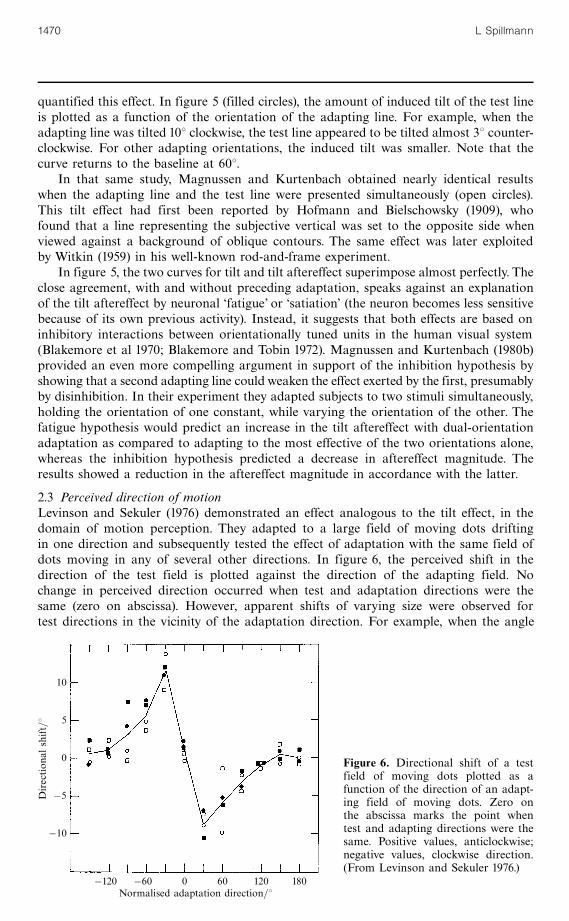
2.3 Perceived direction of motionLevinson and Sekuler (1976) demonstrated an effect analogous to the tilt effect, in thedomain of motion perception. They adapted to a large field of moving dots driftingin one direction and subsequently tested the effect of adaptation with the same field ofdots moving in any of several other directions. In figure 6, the perceived shift in thedirection of the test field is plotted against the direction of the adapting field. Nochange in perceived direction occurred when test and adaptation directions were thesame (zero on abscissa). However, apparent shifts of varying size were observed fortest directions in the vicinity of the adaptation direction. For example, when the angle
10
5
0
ÿ5
ÿ10
Direction
alshift=8
ÿ120 ÿ60 0 60 120 180Normalised adaptation direction=8
Figure 6. Directional shift of a testfield of moving dots plotted as afunction of the direction of an adapt-ing field of moving dots. Zero onthe abscissa marks the point whentest and adapting directions were thesame. Positive values, anticlockwise;negative values, clockwise direction.(From Levinson and Sekuler 1976.)
1470 L Spillmann
between the two kinds of stimuli was 308, the test field appeared to be shifted 108away from the direction of adaptation, resulting in a 408 apparent difference. Thisperceptual displacement is three times larger than for tilt. Note that the positions ofthe maximum and minimum as well as the points where the curve asymptotes arealso three times further away from zero. These differences suggest a much broadertuning range of the motion channel than of the orientation channel.
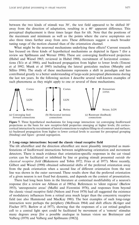
What might be the neuronal mechanisms underlying these effects? Current researchhas focused on three kinds of hypothetical mechanisms as depicted in figure 7 (for areview see Spillmann and Werner 1996). These are: converging feedforward projection(Hubel and Wiesel 1965; reviewed in Hubel 1988), recruitment of horizontal connec-tions (Ts'o et al 1986), and backward propagation from higher to lower levels (Tononiet al 1992; Munk et al 1995) including the lateral geniculate nucleus (Murphy andSillito 1996). The study of these mechanisms represents a shift in paradigm and hascontributed greatly to a better understanding of large-scale perceptual phenomena duringthe last ten years. In the following section I describe several well-known examples ofsuch phenomena as they might apply to one or several of these mechanisms.
3 Long-range interactions: beyond the classic visual receptive fieldThe tilt aftereffect and the direction aftereffect are most plausibly interpreted as mani-festations of feedforward interactions between neighbouring orientation and movementdetectors. There is much evidence that orientation-specific responses in the cat visualcortex can be facilitated or inhibited by line or grating stimuli presented outside theclassical receptive field (Blakemore and Tobin 1972; Fries et al 1977). More recently,Gilbert and Wiesel (1990) obtained substantial shifts of the preferred orientation awayfrom the peak orientation when a second line of different orientation from the testline was shown in the outer surround. These results show that the preferred orientationof a given neuron is not fixed but dynamic, and depends on the context of presentation.
There had long been hints in the literature at contextual modifiability of a neuron'sresponse (for a review see Allman et al 1985a). References to `silent regions' (Barlow1953), `unresponsive areas' (Maffei and Fiorentini 1976), and responses from beyondthe classic visual receptive field (Nelson and Frost 1978) had all suggested the existenceof modulatory influences from a retinal area much larger than the concentric receptivefield (see also Hammond and MacKay 1981). The best examples of such long-rangeinteraction were perhaps the periphery (McIlwain 1964) and shift effects (Kru« ger andFischer 1973; Barlow et al 1977), showing that the responses of a cat retinal ganglioncell to a central light spot could be facilitated by movement of a `remote' stimulusmany degrees away [for a possible analogue in human vision see Breitmeyer andValberg (1979) and Valberg and Spillmann (1982)].
(a) Converging feed- (b) Horizontal intrinsic (c) Reentrant (feedback)forward connections connections connections
Cortex Cortex Extrastriatecortex
LGN LGN
Retina Retina Striate, LGN
Figure 7. Three hypothetical mechanisms for long-range interaction: (a) converging feedforwardprojections as the basis for new receptive-field properties emerging at higher levels; (b) cortico-cortical interaction by recruitment of lateral connections to explain filling-in of contours and surfaces;(c) backward propagation from higher to lower cortical levels to account for perceptual grouping(binding) and figure ^ ground segregation.
Local and global processing in visual neurons 1471
Regrettably, these early ideas did not receive as much attention as they deservedbecause neither the physiological nor the psychological Zeitgeist was ready to deal withthem. A receptive field at that time was considered a fixed structural entity by mostresearchers, limited by the dendritic tree of the neuron, and it was difficult to conceiveof additional inputs from outside the receptive field capable of modifying the responsethrough contextual stimuli. This, however, was a prime requirement if neuronal responsepatterns were to account for large-scale perceptual phenomena. The breakthroughoccurred with the neurophysiological study of illusory contours by Gu« nter Baumgartnerand his co-workers Esther Peterhans and Ru« diger von der Heydt, in 1984. Their discoveryof neurons in area V2 of the macaque responding to what we perceive as partiallyoccluded patterns bounded by illusory contours, was a major achievement that carriedneurophysiology beyond the elementary dot, line, and edge stimuli used in previousdecades.
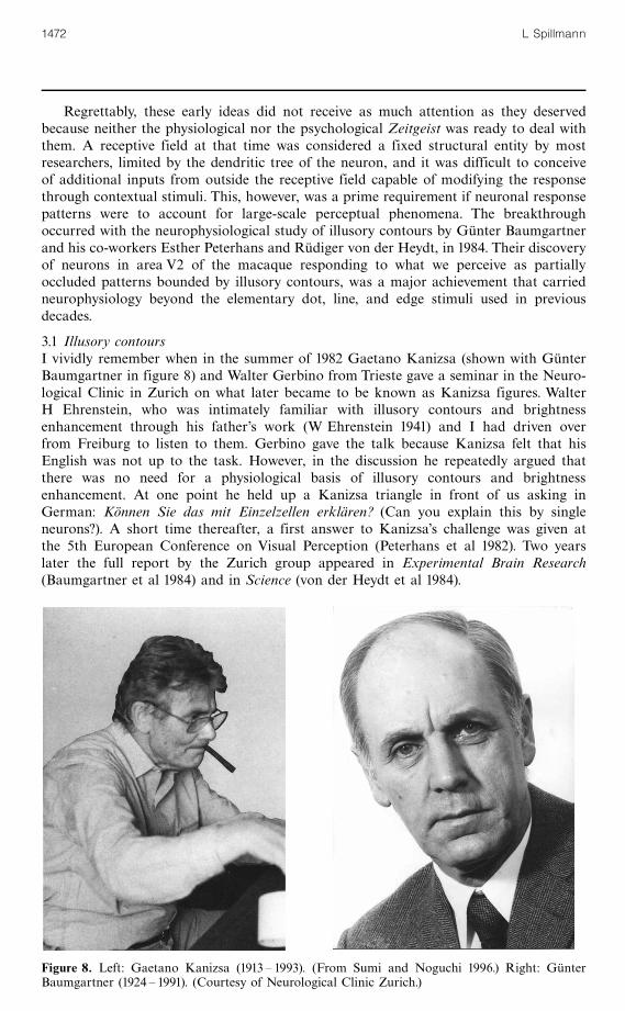
3.1 Illusory contoursI vividly remember when in the summer of 1982 Gaetano Kanizsa (shown with Gu« nterBaumgartner in figure 8) and Walter Gerbino from Trieste gave a seminar in the Neuro-logical Clinic in Zurich on what later became to be known as Kanizsa figures. WalterH Ehrenstein, who was intimately familiar with illusory contours and brightnessenhancement through his father's work (W Ehrenstein 1941) and I had driven overfrom Freiburg to listen to them. Gerbino gave the talk because Kanizsa felt that hisEnglish was not up to the task. However, in the discussion he repeatedly argued thatthere was no need for a physiological basis of illusory contours and brightnessenhancement. At one point he held up a Kanizsa triangle in front of us asking inGerman: Ko« nnen Sie das mit Einzelzellen erkla« ren? (Can you explain this by singleneurons?). A short time thereafter, a first answer to Kanizsa's challenge was given atthe 5th European Conference on Visual Perception (Peterhans et al 1982). Two yearslater the full report by the Zurich group appeared in Experimental Brain Research(Baumgartner et al 1984) and in Science (von der Heydt et al 1984).
Figure 8. Left: Gaetano Kanizsa (1913 ^ 1993). (From Sumi and Noguchi 1996.) Right: Gu« nterBaumgartner (1924 ^ 1991). (Courtesy of Neurological Clinic Zurich.)
1472 L Spillmann
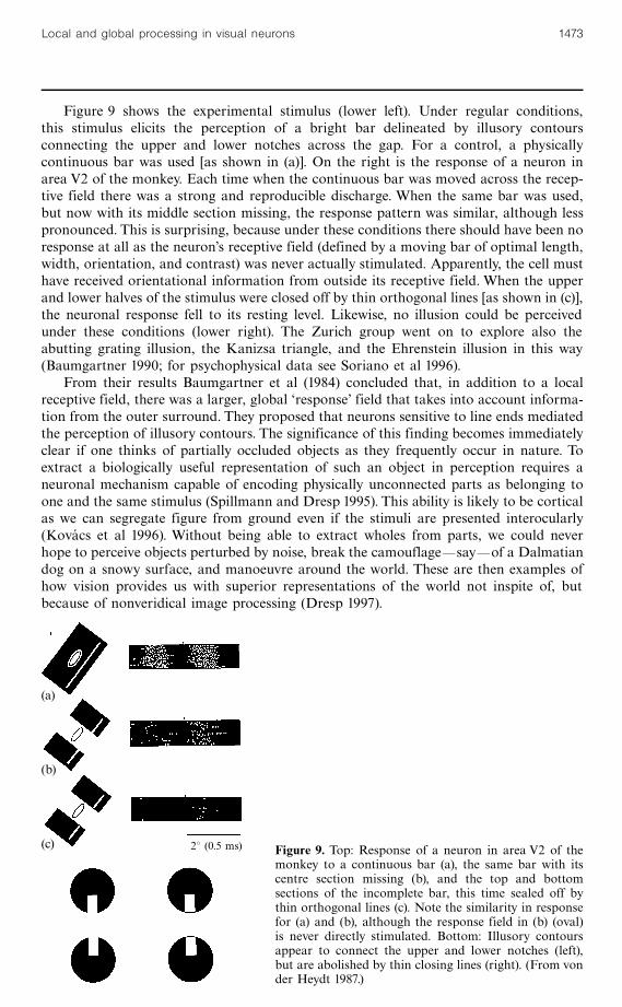
Figure 9 shows the experimental stimulus (lower left). Under regular conditions,this stimulus elicits the perception of a bright bar delineated by illusory contoursconnecting the upper and lower notches across the gap. For a control, a physicallycontinuous bar was used [as shown in (a)]. On the right is the response of a neuron inarea V2 of the monkey. Each time when the continuous bar was moved across the recep-tive field there was a strong and reproducible discharge. When the same bar was used,but now with its middle section missing, the response pattern was similar, although lesspronounced. This is surprising, because under these conditions there should have been noresponse at all as the neuron's receptive field (defined by a moving bar of optimal length,width, orientation, and contrast) was never actually stimulated. Apparently, the cell musthave received orientational information from outside its receptive field. When the upperand lower halves of the stimulus were closed off by thin orthogonal lines [as shown in (c)],the neuronal response fell to its resting level. Likewise, no illusion could be perceivedunder these conditions (lower right). The Zurich group went on to explore also theabutting grating illusion, the Kanizsa triangle, and the Ehrenstein illusion in this way(Baumgartner 1990; for psychophysical data see Soriano et al 1996).
From their results Baumgartner et al (1984) concluded that, in addition to a localreceptive field, there was a larger, global `response' field that takes into account informa-tion from the outer surround. They proposed that neurons sensitive to line ends mediatedthe perception of illusory contours. The significance of this finding becomes immediatelyclear if one thinks of partially occluded objects as they frequently occur in nature. Toextract a biologically useful representation of such an object in perception requires aneuronal mechanism capable of encoding physically unconnected parts as belonging toone and the same stimulus (Spillmann and Dresp 1995). This ability is likely to be corticalas we can segregate figure from ground even if the stimuli are presented interocularly(Kovacs et al 1996). Without being able to extract wholes from parts, we could neverhope to perceive objects perturbed by noise, break the camouflageösayöof a Dalmatiandog on a snowy surface, and manoeuvre around the world. These are then examples ofhow vision provides us with superior representations of the world not inspite of, butbecause of nonveridical image processing (Dresp 1997).
(a)
(b)
(c) 28 (0.5 ms) Figure 9. Top: Response of a neuron in area V2 of themonkey to a continuous bar (a), the same bar with itscentre section missing (b), and the top and bottomsections of the incomplete bar, this time sealed off bythin orthogonal lines (c). Note the similarity in responsefor (a) and (b), although the response field in (b) (oval)is never directly stimulated. Bottom: Illusory contoursappear to connect the upper and lower notches (left),but are abolished by thin closing lines (right). (From vonder Heydt 1987.)
Local and global processing in visual neurons 1473
Up to then, perceptual filling-in of gaps was tentatively explained either by theGestalt factors of good continuation and proximity (bottom ^ up), or by problem solv-ing (top ^ down) strategies (Gregory 1987). It now seems that neural processes such asactivation of end-stopped cells (Baumann et al 1997) may cooperate with mechanismsintegrating information from similarly oriented receptive fields to enable these kindsof perception (Schmidt et al 1997). In support of these ideas, Dresp and Bonnet (1995)demonstrated psychophysically that a near-threshold line when superimposed onto anillusory contour will be more easily detected, in accordance with subthreshold sum-mation. Furthermore, Dresp (1993) showed that a subthreshold target line presentedcollinearly with one or two inducers became visible presumably owing to lateral facili-tation between oriented perceptive fields.
Using the Westheimer function, Yu and Essock (1996) and Yu and Levi (1997) haverecently measured the spatial extent of such interactions in an attempt to define theperceptive fields of end-stopped cells in human observers (see also Soriano et al 1996).In analogy to Westheimer's original study (1965), log threshold for a target line firstincreased with background length, reached a peak, and then decreased again beforelevelling off. The background at which the curve peaked was taken as a psychophysicalmeasure of the perceptive field centre while the background length at which thecurve reached a plateau was interpreted as a measure of the centre plus end zones. Fora target 10 min of arc long presented perifoveally, the length of the centre was found tobe approximately 18 min of arc, while the total length was approximately 45 min of arc.Results suggest that units similar to those found in the monkey may also exist in thehuman visual cortex.
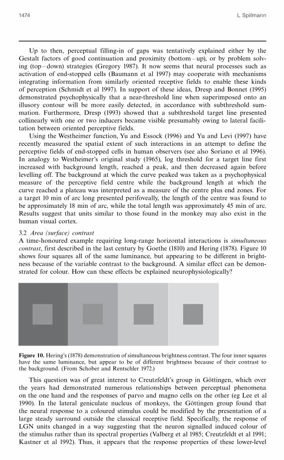
3.2 Area (surface) contrastA time-honoured example requiring long-range horizontal interactions is simultaneouscontrast, first described in the last century by Goethe (1810) and Hering (1878). Figure 10shows four squares all of the same luminance, but appearing to be different in bright-ness because of the variable contrast to the background. A similar effect can be demon-strated for colour. How can these effects be explained neurophysiologically?
This question was of great interest to Creutzfeldt's group in Go« ttingen, which overthe years had demonstrated numerous relationships between perceptual phenomenaon the one hand and the responses of parvo and magno cells on the other (eg Lee et al1990). In the lateral geniculate nucleus of monkeys, the Go« ttingen group found thatthe neural response to a coloured stimulus could be modified by the presentation of alarge steady surround outside the classical receptive field. Specifically, the response ofLGN units changed in a way suggesting that the neuron signalled induced colour ofthe stimulus rather than its spectral properties (Valberg et al 1985; Creutzfeldt et al 1991;Kastner et al 1992). Thus, it appears that the response properties of these lower-level
Figure 10.Hering's (1878) demonstration of simultaneous brightness contrast.The four inner squareshave the same luminance, but appear to be of different brightness because of their contrast tothe background. (From Schober and Rentschler 1972.)
1474 L Spillmann

neurons are consistent with perceptual phenomena such as colour contrast and colourconstancy, features that had previously been attributed to extrastriate V4 neurons(Zeki 1983). A well-known example is Land's (1983) Mondrian pattern where a givenpatch is perceived as `red' regardless of changes in illumination.
In addition to the study of patches of different colour as in a Mondrian one mightask how sustained perception of brightness and colour of large uniform areas is mediated.It is generally assumed that most neurons in areas V1 and V2 respond strongly to linesand edges, but respond poorly or not at all to constant illumination within theirreceptive fields. Why then do we see uniform surfaces and not mere outlines [ie Marr's(1982) primal sketch]? A plausible answer is: because of filling-in through long-rangeinteractions from the edge (Spillmann and Werner 1996; Pessoa et al 1998). Indeed,when a steady stimulus of uniform luminance was shown within the classical receptivefield while the background luminance was modulated well beyond the receptive-fieldarea, the response of the neuron to the uniform stimulus could be modified (Paradisoet al 1996; Rossi et al 1996). This finding indicates long-range interactions betweencortical neurons bridging the size of local receptive fields.
There is a large body of psychophysical studies in human observers showing thatbrightness and colour can be induced from the surround (eg Magnussen and Glad1975b; De Valois et al 1986; Rossi and Paradiso 1996). The stimuli used in these experi-ments were centre ^ surround configurations or black-and-white gratings. By modulatingthe luminance of the surround, the brightness of the enclosed area(s) could be madeto perceptually change in counterphase (Rossi and Paradiso 1996), not unlike an after-image surrounded by a contrast-reversing annulus (Gerling and Spillmann 1987). Lowspatial (0.03 ^ 2 cycles degÿ1 ) and temporal (52:5 Hz) frequencies were best. Induc-tion time depended on distance, suggesting a speed of brightness propagation between80 ^ 180 deg sÿ1 (Paradiso and Nakayama 1991; Paradiso and Hahn 1996; Rossi andParadiso 1996). On the other hand, Davey et al (1998), using the Craik ^O'Brien ^Cornsweet illusion to estimate the speed of brightness propagation, give a much lowervalue of only about 20 deg sÿ1.
A highly simplistic mechanism that could potentially account for these observations isillustrated in figure 11. Shown (on the left) are three neurons in the visual cortex of the cat.Normally, each cell responds only to illumination of its receptive field and, thus, deaffer-entation of a cortical cell by laser photocoagulation of the retina would render this cellunresponsive (right). As a consequence of the laser burn, there would be a small, irrevers-ible scotoma in visual perception. However, we now know that this loss of function canin part be compensated. For example, in the cat, it was found that after only a few minutesa massive reorganisation took place causing the deafferented cell to resume firing whenlight fell onto adjacent sections of the retina (Kaas et al 1990; Gilbert and Wiesel 1992).
Receptive fields
Cortical cellsA B C A B C
retinal lesion
Figure 11. Model for filling-in of a scotoma by intracortical horizontal connections (schematic).Shortly after a retinal lesion, the deafferented cell (B) can be reactivated by illumination of theneighbouring areas through signals from A and C (thick arrows, right). As a consequence, thescotoma becomes invisible. Sustained brightness perception on uniform surfaces (left) may be simi-larly explained by maintained edge signals from the surround. (From Spillmann and Werner 1996.)
Local and global processing in visual neurons 1475

It thus appears as though cells that had their receptive fields in the lesioned areawere now connected to portions of the retina corresponding to regions outside thescotoma (Eysel et al 1998).
One way to explain this reactivation by remapping is by assuming that signals travel-ling from neighbouring receptive fields to cells A and C are passed on in the cortexto cell B through long horizontal (tangential) connections. These cortico-cortical con-nections are assumed to be functionally present all the time; however, in the case of alocal deafferentation, their influence may become potentiated owing to disinhibition.In this manner, the silenced cell B would acquire a receptive field that is displacedrelative to and larger than its former receptive field. As a result of this functionalreorganisation, the visual scotoma is thought to fill in from the surround so that auniform surface would be perceived despite the absence of signals from the lesionedarea (Safran and Landis 1996). Actually, many more cells and receptive fields wouldhave to be considered to provide for this kind of neuroplasticity.(2)
3.3 Filling-in and fading of artificial scotomataHorizontal axons (up to 7 mm long) providing lateral synaptic inputs have indeedbeen found in the cat and monkey cortex (Gilbert 1992; Lund et al 1993). Undernormal stimulus conditions, the same mechanism that is thought to be responsible forthe filling-in of a retinal scotoma may underlie the perception of uniform surfaces.Let us take again cell B (figure 11, left). As long as there is a signal propagating theinformation from the edge (cells A, C), the brightness and colour within the enclosedarea (receptive field of B) would be kept alive. However, if the edge signal decreasesowing to adaptation (no eye movements), the enclosed area would assume the brightnessand colour of the surround and thereby become invisible (ie fading). Curiously, the modeldoes not explain why only the enclosed figure, not the ground, is affected by this process.Only if there are no borders at all as in a Ganzfeld, brightness and colour across the entirevisual field will rapidly fade and ultimately be replaced by one's Eigengrau (Knau andSpillmann 1996, 1997).
Fading and filling-in of enclosed areas are exactly what is perceived with strictfixation. Neumeyer and Spillmann (1977) and Ramachandran and Gregory (1991) showedthat in the absence of eye movements even large uniform areas (so-called artificialscotomata) tend to fill in and within seconds become embedded in the background. Forexample, a centrally fixated, red disk presented on a green background will rapidlydisappear from vision (especially if surrounded by a diffuse border) and be replaced bya uniformly green surface (Krauskopf 1963).
Ramachandran and Gregory (1991) demonstrated that fading was not restrictedto a uniform backgroundöon textured backgrounds it occurred as well. Even moresurprising, a grey target presented on the background of dynamic visual noise, such asa detuned TV set, faded from view within seconds. This was unexpected as the targetcontour was redefined with each new frame and thus should have persisted over time.However, the opposite was the case: On the dynamic noise background fading occurredfaster than on a uniform background (Spillmann and Kurtenbach 1992), possibly owingto adaptation of neurons processing kinetic contours (Allman et al 1985b; Marcar et al1995). Even more intriguing, when observers so adapted viewed a plain surface, theyperceived graininess (`dust') and dynamic twinkle in the area corresponding to thefilled-in patch (eg Hardage and Tyler 1995). This aftereffect spread over an area aslarge as the entire hemifield, suggesting long-range horizontal interactions (Tyler andHardage 1998).
(2) An alternative explanation of short-term plasticity in terms of an overall increase in cell respon-siveness, rather than a dynamic alteration of receptive-field structure, has been proposed (DeAngeliset al 1995). As for long-term plasticity by sprouting see Eysel and Schweigart (1999).
1476 L Spillmann

What may be the neurophysiological mechanism underlying such extensive spreading?Fading and filling-in in monkey and man were studied by De Weerd et al (1995, 1998) whoused the static stimulus shown in figure 12. The monkey had been trained to fixate a fixa-tion point (FP) and to release a lever as soon as the white square disappeared from vision.Figure 13 shows the responses of a neuron in area V3. The continuous line representsthe neuronal response obtained when the artificial scotoma in the stimulus was present,whereas the dotted curve shows the response when the artificial scotoma was absent(control). In both cases, the receptive field of the neuron was located completely insidethe white square. Initially, the neuronal activity elicited by the stimulus plus artificialscotoma was low. However, within seconds the response increased and approachedthe firing level for the control stimulus, at the same time when human observersreported fading of the white square (shaded area). The authors therefore propose thatthe increase of the neural activity reflects perceptual filling-in (De Weerd et al 1995).
3.4 Figure ^ ground segregation by orientation and motion contrastWhat about the opposite of fading: pop-out and salience of a stimulus? Van Essen et al(1991) and Lamme (1995) studied this question in the monkey using orientation contrast.Figure 14 (from Lamme) shows two kinds of stimuli, one that differs from the orientation
FP
Figure 12. Fading and filling-in of anartificial scotoma on a textured back-ground. The white square disappearsfrom view after briefly fixating on FP.(From De Weerd et al 1995.)
40
30
20
10
0
Respo
nse=spikes
sÿ1
0 2 4 6 8 10 12Time=s
no hole filling-in
hole
a
b
Figure 13. Response of a cell in area V3 of the monkey during fixation. The continuous curverefers to the stimulus shown in figure 12 (with the white square), the dotted curve to the samestimulus but without the square (texture only). The receptive field of the neuron was located wellwithin the square. The shaded section marks the time when the square faded for human observers.(From De Weerd et al 1995.)
Local and global processing in visual neurons 1477
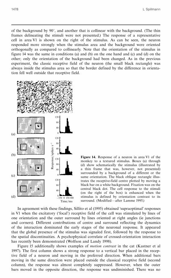
of the background by 908, and another that is collinear with the background. (The thinframes delineating the stimuli were not presented.) The response of a representativecell in area V1 is shown on the right of the stimulus. As can be seen, the neuronresponded more strongly when the stimulus area and the background were orientedorthogonally as compared to collinearly. Note that the orientation of the stimulus infigure 14 was the same in conditions (a) and (b) on the one hand and (c) and (d) on theother; only the orientation of the background had been changed. As in the previousexperiment, the classic receptive field of the neuron (the small black rectangle) wasalways inside the stimulus area so that the border defined by the difference in orienta-tion fell well outside that receptive field.
In agreement with these findings, Sillito et al (1995) obtained `supraoptimal' responsesin V1 when the excitatory (`focal') receptive field of the cell was stimulated by lines ofone orientation and the outer surround by lines oriented at right angles (ie junctionsand corners). Different contributions of centre and surround reflecting the dynamicsof the interaction dominated the early stages of the neuronal response. It appearedthat the global presence of the stimulus was signaled first, followed by the response tothe spatial discontinuities. A psychophysical correlate of crossed-orientation interactionhas recently been demonstrated (Wolfson and Landy 1998).
Figure 15 additionally shows examples of motion contrast in the cat (Kastner et al1997). The first column shows a strong response to a vertical bar placed in the recep-tive field of a neuron and moving in the preferred direction. When additional barsmoving in the same direction were placed outside the classical receptive field (secondcolumn), the response was almost completely suppressed. However, when the outerbars moved in the opposite direction, the response was undiminished. There was no
(a)
(b)
(c)
(d)
120
80
40
0
120
80
40
0
120
80
40
0
120
80
40
0
Respo
nse=spikes
sÿ1
ÿ150 0 150300Time=ms
Figure 14. Response of a neuron in area V1 of themonkey to a textured stimulus. Boxes (a) through(d) show schematically the stimulus (illustrated bya thin frame that was, however, not presented)surrounded by a background of a different or thesame orientation. The black oblique rectangle illus-trates the receptive-field centre plotted by moving ablack bar on a white background. Fixation was on thecentral black dot. The cell response to the stimuli(on the right of the box) is enhanced when thestimulus is defined by orientation contrast to itssurround. (Modifiedöafter Lamme 1995.)
1478 L Spillmann

effect with surround motion alone. These results are consistent with the perceptualsalience of the stimuli. Recent work suggests that under conditions of low salience theinhibitory centre ^ surround interactions for stimuli of this kind may be under thecontrol of extensive feedback from higher areas (Bullier et al 1996; Nowak and Bullier1997). For example, inactivation of area V2 would be expected to diminish the differ-ence between the results for stimuli 0 and 1 on one hand, and 4 and 5 on the other.
The findings by Lamme (1995) and Kastner et al (1997) may be interpreted interms of figure ^ ground segregation occurring as early as the primary visual cortex.Similar results were obtained for borders defined by luminance, colour, texture, and depth(Zipser et al 1996; for visually evoked potentials see Bach and Meigen 1997). To explainthese results, long-range signals arising at the edge of the stimulus and propagated to thereceptive field of the neuron are required.
3.5 Grouping by coherent motionPerhaps the most powerful example of a correlation between single-cell neurophysiologyand perception is perceptual grouping by coherent motion. For instance, a circle consist-ing of regularly spaced dots on a transparent acetate sheet cannot be discerned whensuperimposed onto a field of random dots, as long as both are stationary. However, assoon as the figure (or the ground) is moved, the circle is immediately perceived. Thispop-out effect testifies to the enormous power of coherent motion as a segmentationfactor. The Gestaltists would have attributed this effect to the factor of common fate(Wertheimer 1923; Metzger 1953).
Newsome et al (1989) reported neurons in areas V1 and MT in the behaving monkeythat respond selectively to coherent motion. Such cells have very large receptive fields;and they require only 4%^ 7% of coherently moving dots (relative to the total number)to produce a response, even if the target dots are widely separated in space. Moreimportantly, behavioural and neurophysiological thresholds in the same animal werethe same suggesting that the behavioural response may depend on the activity of arelatively small population of neurons (Britten et al 1992).
This conclusion is made even stronger by the finding that the perceived directionof motion can be biased by electrical microstimulation of a cluster of neurons inarea MT (Salzman et al 1992; Salzman and Newsome 1994). When a small group ofMT neurons was stimulated, the perception of the monkey (as inferred from its behaviour)was shifted towards the motion direction preferred by this group of neurons. Thisstudy convincingly demonstrates that manipulation of a number of cortical cells is
100
80
60
40
20
0
Meanrespon
se=spikes
sÿ1
0 1 2 3 4 5 6 7
C C�S C 6�S S C0 C0 �S0 C0 6�S0 S0
Motion
Stimulus configuration
Figure 15.Neuronal response in areaV1of the cat to directional contrast. Theresponse is strongest, when the centralbar is presented alone (0) or simulta-neously with a surround moving inantiphase (2). In-phase movement ofthe surround (1) inhibits the response.The surround by itself (3) had littleeffect. In the actual experiment manymore bars were presented outside thereceptive field. (From Kastner et al1997.)
Local and global processing in visual neurons 1479
accompanied by a correlated change in the monkey's perceived movement direction.It further suggests that area MT is crucial for movement perception.
In comparison, Peterhans and von der Heydt (1991, 1993) described neurons respond-ing to a row of oscillating dots already in areas V2 and V3/V3A of the awake monkey.The dots were arranged collinearly and moved perpendicularly to their orientation. Twotypes of neurons were found: one that was highly sensitive to misalignments (as smallas 2 min of arc), thus representing a collinearity detector; and another that toleratedmisalignments of up to 1 deg, but was orientation-selective, ie an orientation detector.Coherent motion of the stimuli was crucial.When the dots were moved out of synchrony,the neuronal response was reduced or abolished.
Surprisingly, the neuronal response to the dotted line was comparable to or evenbetter than the response to a continuous line defined by luminance contrast. Theseneurons also responded when the group of dots was moved not on a uniform back-ground, but relative to a dotted background that was either stationary or moved in anti-phase (Peterhans and von der Heydt 1989, 1991; Peterhans 1997). The authors suggestthat by signalling a group of coherently moving dots on a dynamic noise background,these neurons may play an important role at an early stage of figure ^ ground segrega-tion and, ultimately, in the perception of form-from-motion. Uttal et al (1999) in apsychophysical test of human observers have come to similar conclusions. In theirexperiment, performance was best with collinearly aligned dots moving along paralleltrajectories, whereas noncollinear ( crooked') target stimuli greatly impaired detection.
3.6 Binding by synchronisationRegarding the mechanism underlying coherent motion perception, one must ask howindividual dots moving in the same manner are grouped together in the brain. Tworesearch groups (Eckhorn et al 1989; Gray et al 1989) independently suggested thatsuch `binding' may be based on the temporal correlation in the emission of actionpotentials. They observed that (oscillatory) responses of cells stimulated in the samemanner (ie coherently) became synchronised despite their spatial separation. Singer(1989, 1993) speculated that this kind of synchronisation might result from back-propagation (reentry) of signals from higher levels (eg MT) onto similarly tuned unitsat lower levels (V1), thereby linking together like-stimulus features, such as orientation,as a prerequisite for figure ^ ground segregation.
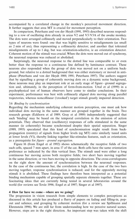
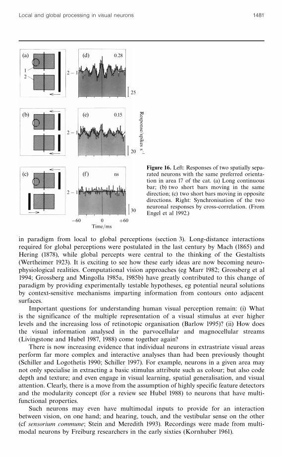
Figure 16 (from Engel et al 1992) shows schematically the receptive fields of twosuch cells, spaced 7 mm apart, in area 17 of the cat. Both cells have the same orientationpreference as indicated by the thin vertical lines. The stimuli used were either a longcontinuous bar moving across both receptive fields; two short bars moving togetherin the same direction; or two bars moving in opposite directions. The cross-correlogramson the right show the amount of synchronisation between the neuronal responses.In the case of the continuous bar, the correlation is high. In the case of the two barsmoving together, response synchronisation is reduced; and for the oppositely movingstimuli it is abolished. These findings have therefore been interpreted as a potentialbinding mechanism capable of grouping spatially separate elements together. These arefascinating ideas that are currently being tested in several laboratories around theworld (for reviews see Tovee 1996; Engel et al 1997; Singer et al 1997).
4 How far have we comeöwhere are we going?The progression in vision research from simple elements to complex perceptions asdiscussed in this article has produced a flurry of papers on fading and filling-in, pop-out and salience, and grouping by coherent motion (for a review see Spillmann andEhrenstein 1996). We are still far from understanding how we perceive natural scenes.However, steps are in the right direction. An important step was taken with the shift
1480 L Spillmann
in paradigm from local to global perceptions (section 3). Long-distance interactionsrequired for global perceptions were postulated in the last century by Mach (1865) andHering (1878), while global percepts were central to the thinking of the Gestaltists(Wertheimer 1923). It is exciting to see how these early ideas are now becoming neuro-physiological realities. Computational vision approaches (eg Marr 1982; Grossberg et al1994; Grossberg and Mingolla 1985a, 1985b) have greatly contributed to this change ofparadigm by providing experimentally testable hypotheses, eg potential neural solutionsby context-sensitive mechanisms imparting information from contours onto adjacentsurfaces.
Important questions for understanding human visual perception remain: (i) Whatis the significance of the multiple representation of a visual stimulus at ever higherlevels and the increasing loss of retinotopic organisation (Barlow 1995)? (ii) How doesthe visual information analysed in the parvocellular and magnocellular streams(Livingstone and Hubel 1987, 1988) come together again?
There is now increasing evidence that individual neurons in extrastriate visual areasperform far more complex and interactive analyses than had been previously thought(Schiller and Logothetis 1990; Schiller 1997). For example, neurons in a given area maynot only specialise in extracting a basic stimulus attribute such as colour; but also codedepth and texture; and even engage in visual learning, spatial generalisation, and visualattention. Clearly, there is a move from the assumption of highly specific feature detectorsand the modularity concept (for a review see Hubel 1988) to neurons that have multi-functional properties.
Such neurons may even have multimodal inputs to provide for an interactionbetween vision, on one hand; and hearing, touch, and the vestibular sense on the other(cf sensorium commune; Stein and Meredith 1993). Recordings were made from multi-modal neurons by Freiburg researchers in the early sixties (Kornhuber 1961).
(a) (d)
(b) (e)
(c) (f)
0.28
0.15
ns
12
2ÿ 1
2ÿ 1
2ÿ 1
25
20
30
ÿ60 0 �60Time=ms
Response/spikes
s ÿ1
Figure 16. Left: Responses of two spatially sepa-rated neurons with the same preferred orienta-tion in area 17 of the cat. (a) Long continuousbar; (b) two short bars moving in the samedirection; (c) two short bars moving in oppositedirections. Right: Synchronisation of the twoneuronal responses by cross-correlation. (FromEngel et al 1992.)
Local and global processing in visual neurons 1481
4.1 Central questions for future researchOther central questions that need to be answered are: How much of our perception isbottom^ up, how much is top ^ down (Kosslyn et al 1995)? The vast majority of fibresin the optic tract of cat carries (feedback) signals from cells in area 17 to the lateralgeniculate nucleus, whereas only about 10% ^ 20% (less than 10% in the monkey)carry (feedforward) signals from retino-geniculate afferents (Sherman and Koch 1986;Peters et al 1994; Murphy and Sillito 1996). A similar prevalence of feedback appearsto exist between extrastriate areas (V2 ^MT) and the primary visual cortex (Perkelet al 1986). Inactivation of any of these higher areas by local cooling drasticallychanges the response pattern of neurons at lower levels (Bullier et al 1996; Payne et al1996; James et al 1997). Apparently, the visual input to any given stage (`bottom^ up')is refined by a cascade of feedback circuits (Lamme et al 1997), and the question ariseswhere does stimulus processing by feedback loops become `top ^ down'.
Another important problem concerns the question of how large-scale surface prop-erties, such as perceived transparency and depth in stratified stimulus patterns, arecomputed from local features (Adelson 1993; Spillmann and Werner 1996) as theymust involve far more than long-range interaction between single cells. Friedman et al(1996) have recently found that many cells in area V2 of the macaque monkey respondto random-dot stereograms by signalling the edges of the cyclopean figure throughedge enhancement. Cells responded maximally if the edge of the figure was centredon their receptive fields, but only weakly when the figure covered the entire receptivefield. However, it remains unclear how edge extrapolation and surface stratificationthrough smoothing and filling-in of depth planes in random-dot stereograms isachieved.
Recent research even suggests that there are `Gestalt' cells that are largely invarianttowards retinal size, position, viewpoint, even partial occlusion (Sary et al 1993; Rolls1994; Kovacs et al 1995; Wallis and Rolls 1997). What about the role of attention andeye movements (Groner and Groner 1989; Fischer and Weber 1993; Treue and Maunsell1996; Kastner et al 1998)? For example, is it conceivable that local enhancement ofthe visual field by a shift of focal attention is comparable to an eye movement that isplanned, but not executed? Other important questions are: How do we coordinate achain of glimpses of a scene into a unified percept when it moves (or when we move)behind a number of apertures such as a hedge? Specifically, how do neurons in areasMT and MSt extract biological motion (Johansson 1964; Oram and Perrett 1994)in optical flow fields (Tanaka et al 1989; Lappe et al 1996; Duffy and Wurtz 1997). Notonly must we correlate stimulus (A) with that same stimulus (A0 ) a moment later, butwe must also account for its different location.
Finally, one might ask with Miyashita (1993): Where does perception meet memory?This is currently a hot debate whether and to what degree memory (imagery) involvesrecruitment of visual areas, as there do not appear to be specific regions for memory(Kosslyn and Ochsner 1994; Roland and Gulyas 1994; Sakai and Miyashita 1994). Rather,memory appears to be intermingled with sensory processing areas.
These are fundamental problems in the study of vision and perception to be tackledby advanced research methods. Among them are long-term multielectrode recordings,microstimulation of nerve cell clusters, and lesion studies in trained animals. Neuro-imaging techniques such as positron emission tomography or functional magneticresonance imaging (eg Zeki 1993; Smith et al 1998) allow us to observe the humanbrain at work while we perceive a given stimulus, execute a given eye movement, or tryto recall or imagine a visual percept. The results shed light on the relative contributionsof individual brain areas to visual perception. Temporal inactivation of cortical functionby transcranial magnetic stimulation (Shimojo and Kamitani 1998) complements thesetechniques.
1482 L Spillmann
It seems like a dream come true that one day one might be able to actually seededicated nerve cells in the human brain lighting up like city lights in the dark. Neverbefore did neuropsychologists have at their disposal such a powerful array of non-invasive research tools to identify the brain loci responsible for visual function. Thesteep rise in the number of publications tackling these and related issues stronglysuggests that some of these questions may soon be answered for a more completeunderstanding of visual perception.
4.2 Epistemological implicationsAlthough we are living in what is perhaps one of the most exciting periods of visionresearch, for some skeptics the gnawing question of an epiphenomenon remains. Yet,even to a reluctant mind, it would appear difficult to assert that the multitude ofcorrelations between neuronal responses and perceptual phenomena described in thispaper is merely coincidental. Take Gru« sser's (1995) article on migraine phosphenes(ie zigzagging line segments of cortical origin resembling the fortifications of a medievalcastle): Who would deny the close relationship between the presumed neuronal activity,cortical magnification, and resultant percepts? It is probably fair to say that manyexperiments that have furthered our understanding of the neuronal bases of visualperception would not have been performed if they had not been prompted by the firmbelief that bridges (analogies) exist between single-cell activity and visual perception(eg Nelson 1985).
Uttal (1997) and others have cautioned us not to fall victim to a naive form ofneuroreductionism (cf Brindley 1970; Teller 1990; Mausfeld 1996; Pessoa et al 1998).Indeed, the progression from correlation!dependence! causation! identity is logi-cally a treacherous one. However, from a pragmatic point of view one need not worryas long as this approach continues to bear fruit. If only we agree that for two differentpercepts there must also be two correlated neural states, we may be safe. The goal thenis to guide single-cell recordings towards biologically important problems (such aspartially occluded contours) and, vice versa, focus psychophysical studies on relevantneuronal mechanisms (such as end-stopping).
It is gratifying to see that the idea of a complementary relationship between neuro-anatomy, neurophysiology, and psychophysics that has marked the beginning of modernresearch into the visual system (Jung and Kornhuber 1961) and that has been followed upat major international conferences (Spillmann and Werner 1990; Valberg and Lee 1990)is also benefitting the European Conference onVisual Perception. Having enjoyed and livedthrough the impressive progress during that time, we are now challenged by the question:Where do visual signals become a perception? The task of the next decade is to tackleconsciousness, the biggest question of all.
Acknowledgements. Preparation of this article was supported by DFG grant SP67/6-1. I thank JeanBullier, Birgitta Dresp, Walter H Ehrenstein, Sabine Kastner, Barry Lee, Svein Magnussen, LarryMaloney, Osvaldo da Pos, Dieter Schmidt, Robert Sekuler, William Uttal, Andrea Vieho« ver, andJohn S Werner for comments and Kristian Donner for providing the impetus to write about history.The comments by two anonymous referees are appreciated. Frank Stu« rzel prepared figure 7.
ReferencesAdelson E H, 1993 ``Perceptual organization and the judgment of brightness'' Science 262
2042 ^ 2044Adrian E D, Matthews R, 1927 ` The action of light on the eye. I. The discharge of impulses in
the optic nerve and its relation to the electrical changes in the retina'' Journal of Physiology(London) 63 378 ^ 414
Adrian E D, Matthews R, 1928 ` The action of light on the eye. III. The interaction of retinalneurones'' Journal of Physiology (London) 65 273 ^ 301
Allman J, Miezin F, McGuinness E, 1985a ``Stimulus specific responses from beyond the classicalreceptive field: Neurophysiological mechanisms for local ^ global comparisons in visual neurons''Annual Review of Neuroscience 8 407 ^ 430
Local and global processing in visual neurons 1483
Allman J, Miezin F, McGuinness E, 1985b ` Direction- and velocity-specific responses from beyondthe classical receptive field in the middle temporal visual area (MT)'' Perception 14 105 ^ 126
Bach M, Meigen T, 1997 ``Similar electrophysiological correlates of texture segregation inducedby luminance, orientation, motion, and stereo'' Vision Research 37 1409 ^ 1414
Barlow H B, 1953 ``Summation and inhibition in the frog's retina'' Journal of Physiology (London)119 69 ^ 88
Barlow H B, 1972 ` Single units and sensation: a neuron doctrine for perceptual psychology?''Perception 1 371 ^ 394
Barlow H B, 1995 ` The neuron doctrine in perception'', in The Cognitive NeurosciencesEd.M Gazzaniga (Cambridge, MA: MIT Press) pp 415 ^ 435
Barlow H B, Derrington A M, Harris I R, Lennie P, 1977 ` The effect of remote retinal stimulationon the responses of cat retinal ganglion cells'' Journal of Physiology (London) 269 177 ^ 194
Baumann R, Zwan R van der, Peterhans E, 1997 ` Figure ^ ground segregation at contours: a neuralmechanism in the visual cortex of the alert monkey'' European Journal of Neuroscience 91290 ^ 1303
BaumgartnerG,1960 ` IndirekteGro« benbestimmung der receptiven Felder derRetinabeimMenschenmittels der Hermannschen Gitterta« uschung'' Pflu« gers Archiv fu« r die gesamte Physiologie 27221 ^ 22
Baumgartner G, 1961 ` Die Reaktionen der Neurone des zentralen visuellen Systems der Katzeim simultanen Helligkeitskontrast'', in Neurophysiologie und Psychophysik des visuellen SystemsEds R Jung, H H Kornhuber (Berlin: Springer) pp 296 ^ 313
Baumgartner G, 1990 ``Where do visual signals become a perception?'', in The Principles of Designand Operation of the Brain Eds J Eccles, O Creutzfeldt (Vatican: Pontificiae AcademiaeScientiarum Scripta Varia) volume 78, pp 99 ^ 114
Baumgartner G, Heydt R von der, Peterhans E, 1984 `Anomalous contours: a tool in studyingthe neurophysiology of vision'' Experimental Brain Research (supplement 9) 413 ^ 419
Blakemore C, Carpenter R H S, Georgeson M A, 1970 ` Lateral inhibition between orientationdetectors in the human visual system'' Nature (London) 228 37 ^ 39
Blakemore C, Tobin E A, 1972 ` Lateral inhibition between orientation detectors in the cat's visualcortex'' Experimental Brain Research 15 439 ^ 440
Braddick O J, Campbell F W, Atkinson J, 1978 ` Channels in vision: Basic aspects'', in Handbook ofSensory Physiology volume VIII Perception Eds R Held, H W Leibowitz, H-L Teuber (Berlin:Springer) pp 1 ^ 38
Breitmeyer B, Valberg A, 1979 ` Local foveal inhibitory effects of global peripheral excitation''Science 203 463 ^ 464
Brindley G S, 1970 Physiology of the Retina and Visual Pathways (London: Edward Arnold)Britten K H, Shadlen M N, Newsome W T, Movshon J A, 1992 ``The analysis of visual motion:
A comparison of neuronal and psychophysical performance'' Journal of Neuroscience 124745 ^ 4765
Bullier J, Hupe J M, James A, Girard P, 1996 ` Functional interactions between areas V1 and V2in the monkey'' Journal of Physiology (Paris) 90 217 ^ 220
Cajal S Ramon y, 1893 ` La retine des vertebres'' La Cellule 9 17 ^ 257 [Translated by D Maguire,R W Rodieck The Vertebrate Retina (San Francisco, CA: Freeman, 1973)]
Creutzfeldt O D, 1990 ` Brain, perception, and mind. A tribute to Richard Jung, DonaldM MacKay, and Hans-Lukas Teuber'', inVisual Perception. The Neurophysiological FoundationsEds L Spillmann, J S Werner (San Diego, CA: Academic Press) pp 449 ^ 463
Creutzfeldt O D, Kastner S, Xing Pei, Valberg A, 1991 ` The neurophysiological correlates ofcolour and brightness contrast in lateral geniculate neurones II. Adaptation and surroundeffects'' Experimental Brain Research 87 22 ^ 45
Davey M P, Maddess T, Srinivasan M V, 1998 ` The spatiotemporal properties of the Craik ^O'Brien ^Cornsweet effect are consistent with `filling in' '' Vision Research 38 2037 ^ 2046
DeAngelis G C, Anzai A, Ohzawa I, Freeman R D, 1995 ` Receptive field structure in the visualcortex: does selective stimulation induce plasticity?'' Proceedings of the National Academy ofSciences of the USA 92 9682 ^ 9686
De Valois R L, De Valois K K, 1990 Spatial Vision (New York: Oxford University Press)De Valois R L, Webster M A, De Valois K K, Lingelbach B, 1986 ` Temporal properties of
brightness and color induction'' Vision Research 26 887 ^ 897De Weerd P, Desimone R, Ungerleider L G, 1998 ` Perceptual filling-in: A parametric study''
Vision Research 38 2721 ^ 2734De Weerd P, Gattas R, Desimone R, Ungerleider L G, 1995 ` Responses of cells in monkey visual
cortex during perceptual filling-in of an artificial scotoma'' Nature (London) 377 731 ^ 734
1484 L Spillmann

Dichgans J, 1998 ` Richard Jung'', in Nervena« rzte ^ Biographien Eds H Schliack, H Hippius(Stuttgart: G Thieme) pp 96 ^ 101
Dresp B, 1993 ` Bright lines and edges facilitate the detection of small light targets'' Spatial Vision7 213 ^ 225
Dresp B, 1997 ` On illusory contours and their functional significance'' Current Psychology ofCognition 16 489 ^ 518
Dresp B, Bonnet C, 1995 ` Subthreshold summation with illusory contours'' Vision Research 351071 ^ 1078
Duffy C J, Wurtz R H, 1997 ` Medial superior temporal area neurons respond to speed patternsin optic flow'' Journal of Neuroscience 17 2839 ^ 2851
Eckhorn R, Bauer R, Jordan W, Brosch M, Kruse W, Munk M, Reitboeck H J, 1988 ` Coherentoscillations: a mechanism of feature linking in the visual cortex? Multiple electrode andcorrelation analysis in the cat'' Biological Cybernetics 60 121 ^ 130
Eckhorn R, Reitboeck H J, Arndt M, Dicke P, 1989 `A neural network for feature linking viasynchronous activity: results from cat visual cortex and from simulations'', in Models of BrainFunction Ed. R M J Cotterill (Cambridge: Cambridge University Press) pp 255 ^ 272
EhrensteinW, 1941 ` Uë ber Abwandlungen der L. Hermannschen Helligkeitserscheinung'' Zeitschriftfu« r Psychologie 150 83 ^ 91 [` Modifications of the brightness phenomenon of L Hermann'';translation by A Hogg, 1987, in The Perception of Illusory Contours Eds S Petry, G E Meyer(1987, New York: Springer) pp 35 ^ 39]
EhrensteinW H, Spillmann L, 1983 ``Time thresholds for increments and decrements in luminance''Journal of the Optical Society of America 73 419 ^ 426
Engel A K, Ko« nig P, Kreiter A K, Schillen T B, Singer W, 1992 ` Temporal coding in the visualcortex. New vistas on integration in the nervous system'' Trends in Neurosciences 15 218 ^ 226
Engel A K, Roelfsema P R, Fries P, Brecht M, Singer W, 1997 ` Role of the temporal domainfor response selection and perceptual binding'' Cerebral Cortex 7 571 ^ 582
Eysel U T, Eyding D, Schweigart G, 1998 ` Repetitive optical stimulation elicits fast receptivefield changes in mature visual cortex'' Neuroreport 9 949 ^ 954
Eysel U T, Schweigart G, 1999 ` Increased receptive field size in the surround of chronic lesionsin the adult cat visual cortex'' Cerebral Cortex 9 101 ^ 109
Fechner G T, 1860 Elemente der Psychophysik (Leipzig: Breitkopf & Ha« rtel)Fiorentini A, Baumgartner G, Magnussen S, Schiller P H, Thomas J P, 1990 ` The perception of
brightness and darkness: Relations to neuronal receptive fields'', in Visual Perception: TheNeurophysiological Foundations Eds L Spillmann, J S Werner (San Diego, CA: Academic Press)pp 129 ^ 161
Fischer B, 1973 ` Overlap of receptive field centers and representation of the visual field in thecat's optic tract'' Vision Research 13 2113 ^ 2120
Fischer B, Weber H, 1993 ` Express saccades and visual attention'' Behavioral and Brain Sciences16 553 ^ 610
Fox P T, Miezin F M, Allman J M, Van Essen D C, Raichle M E, 1987 ` Retinotopic organizationof human visual cortex mapped with positron emission tomography'' Journal of Neuroscience7 913 ^ 922
Friedman H, Zhou H, Poggio G, Heydt R von der, 1996 ` Neural mechanisms for cyclopean formperception in areas V1 and V2 of visual cortex'', Fifth Teuber Symposium, MIT, Cambridge,MA, 25 ^ 27 October 1996 (abstract)
Fries W, Albus K, Creutzfeldt O D, 1977 ``Effects of interacting visual patterns on single cellresponses in cat's striate cortex'' Vision Research 17 1001 ^ 1008
Gerling J, Spillmann L, 1987 ` Duration of visual afterimages on modulated backgrounds: post-receptoral processes'' Vision Research 27 521 ^ 527
Gibson J J, Radner M, 1937 `Adaptation, after-effect and contrast in the perception of tilted lines:I. Quantitative studies'' Journal of Experimental Psychology 20 453 ^ 467
Gilbert C D, 1992 ` Horizontal integration and cortical dynamics'' Neuron 9 1 ^ 13Gilbert C D, Wiesel T, 1990 ` The influence of contextual stimuli on the orientation selectivity
of cells in primary visual cortex of the cat'' Vision Research 30 1689 ^ 1701Gilbert C,Wiesel T, 1992 ` Receptive field dynamics in adult primary visual cortex''Nature (London)
356 150 ^ 152Goethe J W von, 1810 Zur Farbenlehre (Tu« bingen: J G Cotta. Diederich, Jena) [Translated by
R Matthaei Goethe's Color Theory (New York: van Nostrand, 1971)]Gray C M, Ko« nig P, Engel A K, Singer W, 1989 ` Oscillatory responses in cat visual cortex exhibit
inter-columnar synchronization which reflects global stimulus properties'' Nature (London) 338334 ^ 337
Local and global processing in visual neurons 1485
Gregory R L, 1987 ` Illusory contours and occlusion surfaces'', inThe Perception of Illusory ContoursEds S Petry, G E Meyer (New York: Springer) pp 81 ^ 89
Groner R, Groner M T, 1989 `Attention and eye movement control: An overview'' European Archiveof Psychiatry and Neurological Sciences 239 9 ^ 16
Grossberg S, Mingolla E, 1985a ` Neural dynamics of form perception: Boundary completion,illusory figures, and neon color spreading'' Psychological Review 92 173 ^ 211
Grossberg S, Mingolla E, 1985b ` Neural dynamics of perceptual grouping: Texture, boundaries,and emergent segmentation'' Perception & Psychophysics 38 141 ^ 171
Grossberg S, Mingolla E, Ross W D, 1994 `A neural theory of attentive visual search: interactionsof boundary, surface, spatial, and object representations'' Psychological Review 101 470 ^ 489
Gru« sser O-J, 1956 Reaktionen einzelner corticaler und retinaler Neurone der Katze auf Flimmerlichtund ihre Beziehungen zur subjektiven Sinnesphysiologie Dissertation, Albert-Ludwigs-Universita« t,Freiburg im Breisgau
Gru« sser O-J, 1995 ` Migraine phosphenes and the retino-cortical magnification factor'' VisionResearch 35 1125 ^ 1134
Gru« sser O-J, Creutzfeldt O, 1957 ` Eine neurophysiologische Grundlage des Bru« cke ^ Bartley-Effektes: Maxima der Impulsfrequenz retinaler und corticaler Neurone bei Flimmerlichtmittlerer Frequenzen'' Pflu« gers Archiv fu« r die gesamte Physiologie 263 668 ^ 681
Gru« sser O-J, Gru« sser-Cornehls U, 1973 ``Neuronal mechanisms of visual movement perceptionand some psychophysical and behavioral correlations'', in Handbook of Sensory Physiologyvolume VII/3A Central Processing of Visual Information Ed.R Jung (New York: Springer)pp 333 ^ 429
Gru« sser O-J, Gru« sser-Cornehls U, 1976 ` Neurophysiology of the anuran visual system'', in FrogNeurobiology Eds R Llinas, W Precht (Berlin: Springer) pp 297 ^ 451
Gru« sser O-J, Gru« tzner A, 1958 ` Neurophysiologische Grundlagen der periodischen Nachbildphasennach kurzen Lichtblitzen'' Graefes Archiv fu« r Ophthalmologie 160 65 ^ 93
Hammond P, MacKay D M, 1981 ` Modulatory influences of moving textured backgrounds onresponsiveness of simple cells in feline striate cortex'' Journal of Physiology (London) 319431 ^ 442
Hardage L, Tyler C W, 1995 ` Induced twinkle aftereffect as a probe of dynamic visual processingmechanisms'' Vision Research 35 757 ^ 766
Hartline H K, 1938 ` The response of single optic nerve fibers of the vertebrate eye to illumina-tion of the retina''American Journal of Physiology 121 400 ^ 415
Hartline H K, 1940 ` The receptive fields of optic nerve fibers'' American Journal of Physiology130 690 ^ 699
Hering E, 1878 ` Vom simultanen Grenzkontrast'', in Zur Lehre vom Lichtsinne (Wien: Gerold undSoehne). Also 1907 Grundzu« ge der Lehre vom Lichtsinn, in Handbuch der gesamten Augen-heilkunde Eds Graefe-Saemisch, 2nd edition, volume 3, chapter XII (Leipzig: W Engelmann)pp 138 ^ 139 [Translated by L M Hurvich, D Jameson Outlines of the Theory of the Light Sense(Cambridge, MA: Harvard University Press, 1964)]
Hermann L, 1870 ` Eine Erscheinung des simultanen Contrastes'' Pflu« gers Archiv fu« r die gesamtePhysiologie 3 13 ^ 15
Heydt R von der, 1987 `Approaches to visual cortical function'' Reviews of Physiology, Biochemistryand Pharmacology 108 69 ^ 151
Heydt R von der, Peterhans E, Baumgartner G, 1984 ` Illusory contours and cortical neuronresponses'' Science 224 1260 ^ 1262
Hofmann F B, Bielschowsky A, 1909 ` Uë ber die Einstellung der scheinbaren Horizontalen undVertikalen bei Betrachtung eines von schra« gen Konturen erfu« llten Gesichtsfeldes'' Pflu« gersArchiv fu« r die gesamte Physiologie 126 453 ^ 475
Holmes G, 1918 ` Disturbances of vision by cerebral lesions'' British Journal of Ophthalmology 2353 ^ 384
Hubel D H, 1988 Eye, Brain and Vision Scientific American Library (New York: W H Freeman)Hubel D H,Wiesel T, 1959 ``Receptive fields of single neurones in the cat's striate cortex'' Journal
of Physiology (London) 148 574 ^ 591Hubel D H, Wiesel T N, 1965 ` Receptive fields and functional architecture in two nonstriate
visual areas (18 and 19) of the cat'' Journal of Neurophysiology 28 229 ^ 289James A C, Hupe J-M, Payne B R, Lomber S G, Girard P, Bullier J, 1997 ` Feedback connections
from area MT gate information transfer in neurons of areas V1, V2 and V3'' Society forNeuroscience 23 1396 (abstract)
Johansson G, 1964 ``Perception of motion and changing form'' Scandinavian Journal of Psychology5 181 ^ 207
1486 L Spillmann
Jung R, 1959 ``Microphysiology of cortical neurons and its significance for psychophysiology''Anales de la Facultad de Medicina, Montevideo 44 323 ^ 332
Jung R, 1961a ` Neuronal integration in the visual cortex and its significance for visual information'',in Sensory Communication Ed.W A Rosenblith (Cambridge, MA: MIT Press) pp 627 ^ 674
Jung R, 1961b ` Korrelationen von Neuronenta« tigkeit und Sehen'', in Neurophysiologie undPsychophysik des visuellen Systems Eds R Jung, H-H Kornhuber (Berlin: Springer) pp 410 ^ 435
Jung R, 1973 ` Visual perception and neurophysiology'', in Handbook of Sensory Physiologyvolume VII/3ACentral Processing of Visual Information Ed.R Jung (Berlin: Springer) pp 1 ^ 152
Jung R, 1975 ` Some European neuroscientists: A personal tribute'', in The Neurosciences: Paths ofDiscovery Eds FG Worden, J P Swazey, GAdelman (Cambridge, MA: MIT Press) pp 477 ^ 511
Jung R, 1984 ` Sensory research in historical perspective: philosophical foundations of perception'',in Handbook of Physiology, Section 1 The Nervous System volume III/1 Sensory ProcessesEd. I Darian-Smith (Washington, DC: American Physiological Society) pp 1 ^ 74
Jung R, Kornhuber H-H (Eds), 1961 Neurophysiologie und Psychophysik des visuellen Systems(Berlin: Springer)
Jung R, Spillmann L, 1970 ` Receptive-field estimation and perceptual integration in human vision'',in Early Experience and Visual Information Processing in Perceptual and Reading DisordersEds F A Young, D B Lindsley (Washington, DC: National Academic Press) pp 181 ^ 197
Jung R, Baumgarten R von, Baumgartner G, 1952 ` Mikroableitungen von einzelneç n Nervenzellenim optischen Cortex der Katze: Die lichtaktivierten B-Neurone'' Archiv fu« r Psychiatrie undNervenkrankheiten 189 521 ^ 539
Kaas J H, Krubitzer L A, ChinoY M, Langston A L, Polley E H, Blair N, 1990 ` Reorganization ofretinotopic cortical maps in adult mammals after lesions of the retina'' Science 248 229 ^ 231
Kastner S, Crook J M, Pei Xing, Creutzfeldt O D, 1992 ` Neurophysiological correlates of colourinduction on white surfaces'' European Journal of Neuroscience 4 1079 ^ 1086
Kastner S, De Weerd P, Desimone R, Ungerleider L G, 1998 ` Mechanisms of directed attentionin human ventral extrastriate cortex as revealed by functional MRI'' Science 282 108 ^ 111
Kastner S, Nothdurft H C, Pigarev I N, 1997 ` Neuronal correlates of pop-out in cat striatecortex'' Vision Research 37 371 ^ 376
Knau H, Spillmann L, 1996 ` Failure of brightness and color constancy under prolonged Ganzfeldstimulation'' SPIE Proceedings 2657(04) 19 ^ 29
Knau H, Spillmann L, 1997 ` Brightness fading during Ganzfeld adaptation'' Journal of the OpticalSociety of America A 14 1213 ^ 1222
Ko« hler W, Held R, 1949 ` The cortical correlate of pattern vision'' Science 110 414 ^ 419Kornhuber H-H, 1961 ` Convergence of vestibular, visual and auditory afferents at single neurons
of the cat's cortex'' Excerpta Medica (Amsterdam) International Congress Series 37 11 (Abstract)Kornhuber H-H, Spillmann L, 1964 ` Zur visuellen Feldorganisation beim Menschen: Die receptiven
Felder im peripheren und zentralen Gesichtsfeld bei Simultankontrast, Flimmerfusion,Scheinbewegung und Blickfolgebewegung'' Pflu« gers Archiv fu« r die gesamte Physiologie 279 R5(Abstract)
Kosslyn S M, Ochsner K N, 1994 ``In search of occipital activation during visual mental imagery''(Comment) Trends in Neurosciences 17 290 ^ 292
Kosslyn S M, Thompson W L, Kim I J, Alpert N M, 1995 ` Topographical representations ofmental images in primary visual cortex'' Nature (London) 378 496 ^ 498
Kovacs G, Vogels R, Orban G A, 1995 ` Selectivity of macaque inferior temporal neurons forpartially occluded shapes'' Journal of Neuroscience 15 1984 ^ 1997
Kovacs I, Papathomas T V,Yang M, Feher Aè , 1996 ` When the brain changes its mind: Interoculargrouping during binocular rivalry'' Proceedings of the National Academy of Sciences of the USA93 15508 ^ 15511
Krauskopf J, 1963 ` Effect of retinal image stabilization on the appearance of heterochromatictargets'' Journal of the Optical Society of America 53 741 ^ 744
Kru« ger J, 1990 ` Multi-microelectrode investigation of monkey striate cortex: link between corre-lational and neuronal properties in the infragranular layers'' Visual Neuroscience 5 135 ^ 142
Kru« ger J, Becker J D, 1991 ``Recognizing the visual stimulus from neuronal discharges'' Trendsin Neurosciences 14 282 ^ 286
Kru« ger J, Fischer B, 1973 ` Strong periphery effect in cat retinal ganglion cells. Excitatory responsesin ON- and OFF-center neurons to single grid displacements'' Experimental Brain Research18 316 ^ 318
Kuffler S W, 1953 ` Discharge patterns and functional organization of mammalian retina'' Journalof Neurophysiology 16 37 ^ 68
Local and global processing in visual neurons 1487
Lamme V, 1995 ` The neurophysiology of figure ^ ground segregation in primary visual cortex''Journal of Neuroscience 15 1605 ^ 1615
Lamme V A F, Zipser K, Spekreijse H, 1997 ` Figure ^ ground signals in V1 depend on extrastriatefeedback'' Investigative Ophthalmology & Visual Science 38(4) S969 (Abstract 4490)
Land E H, 1983 ` Recent advances in retinex theory and some implications for cortical computa-tions: color vision and the natural image'' Proceedings of the National Academy of Sciencesof the USA 80 5163 ^ 5169
Lappe M, Bremmer F, Pekel M, Thiele A, Hoffmann K P, 1996 ` Optic flow processing inmonkey STS: a theoretical and experimental approach'' Journal of Neuroscience 16 6265 ^ 6285
Lee B B, Pokorny J, Smith V C, Martin P R, Valberg A, 1990 ``Luminance and chromaticmodulation sensitivity of macaque ganglion cells and human observers'' Journal of the OpticalSociety of America A 7 2223 ^ 2236
Lettvin J Y, Maturana H R, McCulloch W S, Pitts W H, 1959 ` What the frog's eye tells the frog'sbrain'' Proceedings of the Institute of Radio Engineers 47 1940 ^ 1951
Levinson E, Sekuler R, 1976 `Adaptation alters perceived direction of motion'' Vision Research16 779 ^ 781
Livingstone M S, Hubel D H, 1987 ` Psychophysical evidence for separate channels for the percep-tion of form, color, movement, and depth'' Journal of Neuroscience 7 3416 ^ 3468
Livingstone M S, Hubel D H, 1988 ` Segregation of form, color, movement and depth: anatomy,physiology and perception'' Science 240 740 ^ 750
Lund J S, Yoshioka T, Levitt J B, 1993 ` Comparison of intrinsic connectivity in different areasof macaque monkey cerebral cortex'' Cerebral Cortex 3 148 ^ 162
Mach E, 1865 ` Uë ber die Wirkung der ra« umlichen Vertheilung des Lichtreizes auf die Netzhaut, I''Sitzungsberichte der Kaiserlichen Akademie der Wissenschaften,Wien, Mathematisch-Naturwissen-schaftliche Classe, Zweite Abteilung 52 303 ^ 322
McIlwain J T, 1964 ` Receptive fields of optic tract axons and lateral geniculate cells. Peripheralextent and barbiturate sensitivity'' Journal of Neurophysiology 27 1154 ^ 1173
Maffei L, Fiorentini A, 1976 ` The unresponsive regions of visual cortical receptive fields'' VisionResearch 16 1131 ^ 1139
Magnussen S, Glad A, 1975a ` Brightness and darkness enhancement during flicker: Perceptualcorrelates of neuronal B- and D-systems in human vision'' Experimental Brain Research 22399 ^ 413
Magnussen S, Glad A, 1975b ``Temporal frequency characteristics of spatial interaction in humanvision'' Experimental Brain Research 23 519 ^ 528
Magnussen S, Kurtenbach W, 1980a ` Linear summation of tilt illusion and tilt aftereffect'' VisionResearch 20 39 ^ 42
Magnussen S, Kurtenbach W, 1980b `Adapting to two orientations: Disinhibition in a visualafter-effect'' Science 207 908 ^ 909
Marcar V L, Xiao D K, Raiguel S E, Maes H, Orban G A, 1995 ` Processing of kineticallydefined boundaries in the cortical motion area MT of the macaque monkey'' Journal ofNeurophysiology 74 1258 ^ 1270
Marr D, 1982 Vision: A Computational Investigation into the Human Representation and Processingof Visual Information (San Francisco, CA: Freeman)
Mausfeld R, 1996 ` Wahrnehmung'' (perception), in Wo« rterbuch der KognitionswissenschaftEd. G Strube (Stuttgart: Klett-Cotta) pp 776 ^ 792
Metzger W, 1953 Gesetze des Sehens 2nd edition (Frankfurt am Main: Waldemar Kramer)Metzger W, 1961 `Aporien der Psychophysik'', in Neurophysiologie und Psychophysik des visuellen
Systems Eds R Jung, H-H Kornhuber (Berlin: Springer) pp 435 ^ 444Miyashita Y, 1993 ` Inferior temporal cortex: Where perception meets memory'' Annual Review
of Neuroscience 16 245 ^ 263Munk M H J, Nowak L G, Nelson J I, Bullier J, 1995 ` Structural basis of cortical synchronization.
II. Effects of cortical lesions'' Journal of Neurophysiology 74 2401 ^ 2414Murphy P C, Sillito A M, 1996 ` Functional morphology of the feedback pathway from area 17 of
the cat visual cortex to the lateral geniculate nucleus'' Journal of Neuroscience 16 1180 ^ 1192Nelson J I, 1985 ` The cellular basis of perception'', in Models of the Visual Cortex Eds D Rose,
V Dobson (Chichester, Sussex: John Wiley and Sons) pp 108 ^ 122Nelson J I, Frost B J, 1978 ` Orientation-selective inhibition from beyond the classic visual
receptive field'' Brain Research 139 359 ^ 365Nelson R, Famiglietti E V, Kolb H, 1978 ` Intracellular staining reveals different levels of stratifica-
tion for on- and off-center ganglion cells in cat retina'' Journal of Neurophysiology 41 472 ^ 483
1488 L Spillmann
Neumeyer C, Spillmann L, 1977 ` Fading of steadily fixated large test fields in extrafoveal vision''Pflu« gers Archiv 368 R40 (Abstract)
Newsome W T, Britten K H, Movshon J A, 1989 ` Neuronal correlates of a perceptual decision''Nature (London) 341 52 ^ 54
Nowak L G, Bullier J, 1997 ` The timing of information transfer in the visual system'', in CerebralCortex volume 12, chapter 5 Eds K S Rockland, J H Kaas, A Peters (New York: Plenum)pp 205 ^ 241
Oram M W, Perrett D I, 1994 ` Responses of anterior superior temporal polysensory (SPa) neuronsto `biological motion' stimuli'' Journal of Cognitive Neuroscience 6 99 ^ 116
Paradiso M A, Hahn S, 1996 ` Filling-in percepts produced by luminance modulation'' VisionResearch 36 2657 ^ 2663
Paradiso M A, Kim W, Nayak S, 1996 ` Cortical representation of surface brightness: influencesfrom beyond the classical receptive field'' Society for Neuroscience 22 951 (Abstract 376.6.)
Paradiso M A, Nakayama K, 1991 ` Brightness perception and filling-in'' Vision Research 311221 ^ 1236
Payne B R, Lomber S G, Villa A E, Bullier J, 1996 ` Reversible deactivation of cerebral networkcomponents'' Trends in Neurosciences 19 535 ^ 542
Perkel D J, Bullier J, Kennedy H, 1986 ` Topography of the afferent connectivity of area 17 of themacaque monkey: a double-labelling study'' Journal of Comparative Neurology 253 374 ^ 402
Pessoa L, Thompson E, Noe Alva, 1998 ` Finding out about filling in: a guide to perceptualcompletion for visual science and the philosophy of perception'' Behavioral and Brain Sciences21 723 ^ 747
Peterhans E, 1997 ` Functional organization of area V2 in the awake monkey'', in Cerebral Cortexvolume 12, chapter 8 Extrastriate Cortex in Primates Eds K S Rockland, J H Kaas, A Peters(New York: Plenum) pp 335 ^ 357
Peterhans E, Heydt R von der, 1989 ` The whole and the piecesöcortical neuron responses to barsand rows of moving dots'', in Seeing Contours and Colour Eds J J Kulikowski, C M Dickinson,I J Murray (Oxford: Pergamon Press) pp 125 ^ 130
Peterhans E, Heydt R von der, 1991 ` Elements of form perception in monkey prestriate cortex'',in Representations of Vision: Trends and Tacit Assumptions in Vision Research Eds A Gorea,Y Fregnac, Z Kapoula, J Findlay (Cambridge: Cambridge University Press) pp 111 ^ 124
Peterhans E, Heydt R von der, 1993 ` Functional organization of area V2 in the alert macaque''European Journal of Neuroscience 5 509 ^ 524
Peterhans E, Heydt R von der, Baumgartner G, 1982 ` Cortical neuron responses and `illusory'contours'' Perception 11(1) A19 (Abstract)
Peters A, Payne B R, Budd J A, 1994 `A numerical analysis of the geniculocortical input to striatecortex in the monkey'' Cerebral Cortex 3 69 ^ 78
Poppelreuter W, 1917 Die Sto« rungen der niederen und ho« heren Sehleistungen durch Verletzungendes Okzipitalhirns (Leipzig: L Voss); [Translated by J Zihl, L Weiskrantz Disturbances of Lowerand Higher Visual Capacities Caused by Occipital Damage (Oxford: Clarendon Press, 1990)]
Przybyszewski A W, 1997 ` Overview of Otto-Joachim Gru« sser's work'' Biological Cybernetics 76317 ^ 320
Rabelo C, Gru« sser O-J, 1961 ` Die Abha« ngigkeit der subjektiven Helligkeit intermittierenderLichtreize von der Flimmerfrequenz (Bru« cke-Effekt, `brightness enhancement'): Untersuchungenbei verschiedener Leuchtdichte und Feldgro« be'' Psychologische Forschung 26 299 ^ 312
Ramachandran V S, Gregory R L, 1991 ` Perceptual filling in of artificially induced scotomas inhuman vision'' Nature (London) 350 699 ^ 702
Ransom-Hogg A, Spillmann L, 1980 ``Perceptive field size in fovea and periphery of the light-and dark-adapted retina'' Vision Research 20 221 ^ 228
Ratliff F, 1965 Mach Bands: Quantitative Studies on Neural Networks in the Retina (San Francisco,CA: Holden-Day)
Rodieck R W, 1965 ` Quantitative analysis of cat retinal ganglion cell response to visual stimuli''Vision Research 5 583 ^ 601
Roland P E, Gulyas B, 1994 ` Visual imagery and visual representation'' Trends in Neurosciences17 281 ^ 287
Rolls E T, 1994 ``Brain mechanisms for invariant visual recognition and learning'' BehaviouralProcesses 33 113 ^ 138
Rosenblith W A (Ed.), 1961 Sensory Communication (Cambridge, MA: MIT Press)Rossi A F, Paradiso M A, 1996 ` Temporal limits of brightness induction and mechanisms of
brightness perception'' Vision Research 36 1391 ^ 1398
Local and global processing in visual neurons 1489
Rossi A F, Rittenhouse C D, Paradiso M A, 1996 ` The representation of brightness in primaryvisual cortex'' Science 273 1104 ^ 1107
Safran A B, Landis T, 1996 ` Plasticity in the adult visual cortex: implications for the diagnosisof visual field defects and visual rehabilitation'' Current Opinion in Ophthalmology 7 53 ^ 64
Sakai K, Miyashita Y, 1994 ` Visual imagery: an interaction between memory retrieved and focalattention'' (Comment) Trends in Neurosciences 17 287 ^ 289
Salzman C D, Murasugi C M, Britten K H, Newsome W T, 1992 ` Microstimulation in visualarea MT: Effects on direction discrimination performance'' Journal of Neuroscience 122331 ^ 2355
Salzman C D, Newsome W T, 1994 ` Neural mechanisms for forming a perceptual decision''Science 264 231 ^ 237
Sary G, Vogels R, Orban G A, 1993 ` Cue-invariant shape selectivity of macaque inferior temporalneurons'' Science 260 995 ^ 997
Schiller P H, 1982 ` Central connections on the retinal ON and OFF pathways'' Nature (London)297 580 ^ 583
Schiller P H, 1997 ` Past and present ideas about how the visual scene is analyzed by the brain'',in Cerebral Cortex volume 12, chapter 8 Extrastriate Cortex in Primates Eds K S Rockland,J H Kaas, A Peters (New York: Plenum) pp 59 ^ 87
Schiller P H, Logothetis N K, 1990 ` The color-opponent and broadband channels of the primatevisual system'' Trends in Neurosciences 13 392 ^ 398
Schiller P H, Sandell J H, Maunsell J H R, 1986 ` Functions of the ON and OFF channels ofthe visual system'' Nature (London) 322 824 ^ 825
Schmidt K E, Goebel R, Loewel S, Singer W, 1997 ` The perceptual grouping criterion of colinearityis reflected by anisotropies of connections in the primary visual cortex'' European Journal ofNeuroscience 9 1083 ^ 1089
Schober H, Rentschler I, 1972 Das Bild als Schein der Wirklichkeit (Mu« nchen: Heinz Moos)Schwartz E L, 1984 ``Spatial mapping and spatial vision in primate striate and infero-temporal
cortex'', in Sensory Experience, Adaptation, and Perception Eds L Spillmann, B R Wooten(Hillsdale, NJ: Lawrence Erlbaum Associates) pp 73 ^ 104
Schwartz E L, 1994 ` Topographical mapping in primate visual cortex: history, anatomy andcomputation'', in Visual Science and Engineering: Models and Applications Ed.D H Kelly(New York: Marcel Dekker) pp 293 ^ 359
Sekuler R, Blake R (Eds), 1994 Perception third edition (New York: McGraw-Hill)Sherman S M, Koch C, 1986 ` The control of retinogeniculate transmission in the mammalian
lateral geniculate nucleus'' Experimental Brain Research 63 1 ^ 20Shimojo S, Kamitani Y, 1998 ` Visualizing suppression induced by transcranial magnetic stimu-
lation. (II) Anisotropy of suppression in oriented patterns'' Investigative Ophthalmology &Visual Science 39(4) S672 (Abstract 3094)
Sillito A M, Grieve K L, Jones H E, Cudeiro J, Davis J, 1995 ` Visual cortical mechanismsdetecting focal orientation discontinuities'' Nature (London) 378 492 ^ 496
Singer W, 1989 ` Search for coherence: A basic principle of cortical self-organization'' Conceptsin Neuroscience 1 1 ^ 25
Singer W, 1993 ` Synchronization of cortical activity and its putative role in information processingand learning''Annual Review of Physiology 55 349 ^ 374
Singer W, Engel A K, Kreiter A K, Munk M H J, Neuenschwander S, Roelfsema P R, 1997` Neuronal assemblies: necessity, significance, and detectability'' Trends in Cognitive Sciences1 252 ^ 261
Smith A T, Greenlee M W, Singh K D, Kraemer F M, Hennig J, 1998 ` The processing of first-and second-order motion in human visual cortex assessed by functional magnetic resonanceimaging (fMRI)'' Journal of Neuroscience 18 3816 ^ 3830
Soriano M, Spillmann L, Bach M, 1996 ` The abutting grating illusion'' Vision Research 36 109 ^ 116Spillmann L, 1971 ` Foveal perceptive fields in the human visual system measured with simulta-
neous contrast in grids and bars'' Pflu« gers Archiv fu« r die gesamte Physiologie 326 281 ^ 299Spillmann L, 1994 ``The Hermann grid illusion: A tool for studying human perceptive field
organization'' Perception 23 691 ^ 708Spillmann L, Dresp B, 1995 ` Phenomena of illusory form: Can we bridge the gap between levels
of explanation?'' Perception 24 1333 ^ 1364Spillmann L, Ehrenstein W H, 1996 ` From neuron to Gestalt-mechanisms of visual perception'',
in Comprehensive Human Physiology. From Cellular Mechanisms to Integration chapter 43,Eds R Greger, U Windhorst (Heidelberg: Springer) pp 861 ^ 893
1490 L Spillmann

Spillmann L, Kurtenbach A, 1992 ` Dynamic noise backgrounds facilitate target fading'' VisionResearch 32 1941 ^ 1946
Spillmann L, Ransom-Hogg A, Oehler R, 1987 `A comparison of perceptive and receptive fieldsin man and monkey'' Human Neurobiology 6 51 ^ 62
Spillmann L, Werner J S (Eds), 1990 Visual Perception. The Neurophysiological Foundations(San Diego, CA: Academic Press)
Spillmann L, Werner J S, 1996 ` Long-range effects in visual perception'' Trends in Neurosciences19 428 ^ 434
Stein B E, Meredith M A (Eds), 1993 The Merging of the Senses (Cambridge, MA: MIT Press)Sumi S, Noguchi K, 1996 New Horizons in the Study of Gestalt Perception (reprinted from Japanese
Psychological Research volumes 36 and 37; distributed gratis by S Sumi, Psychology Laboratory,Keio University, Hiyoshi, Yokohama 223, Japan), p.111
Szily A von, 1918 Atlas der Kriegsaugenheilkunde, samt begleitendemText volume 1 (Stuttgart: Enke)Tanaka K, Fukada Y, Saito H, 1989 ` Underlying mechanisms of the response specificity of
expansion/contraction and rotation cells in the dorsal part of the medial superior temporalarea of the monkey'' Journal of Neurophysiology 62 642 ^ 656
Teller D Y, 1990 ` The domain of visual science'', in Visual Perception. The NeurophysiologicalFoundations Eds L Spillmann, J S Werner (San Diego, CA: Academic Press) pp 11 ^ 21
Teuber H-L, 1967 ` Wolfgang Ko« hler 1887 ^ 1967'' Psychologische Forschung 31 VI ^XVITeuber H-L, Battersby N S, Bender M F, 1960 Visual Field Defects after Penetrating MissileWounds
of the Brain (Cambridge, MA: Harvard University Press)Tononi G, Sporns O, Edelman G M, 1992 ` Reentry and the problem of integrating multiple cortical
areas: simulation of dynamic integration in the visual system'' Cerebral Cortex 2 310 ^ 335Tootell R, Silverman M, Switkes E, De Valois R L, 1982 ` Deoxyglucose analysis of retinotopic
organization in primate striate cortex'' Science 218 902 ^ 904Tovee M J, 1996 An Introduction to the Visual System (Cambridge: Cambridge University Press)
pp 169 ^ 179Treue S, Maunsell J H, 1996 `Attentional modulation of visual motion processing in cortical areas
MT and MST'' Nature (London) 382 539 ^ 541Ts'o D Y, Gilbert C D, Wiesel T N, 1986 ` Relationships between horizontal interactions and
functional architecture in cat striate cortex as revealed by cross-correlation analysis'' Journalof Neuroscience 6 1160 ^ 1170
Tyler C W, Hardage L, 1998 ` Long-range twinkle induction: An achromatic rebound effect inthe magnocellular processing system?'' Perception 27 203 ^ 214
Uttal W R, 1997 ` Do theoretical bridges exist between perceptual experience and neurophysiology?''Perspectives in Biology and Medicine 40 280 ^ 302
Uttal W R, Spillmann L, Sekuler A B, 1999 ` Motion and shape in common fate'' VisionResearch (accepted pending revisions)
Valberg A, Lee B B (Eds), 1990 From Pigments to PerceptionöAdvances in Understanding VisualProcesses NATO ASI Series A, volume 203 (New York: Plenum)
Valberg A, Lee B B, Tigwell D A, Creutzfeldt O D, 1985 `A simultaneous contrast effect of steadyremote surrounds on responses of cells in macaque lateral geniculate nucleus'' ExperimentalBrain Research 58 604 ^ 608
Valberg A, Spillmann L, 1982 ` Foveal inhibition and facilitation caused by remote grating jerks:Interaction between long-range and short-range effects'' Experimental Brain Research 4689 ^ 98
Van Essen D C, Felleman D J, DeYoe E A, Knierim J J, 1991 ``Probing the primate visual cortex:pathways and perspectives'', in From Pigments to PerceptionöAdvances in Understanding VisualProcesses Eds A Valberg, B B Lee (New York: Plenum) pp 227 ^ 237
Wallis G, Rolls E T, 1997 ` Invariant face and object recognition in the visual system'' Progressin Neurobiology 51 167 ^ 194
Wa« ssle H, Peichl L, Boycott B B, 1981 ` Morphology and topography of on- and off-alpha cellsin the cat retina'' Proceedings of the Royal Society of London, Series B 212 157 ^ 175
Wertheimer M, 1923 ` Untersuchungen zur Lehre von der Gestalt II'' Psychologische Forschung 4301 ^ 350
Westheimer G, 1965 ` Spatial interaction in human cone vision'' Journal of Physiology (London) 181881 ^ 894
Witkin H A, 1959 ` The perception of the upright?'' Scientific American 200 51 ^ 70Wohlfarth E, 1932 ` Der Auffassungsvorgang an kleinen Gestalten. Zur Psychologie der Vorgestalt-
erlebnisses'' Neue Psychologische Studien 4 347 ^ 414Wolfe J, 1984 ` Global factors in the Hermann grid illusion'' Perception 13 33 ^ 40
Local and global processing in visual neurons 1491
Wolfson S S, Landy M S, 1998 ` Long-range interactions between texture elements'' InvestigativeOphthalmology & Visual Science 39(4) S650 (Abstract 3018)
Wundt W, 1911 Grundzu« ge der physiologischen Psychologie volume 3, 6th edition (Leipzig:W Engelmann)
Yu C, Essock E A, 1996 ` Psychophysical end-stopping associated with line targets'' Vision Research36 2883 ^ 2896
Yu C, Levi D M, 1997 ` End-stopping and length tuning in psychophysical spatial filters'' Journalof the Optical Society of America A 14 2346 ^ 2354
Zeki S M, 1983 ` Colour coding in the cerebral cortex: The response of wavelength-selective andcolour-coded cells in monkey visual cortex to changes in wavelength composition'' Neuroscience9 767 ^ 781
Zeki S M, 1993 A Vision of the Brain (Oxford: Blackwell Scientific)Zipser K, Lamme V A F, Schiller P H, 1996 ` Contextual modulation in primary visual cortex''
Journal of Neurophysiology 16 7376 ^ 7389
ß 1999 a Pion publication
1492 L Spillmann