from the dipartimento di neurofisiologia, istituto neurologico 'c
TRANSCRIPT

Journal of Physiology (1989), 416, pp. l1l-122 111With 3 text-figuresPrinted in Great Britain
INTRINSIC PROPERTIES OF NUCLEUS RETICULARIS THALAMINEURONES OF THE RAT STUDIED IN VITRO
BY GIULIANO AVANZINI, MARCO DE CURTIS, FERRUCCIO PANZICA ANDROBERTO SPREAFICO
From the Dipartimento di Neurofisiologia, Istituto Neurologico 'C. Besta',via Celoria 11, 20133 Milan, Italy
(Received 16 August 1988)
SUMMARY
1. Neurones of the nucleus reticularis thalami of the rat were studied byintracellular recordings from in vitro slices. The resting membrane potential was-56-28 + 5-86 mV (mean value + S.D.); input resistance was 43 09 + 9 74 MQ; thetime constant r was 16-51 + 3-99 ms. At the resting membrane potential tonic firingis present, while at membrane potentials more negative than -60 mV a burst firingmode gradually prevails.
2. Prolonged depolarizing current pulses superimposed on a steady hyperpo-larization consistently activated sequences of burst-after-hyperpolarization com-plexes. The all-or-none burst response consisted of Na+-mediated, TTX-sensitive fastaction potentials superimposed on a low threshold spike (LTS). The burst wasfollowed by a stereotyped after-hyperpolarization lasting 100-120 ms (BAHP), witha maxima -85 mV. The BAHP was blocked by Cd2' and apamine but not by 8-Brcyclic AMP. The early component of BAHP was significantly attenuated by TEA.The oscillatory rhythmic discharges were abolished by agents which blocked theBAHP.
3. The presence of strong after-hyperpolarizing potentials (SAHP and BAHP) inRTN neurones plays a significant role in determining two different functional states,defined as tonic and oscillatory burst firing modes, respectively.
INTRODUCTION
The nucleus reticularis thalami (RTN) is composed of a sheet-like aggregation ofGABAergic small neurones (Houser, Vaughn, Barber & Roberts, 1980) whichenvelopes the dorsolateral and anterior portions of the dorsal thalamus. It receivescollaterals from the corticothalamic and thalamocortical fibres and projectsexclusively to the dorsal thalamic nuclei (Jones, 1975); moreover, intrareticular axoncollaterals of RTN cells provide an intrinsic GABAergic circuit (Yen, Conley, Hendry& Jones, 1985; Spreafico, Frassoni, Battaglia & Schmechel, 1984; De Biasi, Frassoni& Spreafico, 1986).
Several lines of evidence suggest that the RTN is involved in generation of theoscillatory activity responsible for cortical spindles during states of decreased

112 G. A VANZINI AND OTHERS
vigilance (Steriade & Deschenes, 1984; Steriade, Domich & Oakson, 1986; Steriade,Domich, Oakson & Deschenes, 1987).
Recently Mulle, Madariaga & Deschenes (1986), in an in vivo study in cats,investigated using intracellular recording the spindle-related rhythmic spike burstactivity observed in RTN neurones during barbiturate anaesthesia. The comparisonwith the intrinsic membrane properties previously reported by Deschenes, Paradis,Roy & Steriade (1984) in in vivo cats and by Jahnsen & Llinas (1984a, b) in in vitroguinea-pig thalamic slices led Mulle et al. (1986) to outline some peculiarities in thelocation and relative predominances of conductances, which could support a specificrole for the RTN in pacemaking thalamic spindle oscillation.
In the present study the use of an in vitro preparation of thalamic slices alloweda further electrophysiological and pharmacological characterization of membraneproperties which account for the different firing modes of rat RTN neurones. Ca2+-dependent outward conductances underlying the after-hyperpolarizations whichfollow single spike tonic firing or burst discharges could be differentiated and relatedto the ability of RTN neurones to produce rhythmic activities. A preliminary reportof some of these data has been published in abstract (Avanzini, de Curtis & Spreafico,1987).
METHODS
Experiments were carried out on RTN neurones of young adult Sprague-Dawley rats (150-250g). The animals were decapitated by guillotine under light ether anaesthesia. The brain was quicklyremoved and immersed in artificial cerebrospinal fluid at a temperature of 4 'C. A block of braintissue containing the thalamus was dissected from each hemisphere 400-500 /um thick horizontalthalamic slices were prepared with a vibratome, transferred to the superfusion recording chamberand incubated for at least 1 h before recordings were started. The temperature of the bathingmedium was maintained at 36 'C and a humidified gas mixture of 95% 02- 5% CO2 was bubbledinto the bath during the experiment.Under a binocular microscope the RTN was recognized as a thin grey band located between the
internal capsule and the external medullary lamina; the thalamocortical radiation fibres wereclearly visible as white bundles across the RTN.
Intracellular recordings from the portion of the RTN enveloping the ventrobasal nucleus wereperformed using glass micropipettes (resistance 80-120 mQ) filled with 4 M-potassium acetate.Extracellular stimuli were applied through monopolar tungsten electrodes placed in the internalcapsule and in the dorsal thalamus, close to the recording site, in order to induce a synapticactivation of the neurones. The membrane potentials were recorded with a Neurodata Instrument,Corp. (USA) pre-amplifier provided with a Wheatstone bridge circuit utilized to performintracellular current injections.Input resistance values were calculated on the initial linear portion of current-voltage curves
produced by measuring the membrane voltage changes induced by 80-100 ms hyperpolarizingcurrent pulses applied through the recording microelectrode. The time constant values werecalculated by identifying an exponential function derived from the application of a least-squarescriterion to the logarithms of the data points extrapolated by the membrane deflection induced byinjections of 0-2-0-4 nA current pulses.The data were recorded on magnetic tape (Racal FM4D) and analysed off-line by means of a
PDP 11/34 Digital computer system, together with a Tekronix 5110 digital oscilloscope and a chartrecorder (Linseis type 2045). Only data from those neurones producing prolonged (> 10 min),stable recordings and with a membrane potential over -50 m\' were accepted for analysis.The normal bathing solution contained (in mM): NaCl, 124; KCl, 25; CaCl2, 2; MgSO4, 2;
NaH2PO4, 1-25; NaHCO3, 16; dextrose, 10. When saturated with humidified gas, the pH of thesolution was 7-4.When 1 mM-cadmium (Cd21) was added to the solution, phosphate, sulphate and CaCl2 were
omitted and MgSO4 was replaced with MgCl2. During some experiments pharmacological agents

PROPERTIES OF RTN NEURONES IN VITRO
were added to the perfusion fluid without adjusting the tonicity: I 1uM-tetrodotoxin (TTX) (n =3); 20 mM-tetraethylammonium (TEA) (n = 4); 100 ,uM-apamine (n = 3); 1 puM-8-bromo adenosine3',5'-cyclic monophosphate (8-Br cyclic AMP) (n = 4). All drugs were purchased from Sigma.
RESULTS
The following data are from forty-six neurones from the portion of the RTNrelated to the ventrobasal nucleus. The mean resting membrane potential was-56-28 + 5-86 mV (n = 28). The mean input resistance was 43 09 + 9-74 MQ (n = 21).Rectification was observed in the hyperpolarizing direction for values below -85mV. In resting conditions the membrane time constant (Tm), calculated in theinterval between 10 and 30 ms after the onset of the current pulse, was 16X51 + 3-99ms.No spontaneous activity was recorded from the RTN neurones. Each spike was
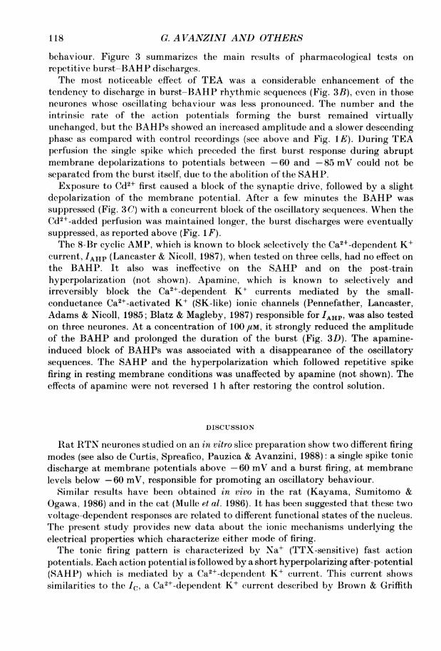
followed by a short (20-60 ms, mean value: 35 28 + 13-34 ms, n = 26) and deep after-hyperpolarizing potential (spike after-hyperpolarizing potential, SA'HP), whichshowed a short time-to-peak (mean value: 2-33 + 0-88 ms, n = 26) and a slower decay.The SAHP persisted during prolonged recordings with KCl-filled microelectrodesand was not blocked by bicuculline. The spike duration was reversibly prolonged byadding TEA to the perfusion fluid; this was due to a slowing down of the spikerepolarization, together with blockage of the SAHP (Fig. 1A). Addition of 1mM-Cd2+ to the perfusion medium also blocked the SAHP and induced a slowingdown of the action potential repolarization; this effect was reversed 40 min after thecontrol solution was restored (Fig. 1B).
Long-lasting depolarizing pulses caused repetitive action potential firing (Fig. 1 C).Each spike was followed by an SAHP.No burst or Ca2+-like action potentials were observed at membrane polarizations
at and above the resting level. The rate of repetitive firing was strictly dependent onthe amplitude of the injected current pulse, and showed a slight early accommodationwhich was manifest during the first 10-20 ms of the pulse. Firing was sustainedthroughout the duration of current injection (see Fig. 1 C and D). The accommodationdid not vary significantly during TEA-added perfusions (Fig. ID).A post-train after-hyperolarizing potential followed the repetitive discharge
evoked by an intracellular depolarizing pulse (Fig. 1D). It lasted about 50-80 ms anddecayed rapidly with a time constant similar to that of the SAHP. The increasednumber of action potentials evoked by increasing the depolarizing pulse durationcaused a mild prolongation of this potential and no significant increase in itsamplitude, which was significantly and reversibly decreased by TEA perfusions (Fig.1D).When the membrane potential was hyperpolarized below -60 mV, a different
firing mode was generated either by orthodromic activation, by depolarizing pulsesor at the break of hyperpolarizing pulses: single spike-SAHPs were followed by anall-or-none burst of spikes superimposed on a slow depolarizing potential (Fig. 1Eand F). The bursts lasted about 25-30 ms (mean value: 34-35 + 9-01 ms) and werecomposed of four to nine fast action potentials, whose duration and amplitude weresimilar throughout the duration of the burst. The initial burst firing rate was 200-300Hz and showed a slight acceleration, which peaked between the third and fifth action
113

114 G. AVANZINI AND OTHERS
TEA TEAControl Wash
A
Cdl2 Cd2+
Control WashB
3 Ms
120 mVC VJb~~~~tLW1iV--~~~~ ~ ~ ~ ~~0.5 nA
25 msD Control TEA Wash
E ~~~ControlTE
40 ms
Control d2+ Ws
F
25 ms
Fig. 1. A-I), intracellular recordings from different RTN neurones in resting conditions(single spike-tonic firing mode). A, single-spike response evoked by internal capsulestimulation; TEA (10 mM) prolonged the spike duration and reduced the SAHP in areversible way. B, Cd2+ (2 mM) has similar effects on the action potential occurring at thebreak of an hyperpolarizing current pulse. TEA is more effective than Cd2+ in prolongingthe spike duration. C, single-spike response and tonic firing obtained by intracellularinjection of depolarizing current pulses of different duration are both followed by short-lasting hyperpolarizations. No important spike accommodation is noted during therepetitive firing. D, the after-hyperpolarization subsequent to a train of action potentialsis blocked by TEA (10 mM). Note that during TEA addition, the same pulse intensityinduced an increase in spike number; this effect is due to the slight depolarization induced

PROPERTIES OF RTN NEURONES IiN VITRO
potentials and was followed by a decrease in frequency (Figs IF and 2B). On twooccasions, during recovery from a low-Ca2+, Cd2+-supplemented perfusion, it waspossible to isolate the single spike from the burst response (Fig. 1 F). When themembrane time constants, obtained by fitting the membrane rectification precedingthe single spike and the burst with an exponential function (using a least-squaresfitting procedure), were compared a significant difference was detected. The pre-spiketime constant of 56+14 ms was significantly different from the pre-burst timeconstant of 14 5 + 0-6 ms (P < 0 05). The pre-spike rectification was not influenced byCd2 , indicating that it was no mediated by a Ca2+ influx.The burst was followed by a pronounced hyperpolarization which lasted 80-120
ms and was composed of a fast, TEA-sensitive (Fig. 1E) repolarizing phase whichpeaked at about 20 ms (mean value: 21P38+ 49 ms) and a slower TEA-insensitive(Fig. 1 E) decay phase (mean value: 96 33 + 12 71 ms) which decreased monotonically.We have called this after-hyperpolarization a 'burst after-hyperpolarization'(BAHP). The BAHP was an all-or-none phenomenon and showed an approximatelyextrapolated equilibrium potential of 85-90 mV. The determination of the reversalpotential by the injection of hyperpolarizing currents was only tentative, because ofthe strict dependence of the burst-BAHP complex on the membrane potential. Theburst-BAHP complex could not be activated below -90 mV or above -60 mV. Itwas abolished by Cd21 (2 mm, Fig. IF) while TTX (1 /aM) sufficient to abolish Na+spikes uncovered a slow all-or-none depolarizing potential underlying the burst (seebelow and Fig. 3A). We have called the all-or-none TTX-resistant Cd2+-sensitive slowpotential of the complex 'low threshold spike' LTS (Llina's & Yarom, 1981 a, b;Deschenes et al. 1984; Jahnsen & Llinas, 1984a, b).
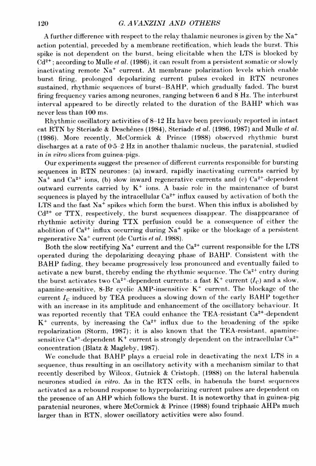
Oscillatory behaviourWhen RTN neurones are stimulated directly with long-lasting (600-1000 ms) and
large (0 3-1 nA) depolarizing current pulses from a hyperpolarized level, repetitiveburst-BAHP complexes are elicited, resulting in a characteristic oscillatorybehaviour. As shown in Fig. 2A and B the decay of the outward current responsiblefor each BAHP results in a depolarizing recovery phase, accompanied by a changein membrane rectification, which leads to a subsequent burst discharge that startsthe oscillatory cycle anew. The time constant of the decaying BAIIP showed asignificant (P < 0-01) increment just before the activation of the second burst (Fig.2B; see further analysis below). A similar set of events could also be obtained at thebreak of a large hyperpolarizing pulse injection superimposed on a steady membrane
by TEA on the membrane potential. E and F, burst-BAHP complexes elicited in twodifferent RTN neurones by depolarizing current pulses superimposed to a steadyhyperpolarizing current which held the membrane potential at 75 and 80 mV,respectively. E, the early phase of the BAHP is markedly slowed down during additionof TEA. The isolated action potential preceding the burst in the control recording couldnot, be distinguished from the following burst during the TEA effect. F, the isolatedpotential is not abolished by prolonged Cd2+ perfusion, which completely and reversiblyblocks the burst, discharge. The variability in spike amplitude shown in the fast-speedsweeps in this and the following figures is due to the numeric reconstruction of the eventsby means of a digital oscilloscope.
115

G. A VANZINI AND OTHERS
hyperpolarization (Fig. 2A, right sweep). As shown in Fig. 2 C the optimal conditionto elicit the oscillatory activity was obtained by stepping up the membrane potentialfrom very hyperpolarized levels (below -90 mV in Fig. 2 C to -85 to -60 mV).Lower depolarizing levels close to the equilibrium potential for BAHP evoked singlebursts followed by only a small hyperpolarization, while at higher depolarizing levelsclose to the resting potential, a first abortive burst was followed by sustained tonicfiring (Fig. 2 C, left sweep).
A
115 mV
20 mV
__ 10.5 nA100 ms 50 ms
120 mV
t~~~~~~~~~~~~~~~~~~~~1. nA
40 ms
Fig. 2. Oscillatory burst-BAAHP sequences in two neurones of the RTN. A, the sequencesare activated either by prolonged depolarizing current pulses (left) or at the break of anhyperpolarizing current pulse (right) when the membrane potential is maintained inhyperpolarized conditions by a steady current. B, the two early bursts in the left trace inA are shown at a greater sweep speed. An isolated action potential precedes the first burst,but not the second and following. C, single responses to long-lasting depolarizing pulsesstarting from a membrane potential kept below -90 mV. Resting potential (dashed line)was -58 mV. When the pulse induced a membrane shift close to resting potential, a tonicresponse preceded by an abortive burst is activated. At -85 mV the BAHP subsequentto the burst is flattened. Between -85 and -60 mV a deep BAHP is present; its risingphase starts the burst-BAHP sequences anew. The [K+]o was 6-25 mm during therecording.
In most of the cells, three to six consecutive oscillations could occur before theoscillatory behaviour faded (Fig. 2A). The fade of the sequences was associated withthe progressive reduction in the amplitude of the BAHP, presumably due to agradual reduction in the transmembrane driving force for the ions responsible for theBAHP (Fig. 2A). The duration of the burst increased slightly and the initial burstspiking rate slowed down during repetitive sequences (Fig. 2A). Note again the singleaction potential followed by SAHP which precedes by 2-10 ms the first burstactivated by a large and abrupt depolarization (Fig. 2B).The addition of TTX to the medium completely abolished the burst Na+ spikes
and isolated the LTS with a rising time constant of 13-34+5 31 ms (Fig. 3A) close tothe pre-burst time constant values. Note that during TTX perfusion no oscillatoryactivity could be activated, suggesting that the TTX-sensitive Na+ component fullyexpressed in the pre-spike rectification could be relevant in the build up of theregenerative inward currents responsible for the activation of the oscillatory
116

PROPERTIES OF RTN NEURONES IN VITRO 117
AControl TTX
20 mV
______________| 1 nA
100 Ms
TEA
Control WashB
J T LLI JLI
Control Cd2+ WashcJJ_
Control Apamine Wash
Fig. 3. Effects ofdifferent compounds on the oscillatory behaviour induced by depolarizingpulses injected when the cell membrane was maintained at a hyperpolarized level (-90mrV) by a DC current. A, when TTX (1 UM) is added to the perfusion fluid, the fast Na+spikes forming the burst are blocked, uncovering the LTS. No oscillatory activity followsthe activation of LTS when a prolonged depolarizing pulse is delivered. B, TEA (10 mM)induces an increase in the oscillating behaviour. The resting potential is -57 mV. C,cadmium (Cd2+; 2 mM) added to the medium virtually abolishes the BAHP in an RTNcell. The membrane resting potential is -55 mV. The blockage of the BAHP prevents theburst sequences. The effect of Cd2+ was recovered after 40 min of wash-out. D, apamine(100 /IM) selectively and irreversibly abolishes the BAHP and prolongs the burst duration.No oscillatory repetitive activation is obtained during BAHP blockage, not even when alarge depolarizing current pulse is injected (right sweep). Membrane resting potential(-58 mV) is unchanged during apamine perfusion.
G. A VANZINI AND OTHERS
behaviour. Figure 3 summarizes the main results of pharmacological tests onrepetitive burst-BAHP discharges.The most noticeable effect of TEA was a considerable enhancement of the
tendency to discharge in burst-BAHP rhythmic sequences (Fig. 3B), even in thoseneurones whose oscillating behaviour was less pronounced. The number and theintrinsic rate of the action potentials forming the burst remained virtuallyunchanged, but the BAHPs showed an increased amplitude and a slower descendingphase as compared with control recordings (see above and Fig. 1E). During TEAperfusion the single spike which preceded the first burst response during abruptmembrane depolarizations to potentials between -60 and -85 mV could not beseparated from the burst itself, due to the abolition of the SAHP.Exposure to Cd2+ first caused a block of the synaptic drive, followed by a slight
depolarization of the membrane potential. After a few minutes the BAHP wassuppressed (Fig. 30) with a concurrent block of the oscillatory sequences. When theCd2+-added perfusion was maintained longer, the burst discharges were eventuallysuppressed, as reported above (Fig. IF).The 8-Br cyclic AMP, which is known to block selectively the Ca2+-dependent K+
current, IAHP (Lancaster & Nicoll, 1987), when tested on three cells, had no effect onthe BAHP. It also was ineffective on the SAHP and on the post-trainhyperpolarization (not shown). Apamine, which is known to selectively andirreversibly block the Ca2+-dependent K+ currents mediated by the small-conductance Ca2+-activated K+ (SK-like) ionic channels (Pennefather, Lancaster,Adams & Nicoll, 1985; Blatz & Magleby, 1987) responsible for 'AHP' was also testedon three neurones. At a concentration of 100 /tM, it strongly reduced the amplitudeof the BAHP and prolonged the duration of the burst (Fig. 3D). The apamine-induced block of BAHPs was associated with a disappearance of the oscillatorysequences. The SAHP and the hyperpolarization which followed repetitive spikefiring in resting membrane conditions was unaffected by apamine (not shown). Theeffects of apamine were not reversed 1 h after restoring the control solution.
DISCUSSION
Rat RTN neurones studied on an in vitro slice preparation show two different firingmodes (see also de Curtis, Spreafico, Pauzica & Avanzini, 1988): a single spike tonicdischarge at membrane potentials above -60 mV and a burst firing, at membranelevels below -60 mV, responsible for promoting an oscillatory behaviour.
Similar results have been obtained in vivo in the rat (Kayama, Sumitomo &Ogawa, 1986) and in the cat (Mulle et al. 1986). It has been suggested that these twovoltage-dependent responses are related to different functional states of the nucleus.The present study provides new data about the ionic mechanisms underlying theelectrical properties which characterize either mode of firing.The tonic firing pattern is characterized by Na+ (TTX-sensitive) fast action
potentials. Each action potential is followed by a short hyperpolarizing after-potential(SAHP) which is mediated by a Ca2+-dependent K+ current. This current showssimilarities to the lc, a Ca2+-dependent K+ current described by Brown & Griffith
118

PROPERTIES OF RTN NEURONES IN VITRO
(1983) on hippocampal neurones, which is responsible for the spike repolarizationand transiently activated by Ca2+ influx during spike activation (Brown, Constanti& Adams, 1982). Like Ic (Lancaster & Nicoll, 1987), the current underlying SAHPin RTN neurones has been found to be: (a) K+ sensitive, (b) blocked by TEA and byCd2' and (c) reversed at -73 mV (de Curtis et al. 1988).A second TEA-sensitive current is known to be responsible for the spike
repolarization, namely the delayed rectifier IK (Hodgkin & Huxley, 1952). Thecontribution of IK to the spike repolarization could be evaluated by subtracting theeffects of Cd2+ (selectively affecting the Ca2+-dependent Ic) from those of TEA(affecting both the Ca2+-dependent Ic and the IK) (Fig. 1). As the TEA effect on therepolarizing phase of the action potential was proved to be substantially greater thanthe Cd2+ effect, we concluded that in RTN neurones the IK significantly contributesto the spike repolarization.Another Ca2+-dependent K+ current, the IAHP, is considered to play an important
role in spike frequency accommodation on different types of neurones where itunderlies long-lasting AHPs (Alger & Nicoll, 1980). In our experiments, the long-lasting SAHPs reported in thalamocortical neurones (Deschenes et al. 1984;McCormick & Prince, 1988) were not observed at resting membrane potential in RTNneurones, which accordingly exhibited a poor spike frequency accommodation.Similar findings have been previously reported by Mulle et al. (1986) in their in vivostudy on cat RTN neurones.The overall result of these data suggests that, in resting conditions, the RTN
neurones have strong spike repolarizing currents and that the mechanismsresponsible for repetitive firing control are inactive. The short refractory periodassociated with the lack of accommodation and slow AHP enable the RTN neuronesto fire tonically at high frequency. GABAergic neurones, located in differentstructures of the CNS, have recently been reported to show electrophysiologicalproperties similar to those described above for the RTN neurones, i.e. a fast, highrate of firing with poor frequency adaptation (Schlag & Waszak, 197 1; Schwartzkroin& Mathers, 1978; McCormick, Connors, Lighthall & Prince, 1985; Lacaille, Mueller,Kunkel & Schwartzkroin, 1987; Nakanishi, Kita & Kitai, 1987).The burst response of RTN neurones differs from that of the other thalamic
neurones (Jahnsen & Llinas, 1984a) in that it is followed by a deep after-hyperpolarization (BAHP). Unlike the AHP described in many CNS neurones, thispotential is not activated in a graded fashion and occurs as a transient all-or-nonepotential. The BAHP was flattened at -85 mV. Its reversal potential was difficultto detect, because of its dependence on the Ca2+ influx associated with LTS; whenthe LTS is inactivated (below -90 mV and above -60 mV) no BAHP was obtained.The BAHP is composed of (a) an early Cd2+- and TEA-sensitive part, probablycarried by an outward current which is the result of the summation of the Ca2+_dependent Ic repeatedly activated during the repolarization of the fast actionpotentials within the burst and (b) a late BAHP due to an outward Ca2+-dependentK+ current, selectively suppressed by apamine, but not by 8-Br cyclic AMP.Therefore, both the early and the late BAHPs are mediated by Ca2+-dependent K+currents, which counteract the strong depolarization underlying the burst. TheBAHP plays an important role in the burst termination.
119
G. AVANZINI AND OTHERS
A further difference with respect to the relay thalamic neurones is given by the Na±action potential, preceded by a membrane rectification, which leads the burst. Thisspike is not dependent on the burst, being elicitable when the LTS is blocked byCd2+; according to Mulle et al. (1986), it can result from a persistent somatic or slowlyinactivating remote Na+ current. At membrane polarization levels which enableburst firing, prolonged depolarizing current pulses evoked in RTN neuronessustained, rhythmic sequences of burst-BAHP, which gradually faded. The burstfiring frequency varies among neurones, ranging between 6 and 8 Hz. The interburstinterval appeared to be directly related to the duration of the BAHP which wasnever less than 100 ms.Rhythmic oscillatory activities of 8-12 Hz have been previously reported in intact
cat RTN by Steriade & Deschenes (1984), Steriade et al. (1986, 1987) and Mulle et al.(1986). More recently, McCormick & Prince (1988) observed rhythmic burstdischarges at a rate of 0-5-2 Hz in another thalamic nucleus, the paratenial, studiedin in vitro slices from guinea-pigs.Our experiments suggest the presence of different currents responsible for bursting
sequences in RTN neurones: (a) inward, rapidly inactivating currents carried byNa+ and Ca2+ ions, (b) slow inward regenerative currents and (c) Ca2+-dependentoutward currents carried by K+ ions. A basic role in the maintenance of burstsequences is played by the intracellular Ca2+ influx caused by activation of both theLTS and the fast Na+ spikes which form the burst. When this influx is abolished byCd2+ or TTX, respectively, the burst sequences disappear. The disappearance ofrhythmic activity during TTX perfusion could be a consequence of either theabolition of Ca2+ influx occurring during Na+ spike or the blockage of a persistentregenerative Na+ current (de Curtis et al. 1988).Both the slow rectifying Na+ current and the Ca2+ current responsible for the LTS
operated during the depolarizing decaying phase of BAHP. Consistent with theBAHP fading, they became progressively less pronounced and eventually failed toactivate a new burst, thereby ending the rhythmic sequence. The Ca2+ entry duringthe burst activates two Ca2+-dependent currents: a fast K+ current (Ic) and a slow,apamine-sensitive, 8-Br cyclic AMP-insensitive K+ current. The blockage of thecurrent Ic induced by TEA produces a slowing down of the early BAHP togetherwith an increase in its amplitude and enhancement of the oscillatory behaviour. Itwas reported recently that TEA could enhance the TEA-resistant Ca2+-dependentK+ currents, by increasing the Ca2+ influx due to the broadening of the spikerepolarization (Storm, 1987); it is also known that the TEA-resistant, apamine-sensitive Ca2+-dependent K+ current is strongly dependent on the intracellular Ca2+concentration (Blatz & Magleby, 1987).We conclude that BAHP plays a crucial role in deactivating the next LTS in a
sequence, thus resulting in an oscillatory activity with a mechanism similar to thatrecently described by Wilcox, Gutnick & Cristoph, (1988) on the lateral habenulaneurones studied in vitro. As in the RTN cells, in habenula the burst sequencesactivated as a rebound response to hyperpolarizing current pulses are dependent onthe presence of an AHP which follows the burst. It is noteworthy that in guinea-pigparatenial neurones, where McCormick & Prince (1988) found triphasic AHPs muchlarger than in RTN, slower oscillatory activities were also found.
120

PROPERTIES OF RTN NEURONES IN VITRO
On the basis of their intracellular recordings from in vivo cat RTN, Mulle et al.(1986) suggested that in addition to the K+-dependent hyperpolarization, GABA-mediated hyperpolarizing IPSPs fulfill an important role in deinactivating LTS inneighbouring neurones. We have found no evidence of any synaptic contribution tothe generation of the oscillatory activity in the in vitro rat RTN. In addition, localapplication of GABA to RTN neurones has proved to depolarize rather than tohyperpolarize the membrane (McCormick & Prince, 1986; Spreafico, de Curtis,Frassoni & Avanzini, 1988) and to be associated with a marked decrease inmembrane resistance; the resulting shunt of the membrane was usually effective indamping the burst-BAHP sequences.
In conclusion, the reported data are consistent with a double function of isolatedRTN neurones studied in vitro: (a) a relay-like behaviour, defined by the tonic firingdischarge modality and characterized by the ability to perform high frequencyresponses in a condition of poor repetitive firing control and (b) a bursting dischargebehaviour, characterized by the ability to maintain rhythmic burst sequences at lowrates (6-8 Hz).
This work was partially supported by C.N.R. grant No. 86.00339.04 and by the AssociazionePaolo Zorzi per le Neuroscienze. We wish to thank Miss Marije de Jager for her assistance in thepreparation of the manuscript.
REFERENCES
ALGER, B. E. & NICOLL, R. A. (1980). Epileptiform burst after hyperpolarization: calcium-dependent potassium potential in hippocampal CAl pyramidal cells. Science 210, 1122-1124.
AVANZINI, G., DE CURTIS, M. & SPREAFICO, R. (1987). Electrophysiological characterization ofsingle neurones of the nucleus reticularis thalami on rat slices. Society for Neuroscience Abstracts3, 271.
BLATZ, A. L. & MAGLEBY, K. L. (1987). Calcium-activated potassium channels. Trends inNeurosciences 10, 463-467.
BROWN, D. A., CONSTANTI, A. & ADAMS, P. R. (1982). Calcium-dependence of a component of atransient outward current in bullfrog ganglion cells. Society for Neuroscience Abstracts 8, 252.
BROWN, D. A. & GRIFFITH, W. H. (1983). Persistent slow inward current in voltage-clampedhippocampal neurones of the guinea-pig. Journal of Physiology 337, 303-320.
DE BIASI, S., FRASSONI, C. & SPREAFICO, R. (1986). GABA immunoreactivity in the thalamicreticular nucleus of the rat. A light and electron microscopical study. Brain Research 339,143-147.
DE CURTIS, M., SPREAFICO, R., PANZICA, F. & AVANZINI, G. (1988). Firing modes of RTN neuronsrecorded from thalamic slices. In Cellular Thalamic Mechanisms, Excerpta Medica InternationalCongress Series 765, ed. BENTIVOGLIO, M. & SPREAFICO, R., pp. 63-75. Amsterdam: Elsevier.
DESCHENES, M., PARADIS, M., Roy, J. P. & STERIADE, M. (1984). Electrophysiology of neurons oflateral thalamic nuclei in cat: resting properties and burst discharges. Journal ofNeurophysiology51, 1196-1219.
HODGKIN, A. L. & HUXLEY, A. F. (1952). A quantitative description of membrane currents and itsapplication to conduction and excitation in nerve. Journal ofPhysiology 117, 500-544.
HOUSER, C. R., VAUGHN, J. E., BARBER, R. P. & ROBERTS, E. (1980). GABA neurons are the majorcell type of the nucleus reticularis thalami. Brain Research 200, 341-354.
JAHNSEN, H. & LLINAS, R. (1984a). Electrophysiological properties of guinea-pig thalamicneurones: an in vitro study. Journal of Physiology 349, 205-226.
JAHNSEN, H. & LLINAiS, R. (1984b). Ionic basis for the electroresponsiveness and oscillatoryproperties of guinea-pig thalamic neurones in vitro. Journal of Physiology 349, 227-247.
JONES, E. G. (1975). Some aspect of the organization of the thalamic reticular complex. Journal ofComparative Neurology 162, 285-308.
KAYAMA, Y., SUMITOMO, I. & OGAWA, T. (1986). Does the ascending cholinergic projection inhibit
121
G. AVANZINI AND OTHERS
or excite neurons in the rat thalamic reticular nucleus? Journal of Neurophysiology 56,1310-1320.
LACAILLE, J. C., MUELLER, A. L., KtTNKEL, D. D. & SCHWARTZKROIN, P. A. (1987). Local circuitinteractions between oriens-alveus interneurons and CA1 pyramidal cells in hippocampal slices:electrophysiology and morphology. Journal ofNeuroscience 7, 1979-1993.
LANCASTER, B. & NICOLL, R. A. (1987). Properties of two calcium-activated hyperpolarizations inrat hippocampal neurones. Journal of Physiology 389, 187-203.
LLINAS, R. & YAROM, Y. (1981 a). Electrophysiology of mammalian inferior olivary neurones invitro: different types of voltage-dependent ionic conductances. Journal of Physiology 315,549-567.
LLINAS, R. & YAROM, Y. (1981 b). Properties and distribution of ionic conductances generatingelectroresponsiveness of mammalian inferior olivary neurones in vitro. Journal of Physiology 315,569-584.
MCCORMIC, D. A., CONNORS, B. WV., LIGHTHALL, J. WV. & PRINCE, D. A. (1985). Comparativeelectrophysiology of pyramidal and sparsely spiny stellate neurons in the neocortex. Journal ofNeurophysiology 54, 782-806.
MCCORMICK, D. A. & PRINCE, D. A. (1986). Acetylcholine induces burst firing in thalamic reticularneurons by activating a potassium conductance. Nature 319, 402-405.
MCCORMIC, D. A. & PRINCE, D. A. (1988). Noradrenergic modulation of firing pattern in guinea pigand cat thalamic neurons, in vitro. Journal ofNeurophysiology 59, 978-996.
MULLE, C., MADARIAGA, A. & DESCHENES, M. (1986). Morphology and electrophysiologicalproperties of reticularis thalami neurons in cat: in vivo study of a thalamic pacemaker. JournalofNeurosciences 6, 2134-2145.
NAKANISHI, H., KITA, H. & KITAI, S. T. (1987). Intracellular study of rat substantia nigra parsreticulata neurons in an in vitro slice preparation: electrical membrane properties and responsecharacteristics to subthalamic stimulation. Brain Research 437, 45-55.
PENNEFATHER, P., LANCASTER, B., ADAMS, P. R. & NICOLL, R. A. (1985). Two distinct Ca-
dependent K currents in bullfrog sympathetic ganglion cells. Proceedings of the National Academyof Sciences of the USA 82, 3040-3044.
SCHLAG, J. & WASZAK, M. (1971). Electrophysiological properties of units in the thalamic reticularcomplex. Experimental Neurology 32, 79-97.
SCHWARTZKROIN, P. A. & MATHERS, L. H. (1978). Physiological and morphological identificationof a non-pyramidal hippocampal cell type. Brain Research 157, 1-10.
SPREAFICO, R., FRASSONI, C., BATTAGLIA, G. & SCHMECHEL, D. E. (1984). The intrinsic organizationof the nucleus reticularis thalami in the rat: a Golgi, HRP and immunocytochemical study.Neuroscience Letters Supplement 18, S25.
SPREAFICO, F., DE CURTIS, M., FRASSONI, C. & AVANZINI G. (1988). Electrophysiologicalcharacteristics of morphologically identified reticular thalamic neurons from rat slices.Neuroscience 27, 629-638.
STERIADE, M. & DESCHENES, M. (1984). The thalamus as a neuronal oscillator. Brain ResearchReview 8, 1-62.
STERIADE, M., DOMICH, L. & OAKSON. G. (1986). Reticularis thalami neurons revisited: activitychanges during shifts in state of vigilance. Journal ofNeuroscience 6, 68-81.
STERIADE, M., DOMICH, L., OAKSON, G. & DESCHENES, M. (1987). The deafferented reticularisthalami nucleus generates spindle rhythmicity. Journal ofNeurophysiology 57, 260-273.
STORM, J. F. (1987). Action potential repolarization and fast after-hyperpolarization in rathippocampal pyramidal cells. Journal of Physiology 385, 733-759.
WILCOX, K. S., GUTNICK, M. J. & CHRISTOPH, G. R. (1988). Electrophysiological properties ofneurons in the lateral habenula nucleus: an in vitro study. Journal of Neurophysiology 59,212-225.
YEN, C. T., CONLEY, M., HENDRY, S. H. C. & JONES, E. G. (1985). The morphology ofphysiologically identified GABAergic neurons in the somatic sensory part of the thalamicreticular nucleus in the cat. Journal of Neuroscience 5, 2254-2268.
122