function and analysis of post- translational protein · pdf file ·...
TRANSCRIPT

Function and Analysis of Post-translational ProteinModifications
Function and Analysis of Post-translational Protein Modifications
Vorlesung 4610
Paul Jenö
Department of Biochemistry
Biozentrum

Function and Analysis of Post-translational ProteinModifications
Posttranslational Modifications ofProteins
http://www.abrf.org/index.cfm/dm.home?AvgMass=all
Approx. naturally occuring 260PTMs listed!

Function and Analysis of Post-translational ProteinModifications
Posttranslational Modifications ofProteins
• Covalent attachment of chemical groups to a protein
• Some modifications occur while the protein istranslated and exit from the ribosome
• Other modifications occur only after proteintranslation
• Require dedicated enzymatic catalysis

Function and Analysis of Post-translational ProteinModifications
Possible Sites of Modifications inProteins
Modificationsinvolving the peptide bonds
Modifications involving the carboxyl- and amino-terminals
Modificationsinvolving individual
amino acid side chains

Function and Analysis of Post-translational ProteinModifications
Some of the Side ChainsEnzymatically Modified by PTMs
• Ser, Thr, Tyr

Function and Analysis of Post-translational ProteinModifications
Some of the Side ChainsEnzymatically Modified by PTMs
• Lys acetylation
• Lys ubiquitination

Function and Analysis of Post-translational ProteinModifications
Some of the Side ChainsEnzymatically Modified by PTMs
• Cys lipidation
• Acylation by long chain fatty acyl CoAs (C14, C16)
• Prenylation by C15 (farnesylation)

Function and Analysis of Post-translational ProteinModifications
Some of the Side ChainsEnzymatically Modified by PTMs
• Glu methylation
• Glu carboxylation (blood coagulationcascades)

Function and Analysis of Post-translational ProteinModifications
General Reaction Scheme for PTMof Proteins
• ATP phosphoproteins
• AcylCoAs acylated lipoproteins
• UDPGlcNAc glycoproteins

Function and Analysis of Post-translational ProteinModifications
General Reaction Scheme for PTMof Proteins
• Farnesyl-PP, Geranylgeranyl-PP prenylatedlipoproteins
• SAM Methylated proteins

Function and Analysis of Post-translational ProteinModifications
Cellular Sites of Major Post-translational Modifications
Addition of palmitoyl groups
O-Glycosylation with GlcNac
Myristoylation of N-terminus
Acetylation of N-terminus
Removal of initiating MetCytoplasm
ModificationSite

Function and Analysis of Post-translational ProteinModifications
Cellular Sites of Major PTM’s
Amidation of C-terminus
Proteolytic processing ofsome precursors
SecretoryVesicles/Granules
Modification of N-glycosylgroups,
O-glycosylation with GalNAcGolgi Apparatus
Cleavage of Signal PeptidesMitochondria/Chloro-plasts
ModificationSite

Function and Analysis of Post-translational ProteinModifications
Cellular Sites of Major PTM’s
Disulfide bond formation
Hydroxylation of Pro/Lys inprocollagen
Addition of palmitoyl andglycosyl-phosphatidylinositol
Core glycosylation of Asnresidues
Cleavage of signal peptidesER
ModificationSite

Function and Analysis of Post-translational ProteinModifications
Functions Enabled byPosttranslational Modification
• Alterations in local folding of proteins– E.g. transitions between unstructured and structured regions– Generation of charge pairs
• Marking proteins for degradation– Proteasome targeting
• Marking chromatin for transcriptional regulation• Changing the intracellular or extracellular adresses of
proteins– Signal peptides direct proteins to …– Plasma membrane– Secretory pathway– Mitochondria– Cytosol
• Inactive apo to active holo forms of enzymes
Function and Analysis of Post-translational ProteinModifications
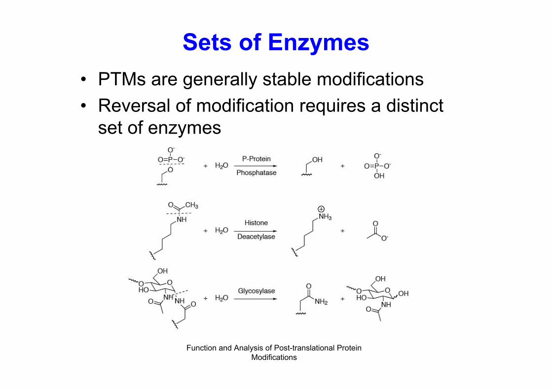
Sets of Enzymes
• PTMs are generally stable modifications
• Reversal of modification requires a distinctset of enzymes
Function and Analysis of Post-translational ProteinModifications
Modifications Involving the α-amino Group
• Nα-acylation– formyl– acetyl– pyruvoyl– α-ketobutyryl– glucuronyl– pyroglutamate– murein

Function and Analysis of Post-translational ProteinModifications
Formylation of the α-Amino Group• Nα-formyl-Met
– forms the start of the nascent protein chain inprokaryotes
– Eukaryotic start signal is Met• Deformylase may remove the CHO group• Aminopeptidase later removes Met from some, but
not all chains• Nα-formyl-Gly
– occurs in honey bee melittin• Murein derivatives
– link E. coli peptidoglycan and membranelipoprotein

Function and Analysis of Post-translational ProteinModifications
Protein Acetylation

Function and Analysis of Post-translational ProteinModifications
Protein N-Acetylation
• In about 50% of yeast proteins andabout 80-90% of higher eukaryoticproteins
• Very rare in E. coli
• S5 (N-Ac-Ala-Arg-…), S18 (N-Ac-Ala-His-…), L12 (N-Ac-Ser-Ile-…)

Function and Analysis of Post-translational ProteinModifications
Protein N-Acetylation

Function and Analysis of Post-translational ProteinModifications
Specificity of Protein N-Acetylation
• Lies in the specificity of MAP• MAP cleaves preferentially
– Met-Gly– Met-Ala– Met-Ser– Met-Cys– Met-Thr– Met-Pro– Met-Val
• Uncovered Ser, Ala, Gly, Thr, get acetylated

Function and Analysis of Post-translational ProteinModifications
N-acetyl Transferases (NATs)
• In yeast, 3 NATs (NAT A, B, and C)
• NATB and NATC acetylate proteins withMet1 still in place
• NATB recognises M-E, M-D, M-Q, M-M
• NATC recognises M-I, M-L, M-W, M-F
• NATA acetylates S, A, G, T

Function and Analysis of Post-translational ProteinModifications
N-acetyl Transferases
•In eukaryotes, hundreds of NATs exist•Involved in K acetylation of histones•Control
-transcriptional activation-Chromatin assembly-DNA replication-Involved in N-acetylation of amino- glycoside antibiotics, results in decreased affinity of the drug for its target

Function and Analysis of Post-translational ProteinModifications
Biological Significance of N-terminal Acetylation
• Unclear in eukaryotes!
• Functions only detected on a case-by-case study
• Actins are known to be acetylated at N-terminus
– N-terminus is M-E…
– E exposed by aminopeptidases
– Further acetylation to yield mature N-acetyl-E
– Strengthens interaction between actin and myosin

Function and Analysis of Post-translational ProteinModifications
N-Acetylation of Lysine-ε-NH2 SideChains
• Interest in protein N-acetylation is onregiospecific modification of K side chains
• Large number of PTMs detected on histones
• Transcriptional coactivators and corepressorsturned out to be HATs and HDACs
• N-terminal regions of histones are flexibleand amenable to PTMs

Function and Analysis of Post-translational ProteinModifications
•Chromatin contains H2A, H2B, H3 and H4•Histone core (H2A)2(H2B)2(H3)2(H4)2
•145-147 bps around core
N-Acetylation of Lysine-ε-NH2 SideChains

Function and Analysis of Post-translational ProteinModifications
Histone Acetylation
Phosphorylation
Methylation
Acetylation

Function and Analysis of Post-translational ProteinModifications
Histone Acetylation
• MS analysis revealed acetylation on…• H2A (K5 and K9)• H2B (K5, K12, K15, K20)• H3 (K9, K14, K18, K23)• H4 (K5, K8, K12, K16)• For two copies each of histone: 28 potential
acetylation sites• Yeast: 13 acetylation sites found/octamer• 50% posttranslational utilization• Immense combinatorial possibilities

Function and Analysis of Post-translational ProteinModifications
Consequences of HistoneAcetylation
• K’s cationic at physiological pH
• N-acetylation quenches positive charges
• Electrostatic weakening of histone/DNA interactions
• Opening of the chromatin
• Allows TFs to bind to promoter regions

Function and Analysis of Post-translational ProteinModifications
Consequences of HistoneAcetylationModification Function
________________________________
Unmodified Silencing
Acetylated Transcription
Acetylated Histone deposition?
Phosphorylated Mitosis/Meiosis
Phos/Acetyl Transcription
Methylated Transcription?
Higher-order ?
combinations

Function and Analysis of Post-translational ProteinModifications
Histone Acetyltransferases - AFamily
• Family of GNATs (Gcn5-related N-acetyltransferases)
HAT Bromo
HATBromo
HAT Bromo Bromo
yGcn5
CBP
TAFII250

Function and Analysis of Post-translational ProteinModifications
Transcriptional Activation byHATs

Function and Analysis of Post-translational ProteinModifications
Histone Deacetylases (HDACS)

Function and Analysis of Post-translational ProteinModifications
Histone Deacetylases (HDACS)
• HDACS are corepressors of transcription
• Maintain histone tails in hypoacetylated state
• Leads to chromosome condensation
• Silence promoters
• 2 distinct families– HDACs that release the acetyl moiety as acetate
– Sirtuins (silent information regulator)

Function and Analysis of Post-translational ProteinModifications
Sirtuin HDACS

Function and Analysis of Post-translational ProteinModifications
Influence of Histone Acetylationand Deacetylation on Nucleosome
Structure

Function and Analysis of Post-translational ProteinModifications
Protein Methylation

Function and Analysis of Post-translational ProteinModifications
Protein Methylation
• Occurs on N- or O-atoms• Methylation of -COO- covers up negative charge• N-methylation of Ks does not alter charge, increases
hydrophobicity• Di- and trimethylation of Ks increases both
hydrophobicity and steric bulk• Affects protein-protein interactions• Occurs on ε-amino group of K, imidazole ring of H,
guanidino group of R, amides of Q and N• N-methylation irreversible

Function and Analysis of Post-translational ProteinModifications
N-Methylations

Function and Analysis of Post-translational ProteinModifications
O-, S-, and C-Methylations

Function and Analysis of Post-translational ProteinModifications
One Carbon Donor

Function and Analysis of Post-translational ProteinModifications
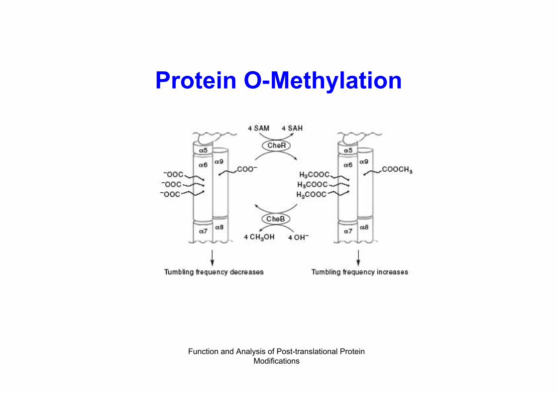
Protein O-Methylation Occurs inBacterial Chemotaxis

Function and Analysis of Post-translational ProteinModifications
Protein O-Methylation• Occurs in bacterial chemotaxis Transmembrane receptors for• Asp (Tar), Ser (Tsr), Peptides (Tap)

Function and Analysis of Post-translational ProteinModifications
Protein O-Methylation
Function and Analysis of Post-translational ProteinModifications
Protein O-Methylation

Function and Analysis of Post-translational ProteinModifications
Histone Methylation

Function and Analysis of Post-translational ProteinModifications
Histone Methylation in H3

Function and Analysis of Post-translational ProteinModifications
Histone Modification
• H3 modification– 3 Rs methylated
– 4 Ks methylated
– 2 Ss phosphorylated
• Results in over 110,000 combinations ofpossible modifications
• approx. 107 possible nucleosomes

Function and Analysis of Post-translational ProteinModifications
Protein Phosphorylation

Function and Analysis of Post-translational ProteinModifications
Protein Phosphorylation• One of the most common PTMs• Estimate that approx. 30% of all eukaryotic proteins
become phosphorylated• Human genome codes for approx. 2,000 protein
kinases• Transient• Regulate a vast number of biological processes
– Enzymatic activity– Metabolism– Motility– Signal transduction– Cell division– Cell growth– Apoptosis

Function and Analysis of Post-translational ProteinModifications
Protein Phosphorylation
• Ser, Thr, Tyr phosphorylation by protein kinases

Function and Analysis of Post-translational ProteinModifications
Protein Phosphorylation• Protein kinase variants defined by the type of protein side chain
modification
a) Ser/Thr protein kinases (e.g. cAMP-dependent PK)
b) Tyr protein kinases (e.g. insulin receptor kinase)
c) His protein kinases (e.g. bacterial two componentsensor/respons regulators)

Function and Analysis of Post-translational ProteinModifications
Protein Phosphorylation
• What is the purpose/effect of protein side chainphosphorylations?
– Protein conformational switching
– Introduction of a dianionic phosphate group (- PO3--) induces
electrostatic reorganization of local regions, loops of proteins
– Reorganization often occurs via charge pairing with a cationic Argside chain

Function and Analysis of Post-translational ProteinModifications
Protein Phosphorylation• What governs specificity of phosphorylation?
– PKs recognise certain motifs

Function and Analysis of Post-translational ProteinModifications
Physiological Importance ofProtein Phosphorylation

Function and Analysis of Post-translational ProteinModifications
Protein Dephosphorylation• Phosphoproteins are dephosphorylated by P-protein
phosphatases
• Phosphatases counteract protein kinases
• Human genome encodes approx. 500 proteinphosphatases
• Ser/Thr- and Tyr-specific phosphatases

Function and Analysis of Post-translational ProteinModifications
Protein Phosphatases
• Phosphatase 1– Ser/Thr-specific phosphatase
– Regulates cell-cycle progression
– Muscle contraction
– Carbohydrate metabolism
• Protein Phosphatase 2A– Controls numerous cellular processes
– Oligomeric enzymes

Function and Analysis of Post-translational ProteinModifications
Protein Phosphatase 2A

Function and Analysis of Post-translational ProteinModifications
Protein Phosphatase 2B,Calcineurin
• Ser/Thr phosphatase
• Controlled by cellular Ca2+
• Multimeric enzyme, bindingto Ca2+/calmodulin requiredfor active enzyme

Function and Analysis of Post-translational ProteinModifications
Ubiquitination

Function and Analysis of Post-translational ProteinModifications
Ubiquitination• Occurs in eukaryotes, not in prokaryotes
• Used for proteolytic destruction of specific proteins
• Ubiquitin is a small 76 residue protein
• Covalent attachment of multiple ubiquitin moleculesto a protein substrate
• Degradation of the tagged protein by the 26Sproteasome (ubiquitin is recycled)

Function and Analysis of Post-translational ProteinModifications
Ubiquitination

Function and Analysis of Post-translational ProteinModifications
Ubiquitination

Function and Analysis of Post-translational ProteinModifications
Ubiquitination ReactionMechanism

Function and Analysis of Post-translational ProteinModifications
Enzymes Involved in theUbiquitination Process
• E1: only one known, consists of one or twopolypeptidic chains, with M.W. 105 kDa perchain, very conservative protein
• E2: 5-12 proteins of this type known(depending on literature), homologous family
• E3: many and structurally unrelated,characterized by ultimate biological specificity,M.W. about 250 kDa
• E4: not known yet, but postulated as existing

Function and Analysis of Post-translational ProteinModifications
Ubiquitination

Function and Analysis of Post-translational ProteinModifications
SUMO (Small Ubiquitin-RelatedModifier)
• SUMO does not have the Lys-48 found in ubiquitin
• SUMO does not make multi-chain forms
• SUMO-1,2,3 are the mammalian form
• SUMO-1: 101 amino acids, C-terminal Gly, 18%identical to ubiquitin

Function and Analysis of Post-translational ProteinModifications
SUMO Substrates
• Many of the known sumoylation substrates arenuclear proteins– p53
– CREB
– STAT1/4
– GATA2, etc

Function and Analysis of Post-translational ProteinModifications
Proteolytic Cleavage

Function and Analysis of Post-translational ProteinModifications
Proteolytic Cleavage
• Following translation, most proteins undergoproteolytic cleavage
• Removal of the initiation Met
• Many proteins are synthesised as inactive precursors
• E.g. pancreatic enzymes, enzymes involved in bloodclotting = proproteins
• Activation occurs via removal of polypeptides

Function and Analysis of Post-translational ProteinModifications
Peptide Preprohormone

Function and Analysis of Post-translational ProteinModifications
Protein Splicing
• Internal protein sequence (intein) Excises itself from a surrounding• External protein • N- and C-terminal exteins are ligated• Protein splicing occurs in bacteria and single-celled eukaryotes• Exteins have no sequence similarity• Inteins have conserved splice junctions:
- Ser/Thr/Cys at the N-terminus- His-Asn/Gly dipeptide at the C-terminus
• inteins encode endonucleases that copy the intein gene into extein sequences• Intein genes propagate themselves

Function and Analysis of Post-translational ProteinModifications
Glycoproteins

Function and Analysis of Post-translational ProteinModifications
Glycoproteins
• Most secreted, or plasma membrane boundproteins are glycosylated
• Extracellular part modified• Cytosolic and/or nuclear proteins also found
to be glycosylated• Predominant sugars found in glycoproteins
are– Glucose, galactose, mannose, fucose, GalNAc,
GlcNAc, NANA

Function and Analysis of Post-translational ProteinModifications
Glycoproteins• Carbohydrates attached either O- or N-glycosidacally• N-glycosidic linkage through amide of Asn
– Carbohydrate attachment within consensus sequence N-X-S(T)
• O-glycosidic linkage is through -OH of Ser, Thr, orOH-Lys

Function and Analysis of Post-translational ProteinModifications
Contains onlymannose outside
the core
GlycoproteinsContains various
sugars
Similar to hybrid,contains sialic acid
Glc-Nac
Mannose
Galactose
Sialic acid

Function and Analysis of Post-translational ProteinModifications
Glycoproteins
• Glycoproteins synthesised at rER
• Sugar attachment cotranslationally in the lumen ofER and continues in the Golgi for N-linked sugars
• O-linked sugars are attached post-translationally inthe Golgi
• Sugars are activated by coupling to nucleotides
• Glc and GlcNAc are coupled to UDP
• Mannose is coupled to GDP

Function and Analysis of Post-translational ProteinModifications
Glycoprotein Synthesis
• N-linked glycoprotein synthesis requiresthe lipid intermediate dolichol phosphate

Function and Analysis of Post-translational ProteinModifications
Glycoprotein Synthesis
+2 GlcNAc
+5 Man
+4 Man +3 Glc
Glc3Man9GlcNac2-P-P-dol

Function and Analysis of Post-translational ProteinModifications
Sugar Trimming
Glucosidase I-1 Glc
Glucosidase II-2 Glc
Core Structure
GlycosyltransferasesGlycosidases
MatureGlycoprotein
GlucosidasesMannosidases

Function and Analysis of Post-translational ProteinModifications
Clinical Significance ofGlycoproteins
• AB0 blood group antigens– AB0 carbohydrates linked to lipids
– AB0 associated with proteins occur in the serum = secreted form
– Some individuals produce secreted AB0
– Used in forensic medicine
• Dystroglycan– Laminin receptor
– alpha-distroglycan serves as receptors for Mycobacterium lepraeand other pathogens
• Helicobacter pylori attaches Lewis blood group antigen on thesurface of gastric mucose
• Etc. …

Function and Analysis of Post-translational ProteinModifications
Further Reading
• Walsh, C.T. (2005) Posttranslational Modifications ofProteins. Expanding Nature’s Inventory. Roberts andCompany Publishers.
• Krishna, R. G. and F. Wold (1998). PosttranslationalModifications. Proteins - Analysis and Design. R. H.Angeletti. San Diego, Academic Press. 1: 121-206.
• Wold, F., (1981) In vivo chemical modification ofproteins (post-translational modification) Ann. Rev.Biochem. 50,, 783-814.