functional dissection of phosphorylation of …jennylab.aecom.yu.edu/paperspdfs/dsh phosphorylation...
TRANSCRIPT
Developmental Biology 360 (2011) 132–142
Contents lists available at SciVerse ScienceDirect
Developmental Biology
j ourna l homepage: www.e lsev ie r .com/deve lopmenta lb io logy
Functional dissection of phosphorylation of Disheveled in Drosophila
Wang A. Yanfeng a, Hebist Berhane b, Marion Mola a, Jaskirat Singh a, Andreas Jenny b,⁎, Marek Mlodzik a,⁎⁎a Dept. of Developmental & Regenerative Biology, Mount Sinai School of Medicine, One Gustave L. Levy Place, New York, NY 10029, USAb Department of Developmental and Molecular Biology, Albert Einstein College of Medicine, Chanin 503, 1300 Morris Park Avenue, Bronx, New York, NY 10461, USA
⁎ Corresponding author.⁎⁎ Corresponding author. Fax: +1 212 860 9279.
E-mail addresses: [email protected] ([email protected] (M. Mlodzik).
0012-1606/$ – see front matter © 2011 Elsevier Inc. Alldoi:10.1016/j.ydbio.2011.09.017
a b s t r a c t
a r t i c l e i n f oArticle history:Received for publication 9 December 2010Revised 23 August 2011Accepted 14 September 2011Available online 22 September 2011
Keywords:Phospho-proteinDisheveledWinglessWnt-signalingSignaling specificityPCP
Disheveled/Dshproteins (Dvl inmammals) are core components of bothWnt/Wg-signaling pathways: canonicalβ-catenin signaling and Frizzled (Fz)-planar cell polarity (PCP) signaling. Although Dsh is a key cytoplasmiccomponent of both Wnt/Fz-pathways, regulation of its signaling specificity is not well understood. Dsh isphosphorylated, but the functional significance of its phosphorylation remains unclear. We have systematicallyinvestigated the phosphorylation of Dsh by combining mass-spectrometry analyses, biochemical studies, and invivo genetic methods in Drosophila. Our approaches identified multiple phospho-residues of Dsh in vivo. Ourdata define three novel and unexpected conclusions: (1) strikingly and in contrast to common assumptions,all conserved serines/threonines are non-essential for Dsh function in either pathway; (2) phosphorylation ofconserved Tyrosine473 in the DEP domain is critical for PCP-signaling— DshY473F behaves like a PCP-specificallele; and (3) defects associated with the PCP specific dsh1 allele, DshK417M, located within a putative ProteinKinase C consensus site, are likely due to a post-translational modification requirement of Lys417, rather thanphosphorylation nearby. In summary, our combined data indicate that while many Ser/Thr and Tyr residuesare indeed phosphorylated in vivo, strikingly most of these phosphorylation events are not critical for Dsh func-tion with the exception of DshY473.
. Jenny),
rights reserved.
© 2011 Elsevier Inc. All rights reserved.
Introduction
Frizzled (Fz) family receptors act as transducers of the Wnt/Wggrowth factor family, commonly signaling through the canonical Wnt(Wg)-Fz/β-catenin pathway (Logan and Nusse, 2004; Polakis, 2000). Inaddition, a distinct Wnt-Fz pathway regulates cellular polarity withintheplane of an epithelium, referred to as non-canonicalWnt or Fz/planarcell polarity (PCP) signaling (Adler, 2002; Keller, 2002; Lawrence et al.,2007; Seifert and Mlodzik, 2007; Wang and Nathans, 2007; Wu andMlodzik, 2009). Fz/PCP signaling diverges from the β-catenin pathwaydownstream of the cytoplasmic component Disheveled (Dsh inDrosophila, Dvl 1–3 in mammals). Since both pathways require Fzand Dsh to transduce signaling information, a key question is how Fzand potential associated factors can differentially activate Dsh. Inmany tissues, both pathways act in the same cells and thus a tightregulation of Fz-Dsh signaling specificity is essential. In deregulated,mutant scenarios, the selection of the wrong Fz-Dsh pathway canlead to disease (i.e. polycystic kidney disease or cancer (Logan andNusse, 2004; Polakis, 2000; Simons and Mlodzik, 2008)). Althougha molecular framework of the two pathways has been established(Klein and Mlodzik, 2005; Logan and Nusse, 2004; Mlodzik, 2002;Strutt, 2003; Veeman et al., 2003), little is known about the molecular
aspects regulating Fz-Dsh pathway selection and signaling specificity.Here we investigate this with focus on Dsh phosphorylation.
Drosophila is a well-suited model system to addressWnt-signalingspecificity mechanisms. For example, in the fly eye, activation of theFz/β-catenin pathway leads to the restriction of the eye field andlater the removal of excess photoreceptor cells through apoptosis(Lin et al., 2004), whereas Fz/PCP signaling is required for correct in-duction of specific photoreceptor fates and their patterning (Mlodzik,1999; Strutt and Strutt, 1999). Similarly, during wing developmentthe two pathways have distinct non-overlapping functions: canonicalWg-signaling is required for correct wing blade/margin formation,while Fz/PCP signaling orients wing cells in the proximal–distal axis.Moreover, as Fz/PCP aspects are universal to metazoans (Adler,2002; Keller, 2002; Lawrence et al., 2007; Seifert and Mlodzik, 2007;Wang and Nathans, 2007; Wu and Mlodzik, 2009), Drosophila iswell suited to study Dsh function, because of lack of Dsh redundancy(a single dsh gene) and its function being testable in rescue assays ofnull mutants.
In Wnt-Fz/β-catenin signaling, the combined action of Fz, LRP5/6-Arrow (a Fz co-receptor), and Dsh is to antagonize the APC/GSK3/Axincomplex that causes β-catenin (Arm in Drosophila) phosphorylationand degradation. Upon Wnt binding, the two transmembrane proteinsassociate, leading to a physical interaction of Dsh and Axin (via an inter-action through their DIX domains) at the membrane receptor complex(Bilic et al., 2007; Zeng et al., 2008). This leads to the stabilization ofβ-catenin/Arm and allows its accumulation in the nucleus, where itinteracts with TCF transcription factors on Wnt-signaling target
133W.A. Yanfeng et al. / Developmental Biology 360 (2011) 132–142
sites (Logan and Nusse, 2004; Polakis, 2000). In contrast, during Fz/PCP signaling, Fz “activates” Dsh through an unknown mechanismin an LRP5/6-Arrow independent manner, with Dsh actng on a dis-tinct set of effectors. The components of Wnt-Fz/β-catenin that actdownstream of Dsh are not required in the Fz/PCP pathway, andvice versa (e.g. (Axelrod et al., 1998; Boutros et al., 1998)). The Fz-Dsh pair thus activates distinct pathways, a feature conserved be-tween flies and vertebrates (Mlodzik, 2002; Sahai et al., 1998; Strutt,2003; Veeman et al., 2003).
The regulation of signaling specificity remains largely obscure. Someinsights have come from studies in Drosophila, with focus on the cyto-plasmic (C−) tails of Fz and Fz2 identified as critical in regulating thespecificity through their effects on subcellular localization (Boutroset al., 2000; Wu et al., 2004). Other domains of Fz and/or Fz2 (e.g. theWnt interacting extracellular CRD) are also important in determiningsignaling outcome (Boutros et al., 2000; Rulifson et al., 2000; Strappsand Tomlinson, 2001;Wu and Mlodzik, 2008). All Dsh-family members(ranging fromnematodes to humans) share three conserved domains: aDIX domain, a central PDZ domain and a C-terminal DEP domain, whichhave been implicated in protein–protein interactions, and thus Dsh like-ly serves as an adapter molecule (rev. in Boutros and Mlodzik, 1999;Wallingford and Habas, 2005). Dsh is recruited by Fz to the membrane(Boutros et al., 2000; Wu et al., 2004). Although a Fz–Dsh interactionsurface has been identified (Wong et al., 2003), additional mechanismshelp to stabilize themembrane complex (Simons et al., 2009). In cell cul-ture, when Dsh is not in a membrane-associated state, it tends to be incytoplasmic aggregates withmultimerizationmediated by DIX domains(Schwarz-Romond et al., 2005). Regulation of Dsh activity and localiza-tion is therefore likely instrumental for signaling outcomes.
PCP specific Dsh recruitment appears more stable than its generalbasolateral membrane recruitment for β-catenin signaling (Wu et al.,2004), suggesting that Dsh receives different “local” input (for examplethrough post-translational modification) for its membrane association(Metcalfe et al., 2010). Dsh/Dvl proteins also use different domains fordownstream pathway functions: DIX and PDZ domains are essential forβ-catenin signaling, while the PDZ and DEP-C terminal domains are crit-ical for PCP signaling (rev. in Boutros and Mlodzik, 1999; Wallingfordand Habas, 2005) (also Fig. 1A). Upon “activation”, Dsh is subject totwo major changes: (1) translocation from the cytosol to membraneand (2) hyper-phosphorylation, usually assessed by changes in gelmobility. Both aspects are often used as ameasure of “Dsh activation”, al-thoughwhat “activation” entails remains unclear. At least for phosphory-lation, it is only a correlated phenomenonwithout a defined contributionto signaling function (Axelrod, 2001; Cong et al., 2004; Klein et al., 2006;Ossipova et al., 2005; Strutt et al., 2006). In Drosophila, both the dsh1 andfz− mutants (with PCP specific defects) show markedly reduced Dshphosphorylation as evident by band-shift (Axelrod, 2001). In vertebratecell culture models, both canonical and non-canonical (PCP)Wnts, inparticular Wnt3A and Wnt5A respectively, lead to induction of Dshphosphorylation as detected with Dsh/Dvl band-shift assays. Multiplekinases have been suggested and shown to phosphorylate Dsh invitro, with potential regulatory requirement(s) in overexpressionexperiments (Cong et al., 2004; Klein et al., 2006; Ossipova et al.,2005; Strutt et al., 2006).
A comprehensive analysis of Dsh phosphorylation has been especiallychallenging since for example over 15% of Dsh residues are serines andtheronines. Here we have explored mass-spectroscopic, biochemical,and genetic approaches to identify phosphorylated residues on Dshand systematically define their functional significance in in vivo inrescue assays. Besides many Ser/Thr residues identified biochemically,our approach also identified tyrosine phosphorylation. Strikingly, incontrast to common assumptions and the fact that a phosphorylationdependent band-shift of Dsh/Dvl-proteins is often used as pathway ac-tivation read-out (Axelrod, 2001; Cong et al., 2004; Klein et al., 2006;Ossipova et al., 2005; Strutt et al., 2006), all of the Ser/Thr residues tested(although conserved) are dispensable for Dsh function in vivo. We also
show that theDsh gel-shift can be uncoupled fromDsh activity. However,Tyr473 located within the DEP domain is essential for Dsh functionfor PCP signaling in vivo. Furthermore, dissection of the dsh1 allele(DshK417M) suggests that K417 could undergo direct post-translationalmodification rather than affect a phosphorylation event in its flankingPKC site consensus. Our data indicate that while many Ser, Thr, and Tyr-residues are phosphorylated, most of these phosphorylation events are,unexpectedly, not critical for Dsh function.
Material and methods
DNA constructs, fly genetics, and rescue assay
dshNDshGFP was kindly provided by Jeff Axelrod. Point mutagenesisof the Dsh ORF was performed by excising it with either KpnI (from278nt of ORF to the end of polyA tail) or EcoRI (from about 135ntahead of start codon to the end of polyA tail), subcloned into pBlueScript,and mutated by standard protocols. For in vivo purification, dshNDsh-3XFlagwasmade by replacing C-terminal GFP sequence from the originalconstruct with 3XFlag sequence (Sigma). To analyze the potentialrequirements of S/T residues in the C-terminal part of Dsh, wehave mutated them to Ala in three blocks (separated by introducedsilent restriction sites) indicated by purple, blue and red (Fig. 2). 2cluster combinations, referred to as C-term#1-2, C-term#1-3 or C-term#2-3 in Table 1 were made by swapping combining individualclusters. The respective mutant clusters in the context of full-lengthDsh were then tested in gel-shift assays after transfection into 293 Tcell with or without Fz. Finally, we also generated a Dsh that lacked allsequences downstreamof the Blp1 site, referred to asΔC-term (Table 1;note that themutant constructs lack all but one Ser, whileΔC-term con-tains no Ser/Thr C-terminal to the DEP domain). A detailed cloningstrategy is available upon request.
To estimate effects of mutants, dshNDsh(mutant)GFP strains werebalanced on the second or third chromosomes and crossed to thedshv26 null mutants. Note that the dshv26 chromosome used was alsomarked with forked (f) to control for non-disjunction events. Rescueindex was calculated as: rescue index (R.I.)=rescued/control. Foreach group of rescue assay, dshNDshwtGFP served as baseline control.R.I. of wild-type Dsh is in theory 1. Practical R.I. (wild-type) was usedto calibrate R.I. of mutants.
y,w,f36a,dshV26/FM6was obtained from the Bloomington Stock center.
Peptides and antibodies
The phospho-peptide sequences used as antigens were:
pS36 CSAQV(pT)LRDFKpY280 RGGDGGI(pY)VGSIpS451 CRREARRIV(pS)AMLRpY473 GGEQC(pY)YVVNE
Peptides were synthesized by ProSci and Tufts University Mass-Spectrometry Core Facility, and antisera were generated by ProSci(California). Anti-Fmi antiserum was from Developmental StudiesHybridoma Bank.
Collection of fly samples and protein purification
There is no documented setup to collectDrosophila larvae and pupaeat large scale. Wemodified a regular trash-can to a “larval farm”. 15–20bottles of fly cultures (larval stages) were transferred to the “farm”.Small amount of water was added to make it moist and a thin layer ofyeast pellets was spread on top, the surface was increased with plasticfoil/film to allow larvae to crawl up and covered with a glass paneland wet towel. The setup was kept at room temperature and inspected

Fig. 1. Identification of phosphorylated residues of Dsh. (A) Schematic of Dsh with the DIX, PDZ, and DEP domains indicated in purple. Light and dark green domains correspond tothe Basic and Proline rich regions, respectively. Dark blue boxes represent the Serine/Threonine clusters that were mutated in Penton et al. (2002). ST8 (light blue box) correspondsto the 8 S/T residues that were mutated in Strutt et al. 2006 and have been identified as potential Par1 sites (Ossipova et al. 2005). S236 corresponds to the CK1ε site in Klein et al.2006. K417 and (*) depict the position of the dsh1 mutation and the BlpI site used to engineer the C-terminal mutations, respectively. Note that re-sequencing of the ST5 region ofthe mutant constructs from Penton et al. (2002) showed that T252 is not mutated in the ST5 containing constructs. The defined functional requirements for the two Wnt-signalingpathways (Boutros and Mlodzik, 1999; Boutros et al., 1998; Wallingford and Habas, 2005) are indicated as yellow (canonical pathway) and red (PCP signaling) bars below thescheme. (B) Purification of transfected Dsh protein from HEK293T cells. Protein staining (Simply Blue, Invitrogen) of SDS-PAGE gel is shown. 3xFlag-tagged Drosophila Dsh wastransfected alone or co-transfected with either Dfz or Dfz2 as indicated. Cell lysates were immunoprecipitated with anti-Flag antibodies. The respective products were analyzedin 4–15% gradient SDS-PAGE gel (Biorad; BSA was used to estimate protein concentration). NT lane: untransfected control. The upper bands/region (hyperphosphorylated Dsh)were cut out, eluted and subjected to mass-spectrometry (MS) studies (see Material and methods). (C) Phosphorylated residues of Drosophila Dsh as identified by mass-spectrom-etry, Drosophila Dsh sequence is aligned with mouse Dvl2, human Dvl2, and Xenopus Dsh. Marked residues were detected as phosphorylated in independent transfections and MSexperiments. The results shown reflect three independent experimental analyses; phosphorylation events detected only in one of the three analyses are shown with open circlesinstead of dots. Green dots: Dsh transfected alone (naïve state); blue dots: Dsh co-transfected with Fz2 (“canonical” signaling state); red dots: Dsh co-transfected with Fz (“PCP”state); the respective signaling states were assigned based on observations of Dsh membrane recruitment (T.J. Klein and MM, unpublished) and activation of the Top-flash Wnt-signaling reporter assay. Orange color bars indicate the extent of the three conserved domains: DIX (residue 35–83), PDZ (252–338) and DEP (404–478). Yellow arrowheads indicateresidues that were chosen for anti-phospho-residue antiserum generation. All four phospho-residues were confirmed by in vivo analyses.
134 W.A. Yanfeng et al. / Developmental Biology 360 (2011) 132–142
every 12 h to adjust for moisture. 48 h after set-up, plastic films weretaken out to collect larvae/pupae (about a 1:1 ration of pupae and larvaewas the preferred stage). A larval “farm” produced about 20 g sampleeach time.
To detect phosphorylation of Dsh protein in vivo, Dshprotein neededto be purified from N500 larvae/pupae. The samplewas homogenized inPBS with 1% SDS, 1% Triton X-100, 1 mMDTT, 1:100 dilution of proteaseinhibitor cocktail, phosphatase inhibitor cocktail I and II (from Sigma).
Lysate was spun at 4 °C 14,000 g for 10 min, supernatant filtered withWhatman paper, followed by (NH4)2SO4 precipitation (Dsh-3Xflagwas enriched in 5–10% saturated fraction). 50–100 ng of pure Dsh-3XFlag was obtained from 50 g larva/pupae.
Transfected Dsh-3Xflag in HEK293T cells was immunoprecipitatedwith M2-agarose gel (Sigma). Cell lysis buffer (same for immunopre-cipitation) contained 50 mM Tris–HCl (pH7.4), 140 mM NaCl, 1% Tri-ton X-100, 1:100 dilution of protease inhibitor cocktail (Sigma), and
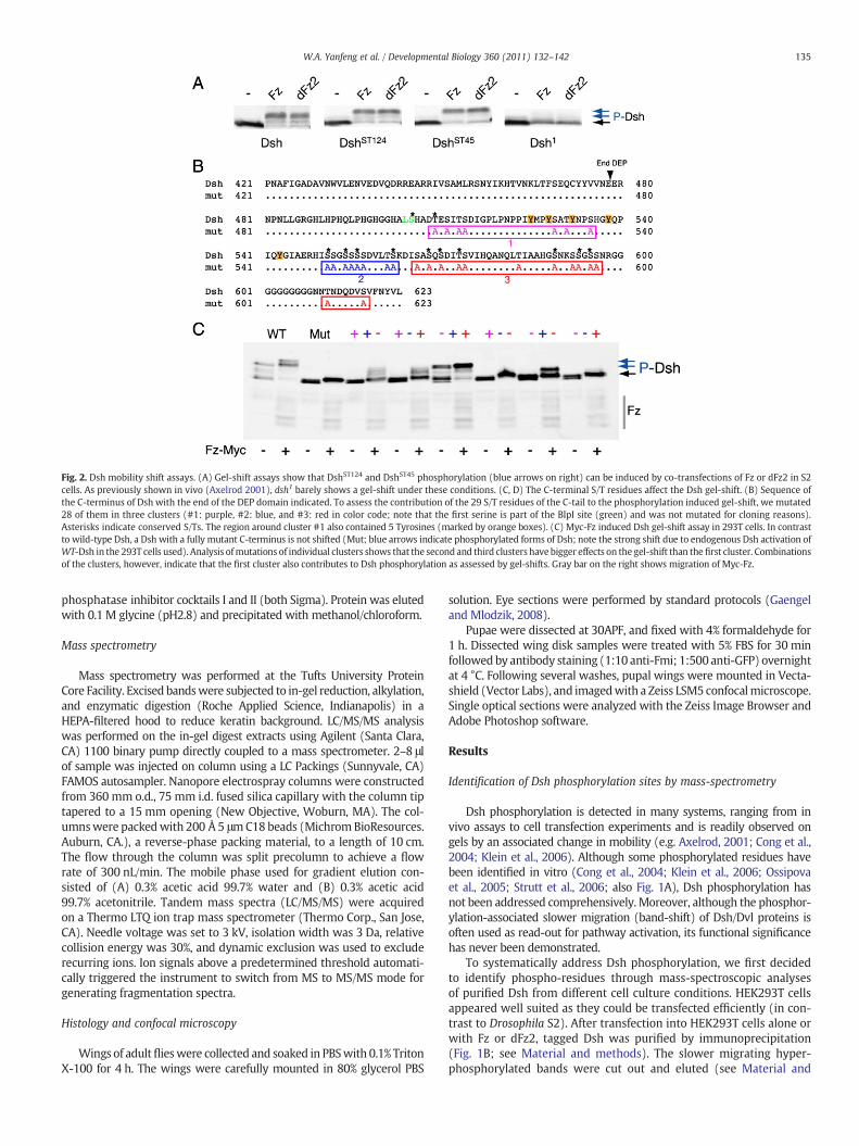
Fig. 2. Dsh mobility shift assays. (A) Gel-shift assays show that DshST124 and DshST45 phosphorylation (blue arrows on right) can be induced by co-transfections of Fz or dFz2 in S2cells. As previously shown in vivo (Axelrod 2001), dsh1 barely shows a gel-shift under these conditions. (C, D) The C-terminal S/T residues affect the Dsh gel-shift. (B) Sequence ofthe C-terminus of Dsh with the end of the DEP domain indicated. To assess the contribution of the 29 S/T residues of the C-tail to the phosphorylation induced gel-shift, we mutated28 of them in three clusters (#1: purple, #2: blue, and #3: red in color code; note that the first serine is part of the BlpI site (green) and was not mutated for cloning reasons).Asterisks indicate conserved S/Ts. The region around cluster #1 also contained 5 Tyrosines (marked by orange boxes). (C) Myc-Fz induced Dsh gel-shift assay in 293T cells. In contrastto wild-type Dsh, a Dsh with a fully mutant C-terminus is not shifted (Mut; blue arrows indicate phosphorylated forms of Dsh; note the strong shift due to endogenous Dsh activation ofWT-Dsh in the 293T cells used). Analysis ofmutations of individual clusters shows that the second and third clusters have bigger effects on the gel-shift than the first cluster. Combinationsof the clusters, however, indicate that the first cluster also contributes to Dsh phosphorylation as assessed by gel-shifts. Gray bar on the right shows migration of Myc-Fz.
135W.A. Yanfeng et al. / Developmental Biology 360 (2011) 132–142
phosphatase inhibitor cocktails I and II (both Sigma). Protein was elutedwith 0.1 M glycine (pH2.8) and precipitated with methanol/chloroform.
Mass spectrometry
Mass spectrometry was performed at the Tufts University ProteinCore Facility. Excised bandswere subjected to in-gel reduction, alkylation,and enzymatic digestion (Roche Applied Science, Indianapolis) in aHEPA-filtered hood to reduce keratin background. LC/MS/MS analysiswas performed on the in-gel digest extracts using Agilent (Santa Clara,CA) 1100 binary pump directly coupled to a mass spectrometer. 2–8 μlof sample was injected on column using a LC Packings (Sunnyvale, CA)FAMOS autosampler. Nanopore electrospray columns were constructedfrom 360 mm o.d., 75 mm i.d. fused silica capillary with the column tiptapered to a 15 mm opening (New Objective, Woburn, MA). The col-umnswere packedwith 200 Å 5 μmC18 beads (MichromBioResources.Auburn, CA.), a reverse-phase packing material, to a length of 10 cm.The flow through the column was split precolumn to achieve a flowrate of 300 nL/min. The mobile phase used for gradient elution con-sisted of (A) 0.3% acetic acid 99.7% water and (B) 0.3% acetic acid99.7% acetonitrile. Tandem mass spectra (LC/MS/MS) were acquiredon a Thermo LTQ ion trap mass spectrometer (Thermo Corp., San Jose,CA). Needle voltage was set to 3 kV, isolation width was 3 Da, relativecollision energy was 30%, and dynamic exclusion was used to excluderecurring ions. Ion signals above a predetermined threshold automati-cally triggered the instrument to switch from MS to MS/MS mode forgenerating fragmentation spectra.
Histology and confocal microscopy
Wings of adultflieswere collected and soaked in PBSwith 0.1% TritonX-100 for 4 h. The wings were carefully mounted in 80% glycerol PBS
solution. Eye sections were performed by standard protocols (Gaengeland Mlodzik, 2008).
Pupae were dissected at 30APF, and fixed with 4% formaldehyde for1 h. Dissected wing disk samples were treated with 5% FBS for 30 minfollowed by antibody staining (1:10 anti-Fmi; 1:500 anti-GFP) overnightat 4 °C. Following several washes, pupal wings were mounted in Vecta-shield (Vector Labs), and imagedwith a Zeiss LSM5 confocalmicroscope.Single optical sections were analyzed with the Zeiss Image Browser andAdobe Photoshop software.
Results
Identification of Dsh phosphorylation sites by mass-spectrometry
Dsh phosphorylation is detected in many systems, ranging from invivo assays to cell transfection experiments and is readily observed ongels by an associated change in mobility (e.g. Axelrod, 2001; Cong et al.,2004; Klein et al., 2006). Although some phosphorylated residues havebeen identified in vitro (Cong et al., 2004; Klein et al., 2006; Ossipovaet al., 2005; Strutt et al., 2006; also Fig. 1A), Dsh phosphorylation hasnot been addressed comprehensively. Moreover, although the phosphor-ylation-associated slower migration (band-shift) of Dsh/Dvl proteins isoften used as read-out for pathway activation, its functional significancehas never been demonstrated.
To systematically address Dsh phosphorylation, we first decidedto identify phospho-residues through mass-spectroscopic analysesof purified Dsh from different cell culture conditions. HEK293T cellsappeared well suited as they could be transfected efficiently (in con-trast to Drosophila S2). After transfection into HEK293T cells alone orwith Fz or dFz2, tagged Dsh was purified by immunoprecipitation(Fig. 1B; see Material and methods). The slower migrating hyper-phosphorylated bands were cut out and eluted (see Material and
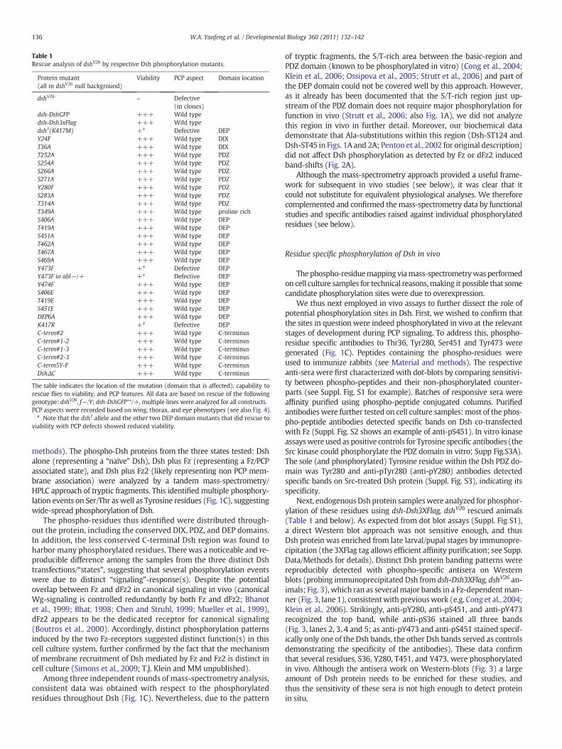
Table 1Rescue analysis of dshV26 by respective Dsh phosphorylation mutants.
Protein mutant(all in dshV26 null background)
Viability PCP aspect Domain location
dshV26 – Defective(in clones)
dsh-DshGFP +++ Wild typedsh-Dsh3xFlag +++ Wild typedsh1(K417M) +a Defective DEPY24F +++ Wild type DIXT36A +++ Wild type DIXT252A +++ Wild type PDZS254A +++ Wild type PDZS266A +++ Wild type PDZS271A +++ Wild type PDZY280F +++ Wild type PDZS283A +++ Wild type PDZT314A +++ Wild type PDZT349A +++ Wild type proline richS406A +++ Wild type DEPT419A +++ Wild type DEPS451A +++ Wild type DEPT462A +++ Wild type DEPT467A +++ Wild type DEPS469A +++ Wild type DEPY473F +a Defective DEPY473F in abl−/+ +a Defective DEPY474F +++ Wild type DEPS406E +++ Wild type DEPT419E +++ Wild type DEPS451E +++ Wild type DEPDEP6A +++ Wild type DEPK417R +a Defective DEPC-term#2 +++ Wild type C-terminusC-term#1-2 +++ Wild type C-terminusC-term#1-3 +++ Wild type C-terminusC-term#2-3 +++ Wild type C-terminusC-term5Y-F +++ Wild type C-terminusDshΔC +++ Wild type C-terminus
The table indicates the location of the mutation (domain that is affected), capability torescue flies to viability, and PCP features. All data are based on rescue of the followinggenotype: dshV26, f−/Y; dsh-DshGFP**/+, multiple lines were analyzed for all constructs.PCP aspects were recorded based on wing, thorax, and eye phenotypes (see also Fig. 4).
a Note that the dsh1 allele and the other two DEP domain mutants that did rescue toviability with PCP defects showed reduced viability.
136 W.A. Yanfeng et al. / Developmental Biology 360 (2011) 132–142
methods). The phospho-Dsh proteins from the three states tested: Dshalone (representing a “naïve” Dsh), Dsh plus Fz (representing a Fz/PCPassociated state), and Dsh plus Fz2 (likely representing non PCP mem-brane association) were analyzed by a tandem mass-spectrometry/HPLC approach of tryptic fragments. This identified multiple phosphory-lation events on Ser/Thr as well as Tyrosine residues (Fig. 1C), suggestingwide-spread phosphorylation of Dsh.
The phospho-residues thus identified were distributed through-out the protein, including the conserved DIX, PDZ, and DEP domains.In addition, the less conserved C-terminal Dsh region was found toharbor many phosphorylated residues. There was a noticeable and re-producible difference among the samples from the three distinct Dshtransfections/“states”, suggesting that several phosphorylation eventswere due to distinct “signaling”-response(s). Despite the potentialoverlap between Fz and dFz2 in canonical signaling in vivo (canonicalWg-signaling is controlled redundantly by both Fz and dFz2; Bhanotet al., 1999; Bhat, 1998; Chen and Struhl, 1999; Mueller et al., 1999),dFz2 appears to be the dedicated receptor for canonical signaling(Boutros et al., 2000). Accordingly, distinct phosphorylation patternsinduced by the two Fz-receptors suggested distinct function(s) in thiscell culture system, further confirmed by the fact that the mechanismof membrane recruitment of Dsh mediated by Fz and Fz2 is distinct incell culture (Simons et al., 2009; T.J. Klein and MM unpublished).
Among three independent rounds of mass-spectrometry analysis,consistent data was obtained with respect to the phosphorylatedresidues throughout Dsh (Fig. 1C). Nevertheless, due to the pattern
of tryptic fragments, the S/T-rich area between the basic-region andPDZ domain (known to be phosphorylated in vitro) (Cong et al., 2004;Klein et al., 2006; Ossipova et al., 2005; Strutt et al., 2006) and part ofthe DEP domain could not be covered well by this approach. However,as it already has been documented that the S/T-rich region just up-stream of the PDZ domain does not require major phosphorylation forfunction in vivo (Strutt et al., 2006; also Fig. 1A), we did not analyzethis region in vivo in further detail. Moreover, our biochemical datademonstrate that Ala-substitutions within this region (Dsh-ST124 andDsh-ST45 in Figs. 1A and 2A; Penton et al., 2002 for original description)did not affect Dsh phosphorylation as detected by Fz or dFz2 inducedband-shifts (Fig. 2A).
Although the mass-spectrometry approach provided a useful frame-work for subsequent in vivo studies (see below), it was clear that itcould not substitute for equivalent physiological analyses. We thereforecomplemented and confirmed themass-spectrometry data by functionalstudies and specific antibodies raised against individual phosphorylatedresidues (see below).
Residue specific phosphorylation of Dsh in vivo
Thephospho-residuemapping viamass-spectrometrywas performedon cell culture samples for technical reasons,making it possible that somecandidate phosphorylation sites were due to overexpression.
We thus next employed in vivo assays to further dissect the role ofpotential phosphorylation sites in Dsh. First, we wished to confirm thatthe sites in question were indeed phosphorylated in vivo at the relevantstages of development during PCP signaling. To address this, phospho-residue specific antibodies to Thr36, Tyr280, Ser451 and Tyr473 weregenerated (Fig. 1C). Peptides containing the phospho-residues wereused to immunize rabbits (see Material and methods). The respectiveanti-sera were first characterized with dot-blots by comparing sensitivi-ty between phospho-peptides and their non-phosphorylated counter-parts (see Suppl. Fig. S1 for example). Batches of responsive sera wereaffinity purified using phospho-peptide conjugated columns. Purifiedantibodieswere further tested on cell culture samples:most of the phos-pho-peptide antibodies detected specific bands on Dsh co-transfectedwith Fz (Suppl. Fig. S2 shows an example of anti-pS451). In vitro kinaseassayswere used as positive controls for Tyrosine specific antibodies (theSrc kinase could phosphorylate the PDZ domain in vitro; Supp Fig.S3A).The sole (and phosphorylated) Tyrosine residue within the Dsh PDZ do-main was Tyr280 and anti-pTyr280 (anti-pY280) antibodies detectedspecific bands on Src-treated Dsh protein (Suppl. Fig. S3), indicating itsspecificity.
Next, endogenousDshprotein sampleswere analyzed for phosphor-ylation of these residues using dsh-Dsh3XFlag, dshV26 rescued animals(Table 1 and below). As expected from dot blot assays (Suppl. Fig S1),a direct Western blot approach was not sensitive enough, and thusDsh protein was enriched from late larval/pupal stages by immunopre-cipitation (the 3XFlag tag allows efficient affinity purification; see Supp.Data/Methods for details). Distinct Dsh protein banding patterns werereproducibly detected with phospho-specific antisera on Westernblots (probing immunoprecipitated Dsh from dsh-Dsh3XFlag, dshV26 an-imals; Fig. 3), which ran as severalmajor bands in a Fz-dependentman-ner (Fig. 3, lane 1), consistentwith previouswork (e.g. Cong et al., 2004;Klein et al., 2006). Strikingly, anti-pY280, anti-pS451, and anti-pY473recognized the top band, while anti-pS36 stained all three bands(Fig. 3, lanes 2, 3, 4 and 5; as anti-pY473 and anti-pS451 stained specif-ically only one of the Dsh bands, the other Dsh bands served as controlsdemonstrating the specificity of the antibodies). These data confirmthat several residues, S36, Y280, T451, and Y473, were phosphorylatedin vivo. Although the antisera work on Western-blots (Fig. 3) a largeamount of Dsh protein needs to be enriched for these studies, andthus the sensitivity of these sera is not high enough to detect proteinin situ.
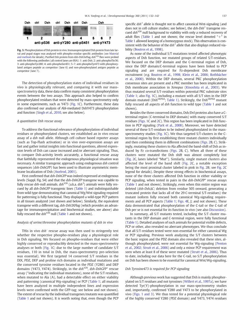
Fig. 3. PhosphorylationofDshprotein in vivo. ImmunoprecipitatedDshprotein from late lar-val and pupal stages was analyzed with phospho-residue specific antibodies (see Materialandmethods for details). Purified Dsh protein from dsh-Dsh3xFlag, dshV26 flies were probedwith the following antibodies (all control lanes are BSA): 1: anti-Dsh; 2: anti-phosphoThr36;3: anti-phosphoTyr280; 4: anti-phosphoSer451; 5–7: anti-phosphoTyr473with phosphory-lated antigen peptide as competitor (lane 6) and non-phosphorylated antigen peptide ascompetitor (lane 7).
137W.A. Yanfeng et al. / Developmental Biology 360 (2011) 132–142
The detection of phosphorylation states of individual residues invivo is physiologically relevant, and comparing it with our mass-spectrometry data, these data confirmmany consistent phosphorylationevents between the two assays. This approach also helped to confirmphosphorylated residues that were detected by mass-spectrometry onlyin some experiments, such as Y473 (Fig. 1C). Furthermore, these dataalso confirmed our analysis of Abl-mediated DshY473 phosphorylationand function (Singh et al., 2010, see also below).
A quantitative Dsh rescue assay
To address the functional relevance of phosphorylation of individualresidues or phosphorylated clusters, we established an in vivo rescueassay of a dsh null allele. Although cell culture based reporter assays(such as Top-Flash activation) or in vivo over-expression assays arefast and gather initial insights into functional questions, altered expres-sion levels of Dsh can cause increased and/or aberrant signaling. Thus,to compare Dsh-activity levels accurately, a functional rescue assaythat faithfully represented the endogenous physiological situation wasnecessary. A similar transgenic approach using endogenous dsh controlsequences (dsh-DshGFP) has been used to illustrate asymmetric mem-brane localization of Dsh (Axelrod, 2001).
First confirmed that dsh-DshGFPwas indeed expressed at endogenouslevels (Suppl. Fig. S4) and that the dsh-DshGFP transgene was capable tofully rescue dsh-null animals. dshV26 (a.k.a. dsh3) animals were fully res-cued by all dsh-DshGFP transgenic lines (Table 1) and indistinguishablefromwild type demonstrating that canonicalWg/Wnt signaling requiredfor patterning is fully functional. They displayed a wild-type PCP patternin all tissues analyzed (not shown and below). Similarly, the equivalenttransgene with a different tag, dsh-Dsh3XFlag (which provides an advan-tage for immunoprecipitation and biochemical studies, see above) alsofully rescued the dshV26 null (Table 1 and not shown).
Analysis of serine/threonine phosphorylation mutants of dsh in vivo
THis in vivo dsh- rescue assay was then used to stringently testwhether the respective phospho-residues play a physiological rolein Dsh signaling. We focused on phospho-residues that were eitherhighly conserved or reproducibly detected in the mass-spectrometryanalyses or both (Fig. 1C; due to the large number of candidate S/Tresidues, 110 in total in Dsh, the mass-spectrometry pre-selectionwas essential). We first targeted 14 conserved S/T residues in theDIX, PDZ, DEP and proline rich domains as individual mutations andthe conserved tyrosine residues located in the PDZ (Y280) and DEPdomains (Y473, Y474). Strikingly, in the dshV26, dsh-DshGFP* rescueassay (*indicating the individual mutations), none of the S/T residues,when mutated to Ala (A), had a detectable effect on either viabilityand patterning (canonical Wg-signaling) or PCP (Table 1; all mutantshave been analyzed in multiple independent lines and expressionlevels were confirmed with the GFP-tag; see below and not shown).The extent of rescue by the individual transgenes/mutantswas quantified(Table 1 and not shown). It is worth noting that, even though the PCP
specific dsh1 allele is thought not to affect canonical Wnt-signaling (anddoes not in cell culture studies; see below), the dsh-Dsh1 transgene res-cued dshV26 null background to viability with only a reduced recovery ofadult flies (Table 1 and not shown; the rescue level denoted “+” inTable 1 allowed keeping of a homozygous stock). This observation is con-sistent with the behavior of the dsh1 allele that also displays reduced via-bility (Boutros et al., 1998).
As none of the individual S/T mutations tested affected phenotypicaspects of Dsh function, we mutated groups of related S/T-residues.We focused on the DEP domain and the C-terminal region of Dsh,since the DEP domain/C-terminal regions have been linked to PCPsignaling and are required for Fz-dependent Dsh membranerecruitment (e.g. Boutros et al., 1998; Klein et al., 2006; Rothbächeret al., 2000). Within the DEP domain, several PKC phosphorylationconsensus sites are present and a PKC member has been implicated inDsh membrane association in Xenopus (Kinoshita et al., 2003). Wethus mutated several S/T-residues within potential PKC substrate sites(Table 1; also Fig. 1C), including a mutant with all S/T sites in the DEPdomain mutated (DshDEP6A; Table 1). Strikingly, the DshDEP6A mutantfully rescued all aspects of dsh function to wild type (Table 1 and notshown).
Besides the three conserved domains, Dsh/Dvl proteins alsohave a C-terminal region (C-terminal to DEP domain) with many conserved S/Tresidues (Figs. 1C and 2C). This region has been implicated in Dsh func-tion in PCP signaling (Park et al., 2008). Moreover, we have detectedseveral of these S/T-residues to be indeed phosphorylated in the mass-spectrometry studies (Fig. 1C). We thus targeted S/T-clusters in the C-terminal region by first establishing three mutant clusters individuallyand then combining them in different combinations (Figs. 2B, C). Strik-ingly, mutating these clusters to Ala affected the band-shift of Dsh as in-duced by Fz co-transfections (Figs. 2B, C). When all three of theseclusters were mutated the Dsh shift was almost completely lost(Fig. 2C, lanes labeled “Mut”). Similarly, single mutant clusters alsoaffected the level of the band shift (Fig. 2C, a notable exceptionbeing the most proximal cluster labeled in pink in Fig. 2B; see Fig. 2Clegend for details). Despite these strong effects in biochemical assays,none of the three clusters affected Dsh function in either viability orPCP signaling, when tested in pairs in the dsh-DshGFP* rescue assays(Table 1 and not shown). Strikingly, even when this entire region wasdeleted (dsh-DshΔC; deletion from residue 505 onward, generating atruncated protein that lacks all of the 29 C-terminal S/T-residues) themutant isoform fully rescued both canonical Wg-signaling require-ments and all PCP aspects (Table 1; Figs. 4E, J; and not shown). Thesedata demonstrated that phosphorylation of the C-tail or the C-tail ofDsh per se is not essential for its function in vivo (see also Discussion).
In summary, all S/T mutants tested, including the S/T cluster mu-tants in the DEP domain and C-terminal region, were fully functional(Table 1). Detailed analyses of such animals for potential visible defects,PCP or other, also revealed no aberrant phenotypes. We thus concludethat all S/T-residues tested were non-essential for either canonical Wgor PCP signaling. Previous work analyzing the S/T clusters betweenthe basic region and the PDZ domain also revealed that these sites, al-though phosphorylated, were not essential for Wg-signaling (Pentonet al., 2002; Strutt et al., 2006) and only a minor PCP requirement wasseen when at least 8 of these were mutated (Strutt et al., 2006). Thusto date, including our data here for the C-tail, no S/T phosphorylationon Dsh has been shown to be essential for canonicalWnt/Wg-signaling.
Dsh Tyrosine473 is required for PCP signaling
Although previouswork has suggested that Dsh ismainly phosphor-ylated on S/T residues and not tyrosines (Willert et al., 1997a), we havedetected Tyr(Y)-phosphorylation in our mass-spectrometry studiesand, importantly, confirmed Y280 and Y473 to be phosphorylated invivo (Figs. 1 and 3). We thus tested for a potential physiological roleof the highly conserved Y280 (PDZ domain) and Y473, Y474 residues

Fig. 4. Canonical Wg-signaling activity of Dsh1, DshY473F, and DshK417R mutants. (A) dsh-DshY473F (in a dsh-background) fully rescues the expression of Wg-signaling targetgenes during wing disk development. Example shown is Distalless (Dll). Senseless/Sens was also tested and rescued (not shown). 3rd instar larval wing disk stained for the clonalmarker lacZ (red, marks wild-type regions) and Distalless (Dll; green), expressed along the dorso-ventral margin flanking the Wg expression stripe and weaker through the wingblade anlage. Dll is a direct target of Wg. dshV26 clones loose Dll expression (not shown). The presence of DshY473F fully rescued the mutant dshV26 effect. Dll expression showed nodifference between mutant and wild-type tissue. (A′) and (A″) show single channels for lacZ and Dll, respectively. (B–B′) Dsh mutants do not affect expression levels of Top-flashreporter. Dsh mutant expression constructs were transfected with Top-flash luciferase reporter (and SV40-renilla luciferase reporter as control) in HEK293T cells. The ratios of Top-flash vs. renilla were normalized against empty vector control at three doses of transfection as indicated. Note that DshY473 showed no significant reduction as compared to wild-typeDsh (DshWT); the same was the case with Dsh1(K417M) and DshK417R mutants. (B″): Western-blot against Dsh to visualize similar protein levels.
138 W.A. Yanfeng et al. / Developmental Biology 360 (2011) 132–142
(DEP domain). DshY280 to phenylalanine (DshY280F) substitution had noeffects on either viability or PCP (Table 1). In contrast, DshY473F displayedthe strongest effect in the viability rescue in the same range as the Dsh1
mutant (Table 1), although a rescued (homozygous) stock was viable,consistent with our previous data in the Abl context (Singh et al., 2010).We did not, however, detect defects inWg-signaling target gene expres-sion or signaling strength during development, and also a quantitativecell culture assay did not reveal a significant difference of DshY473F ascompared to DshWT (Fig. 4). We thus conclude that the Y473 is not re-quired for canonical signaling and the reduced viability might have todo with other Dsh functions not yet described in Drosophila.
Strikingly, the dshY473F flies displayed strong PCP defects in all tissuesanalyzed (Table 1 and Figs. 5C, H). The neighboring Y474 had no effectand the double mutant (DshY473, 474F) behaved like DshY473F alone(Table 1 and not shown). As these PCP defects were strikingly similar tothe dsh1 allele, we compared their phenotypic PCP defects inmore detail.In both cases,dsh1 and dshY473F, the rescued individuals displayed classicalPCP defects in the thorax andwings (Figs. 5A–C′ and not shown), and hadrougheyes,with randomized chirality, symmetrical clusters, andmisrota-tions again resembling dsh1 and classical PCP mutants in general (Figs.5F–H). Moreover, dsh-DshY473F did not rescue the PCP defects of thedsh1 allele (not shown). Taken together, these data indicate thatDshY473F behaves like a strong PCP specific dsh allele and thus Y473appears to be required for PCP regulation.
These data are consistent with our observation that Y473 can bephosphorylated by the Abl kinase, which is also required for PCP
signaling but not canonical Wnt-signaling (Singh et al., 2010). Theneighboring and downstream Tyr residues can also be phosphorylatedby Abl in vitro (Singh et al., 2010), and thus we asked whether any ofthese could be functionally important in the absence Y473 in a redun-dant manner. We thus tested for potential phenotypic consequencesof dsh-, dsh-DshY473F flies in an abl−/+ background. Neither viabilitynor the PCP defects were different in abl−/+ as compared to abl+backgrounds (Table 1, Fig. 5 and not shown), suggesting that none ofthe other Tyr residues in the DEP/C-term region of Dsh is (partially) re-dundant with Y473.
Stable membrane recruitment of Dsh is a hallmark of its role inPCP signaling (Axelrod, 2001; Boutros et al., 2000; Rothbächer et al.,2000) and, for example, reduced in dsh1 mutants or lost altogetherin fz−mutant animals (Axelrod, 2001). We thus analyzed the subcel-lular localization of DshY473F in pupal wings around 30–34 h APF.Strikingly, and in contrast to DshWT (Fig. 6A), DshY473F (in the dshnull background) displayed hardly any membrane association inpupal wings (or elsewhere), very similar to the Dsh1 mutant isoform(Figs. 6B–C″). This defect in membrane association is in contrast withany of the S/T-residue cluster mutants that all localized normally tothe membrane (data not shown), further indicating a specific require-ment of Y473 in PCP signaling.
Similar to the S/T site mutations, expression levels of DshY280F orDshY473F (as detected on Western blots) were very similar to wild-type DshGFP or Dsh1, indicating that protein stability in imaginal diskswas unaffected by the DshY473F mutation (Fig. 7; also Fig. S5). However,

Fig. 5. Functional requirements of specific Dsh residues in vivo. All rescue constructs were expressed under the control of the endogenous dsh promoter in a dshV26 (null), f− background;the only Dsh protein present is provided by the transgenic lines (note that all genotypes are forked [f] to control for dshV26, giving the cellular hairs a wavy appearance). For all constructs,several independent lines behaved identically and displayed very similar expression levels, as expected (see Suppl. Fig. S5). (A–E′)Wings of rescuedflies of indicated genotype are shown;lowmagnification (A–E) highlights normalWg-signaling: developmentwith of normal sizedwing and normalwingmargin development demonstrating correct canonicalWnt signaling.Highmagnification (panels A′–E′) shows PCP arrangements of the respective wings. The highmagnification pictures show a region near the posterior cross vein and L4. Note PCP defects inthe genotypesdsh-Dsh1, dsh-DshY473F, dsh-DshK417R. (F–J) Adult eye sections of the respective genotypeswith schematic presentation in lower panels; black and red arrows represent ommatidiawith chiral arrangements,which are normally arranged in amirror-image across the dorso-ventralmidline (see panel F forwild-type); green arrows represent symmetric ommatidia. Note thatin G–I, the mirror-image arrangement is lost and a random distribution of the two chiral forms and symmetrical ommatidia is detected, reflecting typical PCP defects.
139W.A. Yanfeng et al. / Developmental Biology 360 (2011) 132–142
an obvious difference between wild-type DshGFP and the DshY473F
mutant isoform was their protein migration pattern, as detected onWestern blots with imaginal disk samples (Fig. 7): DshY473F lackedthe slower migrating ‘hyperphosphorylated’ bands, very similar toDsh1 (Fig. 7; Axelrod, 2001; Klein et al., 2006). This was in contrastto DshY280F or DshY474F, which displayed a normal Dsh banding pattern(Fig. 7 and not shown).
DshK417R disrupts PCP signaling very similarly to Dsh1 (DshK417M)
The original PCP specific and first allele of dsh, dsh1, a K toM transitionat residue 417, lies within a Protein Kinase C (PKC) consensus site. Thisobservation led to the hypothesis that it could affect Dsh phosphorylationas it also reduces Dsh phosphorylation in cell culture (Klein et al., 2006).However, as mentioned above, the DEP domain PKC site S/T-mutations,including the respective DshT419A or even the DshDEP6A mutant isoform,affecting all potential DEP-domain PKC sites, had no detectable effect onDsh function. Thus, we reasoned that other types of post-translationalmodification such as acetylation or ubiquitination that occur on lysinesmay be involved. K417 is one of five lysines/arginines (K/R) within theDEP domain. A sequence alignment of Dsh of various species revealedthat K417 and K465 are exclusively lysines, whereas the other three
lysines and arginines appear interchangeable (Fig. 1B). Therefore, wetested whether a replacement of K with R would phenocopy the dsh1 al-lele (DshK417M) or be fully functional, the latter indicating the importanceof the positive charge. DshK417R restores viability/canonical Wg-signalingof dshV26 comparable to dsh1(Table 1 and Fig. 5B) Intriguingly, DshK417R
failed to rescue PCP and revealed PCP defects very similar to dsh1 in all tis-sues analyzed (Figs. 5D, I and not shown). Importantly, in analogy to Dsh1
(DshK417M), subcellular localization/membrane association of DshK417R
was affected as well (Figs. 6D–D″). In 34 h APF pupal wings, DshK417R
was barely found atmembranes, very similar to the Dsh1 protein. Furtheranalysis suggested that DshK417R also lacked the slow migrating Dshbands on Westerns, suggesting reduced activation (Fig. 7). Therefore,we conclude that Lysine417 in theDshDEPdomain is essential, notmere-ly due to a requirement of electric charges, but possibly due to covalentmodification(s) required for PCP function (also Discussion).
Discussion
In vivo, Dsh becomes hyperphosphorylated upon Wnt-pathwayactivation and/or membrane recruitment (PCP signaling) and dueto this correlation, phosphorylation has often been used as a read-out for pathway activation. Previous work has identified a region

Fig. 6. Localization of mutated Dsh isoforms in pupal wings during PCP signaling. Confocal images of DshGFP (green) and Fmi (red) staining in pupal wings at the 30 to 34 h APFstage are shown. The respective genotypes are indicated above. A′–D′ display anti-Fmi monochrome images (highlighting membranes and the PCP complexes localized there), andA″–D″ show monochromes of DshGFP (A–D are the merged channels). Note that in B/B″, C/C″, and D/D″ the membrane levels of DshGFP are reduced or even absent, although theoverall signal and protein levels are not affected (see also Suppl. Figure S5).
140 W.A. Yanfeng et al. / Developmental Biology 360 (2011) 132–142
N-terminal to the PDZdomain,which is rich in Ser/Thr residues and canbe phosphorylated in vitro, but it remained questionablewhether phos-phorylation in this region is functionally significant (Cong et al., 2004;Klein et al., 2006; Ossipova et al., 2005; Strutt et al., 2006). The signifi-cance of other predicted phosphorylation sites and events has notbeen addressed. Our data indicate that, surprisingly, all Ser/Thr residuestested, although highly conserved and phosphorylated, are dispensablefor Dsh function in vivo. In particular, it is a striking observation that thelack of all Ser/Thr residues C-terminal to the DEP domain or within theDEP domain has no effect on Dsh function in either signaling pathway.This, taken together with previous observations that the Ser/Thr-richregion N-terminal to the PDZ domain is not essential for Dsh function(Strutt et al., 2006), strongly suggests that Ser/Thr phosphorylation ofDsh/Dvl proteins is generally (almost) non-essential, which is on itsown a very surprising and unexpected finding.
Fig. 7. Migration behavior and phosphorylation status of mutant DshGFP isoforms asdetected byWestern blots of late 3rd instar/early pupal tissues. The respective Dsh isoformsare indicated above the gel andwere detectedwith an anti-GFP antibody (“ctrl” lane has noDshGFP protein present as control). All lines are expressed from the endogenous dsh-expression cassette and exhibit very similar protein levels (also Suppl. Figure S5). Notethat Dsh1, DshY473F and DshK417R show markedly reduced gel shift, indicative of reducedDsh phosphorylation and correlating with their defective behavior in PCP signaling.
In contrast, our data suggest that tyrosine phosphorylation, whichprevious attempts failed to identify (Willert et al., 1997b), is critical forDsh function. In particular, Y473 within the DEP domain is critical inPCP signaling and likely phosphorylated by Abl type kinases (Singhet al., 2010). Our data further suggest that the originalmutant dsh1 allele(a K417M transition) does not affect phosphorylation, but is likely tocause defects via affecting post-translational modification of Dsh onK417.
Dsh phosphorylation in vivo
Our mass-spectrometry approach identified several phosphorylationsites, including Tyr phosphorylation of Dsh, which has not been previ-ously observed (Willert et al., 1997a). Importantly, several of the individ-ual sites were confirmed to be indeed phosphorylated in vivothrough tissue samples and phospho-specific antibodies. Nevertheless,the respective Ser/Thr residues are functionally non-essential (or possi-bly redundant) for Dsh function in either pathway. Due to the largenumber of Ser/Thr residues in Dsh/Dvl proteins (N110 in approx. 650total residues) it is almost impossible to address phosphorylationredundancy in the whole protein and thus, we have focused mostlyon the C-terminal region including the DEP domain. All mutations ana-lyzed that contained multiple mutant residues, including all Ser/Thrwithin the DEP domain or lacked all C-terminal Ser/Thr residues, werefully functional in rescue assays (Wg-signaling and PCP) of a dsh-nullmutant background. Taken together with our individual mutations inconserved residues in other regions of Dsh/Dvl and the earlier data onthe S/T cluster between the basic region and PDZ domain (Pentonet al., 2002; Strutt et al., 2006), our data suggest that Ser/Thr phosphor-ylation of Dsh is far less important than generally assumed.
141W.A. Yanfeng et al. / Developmental Biology 360 (2011) 132–142
In contrast, Tyr-phosphorylation of DshY473within the DEP domainis essential for Dsh function in PCP signaling (this study and also Singhet al., 2010). The respective DshY473F mutation behaves like the originalPCP specific dsh1 alelle: (1) dsh null flies can be rescued to viability, (2)the mutant protein is not recruited stably to the membrane during PCPsignaling in vivo, and (3) the mutant protein shows an overall reducedphosphorylation as assessed by gel-shift (Fig. 7; compare also to Dsh1
analysis, e.g. Axelrod, 2001; Klein et al., 2006). Strikingly, Y473 is theonly Tyr residue that is functionally required, althoughDsh/Dvl proteinscontain several tyrosines that are absolutely conserved across all spe-cies. For example, Y280 within the PDZ domain, which, although phos-phorylated in vivo and conserved in all Dsh/Dvls, appears dispensable infunctional rescue assays.
Overall, our data suggest that most phosphorylation target residues,although phosphorylated in vivo, are not essential for Dsh functionwiththe notable exception is Y473, which behaves like a PCP specific dshallele when mutated.
The dsh C-terminal region
A striking observation is the lack of a requirement of the C-terminalregion of Dsh (C-terminal to the DEP domain). Not only are all potentialphosphorylation sites, S/T-sites and tyrosines, dispensable (Table 1),but even a complete deletion of the region C-terminal to residue 504has no apparent functional consequences: The respective DshΔCtermisoform fully rescued all aspects of Dsh function. This is surprising asthe equivalent C-terminal region of Xenopus Dsh has been suggestedto be sufficient to localize GFP to the basal body of cilia and to interferewith their polarity when overexpressed (Park et al., 2008). Althoughpatches of Ser/Thr residues are conserved within this C-terminalregion (Figs. 1C and 2B), the sequence alignment of vertebrate andDrosophila Dsh proteins shows overall less conservation in this pro-tein region. The vertebrate Dvl proteins are highly conservedamong themselves in this region, however. Therefore, it is likelythat vertebrate Dsh/Dvls share functional requirements withinthese sequences that are not conserved between Drosophila and ver-tebrates and might lie outside the canonical Wnt-pathway and PCPsignaling.
The nature of the dsh1 allele
The original allele of Dsh, dsh1, a Lys-to-Met transition at position417 within the DEP domain, is a PCP specific allele (Axelrod et al.,1998; Boutros et al., 1998). As this Lysine is located within a potentialPKC consensus site, it has been speculated that it could affect phosphor-ylation of the nearby serine (Boutros and Mlodzik, 1999; Wallingfordand Habas, 2005). We demonstrate that this is not the case and thatpotential PKC phosphorylation within the DEP domain is not essentialfor Dsh function. However, our data indicate that it is the Lysine itselfthat is essential and a mutation to the related Arginine residue (whichis similarly charged and often can substitute for Lys) also causes thesame defects as the original DshK417M mutation. These data suggestthat it is a post-translational modification such as ubiquitination oracetylation of Lys417, as it does not appear to be of structural impor-tance (Wong et al., 2000). This notion is consistent with recent workin mammalian cell culture (Ganner et al., 2009), although we cannotexclude a direct involvement of Lys417.
In summary, our analyses have revealed that many phosphorylationevents on Dsh are functionally dispensable in vivo, even when theyoccur on highly conserved residues. This is in stark contrast tomany pre-vious assumptions and suggestions and thus an important observation.In addition, we have identified that Dsh is phosphorylated on Tyr resi-dues, which has not been observed earlier. Consistent with recent dataon an Abl requirement in PCP (Singh et al., 2010), Tyr-phosphorylationevents on at least one site, DshY473, are critical for Dsh function in PCPsignaling.
Acknowledgments
We thank J. Axelrod, S. Cohen, R. Nusse, and the Bloomington Dro-sophila Stock Center for fly strains and reagents. We are grateful toSophy Okello and Gustavo Garcia for technical help, Melissa Marianiand Katya Serysheva for careful editing of themanuscript, and allMlodzikand Jenny lab members for helpful comments and suggestions through-out these analyses. This work was supported by NIH grants EY13256 toMM and GM088202 to AJ.
Appendix A. Supplementary data
Supplementary data to this article can be found online at doi:10.1016/j.ydbio.2011.09.017.
References
Adler, P.N., 2002. Planar signaling and morphogenesis in Drosophila. Dev. Cell 2,525–535.
Axelrod, J.D., 2001. Unipolar membrane association of Dishevelled mediates Frizzledplanar cell polarity signaling. Genes Dev. 15, 1182–1187.
Axelrod, J.D., Miller, J.R., Shulman, J.M., Moon, R.T., Perrimon, N., 1998. Differentialrequirement of Dishevelled provides signaling specificity in the Wingless andplanar cell polarity signaling pathways. Genes Dev. 12, 2610–2622.
Bhanot, P., Fish, M., Jemison, J.A., Nusse, R., Nathans, J., Cadigan, K.M., 1999. Frizzled andDFrizzled-2 function as redundant receptors for wingless during Drosophila em-bryonic development [In Process Citation]. Development 126, 4175–4186.
Bhat, K.M., 1998. Frizzled and Frizzled2 play a partially redundant role inWingless signalingand have similar requirements to Wingless in neurogenesis. Cell 95, 1027–1036.
Bilic, J., Huang, Y.L., Davidson, G., Zimmermann, T., Cruciat, C.M., Bienz, M., Niehrs, C.,2007. Wnt induces LRP6 signalosomes and promotes dishevelled-dependentLRP6 phosphorylation. Science 316, 1619–1622.
Boutros, M., Mlodzik, M., 1999. Dishevelled: at the crossroads of divergent intracellularsignaling pathways. Mech. Dev. 83, 27–37.
Boutros, M., Paricio, N., Strutt, D.I., Mlodzik, M., 1998. Dishevelled activates JNK and dis-criminates between JNK pathways in planar polarity and wingless signaling. Cell94, 109–118.
Boutros,M.,Mihaly, J., Bouwmeester, T.,Mlodzik,M., 2000. Signaling specificity by Frizzledreceptors in Drosophila. Science 288, 1825–1828.
Chen, C.-N., Struhl, G., 1999. Wingless transduction by the Frizzled and Frizzled2 proteinsof Drosophila. Development 126, 5441–5452.
Cong, F., Schweizer, L., Varmus, H., 2004. Casein kinase Iepsilon modulates the signalingspecificities of dishevelled. Mol. Cell. Biol. 24, 2000–2011.
Gaengel, K., Mlodzik, M., 2008. Microscopic analysis of the adult Drosophila retina usingsemithin plastic sections. Methods Mol. Biol. 420, 277–287.
Ganner, A., Lienkamp, S., Schafer, T., Romaker, D., Wegierski, T., Park, T.J., Spreitzer, S.,Simons, M., Gloy, J., Kim, E., Wallingford, J.B., Walz, G., 2009. Regulation of ciliarypolarity by the APC/C. Proc. Natl. Acad. Sci. U. S. A. 106, 17799–17804.
Keller, R., 2002. Shaping the vertebrate body planbypolarized embryonic cellmovements.Science 298, 1950–1954.
Kinoshita, N., Iioka, H., Miyakoshi, A., Ueno, N., 2003. PKC delta is essential for Dishevelledfunction in a noncanonicalWnt pathway that regulates Xenopus convergent extensionmovements. Genes Dev. 17, 1663–1676.
Klein, T.J., Mlodzik, M., 2005. Planar cell polarization: an emerging model points in theright direction. Annu. Rev. Cell Dev. Biol. 21, 155–176.
Klein, T.J., Jenny, A., Djiane, A., Mlodzik, M., 2006. CKIepsilon/discs overgrown promotesbothWnt-Fz/beta-catenin and Fz/PCP signaling inDrosophila. Curr. Biol. 16, 1337–1343.
Lawrence, P.A., Struhl, G., Casal, J., 2007. Planar cell polarity: one or two pathways? Nat.Rev. Genet. 8, 555–563.
Lin, H.V., Rogulja, A., Cadigan, K.M., 2004. Wingless eliminates ommatidia from theedge of the developing eye through activation of apoptosis. Development 131,2409–2418.
Logan, C.Y., Nusse, R., 2004. The Wnt signaling pathway in development and disease.Annu. Rev. Cell Dev. Biol. 20, 781–810.
Metcalfe, C., Mendoza-Topaz, C., Mieszczanek, J., Bienz, M., 2010. Stability elements inthe LRP6 cytoplasmic tail confer efficient signalling upon DIX-dependent polymer-ization. J. Cell Sci. 123, 1588–1599.
Mlodzik, M., 1999. Planar polarity in the Drosophila eye: a multifaceted view of signalingspecificity and cross-talk. EMBO J. 18, 6873–6879.
Mlodzik, M., 2002. Planar cell polarization: do the same mechanisms regulate Drosophilatissue polarity and vertebrate gastrulation? Trends Genet. 18, 564–571.
Mueller, H., Samanta, R.,Wieschaus, E., 1999.Wingless signaling in theDrosophila embryo:zygotic requirements and the role of the frizzled genes. Development 126, 577–586.
Ossipova, O., Dhawan, S., Sokol, S., Green, J.B., 2005. Distinct PAR-1 proteins function in dif-ferent branches ofWnt signaling during vertebrate development. Dev. Cell 8, 829–841.
Park, T.J., Mitchell, B.J., Abitua, P.B., Kintner, C., Wallingford, J.B., 2008. Dishevelled ControlsApical Docking and Planar Polarization of Basal Bodies in Ciliated Epithelial Cells.
Penton, A., Wodarz, A., Nusse, R., 2002. A mutational analysis of dishevelled in Drosophiladefines novel domains in the dishevelled protein as well as novel suppressing allelesof axin. Genetics 161, 747–762.
Polakis, P., 2000. Wnt signaling and cancer. Genes Dev. 14, 1837–1851.
142 W.A. Yanfeng et al. / Developmental Biology 360 (2011) 132–142
Rothbächer, U., Laurent, M.N., Deardorff, M.A., Klein, P.S., Cho, K.W., Fraser, S.E., 2000.Dishevelled phosphorylation, subcellular localization and multimerization regu-late its role in early embryogenesis. EMBO J. 19, 1010–1022.
Rulifson, E.J., Wu, C.H., Nusse, R., 2000. Pathway specificity by the bifunctional receptorfrizzled is determined by affinity for wingless. Mol. Cell 6, 117–126.
Sahai, E., Alberts, A.S., Treisman, R., 1998. RhoA effector mutants reveal distinct effectorpathways for cytoskeletal reorganization, SRF activation and transformation.EMBO J. 17, 1350–1361.
Schwarz-Romond, T., Merrifield, C., Nichols, B.J., Bienz, M., 2005. TheWnt signalling effectorDishevelled forms dynamic protein assemblies rather than stable associationswith cytoplasmic vesicles. J. Cell Sci. 118, 5269–5277.
Seifert, J.R., Mlodzik, M., 2007. Frizzled/PCP signalling: a conservedmechanism regulatingcell polarity and directed motility. Nat. Rev. Genet. 8, 126–138.
Simons, M., Mlodzik, M., 2008. Planar cell polarity signaling: from fly development tohuman disease. Annu. Rev. Genet. 42, 517–540.
Simons, M., Gault, W.J., Gotthardt, D., Rohatgi, R., Klein, T.J., Shao, Y., Lee, H.J., Wu, A.L.,Fang, Y., Satlin, L.M., Dow, J.T., Chen, J., Zheng, J., Boutros, M., Mlodzik, M., 2009.Electrochemical cues regulate assembly of the Frizzled/Dishevelled complex atthe plasma membrane during planar epithelial polarization. Nat. Cell Biol. 11,286–294.
Singh, J., Yanfeng, W.A., Grumolato, L., Aaronson, S.A., Mlodzik, M., 2010. Abelson familykinases regulate Frizzled planar cell polarity signaling via Dsh phosphorylation.Genes Dev. 24, 2157–2168.
Strapps,W.R., Tomlinson, A., 2001. Transducing properties of Drosophila Frizzled proteins.Development 128, 4829–4835.
Strutt, D., 2003. Frizzled signalling and cell polarisation in Drosophila and vertebrates.Development 130, 4501–4513.
Strutt, H., Strutt, D., 1999. Polarity determination in the Drosophila eye. Curr. Opin.Genet. Dev. 9, 442–446.
Strutt, H., Price, M.A., Strutt, D., 2006. Planar polarity is positively regulated by caseinkinase Iepsilon in Drosophila. Curr. Biol. 16, 1329–1336.
Veeman, M.T., Axelrod, J.D., Moon, R.T., 2003. A second canon. Functions and mecha-nisms of beta-catenin-independent Wnt signaling. Dev. Cell 5, 367–377.
Wallingford, J.B., Habas, R., 2005. The developmental biology of Dishevelled: an enig-matic protein governing cell fate and cell polarity. Development 132, 4421–4436.
Wang, Y., Nathans, J., 2007. Tissue/planar cell polarity in vertebrates: new insights andnew questions. Development 134, 647–658.
Willert, K., Brink, M., Wodarz, A., Varmus, H., Nusse, R., 1997a. Casein kinase 2 associateswith and phosphorylates dishevelled. EMBO J. 16, 3089–3096.
Willert, K., Brink, M., Wodarz, A., Varmus, H., Nusse, R., 1997b. Casein kinase 2 associateswith and phosphorylates dishevelled. EMBO J. 16, 3089–3096.
Wong, H.C., Mao, J., Nguyen, J.T., Srinivas, S., Zhang, W., Liu, B., Li, L., Wu, D., Zheng, J.,2000. Structural basis of the recognition of the dishevelled DEP domain in theWnt signaling pathway. Nat. Struct. Biol. 7, 1178–1184.
Wong, H.-C., Bourdelas, A., Krauss, A., Lee, H.-J., Shao, Y., Wu, D., Mlodzik, M., Shi, D.-L.,Zheng, J., 2003. Direct bindingof thePDZdomainof Dishevelled to a conserved internalsequence in the C-terminal region of Frizzled. Mol. Cell 12, 1251–1260.
Wu, J., Mlodzik, M., 2008. The frizzled extracellular domain is a ligand for Van Gogh/Stbm during nonautonomous planar cell polarity signaling. Dev. Cell 15, 462–469.
Wu, J., Mlodzik, M., 2009. A quest for the mechanism regulating global planar cell polarityof tissues. Trends Cell Biol. 19, 295–305.
Wu, J., Klein, T.J., Mlodzik,M., 2004. Subcellular localization of frizzled receptors, mediatedby their cytoplasmic tails, regulates signaling pathway specificity. PLoS Biol. 2,1004–1014.
Zeng, X., Huang, H., Tamai, K., Zhang, X., Harada, Y., Yokota, C., Almeida, K., Wang, J.,Doble, B., Woodgett, J., Wynshaw-Boris, A., Hsieh, J.C., He, X., 2008. Initiation ofWnt signaling: control ofWnt coreceptor Lrp6 phosphorylation/activation via frizzled,dishevelled and axin functions. Development 135, 367–375.