fundamentals of surgery - the university of tennessee graduate
TRANSCRIPT

Fundamentals of SurgeryUniversity of Tennessee Medical Center at
Knoxville Department of Surgery
Gallbladder and the Extrahepatic BiliarySystem

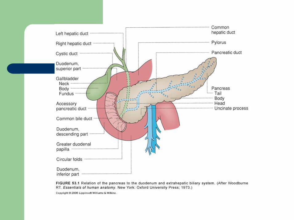
ANATOMYThe Gallbladder
The gallbladder is a pear-shaped sac, about 7 to 10 cm long with an average capacity of 30 to 50 mLSame peritoneal lining covers the liver covers the fundus and the inferior surface of the gallbladder. Lined by a single, highly-folded, tall columnar epithelium that contains cholesterol and fat globules. The epithelial lining of the gallbladder is supported by a lamina propria.The muscle layer has circular longitudinal and oblique fibers, but without well-developed layers. The cystic artery that supplies the gallbladder is usually a branch of the right hepatic artery

The Bile Ducts
The left hepatic duct is longer than the right and has a greaterpropensity for dilatation as a consequence of distal obstructionThe segment of the cystic duct adjacent to the gallbladder neck bears a variable number of mucosal folds called the spiral valves of Heister. The common bile duct is about 7 to 11 cm in length and 5 to 10 mm in diameter. The arterial supply to the bile ducts is derived from the gastroduodenal and the right hepatic arteries, with major trunks running along the medial and lateral walls of the common duct


Anomalies
Small ducts (of Luschka) may drain directly from the liver into the body of the gallbladder. In about 20% of patients the right hepatic artery comes off the superior mesenteric artery
PHYSIOLOGYBile Formation and Composition
Normal adult consuming an average diet produces within the liver 500 to 1000 mL of bile a day. Bile is mainly composed of water, electrolytes, bile salts, proteins, lipids, and bile pigments. The primary bile salts, cholate and chenodeoxycholate, are synthesized in the liver from cholesterol. Bile salts are excreted into the bile by the hepatocyte and aid in the digestion and absorption of fats 95% of the bile acid pool is reabsorbed and returned via the portal venous system to the liverCholesterol and phospholipids synthesized in the liver are the principal lipids found in bile.

Gallbladder Function
In fasting approximately 80% of bile secreted by liver stored ingallbladder. When stimulated GB empties 50 to 70% of its contents within 30 to 40 minutes. Defects in motor activity of gallbladder play a role in cholesterol nucleation and gallstone formationParasympathomimetic drugs contract GB; atropine leads to relaxation. Antral distention of stomach causes both GB contraction and relaxation of sphincter of Oddi.Hormonal receptors on the GB smooth muscle, vessels, nerves, andepithelium CCK is released into the bloodstream by acid, fat, and AA in theduodenum.VIP inhibits contraction and causes GB relaxation. Somatostatin and its analogues are potent inhibitors of GB contraction.

Sphincter of Oddi
The sphincter of Oddi regulates flow of bile (and pancreatic juice) into the duodenum,

Blood Tests
Cholestasis, an obstruction to bile flow, is characterized by an elevation of bilirubinAlkaline phosphatase is released by bile duct cells GGT – gamma glutamyl transferase –sensitive for ductal cells
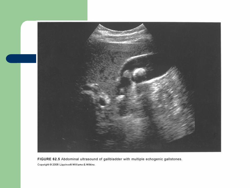
Ultrasonography
An ultrasound is the initial investigation of any patient suspected of disease of the biliary tree. The extrahepatic bile ducts are also well visualized by ultrasound, except for the retroduodenal portion. Ultrasound can be helpful in evaluating tumor invasion and flow in the portal vein, an important guideline for resectability of periampullary tumors
Biliary Radionuclide Scanning (HIDA Scan)
99 m-Technetium-labeled derivatives of dimethyl iminodiacetic acid (HIDA) are injected intravenously, Uptake by the liver is detected within 10 minutes, and the gallbladder, the bile ducts, and the duodenum are visualized within 60 minutes in fasting subjects.
Computed Tomography
Abdominal CT scans are inferior to ultrasonography in diagnosing gallstones. Use of CT scan is an integral part of the differential diagnosis of obstructive jaundice (Fig. 31-7).

Percutaneous TranshepaticCholangiography
An intrahepatic bile duct is accessed percutaneously with a small needle under fluoroscopic guidance.
Magnetic Resonance Imaging
MRI provides anatomic details of the liver, gallbladder, and pancreas similar to CT.

Endoscopic Retrograde Cholangiography and Endoscopic Ultrasound
Common bile duct can be cannulated and a cholangiogram performed using fluoroscopyThe procedure requires intravenous sedation for the patient. Complications of diagnostic ERC include pancreatitis and cholangitis, up to 5% of patients.An endoscopic ultrasound requires a special endoscope with an ultrasound transducer at its tip.

GALLSTONE DISEASEPrevalence and Incidence
Gallstone disease is one of the most common problems affecting the digestive tract. Women are three times more likely to develop gallstones than men, and first-degree relatives of patients with gallstones have a twofold greater prevalence
Natural History
Most patients will remain asymptomatic from their gallstones throughout life. Acute cholecystitis, choledocholithiasis with or without cholangitis, gallstone pancreatitis, cholecystocholedochalfistula, cholecystoduodenal fistula, cholecystoenteric fistula, carcinoma. Gallstones in patients without biliary symptoms are commonly diagnosed incidentally Porcelain gallbladder, a rare premalignant condition in which the wall of the gallbladder becomes calcified, is an absolute indication for cholecystectomy.

Gallstone Formation
Gallstone FormationGallstones form as a result of solids settling out of solution. 80% of gallstones are cholesterol stones and about 15 to 20% are black pigment stones. Cholesterol StonesPure chlolesterol stones are uncommon and account for less than 10% of all stones Pigment StonesPigment stones contain less than 20% cholesterol, dark because of the presence of calcium bilirubinate. Black pigment stones are usually small, brittle, black, and sometimes spiculated. Secondary to hemolytic disorders such as hereditary spherocytosis and sickle cell disease, cirrhosis. Brown stones are usually less than 1 cm in diameter, brownish-yellow, soft, and often mushy. secondary to bacterial infection caused by bile stasis. Brown stones are typically found in the biliary tree of Asian populations and are associated with stasis

Symptomatic Gallstones
Chronic CholecystitisAbout two thirds of patients with gallstone disease present with chronic cholecystitis
Clinical Presentation
The chief symptom associated with symptomatic gallstones is pain. Atypical presentation of gallstone disease is common. When the pain lasts more than 24 hours, an impacted stone in the cystic duct or acute cholecystitis
Diagnosis.
An abdominal ultrasound is the standard diagnostic test for gallstones Gallstones are occasionally identified on abdominal radiographs or CT scans.
Management
Patients with symptomatic gallstones should be advised to have elective laparoscopic cholecystectomy. Pregnant women with symptomatic gallstones who cannot be managed expectantly with diet modifications can safely undergo laparoscopic cholecystectomyduring the second trimester.
Pathogenesis
Acute cholecystitis is secondary to gallstones in 90 to 95% of cases. When the gallbladder remains obstructed and secondary bacterial infection supervenes, an acute gangrenous cholecystitis develops and an abscess or empyema forms within the gallbladder.
Clinical Manifestations
About 80% of patients with acute cholecystitis give a history compatible with chronic cholecystitis. A mild to moderate leukocytosis (12,000 to 15,000 cells/mm3) is usually present. The differential diagnosis for acute cholecystitis includes a peptic ulcer with or without perforation, pancreatitis, appendicitis, hepatitis, perihepatitis(Fitz-Hugh and Curtis syndrome), myocardial ischemia, pneumonia, pleuritis, and herpes zoster involving the intercostal nerve.

Diagnosis
Ultrasonography is the most useful radiologic test for diagnosing acute cholecystitis.

Treatment
Patients who present with acute cholecystitis will need intravenous fluids, antibiotics, and analgesia. Cholecystectomy is the definitive treatment for acute cholecystitisLaparoscopic cholecystectomy is the procedure of choice for acute cholecystitis.CholedocholithiasisCommon bile duct stones may be small or large, single or multiple, and are found in 6 to 12%
Clinical Manifestations
Choledochal stones may be silent and often are discovered incidentally. One third of patients with common bile duct stones, the liver chemistries are normal.PTC is rarely needed in patients with secondary common bile duct stones, but is frequently performed for both diagnostic and therapeutic reasons in patients with primary bile duct stones.

Treatment
Symptomatic gallstones and suspected CBD stones, either preoperative ERCP or an intraop cholangiogramIf an ERCP reveals stones, sphincterotomy and ductalclearance of the stones, followed by a lap chole. Intraoperative cholangiogram will also document the presence or absence of bile duct stonesLap CBDE via the cystic duct or with formal choledochotomyRetained/recurrent stones after lap chole are treated endoscopicallyIf a CBDE done and a T tube left, get T-tube cholangiogram

Clinical Presentation
Cholangitis may present from a mild, intermittent, and self-limited disease to a fulminant septicemia. On abdominal examination, the findings are indistinguishable from those of acute cholecystitis
Cholangitis
Cholangitis is one of the two main complications of choledochal stonesMost common organisms cultured from bile in patients with cholangitis include Escherichia coli, Klebsiella pneumoniae, Streptococcus faecalis, and BacteroidesfragilisCharcot’s Triad ---> Reynolds’ Pentad
Diagnosis and Management
> WBC, > Bi, and > alk phos and transaminasessupport the clinical diagnosis of cholangitisTreatment of cholangitis = iv antibiotics and fluid resuscitationObstructed bile duct must be drained as soon as stabilized. Decompression accomplished endoscopically, via the percutaneous transhepatic route, or surgically. Acute cholangitis is associated mortality rate of 5%.

Biliary Pancreatitis
Gallstones in the common bile duct are associated with acute pancreatitis. Once the pancreatitis has subsided, the gallbladder should be removed during the same admission.Gallstones are present and the pancreatitis is mild and self-limited, the stone has probably passed.
Cholangiohepatitis
Cholangiohepatitis, also known as recurrent pyogenic cholangitis, is endemic to the Orient. The patient usually presents with pain in the right upper quadrant and epigastrium, fever, and jaundice.Recurrence of symptoms is one of the most characteristic features of the disease. Recurrences are common and the prognosis is poor once hepatic insufficiency has developed
OPERATIVE INTERVENTIONS FOR GALLSTONE DISEASE
Cholecystostomy – quick fixICU patient too ill for ORCholecystostomy decompresses and drains the distended, inflamed, hydropic, or purulent gallbladder. Surgical cholecystostomy with a large catheter placed under local anesthesia is rarely required today.
Cholecystectomy
Cholecystectomy = most common major abdominal procedure performed in Western countries. Laparoscopic cholecystectomy is the treatment of choice for symptomatic gallstones.Symptomatic gallstones = main indication.Absolute contraindications are uncontrolled coagulopathy and end-stage liver disease. Conversion to an open not failure and the possibility should be discussed.
Open Cholecystectomy
The same surgical principles apply for laparoscopic and open cholecystectomies.
Intraoperative Cholangiogram or Ultrasound
The bile ducts are visualized under fluoroscopy by injecting contrast through a catheter Routine intraoperative cholangiography will detect stones in approximately 7% of patients

Choledochal Exploration
Common bile duct stones that are detected intraoperatively on intra cholangiogram or U/S may be managed with lap CBDEIf the stones in the duct are small, they may sometimes be flushed into the duodenum with saline irrigation via the cholangiographycatheter after the sphincter of Oddi has been relaxed with glucagon.

Choledochal Drainage Procedures
Choledochoduodenostomy is performed by mobilizing the second part of the duodenum (a Kocher maneuver) and anastomosing it side to side with the common bile duct.A choledochojejunostomy is done by bringing up a 45-cm Roux-en-Y limb of jejunum and anastomosing it end to side to the choledochus.

Transduodenal Sphincterotomy
In the majority of cases, endoscopic sphincterotomy has replaced open transduodenal sphincterotomy.

OTHER BENIGN DISEASES AND LESIONS
Acalculous CholecystitisAcute inflammation of the gallbladder can occur without gallstones.Acalculous cholecystitis typically develops in critically ill patients in the intensive care unit. Symptoms and signs depend on the condition of the patient, but in the alert patient they are similar to acute calculouscholecystitis, with right upper quadrant pain and tenderness, fever, and leukocytosisUltrasonography is usually the diagnostic test of choice, can be done bedside in the intensive care unit.About 90% of patients will improve with the percutaneouscholecystostomy.
Biliary Cysts
Choledochal cysts are congenital cystic dilatations of the extrahepatic and/or intrahepatic biliary tree. Adults commonly present with jaundice or cholangitis.

Sclerosing Cholangitis
Sclerosing cholangitis is an uncommon disease characterized by inflammatory strictures involving the intrahepatic and extrahepaticbiliary tree.The mean age of presentation is 30 to 45 years and men are affected twice as commonly as women.The usual presentation is intermittent jaundice, fatigue, weight loss, pruritus, and abdominal pain. The clinical presentation and elevation of alkaline phosphatase and bilirubin may suggest the diagnosis, but ERC, revealing multiple dilatations and stricturesSclerosing cholangitis is followed by ERC and liver biopsies to provide appropriate management.No known effective medical therapy for primary sclerosing cholangitis, no known curative treatment.Corticosteroids, immunosuppressants, ursodeoxycholic acid, and antibiotics have been disappointing. Primary sclerosing cholangitis recurs in 10 to 20% of patients and may require retransplantation

Stenosis of the Sphincter of Oddi
A benign stenosis of the outlet of the common bile duct is usually associated with inflammation, If the diagnosis is well established, endoscopic or operative sphincterotomy will yield good results
Bile Duct Strictures
Benign bile duct strictures can have numerous causes. Patients with bile duct strictures most commonly present with episodes of cholangitis. Choledochoduodenostomy choice for strictures in the distal-most part of the common bile duct.

INJURY TO THE BILIARY TRACT
The GallbladderInjuries to the gallbladder are uncommon. treatment cholecystectomy and prognosis directly related to the type and incidence of injury.

The Extrahepatic Bile Ducts
Penetrating trauma to the extrahepatic bile ducts is rare and is usually associated with traumaA number of different factors are associated with bile duct injury during laparoscopic cholecystectomy. The routine use of intraoperative cholangiography to prevent bile duct injury is controversial.It may limit the extent of injury, but does not seem to prevent it. Important to check that the whole biliary system fills with contrast and to be sure there are no leaks.
Diagnosis
Only about 25% of major bile duct injuries are recognized at the time of operation. CT scan and ultrasound also are important in the initial evaluation of the jaundiced patient, MRI cholangiography, if available, provides an excellent, noninvasive delineation of the biliary anatomy both proximal and distal to the injury.
Management
If a major injury is discovered and an experienced biliary surgeon is not available, an external drain and, if necessary, transhepatic biliarycatheters are placed, patient is transferred to a referral center.Transected bile ducts smaller than 3 mm or draining a single hepatic segment can safely be ligated. Major bile duct injuries such as transection of the common hepatic or common bile duct are best managed at the time of injury.Critical to perform a tension-free anastomosis to minimize high risk of postop stricture formationCystic duct leaks can usually be managed with percutaneous drainage of intra-abdominal fluid collections followed by an endoscopic biliarystentingMajor injuries diagnosed postoperatively require transhepatic biliarycatheter placement for biliary decompression as well as percutaneousdrainage of intra-abdominal bile collections, if any. Patients with bile duct stricture from an injury or as a sequela of previous repair usually present with either progressive elevation of liver function tests or cholangitis.
Outcome
Good results can be expected in 70 to 90% of patients with bile duct injuriesThe best results are obtained when the injury is recognized during the cholecystectomy and repaired by an experienced biliary tract surgeon.Operative mortality rate varies from 0 to almost 30% in various series, but commonly is about 5 to 8%.Common complications that are specific for bile duct repairs include cholangitis, external biliary fistula, bile leak, subhepatic and subphrenic abscesses, and hemobilia.

TUMORSCarcinoma of the Gallbladder
Cancer of the gallbladder is a rare malignancy that occurs predominantly in the elderly. The overall reported 5-year survival rate is about 5%
Incidence
Gallbladder cancer is the fifth most common gastrointestinal malignancy in Western countries. Overall incidence of gallbladder cancer of 2.5 cases per 100,000 residents in the United States
Etiology
Approximately 90% of patients with carcinoma of the gallbladder have gallstones. Larger stones (3 cm) are associated with a tenfold increased risk of cancer. Risk of developing cancer of gallbladder is higher in patients with symptomatic gallstones.Polypoid lesions of the gallbladder are associated with increased risk of cancer, particularly in polyps larger than 10 mmCalcified "porcelain" gallbladder is associated with 20% incidence of gallbladder carcinoma. These gallbladders should be removed, even if the patients are asymptomatic.

Pathology
Between 80 and 90% of the tumors are adenocarcinomas.Cancer of the gallbladder spreads through the lymphatics, with venous drainage, and with direct invasion into the liver parenchyma. When diagnosed, about 25% of gallbladder cancers are localized to gallbladder wall, 35% have regional nodal involvement and/or extension into adjacent liver, approx 40% have distant metastasis
Clinical Manifestations and Diagnosis
Signs and symptoms of carcinoma of the gallbladder are generally indistinguishable from those associated with cholecystitis and cholelithiasis. Jaundice, weight loss, anorexia, ascites, and abdominal mass are less common presenting symptoms.

Treatment
Surgery remains the only curative option for gallbladder cancer as well as for cholangiocarcinoma. No proven effective options for adjuvant radiation or chemotherapy for gallbladder cancer.Patients without evidence of distant metastasis warrant exploration for tissue diagnosis, pathologic staging, and possible curative resection.Simple cholecystectomy is an adequate treatment for T1 lesions and results in 100% 5-year survival Regional lymphadenectomy is an important part of surgery for T2 cancersAggressive approach in patients who will tolerate surgery has resulted in an increased survival
Prognosis
Most patients with gallbladder cancer have unresectable disease at the time of diagnosis. Median survival for patients with distant metastasis at the time of presentation is only 1 to 3 months.Recurrence of gallbladder cancer most commonly in the liver or the celiac or retropancreatic nodes.

Bile Duct Carcinoma
Bile Duct CarcinomaCholangiocarcinoma is a rare tumor arising from the biliary epithelium Most patients with unresectable disease die within a year of diagnosis.
Incidence
The autopsy incidence of bile duct carcinoma is about 0.3%. The male to female ratio is 1.3:1 and the average age of presentation is between 50 and 70 years.
Etiology
Risk factors associated with cholangiocarcinoma include primary sclerosing cholangitis, choledochal cysts, ulcerative colitis, hepatolithiasis, biliary-enteric anastomosis, and biliary tract infections Factors associated with cholangiocarcinomaare liver flukes, dietary nitrosamines, Thorotrast, dioxin.
Pathology
Over 95% of bile duct cancers are adenocarcinomas. Morphologically they are divided into nodular, scirrhous, diffusely infiltrating, or papillary.Anatomically they are divided into distal, proximal, or perihilartumors. Type I tumors are confined to the common hepatic duct, but type II tumors involve the bifurcation without involvement of the secondary intrahepatic ducts. Type IIIa and IIIb tumors extend into the right and left secondary intrahepatic ducts, respectively. Type IV tumors involve both the right and left secondary intrahepatic ducts.
Clinical Manifestations and Diagnosis
Painless jaundice is the most common presentationPruritus, mild right upper quadrant pain, anorexia, fatigue, and weight loss also may be present. The initial tests are usually ultrasound or CT scan. Either ultrasound or spiral CT can be used to determine portal vein patency. With the newer types of MRI, a single noninvasive test has the potential of evaluating the biliary anatomy, lymph nodes, and vascular involvement, as well as the tumor growth itselfTissue diagnosis may be difficult to obtain nonoperativelyexcept in advanced cases
Treatment
Surgical excision is the only potentially curative treatment forcholangiocarcinoma. No signs of metastasis or locally unresectable disease. For unresectable perihilar cholangiocarcinoma, Roux-en-Y cholangiojejunostomy to either segment II or III bile ducts or to the right hepatic duct can be performed.For curative resection, the location and local extension of the tumor dictates the extent of the resection.Perihilar tumors involving the bifurcation or proximal common hepatic duct with no signs of vascular involvement are candidates for local tumor excision with portal lymphadenectomy, cholecystectomy, common bile duct excision, and bilateral Roux-en-Y hepaticojejunostomiesDistal bile duct tumors are more often resectable. Nonoperative biliary decompression performed for unresectable disease on diagnostic evaluation.Operative intervention is not warranted in patients with metastatic disease.

Treatment
There is no proven role for adjuvant chemotherapy in cholangiocarcinoma. Adjuvant radiation therapy does not increase either quality of life or survival Patients with unresectable disease offered treatment with 5-fluorouracil alone or in combination with mitomycin C and doxorubicin, but the response rates are low, less than 10% and less than 30%,respectively. Radiation/chemotherapy may be more effective than either alone for unresectable disease, but no randomized trialsGiving chemoradiation difficult because of high incidence of cholangitis.External beam radiation not effective treatment for unresecteddisease. Intraop radiation, brachytherapy with iridium 192, and combined interstitial and external beam radiation for unresectablecholangiocarcinoma reported with encouraging results.
Prognosis
Most patients with perihilar cholangiocarcinoma present with advanced, unresectable disease. Median survival between 5 and 8 months. Causes of death are hepatic failure and cholangitis. The overall 5-year survival with resectable perihilarcholangiocarcinoma is 10 and 30%, with negative margins may be as high as 40%. The operative mortality for perihilar cholangiocarcinoma is 6 to 8%.Patients with distal cholangiocarcinoma are more likely to have resectable disease and improved prognosis compared to perihilarcholangiocarcinoma. Overall 5-year survival rate for resectable disease is 30 to 50%, median survival is 32 to 38 months.Risk factors for recurrence are positive margins, lymph node-positive. Therapy for recurrent disease is palliation.