wound healing - university of tennessee college of...
TRANSCRIPT

Wound Healing

Wound Healing
same events, in the same order, in every healing process
regardless tissue type inciting injury

Wound Healing
Wound repairis the effort of tissues to restore normal function and structure after injury.
To reform barriers to fluid loss and infection, Limit further entry of foreign organisms and material, re-establish normal blood and lymphatic flow patterns, restore the mechanical integrity of the injured system, perfect reorganization is sacrificed for urgent return to function.
Regeneration, is the perfect restoration of the preexisting tissue architecture in the absence of scar information.
Ideal wound healing, Only found in embryonic development, in lower organisms, such as the stone crab and the salamander, in certain tissue compartments, such as bone and liver.

Wound Healing
accuracy of regeneration is traded for the speed of repair. divided into specific stages phases overlap in both
time activity.
Acute wounds orderly and timely reparative process to achieve sustained restoration of structure and function.
Chronic wound, does not proceed to a restoration of functional integrity. stalled in the inflammatory phase variety of etiologiesdoes not proceed to closure.

primary, Wound Healing secondary,
tertiary repair
Wound Healing
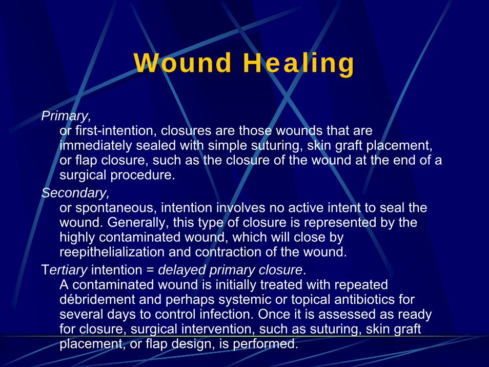
Primary,or first-intention, closures are those wounds that are immediately sealed with simple suturing, skin graft placement, or flap closure, such as the closure of the wound at the end of a surgical procedure.
Secondary,or spontaneous, intention involves no active intent to seal the wound. Generally, this type of closure is represented by the highly contaminated wound, which will close by reepithelialization and contraction of the wound.
Tertiary intention = delayed primary closure. A contaminated wound is initially treated with repeated débridement and perhaps systemic or topical antibiotics for several days to control infection. Once it is assessed as ready for closure, surgical intervention, such as suturing, skin graftplacement, or flap design, is performed.

Wound Healing Phases

Wound Healing Phases
inflammatory (reactive) the body’s defenses are aimed at limiting the amount of damage and preventing further injury. proliferative (regenerative or reparative)is the reparative process with
reepithelialization, matrix synthesis, neovascularization
to relieve the ischemia of the trauma itself.
maturational (remodeling) is the period of scar contraction with
collagen cross-linking, shrinking, loss of edema.

Wound Healing Phases in a Large Wound
i.e. pressure sore eschar or fibrinous exudateinflammatory phase; granulation tissue proliferative phase;the contracting or advancing edgematurational phase.
All three phases may occur simultaneously,
Phases with their individual processes may overlap

Inflammatory PhaseHemostasis and inflammation
tissue’s attempt to limit damage stopping the bleeding, sealing the surface of the wound, removing any necrotic tissue, foreign debris, or bacteria
Characterized byincreased vascular permeability,migration of cells into the wound by chemotaxis, secretion of cytokines and growth factors into the wound,activation of the migrating cells

Hemostasis and Inflammation
Blood vessel damage →exposure of subendothelial collagen to platelets, →
platelet aggregation and activation of the coagulation pathway.
Initial intense local vasoconstriction of arterioles and capillaries followed by vasodilation and increased vascular permeability.
Cessation of hemorrhage is aided by plugging of capillaries witherythrocytes and platelets, which adhere to the damaged capillary endothelium.
Exposure of types IV and V collagen promotes platelet aggregation as platelets bind to these proteins and become activated.
The initial contact between platelets and collagen requires the von Willebrand factor (vWF) VIII

Hemostasis and Inflammation
Platelet adhesion to the endothelium interaction between high-affinity glycoprotein receptors and the integrin receptor GPIIb-IIIa (αIIbβ3). platelets express other integrin receptors
direct binding of collagen, laminin, indirectly by attaching to subendothelial matrix-bound fibronectin, vitronectin, and other ligands.
The clotting cascade initiated by:intrinsic extrinsic pathways.
The fibrin strands trap red blood cells, forming the clot, and seal Lattice framework will be the scaffold for
endothelial cells, inflammatory cells,fibroblasts.
Thromboxane A2 and prostaglandin F2 α, assist with platelet aggregation and vasoconstriction.

Hemostasis and Inflammation
Increased Vascular Permeability Platelet binding → changes in conformation→ intracellular signal transduction pathways → platelet activation and the release of biologically active proteins.
α granules and, dense bodiesMast cells:
release histamine and serotonin, → permeability of endothelial cells → leakage of plasma from the intravascular space to the extracellular compartment.

Hemostasis and Inflammation
Polymorphonuclear Cellsadherence, chemoattraction:
Complement factors:C5a, leukotriene B4
Adhesion by Plt-aggregating factor:
thrombin + leukotriene C4 and D4
endothelial-neutrophiladherence: Monocytes and endothelial cells inflammatory mediators:
interleukin (IL)-1 tumor necrosis factor (TNF)-α
All these facilitates: diapedesis of neutrophilsinto the inflammatory site
Neutrophils begin their migration:release the contents of their lysosomes and enzymeselastase and other proteases into the extracellular matrix (ECM), facilitating the migration of the neutrophils.

Hemostasis and Inflammation
intense vasodilationincreased vascular permeability
Clinical findings:rubor (redness), tumor (swelling), calor (heat), dolor (pain)functio lesa (loss of function)

Hemostasis and Inflammation
Following migration of the PMNs :Functional activation:may induce new cell surface antigen expression, increased cytotoxicity, increased production and release of cytokines.
Activated neutrophils: scavenge necrotic debris, foreign material, and bacteria.
Stimulated neutrophilsgenerate free oxygen radicals with electrons donated by the NADPH electrons across the membrane into lysosomessuperoxide anion (O2
− ) is formedsuperoxide dismutase catalyzes formation of H2 O2, degraded by myeloperoxidase in the azurophilic

Hemostasis and Inflammation
MacrophagesThe macrophage is central to wound healing
orchestrates the release of cytokines and stimulate many of the subsequent processes of wound healing
appear at the same time that neutrophils disappearinduce PMN apoptosisChemotaxis of migrating blood monocytes occurs within 24 to 48 hours.
Chemotactic factors specific for monocytes:bacterial products, complement degradation products (C5a), thrombin, fibronectin, collagen, TGF-β, and PGDF-BB. also facilitated by the interaction of the integrin receptors on the monocytesurface with ECM proteins, such as fibrin and fibronectin.

Hemostasis and Inflammation

TABLE 8-1 -- Cytokine Activity in Wound Healing
Cytokine Cell Source Biological Activity
Proinflammatory Cytokines
TNF-α Macrophages PMN margination and cytotox., ± collagen synth.; prov. metab. subst.
IL-1 Macrophages Fibroblast and keratinocyte chemotaxis, collagen synthesis
Keratinocytes
IL-2 T lymphocytes Increases fibroblast infiltration and metabolism
IL-6 Macrophages Fibroblast proliferation, hepatic acute-phase protein synthesis
PMNs
Fibroblasts
IL-8 Macrophages Macrophage and PMN chemotaxis, keratinocyte maturation
Fibroblasts
IFN-γ T lymphocytes Macrophage and PMN activation; retards collagen synthesis and cross-linking; stimulates collagenase activity
Macrophages
Anti-inflammatory Cytokines
IL-4 T lymphocytes Inhibition of TNF, IL-1, IL-6 production; fibroblast prolif., collagen synth.
Basophils
Mast cells
IL-10 T lymphocytes Inhibition of TNF, IL-1, IL-6 prod.; inhib. macrophage and PMN activ.
Macrophages
Keratinocytes

TABLE 8-2 -- Cytokines that Affect Wound Healing
Cytokine Abbreviation Source Functions
Platelet-derived growth factor
PDGF Platelets, macrophages, endothelial cells, keratinocytes
Chemotactic for PMNs, macrophages, fibroblasts, and smooth muscle cells; activates PMNs, macroph., and fibrobl.; mitogenic for fibrobl., endoth. cells; stim. Prod. of MMPs, fibronectin, and HA; stim. angiogenesis and wound contraction; remodeling
Transforming growth factor-beta (including isoforms β1 , β2 , and β3 )
TGF-β Platelets, T lymphocytes, macrophages, endothelial cells, keratinocytes, fibroblasts
Chemotactic for PMNs, macrophages, lymphocytes, fibroblasts; stim. TIMP synthesis, keratinocyte migration, angiogenesis, and fibroplasia; inhib. Prod. of MMPs and keratinocyte proliferation; induces TGF-β production
Epidermal growth factor EGF Platelets, macrophages Mitogenic for keratinocytes and fibroblasts; stimulates keratinocyte migration
Transforming growth factor-alpha
TGF-α Macrophages, T lymphoc., keratinocytes
Similar to EGF
Fibroblast growth factor-1 and -2 family
FGF Macrophages, mast cells, T lymphocytes, endoth. cells, fibroblasts
Chemotactic for fibroblasts; mitogenic for fibroblasts and keratinocytes; stimulates keratinocyte migration, angiogenesis, wound contraction, and matrix deposition
Keratinocyte growth factor (also called FGF-7)
KGF Fibroblasts Stimulates keratinocyte migration, proliferation, and differentiation
Insulin-like growth factor IGF-1 Macrophages, fibroblasts Stimulates synthesis of sulfated proteoglycans, collagen, keratinocyte migration, and fibroblast proliferation; endocrine effects similar to those of growth hormone
Vascular endothelial cell growth factor
VEGF Keratinocytes Increases vasopermeability; mitogenic for endoth. cells
HA, hyaluronic acid; MMPs, matrix metalloproteinases; PMNs, polymorphonuclear leukocytes; TIMP, tissue inhibitor of matrix metalloproteinase.

Hemostasis and Inflammation
LymphocytesAppear 5th day, peak at around the 7th day. Initially, lymphocytes were thought to play a minimal role in acute wound healing
B lymphocytes:downregulating healing as the wound closes.
Effects on fibroblasts stimulatory cytokines
IL-2 and fibrobl. activ. factorinhibitory cytokines:
TGF-β, TNF-α, and IFN-γ.
T cells produce IFN-γ, Stim. the macroph. to release a cascade of cytokines TNF-α and IL-1decreased synthesis of prostaglandins, which enhances the effect of inflam. mediators. suppresses collagen synthesis inhibits macrophages from leaving the site of injury. IFN-γ appears to be an important mediator of the chronic nonhealing wound

Hemostasis and Inflammation
Role of lymphocytescyclosporine, tacrolimus and steroids
suppress T-lymphocyte function and proliferationimpair wound healing in experimental wound models
possibly through decreased nitric oxide synthesis. In vivo
lymphocyte depletion suggests the existence of an incompletely characterized T-cell lymphocyte population neither CD4+ nor CD8+
this subset that seems to be responsible for the promotion of wound healing

Proliferative Phase
acute responses of hemostasis and inflammation begin to resolvescaffolding is laid for repair of the wound:
angiogenesis, fibroplasia, epithelialization.
characterized by formation of granulation tissue: capillary bed, fibroblasts, macrophages, loose arrangement of collagen, fibronectin, and hyaluronic acid.

Proliferative Phase
AngiogenesisActivated endothelial cells degrade the basement membrane of postcapillary venules, allowing migration of cells through this gapDivision of these migrating endothelial cells results in tubule or lumen formationEventually, deposition of the basement membrane occurs, resulting in capillary maturationAppears to be stimulated and manipulated by a variety of cytokines, predominantly produced by macrophages and platelets

Proliferative Phase
FibroplasiaFibroblasts differentiate from resting mesenchymal cells in
connective tissue; the normally quiescent and sparse fibroblasts are chemoattracted to the inflammatory sitethey divide and produce the components of the ECM
Fibroblast, normally arrested in the G0 phaseundergoes replication and proliferation after stimulation by macrophage and platelet-derived cytokines and growth factors
Proliferative Phase
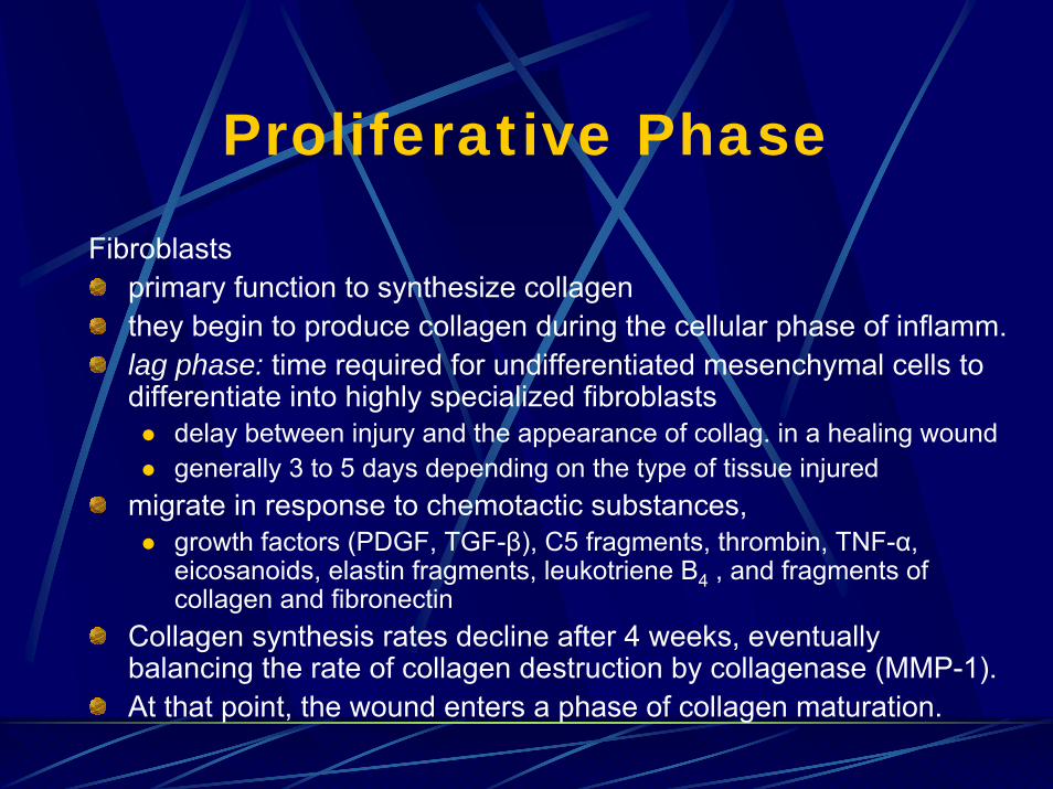
Fibroblastsprimary function to synthesize collagen they begin to produce collagen during the cellular phase of inflamm.lag phase: time required for undifferentiated mesenchymal cells to differentiate into highly specialized fibroblasts
delay between injury and the appearance of collag. in a healing woundgenerally 3 to 5 days depending on the type of tissue injured
migrate in response to chemotactic substances,growth factors (PDGF, TGF-β), C5 fragments, thrombin, TNF-α, eicosanoids, elastin fragments, leukotriene B4 , and fragments of collagen and fibronectin
Collagen synthesis rates decline after 4 weeks, eventually balancing the rate of collagen destruction by collagenase (MMP-1). At that point, the wound enters a phase of collagen maturation.

Proliferative PhaseEpithelialization
epidermis serves as a physical barrier to prevent fluid loss andbacterial invasiontight cell junctions contribute to its impermeability the basement membrane zone
gives structural support attachment between the epidermis and the dermis
The basement membrane zone:(1) the lamina lucida (electron clear), laminin and heparan sulfate; (2) lamina densa (electron dense), type IV collagen;(3) anchoring fibrils, type IV collagen, secure the epidermodermal interface connect from the lamina densa into the dermis.
The basal layer of the epidermis attaches to the basement membrane zone by hemidesmosomes.
Proliferative Phase
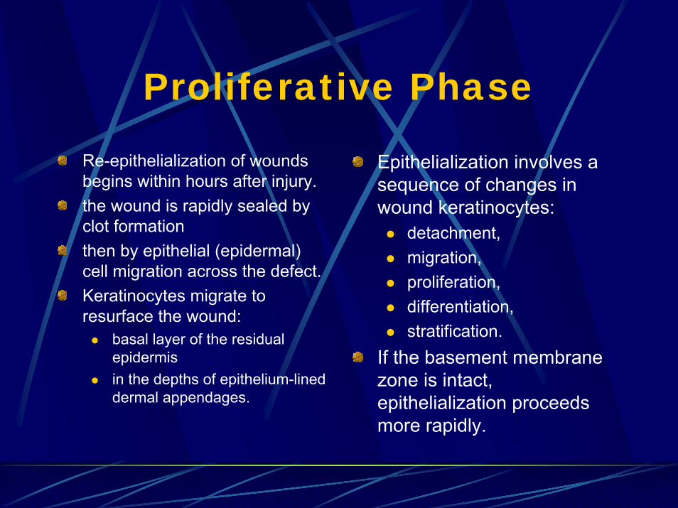
Re-epithelialization of wounds begins within hours after injury.the wound is rapidly sealed by clot formationthen by epithelial (epidermal) cell migration across the defect. Keratinocytes migrate to resurface the wound:
basal layer of the residual epidermis in the depths of epithelium-lined dermal appendages.
Epithelialization involves a sequence of changes in wound keratinocytes:
detachment, migration, proliferation, differentiation,stratification.
If the basement membrane zone is intact, epithelialization proceeds more rapidly.

Proliferative Phase
The epithelial cells move in a leapfrog and tumbling fashionThey move until the edges establish contact. If the basement membrane zone is not intact, it will be repairedfirst.The absence of neighbor cells at the margin may be a signal for the migration and proliferation of epidermal cellsAfter the wound is completely re-epithelialized,
the cells become columnar and stratified again, while firmly attaching to the re-established basement membrane and underlying dermis.

Proliferative Phase
Extracellular Matrix The ECM exists as a scaffold
to stabilize the physical structure of tissues complex role by regulating the behavior of the cells that contact it.
Cells within it produce the macromolecular constituents, (1) glycosaminoglycans (GAGs), polysaccharide chains, usually found covalently linked to protein in the form of proteoglycans; (2) fibrous proteins, such as collagen, elastin, fibronectin, and laminin.

Proliferative Phase
Proteoglycan molecules: a gel-like “ground substance.”highly hydrated gel allows the matrix to withstand compressive forceswhile permitting between the blood and the tissue cells the rapid diffusion of:
nutrients, metabolites, hormones
Collagen fibers to organize and strengthen itElastin fibers to give resilience Matrix proteins to have adhesive functions
Proliferative Phase
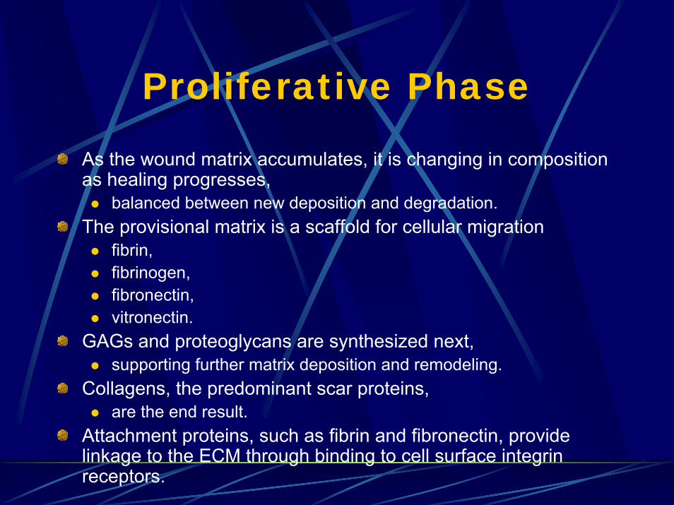
As the wound matrix accumulates, it is changing in composition as healing progresses,
balanced between new deposition and degradation. The provisional matrix is a scaffold for cellular migration
fibrin, fibrinogen, fibronectin, vitronectin.
GAGs and proteoglycans are synthesized next, supporting further matrix deposition and remodeling.
Collagens, the predominant scar proteins, are the end result.
Attachment proteins, such as fibrin and fibronectin, provide linkage to the ECM through binding to cell surface integrinreceptors.

Proliferative PhaseWound matrix deposition over time.
Fibronectin and type III collagen constitute the early matrix.
Type I collagen accumulates later and corresponds to the increase in wound-breaking strength.

Proliferative PhaseCollagen Structure
secreted by a variety of cell types. major component of skin and bone25% of the total protein mass in mammals.
The collagen molecule proline and glycine-rich long, stiff, triple-stranded helical structure, comprises three collagen polypeptide α chains wound around one another in a ropelike superhelix. proline (ring) provides stability to the helical conformation,glycine, (small size) allows tight packing of the three alpha chains to form the final superhelix. There are at least 20 types of collagen; main of connective tissue are types I, II, III, V, and XI

Proliferative Phase
The formation of a collagen fibril. Collagen fibrils assembling in the extracellular space contained within a large infolding in the plasma membrane.

Proliferative Phase
CollagenType I is the principal collagen of skin and bone, the most common.In the adult, the skin is approximately 80% type I and 20% type III. In newborns, the content of type III collagen is greater than that found in the adult. In early wound healing, there is also an increased expression of type IIIType I collagens are the fibrillar collagens, or the fibril-forming collagens.
They are secreted into the extracellular space where they assemble into collagen fibrils (10 to 300 nm in diameter), which then aggregate into larger, cablelike bundles called collagen fibers (several micrometers in diameter).

Proliferative Phase
Collagencollagen synthesis increased:
Vitamin C (ascorbic acid), TransformingGF-β, Insulin like GF-1, and IGF-2.
collagen synthesis decreased IFN-γ type I procollagen mRNA synthesis, glucocorticoids inhibit procollagen gene transcription
Genetic disorders:osteogenesis imperfecta, deletion of one procollagen α1 allele results in weak and easily fractured bones. Ehlers-Danlos syndrome is a result of mutations affecting type III collagen and is characterized by fragile skin and blood vessels and hypermobile joints.

Proliferative PhaseElastic Fibers
skin, blood vessels, and lungs require strength and elasticity to function, for recoil after transient stretch.Elastic fibers are predominantly composed of elastin,
highly hydrophobic protein (about 750 amino acids long). Soluble tropoelastin is secreted into the extracellular space where it forms lysine cross-links to generate a large network of elastin fibers and sheets. The predominant theory is that the elastin polypeptide chain adopts a “random coil” conformation that allows the network to stretch and recoil like a rubber band.Elastic fibers consist of an elastin core covered with a sheath of microfibrils (fibrillin), essential for the integrity of the elastic fibers.Elastin is produced early in life, stabilizes, and does not undergo much further synthesis or degradation, with a turnover that approaches the life spanFibrillin gene mutations result in Marfan’s syndrome; severely affected individuals are prone to aortic rupture

Proliferative Phase
Glycosaminoglycans and ProteoglycansGAGs are unbranched polysaccharide chains composed of repeating disaccharide units:
a sulfated amino sugar (N-acetylglucosamine or N-acetylgalactosamine), uronic acid (glucuronic or iduronic).
The GAGs are highly negatively charged because of the sulfate or carboxyl groups on most of their sugars.Four types of GAGS exist:
(1) hyaluronan (HA), (2) chondroitin sulfate and dermatan sulfate, (3) heparan sulfate, (4) keratan sulfate.

Proliferative Phase
The GAGs in connective tissue usually constitute less than 10% of the weight of the fibrous proteins. highly negative charge attracts osmotically active cations, such as Na+
causing large amounts of water to be incorporated into the matrix. This results in porous hydrated gels and is responsible for the turgor that enables the matrix to withstand compressive forces. Hyaluronan is the simplest of the GAGs.
prevalent in fetal tissues, is believed to be a factor in the scarlesswound healing seen in fetal tissues.during wound healing facilitates cell migration by physically expanding the ECMcreates a cell-free space for cell migration, during embryogenesis and formation of the heart and other organs. when cell migration finishes, the excess HA is degraded by hyaluronidase.

Proliferative PhaseProteoglycans
diverse group with functions mediated by both their core proteins and GAG chains. provide hydrated space around and between cells. form gels of different pore size and charge density to regulate movement of cells and molecules.
Perlecan, a heparan sulfate proteoglycan, serves this role in the basal lamina of the kidney glomerulus. Decreased levels of perlecan are believed to play a role in diabetic albuminuria.
bind signal molecules (growth factors), proteases and protease inhibitors.
(1) immobilizing the protein and restricting its range of action; (2) providing a reservoir of the protein for delayed release; (3) altering the protein, more effective presentation to cell surface recept. (4) prolonging the protein’s action by protecting it from degradation; (5) blocking the activity of the protein

Proliferative PhaseBasal Lamina
flexible, thin (40- to 120-nm thick) mats of specialized ECM separate cells and epithelia from the underlying or surrounding connective tissue. functions;
(1) as a molecular filter, preventing passage of macromolecules (i.e., in kidney glomerulus); (2) as a selective barrier to certain cells (i.e., the lamina beneath the epithelium prevents fibroblasts from contacting epithelial cells, but does not stop macrophages or lymphocytes); (3) as a scaffold for regenerating cells to migrate; (4) is important in tissue regeneration where the basal lamina survives.

Proliferative Phase
Basal LaminaIn the skin, the basal lamina is tethered to the underlying connective tissue by specialized anchoring fibrils. This composite of basal lamina and collagen is the basement membrane. Most mature basal laminae contain type IV collagen, perlecan, and the glycoproteins laminin and nidogen. Mice lacking the laminin-γ1 chain die during embryogenesis because they cannot make a basal lamina.

Maturational PhaseWound contraction is the centripetal movement of the whole thickness of the surrounding skin, reducing the amount of disorganized scar. Wound contracture, in contrast, is a physical constriction or limitation of function and is the result of the process of woundcontraction. Contractures occur when excessive scar exceeds normal wound contraction and results in a functional disability. Scars that traverse joints and prevent extension or scars that involve the eyelid or mouth and cause ectropionsWound contraction appears to occur by a complex interaction of the extracellular materials and the fibroblast (not completely understood)Myofibroblasts: fibroblasts in a contracting wound, which undergo change for stimulation
both function and structure in common with fibroblasts and smooth muscle cells

Maturational PhaseRemodeling
The fibroblast population decreases and the dense capillary network regresses. Wound strength increases rapidly within 1 to 6 weeks and then appears to plateau up to 1 year after the injuryCompared with unwounded skin, the tensile strength is only 30% in the scar. There is an increase in breaking strength after approximately 21days, which is mostly a result of cross-linking. Although collagen cross-linking causes further wound contraction and increase in strength, it also results in a scar that is more brittle and less elastic than normal skin. Unlike normal skin the epidermodermal interface in the healed wound is devoid of rete pegs, the undulating projections of epidermis that penetrate into the papillary dermis. Loss of thisanchorage results in increased fragility and predisposes the neoepidermis to avulsion after minor trauma.

Abnormal Wound HealingFactors that Inhibit Wound Healing
InfectionIschemia
CirculationRespirationLocal tension
Diabetes mellitusIonizing radiationAdvanced ageMalnutrition
Vitamin deficienciesVitamin CVitamin A
Mineral deficienciesZincIron
Exogenous drugsDoxorubicin (Adriamycin)
Glucocorticosteroids

Hypertrophic Scars and Keloids
Keloids are defined as scars that grow beyond the borders of the original woundsrarely regress with time. more prevalent among patients with darker pigmented skin, occurring in 15% to 20% of African Americans, Asians, and Hispanics.appears to have a genetic predisposition.
tends to occur above the clavicles on the trunk, in the upper extremities, and on the face. cannot be prevented at this time and are refractory to medical and surgical intervention. have thicker, more abundant collagen bundles that form acellular nodelike structures in the deep dermal portion of the keloid lesion.
Both keloids and hypertrophic scars are characterized by excessive collagen deposition versus collagen degradation.

Hypertrophic Scars and Keloids
Hypertrophic scars, are raised scars that remain within the confines of the original woundfrequently regress spontaneously. can occur anywhere on the body. differ histologically from normal scars. in many cases preventable
Prolonged inflammation and insufficient resurfacing, such as can occur with a burn wound, lead to hypertrophicscar.It appears that the tension that signals formation of activated fibroblasts also causes deposition of excessive collagen.

Hypertrophic Scars and Keloids
Both keloids and hypertrophic scars have stretched collagen bundles aligned in the same plane as the epidermis as opposed to normal scar tissue, where the collagen bundles are randomly arrayed and relaxed.
The center of keloid lesions also contains a paucity of cells compared to that of the hypertrophic scar that has islands composed of aggregates of fibroblasts, small vessels, and collagen fibers throughout the dermis.

Chronic NonhealingWounds
Chronic wounds, like other abnormal wounds, appear to have derangements in various stages of wound healing and unusually elevated or depressed levels of cytokines, growth factors, or proteinases. Chronic wound fluid, unlike acute wound fluid, has been demonstrated to have greater levels of IL-1, IL-6, and TNF-α; Levels of these proinflammatory cytokines decreased as the wound healed.An inverse relationship between TNF-α and essential growth factors such as EGF and PDGF has been demonstrated.

Chronic NonhealingWounds
Wounds that are chronically inflamed and do not proceed to closure can develop squamous cell carcinoma
chronic burn scars, Marjolinosteomyelitis, pressure sores, venous stasis ulcers, hidradenitis.
The wound appears irregular, raised above the surface, with a white, pearly discoloration. premalignant state is pseudoepitheliomatous hyperplasia.The above biopsy result, biopsy should be repeated because there may be squamous cell carcinoma present in other areas

Chronic NonhealingWounds
InfectionProbably the most common cause of healing delays is wound infection.If the bacterial count in the wound exceeds 105 organisms per gram of tissue, if any β-hemolytic streptococci are present

Chronic NonhealingWounds
HypoxiaMolecular oxygen is essential for collagen formation. Ischemia can be caused by atherosclerosis, cardiac failure, or simple wound tension preventing localized perfusion. glycolysis may be sufficient to initiate collagen synthesis,presence of molecular oxygen is critical for the post-translational hydroxylation of prolyl and lysyl residues required for triple-helix formation and cross-linking of collagen fibrils.Use of tobacco products has a similar impact on wound healing due to
vasoconstriction that occurs with smoking the elevated carbon monoxide serum levels that can limit the oxygen-carrying capacity of the blood

Chronic NonhealingWounds
Diabetesimpairs wound healing at all stages of the process. associated neuropathy and atherosclerosis is prone to tissue ischemia, repetitive trauma, and infection.
large-vessel disease, microvascular level.
basement membrane of the capillaries is thickened, causing decreased perfusion in the microenvironment, increased perivascular localization of albumin, suggesting that these capillaries are leaky. diabetic patients are prone to repeated trauma as a result of the diabetic neuropathy that affects both sensory and motor functions both in somatic and autonomic pathways.

Chronic NonhealingWounds
Diabetessusceptible to infection because of an attenuated inflammatory response, impaired chemotaxis, and inefficient bacterial killing.Infection also increases local tissue metabolism, further imposing a burden on an already tenuous blood supply lymphocyte and leukocyte function is impaired, increased collagen degradation and decreased collagen deposition. The collagen that is formed is more brittle than normal collagen,
Chronic NonhealingWounds
Ionizing Radiationcauses endothelial cell injury with endarteritis resulting in atrophy, fibrosis, and delayed tissue repair. unlike most hypoxic wound beds, angiogenesis is not initiated. greatest effect is on cells in the G2 through M phase, rapidly dividing cell populations are most sensitive to radiation:
keratinocytesfibroblasts
impairing epithelialization and granulation tissue formation.

Chronic NonhealingWounds
Agingelderly patients are more likely to have surgical wound rupturesand delayed healingthe same patient as he or she ages will heal more slowly. collagen undergoes qualitative and quantitative changes. dermal collagen content decreasesaging collagen fibers show distorted architecture and organizationdecreased re-epithelialization, depressed collagen synthesis, and impaired angiogenesis with decreased levels of multiple growth factorsimpaired macrophage activity, with reduced phagocytosis and delayed infiltration of macrophages and B lymphocytes into woundsdecrease in response to hypoxia

Chronic NonhealingWounds
Malnutritionprotein catabolism can result in a delay in wound healing. hypoalbuminemic patient can experience wound healing delay or even dehiscence, (below 2.0 g/dL)Vitamin deficiencies primarily owing to their effect as cofactors:
3 months of vitamin C deprivationvitamin A impedes monocyte activation, fibronectin deposition that further affects cellular adhesion, and impairment of the TGF-βreceptors.vitamin K deficiency is to limit the synthesis of prothrombin and factors VII, IX, and X. Vitamin K metabolism is impeded by antibiotics. Those patients who have chronic or recurrent infections should have their clotting parameters checked before surgical procedures.
Chronic NonhealingWounds
MalnutritionMinerals:
Zinc deficiency is rare, except in cases such as large burns, severe multiple trauma, and hepatic cirrhosis. Zinc deficiency results in early wound healing delaysIron deficiency anemia is a debatable cause of wound healing delay. Although the ferrous ion is a cofactor necessary to convert hydroxyproline to proline, there are conflicting reports as to the effects that acute and chronic anemia have on wound healing

Chronic NonhealingWounds
DrugsSome directly inhibit wound healing.
Doxorubicin (Adriamycin) is a potent inhibitorclinical studies show little impairment, experimental models:
nitrogen mustard, cyclophosphamide, methotrexate, bis-chloroethyl-nitrosourea (BCNU), and doxorubicin are the most potent wound inhibitors.
Tamoxifen, an antiestrogen:decrease cellular proliferationdose-dependent decrease in wound breaking strength
Glucocorticosteroidimpairs fibroblast proliferation and collagen synthesis. amount of granulation tissue formed is also decreased.
Steroids stabilize the lysosomal membranes, can be reversed by the administration of vitamin A.

Fetal Wound Healing
heal rapidly and without the scarring and inflammation re-epithelialize faster with less neovascularization and faster increase in strength. differ from adults
in inflammatory responses, ECM components, growth factor expression and responses.
gestational-age and wound-size dependent wounds created early in gestation heal scarlessly and with dermal appendages