galactosidase activity a modified chitosan (water … · and vmax of the michaelis-menten equation...
TRANSCRIPT

Full Terms & Conditions of access and use can be found athttp://www.tandfonline.com/action/journalInformation?journalCode=ldrt20
Download by: [b-on: Biblioteca do conhecimento online UP] Date: 17 October 2016, At: 04:05
Drying TechnologyAn International Journal
ISSN: 0737-3937 (Print) 1532-2300 (Online) Journal homepage: http://www.tandfonline.com/loi/ldrt20
The Influence of Microencapsulation witha Modified Chitosan (Water Soluble) on β-Galactosidase Activity
Berta N. Estevinho , Ana M. Damas , Pedro Martins & Fernando Rocha
To cite this article: Berta N. Estevinho , Ana M. Damas , Pedro Martins & FernandoRocha (2014) The Influence of Microencapsulation with a Modified Chitosan (WaterSoluble) on β-Galactosidase Activity, Drying Technology, 32:13, 1575-1586, DOI:10.1080/07373937.2014.909843
To link to this article: http://dx.doi.org/10.1080/07373937.2014.909843
Accepted author version posted online: 15Apr 2014.Published online: 15 Apr 2014.
Submit your article to this journal
Article views: 101
View related articles
View Crossmark data
Citing articles: 6 View citing articles

The Influence of Microencapsulation with a ModifiedChitosan (Water Soluble) on b-Galactosidase Activity
Berta N. Estevinho,1 Ana M. Damas,2 Pedro Martins,1,2 and Fernando Rocha11LEPABE, Departamento de Engenharia Quımica, Faculdade de Engenharia da Universidade doPorto, Porto, Portugal2ICBAS, Instituto de Ciencias Biomedicas Abel Salazar da Universidade do Porto, Porto, Portugal
The aim of this work was to investigate the possibility ofproducing microparticles containing b-galactosidase, using a modi-fied chitosan (water soluble) as encapsulating agent. b-galactosidasemicroparticles were prepared by a spray-drying method and werecharacterized in terms of particle size, surface morphology, zetapotential, and stability over a storage period of six months. Micro-particles were also analyzed by FTIR spectroscopy and thermogravi-metry techniques. Structural analysis of the surface of the particleswas performed by scanning electron microscopy (SEM). SEMresults show that the obtained microparticles have an average diam-eter smaller than 3.5lm and a regular shape. The b-galactosidaseactivity decreases when microencapsulated. The parameters Kmand Vmax of the Michaelis-Menten equation were calculated forthe different experimental conditions. The optimal pH ranges from6.4 to 7.2, approximately, depending on the enzyme concentrationin the microcapsule. After six months of storage, the enzyme activitypresents a small decrease, although no significant differences in theappearance, color, and particle size distribution were identified.
Keywords b-Galactosidase; Immobilization; Michaelis-Menten;Spray drying; Water-soluble chitosan
INTRODUCTION
Entrapment of bioactive molecules, such as enzymes,drugs, vitamins, flavors, and peptides, in stabilizingmatrices has been practiced by the pharmaceutical, food,biomedical, chemical, and waste-treatment industries formany years.[1–4]
In the present work, we propose the use of a modifiedchitosan (water soluble) to encapsulate the enzymeb-galactosidase. This study focuses on this enzyme due toits importance in health and industry. There are severalwell-known problems where the enzyme b-galactosidasehas a primordial role in their solution. For example, a largepercentage of the world’s population suffers from lactoseintolerance caused by the lack of b-galactosidase activity.This problem is avoided if lactose is hydrolyzed by
b-galactosidase to the readily utilizable sugars, glucose,and galactose.[5] Another example is the pollution problemcaused by the cheese whey that is disposed in wastewater,causing serious environmental problems on account of highorganic matter content. Whey disposal is a serious problemfor the dairy industry. In order to reduce pollution load,whey should be treated to obtain commercial products.b-galactosidase hydrolyzes lactose found in the whey,allowing the recovery of glucose and galactose.[6–8] Further,the functional galacto-oligosaccharides (GOS), which pro-mote the growth of bifido-bacteria in vivo, can be producedby enzymatic reaction with b-galactosidase.[9,10]
b-galactosidase has been used in large-scale processes inboth free and immobilized forms.[5,11,12] Enzyme immobili-zation has attracted a wide range of interest to many differ-ent industrial applications.[13] The enzyme immobilizationcan be obtained using a variety of techniques[5,14,15] thatshould be selected according to the physicochemical proper-ties of the enzyme (such as molecular weight, protein chainlength, and position of the active site), immobilizationmatrix, reaction conditions, type of reactor, amongothers.[5,16] An important concern in the production ofcommercial proteins and enzymes is the preservation oftheir properties, namely stability and activity, during stor-age. The stability of immobilized enzymes is the subject ofcontinuous research.[17–21] The results of the immobilizedenzymes are compared with those of the free enzymes. Insome cases, immobilized enzymes are considerably morestable against different effects (temperature, pH) incomparison with the free one.[17] It is assumed that enzymeimmobilization on a carrier saves enzyme activity byincreasing its conformational stability.[22] Several authorsproposed mechanisms to explain the enzymes’ immobiliza-tion and how the immobilization can affect the activity ofthe enzyme.[18] For example, Bernal et al.[18] studied theimmobilization of b-galactosidase from Kluyveromyceslactis. The enzyme was covalently immobilized on a glyoxylsepharose support by multi-point attachment. In the multi-point mechanism, the enzymes are immobilized through theregion of the protein surface richest in lysines and generally
Correspondence: Berta N. Estevinho, LEPABE, Departa-mento de Engenharia Quımica, Faculdade de Engenharia daUniversidade do Porto, Rua Dr. Roberto Frias, 4200-465 Porto,Portugal; E-mail: [email protected]
Drying Technology, 32: 1575–1586, 2014
Copyright # 2014 Taylor & Francis Group, LLC
ISSN: 0737-3937 print=1532-2300 online
DOI: 10.1080/07373937.2014.909843
1575

have to be immobilized at alkaline pH. Bortone et al.[19] alsoreported that a way to have multi-point interaction betweenthe enzyme and support seems to consist of favoring thereactivity of the enzyme nucleophiles (i.e., Lys) with properactive groups of the support of choice by incubation at alka-line pH values. Verma et al.[20] immobilized b-galactosidase(from Kluyveromyces lactis) by covalent binding toglutaraldehyde-activated silica nanoparticles. The kineticsof the free and immobilized enzyme suggested that theenzyme undergoes a conformational change during theimmobilization process, resulting in a change in the opti-mum pH and temperature, as well as kinetic constants.
Regarding the immobilization agent, there are severalpossible substances, even though biopolymers like chitosanhave attracted special interest as a matrix for immobiliza-tion and controlled release of cells, protein drugs, DNA,and bioactive compounds like enzymes.[21,23]
Chitosan beads have been widely used as enzyme immo-bilization carriers in food processing, owing to their lowcost, lack of toxicity, and high protein affinity.[2,10,14,24]
Chitosan is obtained by partial alkaline deacetylation ofchitin, which is the second most abundant natural polymerin nature after cellulose and it is found in the structure ofa wide number of invertebrates (crustaceans, exoskeletoninsects, cuticles), among others.[25] The chitosan molecule isa copolymer of N-acetyl-D-glucosamine andD-glucosamineand differs in the degree of N-acetylation (40–98%) andmolecular weight (50–2000 kDa).[25,26] Other properties withspecial interest for the food, medical, and pharmaceuticalindustries are related to the anticholesterolemic propertiesand bacteriostatic effects of the chitosan.[27,28] Althoughchitosan is an attractive biomacromolecule, it is a water-insoluble material, only soluble in acidic solutions becauseof its rigid crystalline structure and deacetylation, thuslimiting its application to bioactive agents such as drugcarriers.[29,30] It is possible, however, to modify the chitosanstructure in order to produce easily soluble chitosan inneutral aqueous solutions. The chitosan solubility can beimproved by chemical modifications involving the introduc-tion of hydrophilic functional groups, and also by a depoly-merization process which leads to low-molecular-weightchitosans or oligomers.[29–31] The advantage of using awater-soluble chitosan is that it is useful for drug carriersand for food industrial applications.[32]
Another advantage of the immobilization (microencapsu-lation) is the protection against humidity. Water facilitatesor mediates a variety of physical and chemical degradations.Consequently, dry solid formulations of immobilizedenzymes are often developed to provide an acceptable pro-tein shelf-life.[33] A way to obtain this dry solid formulationinvolves a spray-drying methodology, which has been usedextensively with heat-sensitive materials including enzymes,blood products, microorganisms, and foods.[34–37] Spray-dried products exhibit more attractive properties than their
pure forms and broaden their application range.[38] Bydefinition, spray drying is the conversion of a material froma fluid state into a dried particulate form by spraying thematerial into a hot drying gas medium. It is a continuousand single-cycle process that has the following main steps:the feeding of a solution or suspension to the atomizer;atomization; mixing of the spray with drying air; solventevaporation; dried product separation and collection.[2]
The efficiency of spray drying can be easily improved bymodifying the design of the spray dryer. The particle sizeof the microspheres prepared by spray drying ranged frommicrons to several tens of microns and had a relatively nar-row distribution. Thus, spray-drying techniques have beenwidely used in the food and pharmaceutical industries.[38]
In the present work, b-galactosidase microparticles wereformed by a spray-drying technique using a modified,water-soluble chitosan. The main objective was to microen-capsulate the enzyme in a healthy form; for example, to beadded to food products at the consumption time. A series ofb-galactosidase-chitosan particles were prepared, and theirphysicochemical structures were analyzed by laser granulo-metry analysis, zeta potential analysis, and SEM. FTIRspectroscopy and the thermogravimetry technique were alsoused to characterize the microparticles. In addition, theactivity of b-galactosidase from the particles was also testedat different pH levels. A new b-galactosidase microparticlessystem was created, and water-soluble chitosan potentialevaluated as an enzyme-encapsulating material.
MATERIALS AND METHODS
Reagents
High-purity reagents were used. Water-soluble chitosan(pharmaceutical-grade, water-soluble chitosan) was obtainedfrom China Eastar Group (Dong Chen) Co., Ltd. (Batch no.SH20091010). Water-soluble chitosan was produced bycarboxylation and had a deacetylation degree of 96.5% anda viscosity (1%, 25�C) of 5mPa � s.
b-Galactosidase enzyme (Escherichia coli) from Calbio-chem (Cat 345,788; EC number: 3.2.1.23) with a specificactivity¼ 955U=mg protein and BSA (bovine serum albu-min) were purchased from Sigma Aldrich (A7906–100g).The enzyme substrate O-nitrophenyl b, D-galactopyranoside(ONPG), was purchased from Merck (ref. 8.41747.0001).
Experimental Conditions: Spray-Drying Process
The same type of procedure was followed for all the typesof microparticles prepared. All the solutions were preparedwith deionized water at room temperature. Water-solublechitosan 1% (w=v) solution was prepared after two hours’agitation at 1200 rpm. Solutions with different concentra-tions of enzyme (0.1mg=mL and 0.01mg=mL) wereprepared from stock solution in phosphate buffer 0.08Mat pH 7.7. To the enzyme stock solution was added BSA
1576 ESTEVINHO ET AL.

in order to obtain a final concentration of 1mg BSA=mL.BSA is used to stabilize some enzymes and to preventadhesion of the enzyme to reaction tubes, pipet tips, andother vessels. The solution containing the enzyme wasadded and mixed with the chitosan aqueous solution atconstant agitation speed of 1200 rpm, during 10min atroom temperature.
The prepared chitosan-enzyme solution was spray driedusing a spray dryer BUCHI B-290 advanced (Flawil,Switzerland) with a standard 0.5mm nozzle. The spray-drying conditions, solution, and air-flow rates, air pressure,and inlet temperature were set at 4mL=min (15%), 32m3=h(80%), 6.5 bar, and 115�C, respectively. The outlet tempera-ture, a consequence of the other experimental conditionsand of the solution properties, was around 54�C. The micro-particles prepared had the following enzyme composition:assay A, no enzyme; assay B, 0.02% (w=w) enzyme; assayC, 0.1% (w=w) enzyme; and assay D, 0.2% (w=w) enzyme.
Scanning Electron Microscopy Characterization
Structural analysis of the surface of the particles wasperformed by Scanning Electron Microscopy, SEM (FeiQuanta 400 FEG ESEM=EDAX Pegasus X4M). Thesurface structure of the particles was observed by SEM aftersample preparation by pulverization of gold in a Jeol JFC100 apparatus at Centro de Materiais da Universidade doPorto (CEMUP).
Particle Size Distribution
The size distribution of the microparticles was assessedby laser granulometry using a Coulter Counter-LS 230Particle Size Analyser (Miami, FL, USA). The particleswere characterized by number and volume average. Theresults were obtained as an average of three 60-second runs.To avoid the particle agglomeration, ethanol was used asdispersant and the samples were previously ultrasound-irradiated.
Zeta Potential
The zeta potential is representative of the particlecharge. The zeta potential of the particles was measuredby zeta Sizer–Nano ZS (Malvern Instruments, UK). Thezeta potential for all the samples was evaluated indeionized water, and every measured value is an averageof 12 runs.
Fourier Transform Infrared Spectroscopy (FTIR) Analysis
The FTIR analysis of the microparticles with andwithout b-galactosidase was performed in order to confirmthe encapsulation of b-galactosidase with chitosan, in aBomem–MB Series, Arid-ZoneTM (Quebec, Canada). Thespectra were obtained with KBr at 99%, at 21 scans=min,with a resolution of 4 cm�1 and expressed in transmittancein the 4000–650 cm�1 range.
Thermogravimetric Analysis
Thermogravimetric analysis is a method of thermal analy-sis in which changes in physical and chemical properties ofmaterials are measured as a function of increasing tempera-ture (with constant heating rate). Thermogravimetric analy-sis can provide information about physical and chemicalphenomena. Thermogravimetric analysis of the microparti-cles were made in a Setaram 92 16.18. The initial massof the samples was generally in the range of 7–9mg. Thesample pan was placed in the balance system equipmentand the temperature was raised from 25 to 550�C at aheating rate of 10�C per min. The mass of the sample panwas continuously recorded as a function of temperature.
b-Galactosidase Activity
The activity of the b-galactosidase enzyme wasmeasured according to the methodology described inSwitzer and Garrity.[39] The enzyme activity was evaluated,based on absorbance values, by UV-visible spectrophoto-metry (UV-1700, PharmaSpec, Shimadzu) at 420 nm andat room temperature.
The enzyme activity was tested with the substrate ONPG.A stock solution of ONPG was prepared with a concen-tration of 2.25mM. After this, the enzyme was exposed todifferent ONPG concentrations (0.225mM, 0.198mM,0.180mM, 0.135mM, 0.090mM, 0.068mM, 0.045mM,and 0.018mM). Different solutions of phosphate buffer(0.08M) with different pHs (6.4, 6.8, 7.2, 7.7, and 8.0) wereprepared.
The enzymatic reaction started by adding the enzyme sol-ution (either in the free form, or in the microencapsulatedform) to the cuvette containing the buffer solution (phos-phate buffer 0.08M at different pH (6.4, 6.8, 7.2, 7.7, and8.0)) and the substrate ONPG. The reaction volume waskept constant in all the experiments and equal to 2.5mL.The cuvette was stirred for 20 s. The formation of an orange-colored product (O-nitrophenol (ONP)) that absorbs at420 nm allows the monitoring of the enzymatic reaction.The value of the absorbance was recorded at time intervalsof 30 s. The enzyme concentration, in the microencapsulatedenzyme assays, is estimated by mass balance and corre-sponds to the same value used in the free enzyme assays.
Determination of Optimum pH for b-Galactosidase
The activity of the enzyme (free and microencapsulatedform) has been tested at diferent pHs. For eachb-galactosidase reaction curve, at different pH, the corre-sponding enzymatic activity was determined dividing theinitial velocity by the maximum value of the absorbance ofthe respective set of assays.[39] The relative activity, calculatedas the ratio between the activity of every sample and themaximum activity of samples, was determined for the pH
b-GALACTOSIDASE MICROENCAPSULATION WITH A MODIFIED CHITOSAN 1577

6.4, 6.8, 7.2, 7.7, and 8.0 and for an enzyme concentration of0.001mg=mL and ONPG concentration of 0.225mM.[39]
Determination of b-Galactosidase Kinetics Parameters
For an enzyme concentration of 0.001mg=mL, severalconcentrations of ONPG have been tested between0.018mM and 0.225mM. For each b-galactosidase reac-tion curve, the initial velocity was calculated according tothe methodology described in Switzer and Garrity[39] anda nonlinear regression method was performed to determinethe Michaelis-Menten parameters.[40] Nonlinear curve fitwas performed using a Microcal Origin 6.0 softwarepackage based on the Levenberg-Marquardt algorithm ofx2 minimization.
RESULTS AND DISCUSSION
It was necessary to select and optimize the experimentalconditions to maximize the activity of the enzymeb-galactosidase. These tests were done with the free enzyme,at different substrate and enzyme concentrations. Then,experiments were made to microencapsulate b-galactosidasewith a modified chitosan (water soluble), characterize themicroparticles formed, and evaluate the kinetic mechanismand the pH effect of the microencapsulated enzyme.
Optimization of the Experimental Conditions
The optimization of the experimental conditions wasperformed with regard to the enzyme activity. The first stepof this study was to select the enzyme concentration bestadapted to the intended assays with microencapsulateenzyme. Several enzyme concentrations have been testedbetween 0.00001mg=mL and 0.002mg=mL (Fig. 1). Theseexperiments have been done at pH 7.7, with a substrateconcentration of 0.225mM.
In order to choose the enzyme concentration in solutionfor the experiments with microcapsules, the time required toachieve the end of the enzymatic reaction (i.e., when all thesubstrate is converted to ONP) was considered. The chosenconcentration of 0.001mg=mL warrants a sufficiently fastsolution to accurately estimate reaction rates. This choiceis a compromise between the time of reaction and the timeto have reliable results. If we have long reactions, we willneed more time to do the experiments and we will riskhaving material changes (the same stock solution was usedfor all the experiments). Thus, we tried to perform all theexperiments in the smallest possible period of time.
Enzyme Microencapsulation and Characterization of theMicroparticles
Different b-galactosidase microparticles systems with amodified chitosan (water soluble) were created. The enzymemicroencapsulation was made by a spray-drying process.The operation conditions are optimized based on prelimi-nary studies (Estevinho et al.[41]) and considering the studiesof other authors, namely Namaldi et al.[33] and Samborskaet al.[42] This technique was successfully used by otherauthors for thermosensitive compounds; for example, Goulaand Adamopoulos[3] applied a spray-drying technique tomicroencapsulate lycopene, a carotenoid, and Jiang et al.[43]
to microencapsulate a-amylase. Enzymes can be spray driedwithout significant activity losses, as reported by someauthors.[33] The spray-drying process is very rapid, so theenzyme will be exposed only for a few milliseconds at elev-ated temperatures (115–54�C). The highest temperature willbe 115�C in the atomizer of the spray dryer; after this, thetemperature decreases very fast and at the entrance of thecyclone it is 54�C, where it continue decreasing until reach-ing the ambient temperature. The thermal denaturation data
FIG. 1. Evolution of the enzymatic reaction (pH 7.7, with a substrate concentration of 0.225mM) with time for different concentrations of free enzyme
(0.00001–0.002mg=mL). The enzymatic reaction was evaluated, based on absorbance values, by UV-visible spectrophotometry at 420 nm and at room
temperature.
1578 ESTEVINHO ET AL.

for b-galactosidase presented by Branchu et al.[44] reportedthat the calorimetric profile for commercial b-galactosidasedisplayed a single endotherm with a transition temperature,Tm, of 69.7�C, and all samples exhibited a similar profile andTm values in the range 68.4–70.0�C. Other authors[45,46] con-cluded that the enzymatic activity of the b-galactosidaseswas found to be unperturbed at temperatures near 50�C.The enzyme loses 68% residual enzyme activity within5min at 60�C, whereas complete loss in enzyme activitywas seen around 65�C.
The microencapsulation process has been performed inorder to obtain microparticles with different formulations(Table 1). The product yield (quantity of powder recoveredreported to the amount of raw materials) ranged between30 and 40%. The as-prepared microparticles were analyzedby laser granulometry, zeta potential, and scanning elec-tron microscopy (SEM). Also, FTIR and TG analyzes havebeen made. Spherical microparticles, with regular shape,were produced in all of the cases with an average size indifferential volume less than 3.5 mm and an average sizein differential number around 0.1 mm.
The size of the microparticles that contained enzymes(Table 1: B, C, D) was similar to the size of the microparti-cles obtained without enzymes (Table 1: A). It can beconcluded that b-galactosidase does not influence the sizeof the particles formed. It was observed that, for higherconcentrations of enzyme (Table 1: D), a slight roughnessappeared at the surface of the microparticles.
The stability of many colloidal systems is directly relatedto the magnitude of their zeta potential. In general, if thevalue of the particle zeta potential is large, the colloidal sys-tem will be stable. Conversely, if the particle zeta potentialis relatively small, the colloid system will tend to agglomer-ate. Table 1 shows zeta potential values measured in waterfor the microparticles formed with different formulations. Itwas observed that the microparticles formed with theenzyme had a smaller value of zeta potential than the parti-cles formed only with chitosan, but in all cases the valueobtained is associated with a stable colloidal system. Thelower potential value was associated with the incorporationof the enzyme.
FTIR studies (Fig. 2) give information about the molecu-lar structure of chemical compounds and are useful for thecharacterization of biopolymers and to evaluate, in thiscase, the success of the microencapsulation and the presenceof the enzyme in the microparticles. FTIR was performed inorder to confirm binding of b-galactosidase with chitosan.Analyzing FTIR spectra of all the samples (with andwithout enzyme), it was possible to verify the presence ofthe enzyme associated with new bonds in the spectra.
For chitosan microparticles (sample A—withoutenzyme), the more important absorption bands at fre-quency values that justify the existence of the correspondingfunctional groups (bonds) were observed at approximately
3400 cm�1, corresponding to the vibrational stretching ofthe hydroxyl groups, at 1656 cm�1, corresponding to theamide I stretching of C=O, and at 1410 cm�1 correspondingto CH2 bending. The band at 1560 cm�1 has a larger inten-sity than at 1655 cm�1, which suggests effective deacetyla-tion of chitosan.
The spectra for the samples containing enzyme (B, C, andD) have new bands at 950 cm�1 C-H bend (disubstituted—E) and 860 cm�1 C-H bend (disubstituted—1,1). Otherbands should probably appear in samples, but the overlap-ping of the bands of chitosan made it difficult to distinguishother bands. However, these results confirm that new bondsappear in the spectra of those samples, thus confirming thepresence of enzyme in the microparticles.
Thermogravimetric analysis is presented in Fig. 3and was performed in order to confirm the influence ofb-galactosidase in the chitosan sample. Analyzing the ther-mogram of all the samples (with and without enzyme), itwas possible to verify differences associated with thepresence of the enzyme. The sample A with chitosan andwithout enzyme has a strong weight loss at 371�C. Thesamples containing enzyme (B, C, and D) did not have thispronounced decrease at this temperature; however, itpresented a more intense loss of weight for temperatures50–85�C, associated with the enzyme decomposition.
Optimum pH Determination
In this work, the influence of pH on b-galactosidaseactivity is analyzed to determine the optimal pH range.The effect of pH on enzymatic activity is usually explainedby a kinetic model in which the enzyme undergoes deproto-nation.[47] Different pH values have been tested between 6.4and 8 (Fig. 4) for free and microencapsulated enzyme. Thereaction product ONP, based on which the enzymaticreaction is followed, is a stronger chromophore in alkalinesolutions, which justifies the higher values of absorbance.The pH effect was studied for substrate and enzyme concen-tration of 0.225mM and 0.001mg=mL, respectively. Theactivity of the enzyme decreases with the microencapsula-tion process for all of the pH values investigated.
The highest value of the free enzyme activity was obtainedfor pH 6.8 (Fig. 5), which is in agreement with the modelsand results obtained by other authors.[47] The enzyme rela-tive activity decreases to 92%, 78%, and 67% of the activityat pH 6.8 as pH increases to 7.2, 7.7, and 8.0, respectively. AtpH 6.4, the enzyme relative activity was 97% of the activityat pH 6.8. Comparing the values obtained with free andencapsulated enzyme (Fig. 5), one concludes that the lossof activity is more pronounced for pH 8.0. The optimalpH ranges from 6.4 to 7.2, approximately, depending onthe enzyme concentration in the microcapsule. The microen-capsulated formulations with enzyme concentration of 0.2and 0.02% w=w present a decrease of the activity for pHhigher than 7.2. Until pH 7.7, this decrease is smaller than
b-GALACTOSIDASE MICROENCAPSULATION WITH A MODIFIED CHITOSAN 1579
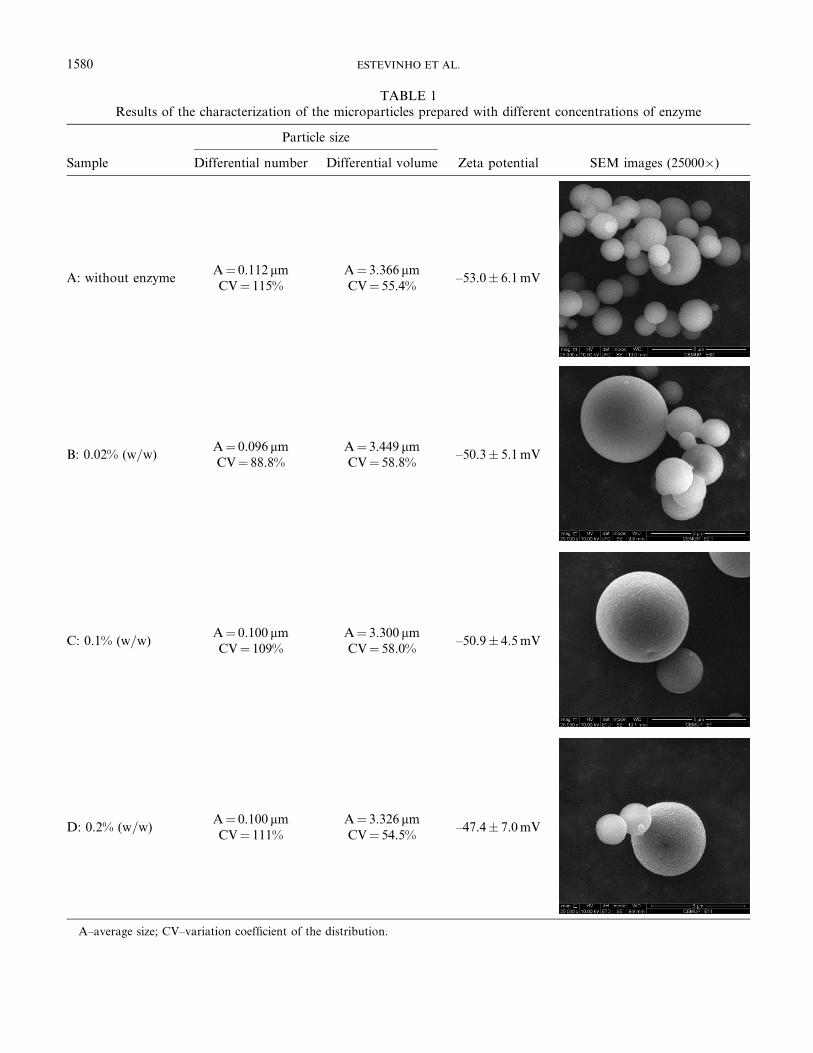
TABLE 1Results of the characterization of the microparticles prepared with different concentrations of enzyme
Sample
Particle size
Zeta potential SEM images (25000�)Differential number Differential volume
A: without enzymeA¼ 0.112 mmCV¼ 115%
A¼ 3.366 mmCV¼ 55.4%
–53.0� 6.1mV
B: 0.02% (w=w)A¼ 0.096 mmCV¼ 88.8%
A¼ 3.449 mmCV¼ 58.8%
–50.3� 5.1mV
C: 0.1% (w=w)A¼ 0.100 mmCV¼ 109%
A¼ 3.300 mmCV¼ 58.0%
–50.9� 4.5mV
D: 0.2% (w=w)A¼ 0.100 mmCV¼ 111%
A¼ 3.326 mmCV¼ 54.5%
–47.4� 7.0mV
A–average size; CV–variation coefficient of the distribution.
1580 ESTEVINHO ET AL.

that for the free enzyme, only presenting an equal or biggerloss of activity at pH 8. The enzyme microencapsulated withconcentration of 0.1% w=w presented a decrease of theactivity similar to the free enzyme for pH values between6.4 and 7.2 and higher losses for pH 7.8 and 8.
b-Galactosidase has two active-site carboxyl groups thatcan exist as –COO– (as nucleophile) and –COOH (asproton donor) simultaneously at neutral pH.[48] The shiftof the optimum pH to a basic pH may depend on thecharge of the enzyme and of the immobilizing agentsurface. Similar results, with an alteration of the pH rangeafter enzyme immobilization, have been reported by Zhouand Chen.[48]
However, the chosen pH was 7.7, since it minimizes theexperimental errors during the measurements of ONPconcentration. The enzyme activity decreases but themagnitude of the variation range of absorbance is higherfor pH 7.7. Thus, the errors involved in the determinationof the activity are smaller.
The ratio of the activity of immobilized enzyme to theactivity of the free enzyme at pH 7.7 and with a substrateconcentration of 0.225mM was 0.138, 0.115, 0.160, forenzymemicroencapsulated 0.02, 0.1, and 0.2% w=w, respect-ively. This confirms the observations of other authors.[48,49]
Carrara and Rubiolo[50] obtained activity values of theimmobilized enzyme in chitosan of 10.7% of the free enzyme
FIG. 3. Thermogram of the microparticles prepared with the composition: assay A, no enzyme; assay B, 0.02% (w=w) enzyme; assay C, 0.1% (w=w)
enzyme; and assay D, 0.2% (w=w) enzyme. Temperature raised from 25 to 550�C at a heating rate of 10�C per min.
FIG. 2. FTIR spectra of the microparticles prepared with the composition: assay A, no enzyme; assay B, 0.02% (w=w) enzyme; assay C, 0.1% (w=w)
enzyme; and assay D, 0.2% (w=w) enzyme. Spectra obtained with KBr at 99%, at 21 scans=min, with a resolution of 4 cm�1 and expressed in transmit-
tance in the 4000–650 cm�1 range.
b-GALACTOSIDASE MICROENCAPSULATION WITH A MODIFIED CHITOSAN 1581

values. Branchu et al.[44] also report a loss of catalyticactivity of the b-galactosidase enzyme spray dried whencompared with the commercial one. It should be noted thatthe microencapsulated enzyme activity is also influenced bymechanisms of enzyme release through the microparticleand, eventually, by conformational changes.[51,52]
Determination of Kinetic Parameters
The evolution of the enzymatic reaction (free enzyme)for different substrate concentrations is presented in Fig. 6for an enzyme concentration of 0.001mg=mL. For eachb-galactosidase reaction curve, the initial velocity was cal-culated, and a nonlinear regression method was performed
FIG. 5. Effect of the pH on the relative activity of free and microencapsulated b-galactosidase (with a substrate concentration of 0.225mM and an
enzyme concentration in solution of 0.001mg=mL).
FIG. 4. Evolution of the enzymatic reaction with time for free and microencapsulated enzyme at different pHs (6.4, 7.2, 7.7, 8.0). The enzymatic
reaction was studied for substrate and enzyme concentration of 0.225mM and 0.001mg=mL, respectively, based on absorbance values, by UV-visible
spectrophotometry at 420 nm and at room temperature.
1582 ESTEVINHO ET AL.
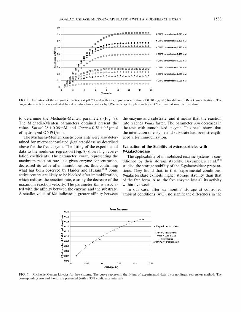
to determine the Michaelis-Menten parameters (Fig. 7).The Michaelis-Menten parameters obtained present thevalues Km¼ 0.28� 0.06mM and Vmax¼ 0.38� 0.5 mmolof hydrolyzed ONPG=min.
The Michaelis-Menten kinetic constants were also deter-mined for microencapsulated b-galactosidase as describedabove for the free enzyme. The fitting of the experimentaldata to the nonlinear regression (Fig. 8) shows high corre-lation coefficients. The parameter Vmax, representing themaximum reaction rate at a given enzyme concentration,decreased its value after immobilization, thus confirmingwhat has been observed by Haider and Husain.[53] Someactive centers are likely to be blocked after immobilization,which reduces the reaction rate, causing the decrease of themaximum reaction velocity. The parameter Km is associa-ted with the affinity between the enzyme and the substrate.A smaller value of Km indicates a greater affinity between
the enzyme and substrate, and it means that the reactionrate reaches Vmax faster. The parameter Km decreases inthe tests with immobilized enzyme. This result shows thatthe interaction of enzyme and substrate had been strength-ened after immobilization.
Evaluation of the Stability of Microparticles withb-Galactosidase
The applicability of immobilized enzyme systems is con-ditioned by their storage stability. Bayramoglu et al.[54]
studied the storage stability of the b-galactosidase prepara-tions. They found that, in their experimental conditions,b-galactosidase exhibits higher storage stability than thatof the free form. Also, the free enzyme lost all its activitywithin five weeks.
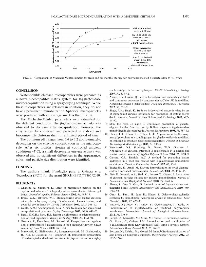
In our case, after six months’ storage at controlledambient conditions (4�C), no significant differences in the
FIG. 6. Evolution of the enzymatic reaction (at pH 7.7 and with an enzyme concentration of 0.001mg=mL) for different ONPG concentrations. The
enzymatic reaction was evaluated based on absorbance values by UV-visible spectrophotometry at 420 nm and at room temperature.
FIG. 7. Michaelis-Menten kinetics for free enzyme. The curve represents the fitting of experimental data by a nonlinear regression method. The
corresponding Km and Vmax are presented (with a 95% confidence interval).
b-GALACTOSIDASE MICROENCAPSULATION WITH A MODIFIED CHITOSAN 1583

appearance, color, and particle size distribution of all theprepared formulations were observed. The formulation ofmicroencapsulated b-galactosidase 0.1% (w=w) was selectedto reassess the stability after six months (Fig. 9). There wasa small decrease in the enzyme activity. The parameter Kmpresents a small increase and Vmax decreased from 0.07 to0.05 mmol of hydrolyzed ONPG=min.
Dwevedi and Kayastha[55] microencapsulatedb-galactosidase (from Pisum sativum) (PsBGAL) withchitosan and concluded that chitosan-PsBGAL hasdesirable properties like good stability, reusability, broadtemperature, and pH optima. These authors also reporteda loss of about 50% in activity of chitosan-PsBGAL,observed after 46 days at 4�C.
FIG. 8. Michaelis-Menten kinetics for microencapsulated b-galactosidase. The microparticles were prepared with different compositions: 0.02% (w=w)
enzyme, 0.1% (w=w) enzyme, and 0.2% (w=w) enzyme. The curves represent the fitting of experimental data by a nonlinear regression method. The
corresponding Km and Vmax are presented (with a 95% confidence interval).
1584 ESTEVINHO ET AL.
CONCLUSION
Water-soluble chitosan microparticles were prepared asa novel biocompatible matrix system for b-galactosidasemicroencapsulation using a spray-drying technique. Whilethese microparticles are released in solution, they do nothave a permanent immobilization. Spherical microparticleswere produced with an average size less than 3.5 mm.
The Michaelis-Menten parameters were estimated forthe different conditions. The b-galactosidase activity wasobserved to decrease after encapsulation; however, theenzyme can be conserved and protected in a dried andbiocompatible chitosan shell for a limited period of time.
The optimum pH ranges from 6.4 to 7.2 approximately,depending on the enzyme concentration in the microcap-sule. After six months’ storage at controlled ambientconditions (4�C), a small decrease in enzyme activity wasobserved and no significant differences in the appearance,color, and particle size distribution were identified.
FUNDING
The authors thank Fundacao para a Ciencia e aTecnologia (FCT) for the grant SFRH=BPD=73865=2010.
REFERENCES
1. Ghanem, A.; Skonberg, D. Effect of preparation method on the
capture and release of biologically active molecules in chitosan gel
beads. Journal of Applied Polymer Science 2002, 84, 405–13.
2. Braga, G.K.; Oliveira, W.P. Manufacturing drug loaded chitosan
microspheres by spray drying: Development, characterization, and
potential use in dentistry. Drying Technology 2007, 25(2), 303–10.
3. Goula, A.M.; Adamopoulos, K.G. A new technique for spray-dried
encapsulation of lycopene. Drying Technology 2012, 30(6), 641–52.
4. Desai, K.G.H.; Park, H.J. Recent developments in microencapsula-
tion of food ingredients. Drying Technology 2005, 23, 1361–94.
5. Grosova, Z.; Rosenberg, M.; Rebro�ss, M. Perspectives and applica-
tions of immobilised b-galactosidase in food industry: A review. Czech
Journal of Food Science 2008, 26, 1–14.
6. Makowski, K.; Białkowska, A.; Szczesna-Antczak, M.; Kalinowska,
H.; Kur, J.; Cieslinski, H.; Turkiewicz, M. Immobilized preparation
of cold-adapted and halotolerant Antarctic b-galactosidase as a highly
stable catalyst in lactose hydrolysis. FEMS Microbiology Ecology
2007, 59, 535–42.
7. Ansari, S.A.; Husain, Q. Lactose hydrolysis from milk=whey in batch
and continuous processes by concanavalin A-Celite 545 immobilized
Aspergillus oryzae b galactosidase. Food and Bioproducts Processing
2012, 90, 351–9.
8. Singh, A.K.; Singh, K. Study on hydrolysis of lactose in whey by use
of immobilized enzyme technology for production of instant energy
drink. Advance Journal of Food Science and Technology 2012, 4(2),
84–90.
9. Shin, H.; Park, J.; Yang, J. Continuous production of galacto-
oligosaccharides from lactose by Bullera singularis b-galactosidaseimmobilized in chitosan beads. Process Biochemistry 1998, 33, 787–92.
10. Cheng, T.-C.; Duan, K.-J.; Sheu, D.-C. Application of tris(hydroxy-
methyl)phosphine as a coupling agent for b-galactosidase immobilized
on chitosan to produce galactooligosaccharides. Journal of Chemical
Technology & Biotechnology 2006, 81, 233–6.
11. Wentworth, D.S.; Skonberg, D.; Darrel, W.D.; Ghanem, A.
Application of chitosan-entrapped b-galactosidase in a packed-bed
reactor system. Journal of Applied Polymer Science 2004, 91, 1294–9.
12. Carrara, C.R.; Rubiolo, A.C. A method for evaluating lactose
hydrolysis in a fixed bed reactor with b-galactosidase immobilized
on chitosan. Chemical Engineering Journal 1997, 65, 93–8.
13. Taqieddin, E.; Amiji, M. Enzyme immobilization in novel alginate–
chitosan core-shell microcapsules. Biomaterials 2004, 25, 1937–45.
14. Biro, E.; Nemeth, A.S.; Sisak, C.; Feczko, T.; Gyenis, J. Preparation
of chitosan particles suitable for enzyme immobilization. Journal of
Biochemical and Biophysical Methods 2008, 70, 1240–6.
15. Zhang, S.; Gao, S.; Gao, G. Immobilization of b-galactosidase onto
magnetic beads. Applied Biochemistry and Biotechnology 2010, 160,
1386–93.
16. Gaur, R.; Pant, H.; Jain, R.; Khare, S. Galacto-oligosaccharide
synthesis by immobilized Aspergillus oryzae b-galactosidase. Food
Chemistry 2006, 97, 426–30.
17. Vasileva, N.; Iotov, V.; Ivanov, Y.; Godjevargova, T.; Kotia, N.
Immobilization of b-galactosidase on modified polypropilene
membranes. International Journal of Biological Macromolecules
2012, 51, 710–9.
18. Bernal, C.; Marciello, M.; Mesa, M.; Sierra, L.; Fernandez-Lorente,
G.; Mateo, C.; Guisan, J.M. Immobilisation and stabilisation of
b-galactosidase from Kluyveromyces lactis using a glyoxyl support.
International Dairy Journal 2013, 28, 76–82.
19. Bortone, N.; Fidaleo, M.; Moresi, M. Immobilization=stabilization of
acid urease on Eupergit V supports. Biotechnology Progress 2012, 28,
1232–1244.
FIG. 9. Comparison of Michaelis-Menten kinetics for fresh and six months’ storage for microencapsulated b-galactosidase 0.1% (w=w).
b-GALACTOSIDASE MICROENCAPSULATION WITH A MODIFIED CHITOSAN 1585

20. Verma, M.L.; Barrow, C.J.; Kennedy, J.F.; Puri, M. Immobilization
of b-d-galactosidase from Kluyveromyces lactis on functionalized
silicon dioxide nanoparticles: Characterization and lactose hydrolysis.
International Journal of Biological Macromolecules 2012, 50, 432–7.
21. Singh, N.; Kayastha, A.M. Cicer a-galactosidase immobilization onto
chitosan and Amberlite MB-150: Optimization, characterization, and
its applications. Carbohydrate Research 2012, 358, 61–6.
22. Tavares, A.P.M.; Rodrıguez, O.; Fernandez-Fernandez, M.;
Domınguez, A.; Moldes, D.; Sanroman, M.A.; Macedo, E.A. Immobi-
lization of laccase onmodified silica: Stabilization, thermal inactivation
and kinetic behaviour in 1-ethyl-3-methylimidazolium ethylsulfate
ionic liquid. Bioresource Technology 2013, 131, 405–12.
23. Oliveira, B.F.; Santana, M.H.A.; Re, M.I. Spray-dried chitosan
microspheres as a pDNA carrier. Drying Technology 2006, 24(3),
373–82.
24. Liu, Y.; Sun, Y.; Li, Y.; Xu, S.; Tang, J.; Ding, J.; Xu, Y. Preparation
and characterization of a-galactosidase-loaded chitosan nanoparticles
for use in foods. Carbohydrate Polymers 2011, 83, 1162–8.
25. Kumar, M.N.V.R. A review of chitin and chitosan applications.
Reactive and Functional Polymers 2000, 46, 1–27.
26. Guliyeva, U.; Oner, F.; Ozsoy, S.; Haziroglu, R. Chitosan micro-
particles containing plasmid DNA as potential oral gene delivery
system. European Journal of Pharmaceutics and Biopharmaceutics
2006, 62, 17–25.
27. Aranaz, I.;Mengıbar,M.; Harris, R.; Panos, I.;Miralles, B.; Acosta, N.;
Galed, G.; Heras, A. Functional characterization of chitin and chitosan.
Current Chemical Biology 2009, 3, 203–30.
28. Fernandez-Pan, I.; Ziani, K.; Pedroza-Islas, R.; Mate, J.I. Effect of
drying conditions on the mechanical and barrier properties of films
based on chitosan. Drying Technology 2010, 28(12), 1350–8.
29. Sashiwa, H.; Kawasaki, N.; Nakayama, A. Chemical modification
of chitosan. 14:1. Synthesis of water-soluble chitosan derivatives by
simple acetylation. Biomacromolecules 2002, 3, 1126–8.
30. Zhang, H.; Wu, S.; Tao, Y.; Zang, L.; Su, Z. Preparation and
characterization of water-soluble chitosan nanoparticles as protein
delivery system. Journal of Nanomaterials 2010, 1–5.
31. Choi, W.; Ahn, K.; Lee, D.; Byun, M.; Park, H. Preparation of
chitosanoligomers by irradiation. Polymer Degradation and Stability
2002, 78, 533–8.
32. Estevinho, B.N.; Rocha, F.; Santos, L.; Alves, A. Using water soluble
chitosan for flavour microencapsulation in food industry. Journal of
Microencapsulation 2013, 30, 571–9.
33. Namaldi, A.; Calik, P.; Uludag, Y. Effects of spray drying tempera-
ture and additives on the stability of serine alkaline protease powders.
Drying Technology 2006, 24, 1495–500.
34. Kurozawa, L.E.; Park, K.J.; Hubinger, M.D. Effect of carrier agents
on the physicochemical properties of a spray dried chicken meat
protein hydrolysate. Journal of Food Engineering 2009, 94, 326–33.
35. Cakaloz, T.; Akbaba, H.; Yesugey, E.T.; Periz, A. Drying model for
a-amylase in a horizontal spray dryer. Journal of Food Engineering
1997, 31, 499–510.
36. Sloth, J.; Bach, P.; Jensen, A.D.; Kiil, S. Evaluation method for the
drying performance of enzyme containing formulations. Biochemical
Engineering Journal 2008, 40, 121–9.
37. Tonon, R.V.; Pedro, R.B.; Grosso, C.R.F.; Hubinger, M.D. Microen-
capsulation of flaxseed oil by spray drying: Effect of oil load and type
of wall material. Drying Technology 2012, 30(13), 1491–501.
38. Liu, C.; Desai, K.G.; Tang, X.; Chen, X. Drug release kinetics of
spray-dried chitosan microspheres. Drying Technology 2006, 24(6),
769–76.
39. Switzer, R.; Garrity, L.; Experimental Biochemistry, 3rd Ed.;
Freeman: New York, 1999.
40. Johnson, K.A.; Goody, R.S. The original Michaelis constant:
Translation of the 1913 Michaelis-Menten paper. Biochemistry 2011,
50, 8264–9.
41. Estevinho, B.N.; Damas, A.M.; Martins, P.; Rocha, F. Study of the
inhibition effect on the microencapsulated enzyme b-galactosidase.Environmental Engineering andManagement Journal 2012, 11, 1923–30.
42. Samborska, K.; Witrowa-Rajchert, D.; Goncalves, A. Spray-drying of
a-amylase: The effect of process variables on the enzyme inactivation.
Drying Technology 2005, 23, 941–53.
43. Jiang, H.; Zhang, M.; McKnight, S.; Adhikari, B. Microencapsulation
of a-amylase by carrying out complex coacervation and drying in
a single step using a novel three-fluid nozzle spray drying. Drying
Technology 2013, 31(16), 1901–10.
44. Branchu, S.; Forbes, R.; York, P.; Petren, S., Nyqvist, H., Camber, O.
Hydroxypropyl-b-cyclodextrin inhibits spray-drying-induced inacti-
vation of b-galactosidase. Journal of Pharmaceutical Sciences 1999,
88, 907–11.
45. Kishore, D.; Kayastha, A.M. Optimisation of immobilisation
conditions for chick pea b-galactosidase (CpGAL) to alkylamine glass
using response surface methodology and its applications in lactose
hydrolysis. Food Chemistry 2012, 134, 1650–7.
46. Kishore, D.; Kundu, S.; Kayastha, A.M. Thermal, chemical and pH
induced denaturation of a multimeric b-galactosidase reveals multiple
unfolding pathways. PloS One 2012, 7, e50380.
47. Jurado, E.; Camacho, F.; Luzon, G.; Vicaria, J.M. Kinetic models of
activity for b-galactosidases: Influence of pH, ionic concentration and
temperature. Enzyme and Microbial Technology 2004, 34, 33–40.
48. Zhou, Q.Z.K.; Chen, X.D. Effects of temperature and pH on the cata-
lytic activity of the immobilized b-galactosidase from Kluyveromyces
lactis. Biochemical Engineering Journal 2001, 9, 33–40.
49. Tanriseven, A.; Dogan, S. A novel method for the immobilization of
b-galactosidase. Process Biochemistry 2002, 38, 27–30.
50. Carrara, C.R.; Rubiolo, A.C. Immobilization of b-galactosidase on
chitosan. Biotechnology Progress 1994, 10, 220–4.
51. Gonzalez Siso, M.I.; Lang, E.; Carreno-Gomez, B.; Becerra, M.;
Otero Espinar, F.; Blanco Mendez, J. Enzyme encapsulation on
chitosan microbeads. Process Biochemistry 1997, 32, 211–6.
52. Haider, T.; Husain, Q. Concanavalin A layered calcium alginate-
starch beads immobilized b-galactosidase as a therapeutic agent for
lactose intolerant patients. International Journal of Pharmaceutics
2008, 359, 1–6.
53. Haider, T.; Husain, Q. Hydrolysis of milk=whey lactose by b galacto-
sidase: A comparative study of stirred batch process and packed bed
reactor prepared with calcium alginate entrapped enzyme. Chemical
Engineering and Processing 2009, 48, 576–80.
54. Bayramoglu, G.; Tunali, Y.; Arica, M.Y. Immobilization of
b-galactosidase ontomagnetic poly(GMA–MMA) beads for hydrolysis
of lactose in bed reactor. Catalysis Communications 2007, 8, 1094–101.
55. Dwevedi, A.; Kayastha, A.M. Optimal immobilization of beta-
galactosidase from Pea (PsBGAL) onto Sephadex and chitosan beads
using response surface methodology and its applications. Bioresource
Technology 2009, 100, 2667–75.
1586 ESTEVINHO ET AL.