gene expression profile testing of cancer tissue expression profile testing of cancer tissue draft...
TRANSCRIPT

Gene expression profile testing of cancer tissue
Draft evidence report
January 3, 2018
Health Technology Assessment Program (HTA)
Washington State Health Care Authority
PO Box 42712
Olympia, WA 98504-2712
(360) 725-5126
www.hca.wa.gov/hta


Gene Expression Profile Testing of Cancer Tissue
Draft Evidence Report
January 3, 2018
Prepared by:
Center for Evidence-based Policy
Oregon Health & Science University
3030 SW Moody, Suite 250
Portland, OR 97201
Phone: 503.494.2182
Fax: 503.494.3807
http://centerforevidencebasedpolicy.org/
Authors:
Valerie King, MD, MPH, Craig Mosbaek, MPH, Susan Carson, MPH, Brittany Lazur, MPH, Allison
Leof, PhD, Robyn Liu, MD, MPH, FAAFP
The authors would like to acknowledge the contributions of the following:
Galen Gamble, BA, Andrew Hamilton, MLS, MS, Joan Holup, MA, Anitra Ingham, MFA, Chris
Kelleher, BA, Adam Obley, MD, and the independent peer reviewers of this report

This health technology assessment report is based on research conducted by the Center for
Evidence-based Policy under contract to the Washington State Health Care Authority (HCA). This
report is an independent assessment of the technology question(s) described based on
accepted methodological principles. The findings and conclusions contained herein are those of
the authors, who are responsible for the content. These findings and conclusions do not
necessarily represent the views of the Washington HCA and thus, no statement in this report
shall be construed as an official position or policy of the HCA.
The information in this assessment is intended to assist health care decision makers, clinicians,
patients, and policy makers in making evidence-based decisions that may improve the quality
and cost-effectiveness of health care services. Information in this report is not a substitute for
sound clinical judgment. Those making decisions regarding the provision of health care services
should consider this report in a manner similar to any other medical reference, integrating the
information with all other pertinent information to make decisions within the context of
individual patient circumstances and resource availability.
About the Center for Evidence-based Policy
The Center for Evidence-based Policy (Center) is recognized as a national leader in evidence-
based decision making and policy design. The Center understands the needs of policymakers
and supports public organizations by providing reliable information to guide decisions,
maximize existing resources, improve health outcomes, and reduce unnecessary costs. The
Center specializes in ensuring that diverse and relevant perspectives are considered and
appropriate resources are leveraged to strategically address complex policy issues with high-
quality evidence and collaboration. The Center is based at Oregon Health & Science University
in Portland, Oregon.
Conflict of Interest Disclosures: No authors have conflicts of interest to disclose. All authors have
completed and submitted the Oregon Health & Science University form for Disclosure of
Potential Conflicts of Interest, and none were reported.

WA – Health Technology Assessment January 3, 2018
Gene expression profile testing for cancer tissue: draft report Page i
Table of Contents
List of Tables ..................................................................................................................................................................... ii
List of Figures ................................................................................................................................................................... iii
List of Abbreviations ..................................................................................................................................................... iii
Executive Summary ........................................................................................................................................................ 1
Structured Abstract .................................................................................................................................................... 1
Background ................................................................................................................................................................... 4
Methods ......................................................................................................................................................................... 5
Results ............................................................................................................................................................................. 9
Clinical Practice Guidelines .................................................................................................................................. 21
Selected Payer Coverage Determinations ...................................................................................................... 24
Conclusions ................................................................................................................................................................ 26
Technical Report ........................................................................................................................................................... 28
Background ................................................................................................................................................................ 28
Technology Description ........................................................................................................................................ 28
Policy Context ........................................................................................................................................................... 35
Washington State Utilization and Cost Data ................................................................................................. 36
Methods ...................................................................................................................................................................... 37
Search Results ........................................................................................................................................................... 43
Evidence Summary .................................................................................................................................................. 48
Clinical Practice Guidelines .................................................................................................................................. 68
Selected Payer Coverage Determinations ...................................................................................................... 72
Conclusions ................................................................................................................................................................ 78
References....................................................................................................................................................................... 80
Appendix A. Search Strategy ................................................................................................................................... 93
Appendix B. Additional Methods ......................................................................................................................... 100
Appendix C. Evidence Tables ................................................................................................................................. 109
Appendix D. Risk of Bias Assessments ............................................................................................................... 169
Appendix E. GRADE Profile Ratings ..................................................................................................................... 176
Appendix F. Studies Registered at ClinicalTrials.gov .................................................................................... 184
Appendix G. Detailed Clinical Practice Guidelines ......................................................................................... 190
Appendix H. See Attachment for Excluded Studies ...................................................................................... 193

WA – Health Technology Assessment January 3, 2018
Gene expression profile testing for cancer tissue: draft report Page ii
List of Tables
Table 1. CPT Codes and Descriptions for Gene Expression Profile Tests ................................................ 36
Table 2. PEBB/UMP (No Medicare) Claims for Gene Expression Profile Testing: 2013-2016 .......... 37
Table 3. Study Inclusion and Exclusion Criteria ................................................................................................ 39
Table 4. Recommendations for Lymph Node Status in Guidelines on the Use of Gene Expression
Tests in Early State Breast Cancer .......................................................................................................................... 70
Table 5. Evidence Table: Breast Cancer Systematic Reviews ...................................................................... 110
Table 6. Evidence Table: Breast Cancer Randomized Controlled Trials ................................................. 114
Table 7. Evidence Table: Breast Cancer Observational Studies ................................................................. 118
Table 8. Evidence Table: Breast Cancer Economic Studies ......................................................................... 144
Table 9. Evidence Table: Prostate Cancer Observational Studies ............................................................ 147
Table 10. Evidence Table: Prostate Cancer Economic Studies .................................................................. 159
Table 11. Evidence Table: Colon Cancer Observational Studies ............................................................... 163
Table 12. Evidence Table: Colon Cancer Economic Studies ....................................................................... 168
Table 13. Risk of Bias: Breast Cancer Systematic Reviews and Meta-analyses ................................... 169
Table 14. Risk of Bias: Breast Cancer Randomized Controlled Trial ........................................................ 169
Table 15. Risk of Bias: Breast Cancer Observational Studies ..................................................................... 170
Table 16. Risk of Bias: Breast Cancer Economic Studies .............................................................................. 171
Table 17. Risk of Bias: Breast Cancer Guidelines ............................................................................................ 172
Table 18. Risk of Bias: Prostate Cancer Observational Studies ................................................................. 172
Table 19. Risk of Bias: Prostate Cancer Economic Studies .......................................................................... 173
Table 20. Risk of Bias: Prostate Cancer Guidelines ........................................................................................ 174
Table 21. Risk of Bias: Colon Cancer Observational Studies ...................................................................... 174
Table 22. Risk of Bias: Colon Cancer Economic Studies .............................................................................. 175
Table 23. Risk of Bias: Colon Cancer Guidelines ............................................................................................. 175
Table 24. Risk of Bias: Multiple Myeloma Guidelines ................................................................................... 175
Table 25. GRADE Quality of Evidence for Breast Cancer ............................................................................. 176
Table 26. GRADE Quality of Evidence for Prostate Cancer ......................................................................... 179
Table 27. GRADE Quality of Evidence for Colon Cancer ............................................................................. 181
Table 28. GRADE Quality of Evidence for Multiple Myeloma .................................................................... 183

WA – Health Technology Assessment January 3, 2018
Gene expression profile testing for cancer tissue: draft report Page iii
List of Figures
Figure 1. Analytic Framework .................................................................................................................................. 39
Figure 2. Study Flow Diagram: Breast Cancer.................................................................................................... 44
Figure 3. Study Flow Diagram: Prostate Cancer ............................................................................................... 45
Figure 4. Study Flow Diagram: Colon Cancer .................................................................................................... 46
Figure 5. Study Flow Diagram: Multiple Myeloma .......................................................................................... 47
List of Abbreviations
aOR adjusted odds ratio
ASCO American Society of Clinical Oncology
BCI Breast Cancer Index
CI confidence interval
ER estrogen receptor
HER2 human epidermal growth factor receptor 2
HR hazards ratio
HTA health technology assessment
LN lymph node
NCCN National Comprehensive Cancer Network
NR not reported
PR progesterone receptor
RCT randomized controlled trial
RR risk ratio
PSA prostate-specific antigen
U.S. United States

WA – Health Technology Assessment January 3, 2018
Gene expression profile testing for cancer tissue: draft report Page 1
Executive Summary
Structured Abstract
Purpose
The purpose of this evidence report is to review the clinical utility and cost-effectiveness of
selected gene expression profile tests used to guide treatment decisions for breast, prostate,
and colon cancers and multiple myeloma. There are a growing number of gene expression
profile tests for cancers designed to help inform treatment after diagnosis. The potential
benefits of these tests could involve more appropriate treatment decisions and better patient
outcomes, including avoiding treatment-related side effects by forgoing unnecessary
treatments. This evidence review will help to inform Washington’s independent Health
Technology Clinical Committee as it determines coverage regarding the use of the Oncotype
DX, MammaPrint, Prosigna, Endopredict, Breast Cancer Index (BCI) and Mammostrat tests for
early invasive breast cancer; the Decipher, Prolaris and Oncotype DX tests for prostate cancer;
the ColoPrint and Oncotype DX tests for stage 2 or 3 colon cancers; and the Myeloma
Prognostic Risk Signature (MyPRS) and SKY92 tests for multiple myeloma.
Data Sources
Searches of OVID Medline, the Cochrane Database of Systematic Reviews, and the Cochrane
Central Register of Controlled Trials were conducted for English-language studies published
from January 2007 to the present (end search dates for the draft report were in early November
2017). Searches were also conducted for eligible health technology assessment (HTA) and
evidence reviews from the Agency for Healthcare Research and Quality (AHRQ), the Blue
Cross/Blue Shield HTA program, the U.K. National Institute for Health and Care Excellence, and
the Veterans Administration Evidence-based Synthesis Program. Studies were also identified
from the reference lists of included studies, test manufacturer websites, and a dossier that had
been submitted to the Washington State Agency Medical Directors’ Group in December 2016
for one of the tests. Center researchers also searched for ongoing and recently completed
registered trials using the ClinicalTrials.gov database and the AHRQ National Guideline
Clearinghouse for clinical practice guidelines published in the past five years. The Centers for
Medicare & Medicaid Services (CMS) website for the Medicare Coverage Database was searched
for NCDs and LCDs applying to the state of Washington. The Aetna, Cigna, and Regence
websites were searched for coverage policies for these private payers.
Study Selection
Two independent Center researchers screened all titles and abstracts for potential inclusion
based on pre-specified inclusion and exclusion criteria. Eligible study designs for Key Questions
(KQ) 1, 2 and 3 were systematic reviews (with and without meta-analysis), randomized controlled
trials (RCTs), and comparative observational studies that reported clinical outcomes, including
survival, decision impact, quality of life, and harms. These study designs were also eligible for KQ
4 with the addition of economic modeling studies for cost-effectiveness. Dual full-text review for

WA – Health Technology Assessment January 3, 2018
Gene expression profile testing for cancer tissue: draft report Page 2
inclusion criteria was done. In instances of disagreement, an independent third screener
resolved disputes.
Data Extraction
Using standardized processes and forms, one researcher extracted data and a second one
checked the extraction for accuracy. Two researchers independently assessed the risk of bias of
included studies and clinical practice guidelines. A rating of high, moderate, or low risk of bias
was assigned to each study or review based on adherence to recommended methods and
potential for internal and external validity biases.
Data Synthesis
Database searches and other sources yielded a total of 2,949 citations, including 2,005 for breast
cancer, 266 for prostate cancer, 431 for colon cancer, and 247 for multiple myeloma. A total of
35 studies met inclusion criteria with 22 for breast cancer, 10 for prostate cancer, 3 for colon
cancer, and no studies for multiple myeloma. Researchers applied the Grading of
Recommendations, Assessment, Development, and Evaluation Working Group (GRADE) system
to rate the overall quality of evidence on key outcomes for each genomic test. There was no
high-quality evidence of clinical utility to guide decisions about the use of gene expression
profile tests for breast, prostate, and colon cancers. There is moderate quality evidence that
women with early invasive breast cancer who are at high clinical risk based on the Adjuvant!
Online risk assessment tool may safely forego adjuvant systemic chemotherapy if their
MammaPrint risk score is low. Moderate quality of evidence supports the use of Oncotype DX
because of its impact on clinical treatment recommendations and chemotherapy use for women
with early invasive breast cancer (particularly its ability to identify low-risk women who would
not benefit from adjuvant systemic chemotherapy). There is low quality evidence about the
decision impact of using MammaPrint and low quality evidence that both Oncotype DX and
MammaPrint are cost-effective at conventional thresholds when used to guide treatment
decisions among women with early invasive breast cancer. Among the remaining conditions and
tests there is a mix of very low quality or an absence of evidence to support use of these tests to
improve clinical decision-making and important patient outcomes.
Limitations
The risk of bias of included studies varied, but was often high. The evidence base was very
limited for assessing the clinical utility, harms and cost-effectiveness of most of the tests.
Populations included in studies were generally not diverse in terms of race, ethnicity, and
socioeconomic factors. Although a large number of the studies were conducted in the U.S.,
many were also conducted in Europe which may limit generalizability to the U.S. context. Given
limited evidence regarding effectiveness, economic modeling studies did not have adequate
quality estimates of effectiveness to include in these modeling exercises. Economic studies also
used a variety of modeling techniques and other assumptions, making direct comparisons
among them difficult.

WA – Health Technology Assessment January 3, 2018
Gene expression profile testing for cancer tissue: draft report Page 3
Conclusions
There is moderate quality of evidence to support the use of MammaPrint (for clinically high risk
individuals based on the Adjuvant! Online assessment tool) and Oncotype DX breast cancer
assay for important outcomes related to early invasive breast cancer with negative or positive
lymph nodes. Based on limited economic modeling studies, these tests are likely supported at
current conventional economic thresholds for use. There is a mix of low, very low, and no quality
of evidence to support the other included tests across the conditions of prostate cancer, colon
cancer and multiple myeloma. There are multiple ongoing clinical trials on most of the tests that
will be reporting out in the next few years and will hopefully improve the evidence base for
decision making regarding the clinical usefulness and economic impacts of these tests.

WA – Health Technology Assessment January 3, 2018
Gene expression profile testing for cancer tissue: draft report Page 4
Background
The lifetime risk of developing cancer is about 40%, and one in every five Americans will die
from cancer.1 Strategies for reducing the burden of cancer include prevention, early diagnosis,
and appropriate treatments.2 Common treatments for cancer are surgery, chemotherapy,
radiation therapy, hormone therapy, and immunotherapy.3 The most appropriate treatments for
a particular cancer depend on the cancer’s characteristics (e.g., cancer stage and grade), the
patient’s age and health status, response to previous treatments, and other factors.4
In recent years, gene expression profile testing of cancer tissue has been used to help inform
decisions on appropriate treatments. Gene expression profile testing identifies the genes in a
cancer cell or tissue that are making messenger RNA (mRNA), which carry the genetic
information that cancer cells need to make proteins. Some gene expression profile tests are
designed to increase the accuracy of the prognosis for a patient with cancer. If a test predicts
that a cancer is slow growing or is unlikely to metastasize, then active surveillance of the cancer
could be the most appropriate course. If a test predicts that a cancer is likely to progress and
metastasize, then more aggressive treatments could be warranted.5 Other gene expression tests
may identify specific mutations for which there are targeted treatments (i.e., the Anaplastic
lymphoma kinase [ALK] mutation in lung cancer can be treated with a specific drug). However,
to have usefulness in clinical practice, a gene expression profile test must do more than predict
better or worse prognosis. Clinical utility means that a test has an impact on clinical decision
making or can help direct therapy in an actionable way to improve patient outcomes. Gene
expression profile tests have been developed for patients with breast cancer, prostate cancer,
colon cancer, and multiple myeloma, among others. This report examines the clinical utility and
cost-effectiveness of selected genomic tests for these four cancers.
Technology Description
This report reviews gene expression profile tests for breast, prostate, and colon cancers and
multiple myeloma. The six breast cancer tests are indicated for women with early stage, invasive
breast cancer: Oncotype DX breast cancer assay, MammaPrint, EndoPredict, Prosigna Breast
Cancer Prognostic Gene Signature Assay (PAM50), Breast Cancer Index (BCI), and Mammostrat.
These tests are used to predict the cancer’s aggressiveness and thus inform treatment decisions.
Decipher, Prolaris, and Oncotype DX prostate cancer assay are gene expression tests for
prostate cancer. The Decipher test is used after radical prostatectomy to predict the probability
of metastasis and inform clinical decisions on the use of adjuvant prostate cancer treatments.
The other two tests, Prolaris and Oncotype DX for prostate cancer, are used after an initial
diagnosis of prostate cancer to predict the cancer’s aggressiveness and thus inform treatment
decisions.
ColoPrint and Oncotype DX colon cancer assay are gene expression tests for colon cancer.
ColoPrint is performed using fresh or frozen tumor samples from stage 2 colon cancer patients

WA – Health Technology Assessment January 3, 2018
Gene expression profile testing for cancer tissue: draft report Page 5
who have undergone surgery. The Oncotype DX colon cancer assay is indicated for patients with
anatomic stage 2, mismatch repair proficient (MMR-P) and stage 3 A/B colon cancers.
My Prognostic Risk Signature (MyPRS) and SKY92 (formerly EMC92) are gene expression tests
for multiple myeloma. These tests are used to predict the risk of disease relapse and survival
outcomes in patients with multiple myeloma.
Molecular diagnostic tests are regulated by the U.S. Food and Drug Administration (FDA) and
CMS.6 The FDA has exercised discretion in its requirements for approval of in vitro diagnostic
assays.6 In vitro tests developed, validated, and performed in-house by a specific reference
laboratory are required to abide by the Clinical Laboratory Improvement Amendments (CLIA),
but FDA clearance and approval is currently not required for these laboratory-developed tests
(LDTs).6 Most of the tests described here are regulated as LDTs. Two of the breast cancer tests,
MammaPrint and Prosigna, have received FDA pre-market approval. SkylineDx plans to make
MMprofiler, based on the SKY92 test for multiple myeloma, available soon as a LDT in the U.S.7
MMprofiler is available in Europe, but is only available in the U.S. for research use.8
Policy Context
There are a growing number of gene expression profile tests for cancer tissue designed to
inform treatment decisions after cancer diagnoses. Potential benefits of these tests are more
appropriate treatment decisions, better patient outcomes, including avoiding treatment-related
side effects, and the potential cost and side-effect savings from avoiding unnecessary
treatments. This topic was selected for a health technology assessment because of medium
concerns for the safety of these tests, medium to high concerns for efficacy, and high concerns
for cost.
This evidence review will help to inform Washington’s independent Health Technology Clinical
Committee as the committee determines coverage regarding selected gene expression profile
tests for patients with eligible breast, prostate, or colon cancers or multiple myeloma.
Methods
This evidence review is based on the final key questions published on November 14, 2017.
Population: Adults with breast, prostate, or colon cancers or multiple myeloma
Interventions: Gene expression profile testing of cancer tissue to inform treatment decisions,
including the following tests by cancer type:
Breast Cancer—Oncotype DX breast cancer assay, MammaPrint, EndoPredict, Prosigna
Breast Cancer Prognostic Gene Signature Assay (PAM50), Breast Cancer Index (BCI),
Mammostrat
Prostate Cancer— Decipher, Prolaris, Oncotype DX prostate cancer assay
Colon Cancer—ColoPrint, Oncotype DX colon cancer assay

WA – Health Technology Assessment January 3, 2018
Gene expression profile testing for cancer tissue: draft report Page 6
Multiple Myeloma—Myeloma Prognostic Risk Signature (MyPRS), SKY92-signature
(formerly EMC92)
Comparators: Usual care without gene expression profile testing of cancer tissue, alternate
gene expression profile tests (i.e., one test intervention listed above versus another)
Outcomes:
Patient management decisions (including selection of active surveillance rather than
active treatment)
Clinical outcomes (e.g., morbidity, mortality, quality of life)
Harms, such as consequences of false-positive or false-negative test results
Cost-effectiveness and other economic outcomes
Time period for literature search: January 2007 to November 2017
Key Questions
1. Effectiveness: What is the clinical utility of gene expression profile testing of cancer tissue
to inform treatment decisions for patients with breast, prostate, and colon cancers and
multiple myeloma?
a. Is there evidence that test results affect treatment decisions?
b. Do treatment decisions guided by gene expression profile testing of cancer tissue
result in clinically meaningful improvements in patient outcomes?
2. Harms: What harms are associated with conducting gene expression profile testing of
cancer tissue?
3. Special populations: Compared with usual care, do treatment decisions, patient
outcomes, or harms after gene expression profile testing of cancer tissue vary by:
a. Patient demographics (e.g., age, sex, race/ethnicity)?
b. Clinical history (e.g., means of diagnosis, stage or grade of cancer, results of other
testing, previous treatments, chronicity)?
c. Medical comorbidities?
d. Provider type or care setting?
4. What are the cost-effectiveness and other economic outcomes of gene expression
profile testing used to inform treatment management decisions?
Eligible Studies
Randomized controlled trials (RCTs), nonrandomized comparative studies, and systematic
reviews (with and without a meta-analysis) of these two types of studies that assess clinical
utility were considered for Key Questions 1, 2, and 3. Cost-effectiveness studies and other
comparative economic evaluations, along with systematic reviews (with and without a meta-
analysis) of these types of studies, were considered for Key Question 4.

WA – Health Technology Assessment January 3, 2018
Gene expression profile testing for cancer tissue: draft report Page 7
Data Sources and Searches
A search of the peer-reviewed published literature was conducted using Ovid MEDLINE. RCTs,
nonrandomized comparative studies, and systematic reviews (with and without meta-analysis)
and health technology assessments of these studies that assess clinical utility were considered
for Key Questions 1, 2, and 3. Cost-effectiveness studies and other comparative economic
evaluations, along with systematic reviews (with and without meta-analysis) reporting economic
outcomes, were considered for Key Question 4. The following electronic databases were
searched to identify relevant peer-reviewed studies:
Medline
Cochrane Database of Systematic Reviews
Cochrane Central Register of Controlled Trials
The full Ovid MEDLINE search strategies for each of the indications (i.e., breast cancer, prostate
cancer, colon cancer, and multiple myeloma) are in Appendix A. Center researchers also
screened reference lists of relevant studies and used lateral search functions such as "related
articles" and "cited by." Citations from the Myriad Genetic Laboratories dossier for coverage of
EndoPredict, which was submitted to the Washington State Agency Medical Directors’ Group in
December 2016, were also considered for inclusion. In addition, these core sources were
searched:
Agency for Healthcare Research and Quality
Blue Cross/Blue Shield HTA program
National Institute for Health and Care Excellence – Evidence
Veterans Administration Evidence-based Synthesis Program
Center researchers scanned manufacturer websites and conducted a general Internet search for
appropriate published studies and relevant gray literature.
Two independent Center researchers screened titles and abstracts. For studies that could not be
excluded by title and abstract screening, a full-text review for inclusion criteria was performed. In
instances of disagreement, an independent third screener resolved disputes.
If Center researchers identified a high-quality systematic review (with or without meta-analysis)
addressing any of the key questions, a search was conducted to find eligible studies published
after the search dates of the systematic review. Center researchers excluded systematic reviews if
all of the included studies were also summarized by a more comprehensive systematic review, a
systematic review of a higher methodological quality, or a more recently published systematic
review.
Center researchers also searched the sources listed above for clinical practice guidelines. In
addition, searches of the Agency for Healthcare Research and Quality’s National Guideline
Clearinghouse (guidelines.gov) and websites of relevant professional organizations were
conducted. Guidelines published in the past five years were considered for inclusion.

WA – Health Technology Assessment January 3, 2018
Gene expression profile testing for cancer tissue: draft report Page 8
The CMS website for the Medicare Coverage Database was searched for NCDs and LCDs
applying to the state of Washington. The Aetna, Cigna, and Regence websites were searched for
coverage policies for these private payers.
Center researchers searched the online database of clinical trials (clinicaltrials.gov) maintained
by the National Library of Medicine at the National Institutes of Health. Information in this
database is provided by the sponsor or principal investigator of clinical studies. Studies are
generally registered in the database when they begin, with information updated as the study
progresses. The search included the names of the gene expression profile tests and other
common names for them: Oncotype breast (21-gene), MammaPrint (70-gene), EndoPredict (12-
gene), Prosigna (PAM50, 50-gene), Breast Cancer Index (BCI), Mammostrat, Decipher (22-gene),
Prolaris (46-gene), Oncotype prostate (17-gene), ColoPrint (18-gene), Oncotype colon (12-gene),
Myeloma Prognostic Risk Signature (MyPRS), Myeloma (MyPRS, 70-gene), and SKY92 (EMC92,
92-gene).
Data Abstraction and Quality Assessment
Standardized procedures were used to extract relevant data from each of the included studies
by one investigator and cross-checked for accuracy by at least one other investigator. Two
independent Center researchers evaluated studies for methodological risk of bias, and critical
outcomes disagreement among these assessments were settled by a third independent Center
researcher. Each study was assessed using Center instruments adapted from international
standards and assessments for methodological quality.9-14 A rating of high, moderate, or low risk
of bias was assigned to each study or review based on adherence to recommended methods
and potential for bias affecting internal and external biases. The risk of bias criteria for all the
study types are in Appendix B.
Center researchers assigned each outcome a summary judgment for the overall quality of
evidence based on the system developed by the Grading of Recommendations, Assessment,
Development, and Evaluation Working Group (GRADE).15,16 The GRADE system defines the
overall quality of a body of evidence for an outcome in the following manner:
High: Raters are very confident that the estimate of the effect of the intervention on the
outcome lies close to the true effect. Typical sets of studies are RCTs with few or no
limitations, and the estimate of effect is likely stable.
Moderate: Raters are moderately confident in the estimate of the effect of the
intervention on the outcome. The true effect is likely to be close to the estimate of the
effect, but there is a possibility that it is different. Typical sets of studies are RCTs with
some limitations or well-performed nonrandomized studies with additional strengths
that guard against potential bias and have large estimates of effects.
Low: Raters have little confidence in the estimate of the effect of the intervention on the
outcome. The true effect may be substantially different from the estimate of the effect.

WA – Health Technology Assessment January 3, 2018
Gene expression profile testing for cancer tissue: draft report Page 9
Typical sets of studies are RCTs with serious limitations or nonrandomized studies
without special strengths.
Very low: Raters have no confidence in the estimate of the effect of the intervention on
the outcome. The true effect is likely to be substantially different from the estimate of
effect. Typical sets of studies are nonrandomized studies with serious limitations or
inconsistent results across studies.
Not applicable: Researchers did not identify any eligible articles.
Results
Across all four cancer indications and 13 tests of interest, searches and other sources retrieved a
total of 2,949 citations and abstracts, including 2,005 for breast cancer, 266 for prostate cancer,
431 for colon cancer, and 247 for multiple myeloma. Ultimately, after applying predefined
exclusion criteria, there were a total of 35 studies that met inclusion criteria (breast cancer: 22
studies; prostate cancer: 10 studies; colon cancer: 3 studies; multiple myeloma; no studies).
KQ1: Clinical Utility
Only two included studies provided information about morbidity and mortality outcomes
related to tests used for breast cancer.17,18 There was no direct information and little indirect
information about quality of life related to use of these tests in clinical decision making. A few
studies for breast cancer, colon cancer and prostate cancer reported outcomes such as changes
in decisional conflict, confidence in the decision, or anxiety. Most of the evidence about clinical
utility outcomes was based on the test affecting treatment recommendations or decisions for
breast, prostate, and colon cancer. These results, along with any available information about
indirect quality of life findings are summarized in the following paragraphs, by cancer type. No
studies met inclusion criteria for multiple myeloma.
Breast Cancer
The majority of research evidence on clinical utility outcomes (22 of 35 studies) included in this
report pertains to the use of gene expression profile tests for early invasive breast cancer. These
tests have been available for the longest amount of time and simply have a larger research base.
However, even within this group of studies, the majority of evidence relates to the Oncotype DX
test (38 primary studies from three SRs and 10 additional studies), with a moderate amount of
research about MammaPrint (seven primary studies from two SRs and four additional studies), a
small amount of research on Prosigna (one primary study from a SR and two additional studies),
and very small research bases consisting of one study each for the Endopredict, BCI and
Mammostrat tests.
Systematic Reviews
A systematic review and meta-analysis by Augustovski and colleagues reported the global
pooled decision impact (defined as the proportion of patients whose treatment decision was
altered with use of the Oncotype DX test), and global pooled net chemotherapy change (defined

WA – Health Technology Assessment January 3, 2018
Gene expression profile testing for cancer tissue: draft report Page 10
as the difference in the number of patients who were assigned to receive chemotherapy before
versus after the test), from 15 primary studies of women with lymph node (LN)-negative early
invasive breast cancer.19 Among a group of studies that used universal rather than selective
enrollment, they reported a pooled decision impact of 28.97% (95% CI, 26.65% to 31.34%),
I2=0.00%, and a corresponding net chemotherapy change of 9.00% (95% CI, 4.00% to 14.00%),
I2=89.00%.19
The systematic review by Blok and colleagues also presented pooled estimates for the
proportion of patients who had a change in treatment recommendation to a more or less
intensive recommended treatment strategy (i.e., chemotherapy, endocrine therapy, or no
treatment).20 These pooled estimates combined studies of patients with LN-negative and LN-
positive tumors, although the majority of included studies were of patients with LN-negative
tumors.20 Four included studies, with 790 patients, used the MammaPrint test; 22 studies (n =
3,743) examined the Oncotype DX test; and one study each involved the use of the Prosigna (n
= 200) and EndoPredict (n = 167) tests.20 Blok and colleagues reported that lower proportions of
patients received recommendations for or treatments that were more invasive (MammaPrint:
-17%; Oncotype DX: -14.6%; Prosigna: -12.9%; and EndoPredict: -34%).20 Correspondingly, the
proportion of patients who were recommended to have less intensive treatment increased with
use of all the tests (MammaPrint: +32.2%; Oncotype DX: +51.1%; Prosigna: +37.3%; and
EndoPredict: +53.2%).20 This review did not provide any risk of bias assessment for the included
studies and did not perform any assessment of statistical heterogeneity across the groups of
studies or provide confidence intervals for the estimates.
The systematic review by Scope and colleagues did not present any pooled estimates because
the authors were concerned about heterogeneity among studies.21 In a narrative synthesis the
authors reported that the use of Oncotype DX led to changes in treatment recommendations for
21 to 74% of patients enrolled in these studies.21 Change from a recommendation of
chemotherapy to no chemotherapy ranged from 6% to 51% of patients after Oncotype DX use,
but in one study the proportion of patients who were recommended to receive chemotherapy
actually increased after use of the test.21 Similarly, the authors stated that the use of
MammaPrint led to treatment recommendation changes of 10% to 40% of patients, and that
between 2% and 32% of patients would have a recommendation that went from chemotherapy
to no chemotherapy after the test was used.21
The search also identified two RCTs and 15 observational studies which were published after the
search dates for the included systematic reviews. One18 of the RCTs was conducted using the
MammaPrint test and the other17 involved the Oncotype DX test alone. Nine22-30 of the 15
additional observational studies used the Oncotype DX test, three31-33 used MammaPrint, two34,35
were on the use of Prosigna/PAM50 and one36 study involved the BCI test.

WA – Health Technology Assessment January 3, 2018
Gene expression profile testing for cancer tissue: draft report Page 11
Additional Studies
Oncotype DX
Bear and colleagues conducted a small RCT that was assessed as having a high risk of bias.17
Thirty-three women with Oncotype DX scores of 11 to 25 were randomized to neoadjuvant
hormone therapy (NHT) or neoadjuvant chemotherapy (NCT), however two patients crossed
over from the NCT to NHT group and no ITT results were presented.17 The randomized groups
were not similar at baseline in terms of race, tumor stage, or menopausal status.17 The authors
reported that the clinical and partial response rates by treatment received were significantly
different in an ordinal regression that controlled for age, race, menopausal status, and study
site.17 Women who received NHT had a lower clinical response rate than did women who
received NCT (22.2% vs. 36.4% (p = .034).17 However, clinical response rate is a poor surrogate
for survival and this study provides very little evidence about the clinical utility of the test for
important patient outcomes.
Friese and colleagues conducted a retrospective cohort study using the Surveillance,
Epidemiology, and End Results (SEER) registries from Los Angeles County and Georgia, and
included 1,527 women with estrogen receptor (ER)-positive, human epidermal growth factor
receptor 2 (HER2)-negative early invasive breast cancer (60% were LN-negative), 778 of whom
had a treatment recommendation made on the basis of their Oncotype DX test result compared
to a group who had treatment recommendations on other factors.23 The study population was
racially and ethnically diverse and included women from a wide range of educational and
income levels.23 Women with low risk Oncotype DX scores were less likely to receive
chemotherapy than women who were not tested (OR, 0.1; 95% CI, 0.1 to 0.2), while women with
high- and medium-risk Oncotype DX scores were more likely to receive chemotherapy
compared to women who were not tested (OR for high-risk RS 2.8 (95% CI, 2.0 to 4.0); OR for
medium-risk RS 1.4 (95% CI, 1.1 to 1.7). This study also reported that 64% of patients who
received the Oncotype DX test found it “very” or “extremely” helpful in making their treatment
decision.23 Just under 65% of women with a low-risk score reported that it shifted their opinion
away from chemotherapy and slightly over 73% of women with a high-risk result reported that
they shifted toward wanting chemotherapy.23 This study was assessed as having a moderate risk
of bias.
Jasem and colleagues conducted two retrospective cohort studies using data from the U.S.
National Cancer Data Base (NCDB).24,25 The NCDB captures approximately 70% of newly
diagnosed cancers in the nation, from more than 1,500 Commission on Cancer accredited
facilities.24,25 Receipt of adjuvant chemotherapy was highly associated with having an
intermediate- or high-risk RS (aORs of 12 and 83, respectively), although the analysis did not
report the proportions of women with low-risk tests, or who were untested who received
adjuvant chemotherapy.24 The second study by Jasem and colleagues included over 30,000
patients with a diagnosis of breast cancer in 2010 through 2012, about a third of whom had the
Oncotype DX test ordered.25 Patients who had the test ordered received chemotherapy less

WA – Health Technology Assessment January 3, 2018
Gene expression profile testing for cancer tissue: draft report Page 12
often than did those who did not have the test ordered (38% vs. 75%), with an adjusted odds
ratio of 0.21 (95% CI, 0.20 to 0.22).25 As with the previous study, patients with intermediate- and
high-risk scores were substantially more likely to receive chemotherapy (aOR 4.5 and 19.8,
respectively) compared to those with low-risk scores.25 Both of these studies were assessed as
having a high risk of bias due to potential for miscoding of tests ordered and incomplete control
for confounding variables.
Parsons and colleagues conducted a retrospective cohort study that also used the NCDB
described above.28 They included over 132,000 women diagnosed between 2010 and 2013 who
were aged 18 to 70 years, and had ER-positive (or borderline), HER2-negative (or borderline)
early invasive breast cancer.28 Patients who did not receive the test were more likely to have
chemotherapy treatment, OR 1.21 (95% CI, 1.17 to 1.25).28 Patients with intermediate- and high-
risk Oncotype DX scores were also more likely to receive chemotherapy (OR 12.9 and OR 87.2)
than those who had a low-risk result.28 This study was also assessed as having a high risk of bias.
O’Neill and colleagues conducted a retrospective cohort study using a five-state (CA, GA, KY, NY,
OH) commercial insurance claims database with linkage to Oncotype DX test results from the
patent holder and provider of the test.27 The study included approximately 5,000 women age 65
years and under, and aimed to enroll those diagnosed with stage 1 or 2 hormone receptor-
positive, HER2-negative cancers between 2006 and 2010.27 However, the HER2 status was
missing for over half of the subjects and about a third of them had histologic grade 3 tumors.27
The authors reported the initiation of endocrine therapy within six months of diagnosis and
continuation of the medication.27 Oncotype DX test receipt was associated with endocrine
therapy initiation (aOR 2.48; 95% CI, 2.03 to 3.04) and was not associated with medication
discontinuation (aOR, 0.93; 95% CI, 0.85 to 1.02).27 This study was rated as having a high risk of
bias.
Ray and colleagues used the Kaiser Permanente’s Northern California tumor registry to examine
the relationship between use of the Oncotype DX test and receipt of chemotherapy for the
treatment of women with ER-positive, HER2-negative, stage 1 and 2 breast cancers who received
treatment from 2005 to 2012.30 The racial and ethnic composition of the group was fairly
diverse; 71% Caucasian, 6% African-American, 9% Hispanic, and 14% Asian.30 The database
included 1,567 women who received Oncotype DX testing and 5,437 who did not.30 In a
propensity score matched analysis (n = 2,923), women who had received the Oncotype DX test
were less likely to receive chemotherapy (OR, 0.74; 95% CI, 0.63 to 0.87).30 The absolute
reduction in chemotherapy use for those who were tested was 6.2% (95% CI, 5.3% to 13.6%).30
This study was assessed as having a moderate risk of bias.
Two small before and after studies did not contribute new or different decision impact
information and are detailed in the main body of this report.26,37 The study by Evans and
colleagues reported some information that is indirectly related to quality of life that are
discussed in the main report and is also discussed below in the section related to harms.22

WA – Health Technology Assessment January 3, 2018
Gene expression profile testing for cancer tissue: draft report Page 13
MammaPrint
Cardoso and colleagues conducted a large European RCT enrolling women aged 18 to 70 years
with early invasive breast cancer (most women were post-menopausal and had ER-positive,
HER2-negative tumors with negative lymph nodes and, grade 1 or 2 tumors). All 6,693 enrolled
women underwent clinical risk assessment using a modified version of the Adjuvant! Online tool,
and genomic risk assessment using the 70-gene signature test (MammaPrint). Only women with
discordant clinical and genomic risks were randomized to receive or not receive chemotherapy
(n = 2,187 randomized and in the intent-to-treat [ITT] analysis).18 The groups randomized to
chemotherapy or no chemotherapy had very similar five-year survival without distant metastasis
rates (95.9% in the chemotherapy group, 94.4% in the no chemotherapy group) with an adjusted
HR of 0.78 (95% CI, 0.50 to 1.21).18 Among the group of randomized women who had low
clinical risk, but high genomic risk, the risk of death or distant metastases were also very similar
in the chemotherapy versus no chemotherapy groups (95.8% vs. 95.0%; aHR 1.17 (95% CI, 0.59
to 2.28). This RCT was assessed as having a moderate risk of bias because of test changes that
caused different risk classification of patients for a period during the trial.18
The retrospective cohort study by Kuijer and colleagues included 2,043 women with early
invasive breast cancer surgically treated in the Netherlands between November 2011 and
October 2013.32 Nearly 300 of them received the 70-GS (MammaPrint) test to inform treatment
decisions, and for 1,745 women, treatment was determined by standard clinicopathologic
factors.32 In a mixed-effects linear regression model, use of the test, after control for age,
incidence year, size and grade of tumor and axillary node involvement, was associated with an
absolute 9.5% reduction in the use of chemotherapy (95% CI, -15.7% to -3.3%).32 This study’s risk
of bias was rated as high due to the design and the author’s concerns about the inability to
control for confounding by indication.
Kuijer and colleagues also published a prospective before-after study involving similar 660
women in the Netherlands who were treated at 33 hospitals during calendar years 2013 through
2015.31 Women enrolled had surgically treated ER-positive early invasive breast cancer and were
eligible for adjuvant chemotherapy treatment.31 About 40% of all patients had a
recommendation for chemotherapy before the test was performed.31 After the test results were
available, treatment recommendations changed for more than half of the women, (51%; 95% CI,
46 to 56), and actual treatment received changed for 52%.31 Oncologists’ initial treatment
recommendations did not predict the test result well (no to slight agreement, based on a kappa
of 0.02).31 This before-after study was assessed as being at high risk of bias.
Tsai and colleagues conducted a before-after study of 840 women with early invasive breast
cancer treated between May 2012 and December 2015 at 58 participating U.S. institutions.33 The
study was designed to test whether application of the 70-GS (MammaPrint) test had an impact
on treatment decisions among women with an intermediate Oncotype DX score (score of 18 to
30).33 The 70-GS result was highly associated with changes in recommendations to add or
remove chemotherapy treatment.33 For patients initially recommended to have chemotherapy,

WA – Health Technology Assessment January 3, 2018
Gene expression profile testing for cancer tissue: draft report Page 14
but who had a low-risk 70-GS score, the odds for withdrawing the chemotherapy
recommendation were high, although with a wide CI (OR, 108; 95% CI, 18.98 to 43.4.77).33 Not
surprisingly, for those with an initial recommendation of chemotherapy, with a high-risk 70-GS
score, the odds of having that chemotherapy recommendation withdrawn were very low (OR,
0.01; 95% CI, 0.001 to 0.04).33 Among all patients, the odds of chemotherapy treatment
recommendation withdrawal were 0.64 (95% CI, 0.50 to 0.82).33 Overall, 33.6% of treatment
recommendations changed after the 70-GS test was done.33 Physicians were surveyed about
how the addition of the 70-GS test influenced their decision, and reported that it increased their
confidence in the final treatment plan in 78.6%, reduced it in 5.8%, and had no influence in
15.6% of cases.33 This study was assessed as having a high risk of bias.
Prosigna
Two additional observational studies on the Prosigna (PAM50) test met inclusion criteria34,35 and
are in broad agreement with the findings of the systematic review by Blok and colleagues.20
Both were before-after studies using a single group of patients for whom a treatment
recommendation was available prior to the test result being provided, and the final treatment
recommendation after the additional information from the test was available.34,35 Hequet and
colleagues studied 210 postmenopausal women with ER-positive, HER2-negative, LN-negative
breast cancer. Overall, treatment recommendation changes occurred for 18% of the women.34
The direction of change was from a recommendation of no adjuvant chemotherapy to adjuvant
chemotherapy for 13% of the women, and from adjuvant chemotherapy to no chemotherapy
among 5% of them.34 Physicians reported increased confidence in 39%, decreased confidence in
11%, and no change in confidence in 51% of cases.34 The study also gathered information on
women’s decisional conflict using the DCS measure and function using a functional assessment
measure (the Functional Assessment of Cancer Therapy-General, or FACT-G).34 Patients’ overall
scores on the DCS decreased 3.5 points after the test results, from a mean of 9.8 to 6.2 (p <
.001).34 The overall mean FACT-G increased from a baseline level of 79.4 to a post-test mean of
80.2 (p = .264) and then decreased at six months post-diagnosis to 76.77 (no statistical testing
reported).34 The changes on both of these scales are minor and do not likely represent clinically
meaningful differences.34 The risk of bias for this study was rated as high.
Wuerstlein and colleagues reported treatment recommendations before and after Prosigna
study results were available in the West German Study Group (WSG) Breast Cancer Intrinsic
Subtype study.35 Overall, for 18% of cases (n = 198) test results were associated with any change
in treatment recommendation.35 In 11% of cases there was a change from a no adjuvant
chemotherapy to an adjuvant chemotherapy recommendation, and for 2% there was a change
from an adjuvant chemotherapy recommendation to one against adjuvant chemotherapy.35 For
5% of women there was a change in the particular type of chemotherapy regimen.35 Physicians
reported increased confidence in their treatment recommendation after test results in 89% of
cases.35 Patients reported a decrease in “state-anxiety” scores from a mean of 40.5 before the
test to 38.5 after the test (p = .082).35 This study was assessed as being at a high risk of bias.

WA – Health Technology Assessment January 3, 2018
Gene expression profile testing for cancer tissue: draft report Page 15
Breast Cancer Index (BCI)
One additional study on the BCI was found with the search, a small before-after study of 96
women from a single U.S. institution who had completed at least 3.5 years of adjuvant endocrine
therapy and were eligible for extended endocrine treatment.36 Overall, 26% of women had a
change of treatment recommendation after use of the test, with an overall decline in
recommendations for extended adjuvant chemotherapy (74% before the test versus 54% after
the test; OR, 0.14; 95% CI, 0.04 to 0.46).36 More physicians felt “strongly confident” in their
treatment recommendation after the test (8% vs. 24%; OR, 4.75; 95% CI, 1.62 to 13.96).36 The risk
of bias for this study was rated as high.
Prostate Cancer
There were eight included studies that contributed data for this KQ.38-45 All included studies
were before-after designs that reported treatment recommendations before and after the test
result was available. Four of these studies used a single group of patients and tracked decision
outcomes before and after the test results were provided.39,43-45 The other four employed a
historical comparator group from a time period where treatment decisions were made without
the assistance of genomic testing.38,40-42 Four included studies provided evidence about the
Oncotype DX prostate cancer test38,39,41,42 and two provided information about the Prolaris
test.40,45,46 For both of these tests, men were generally categorized as having very low to
intermediate risk disease based on clinicopathological criteria. All of these studies were
conducted in the U.S. and predominantly enrolled Caucasian men with an average age over 60
years. All of these studies were at a high risk of bias due to their study designs. For both the
Oncotype DX prostate cancer and Prolaris tests there were, however, consistent findings that use
of the tests was associated with decreased treatment intensity. Two studies on the Prolaris test
found that for between 40% and 70% of patients, the recommended or actual treatments were
less invasive or intensive with the use of the test than before test results were available.40,45,46
Similar results were reported for all four of the Oncotype DX prostate cancer test studies, with
findings that more men had recommendations for watchful waiting or active surveillance rather
than more intensive forms of treatment in three of the studies.38,41,42 The magnitude of these
changes to non-invasive forms of treatment varied by study, but ranged from 21% to 51% of
subjects compared to the group without the test. The fourth study reported that treatment
intensity decreased for 15.8%, increased for 8.9% and was unchanged for 38.7%.39 Two of these
studies also reported that physicians found the test useful and that Oncotype DX prostate test
results increased their confidence in treatment recommendations.39,42 In addition, Eure and
colleagues reported that 96% of patients found the test to be useful in decision making.42
The Decipher test is used differently for men with prostate cancer. It is indicated for men who
have been treated with radical prostatectomy and are faced with making a decision about
subsequent additional therapies such as hormonal treatment, adjuvant or salvage radiotherapy.
Enrolled subjects generally had similar demographics to those in the studies on the Prolaris and
Oncotype DX prostate tests. Two before-after studies using a single group of patients were
available on this test.43,44 Gore and colleagues estimated, using multivariable regression models,

WA – Health Technology Assessment January 3, 2018
Gene expression profile testing for cancer tissue: draft report Page 16
that an independent association between Decipher test use and changes in treatment
recommendations was present both for groups of men considering adjuvant radiotherapy (OR,
1.48; 95% CI, 1.19 to 1.85), and for a group encountering a decision about salvage radiotherapy
(OR, 1.30; 95% CI, 1.03 to 1.65).43 The second study reported that 42% of patients who had a
recommendation of any active treatment experienced a change to observation only, and nearly
18% with an initial recommendation of observation had a post-test recommendation of an
active treatment strategy.44 Gore and colleagues also found that median patient Decisional
Conflict Scale scores for both the adjuvant and salvage radiotherapy groups had a statistically
significant decrease after the test results were known.43
Although the entire group of studies on gene tests to inform treatment of prostate cancer have
a high risk of bias, their findings are consistent regarding an association between test use and
decreased treatment intensity and increased decision confidence for both patients and
physicians. The overall quality of evidence for these findings is, however, very low due to
substantial limitations, including use of before-after designs, recommended rather than actual
treatments, and lack of important patient outcomes such as survival or treatment-related
morbidity.
Colon Cancer
Although there was limited data, the two included studies reported decreased treatment
intensity with the use of the Oncotype DX colon cancer test. One study had outcomes reported
in two papers.47,48 The paper led by Renfro reported on patient and physician decisional conflict
and perceptions of the test.47 Regarding clinical decision-making outcomes, Svrivastava and
colleagues reported that use of the test resulted in no treatment recommendation change for
55.7% of patients, increased intensity therapy recommendations for 11.4%, and decreased
intensity recommendations for 32.9%.48 Brenner and colleagues reported actual treatment
received compared to initial treatment recommended before the test results were known, and
found that there was no change in treatment intensity for 62.1% of patients, increased intensity
for 9.7%, and decreased intensity for 28.3%.49 Thus, across both studies, patients were more
likely to receive decreased rather than increased treatments or recommendations for treatments
after use of the Oncotype DX test, but the majority did not have a change.48,49 Renfro and
colleagues reported that there was a statistically significant decrease in the mean overall
Decisional Conflict Scale score as well as in each subscale scores after receipt of the test (mean
overall change, -7.88; 95% CI, -11.25 to -4.50; p < .001).47 However, initial scores were fairly low
even before the test and below the threshold generally accepted as associated with decisional
delay or uncertainty. There were no decision impact studies for the ColoPrint test.
KQ2: Harms
There was little information regarding harms of gene expression profile testing related to clinical
utility for any of the tests. One study on the Oncotype DX test for women with breast cancer did
find that level of pre- and post-test emotional distress were similar and attributed increased

WA – Health Technology Assessment January 3, 2018
Gene expression profile testing for cancer tissue: draft report Page 17
levels of distress among particular groups of women to concurrent receipt of chemotherapy.22
Knowledge about the benefits and harms of chemotherapy increased while perceived risk of
recurrence decreased after receipt of the test.22 This study was assessed as having a high risk of
bias.
KQ3: Special Populations
Clinical utility studies included in the report did not often report findings stratified by subgroups
defined by age, gender, race or ethnicity, or clinical history. No study, for any cancer, reported
subgroup results according to medical comorbidity, provider type or care setting.
Breast Cancer
While some studies reported differences in subgroups that were more or less likely to receive
testing, these studies did not relate differences in who was tested and clinical utility outcomes
such as decision making.25,30 For example, Jasem and colleagues reported that older patients
were more likely to receive testing than younger patients, that women were more likely to be
tested than men, and that African-American women and those without insurance were less likely
to be tested, but there was no analysis about whether these factors were also associated with
subgroup differences for treatment recommendations or actual treatments received.25 The
included studies tended to enroll similar populations in terms of clinical characteristics, although
some studies enrolled women with only positive or only negative lymph nodes, and some
studies had populations with mixed nodal status.
When specific results were reported for a subgroup of interest, the effect of confounding could
usually not be ruled out. For example, Jasem and colleagues, in a study that included only LN-
negative women, reported that younger African-American women were more likely to receive a
recommendation for chemotherapy even with a low Oncotype DX score after controlling for
other factors, but the adjusted OR was 1.33 and although statistically significant, may not
represent a clinically significant difference given the potential for bias inherent in the study and
small difference in the effect.24 Although Jasem and colleagues reported small differences in use
of adjuvant chemotherapy among groups who received and did not receive the Oncotype DX
test, these differences are accompanied by overlapping CIs or are not clinically surprising (i.e., it
would be expected that women with stage 3 disease would more often be recommended to
have adjuvant chemotherapy, even if they had the test).24 Evans and colleagues reported
younger women more likely to receive chemotherapy even when controlling for test result.22
Prostate Cancer
As described above, the Prolaris and Oncotype DX prostate tests are used on biopsy specimens
and the Decipher test is used on surgical prostatectomy tissue. The clinical characteristics of
patients in these different clinical situations and who therefore have these tests are likely
different, but no study describing subgroup differences related to clinical utility met inclusion
criteria.

WA – Health Technology Assessment January 3, 2018
Gene expression profile testing for cancer tissue: draft report Page 18
Colon Cancer
Patients included in these studies had resected stage 2 or 2A colon cancer with mismatch repair-
proficient (MMR-P) tumors. No included study presented an analysis of any outcome of interest
by special population or subgroup.
KQ4: Cost-effectiveness and other economic outcomes
Breast Cancer
The systematic review by Blok and colleagues included studies with economic outcomes.20 The
search retrieved one additional economic study by Hall and colleagues,50 published after the
systematic review by Blok and colleagues20 as well as one decision analytic study51 published
prior to that date, but that reported on the BCI test which was not included in the Blok
systematic review. In addition, the study by Loncaster and colleagues, which is included for KQ1
reported some cost outcomes related to use of the Oncotype DX test.26
Blok and colleagues20 did not conduct individual study risk of bias assessment in their systematic
review, but given the descriptions of the modeling studies included in their review most would
likely be rated as having a moderate to high risk of bias. There is more and higher quality
information available for the Oncotype DX test than any of the other tests in this study20 and the
only two included studies52,53 using actual (rather than modeled) patient group inputs both
studied the economic impact of the Oncotype DX test and estimated that its use would increase
costs. However, given that some modeling studies suggest Oncotype DX dominates treatments
strategies without genetic testing, it is difficult to say with any certainty what the economic
impact in the U.S. would be. The economic study done for the U.K. Health Technology
Assessment program by Hall and colleagues was assessed as having a moderate risk of bias, and
included three other tests of interest (Oncotype DX, MammaPrint, and Prosigna [subtype]).50
Taking the systematic review by Blok and colleagues20 and the additional economic analysis by
Hall and colleagues50 together, the overall quality of economic evidence about the Prosigna,
Endopredict and Mammostrat tests is very low, while the quality of economic evidence for the
Oncotype DX and MammaPrint tests is low.
Prostate Cancer
The search located one cost-effectiveness modeling study54 on the use of Decipher for men who
have had a prostatectomy and one55 on the budget impact of the Prolaris test for men who have
received the diagnosis of localized prostate cancer after a prostate biopsy. In addition, the study
by Abala and colleagues reported that the aggregate total cost of care related to prostate
cancer was $2,286 less for men who had received the Oncotype DX test compared to historical
costs.38
The Ontario Health Technology Advisory Committee commissioned an economic report on
adding the Prolaris test to care for men with localized prostate cancer.55 The authors found that
there was insufficient data to support a primary economic analysis because the impact of the
test upon patient-important outcomes such as survival or need for subsequent radical

WA – Health Technology Assessment January 3, 2018
Gene expression profile testing for cancer tissue: draft report Page 19
prostatectomy is not known.55 Therefore, the authors conducted a budget impact analysis of
adding the test to public coverage in the provincial health service.55 This study was assessed to
be at moderate risk of bias.55 Taking into account the cost of the test and physician visits the
savings from treatment changes, the net budget impact was estimated to add approximately
CAN$ 8 million in costs per year.55
Lobo and colleagues conducted a cost-effectiveness study of care guided by the Decipher test
for men who have had a radical prostatectomy.54 Compared to usual care, this cost-effectiveness
analysis found that test-based care increased the average per-person cost of care from $18,370
to $23,823, but increased the mean QALY per individual by 0.066 (95% CI, 0.016 to 0.117).54 The
incremental cost-effectiveness ratio was $90,88354 which is in the middle of the range generally
proposed by the Institute for Clinical and Economic Review for ICER interpretation in the U.S.56
There were no economic studies of Oncotype DX, and the overall quality of evidence regarding
economic outcomes for the Decipher and Prolaris tests is very low because of the risk of bias in
the evidence that informs the assumptions and limitations associated with these type of
modeling studies.
Colon
One study was identified that addressed economic outcomes the colon cancer test. The study by
Alberts and colleagues included a decision analysis to determine the cost-effectiveness of using
the Oncotype DX colon cancer test to guide therapy for patients with resected stage 2 MMR-P
colon cancer.57 This cost-effectiveness used information from the study by Srivastava and
colleagues to populate the assumptions for the clinical decision-making for the model.48 Overall,
this study was at moderate risk of bias due to limited information about treatment effectiveness
and outcome data from a single source and lack of complete cost modeling.57 Alberts and
colleagues reported slightly lower total lifetime costs ($991 less) with the test ($103,775) than
without it ($104,767).57
The overall quality of evidence for this outcome is very low for Oncotype DX for colon cancer,
and there was no evidence included that pertained to the ColoPrint test.
Multiple Myeloma
No studies met inclusion criteria for this key question.
Summary
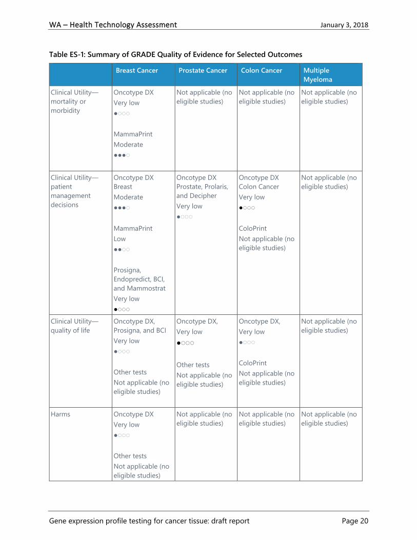
Table ES-1 summarizes the GRADE quality of evidence for key outcomes, by cancer type and for
each test considered for that cancer.
WA – Health Technology Assessment January 3, 2018
Gene expression profile testing for cancer tissue: draft report Page 20
Table ES-1: Summary of GRADE Quality of Evidence for Selected Outcomes
Breast Cancer Prostate Cancer Colon Cancer Multiple
Myeloma
Clinical Utility—
mortality or
morbidity
Oncotype DX
Very low
●◌◌◌
MammaPrint
Moderate
●●●◌
Not applicable (no
eligible studies)
Not applicable (no
eligible studies)
Not applicable (no
eligible studies)
Clinical Utility—
patient
management
decisions
Oncotype DX
Breast
Moderate
●●●◌
MammaPrint
Low
●●◌◌
Prosigna,
Endopredict, BCI,
and Mammostrat
Very low
●◌◌◌
Oncotype DX
Prostate, Prolaris,
and Decipher
Very low
●◌◌◌
Oncotype DX
Colon Cancer
Very low
●◌◌◌
ColoPrint
Not applicable (no
eligible studies)
Not applicable (no
eligible studies)
Clinical Utility—
quality of life
Oncotype DX,
Prosigna, and BCI
Very low
●◌◌◌
Other tests
Not applicable (no
eligible studies)
Oncotype DX,
Very low
●◌◌◌
Other tests
Not applicable (no
eligible studies)
Oncotype DX,
Very low
●◌◌◌
ColoPrint
Not applicable (no
eligible studies)
Not applicable (no
eligible studies)
Harms Oncotype DX
Very low
●◌◌◌
Other tests
Not applicable (no
eligible studies)
Not applicable (no
eligible studies)
Not applicable (no
eligible studies)
Not applicable (no
eligible studies)

WA – Health Technology Assessment January 3, 2018
Gene expression profile testing for cancer tissue: draft report Page 21
Breast Cancer Prostate Cancer Colon Cancer Multiple
Myeloma
Cost-effectiveness
and other
economic
outcomes
Oncotype DX and
MammaPrint:
Low
●●◌◌
Endopredict,
Mammostrat,
Prosigna, BCI:
Very low
●◌◌◌
Oncotype DX
Very low
●◌◌◌
Prolaris and
Decipher
Not applicable (no
eligible studies)
Oncotype DX
Very low
●◌◌◌
ColoPrint
Not applicable (no
eligible studies)
Not applicable (no
eligible studies)
Clinical Practice Guidelines
Breast Cancer
The most detailed clinical practice guideline, Use of Biomarkers to Guide Decisions on Adjuvant
Systemic Therapy for Women with Early-Stage Invasive Breast Cancer, was published by the
American Society of Clinical Oncology (ASCO) in 2016.58 ASCO published a guideline update in
2017 modifying the recommendations regarding MammaPrint, which draws upon recently
published studies.59 Both of these guidelines were rated as having good methodological quality.
The ASCO guidelines outlined recommendations for when Oncotype DX breast cancer assay,
MammaPrint, EndoPredict, Prosigna, BCI, and Mammostrat should or should not be used in
patients with early-stage breast cancer. All of these tests, except for Mammostrat, are
recommended for use in patients that have ER-positive/progesterone receptor (PR)-positive,
HER2-negative, LN-negative breast cancer.58,59 The guidelines recommended against the use of
Mammostrat for the following categories of breast cancer: ER-positive/PR-positive, HER2-
negative (LN-positive or negative); HER2-positive; or ER-negative/PR-negative, HER2-negative,
LN-negative.58
According to the ASCO guidelines, MammaPrint should not be used in patients with low clinical
risk (as defined by the Adjuvant! Online tool as used in the MINDACT study),18 because women
in the low clinical risk category had very good outcomes and did not appear to benefit from
chemotherapy even with a genomic high-risk cancer.59 MammaPrint is recommended for use in
patients with ER-positive/PR-positive, HER2-negative, LN-positive breast cancer who have one
to three positive nodes and are at high clinical risk per MINDACT categorization.59 Still, these
patients should be informed that a benefit of chemotherapy cannot be excluded, particularly
among patients with more than one involved lymph node.59 The ASCO guidelines recommend
against using the other tests in patients with ER-positive/PR-positive, HER2-negative, LN-

WA – Health Technology Assessment January 3, 2018
Gene expression profile testing for cancer tissue: draft report Page 22
positive breast cancer; HER2-positive breast cancer; or ER-negative/PR-negative, HER2-negative,
LN-negative breast cancer.58
The 2017 National Comprehensive Cancer Network (NCCN) clinical practice guidelines on breast
cancer discuss the evidence for Oncotype DX (21-gene breast cancer assay), MammaPrint (70-
gene assay), and Prosigna (50-gene assay).60 According to the guidelines, Oncotype DX may be
considered for ER-positive/PR-positive, HER2-negative cancers with pT1, pT2, or pT3, and pN0
or pN1mi ≤ 2 mm axillary node metastasis and a tumor greater than 0.5 cm.60 Oncotype DX can
also be considered in certain patients with one to three involved ipsilateral axillary lymph nodes
to guide the addition of combination chemotherapy to standard hormone therapy.60 The NCCN
guidelines state that the other gene expression profile tests can be considered to assess risk of
cancer recurrence, but that they have not been validated to predict response to chemotherapy.60
The NCCN guidelines were rated as having fair methodological quality.
The National Institute for Health Care Excellence (NICE) published guidelines in 2013 that
assessed the use of Oncotype DX breast cancer assay, MammaPrint, Mammostrat, and
immunohistochemical 4 (IHC4) score in early-stage breast cancer.61 The guidelines
recommended Oncotype DX as an option for guiding adjuvant chemotherapy decisions for
people with ER-positive, HER2-negative, LN-negative early-stage breast cancer when the patient
is assessed as being at intermediate risk.61 According to the guidelines, Oncotype DX should
only be used when the test results are likely to help in predicting the course of the disease, and
therefore in the decision of whether to prescribe chemotherapy.61 MammaPrint and
Mammostrat are only recommended for use in research in patients with ER-positive, HER2-
negative, LN-negative early-stage breast cancer.61 The NICE guidelines were rated as having
good methodological quality.
The European Society for Medical Oncology (ESMO) published breast cancer clinical practice
guidelines in 2015.62 The ESMO guidelines recommended that gene expression profile tests,
such as Oncotype DX breast cancer assay, MammaPrint, Endopredict, and Prosigna, can be used
to complement pathology assessments to predict the benefit of adjuvant chemotherapy.62 The
ESMO guidelines were rated as having poor methodological quality. The European Group on
Tumor Markers published a guideline in 2017 on the use of biomarkers in breast cancer.63 These
guidelines recommended that the Oncotype DX breast cancer assay, MammaPrint, EndoPredict,
Prosigna, and BCI can be used to aid in adjunct therapy decision-making in ER-positive, HER2-
negative, LN-negative patients.63 In addition, Oncotype DX, MammaPrint, EndoPredict, and
Prosigna can be used in patients with one to three metastatic lymph nodes.63 The European
Group on Tumor Markers guidelines were also rated as having poor methodological quality.
Table ES-2 summarizes these five guidelines on breast cancer, indicating whether the gene
expression profile tests are recommended for LN-negative and/or LN-positive cancers.

WA – Health Technology Assessment January 3, 2018
Gene expression profile testing for cancer tissue: draft report Page 23
Table ES-2. Recommendations for Lymph Node Status in Guidelines on the
Use of Gene Expression Profile Tests in Early State Breast Cancer
ASCO NCCN NICE ESMO** EGTM
Oncotype DX LN-negative LN-negative
LN-positive
LN-negative LN-negative
LN-positive
LN-negative
LN-positive
MammaPrint LN-negative
LN-positive
Not
recommended*
Not
recommended
LN-negative
LN-positive
LN-negative
LN-positive
EndoPredict LN-negative Not
recommended*
No guideline
recommendation
LN-negative
LN-positive
LN-negative
LN-positive
Prosigna LN-negative Not
recommended*
No guideline
recommendation
LN-negative
LN-positive
LN-negative
LN-positive
Breast Cancer
Index
LN-negative Not
recommended*
No guideline
recommendation
No guideline
recommendation
LN-negative
Mammostrat Not
recommended
Not
recommended*
Not
recommended
No guideline
recommendation
No guideline
recommendation
*The NCCN guidelines recommend Oncotype DX and conclude that the other multigene assays have not
been validated to predict response to chemotherapy.
**The ESMO guidelines do not distinguish between LN-negative and LN-positive cancers in their
recommendations.
Prostate Cancer
Two clinical practice guidelines were identified that included recommendations on the use of
Decipher, Prolaris, and Oncotype DX prostate cancer assay. The 2017 NCCN guidelines on
prostate cancer stated that men with clinically localized prostate cancer may consider the use of
tumor-based molecular assays and made specific recommendations on the use of Decipher,
Prolaris, and Oncotype DX for prostate cancer.64 The guidelines recommended Decipher after a
radical prostatectomy for patients with pT2 (confined to prostate) with positive margins, any pT3
(extraprostatic extension) disease, and a rising prostate specific antigen (PSA) level.64 Prolaris
and Oncotype DX are recommended post-biopsy for low- and very low-risk prostate cancer in
patients with at least 10 years of life expectancy who have not received other active treatment
for prostate cancer and who are candidates for active surveillance or definitive therapy.64 The
NCCN guidelines were rated as having fair methodological quality.
A guideline on clinically localized prostate cancer has been published by the American
Urological Association, American Society for Radiation Oncology, and Society of Urologic
Oncology in 2017.65 These guidelines included the following recommendation based on expert
opinion: “Tissue-based genomic biomarkers have not shown a clear role in active surveillance for
localized prostate cancer and are not necessary for follow up.”65(p. 4) These guidelines were rated
as having good methodological quality.

WA – Health Technology Assessment January 3, 2018
Gene expression profile testing for cancer tissue: draft report Page 24
Colon Cancer
No clinical practice guidelines were found that included recommendations for the use of
ColoPrint or Oncotype DX for colon cancer. The American Society for Clinical Pathology, College
of American Pathologists, Association for Molecular Pathology, and ASCO published a guideline
on molecular biomarkers for colorectal cancer in 2017.66 This guideline stated, “A problem of
quantitative assays, such as gene expression, microRNA expression, and methylation levels,
tested in solid tumors, results from the intrinsic mixed nature of the tissue with significant
variability of tumor and nontumor tissue content. Another limitation of molecular biomarker
discovery approaches that rely on expression levels is that these biomarkers have not been
evaluated in the context of complex molecular regulation of individual cancer subtypes.”66(p. 1482)
These guidelines were rated as having good methodological quality.
The fair methodological quality 2017 NCCN guidelines on colon cancer discussed multigene
assays, including ColoPrint and Oncotype DX colon cancer assay, and concluded that there is no
evidence of predictive value in terms of the potential benefit of chemotherapy for any of the
multigene assays.67 Similarly, the 2016 guidelines on metastatic colon cancer from ESMO
concluded that gene expression signatures have failed to accurately predict disease recurrence
and prognosis.68 The ESMO guidelines were rated as having poor methodological quality.
Multiple Myeloma
The 2017 NCCN guidelines on multiple myeloma discussed gene expression profiling tests,
including MyPRS and SKY92, but did not make any recommendations about the use of these
tests.69 The NCCN panel unanimously agreed that although gene expression profile tests are not
routinely used, they could be helpful in selected patients to estimate the aggressiveness of the
disease and to individualize treatment.69 The NCCN guidelines were rated as having fair
methodological quality. The 2017 guidelines on multiple myeloma from the ESMO stated that
gene-expression profiling is not currently used routinely, and more research is needed to
identify molecular markers, which could lead to advances in this area.70 70 The ESMO guidelines
were rated as having fair methodological quality. No other clinical practice guidelines were
found that included recommendations for the use of My Prognostic Risk Signature (MyPRS) or
SKY92 tests.
Selected Payer Coverage Determinations
Breast Cancer
No Medicare National Coverage Determinations (NCDs) were found for any of the gene
expression profile tests for breast cancer. Center researchers identified Local Coverage
Determinations (LCDs) by Noridian Healthcare Solutions, which apply to Washington, that
provide coverage for Prosigna and BCI. The Prosigna LCD provides coverage for
postmenopausal women with either:

WA – Health Technology Assessment January 3, 2018
Gene expression profile testing for cancer tissue: draft report Page 25
ER-positive, LN-negative, stage 1 or 2 breast cancer or
ER-positive, LN-positive (one to three positive nodes), stage 2 breast cancer71
The BCI LCD provides coverage for patients have non-relapsed, ER-positive, LN-negative breast
cancer, among other criteria.72 No Medicare LCDs covering Washington were found for the
Oncotype DX breast cancer assay, MammaPrint, EndoPredict, or Mammostrat.
Private payer policies were assessed for Aetna, Cigna, and Regence. The Aetna policy on tumor
markers provides coverage for the Oncotype DX breast cancer assay, MammaPrint, EndoPredict,
Prosigna, and BCI to assess the necessity of adjuvant chemotherapy in females or males with
recently diagnosed breast tumors.73 Oncotype DX and MammaPrint are covered for breast
cancers that are LN-negative or with one to three involved ipsilateral axillary lymph nodes.73
EndoPredict, Prosigna, and BCI are covered for only LN-negative cancers.73 Coverage for all
these tests requires that adjuvant chemotherapy is not precluded by any other factor (e.g.,
advanced age or significant comorbidities), and that the patient and physician have discussed
the potential results of the test and agree to use the results to guide therapy, among other
criteria.73 Aetna does not cover Mammostrat.73
The Cigna policy on gene expression assays covers the Oncotype DX breast cancer assay,
MammaPrint, and Prosigna under certain conditions, and does not provide coverage for
EndoPredict, BCI, or Mammostrat.74 Oncotype DX and MammaPrint are covered for LN-negative
cancers and for cancers with up to three positive nodes, and Mammostrat is covered for only
LN-negative cancers.74
The Regence policy on gene expression test for breast cancer provides coverage for the
Oncotype DX breast cancer assay, EndoPredict, and BCI under certain conditions, and does not
cover MammaPrint, Prosigna, or Mammostrat.75 Regence covers the Oncotype DX breast cancer
assay, EndoPredict, and BCI for women with primary breast cancer, stages 1, 2, or 3, that are LN-
negative, among other criteria.75
Prostate Cancer
No Medicare NCDs were found for Decipher, Prolaris, or Oncotype DX for prostate cancer. There
are LCDs for Noridian Healthcare Solutions, applying to the state of Washington, that provide
coverage for Decipher, Prolaris, and Oncotype DX for prostate cancer under certain conditions.
The LCD for Decipher provides coverage after radical prostatectomy when certain conditions are
met.76 There are two LCDs providing coverage for Prolaris, under certain conditions, one for
patients with early stage, needle-biopsy-proven prostate cancer77 and the other for patients with
favorable intermediate risk, needle-biopsy-proven prostate cancer.78 The LCD for Oncotype DX
for early stage, needle biopsy proven prostate cancer provides coverage with specified
condictions.79
The coverage policies for Aetna73 and Regence80 consider Decipher, Prolaris, and Oncotype DX
prostate cancer assay to be experimental or investigational. Cigna does not include Decipher,

WA – Health Technology Assessment January 3, 2018
Gene expression profile testing for cancer tissue: draft report Page 26
Prolaris, or Oncotype DX prostate cancer assay in the list of medically necessary prostate cancer
prognostic tests.74
Colon Cancer
No Medicare National or Local Coverage Determinations were found for ColoPrint or the
Oncotype DX colon cancer assay. The policies for Aetna,73 Cigna,74 and Regence81 do not cover
ColoPrint or Oncotype DX colon cancer assay.
Multiple Myeloma
No Medicare National or Local Coverage Determinations were found for MyPRS or SKY92. The
policy for Aetna does not cover MyPRS, and this coverage policy does not mention SKY92.73
Cigna’s coverage policy on tumor markers does not mention MyPRS or SKY92.74 The Regence
coverage policy states that all microarray-based gene expression profile testing for multiple
myeloma is considered investigational.82
Conclusions
There was no high-quality evidence of clinical utility to guide decisions about the use of gene
expression profile tests for breast, prostate, and colon cancers. The only condition with quality of
evidence ratings above very low was breast cancer and only for the MammaPrint and Oncotype
DX tests. Based on a single RCT, there is moderate quality evidence that women with early
invasive breast cancer who are considered to be at high clinical risk by the Adjuvant! Online risk
assessment tool may safely forego adjuvant systemic chemotherapy if their MammaPrint
genomic risk score is low. There was no statistically significant difference in distant metastasis-
free survival at five years.
Moderate quality of evidence supports the use of Oncotype DX because of its impact on clinical
treatment recommendations and chemotherapy use for women with early invasive breast
cancer, particularly its ability to identify low risk women who would not benefit from adjuvant
systemic chemotherapy. There is also low quality of evidence about the decision impact of using
MammaPrint for women with early invasive breast cancer. Based primarily on modeling studies,
there is low quality of evidence that both Oncotype DX and MammaPrint are cost-effective at
conventional thresholds when used to guide treatment decisions among women with early
invasive breast cancer. Among the remaining conditions and tests there is a mix of very low
quality or a complete absence of evidence to support use of these tests to improve clinical
decision-making and important patient outcomes.
Clinical practice guidelines are generally more liberal in their recommendations about test use,
largely due to the ability of most of these tests to give information about overall prognosis even
if they lack evidence of clinical utility. For example, ASCO endorses all of the breast cancer tests
(with the exception of Mammostrat) for use in LN-negative patients, and also the use of
MammaPrint for those with limited lymph node involvement. However, NCCN has adopted a

WA – Health Technology Assessment January 3, 2018
Gene expression profile testing for cancer tissue: draft report Page 27
more rigorous standard and advises the exclusive use of Oncotype DX for both LN-negative and
LN-positive patients.
There are multiple ongoing clinical trials on most of the tests included in this report (see
Appendix F), including some large and important trials that will be reporting out in the next few
years. Among these are the TAILORx and RxPONDER breast cancer trials for Oncotype DX,
additional results for the MINDACT trial involving MammaPrint for LN-positive women.

WA – Health Technology Assessment January 3, 2018
Gene expression profile testing for cancer tissue: draft report Page 28
Technical Report
Background
The lifetime risk of developing cancer is about 40%, and one in every five Americans will die
from cancer.1 Strategies for reducing the burden of cancer include prevention, early diagnosis,
and appropriate treatments.2 Common treatments for cancer are surgery, chemotherapy,
radiation therapy, hormone therapy, and immunotherapy.3 The most appropriate treatments for
a particular cancer depend on the cancer’s characteristics (e.g., cancer stage and grade), the
patient’s age and health status, response to previous treatments, and other factors.4
In recent years, gene expression profile testing of cancer tissue has been used to help inform
decisions on appropriate treatments. Gene expression profile testing identifies the genes in a
cancer cell or tissue that are making messenger RNA (mRNA), which carry the genetic
information that cancer cells need to make proteins. Some gene expression profile tests are
designed to increase the accuracy of the prognosis for a patient with cancer. If a test predicts
that a cancer is slow growing or is unlikely to metastasize, then active surveillance of the cancer
could be the most appropriate course. If a test predicts that a cancer is likely to progress and
metastasize, then more aggressive treatments could be warranted.5 Other gene expression tests
may identify specific mutations for which there are targeted treatments (i.e., the Anaplastic
lymphoma kinase [ALK] mutation in lung cancer can be treated with a specific drug). However,
to have usefulness in clinical practice, a gene expression profile test must do more than predict
better or worse prognosis. Clinical utility means that a test has an impact on clinical decision
making or can help direct therapy in an actionable way to improve patient outcomes. Gene
expression profile tests have been developed for patients with breast cancer, prostate cancer,
colon cancer, and multiple myeloma, among others. This report examines the clinical utility and
cost-effectiveness of selected genomic tests for these four cancers.
Technology Description
This section contains descriptions of gene expression profile tests, along with recent studies on
their clinical validity, for breast, prostate, and colon cancers and multiple myeloma. Molecular
diagnostic tests are regulated by the U.S. Food and Drug Administration (FDA) and the Centers
for Medicare & Medicaid Services (CMS).6 The FDA has exercised discretion in its requirements
for approval of in vitro diagnostic assays.6 In vitro tests developed, validated, and performed in-
house by a specific reference laboratory are required to abide by the Clinical Laboratory
Improvement Amendments (CLIA), but FDA clearance and approval is currently not required for
these laboratory-developed tests (LDTs).6 Most of the tests described in this report are regulated
as LDTs. Two of the tests, MammaPrint and Prosigna, have received FDA pre-market approval.

WA – Health Technology Assessment January 3, 2018
Gene expression profile testing for cancer tissue: draft report Page 29
Breast Cancer
This evidence review considered six tests that are indicated for women with early stage, invasive
breast cancer: Oncotype DX breast cancer assay, MammaPrint, EndoPredict, Prosigna, Breast
Cancer Index (BCI), and Mammostrat.
Oncotype DX
The Oncotype DX breast cancer assay uses reverse transcription polymerase chain reaction (RT-
PCR, a common technique to detect and quantify mRNA, to measure gene expression in
formalin fixed paraffin-embedded (FFPE) cancer tissue.83 The Oncotype DX breast recurrence
score test is used to predict the likely benefit of chemotherapy and the risk of distant recurrence
among patients newly diagnosed with early-stage estrogen receptor (ER)-positive, human
epidermal growth factor receptor 2 (HER2)-negative invasive breast cancer, both lymph node
(LN)-negative and LN-positive.83 The test measures the expression of 21 genes, including 16
cancer-related genes and five reference genes.83 These genes were selected out of 250 rationally
selected candidate genes bases on prognostic capacity and consistency in test performance.20
The Oncotype DX recurrence score ranges from 0 to 100, with categories of low risk (0 to 17),
intermediate risk (18 to 30), and high risk (31 to 100).20 The Oncotype DX report also provides a
quantitative ER score to help assess the magnitude of hormonal therapy benefit.20 Oncotype DX
is available as a LDT from Genomic Health, licensed under CLIA, and does not require FDA
approval.84
The 2017 systematic review by Blok and colleagues included 20 studies assessing the clinical
validity of Oncotype DX.20 All the included studies in patients with LN-negative disease showed
significant differences in outcomes (usually recurrence free survival) among low-, intermediate-,
and high-risk patients.20 Most of the studies that included patients with LN-positive cancer
showed significant differences in outcomes among the three risk groups.20
MammaPrint
Using microarray technology, MammaPrint measures the mRNA expression of 70 genes in fresh
tissue or FFPE tissue.85 The 70 genes were identified in 2002 from a total of 25,000 genes using
supervised clustering.20 MammaPrint categorizes patients as having a low or high risk of breast
cancer recurrence among women with stage 1 or stage 2 invasive breast cancer, with a tumor
size ≤ 5.0 cm, that is LN-negative, ER-positive or negative, and HER2-positive or negative.85 In
2007, Agendia received 510(k) clearance from the FDA for MammaPrint and received clearance
for its use in FFPE tissue in 2015.20
The systematic review by Blok and colleagues included 21 studies assessing the clinical validity
of MammaPrint.86 In studies of patients with LN-negative breast cancer, MammaPrint was
reported to be of significant prognostic value for risk of recurrence outcomes.86 For LN-positive
disease, the studies reported statistically significant hazard ratios for distant metastases-free
survival and breast cancer-specific survival between low- and high-risk MammaPrint groups.86 In
the remaining studies, without specific classification or with both LN-positive and LN-negative

WA – Health Technology Assessment January 3, 2018
Gene expression profile testing for cancer tissue: draft report Page 30
combined, most of the results showed a significant difference in outcomes between
MammaPrint risk groups.86
EndoPredict
EndoPredict tests FFPE tissue to estimate the 10-year risk of distant recurrence of early-stage
ER-positive, HER2-negative invasive breast cancer, both LN-negative and LN-positive.87
EndoPredict produces a 12-gene molecular score using quantitative RT-PCR on eight signature
genes, three normalization genes, and one control gene, with the score ranging from 0 to 15.20
This 12-gene molecular score is combined in an algorithm with tumor size and nodal status to
calculate an EPclin score, which has a clinically reportable range of 1.1 to 6.2.87 EPclin scores
from 1.1 to 3.3 indicate a low 10-year risk for distant recurrence, and scores from 3.4 to 6.1
indicate a high risk for 10-year risk for distant recurrence.87 EndoPredict is available as a LDT
from Myriad Genetics, licensed under CLIA, and does not require FDA approval.88
Four studies included in the systematic review by Blok and colleagues assessed the clinical
validity of EndoPredict.20 Three of these studies compared low-risk patients to high-risk patients
on risk of recurrence outcomes, and all three studies found statistically significant hazard ratios,
ranging from 1.19 to 1.31.20 The fourth study found a low proportion of distant metastases in
the low risk group, but did not compare outcomes between low-risk and high-risk groups.20
Prosigna
The Prosigna breast cancer gene signature assay, based on the PAM50 gene signature, uses
quantitative PCR performed on RNA isolated from FFPE breast tumor tissue to classify tumors
into four intrinsic subtypes: luminal A, luminal B, HER2-negative enriched, and basal-like.89,90
Prosigna is indicated for use in postmenopausal women with hormone receptor-positive, LN-
negative (stage 1 or 2), or LN-positive (stage 2) breast cancer with a clinicopathological
indication to be treated with adjuvant endocrine therapy.90 The Prosigna algorithm produces a
risk of recurrence score using a measure of the expression of 46 genes, intrinsic subtype, tumor
size, nodal status, and proliferation score.20 The risk of recurrence score is on a zero to 100 scale,
which correlates with the probability of distant recurrence in a 10-year period.90 In 2013,
NanoString Technologies received 510(k) clearance from the FDA for Prosigna.86
Five studies included in the systematic review by Blok and colleagues assessed the clinical
validity of Prosigna.20 Four of the five included studies showed that risk of recurrence outcomes
were significantly different among risk categories as determined by the Prosigna test, while the
fifth study did not find a statistically significant difference.20
Breast Cancer Index
Breast Cancer Index (BCI) predicts the likelihood of benefit from extended endocrine therapy
among women with ER-positive, LN-negative or LN-positive (with one to three positive nodes),
early-stage, invasive breast cancer.91 BCI measures the expression of 11 genes using quantitative
RT-PCR performed on FFPE tissue.92 BCI was developed from two gene expression-based
WA – Health Technology Assessment January 3, 2018
Gene expression profile testing for cancer tissue: draft report Page 31
biomarkers: the HOXB13:IL17BR ratio, which is associated with tumor responsiveness to
endocrine therapy in breast cancer, and the Molecular Grade Index, which consists of the
average expression of five cell cycle-associated genes.92 The BCI score ranges continuously from
0 to 10. The categories for risk of late (post-five year) distant recurrence are low risk (< 5.0825)
and high risk (≥ 5.0825).92 The categories for risk of overall (0 to 10 year) distant recurrence are
low risk (< 5.0825), intermediate risk (≥ 5.0825 to 6.5025), and high risk (> 6.5025).92 BCI also
predicts the likelihood of benefit from extended (> 5 year) endocrine therapy.91 BCI is available
as a LDT from Biotheranostics, licensed under CLIA, and does not require FDA approval.88
In a 2013 study by Zhang and colleagues, the BCI model was validated by retrospective analyses
of tumor samples from tamoxifen-treated patients from a randomized prospective trial (n =317)
and a multi-institutional cohort (n = 358).93 In the first cohort, BCI categorized 65% of patients
as low risk, and less than 3% of low-risk patients had distant recurrence in years 0 to 5 and in
years 5 to 10.93 The second cohort generally had larger tumors, and 55% of patients were
classified as BCI low risk.93 This low-risk group had less than 5% distant recurrence in years 0 to 5
and years 5 to 10.93
Mammostrat
The Mammostrat test, launched by Clarient in 2010, is based on the immunohistochemical (IHC)
assay of the expression of five genes in hormone receptor positive, stage 1 or 2 breast cancer
cells.94 An algorithm is used to calculate a risk index score, with patients categorized as low (≤ 0),
moderate (0 to ≤ 0.7) or high (> 0.7) risk.21 This test is not technically a molecular gene
expression profile test but is included in this group because it is used for similar purposes in
practice. In this report we do not make this distinction. The five genes were selected from 700
gene targets in gene expression assays in three patient cohorts.94 Mammostrat was developed
and validated using clinical samples from patients enrolled in the National Surgical Adjuvant
Breast and Bowel Project B14 and B20 trials.95 Mammostrat is available as a LDT from Clarient,
licensed under CLIA, and does not require FDA approval.96
Prostate Cancer
Decipher is a gene expression profile test used after prostatectomy to predict the probability of
metastasis and inform clinical decisions on the use of adjuvant prostate cancer treatments. Two
other tests, Prolaris and Oncotype DX for prostate cancer, are used with biopsy tissue after an
initial diagnosis of prostate cancer to predict the cancer’s aggressiveness and thus inform initial
treatment decisions.
Decipher
Decipher is a test designed to predict the probability of metastasis within five years of radical
prostatectomy.97 Decipher analyzes a tissue sample removed during surgery that is routinely
archived or stored by the pathology laboratory.97 Decipher measures the expression levels of 22
RNA biomarkers involved in biological pathways across the genome that are associated with
aggressive prostate cancer.97 The panel is designed to evaluate the expression of genetic

WA – Health Technology Assessment January 3, 2018
Gene expression profile testing for cancer tissue: draft report Page 32
markers associated with specific biological processes, including the cell proliferation and
differentiation processes, cell structure, adhesion, motility, immune response, and mitosis
processes, cell cycle progression (CCP), and other functional processes.98 The Decipher Genomic
Classifier score ranges continuously from zero to one, where a higher score indicates a higher
probability of clinical metastasis.99 The risk categories for Decipher scores are low (< 0.45),
intermediate (0.45 to 0.60), and high (> 0.60).100 Decipher is available as a LDT from GenomeDx,
licensed under CLIA, and does not require FDA approval.6
A recent systematic review by Spratt and colleagues included a meta-analysis of 855 patients
who had the Decipher test and were followed-up for a median of eight years.101 Patients were
classified by Decipher as low, intermediate, or high risk.101 The 10-year cumulative incidence
metastases rates for the low, intermediate, and high risk groups were 5.5%, 15.0%, and 26.7% (p
= .001), respectively (HR per 0.1 unit change in Decipher score, 1.52; 95% CI, 1.39 to 1.67).101
After adjusting for clinicopathologic variables, Decipher remained a statistically significant
predictor of metastasis risk (HR per 0.1 unit, 1.30; 95% CI, 1.14 to 1.47; p = .001)101 The authors
concluded that Decipher has good clinical validity and can independently and significantly
improve prognostic accuracy for patients after a prostatectomy, within nearly all
clinicopathologic, demographic, and treatment subgroups.101
Prolaris
The Prolaris CCP test uses a quantitative RT-PCR assay on FFPE biopsy tissue to determine
cancer aggressiveness and predict the probability of disease progression in patients with low-
and intermediate-risk prostate cancer.102,103 Prolaris uses a 46-gene expression signature,
including 31 CCP genes and 15 housekeeping genes.104 The CCP score ranges from 0 to 10, with
each unit increase representing a doubling of risk of disease progression.102 The CCP report
provides an estimate of a patient's 10-year prostate cancer–specific mortality risk, from an
algorithm combining the CCP score and clinicopathological variables.55 Prolaris is available as a
LDT from Myriad Genetic Laboratories, licensed under CLIA, and does not require FDA
premarket review.6
A 2016 systematic review and meta-analysis by Sommariva and colleagues assessed the clinical
validity of Prolaris.105 In a meta-analysis of four studies (n = 1,489), the hazard ratio for
biochemical recurrence after prostatectomy per 1-unit increase in the CCP score was 1.88 (p <
.001) in a univariate model and 1.63 (p < .001) in a multivariable model adjusted for clinical and
pathologic variables, such as extent of disease, age, clinical stage, and use of hormones.105 For
example, a patient with a CCP score of five has a 1.63 times greater likelihood of biochemical
recurrence than a patient with a score of four, if all other factors are equal.
Oncotype DX for Prostate Cancer
The Oncotype DX genomic prostate score assay analyzes prostate cancer gene activity to predict
disease aggressiveness among men with clinically low-risk prostate cancer.106 This test was
developed by Genomic Health and was based on studies evaluating 727 genes among patients

WA – Health Technology Assessment January 3, 2018
Gene expression profile testing for cancer tissue: draft report Page 33
undergoing radical prostatectomy between 1987 and 2004 in Cleveland, Ohio.98 Oncotype DX
uses a quantitative RT-PCR assay performed on approximately 1 mm FFPE tissue from a prostate
needle biopsy.104 Oncotype DX measures the expression of 17 genes, 12 cancer-related genes
across four genetic pathways (stromal response, androgen signaling, proliferation, and cellular
organization) and five housekeeping genes.106 Gene expression in conjunction with clinical risk
factors are used to predict the likelihood of adverse pathology, with Genomic Prostate Scores
ranging from zero to 100.106 Oncotype DX is available as a LDT from Genomics Health, licensed
under CLIA, and does not require FDA approval.6
In 2016, Brand and colleagues published a patient-level meta-analysis of two independent
clinical validation studies (n = 732) of Oncotype DX for prostate cancer, along with the Cancer of
the Prostate Risk Assessment (CAPRA) score and National Comprehensive Cancer Network
(NCCN) risk group, as predictors of the likelihood of favorable or adverse pathologic staging
following surgery.107 In a decision curve analysis, greater net benefit was shown when combining
Oncotype DX score with each clinical classifier compared with the classifier alone.107 By adding
Oncotype DX score to CAPRA score, the area under the receiver operating characteristic curve
(AUROC) improved from 0.68 to 0.73 (p < .001), and adding Oncotype DX to NCCN risk group
improved the AUROC from 0.64 to 0.70 (p < .001).107 These AUROCs are generally considered by
biostatisticians to represent poor to fair test accuracy.
Colon Cancer
Two gene expression profile tests are described below: ColoPrint, which can be used in stage 2
colon cancer patients, and Oncotype DX for stage 2 and 3 colon cancer patients.
ColoPrint
Released by Agendia in 2012, ColoPrint was developed using whole-genome expression data to
identify genes that have the highest correlation to a tumor-relapse in stage 2 colon cancer
patients.108 ColoPrint’s microarray-based 18-gene expression signature is performed using fresh
or frozen tumor samples from stage 2 colon cancer patients who have undergone surgery.109
ColoPrint categorizes patients as being at low or high risk of distant recurrence.108 ColoPrint is
available as a LDT from Agendia, licensed under CLIA, and does not require FDA approval.110
In a 2015 review by Kopetz and colleagues, the authors conducted a pooled analysis of studies
assessing the risk of recurrence in 416 stage 2 colon cancer patients who had available ColoPrint
results.109 High-risk patients, as identified by ColoPrint, had a statistically significantly higher risk
of recurrence compared to low-risk patients (HR, 2.16; 95% CI, 1.28 to 3.65; p = .004).109 High-
risk patients had a five-year risk of recurrence of 20.9% (95% CI, 14.2% to 27.6%) and low-risk
ColoPrint patients had a five-year risk of recurrence of 10.3% (95% CI, 6.6% to 14%).109 The
hazard ratio remained statistically significant in a multivariable model that included number of
lymph nodes retrieved and a measure of microsatellite instability.109

WA – Health Technology Assessment January 3, 2018
Gene expression profile testing for cancer tissue: draft report Page 34
Oncotype DX for Colon Cancer
The Oncotype DX colon cancer assay, by Genomic Health, is indicated for patients with anatomic
stage 2, mismatch repair proficient (MMR-P) and stage 3 A/B (cancer has not spread to distant
organs, but cancer has spread to one to three lymph nodes and grown into the submucosa or
into the outermost layer of the colon) colon cancers.111 The test was developed from four
studies that explored the expression of 761 genes in patient tumor samples that were obtained
at the time of initial diagnosis of colon cancer.112 The 12-gene test (seven cancer-related genes
and five reference genes) produces a score of 0 to 100 and then categorizes patients as having
low risk (< 30), intermediate risk (30 to 40), or high risk (≥ 41) of recurrence.111 Oncotype DX is
available as a LDT from Genomic Health, licensed under CLIA, and does not require FDA
premarket review.110
A 2016 study by Yamanaka and colleagues assessed the predictive ability of Oncotype DX in 597
stage 2 and 3 colon cancer patients.113 The continuous Oncotype DX recurrence score was
significantly associated with recurrence-free interval after adjustment for disease stage (HR for
25-unit increase in recurrence score, 2.05; 95% CI, 1.47 to 2.86; p = .001).113
Multiple Myeloma
The two tests described below, My Prognostic Risk Signature (MyPRS) and SKY92, are used as
prognostic tests in patients with multiple myeloma.
My Prognostic Risk Signature (MyPRS)
MyPRS was developed at the University of Arkansas for Medical Science and then licensed to
Signal Genetics.114 The MyPRS test includes a 70-gene Prognostic Risk Score (GEP-70), which
quantifies the expression of 70 genes commonly altered in multiple myeloma.115 Using a scale of
0 to 100, the test predicts the risk of disease relapse and survival outcomes and classifies
patients as low or high risk.115 MyPRS was available as a LDT from Signal Genetics, licensed
under CLIA, and does not require FDA approval.116 In November 2016, Signal Genetics sold its
MyPRS assets to Quest Diagnostics.116
A 2007 study by Shaughnessy and colleagues analyzed tumor cells from 532 newly diagnosed
patients with multiple myeloma, using the 70-gene test.117 A high-risk score was an independent
predictor of event-free (HR, 3.41; p = .002) and overall survival (HR, 4.75; p < .001).117
SKY92
The SKY92-signature (formerly EMC92) was discovered at the Erasmus Medical Centre in the
Netherlands, and the test measures the expression level of 92 genes that are directly or
indirectly related multiple myeloma.8 The SKY92 test is used to predict how likely multiple
myeloma is to progress and to aid in treatment decision-making.8 Starting in 2015, SkylineDx
began to produce the MMprofiler test which uses the SKY92 gene signature to classify multiple
myeloma patients into high or standard risk groups.8 MMprofiler is available in Europe, but is

WA – Health Technology Assessment January 3, 2018
Gene expression profile testing for cancer tissue: draft report Page 35
only available in the U.S. for research use and currently not permitted for clinical diagnostic use.8
SkylineDx plans to make MMprofiler available soon as a LDT in the U.S.7
A 2015 study by Kuiper and colleagues of 4,750 multiple myeloma patients assessed the value of
20 markers for predicting patients overall survival.118 The study found that the combination of
SKY92 and the International Staging System provided the strongest predictor for overall survival
among the 20 markers tested.118 Median survival was 24 months for the highest risk group, 47
months for the intermediate-high risk group, 61 months for the intermediate-low risk group,
and the median was not reached after 96 months for the lowest risk group.118 Relative to the
lowest risk group, the hazard ratios were significantly higher in the intermediate-low group (HR,
2.6; 95% CI, 1.6 to 4.5), intermediate-high group (HR, 3.2; 95% CI, 1.9 to 5.4), and high group
(HR, 6.9; 95% CI, 4.1 to 11.7).118
Policy Context
There are a growing number of gene expression profile tests for cancer tissue designed to
inform treatment decisions after cancer diagnoses. Potential benefits of these tests are more
appropriate treatment decisions, better patient outcomes, including avoiding treatment-related
side effects, and the potential cost and side-effect savings from avoiding unnecessary
treatments. This topic was selected for a health technology assessment because of medium
concerns for the safety of these tests, medium to high concerns for efficacy, and high concerns
for cost.
This evidence review will help to inform Washington’s independent Health Technology Clinical
Committee as the committee determines coverage regarding selected gene expression profile
tests for patients with eligible breast, prostate, or colon cancers or multiple myeloma.

WA – Health Technology Assessment January 3, 2018
Gene expression profile testing for cancer tissue: draft report Page 36
Washington State Utilization and Cost Data
Populations
The Gene Expression Profile Testing of Cancer Tissue analysis includes member utilization and
cost data from the Public Employees Benefit Board Uniform Medical Plan (PEBB/UMP). There
were no claims from the Department of Labor and Industries workers’ compensation plan. PEBB
Medicare claims were excluded. Note: HCA Medicaid (formerly Fee-for-Service) and the Managed
Care (MCO) Medicaid program analysis will be included in the final report.
The analysis period was four calendar years, 2013 to 2016. Primary population inclusion criteria
included age 18 years old or older, having a diagnosis of breast, prostate, or colon cancers or
multiple myeloma, and experiencing at least one of the specific CPT/HCPCS codes from Table 1.
Denied claims were excluded from the analysis.
Table 1. CPT Codes and Descriptions for Gene Expression Profile Tests
CPT Code Description
81519 Breast Cancer—Oncotype DX Breast Cancer Assay
81599 EndoPredict
88381 Prosigna Breast Cancer Prognostic Gene Signature Assay (PAM50)
81525 Colon Cancer—Oncotype DX Colon Cancer Assay
81479 Unlisted molecular pathology procedure
Methods
Utilization was calculated based on an individual having: 1) a claims coded diagnosis of breast,
prostate, or colon cancer or multiple myeloma; and 2) having a paid claim for one of the
identified tests from Table 1. Codes for specific tests were identified only for Oncotype DX
breast cancer assay, EndoPredict, Prosigna, and Oncotype DX colon cancer assay. Other tests in
table are for an “unlisted molecular pathology procedure.” It is not possible to ascertain whether
this code refers to one of the tests in this report. Data evaluation included examining average
utilization and costs by member.
Table 2 below shows the total number of claims and total allowed charges for the four calendar
years, 2013 to 2016 from PEBB/UMP, excluding Medicare.

WA – Health Technology Assessment January 3, 2018
Gene expression profile testing for cancer tissue: draft report Page 37
Table 2. PEBB/UMP (No Medicare) Claims for Gene Expression Profile Testing: 2013-2016
CPT Code Description Services Allowed
Breast Cancer
81519 Breast Cancer—Oncotype DX Breast Cancer Assay 10 $39,328
81599 EndoPredict 0 --
88381 Prosigna (PAM50) 0 --
81479 Unlisted molecular pathology procedure 4 $964
Prostate Cancer
81479 Unlisted molecular pathology procedure 1 $165
Colon Cancer
81525 Colon Cancer—Oncotype DX Colon Cancer Assay 0 --
81479 Unlisted molecular pathology procedure 10 $6,662
Multiple Myeloma
81479 Unlisted molecular pathology procedure 3 $2,674
Methods
This evidence review is based on the final key questions published on November 14, 2017.
Population: Adults with breast, prostate, or colon cancers or multiple myeloma
Interventions: Gene expression profile testing of cancer tissue to inform treatment decisions,
including the following tests by cancer type:
Breast Cancer—Oncotype DX breast cancer assay, MammaPrint, EndoPredict, Prosigna
Breast Cancer Prognostic Gene Signature Assay (PAM50), Breast Cancer Index (BCI),
Mammostrat
Prostate Cancer— Decipher, Prolaris, Oncotype DX prostate cancer assay
Colon Cancer—ColoPrint, Oncotype DX colon cancer assay
Multiple Myeloma—Myeloma Prognostic Risk Signature (MyPRS), SKY92-signature
(formerly EMC92)
Comparators: Usual care without gene expression profile testing of cancer tissue, alternate
gene expression profile tests (i.e., one test intervention listed above versus another)
Outcomes:
Patient management decisions (including selection of active surveillance rather than
active treatment)
Clinical outcomes (e.g., morbidity, mortality, quality of life)
Harms, such as consequences of false-positive or false-negative test results
Cost-effectiveness and other economic outcomes

WA – Health Technology Assessment January 3, 2018
Gene expression profile testing for cancer tissue: draft report Page 38
Time period for literature search: January 2007 to November 2017
Key Questions
1. Effectiveness: What is the clinical utility of gene expression profile testing of cancer tissue
to inform treatment decisions for patients with breast, prostate, and colon cancers and
multiple myeloma?
a. Is there evidence that test results affect treatment decisions?
b. Do treatment decisions guided by gene expression profile testing of cancer tissue
result in clinically meaningful improvements in patient outcomes?
2. Harms: What harms are associated with conducting gene expression profile testing of
cancer tissue?
3. Special populations: Compared with usual care, do treatment decisions, patient
outcomes, or harms after gene expression profile testing of cancer tissue vary by:
a. Patient demographics (e.g., age, sex, race/ethnicity)?
b. Clinical history (e.g., means of diagnosis, stage or grade of cancer, results of other
testing, previous treatments, chronicity)?
c. Medical comorbidities?
d. Provider type or care setting?
4. What are the cost-effectiveness and other economic outcomes of gene expression
profile testing used to inform treatment management decisions?
Analytic Framework
The analytic framework shown in
Figure 1 guided the selection, synthesis, and interpretation of available evidence.

WA – Health Technology Assessment January 3, 2018
Gene expression profile testing for cancer tissue: draft report Page 39
Figure 1. Analytic Framework
Eligible Studies
Randomized controlled trials (RCTs), nonrandomized comparative studies, and systematic
reviews (with and without a meta-analysis) of these two types of studies that assess clinical
utility were considered for Key Questions 1, 2, and 3. Cost-effectiveness studies and other
comparative economic evaluations, along with systematic reviews (with and without a meta-
analysis) of these types of studies, were considered for Key Question 4. The study inclusion and
exclusion criteria are summarized in Table 3.
Table 3. Study Inclusion and Exclusion Criteria
For clinical utility studies (KQ 1, 2, and 3), include
study if all of the following criteria are met:
Rationale
Study population is made up of adults with
multiple myeloma, or breast, prostate, or colon
cancer.
The clinical population of interest for whom the
test may be used.
The intervention is one of the selected gene
expression profile tests.
Breast Cancer:
Oncotype DX breast cancer assay
MammaPrint
These tests were of interest to the requester and
were vetted by posting and requesting public
comment before being finalized. These tests are
available and being used in the U.S.

WA – Health Technology Assessment January 3, 2018
Gene expression profile testing for cancer tissue: draft report Page 40
For clinical utility studies (KQ 1, 2, and 3), include
study if all of the following criteria are met:
Rationale
EndoPredict
Prosigna (PAM50)
Breast Cancer Index (BCI)
Mammostrat
Prostate Cancer:
Decipher
Prolaris
Oncotype DX prostate cancer assay
Colon Cancer:
ColoPrint
Oncotype DX colon cancer assay
Multiple Myeloma:
Myeloma Prognostic Risk Signature (MyPRS)
SKY92 (EMC92)
Comparator included usual care without gene
expression profiling of cancer tissue or another
gene expression profile test listed.
Without a comparison group, a study is not able
to measure the effect of the intervention test on
the outcomes of interest. Concurrent prospective
comparison groups offer the most valid
comparison because they more accurately reflect
the temporal change that occurs in clinical
practice and among populations.
At least one outcome is a measure of direct
clinical utility, including patient management
decision (by care provider and/or patient); clinical
outcomes such as mortality, morbidity, or quality
of life measures resulting from patient
management decisions resulting from the test;
harms such as inaccurate test results influencing
patient management decisions; and (for KQ4 only)
cost-effectiveness or other economic outcomes
resulting from use of the test.
Outcomes of interest are defined to inform the
requestor’s decision-making needs. Patient-
oriented end outcomes rather than surrogate or
intermediate outcomes are required to inform this
decision. Studies that assess only analytic or
clinical validity are excluded as they do not yield
information about their usefulness to patients and
clinicians in practice. Outcomes reported based on
hypothetical or scenario-based studies were not
eligible for inclusion because they do not provide
direct evidence of clinical utility.
Settings for data collection included clinical
facilities (inpatient or outpatient) in any country
with substantial applicability to the U.S. setting.
The clinical utility of a gene expression profile test
could be expected to vary based on the
underlying health system and cancer care within
that system and so only settings with direct
applicability to the U.S. are included.
Study designs include systematic reviews (with
and without meta-analysis) and health technology
assessments that meet inclusion criteria for this
review; randomized controlled trials, non-
randomized studies with appropriate comparison
groups. Systematic reviews, meta-analyses, and
Study designs are selected to minimize bias.
Randomized controlled trials and systematic
reviews and meta-analyses of them generally offer
the lowest risk of bias because they are designed
to minimize the effects of confounding factors on
the outcomes. Prospective non-randomized
studies with appropriate control groups may also

WA – Health Technology Assessment January 3, 2018
Gene expression profile testing for cancer tissue: draft report Page 41
For clinical utility studies (KQ 1, 2, and 3), include
study if all of the following criteria are met:
Rationale
health technology assessments must be of
moderate or high methodological quality.
give valuable, although generally less biased
information. Retrospective non-randomized
studies often suffer from biases related to subject
selection, and exposure and outcome
measurement, but may be useful to decision-
makers when studies with fewer biases are not
available. Thus, we include some study designs
with high risk of bias that may be useful to
decision-makers. Cross sectional studies do not
allow evaluation of whether the intervention
preceded the outcome occurrence and are not
included because of the uncertainty of this
temporal relationship. Case reports and case
series are not included because they do not
include a comparison group.
Other criteria:
Publication in English
Publication date 2007 or later
Publication available for full text review
Data from study publication is extractable
Scoping indicated that likely eligible studies of
gene expression profile tests were not published
prior to 2007 or were captured in systematic
reviews published after that date. Studies must be
available for review. Report authors and users of
report use English language. Data included in
publication must be reported in a way that can be
used and analyzed in the report.
Data Sources and Searches
A search of the peer-reviewed published literature was conducted using Ovid MEDLINE. RCTs,
nonrandomized comparative studies, and systematic reviews (with and without meta-analysis)
and health technology assessments of these studies that assess clinical utility were considered
for Key Questions 1, 2, and 3. Cost-effectiveness studies and other comparative economic
evaluations, along with systematic reviews (with and without meta-analysis) reporting economic
outcomes, were considered for Key Question 4. The following electronic databases were
searched to identify relevant peer-reviewed studies:
Medline
Cochrane Database of Systematic Reviews
Cochrane Central Register of Controlled Trials
The full Ovid MEDLINE search strategies for each of the indications (i.e., breast cancer, prostate
cancer, colon cancer, and multiple myeloma) are in Appendix A. Center researchers also
screened reference lists of relevant studies and used lateral search functions such as "related
articles" and "cited by." Citations from the Myriad Genetic Laboratories dossier for coverage of
EndoPredict, which was submitted to the Washington State Agency Medical Directors’ Group in

WA – Health Technology Assessment January 3, 2018
Gene expression profile testing for cancer tissue: draft report Page 42
December 2016, were also considered for inclusion. In addition, these core sources were
searched:
Agency for Healthcare Research and Quality
Blue Cross/Blue Shield HTA program
National Institute for Health and Care Excellence – Evidence
Veterans Administration Evidence-based Synthesis Program
Center researchers scanned manufacturer websites and conducted a general Internet search for
appropriate published studies and relevant gray literature.
Two independent Center researchers screened titles and abstracts. For studies that could not be
excluded by title and abstract screening, a full-text review for inclusion criteria was performed. In
instances of disagreement, an independent third screener resolved disputes.
If Center researchers identified a high-quality systematic review (with or without meta-analysis)
addressing any of the key questions, a search was conducted to find eligible studies published
after the search dates of the systematic review. Center researchers excluded systematic reviews if
all of the included studies were also summarized by a more comprehensive systematic review, a
systematic review of a higher methodological quality, or a more recently published systematic
review.
Center researchers also searched the sources listed above for clinical practice guidelines. In
addition, searches of the Agency for Healthcare Research and Quality’s National Guideline
Clearinghouse (guidelines.gov) and websites of relevant professional organizations were
conducted. Guidelines published in the past five years were considered for inclusion.
The CMS website for the Medicare Coverage Database was searched for NCDs and LCDs
applying to the state of Washington. The Aetna, Cigna, and Regence websites were searched for
coverage policies for these private payers.
Center researchers searched the online database of clinical trials (clinicaltrials.gov) maintained
by the National Library of Medicine at the National Institutes of Health. Information in this
database is provided by the sponsor or principal investigator of clinical studies. Studies are
generally registered in the database when they begin, with information updated as the study
progresses. The search included the names of the gene expression profile tests and other
common names for them: Oncotype breast (21-gene), MammaPrint (70-gene), EndoPredict (12-
gene), Prosigna (PAM50, 50-gene), Breast Cancer Index (BCI), Mammostrat, Decipher (22-gene),
Prolaris (46-gene), Oncotype prostate (17-gene), ColoPrint (18-gene), Oncotype colon (12-gene),
Myeloma Prognostic Risk Signature (MyPRS), Myeloma (MyPRS, 70-gene), and SKY92 (EMC92,
92-gene).

WA – Health Technology Assessment January 3, 2018
Gene expression profile testing for cancer tissue: draft report Page 43
Data Abstraction and Quality Assessment
Standardized procedures were used to extract relevant data from each of the included studies
by one investigator and cross-checked for accuracy by at least one other investigator. Two
independent Center researchers evaluated studies for methodological risk of bias, and critical
outcomes disagreement among these assessments were settled by a third independent Center
researcher. Each study was assessed using Center instruments adapted from international
standards and assessments for methodological quality.9-14 A rating of high, moderate, or low risk
of bias was assigned to each study or review based on adherence to recommended methods
and potential for bias affecting internal and external biases. The risk of bias criteria for all the
study types are in Appendix B.
Center researchers assigned each outcome a summary judgment for the overall quality of
evidence based on the system developed by the Grading of Recommendations, Assessment,
Development, and Evaluation Working Group (GRADE).15,16 The GRADE system defines the
overall quality of a body of evidence for an outcome in the following manner:
High: Raters are very confident that the estimate of the effect of the intervention on the
outcome lies close to the true effect. Typical sets of studies are RCTs with few or no
limitations, and the estimate of effect is likely stable.
Moderate: Raters are moderately confident in the estimate of the effect of the
intervention on the outcome. The true effect is likely to be close to the estimate of the
effect, but there is a possibility that it is different. Typical sets of studies are RCTs with
some limitations or well-performed nonrandomized studies with additional strengths
that guard against potential bias and have large estimates of effects.
Low: Raters have little confidence in the estimate of the effect of the intervention on the
outcome. The true effect may be substantially different from the estimate of the effect.
Typical sets of studies are RCTs with serious limitations or nonrandomized studies
without special strengths.
Very low: Raters have no confidence in the estimate of the effect of the intervention on
the outcome. The true effect is likely to be substantially different from the estimate of
effect. Typical sets of studies are nonrandomized studies with serious limitations or
inconsistent results across studies.
Not applicable: Researchers did not identify any eligible articles.
Search Results
The figures on the following pages show the study flow diagrams for each of the indications:
breast cancer, prostate cancer, colon cancer, and multiple myeloma. A list of the excluded
studies and reasons for exclusion are in Appendix H.

WA – Health Technology Assessment January 3, 2018
Gene expression profile testing for cancer tissue: draft report Page 44
Figure 2. Study Flow Diagram: Breast Cancer
Records identified through
database searching
(n = 1,983)
Additional records identified
through other sources
(n = 22)
Total records
(n = 2,005)
Records after duplicates removed
(n = 1,675)
Records excluded by title/abstract
(n = 1,454)
Not intervention of interest: 592
No outcome of interest: 463
Publication type: 191
Prior to 2007: 190
Cannot be found: 1
Not English: 17
Full-text articles assessed
for eligibility
(n = 221)
Full-text articles excluded (n = 199)
Not intervention of interest: 4 No outcome of interest: 50
Publication type: 40 Date: 4
Not English: 6 No comparator: 12
Not appropriate comparator: 3 Ecological study: 1
Cross-sectional design: 1 Hypothetical or scenario-based outcome: 1 Included in eligible systematic review: 77 Studies included in
qualitative synthesis
(n = 22)
3 systematic reviews
17 additional individual studies
2 additional economic individual studies

WA – Health Technology Assessment January 3, 2018
Gene expression profile testing for cancer tissue: draft report Page 45
Figure 3. Study Flow Diagram: Prostate Cancer
Records identified through
database searching
(n = 257)
Additional records identified
through other sources
(n = 9)
Total records
(n = 266)
Records after duplicates removed
(n = 231)
Records excluded by title/abstract (n = 198)
Not intervention of interest: 135 No outcome of interest: 26
Publication type: 7 Prior to 2007: 30
Full-text articles assessed
for eligibility
(n = 33)
Full-text articles excluded (n = 23)
No outcome of interest: 8 Publication type: 11
No comparator: 1 Hypothetical or scenario-
based outcome: 2 Individual studies from systematic review are
included: 1 Studies included in
qualitative synthesis
(n = 10)
8 individual studies
2 economic studies

WA – Health Technology Assessment January 3, 2018
Gene expression profile testing for cancer tissue: draft report Page 46
Figure 4. Study Flow Diagram: Colon Cancer
Records identified through
database searching
(n = 426)
Additional records identified
through other sources
(n = 5)
Total records
(n = 431)
Records after duplicates removed
(n = 369)
Records excluded by title/abstract (n = 334)
Not intervention of interest: 243 No outcome of interest: 13
Publication type: 9 Prior to 2007: 69
Full-text articles assessed
for eligibility
(n = 35)
Full-text articles excluded (n = 32)
Not intervention of interest: 9 No outcome of interest: 13
Publication type: 9 Individual studies from systematic
review are included: 1
Studies included in
qualitative synthesis
(n = 3)
2 individual studies
1 economic study

WA – Health Technology Assessment January 3, 2018
Gene expression profile testing for cancer tissue: draft report Page 47
Figure 5. Study Flow Diagram: Multiple Myeloma
Records identified through
database searching
(n = 247)
Additional records identified
through other sources
(n = 0)
Total records
(n = 247)
Records after duplicates removed
(n = 215)
Records excluded by title/abstract (n = 196)
Not intervention of interest: 153 No outcome of interest: 15
Publication type: 2 Prior to 2007: 26
Full-text articles assessed
for eligibility
(n = 19)
Full-text articles excluded (n = 19)
Not intervention of interest: 7 No outcome of interest: 7
Publication type: 5
Studies included in
qualitative synthesis
(n = 0)

WA – Health Technology Assessment January 3, 2018
Gene expression profile testing for cancer tissue: draft report Page 48
Evidence Summary
The included studies are summarized below by indication and by key question. Detailed
evidence tables can be found in Appendix C. Risk of bias assessments for all the included studies
are in Appendix D.
Breast Cancer
The search located 1,675 unduplicated records. After excluding 1,454 citations during title and
abstract review, a total of 221 were reviewed in full text. A total of 22 studies met final inclusion
criteria including, three systematic reviews (all three contributed data for KQ1 and one also
contributed to KQ4). Two RCTs and 15 additional observational studies (one with two
publications), published after the last search dates of the included systematic reviews were
included for KQ1 and two additional economic modeling studies were included for KQ4. Studies
on each of the six genetic tests (Oncotype DX for early-stage, invasive breast cancer [also known
as the Oncotype DX recurrence score], EndoPredict, MammaPrint, Prosigna Breast Cancer Assay
[also known as the PAM50], Mammostrat, and the Breast Cancer Index [or BCI]) were available
for inclusion in this analysis.
Key Question 1: Effectiveness
Systematic Reviews
The three systematic reviews included a high proportion of overlapping primary studies for the
Oncotype DX and MammaPrint tests, but each examined different groups of tests. The
systematic review and meta-analysis by Augustovski and colleagues19 only included the
Oncotype DX test, while Blok and colleagues20 included Oncotype DX, MammaPrint, Prosigna,
and Endopredict tests, and Scope21 included multiple overlapping studies for Oncotype DX and
MammaPrint, and one study for Mammostrat. Overall, the systematic review by Blok and
colleagues was the most comprehensive of the three and was assessed as having a moderate
risk of bias.20 The review by Augustovski19 and colleagues was rated as having a low risk of bias,
and the review by Scope21 and colleagues was assessed as being at high risk of bias.
Augustovski and colleagues reported the global pooled decision impact (defined as the
proportion of patients whose treatment decision was altered with use of the Oncotype DX test),
and global pooled net chemotherapy change (defined as the difference in the number of
patients who were assigned to receive chemotherapy before versus after the test), from 15
primary studies of women with early invasive breast cancer.19 Because of the clinical
heterogeneity and risk of bias among these 15 studies, Augustovski and colleagues also
conducted subgroup analyses for the eight studies with universal enrollment of subjects versus
the seven with selective enrollment.19 The authors reported a global pooled decision impact of
29.52% (95% CI, 26.29 to 32.86%), I2=59.5%, and a global pooled net chemotherapy change of
12.00% (95% CI, 8.00 to 16.00%), I2=92.00%.19 For the subgroup of studies at lower risk of bias
because they used universal subject enrollment, the corresponding decision impact estimate
was 28.97% (95% CI, 26.65% to 31.34%), I2=0.00%, and the corresponding net chemotherapy

WA – Health Technology Assessment January 3, 2018
Gene expression profile testing for cancer tissue: draft report Page 49
change was 9.00% (95% CI, 4.00% to 14.00%), I2=89.00%.19 Among the subgroup of studies with
selective enrollment, the pooled decision impact was 29.43% (95% CI, 22.88% to 36.45%),
I2=74.09%, and pooled net chemotherapy change was 16.00% (95% CI, 7.00% to 24.00%),
I2=93.00%.19 The I2 statistic is a measure of statistical heterogeneity and is commonly interpreted
as indicating high, moderate, or low levels of heterogeneity among the pooled studies for cut
off values of 75%, 50%, and 25%, respectively.119 These analyses indicate that while the estimate
for decision impact is quite stable at about 28 to 29%, the pooled estimate for net
chemotherapy change of 12% is more variable and subject to high levels of heterogeneity.
The systematic review by Blok and colleagues also presented pooled estimates for the
proportion of patients who had a change in treatment recommendation to a more intensive
recommended treatment strategy (adjuvant systemic chemotherapy), and the proportion with a
change to a less intensive treatment regimen (endocrine therapy or no treatment).20 These
pooled estimates included studies of patients with either LN-negative or LN-positive tumors,
although the majority of included studies were of patients with LN-negative tumors.20 Four
included studies, with 790 patients used the MammaPrint test; 22 studies (n = 3,743) examined
the Oncotype DX test; and one study each involved the use of the Prosigna (n = 200) and
EndoPredict (n = 167) tests.20 Blok and colleagues reported a lower proportion of patients
received higher intensity treatment recommendations after testing for all genomic tests included
in the review (MammaPrint: -17%; Oncotype DX: -14.6%; Prosigna: -12.9%; and EndoPredict:
-34%).20 Correspondingly, the proportions of patients who were recommended to have less
intensive treatment increased with use of all the tests (MammaPrint: +32.2%; Oncotype DX:
+51.1%; Prosigna: +37.3%; and EndoPredict: +53.2%).20 This review did not provide any risk of
bias assessment for the included studies and did not perform any assessment of statistical
heterogeneity across the groups of studies or provide confidence intervals for the estimates.
The systematic review by Scope and colleagues, rated as having a high risk of bias due to
unclear methodological criteria and detail, did not present any pooled estimates because the
authors were concerned about heterogeneity among studies.21 The systematic review included
28 studies reporting outcomes on changes in recommended treatment after Oncotype DX
testing and six that used the MammaPrint test.21 The review of included studies did not clearly
distinguish between treatments recommended and treatments selected or rendered. Although
the review searched for studies on the Mammostrat test, none were identified for this
outcome.21 In a narrative synthesis the authors reported that the use of Oncotype DX led to
changes in treatment recommendations for 21% to 74% of patients enrolled in these studies.21
Change from a recommendation of chemotherapy to no chemotherapy ranged from 6% to 51%
of patients after Oncotype DX use, but in one study the proportion of patients who were
recommended to receive chemotherapy increased after use of the test.21 Similarly, the authors
stated that the use of MammaPrint led to treatment recommendation changes in 10% to 40% of
patients, and that between 2% and 32% of patients would have a recommendation that went
from chemotherapy to no chemotherapy after the test was used.21

WA – Health Technology Assessment January 3, 2018
Gene expression profile testing for cancer tissue: draft report Page 50
Additional Studies
The search identified two RCTs and 15 observational studies which were published after the
search dates for the included systematic reviews (with the exception of one study36 that was
published on the BCI test and that was not included in any of the systematic reviews). One of the
RCTs was conducted using the MammaPrint test18 and the other involved the Oncotype DX test
alone.17 Nine22-28,30,37 of the 15 additional observational studies used the Oncotype DX test,
three31-33,120 used MammaPrint, two34,35 were on the use of Prosigna/PAM50 and one study
involved the BCI test. One of the additional observational studies for Oncotype DX was reported
in two publications.23,121
Randomized Controlled Trials
The additional RCTs that met inclusion criteria are described below, by test.
Oncotype DX
Bear and colleagues reported the results of a small RCT that was assessed as having a high risk
of bias.17 The 33 women with Oncotype DX scores of 11 to 25 were randomized to neoadjuvant
hormone therapy (NHT) or neoadjuvant chemotherapy (NCT), but two patients refused
assignment and crossed over from the NCT to NHT group.17 The randomized groups were
dissimilar at baseline in terms of race, tumor stage, or menopausal status.17 No intention-to-
treat (ITT) results were presented.17 The authors reported that the clinical and partial response
rates, by treatment received were significantly different in an ordinal regression that controlled
for age, race, menopausal status, and study site. Women who received NHT had a lower clinical
response rate than did women who received NCT (22.2% vs. 36.4% (p = .034).17
MammaPrint
Cardoso and colleagues conducted a large RCT enrolling women aged 18 to 70 years with early
invasive breast cancer.18 This publication is the first report from the international, phase 3 RCT
called the Microarray in Node-negative and 1 to 3 Positive Lymph Node Disease May Avoid
Chemotherapy (EORTC 10041/BIG 3-04 MINDACT) study, also referred to as the MINDACT
study.18 All 6,693 enrolled women underwent clinical risk assessment using a modified version of
the Adjuvant! Online tool, and genomic risk assessment using the 70-gene signature test
(MammaPrint).18 Only women with discordant clinical and genomic risks were randomized to
receive or not receive chemotherapy (n = 2,187 randomized and in the intent-to-treat [ITT]
analysis).18 The specific demographic and clinical characteristics of this randomized group were
not reported, but the overall group had an average age of 55 years and most women had ER-
positive, HER2-negative, negative lymph nodes and, grade 1 or 2 tumors.18 A protocol
amendment in August 2009 allowed inclusion of women with up to three positive lymph nodes
and 21 percent of the final total study population had one to four positive nodes.18 In the
randomized groups (with discordant clinical and genomic risk), 35 percent of subjects had at
least one positive lymph node.18 Racial composition of the study group was not reported,
although it was conducted across 112 centers in nine European countries.18 Among women with
high clinical risk, but low genomic risk who were randomized to treatment based on clinical risk

WA – Health Technology Assessment January 3, 2018
Gene expression profile testing for cancer tissue: draft report Page 51
(adjuvant chemotherapy) or genomic risk (no adjuvant chemotherapy) there were similar rates
of five-year survival without distant metastasis (95.9% in the chemotherapy group, 94.4% in the
no chemotherapy group) with an adjusted HR of 0.78 (95% CI, 0.50 to 1.21).18 Among the group
of women with low clinical risk but high genomic risk, the rates of death or distant metastases at
five years were also similar for those randomized to treatment on the basis of genomic risk
(adjuvant systemic chemotherapy) compared to those randomized to treatment on the basis of
clinical risk (no adjuvant systemic chemotherapy) (95.8% vs. 95.0%; aHR 1.17, 95% CI, 0.59 to
2.28). This RCT was assessed as having at moderate risk of bias because of test changes that
caused different risk classification of patients for a period during the trial.18 Other analyses from
the MINDACT study are expected to be released in the future,122 including an analysis involving
women with more than three positive lymph nodes that is due to be completed in 2020,
according to the NLM/NIH clinical trials registry from the National Library of Medicine at the
National Institutes of Health (ClinicalTrials.gov). A list of the ongoing clinical trials in this registry
on the gene expression profile tests is in Appendix F.
Observational Studies
The additional observational studies that met inclusion criteria are described below, by test.
Oncotype DX
Nine of the 15 additional observational studies involved the use of the Oncotype DX test.22-28,30,37
Of these, one was a prospective cohort study,23 five were retrospective cohort studies,24,25,27,28,123
three22,26,37 were before-after studies using a single group of patients. One of the studies37
reported on the decision impact of the test based on treatment recommendations and six others
reported the actual treatment received.23-28,30 One study reported women’s reported distress,
perceived benefits and harms of treatment and recurrence risk both before and after having the
test.22
Friese and colleagues conducted a retrospective cohort study (using a database with prospective
data collection) involving 1,527 women with ER-positive, HER2-negative early invasive breast
cancer (60% were LN-negative), 778 of whom had a treatment recommendation made on the
basis of their Oncotype DX test result compared to a group who had treatment
recommendations based on other factors.23 Some aspect of this study were also reported in
another publication121 but the article by Friese23 is more comprehensive and is summarized here.
This study used the Surveillance, Epidemiology, and End Results (SEER) registries from Los
Angeles County and Georgia for eligible women treated in 2013 and 2014.23 The study
population was racially and ethnically diverse (57% Caucasian, 15% African-American, 18%
Latina, and 7% Asian) and included women from a wide range of educational and income
levels.23 Women with low risk Oncotype DX scores were less likely to receive chemotherapy than
women who were not tested (OR, 0.1; 95% CI, 0.1 to 0.2), while women with high- and medium-
risk Oncotype DX scores were more likely to receive chemotherapy compared to women who
were not tested (OR for high-risk RS, 2.8; 95% CI, 2.0 to 4.0; OR for medium-risk RS, 1.4; 95% CI,
1.1 to 1.7).23 This study also reported that 64% of patients who received the Oncotype DX test

WA – Health Technology Assessment January 3, 2018
Gene expression profile testing for cancer tissue: draft report Page 52
found it “very” or “extremely” helpful in making their treatment decision.23 Just under 65% of
women with a low-risk score reported that it shifted their opinion away from chemotherapy and
slightly over 73% of women with a high-risk result reported that they shifted toward wanting
chemotherapy.23 This study was assessed as having a moderate risk of bias.
Jasem and colleagues conducted two retrospective cohort studies using data from the U.S.
National Cancer Data Base (NCDB).24,25 The NCDB captures approximately 70% of newly
diagnosed cancers in the nation, from more than 1,500 Commission on Cancer accredited
facilities.24,25 The first study reported on receipt of adjuvant chemotherapy for over 120,000
women with ER+, LN-negative early invasive breast cancer diagnosed from 2004 to 2012, based
on whether they received the Oncotype DX test.24 Women who did not receive the test were
more likely to be older, Caucasian, have non-commercial insurance coverage, and grade 1
tumors.24 Receipt of adjuvant chemotherapy was highly associated with having an intermediate-
or high-risk RS (aORs of 12 and 83, respectively), although the analysis did not report the
proportions of women with low-risk tests, or who were untested who received adjuvant
chemotherapy.24 However, the authors did report the adjusted odds of chemotherapy receipt for
women with and without the test, by various demographic and clinical characteristics to assess
patterns of care associated with test receipt.24 The authors found that younger African-American
women were more likely to receive adjuvant chemotherapy even when they had low-risk
Oncotype DX test results (aOR, 1.33; 95% CI, 1.16 to 1.54).24 This study was assessed as having a
high risk of bias due to potential for miscoding of tests ordered and incomplete control for
confounding variables.
The second study by Jasem and colleagues also used data from the NCDB registry, and included
over 30,000 patients with a diagnosis of breast cancer in 2010 through 2012, about a third of
whom had the Oncotype DX test ordered.25 Jasem and colleagues reported differences in receipt
of chemotherapy among women who had or did not have the test ordered.25 Patients who had
the test ordered received chemotherapy less often than did those who did not have the test
ordered (38% vs. 75%), with an adjusted odds ratio of 0.21 (95% CI, 0.20 to 0.22).25 As with the
previous study, patients with intermediate- and high-risk scores were substantially more likely to
receive chemotherapy (aOR 4.5 and 19.8, respectively) compared to those with low-risk scores.25
This study was also assessed as having a high risk of bias due to potential for miscoding of tests
ordered and incomplete control for confounding variables.
O’Neill and colleagues conducted a retrospective cohort study using a five-state (CA, GA, KY, NY,
OH) commercial insurance claims database with linkage to a registry of Oncotype DX test results
from the patent holder and provider of the test.27 The study included approximately 5,000
women age 65 years and under, and aimed to enroll those diagnosed with stage 1 or 2
hormone receptor-positive, HER2-negative cancers from 2006 to 2010.27 However, the HER2
status was missing for over half of the subjects and about a third of them had histologic grade 3
tumors.27 The study sample was predominantly Caucasian (85%), and 75% had LN-negative
tumors.27 The authors reported the initiation of endocrine therapy within six months of

WA – Health Technology Assessment January 3, 2018
Gene expression profile testing for cancer tissue: draft report Page 53
diagnosis and continuation of the medication.27 Oncotype DX test receipt was associated with
endocrine therapy initiation (aOR, 2.48; 95% CI, 2.03 to 3.04) and was not associated with
medication discontinuation (aOR, 0.93; 95% CI, 0.85 to 1.02).27 This study was rated as having a
high risk of bias because of misclassification due to missing data, potential for coding errors in
claims data, and the inability to control for important confounders not recorded in the database.
Parsons and colleagues28 conducted a retrospective cohort study using the NCDB described
above in the description of the studies by Jasem and colleagues.24,25 The study included over
132,000 women diagnosed between 2010 and 2013 who were aged 18 to 70 years, and had ER-
positive (or borderline), HER2-negative (or borderline) early invasive breast cancer.28 The study
compared receipt of chemotherapy among patients who had or did not have the Oncotype DX
test performed.28 Patients who did not receive the test were more likely to have chemotherapy
treatment (OR, 1.21; 95% CI, 1.17 to 1.25).28 Patients with intermediate- and high-risk Oncotype
DX scores were also more likely to receive chemotherapy (OR 12.9 and OR 87.2, respectively)
than those who had a low-risk result.28 These results are, not surprisingly, similar to those
reported by Jasem and colleagues24,25 given that the analyses were performed on substantially
overlapping datasets. Accordingly, this study was also assessed as having a high risk of bias due
to potential for miscoding of tests ordered and incomplete control for confounding variables.
Ray and colleagues used the Kaiser Permanente’s Northern California tumor registry to examine
the relationship between use of the Oncotype DX test and receipt of chemotherapy for the
treatment of women with ER-positive, HER2-negative, stage 1 and 2 breast cancers who received
treatment between 2005 and 2012.30 The authors included women with primary tumors larger
than 0.5 cm and with either no nodal involvement or only micrometastases.30 The racial and
ethnic composition of the group was diverse; 71% Caucasian, 6% African-American, 9% Hispanic
and 14% Asian.30 The database included1567 women who received Oncotype DX testing and
5,437 who did not.30 In a propensity score matched analysis (n = 2,923), women who had
received the Oncotype DX test were less likely to receive chemotherapy (OR, 0.74; 95% CI, 0.63
to 0.87).30 The absolute reduction in chemotherapy use for those who were tested was 6.2%
(95% CI, 5.3% to 13.6%).30 This study was assessed as having a moderate risk of bias because of
its design and calculation of some outcome measures using an OR that would likely inflate the
effect estimates, although the authors did attempt to control confounding by conducting a
propensity matched analysis.
Loncaster and colleagues performed a small (n = 201) before-after study of a single group of
patients with early invasive breast cancer in Manchester, U.K. who were referred for
chemotherapy and then had Oncotype DX testing performed before a final decision about the
use of adjuvant treatment.26 Women with both LN-negative (n = 136) and LN-positive (n = 65)
tumors were included and many had grade 3 cancers (46% vs. 52% with grade 2 cancers).26 After
receipt of the test, 63% of subjects overall and 69% of women with LN-positive tumors did not
receive chemotherapy.26 This study also reported costs associated with chemotherapy avoidance

WA – Health Technology Assessment January 3, 2018
Gene expression profile testing for cancer tissue: draft report Page 54
which are detailed below in the section on economic outcomes for KQ4. The risk of bias for this
study was assessed as high due to its design.
Pestalozzi and colleagues also conducted a small (n = 229) before-after study of a single group
of patients drawn from 18 sites in Switzerland.37 Patients had completely resected early invasive
breast cancer (ER-positive, HER2-negative, one to three positive lymph nodes).37 The study
compared the treatment recommendations of a multidisciplinary tumor board before and then
after receipt of the Oncotype DX test.37 Overall, 20% of subjects had a treatment
recommendation change after the test results were known with 15% of the low-risk group and
32% of the non-low-risk group having a change of recommendation.37 For 44% of the low-risk
group and 40% of the non-low-risk group, the recommendation went from adjuvant
chemotherapy to no chemotherapy.37 The addition of adjuvant chemotherapy was
recommended for 4% of the low-risk group and 16% of the non-low-risk group after testing.37
The risk of bias for this study was rated as high due to its study design.
Evans and colleagues reported women’s views and perceptions before and after receipt of the
Oncotype DX test.22 The 193 women included in the results were enrolled from a group of 352
who were eligible.22 Perceptions about the benefits and harms of chemotherapy increased while
perceived risk of recurrence decreased after receipt of the test.22 Distress levels were not
statistically different in the pre- and post-test assessments, although higher levels of post-test
distress were associated with increased receipt of chemotherapy.22 This study was assessed as
having a high risk of bias due to its design.22
MammaPrint
In addition to the RCT by Cardoso and colleagues18 described in the section above, the search
located one retrospective cohort study32 and two before-after studies with single groups of
patients.31,33
The retrospective cohort study by Kuijer and colleagues included 2,043 women with breast
cancer surgically treated in the Netherlands between November 2011 and October 2013.32
Nearly 300 of them received the 70-GS (MammaPrint) test to inform treatment decisions and for
1,745 women treatment was determined by standard clinicopathologic factors.32 All women in
this study would have been eligible for MammaPrint testing according to national guidelines at
that time.32 Most women (86% in each group had grade 2 tumors) and most had negative lymph
nodes (80% in tested group and 77% in comparator group).32 In a mixed-effects linear
regression model, use of the test (after controlling for age, incidence year, size and grade of
tumor, and axillary node involvement) was associated with a 9.5% absolute reduction in the use
of chemotherapy (95% CI, -15.7% to -3.3%).32 In subgroup analyses, this effect was most
pronounced among women with grade 1 tumors larger than 2 cm without nodal involvement
and for those younger than 50 years old.32 Given the nature of the study design, the authors
noted that confounding by indication was a concern and that no causal relationship between
testing and chemotherapy use should be inferred.32 This study’s risk of bias was rated as high

WA – Health Technology Assessment January 3, 2018
Gene expression profile testing for cancer tissue: draft report Page 55
due to the design and concerns about the inability to control for other important sources of
confounding.
Kuijer and colleagues also published a prospective before-after study involving 660 women in
the Netherlands who were treated at 33 hospitals during calendar years 2013 through 2015.31
Women enrolled had surgically treated ER-positive early invasive breast cancer and were eligible
for adjuvant chemotherapy treatment.31 Women entered into the database most often had
grade 2 tumors (73%), negative axillary lymph nodes (84%) and were HER2-negative (97%).31
During the post-operative multidisciplinary treatment planning meeting, a treatment
recommendation which was based on clinicopathologic features and informed by use of the U.K.
NHS PREDICT tool.31 Overall, 41% of all patients had a recommendation for adjuvant systemic
chemotherapy before the test was performed.31 The 70-GS (MammaPrint) test was done and
results sent back to the treating physician who recorded both the treatment recommendation
and the actual treatment administered.31 Treatment recommendations changed for more than
half of the women, 51% (95% CI, 46% to 56%), and actual treatment received changed for 52%.31
Chemotherapy actually administered comported with what was recommended based on the test
90 to 91% of the time.31 Oncologists’ initial treatment recommendations did not predict the test
result well (no to slight agreement, based on a kappa statistic of 0.02).31 While tests were
ordered in accord with the national guideline, ordering was selective and not universal. Thus it is
possible that tests were ordered more for women where adjuvant treatment was uncertain or
there were other unmeasured confounding factors. This study was assessed as being at high risk
of bias due to this, and general issues with the before-after design.
Tsai and colleagues conducted a before-after study of 840 women with early invasive breast
cancer treated between May 2012 and December 2015 at 58 participating U.S. institutions.33 This
study was a secondary analysis of the Prospective Study of MammaPrint in Breast Cancer
Patients with an Intermediate Recurrence Score (PROMIS) study.33 Inclusion criteria required ER-
positive, HER2-negative, LN-negative cancers, but a protocol amendment allowing LN-positive
patients with one to three positive nodes midway through the study.33 Among the final enrolled
population, 99.9% were ER-positive, 97.7% were HER2-negative, and 88.6% had no nodal
involvement.33 The mean age of the study population was 59 and 87% were Caucasian.33 The
study was designed to test whether application of the 70-GS (MammaPrint) test had an impact
on treatment decisions among women with an intermediate Oncotype DX score (score of 18 to
30).33 The median Oncotype DX was 22 (IQR 20 to 25).33 The 70-GS result was highly associated
with changes in recommendations to add or remove chemotherapy treatment.33 For patients
initially recommended to have chemotherapy, but who had a low-risk 70-GS score, the odds for
withdrawing the chemotherapy recommendation were high, although with a wide CI (OR, 108;
95% CI, 18.98 to 434.77).33 For those with an initial recommendation of chemotherapy who
received a high-risk 70-GS score, the odds of having that chemotherapy recommendation
withdrawn was very low (OR, 0.01; 95% CI, 0.001 to 0.04).33 Among all patients, the odds of
chemotherapy treatment withdrawal were 0.64 (95% CI, 0.50 to 0.82).33 Overall, 33.6% of

WA – Health Technology Assessment January 3, 2018
Gene expression profile testing for cancer tissue: draft report Page 56
treatment recommendations changed after the 70-GS test was done.33 Physicians were surveyed
about how the addition of the 70-GS test influenced their decision and reported that it
increased their confidence in the final treatment plan in 78.6%, reduced it in 5.8%, and had no
influence in 15.6% of cases.33 This study is limited by its before-after design and had extensive
involvement by the manufacturer of the 70-GS test in the design and conduct of the study and
writing of the report.33 The study was assessed as having a high risk of bias.
Prosigna
Two additional observational studies on the Prosigna (PAM50) test met inclusion criteria.34,35
Both were before-after studies using a single group of patients for whom a treatment
recommendation was available prior to the test result being available and the final treatment
recommendation with the additional information from the test was also available.34,35
Hequet and colleagues studied 210 postmenopausal women with ER-positive, HER2-negative,
LN-negative breast cancer diagnosed between March 2015 and January 2016 across eight
centers in France.34 Seventy-nine percent of enrollees had stage 1 disease.34 Overall, treatment
recommendation changes occurred for 18% of the women.34 The direction of change was from a
recommendation of no adjuvant chemotherapy to adjuvant chemotherapy for 13% of the
women, and from adjuvant chemotherapy to no chemotherapy among 5% of them.34 Physician
confidence in the treatment recommendation was also reported, with physicians reporting
increased confidence in 39% of cases, decreased confidence in 11% of cases, and no change in
confidence in 51% of cases.34 The study also gathered information on women’s decisional
conflict using the DCS measure and function using a functional assessment measure (the
Functional Assessment of Cancer Therapy-General [FACT-G]).34 Patient’s overall scores on the
DCS decreased 3.5 points after the test results, from a mean of 9.8 to 6.2 (p < .001).34 The overall
mean FACT-G increased from a baseline level of 79.4 to a post-test mean of 80.2 (p = .264) and
then decreased at six months post-diagnosis to 76.77 (no statistical testing reported).34 The
changes on both of these scales are minor and do not likely represent patient-important
differences.34 The risk of bias for this study was rated as high due to the study design, selective
reporting of outcomes, and funding and authorship by the test manufacturer.
Wuerstlein and colleagues reported treatment recommendations before and after Prosigna
study results were available in the West German Study Group (WSG) Breast Cancer Intrinsic
Subtype study.35 Women were enrolled consecutively between October 2013 and October 2014
across 11 centers in Germany.35 The study population included 198 post-menopausal women
with ER-positive, HER2-negative, predominantly stage 1 (77%) tumors with evaluable test
results.35 Overall, for 18% of cases test results were associated with any change in treatment
recommendation.35 In 11% of cases there was a change from a no adjuvant chemotherapy to an
adjuvant chemotherapy recommendation, and for 2% there was a change from an adjuvant
chemotherapy recommendation to a recommendation against adjuvant chemotherapy.35 For 5%
of women there was a change in the particular type of chemotherapy regimen.35 Physicians
reported increased confidence in their treatment recommendation after test results in 89% of

WA – Health Technology Assessment January 3, 2018
Gene expression profile testing for cancer tissue: draft report Page 57
cases.35 Patients reported a decrease in “state-anxiety” scores from a mean of 40.5 before the
test to 38.5 after the test (p = .082).35 This study was assessed as being at a high risk of bias due
to the study design. The test manufacturer funded the study, and several authors had financial
relationships with the company.35
Breast Cancer Index (BCI)
Sanft and colleagues conducted a before-after study of 96 women with ER- or PR-positive, stage
1 through 3 breast cancer who had completed at least 3.5 years of adjuvant endocrine therapy
and were eligible for extended endocrine treatment.36 Subjects were patients and their
physicians from a single U.S. institution, enrolled between February and August 2014.36 Enrollees
were predominantly Caucasian (91%), most women had stage 1 cancer (60%) and the majority
did not have nodal involvement (73%).36 Overall, 26% of women had a change of treatment
recommendation after the test, with an overall decline in recommendations for extended
adjuvant chemotherapy (74% before the test vs. 54% after the test; OR, 0.14; 95% CI, 0.04 to
0.46).36 More physicians felt “strongly confident” in their treatment recommendation after the
test (8% vs. 24%; OR, 4.75; 95% CI, 1.62 to 13.96).36 The risk of bias for this study was rated as
high due to the study design. In addition, the use of an OR to estimate the effect of an outcome
which is not rare, particularly with a small sample size as was seen in this study, will overstate the
estimate of effect.
Summary
Although the majority of studies on gene tests to inform treatment of breast cancer have a high
risk of bias, their findings are consistent regarding an association between test use and changes
in recommended or actual treatment based on the test result. There is evidence regarding
usefulness of the tests in terms of patient management decisions, particularly the decision for or
against adjuvant systemic chemotherapy. The largest body of evidence pertains to the Oncotype
DX test, with a moderate amount for the MammaPrint and Prosigna tests and very little for the
BCI, Endopredict, and Mammostrat tests. The overall quality of evidence for these findings is,
however, limited due to the lack of information on important patient outcomes such as survival
(with the exception of the MammaPrint test based on the MINDACT study),18 and the absence of
high quality study designs. The overall quality of evidence varies by test with moderate quality
of evidence for the Oncotype DX test, low quality of evidence for the MammaPrint test, and very
low quality of evidence for the EndoPredict, Prosigna, Mammostrat and BCI tests. Based on a
single RCT, there is moderate quality of evidence that women with early invasive breast cancer
who are considered to be at high clinical risk by the Adjuvant! Online risk assessment tool may
safely forego adjuvant systemic chemotherapy if their MammaPrint genomic risk score is low (no
statistically significant difference in distant metastasis free survival at five years). Moderate
quality of evidence supports the use of Oncotype DX because of its impact on clinical treatment
recommendations and chemotherapy use for women with early invasive breast cancer,
particularly for low risk women who would not benefit from adjuvant systemic chemotherapy.

WA – Health Technology Assessment January 3, 2018
Gene expression profile testing for cancer tissue: draft report Page 58
Key Question 2: Harms
No studies reported outcomes related to false reassurance or false alarm from the test. A few
studies reported patient’s perceptions of or experiences with the test that may be indirectly
related to quality of life, such as anxiety. The study on the use of Oncotype DX testing by Evans
and colleagues did report that women had similar levels of distress before and after receiving
the test results, but that any observed differences in the levels of distress were likely due to
differences among women who were receiving chemotherapy as opposed to the test, per se.22 In
general, studies that reported on decisional conflict, anxiety, function or patient-perceived
usefulness of the test, found small differences in favor of testing.23,34,35 Similarly, physicians
reported finding the test useful or that it increased their confidence in treatment
recommendations.34-36 These details are presented under the results for KQ1 and in the evidence
tables found in Appendix C No other studies met specific inclusion criteria for this key question.
Overall, there is no compelling evidence of harm from the tests, but the research is sparse and
not likely reported in ways that allow for full evaluation of these types of outcomes. For tests
where there is evidence regarding harms, the quality of that evidence is very low. For other tests,
there is no evidence included in this report to inform KQ2.
Key Question 3: Subpopulations
Few studies reported results stratified by subpopulations of interest such as age, race, or disease
characteristics. When available, results are presented above in the results for KQ1 or below in
the results for KQ4. Some studies reported differences in groups that received testing.25,30 For
example, Jasem and colleagues reported that older patients were more likely to receive testing
than younger patients, that women were more likely to be tested than men, and that African-
American women and those without insurance were less likely to be tested.25 However, these
factors were not analyzed for subgroup differences with respect to the outcomes of interest in
this report. The included studies tended to enroll similar populations in terms of clinical
characteristics, although some studies did not enroll women with positive lymph nodes or had
variable proportions of women with positive nodes or with higher stage disease.
Most studies enrolled a mixed group of patients with both LN-negative and LN-positive status.
Two studies on Oncotype DX24,30 enrolled exclusively LN-negative subjects and one25 enrolled
only LN-positive patients. For the MammaPrint test, the Blok systematic review, contained one
small study of LN-negative patients, but the remaining four studies in that systematic review, as
well as all four of the additional studies had mixed nodal status enrollment. All of the studies on
the Prosigna test enrolled exclusively LN-negative populations.20,34,35 The MINDACT study by
Cardoso and colleagues enrolled both LN-negative and LN-positive patients, but did not report
results stratified by nodal status.18 They found a mortality benefit from using MammaPrint only
among those with high clinical risk (based on the Adjuvant! Online risk assessment tool).18 In the
one study of an exclusively LN-positive population, the use of Oncotype DX remained associated
with lower use of chemotherapy (aOR, 0.21; 95%CI, 0.02 to 0.22).25

WA – Health Technology Assessment January 3, 2018
Gene expression profile testing for cancer tissue: draft report Page 59
When specific results were reported for a subgroup of interest, the effect of confounding could
usually not be ruled out. For example, Jasem and colleagues, in a study that included only LN-
negative women, reported that younger African-American women were more likely to receive a
recommendation for chemotherapy even with a low Oncotype DX score after controlling for
other factors, but the adjusted OR was 1.33 and although statistically significant, may not
represent a clinically significant difference given the potential for bias inherent in the study and
small difference in the effect.124 Although Jasem and colleagues reported small differences in
use of adjuvant chemotherapy among groups who received and did not receive the Oncotype
DX test these differences are accompanied by overlapping CIs or may reflect decisions based on
clinicopathologic factors (i.e., it is expected that women with stage 3 disease would more often
receive recommendations to have adjuvant chemotherapy, even if they had the gene expression
test).124 Evans and colleagues reported younger women more likely to receive chemotherapy
even when controlling for test result.22
Key Question 4: Cost and Cost-Effectiveness
Systematic Review
The systematic review by Blok and colleagues, described under the section on KQ 1 results,
included 44 studies that reported some type of economic analysis of the four tests included in
their review (MammaPrint, Oncotype DX, Prosigna/PAM50, and EndoPredict).20 Of these, 26
reported outcomes in costs per QALY and are summarized below, giving the ranges as reported.
Studies adopted different perspectives, included different costs, made varying assumptions,
used a variety of analytic techniques, and reported results in differing currency units. The
remaining 18 economic studies reported outcomes in terms of whether the test strategy was
“dominant” (both clinically effective and cost saving compared to current care), “cost increasing,”
or “cost saving.”20 The only two studies52,53 that compared measured outcomes associated with
testing or not testing using actual patient groups found increased costs associated with the use
of Oncotype DX compared to no testing.20
The remaining 42 studies summarized by Blok and colleagues used mathematical modeling to
estimate economic outcomes of testing strategies.20 For better comparability within this
narrative only the 26 cost-utility analyses that reported results in costs per QALY are
summarized. Blok and colleagues included five studies that evaluated the costs of MammaPrint
compared to no testing.20 The authors found cost estimates per QALY across these studies
ranging from $10,000 to $43,044 in U.S. dollars and €4,614 to €134,000 in euros for women with
LN-negative cancers.20 One study reported costs per QALY of $13,723 for women with positive
lymph nodes. There were 14 studies that compared Oncotype DX to no testing and reported
costs per QALY ranges of $3,843 to $43,044, CAN$3,206 to CAN$63,064, or £29,502 for women
with LN-negative tumors.20 Five studies reported costs per QALY $1,914 to $49,059, CAN$464 to
CAN$14,844, and £5,529 for women with LN-positive tumors (or studies with mixed LN-negative
and LN-positive populations).20 There were two head-to-head economic analyses that reported
on economic comparisons of Oncotype DX versus either Mammostrat or MammaPrint among

WA – Health Technology Assessment January 3, 2018
Gene expression profile testing for cancer tissue: draft report Page 60
women with LN-negative tumors.20 Mammostrat was estimated to be more cost-effective than
Oncotype DX (although at $453,600 per QALY which would not meet conventional cost-
effectiveness thresholds) and MammaPrint was also estimated to be more cost effective than
Oncotype DX (€1,457).20
Additional economic studies
The search retrieved one additional economic study50 published after the systematic review by
Blok and colleagues20 as well as one decision analytic study51 published prior to that date, but
which reported on the BCI test which was not included in that systematic review. In addition, one
other study described above in the section for KQ1, included cost outcomes related to use of
the Oncotype DX test.26
Hall and colleagues conducted a modified Markov modeling study for the U.K. health
technology assessment program to accompany the OPTIMA preliminary feasibility study.50 In
that trial, Oncotype DX testing using a cutoff of greater than 25, was compared to standard risk
assessment to guide treatment recommendations, and remaining biopsy tissue was also tested
with the MammaPrint and Prosigna (subtype) tests (as well as other tests not included in the
scope of this review) to determine the final selection of test(s) to be used in the full trial.50 This
economic study was assessed as being at moderate risk of bias. In the base case analysis, the
lifetime per patient cost without testing, assuming chemotherapy treatment of all patients was
£13,961 (95% CI, £10,535 to £21, 203) and the lifetime per patient QALYs were 7.69 (95% CI, 5.06
to 9.58).50 The mean incremental per-person cost changes for each of the three tests compared
to this base case were reported as follows: Oncotype DX -£180 (95% CI, -£4,610 to £4,592);
MammaPrint £195 (95% CI, -£3.206 to £3,430); and for Prosigna -£281 (95% CI, -£3,553 to
£2,774).50 Mean incremental QALYs per person compared to the base case for each test were
also calculated as follows: Oncotype DX 0.2 (95% CI, -1.07 to 1.40); MammaPrint 0.18 (95% CI,
-0.87 to 1.1); and Prosigna 0.18 (95% CI, -0.91 to 1.15).50 The probabilities that each test would
be cost-effective within the U.K. health system compared to the base case were nearly identical,
with estimates of 77% for Oncotype DX, 75% for MammaPrint, and 77% for Prosigna.50
Two sensitivity analyses using Monte Carlo simulation to determine the probability of cost-
effectiveness under different assumptions were performed; the first with an assumption of a
constant relative effect of chemotherapy and the second modeling variable survival after
recurrence.50 When the relative effect of chemotherapy was held constant the probabilities of
cost-effectiveness decreased for all tests, with estimates of 33% for Oncotype DX, 31% for
MammaPrint, and 41% for Prosigna.50 This means that a strategy of treating all women with
chemotherapy was more cost-effective than use of any of the tests under the assumption that
the relative benefit of adjuvant chemotherapy is constant across risk groups.50 The second
Monte Carlo simulation modeled variation in survival after breast cancer recurrence and found
increased cost-effectiveness estimates for all tests compared to the base case, with 97% for
Oncotype DX, 94% for MammaPrint, and 94% for Prosigna.50 This finding means that under this

WA – Health Technology Assessment January 3, 2018
Gene expression profile testing for cancer tissue: draft report Page 61
assumption, each test offered better cost-effectiveness compared to a strategy of treating all
women initially diagnosed without using genomic testing.50
Hall and colleagues were careful to say that, given the uncertainty of much of the information
contributing to the analysis, it would be incorrect to draw conclusions about either the
effectiveness or cost-effectiveness of any of the tests included in the analysis.50 The analysis was
done for the express purpose of guiding selection of tests, based on the value of information to
be gained, for a future randomized trial with the primary end point of five-year recurrence-free
survival.50 On a value of information basis, the Prosigna ROR, IHC4, IHC4 AQUA, and
MammaTyper tests were all preferred over MammaPrint, Prosigna (subtype) and Oncotype DX
for future research.50 This finding is likely due to the more mature research bases for these three
tests.50
Gustavsen and colleagues conducted a decision-analysis evaluation of the BCI test for two
scenarios compared to usual care.51 The first scenario involved the use of the test for newly
diagnosed women with ER-positive, LN-negative breast cancer who were making decisions
about the use of adjuvant chemotherapy.51 In the second scenario, the test was used for women
who were cancer-free at five years after initial treatment and were making a decision about
whether to pursue extended endocrine therapy.51 The study assumed a U.S. commercial payer
perspective and based model treatment assumptions on the 2012 NCCN guidelines.51 This study
was assessed to be at high risk of bias because of the use of a modeling technique that is no
longer considered best practice, questionable model assumptions due to a limited evidence
base, and the use of one-way sensitivity analyses which would not account for interrelationships
between either the model inputs or outcomes. The authors reported a mean cost difference per
patient of $3,803 ($41,634 vs. $45,437) for the first scenario, with sensitivity analyses ranging
from $2,800 to $4,000, in favor of the BCI test over usual care.51 In the second scenario, BCI
testing was also associated with a lower mean cost per patient difference estimate of $1,803
($20,904 vs. $22,708), with a sensitivity analysis range of $300 to $2,300.
Loncaster and colleagues described the projected budget impact for a single group of 201
women in the U.K. who received treatment guided by results from the Oncotype DX test
compared to what the costs would have been if the test had not been used.26 The estimated
overall budget savings from patients not receiving chemotherapy, including the costs associated
with testing all patients) was £266,427, or approximately £1,325 per patient.26 This study was
assessed as having a high risk of bias due to being a before-after design with no true
comparator group. Of interest, this a slightly higher estimate of increased cost on a per patient
basis ($1,367 and $400, respectively) as was seen in the two U.S. non-model based studies52,53
described in the systematic review by Blok and colleagues.20 However, the systematic review did
not provide details about what costs were included in these estimates.20

WA – Health Technology Assessment January 3, 2018
Gene expression profile testing for cancer tissue: draft report Page 62
Summary
Blok and colleagues20 did not conduct individual study risk of bias assessment in their systematic
review, but given the descriptions of the modeling studies included in their review most would
likely be rated as having a moderate to high risk of bias. There is more and higher quality
information available for the Oncotype DX test than any of the others in this study20 and the
only two included studies52,53 using actual (rather than modeled) patient group inputs both
studied the economic impact of the Oncotype DX test and estimated that its use would increase
costs. This is consistent with the findings by Loncaster and colleagues.26 However, given that
some modeling studies suggest Oncotype DX dominates treatments strategies without genetic
testing, it is difficult to say with any certainty what the economic impact in the U.S. would be.
Similarly, two of the additional studies described above range from a moderate to a low risk of
bias. The quality of evidence about the economic outcomes of the BCI test can be rated as very
low, given that there was only one study, found to be a high risk of bias, included in this review.
The economic study done for the U.K. Health Technology Assessment program by Hall and
colleagues was assessed as having a moderate risk of bias, and included three other tests of
interest (Oncotype DX, MammaPrint, and Prosigna [subtype]).50 Taking the systematic review by
Blok and colleagues20 and the additional economic analysis by Hall and colleagues50 together,
the overall quality of economic evidence about the Prosigna, Endopredict and Mammostrat tests
is also very low, while the quality of economic evidence for the Oncotype DX and MammaPrint
tests is low.
Prostate Cancer
The search located 231 unduplicated records and after excluding 198 citations on the basis of
abstract review, a total of 33 full text articles were reviewed. Of these, eight additional
observational studies (one of which also reported cost outcomes) were included for KQ1 and
two economic modeling studies were included for KQ4. No systematic reviews that met
inclusion criteria were available. Studies on each of the three genetic tests (Oncotype DX for
prostate cancer, Prolaris, and Decipher) were available for inclusion in the analysis.
Key Question 1: Effectiveness
All included studies were a before-after design that reported treatment decisions before and
after test results and all judged to be at high risk of bias.38-45 Four studies employed a historical
comparator groups38,40-42 and the remaining four39,43-45 used a group of patients for whom test
recommendations were available before and after the test was performed.
The Oncotype DX for prostate cancer and Prolaris tests are applied to needle biopsy tissue and
are indicated for men who have lower risk disease and are candidates for active surveillance or
definitive therapy such as radical prostatectomy or radiotherapy.102,106 Four of the eight included
studies used the Oncotype DX for prostate cancer test38,39,41,42 and two40,45 studied Prolaris. Men
enrolled in the studies using the Oncotype DX38,39,41,42 and Prolaris40,45 tests were predominantly
categorized as having very low, low or intermediate risk disease, based on clinicopathological

WA – Health Technology Assessment January 3, 2018
Gene expression profile testing for cancer tissue: draft report Page 63
considerations such as PSA level, Gleason score, and biopsy result prior to testing. Patients
enrolled in these studies had an average age of 60 or greater and study populations were
predominantly Caucasian. All of the studies were conducted in the U.S.
Crawford and colleagues and Shore and colleagues both reported changes in treatment
intensity after Prolaris testing,40,45 although Crawford used a historical comparison group.
Crawford found that 40% of study subjects had decreased treatment intensity
recommendations, including a recommendation away from interventional treatment in favor of
active surveillance or watchful waiting for 37% of subjects.40 Shore reported that treatment
options were altered for 47.8 percent (95% CI, 45.0 to 50.6%) of patients from the initial to the
final treatment recommendation.45 For the 576 of 1,206 patients in this group with a treatment
change, the treatment intensity decreased for nearly three-quarters of those.45 Overall, 17.6
percent of the study population had a change in treatment modality, with 24.2 percent changing
from noninterventional to interventional treatment and 14.2% changing from interventional to
non-interventional treatments.45 At the end of the study, nearly the same proportion of patients
were recommended to have interventional and noninterventional treatments, but the treatments
recommended to individual patients had shifted.45
The Oncotype DX prostate cancer test was studied using a historical comparison group in
three38,41,42 of the four studies, while Badini39 and colleagues employed a single group of
patients with recommended treatment strategy reported both before and after the test result
was available. Abala and colleagues reported that more men received active surveillance after
the test results became available (59% vs. 38%) and that use of any active treatment (radical
prostatectomy, intensity modulated radiation therapy, or other therapies) decreased by a
corresponding proportion when the test was added to practice.38 Using another historically
compared study design, Dall-Era41 and colleagues reported that actual use of active surveillance
or watchful waiting increased from 43% in the historical group to 67% in the testing group,
while use of radical prostatectomy decreased from 27% to 14%. Eure and colleagues reported
recommended treatment changes with the availability of testing, including that 51% switch from
interventional treatment to active surveillance while 14% had a change from active surveillance
to interventional treatment recommendations.42 This study also examined the proportion of men
that persisted in active surveillance one year after the recommendation and found that higher
proportions of men in the testing group (53%) continued with active surveillance at one year
compared to the historical group (34%), but that among men who actually initiated active
surveillance the persistence rate was quite similar (89% and 86%, respectively).42 The study by
Badani and colleagues reported changes in recommended treatment intensity, and found that
recommended intensity of therapy decreased for 15.8% of men and increased for 8.9%, but that
the majority (38.7%) had no or equivocal changes.39
Physician assessment of their confidence in recommended treatment and perceived usefulness
of the test was reported by two studies.39,42 Both studies found that a majority of physicians
reported the test to be useful and that it increased their confidence in their treatment

WA – Health Technology Assessment January 3, 2018
Gene expression profile testing for cancer tissue: draft report Page 64
recommendation.39,42 In addition, Eure and colleagues reported that 96% of patients found the
test to be useful in decision making.42
The Decipher test, studied by Gore and colleagues43 and Michalopoulos and colleagues44 is used
in men who have been treated with radical prostatectomy and are making decisions about
subsequent treatments such as hormonal therapy, and adjuvant radiotherapy (ART) versus
salvage radiotherapy (SRT). Both of these studies were conducted in the U.S. (with the exception
of one study site in Canada43). The mean age of patients enrolled in these studies was around
60.43,44 More than 80% of both the ART and SRT groups in the Gore43 study were Caucasian, but
race was not reported in the study by Michalopoulos.44 The spectrum of disease for patients in
the study by Michalopoulos44 was somewhat less severe, with a larger proportion of patients
having lower Gleason score than those in the Gore43 study. This is reflective of the Gore study
enrolling men who had already had a biochemical recurrence of their prostate cancer and were
therefore considering SRT.43 Men enrolled into the ART arm in the Gore study were not eligible if
they were receiving adjuvant systemic chemotherapy which is a somewhat unusual clinical
scenario.43 Both studies enrolled a single group of men with known treatment recommendations
before and then after testing, rather than actual treatment received, and also reported physician
decisional conflict scores.43,44
Gore and colleagues reported that 88.7% of the ART group and 58.3% of the SRT group had
observation recommended prior to testing and that there was a change in recommended
treatment strategy for 18% of the ART group and 32% of the SRT group.43 Gore and colleagues
used multivariable regression models to estimate the independent association between the
Decipher test use and change in treatment recommendation, controlling for age, pre-
prostatectomy PSA level, Gleason score, extraprostatic extension, seminal vesicle invasion,
surgical margin (and for the SRT group the time to biochemical recurrence or PSA rise).43 The
adjusted OR for the ART group was 1.48 (95% CI, 1.19 to 1.85) and for the SRT group it was 1.30
(95% CI, 1.03 to 1.65).43 Michalopoulos and colleagues found that before the test result was
known 69.9% of patients were recommended to have observation and 27.4% were
recommended to have adjuvant treatment.44 After the test result, 27.4% were still recommended
to have adjuvant treatment, but recommendations changed for individual patients.44 Over 42%
of patients who had a recommendation of any active treatment had a change to observation
only and nearly 18% with an initial recommendation of observation had a post-test
recommendation of an active treatment strategy.44 Both studies reported that there was a
statistically significant decrease in physician’s decisional conflict scores after the test results were
available.43,44 Similarly, Gore and colleagues found that median patient Decisional Conflict Scale
scores in both the ART and SRT groups had statistically significantly decreased after the test
results were known.43
Summary
Although the entire group of studies on gene tests to inform treatment of prostate cancer have
a high risk of bias, their findings are consistent regarding an association between test use and
WA – Health Technology Assessment January 3, 2018
Gene expression profile testing for cancer tissue: draft report Page 65
decreased treatment intensity and decisional conflict for both patients and physicians. The
overall quality of evidence for these findings is, however, very low due to substantial limitations
including use of before-after designs, recommended rather than actual treatments, uncertainty
about the persistence of treatment choices, and lack of information about important patient
outcomes such as survival or treatment-related morbidity.
Key Question 2: Harms
No studies met inclusion criteria for this key question.
Key Question 3: Subpopulations
No studies met inclusion criteria for this key question, with the exception that the Oncotype DX
for prostate cancer and Prolaris are used in different clinical situations than is the Decipher test.
Those differences are described above under Key Question 1.
Key Question 4: Cost and Cost-Effectiveness
The search located one cost-effectiveness modeling study54 on the use of Decipher for men who
have had a prostatectomy and one55 on the budget impact of the Prolaris test for men who have
received the diagnosis of localized prostate cancer after a prostate biopsy. In addition, the study
by Abala and colleagues mentioned above, reported that the aggregate total cost of care
related to prostate cancer was $2,286 less for men who had received the Oncotype DX test
compared to a historical group of men who had not.38
The Ontario Health Technology Advisory Committee commissioned an economic report on
adding the Prolaris test to care for men with localized prostate cancer and to make a
recommendation to the Ontario Ministry of Health about public funding of the service.55The
authors found that there was insufficient data to support a primary economic analysis because
the impact of the test upon patient-important outcomes such as survival or need for subsequent
radical prostatectomy is not known.55 Therefore, the authors conducted a budget impact
analysis of adding the test to public coverage in the provincial health service.55 This study was
assessed to be at moderate risk of bias, largely because of the very limited evidence base of the
inputs and the small number of sensitivity analyses that can be considered in this type of
analysis.55 The authors reported, using units of 2016 Canadian dollars, that the estimated cost of
the test itself over the first five years of use would be CAN$ 47.9 million and that additional
physician visits required to interpret the test would add another CAN$ 0.7 million in cost.55
Savings due to treatment changes (increased use of active surveillance and decreased use of
interventional therapies) was estimated to decrease by CAN$ 7.3 million over the same five-year
time horizon.55 The net budget impact was estimated to add CAN$ 41.3 million of costs, or
approximately CAN$ 8 million additional cost per year.55 The impact analysis was most sensitive
to assumptions regarding uptake of the test, unit cost of the test, and the distribution of
treatments used after test introduction, but was also influenced by the spectrum of tumor risk
groups, changes in prostate cancer incidence, and the proportion of patients electing watchful
waiting among those receiving non-interventional treatment.55

WA – Health Technology Assessment January 3, 2018
Gene expression profile testing for cancer tissue: draft report Page 66
Lobo and colleagues conducted a cost-effectiveness study of care guided by the Decipher test
for men who have had a radical prostatectomy.54 The Markov model’s effectiveness estimates
were provided by two non-comparative before-after studies of treatment recommendations
rather than actual treatments and other model inputs were generally informed by single studies
rather than systematic reviews or multiple studies.54 The manufacturer of the Decipher test
funded the study and several authors had financial relationships with the company.54 The study
was assessed to be at high risk of bias due to these concerns. Compared to usual care, this cost-
effectiveness analysis found that test-based care increased the average per-person cost of care
from $18,370 to $23,823, but increased the mean QALY per individual by 0.066 (95% CI, 0.016 to
0.117).54 The authors stated that genetic-based care improved clinical outcomes, including 16%
lower incidence of metastases at five years after protastectomy.54 The incremental cost-
effectiveness ratio was $90,88354 which is in the middle of the range generally proposed in 2017
by the Institute for Clinical and Economic Review for ICER interpretation in the U.S.56 When
genetic-based care was compared to a scenario of 100% of patients receiving adjuvant therapy,
the average per-person cost of care was lower with genetic testing ($23,823) compared to
adjuvant treatment ($31,125) and the mean difference in QALYs was somewhat higher at 0.140
(95% CI, 0.080 to 0.201).
Summary
The overall quality of evidence regarding economic outcomes for the Decipher and Prolaris tests
is very low because of the risk of bias in the evidence that informs the assumptions and
limitations associated with these type of modeling studies.
Colon
The search located 369 unduplicated citations of which 35 underwent full text review.
There were no systematic reviews that met final inclusion criteria. Three primary studies
(one of these with two separate publications that reported different outcomes) were
included with two contributing to KQ1, and one to KQ4. All of these studies used the
Oncotype DX for colon cancer test. No studies using ColoPrint, the other test of interest for
the State of Washington, were identified and meet final inclusion criteria.
Key Question 1: Effectiveness
One study was reported in two papers47,48 with Srivastava and colleagues reporting treatment
recommendation change and Renfro and colleagues reporting patient decisional conflict change
and physician ratings of Oncotype DX colon cancer test usefulness. Both this study47,48 and the
other included study by Brenner and colleagues49 included patients with resected stage 2 or 2A
colon cancer with mismatch repair-proficient (MMR-P) tumors. Tumors that are MMR-deficient
are also said to have microsatellite instability (MSI). MSI is a predisposition to mutation that
results from impaired DNA mismatch repair function. MMR-deficient tumors generally have a
better prognosis, but do not respond to adjuvant 5-fluorouracil chemotherapy.48 A total of 410
patients and 105 physicians were included in these two studies. Both studies are at high risk of
bias, largely due to the non-comparative before-after design.47-49 Additionally, measuring

WA – Health Technology Assessment January 3, 2018
Gene expression profile testing for cancer tissue: draft report Page 67
changes in treatment recommendations rather than actual treatments received, and funding by
the test manufacturer and financial relationships of some authors with the manufacturer
increase the risk of bias. Srivastava and colleagues reported that use of the test resulted in no
treatment recommendation change for 55.7% of patients, increased intensity therapy
recommendations for 11.4%, and decreased intensity recommendations for 32.9%. Brenner and
colleagues reported actual treatment received compared to initial treatment recommended
before the test results were known and found that there was no change in treatment intensity
for 62.1% of patients, increased intensity for 9.7%, and decreased intensity for 28.3%.49 Thus,
across both studies, patients were more likely to receive lower intensity adjuvant treatments or
recommendations for treatments after use of the Oncotype DX test, but the majority did not see
a change.48,49
Renfro and colleagues reported patients’ Decisional Conflict Scale scores before and after
receiving the Oncotype DX for colon cancer test. The Decisional Conflict Scale measures five
subscales related to patient perception of the effectiveness of the decision; feeling informed,
supported or uncertain about the decision, and whether their values regarding the decision have
been clarified and is reported on a scale of 0 (no conflict) to 100 (extreme conflict).125 There was
a statistically significant decrease in mean overall and each subscale scores after receipt of the
test (mean overall change -7.88; 95% CI, -11.25 to -4.50; p < .001).47 However, initial scores were
fairly low even before the test and below the threshold generally accepted as associated with
decisional delay or uncertainty.125
Renfro and colleagues also surveyed 150 physicians who cared for the patients in the study and
found that most agreed (55.3%) or strongly agreed (28.7%) that they felt more confident in their
recommendation after ordering the test.47 The physicians also reported that they agreed (55.3%)
or strongly agreed (30.7%) that the test provided additional clinically relevant information.47
Fewer physicians reported that the test influenced their decision (25.4% agreed and 24.0%
strongly agreed).47 This study was at high risk of bias, but provides limited information that both
patients and physicians feel the Oncotype DX colon cancer test is helpful in making their
decision about treatment for stage 2 colon cancer.47
The overall quality of evidence for this outcome is very low for Oncotype DX for colon cancer.
There was no evidence included that pertained to any other test.
Key Question 2: Harms
No studies met inclusion criteria for this key question.
Key Question 3: Subpopulations
No studies met inclusion criteria for this key question.
Key Question 4: Cost and Cost-Effectiveness
Alberts and colleagues conducted a decision analysis to determine the cost-effectiveness of
using the Oncotype DX colon cancer test to guide therapy for patients with resected stage 2

WA – Health Technology Assessment January 3, 2018
Gene expression profile testing for cancer tissue: draft report Page 68
MMR-P colon cancer.57 This cost-effectiveness used information from the study by Srivastava
and colleagues that is described above to populate the assumptions for the clinical decision-
making for the model.48 Overall, this study was at moderate risk of bias due to limited
information about treatment effectiveness and outcome data from a single source and lack of
complete cost modeling.57 This study was also funded by the manufacturer and a minority of its
authors had financial relationships with the manufacturer.57 Alberts and colleagues reported
slightly lower total lifetime costs ($991 less) with the test ($103,775) than without it ($104,767).57
One-way sensitivity analysis found 76.1% probability of cost savings and QALY improvement
with use of the test.57 Quality adjusted survival increased by 0.114 QALY and at a QALY threshold
of $50,000 the probability of cost-effectiveness was reported to be 95.5%.57
The overall quality of evidence for this outcome is very low for Oncotype DX for colon cancer.
There was no evidence included that pertained to any other test.
Multiple Myeloma
The search located 215 unduplicated studies of which 197 were eliminated after dual
citation and abstract screening. Full text review was done for 19 studies and after that
none remained eligible for final inclusion.
Key Question 1: Effectiveness
No studies met inclusion criteria for this key question.
Key Question 2: Harms
No studies met inclusion criteria for this key question.
Key Question 3: Subpopulations
No studies met inclusion criteria for this key question.
Key Question 4: Cost and Cost-Effectiveness
No studies met inclusion criteria for this key question.
The overall quality of evidence for outcomes of gene expression profile tests for multiple
myeloma is not rated because no evidence met inclusion criteria.
Clinical Practice Guidelines
Breast Cancer
The most detailed clinical practice guideline, Use of Biomarkers to Guide Decisions on Adjuvant
Systemic Therapy for Women with Early-Stage Invasive Breast Cancer, was published by the
American Society of Clinical Oncology (ASCO) in 2016.58 ASCO published a guideline update in
2017 modifying the recommendations regarding MammaPrint, which draws upon recently
published studies.59 Both of these guidelines were rated as having good methodological quality.
The detailed ASCO recommendations for the use of biomarkers in early-stage breast cancer is in
Appendix G.

WA – Health Technology Assessment January 3, 2018
Gene expression profile testing for cancer tissue: draft report Page 69
The ASCO guidelines outlined recommendations for when Oncotype DX breast cancer assay,
MammaPrint, EndoPredict, Prosigna, BCI, and Mammostrat should or should not be used in
patients with early-stage breast cancer. All of these tests, except for Mammostrat, are
recommended for use in patients that have ER-positive/PR-positive, HER2-negative, LN-negative
breast cancer.58,59 The guidelines recommended against the use of Mammostrat for the
following categories of breast cancers: ER-positive/PR-positive, HER2-negative (LN-positive or
negative); HER2-positive; or ER-negative/PR-negative, HER2-negative, LN-negative.58
According to the ASCO guidelines, MammaPrint should not be used in patients with low clinical
risk (as defined by the Adjuvant! Online tool as used in the MINDACT study18), because women
in the low clinical risk category had very good outcomes and did not appear to benefit from
chemotherapy even with a genomic high-risk cancer.59 MammaPrint can be used in patients with
ER-positive/PR-positive, HER2-negative, LN-positive breast cancer who have one to three
positive nodes and are at high clinical risk per MINDACT categorization.59 Still, these patients
should be informed that a benefit of chemotherapy cannot be excluded, particularly among
patients with more than one involved lymph node.59 The ASCO guidelines recommended
against using the other tests in patients with ER-positive/PR-positive, HER2-negative, LN-
positive breast cancer; HER2-positive breast cancer; or ER-negative/PR-negative, HER2-negative,
LN-negative breast cancer.58
The 2017 NCCN clinical practice guidelines on breast cancer discussed the evidence for
Oncotype DX (21-gene breast cancer assay), MammaPrint (70-gene assay), and Prosigna (50-
gene assay).60 According to the guidelines, Oncotype DX may be considered for ER-positive/PR-
positive, HER2-negative cancers with pT1, pT2, or pT3, and pN0 or pN1mi ≤ 2 mm axillary node
metastasis and a tumor greater than 0.5 cm.60 Oncotype DX can also be considered in certain
patients with one to three involved ipsilateral axillary lymph nodes to guide the addition of
combination chemotherapy to standard hormone therapy.60 The NCCN guidelines stated that
the other gene expression profile tests can be considered to assess risk of cancer recurrence, but
that they have not been validated to predict response to chemotherapy.60 The NCCN guidelines
were rated as having fair methodological quality.
The National Institute for Health Care Excellence (NICE) published guidelines in 2013 that
assessed the use of Oncotype DX breast cancer assay, MammaPrint, Mammostrat, and
immunohistochemical 4 (IHC4) score in early-stage breast cancer.61 The guidelines
recommended Oncotype DX as an option for guiding adjuvant chemotherapy decisions for
people with ER-positive, HER2-negative, LN-negative early-stage breast cancer when the patient
is assessed as being at intermediate risk.61 According to the guidelines, Oncotype DX should
only be used when the test results are likely to help in predicting the course of the disease, and
therefore in the decision of whether to prescribe chemotherapy.61 MammaPrint and
Mammostrat are only recommended for use in research in patients with ER-positive, HER2-
negative, LN-negative early-stage breast cancer.61 The NICE guidelines were rated as having
good methodological quality.

WA – Health Technology Assessment January 3, 2018
Gene expression profile testing for cancer tissue: draft report Page 70
The European Society for Medical Oncology (ESMO) published breast cancer clinical practice
guidelines in 2015.62 The ESMO guidelines recommended that gene expression profile tests,
such as Oncotype DX breast cancer assay, MammaPrint, Endopredict, and Prosigna, can be used
to complement pathology assessments to predict the benefit of adjuvant chemotherapy.62 In
cases when decisions might be challenging, such as luminal B HER2-negative and LN-negative
breast cancer, Oncotype DX, EndoPredict, and Prosigna can be used.62 For all types of breast
cancer (pN0–1), MammaPrint can be used in conjunction with clinicopathological factors, to help
in decision making about treatment.62 The ESMO guidelines were rated as having poor
methodological quality.
The European Group on Tumor Markers published a guideline in 2017 on the use of biomarkers
in breast cancer.63 These guidelines recommended that the Oncotype DX breast cancer assay,
MammaPrint, EndoPredict, Prosigna, and BCI can be used to aid in adjunct therapy decision-
making in ER-positive, HER2-negative, LN-negative patients.63 In addition, Oncotype DX,
MammaPrint, EndoPredict, and Prosigna can be used in patients with one to three metastatic
lymph nodes.63 The European Group on Tumor Markers guidelines were also rated as having
poor methodological quality. The detailed recommendations from the European Group on
Tumor Markers are in Appendix G.
Table 4 below summarizes these five guidelines on breast cancer, indicating whether the gene
expression profile tests were recommended for LN-negative and/or LN-positive cancers.
Table 4. Recommendations for Lymph Node Status in Guidelines on the Use of Gene
Expression Tests in Early State Breast Cancer
ASCO NCCN NICE ESMO** EGTM
Oncotype DX LN-negative LN-negative
LN-positive
LN-negative LN-negative
LN-positive
LN-negative
LN-positive
MammaPrint LN-negative
LN-positive
Not
recommended*
Not
recommended
LN-negative
LN-positive
LN-negative
LN-positive
EndoPredict LN-negative Not
recommended*
No guideline
recommendation
LN-negative
LN-positive
LN-negative
LN-positive
Prosigna LN-negative Not
recommended*
No guideline
recommendation
LN-negative
LN-positive
LN-negative
LN-positive
Breast Cancer
Index
LN-negative Not
recommended*
No guideline
recommendation
No guideline
recommendation
LN-negative
Mammostrat Not
recommended
Not
recommended*
Not
recommended
No guideline
recommendation
No guideline
recommendation
**The NCCN guidelines recommend Oncotype DX and conclude that the other multigene assays have not
been validated to predict response to chemotherapy.
*The ESMO guidelines do not distinguish between LN-negative and LN-positive cancers in their
recommendations.

WA – Health Technology Assessment January 3, 2018
Gene expression profile testing for cancer tissue: draft report Page 71
Prostate Cancer
Two clinical practice guidelines were identified that included recommendations on the use of
Decipher, Prolaris, and Oncotype DX prostate cancer assay. The 2017 NCCN guidelines on
prostate cancer stated that men with clinically localized prostate cancer may consider the use of
tumor-based molecular assays and made specific recommendations on the use of Decipher,
Prolaris, and Oncotype DX for prostate cancer.64 The guidelines recommended Decipher after a
radical prostatectomy for patients with pT2 (confined to prostate) with positive margins, any pT3
(extraprostatic extension) disease, and a rising PSA level.64 Prolaris and Oncotype DX are
recommended post-biopsy for low- and very low-risk prostate cancer in patients with at least 10
years of life expectancy who have not received other active treatment for prostate cancer and
who are candidates for active surveillance or definitive therapy.64 The NCCN guidelines were
rated as having fair methodological quality.
A guideline on clinically localized prostate cancer has been published by the American
Urological Association, American Society for Radiation Oncology, and Society of Urologic
Oncology in 2017.65 These guidelines included the following recommendation based on expert
opinion: “Tissue-based genomic biomarkers have not shown a clear role in active surveillance for
localized prostate cancer and are not necessary for follow up.”65(p. 4) These guidelines were rated
as having good methodological quality.
Colon Cancer
No clinical practice guidelines were found that included recommendations for the use of
ColoPrint or Oncotype DX for colon cancer. The American Society for Clinical Pathology, College
of American Pathologists, Association for Molecular Pathology, and ASCO published a guideline
on molecular biomarkers for colorectal cancer in 2017.66 This guideline stated, “A problem of
quantitative assays, such as gene expression, microRNA expression, and methylation levels,
tested in solid tumors, results from the intrinsic mixed nature of the tissue with significant
variability of tumor and nontumor tissue content. Another limitation of molecular biomarker
discovery approaches that rely on expression levels is that these biomarkers have not been
evaluated in the context of complex molecular regulation of individual cancer subtypes.”66(p. 1482)
These guidelines were rated as having good methodological quality.
The fair methodological quality 2017 NCCN guidelines on colon cancer discussed multigene
assays, including ColoPrint and Oncotype DX colon cancer assay, and concluded that there is no
evidence of predictive value in terms of the potential benefit of chemotherapy for any of the
multigene assays.67 Similarly, the 2016 guidelines on metastatic colon cancer from ESMO
concluded that gene expression signatures have failed to accurately predict disease recurrence
and prognosis.68 The ESMO guidelines were rated as having poor methodological quality.

WA – Health Technology Assessment January 3, 2018
Gene expression profile testing for cancer tissue: draft report Page 72
Multiple Myeloma
The 2017 NCCN guidelines on multiple myeloma discussed gene expression profiling tests,
including MyPRS and SKY92, but did not make any recommendations about the use of these
tests.69 The NCCN panel unanimously agreed that although gene expression profile tests are not
routinely used, they could be helpful in selected patients to estimate the aggressiveness of the
disease and to individualize treatment.69 The NCCN guidelines were rated as having fair
methodological quality. The 2017 guidelines on multiple myeloma from the ESMO stated that
gene-expression profiling is not currently used routinely, and more research is needed to
identify molecular markers, which could lead to advances in this area.70 70 The ESMO guidelines
were rated as having fair methodological quality. No other clinical practice guidelines were
found that included recommendations for the use of My Prognostic Risk Signature (MyPRS) or
SKY92 tests.
Selected Payer Coverage Determinations
Breast Cancer
Medicare
No Medicare National Coverage Determinations (NCDs) were found for any of the gene
expression profile tests for breast cancer. Center researchers identified Local Coverage
Determinations (LCDs) by Noridian Healthcare Solutions, a Medicare contractor in Washington,
that provide coverage for Prosigna and BCI.
The Prosigna LCD provides coverage for postmenopausal women with either:
ER-positive, LN-negative, stage 1 or 2 breast cancer or
ER-positive, LN-positive (one to three positive nodes), stage 2 breast cancer71
The BCI LCD provides coverage for patients who meet these criteria:
Postmenopausal female with non-relapsed, ER-positive breast cancer
LN-negative
Completing five years of tamoxifen therapy
Must be eligible for consideration of extended endocrine therapy based on published
clinical trial data or practice guidelines
Physician or patient is concerned about continuing anti-hormonal therapy because of
documented meaningful toxicity or possible significant patient-specific side effects
The test results will be discussed with the patient (including the limitations of the testing
method, the risks and benefits of either continuing or stopping the therapy based on the
test, and current cancer management guidelines).72
No Medicare LCDs covering Washington were found for Oncotype DX breast cancer assay,
MammaPrint, EndoPredict, or Mammostrat.

WA – Health Technology Assessment January 3, 2018
Gene expression profile testing for cancer tissue: draft report Page 73
Aetna
The Aetna policy on tumor markers provides coverage for the Oncotype DX breast cancer assay,
MammaPrint, EndoPredict, Prosigna, and BCI to assess the necessity of adjuvant chemotherapy
in females or males with recently diagnosed breast tumors.73 Coverage for these tests requires
that adjuvant chemotherapy is not precluded by any other factor (e.g., advanced age or
significant comorbidities), and that the patient and physician have discussed the potential
results of the test and agree to use the results to guide therapy.73 Aetna does not cover
Mammostrat.73
The following are Aetna’s additional criteria for coverage of the Oncotype DX breast cancer
assay:
Breast cancer is nonmetastatic (LN-negative) or with one to three involved ipsilateral
axillary lymph nodes
Breast tumor is ER-positive
Breast tumor is HER2-negative or breast tumor is HER2-positive and less than 1 cm in
diameter73
The following are Aetna’s additional criteria for coverage of MammaPrint:
Breast cancer is nonmetastatic (LN-negative) or with one to three involved ipsilateral
axillary lymph nodes
Breast tumor is ER-positive or PR-positive
Breast tumor is HER2-negative
Member is determined to be at "high clinical risk" of recurrence using Adjuvant! Online73
The following are Aetna’s additional criteria for coverage of EndoPredict:
Breast cancer is nonmetastatic (LN-negative)
Breast tumor is ER-positive
Breast tumor is HER2-negative73
The following are Aetna’s additional criteria for coverage of Prosigna:
Breast cancer is nonmetastatic (LN-negative)
Breast tumor is ER-positive
Breast tumor is HER2-negative73
The following are Aetna’s additional criteria for coverage of BCI:
Breast cancer is nonmetastatic (LN-negative)
Breast tumor is ER-positive
Breast tumor is HER2-negative73

WA – Health Technology Assessment January 3, 2018
Gene expression profile testing for cancer tissue: draft report Page 74
Cigna
The Cigna policy on gene expression assays covers the Oncotype DX breast cancer assay,
MammaPrint, and Prosigna under certain conditions, and does not provide coverage for
EndoPredict, BCI, or Mammostrat.74
The following are Cigna’s coverage criteria for the Oncotype DX breast cancer assay:
Recently diagnosed stage 1 or stage 2 breast cancer
ER-positive
HER2-negative
No evidence of distant metastasis
Either of the following criteria:
o Axillary-node status is negative (micrometastasis is no greater than 2.0 mm)
whether the woman is pre- or postmenopausal
o Up to three positive axillary nodes in a postmenopausal woman74
The following are Cigna’s coverage criteria for MammaPrint:
Stage 1 or stage 2 invasive breast cancer and being considered for adjuvant systemic
therapy
High clinical risk of recurrence
ER-positive/PR-positive
HER2-negative
Up to three positive nodes74
The following are Cigna’s coverage criteria for Prosigna:
Recently diagnosed stage 1 or stage 2 breast cancer
ER-positive
HER2-negative
Postmenopausal
No evidence of distant metastasis
Axillary node status is negative (micrometastasis is no greater than 2.0 mm)74
Regence
The Regence policy on gene expression test for breast cancer provides coverage for the
Oncotype DX breast cancer assay, EndoPredict, and BCI under certain conditions, and does not
cover MammaPrint, Prosigna, or Mammostrat.75 Regence covers the Oncotype DX breast cancer
assay, EndoPredict, and BCI for women with primary breast cancer, stage 1, 2, or 3, to determine
recurrence risk for deciding whether or not to undergo adjuvant chemotherapy when these
criteria are met:
Individual has had excision of breast mass and full pathologic evaluation of the specimen
has been completed (i.e., the test should not be ordered on a preliminary core biopsy)

WA – Health Technology Assessment January 3, 2018
Gene expression profile testing for cancer tissue: draft report Page 75
Primary tumor size 0.6 cm to 1 cm with moderate/poor differentiation or unfavorable
features, OR tumor size of 1 cm or greater
If there are multiple ipsilateral primary tumors, a specimen from the tumor with the most
aggressive histological characteristics should be submitted for testing
ER-positive/PR-positive
HER2-negative
Negative lymph nodes (nodes with micrometastases of 2 mm or less in size are
considered node negative)
The test result will aid the patient in making a decision regarding chemotherapy when
chemotherapy is a therapeutic option75
Prostate Cancer
No Medicare NCDs were found for Decipher, Prolaris, or Oncotype DX for prostate cancer. There
are LCDs for Noridian Healthcare Solutions, applying to the state of Washington, that provide
coverage for Decipher, Prolaris, and Oncotype DX for prostate cancer under certain conditions.
The LCD for Decipher provides coverage after radical prostatectomy when all these conditions
are met:
Patient with prostate cancer who has undergone a radical prostatectomy within the
previous 60 months and is being considered for postoperative secondary therapy due to
one or more cancer-recurrence risk factors
Patient must have achieved initial PSA nadir (defined as undetectable PSA) within 30
days of radical prostatectomy surgery
Patient must not have any evidence of distant metastasis
Patient must not have received any neo-adjuvant treatment prior to surgery
Decipher test is performed on a patient’s radical prostatectomy specimen
Patient’s surgical pathology report or medical records must have documented presence
of adverse pathology:
o Pathological stage T2 disease with a positive surgical margin, or
o Pathological stage T3 disease (e.g., extraprostatic extension, seminal vesicle
invasion, bladder neck invasion), or
o Rising PSA after initial PSA nadir
Testing has been ordered by a physician who is certified in the GenomeDx Decipher
Certification and Training Registry76
There are two LCDs providing coverage for Prolaris, one for patients with early stage, needle-
biopsy-proven prostate cancer77 and the other for patients with favorable intermediate risk,
needle-biopsy-proven prostate cancer.78 Coverage for Prolaris for early-stage prostate cancer
requires these conditions to be met:
Needle biopsy with localized adenocarcinoma of prostate (no clinical evidence of
metastasis or lymph node involvement)

WA – Health Technology Assessment January 3, 2018
Gene expression profile testing for cancer tissue: draft report Page 76
Formalin-fixed paraffin-embedded prostate biopsy specimen with at least 0.5 mm of
cancer length
Patient stage as defined by one of the following:
o Very Low Risk Disease (T1c and Gleason Score ≤ 6 and PSA ≤ 10 ng/mL and < 3
prostate cores with tumor and ≤ 50% cancer in any core and PSA density of
< 0.15 ng/mL/g) OR
o Low Risk Disease (T1-T2a and Gleason Score ≤ 6 and PSA ≤ 10 ng/mL)
Patient has an estimated life expectancy of greater than or equal to 10 years
Patient is a candidate for and is considering conservative therapy and yet and would be
eligible for definitive therapy (radical prostatectomy, radiation therapy, or brachytherapy)
Result will be used to determine treatment between definitive therapy and conservative
management
Patient has not received pelvic radiation or androgen deprivation therapy prior to the
biopsy
Test is ordered by a physician certified in the Myriad Prolaris™ Certification and Training
Registry
Patient is monitored for disease progression according to established standard of care
Physician must report the development of prostate cancer metastasis or prostate cancer
deaths in patients not treated definitively who were deemed low risk by the assay77
Prolaris coverage for favorable, intermediate-risk prostate cancer requires these conditions to be
met:
Needle biopsy with localized adenocarcinoma of prostate (no clinical evidence of
metastasis or lymph node involvement)
Formalin-fixed paraffin-embedded prostate biopsy specimen with at least 0.5 mm of
cancer length
Patients with favorable intermediate-risk disease, defined by the NCCN as follows:
o Predominant Gleason grade 3 (i.e., Gleason score 3 + 4 = 7), percentage of
positive cores < 50%, and no more than one NCCN intermediate-risk factor)
o NCCN intermediate risk factors include T2b-T2c, Gleason score 7, and PSA 10-20
ng/mL
Patient has an estimated life expectancy of greater than or equal to 10 years
Patient is a candidate for and is considering conservative therapy and would be eligible
for definitive therapy (radical prostatectomy, radiation therapy, or brachytherapy)
Result will be used to determine treatment between definitive therapy and conservative
management
Patient has not received pelvic radiation or androgen deprivation therapy prior to the
biopsy
Patient is monitored for disease progression according to established standard of care78

WA – Health Technology Assessment January 3, 2018
Gene expression profile testing for cancer tissue: draft report Page 77
The LCD for Oncotype DX for early stage, needle biopsy proven prostate cancer provides
coverage when all these conditions are met:
Needle biopsy with localized adenocarcinoma of prostate (no clinical evidence of
metastasis or lymph node involvement)
Patient stage as defined by the one of the following:
o Very Low Risk Disease (T1c and Gleason Score = 6 and PSA = 10 ng/mL and < 3
prostate cores with tumor and = 50% cancer in any core and PSA density of
< 0.15 ng/mL/g) OR
o Low Risk Disease (T1-T2a and Gleason Score = 6 and PSA = 10 ng/mL)
Patient has an estimated life expectancy of ≥ 10 years
Patient has a life expectancy of 10 to 20 years
Patient is a candidate for and is considering conservative therapy and would be eligible
for definitive therapy (radical prostatectomy, radiation therapy, or brachytherapy)
Patient has not received pelvic radiation or androgen deprivation therapy prior to the
biopsy
Test is ordered by a physician certified in the Oncotype DX prostate cancer assay
Certification and Training Registry
Patient is monitored for disease progression according to active surveillance guidelines
as recorded in NCCN guidelines
Physician must report the development of metastasis or prostate cancer deaths in
patients not treated definitively who were deemed low risk by the assay79
The coverage policies for Aetna73 and Regence80 consider Decipher, Prolaris, and Oncotype DX
prostate cancer assay to be experimental or investigational. Cigna does not include Decipher,
Prolaris, or Oncotype DX prostate cancer assay in the list of medically necessary prostate cancer
prognostic tests.74
Colon Cancer
No Medicare National or Local Coverage Determinations were found for ColoPrint or the
Oncotype DX colon cancer assay. The policies for Aetna,73 Cigna,74 and Regence81 do not cover
ColoPrint or Oncotype DX colon cancer assay.
Multiple Myeloma
No Medicare National or Local Coverage Determinations were found for MyPRS or SKY92. The
policy for Aetna does not cover MyPRS, and this coverage policy does not mention SKY92.73
Cigna’s coverage policy on tumor markers does not mention MyPRS or SKY92.74 The Regence
coverage policy states that all microarray-based gene expression profile testing for multiple
myeloma is considered investigational.82

WA – Health Technology Assessment January 3, 2018
Gene expression profile testing for cancer tissue: draft report Page 78
Conclusions
The GRADE quality of evidence tables in Appendix E summarize the strength of evidence for the
gene expression profile tests for the outcomes of interest. There was no high-quality evidence of
clinical utility to guide decisions about the use of gene expression profile tests for breast,
prostate, and colon cancers. The only condition with quality of evidence ratings above very low
was breast cancer and only for the MammaPrint and Oncotype DX tests. Based on a single RCT,
there is moderate quality evidence that women with early invasive breast cancer who are
considered to be at high clinical risk by the Adjuvant! Online risk assessment tool may safely
forego adjuvant systemic chemotherapy if their MammaPrint genomic risk score is low, given
that there was no statistically significant difference in distant metastasis-free survival at five
years. Moderate quality of evidence supports the use of Oncotype DX because of its impact on
clinical treatment recommendations and chemotherapy use for women with early invasive breast
cancer, and particularly because of its ability to identify low risk women who would not benefit
from adjuvant systemic chemotherapy. Moderate quality of evidence supports the use of
Oncotype DX because of its impact on clinical treatment recommendations and chemotherapy
use for women with early invasive breast cancer. There is also low quality of evidence about the
decision impact of using MammaPrint for women with early invasive breast cancer. Based
primarily on modeling studies, there is low quality of evidence that both Oncotype DX and
MammaPrint are cost-effective at conventional thresholds when used to guide treatment
decisions among women with early invasive breast cancer. Among the remaining conditions and
tests there is a mix of very low quality or a complete absence of evidence to support use of
these tests to improve clinical decision-making and important patient outcomes. While there are
minimal amounts of evidence on the clinical utility of gene expression profile tests for prostate
and colon cancers, there was none for multiple myeloma.
However, there is also little evidence of direct patient harm from the use of these tests. While
some patients may experience increased anxiety and distress from any test result, more patients
were likely find them useful and reassuring, based on very low quality evidence for breast,
prostate, and colon cancer tests.
Clinical Practice Guidelines are generally more liberal in their recommendations about test use,
largely due to the ability of most of these tests to give information about overall prognosis even
if they lack evidence of clinical utility. For example, ASCO endorses all of the breast cancer tests
(with the exception of Mammostrat) for use in LN-negative patients, and also the use of
MammaPrint for those with limited lymph node involvement. However, NCCN has adopted a
more rigorous standard and advises the exclusive use of Oncotype DX for both LN-negative and
LN-positive patients.
There are multiple ongoing clinical trials on most of the tests included in this report (see
Appendix F), including some large and important trials that will be reporting out in the next few

WA – Health Technology Assessment January 3, 2018
Gene expression profile testing for cancer tissue: draft report Page 79
years. Among these are the TAILORx and RxPONDER breast cancer trials for Oncotype DX,
additional results for the MINDACT trial involving MammaPrint for LN-positive women.

WA – Health Technology Assessment January 3, 2018
Gene expression profile testing for cancer tissue: draft report Page 80
References
1. American Cancer Society. Lifetime risk of developing or dying from cancer. 2016;
https://www.cancer.org/cancer/cancer-basics/lifetime-probability-of-developing-or-
dying-from-cancer.html.
2. Centers for Disease Control and Prevention. Cancer. 2016;
https://www.cdc.gov/chronicdisease/resources/publications/aag/dcpc.htm.
3. National Cancer Institute. Types of cancer treatment. 2017;
https://www.cancer.org/treatment/treatments-and-side-effects/treatment-types.html.
4. American Cancer Society. Making treatment decisions. 2017;
https://www.cancer.org/treatment/understanding-your-diagnosis/after-
diagnosis/making-treatment-decisions.html.
5. Meleth S, Reeder-Hayes K, Ashok M, et al. Technology assessment of molecular pathology
testing for the estimation of prognosis for common cancers. Rockville (MD): Agency for
Healthcare Research and Quality (US);2014.
6. Canfield SE, Kibel AS, Kemeter MJ, Febbo PG, Lawrence HJ, Moul JW. A guide for
clinicians in the evaluation of emerging molecular diagnostics for newly diagnosed
prostate cancer. Rev Urol. 2014;16(4):172-180.
7. SkylineDx. About MMprofiler™ with SKY92. 2017; http://www.skylinedx.com/news-
events.html.
8. SkylineDx. News and press releases. 2017; http://skylinedx.com/news-
events/93/SKY92_ISS_Superior_to_current_markers_in_risk_classification_for_myeloma_sur
vival.html.
9. Brouwers MC, Kho ME, Browman GP, et al. AGREE II: Advancing guideline development,
reporting and evaluation in health care. Canadian Medical Association Journal.
2010;182(18):E839-E842.
10. Canadian Agency for Drugs and Technologies in Health. Guidelines for the economic
evaluation of health technologies: Canada. 2017; https://www.cadth.ca/about-
cadth/how-we-do-it/methods-and-guidelines/guidelines-for-the-economic-evaluation-
of-health-technologies-canada.
11. Higgins JPT, Green S, eds. Cochrane handbook for systematic reviews of interventions.
http://handbook.cochrane.org/2017.

WA – Health Technology Assessment January 3, 2018
Gene expression profile testing for cancer tissue: draft report Page 81
12. Scottish Intercollegiate Guidelines Network. Methodology checklist 1: Systematic reviews
and meta-analyses. 2015; http://www.sign.ac.uk/checklists-and-notes.html.
13. Scottish Intercollegiate Guidelines Network. Methodology checklist 2: Randomised
controlled trials. 2015; http://www.sign.ac.uk/checklists-and-notes.html.
14. Scottish Intercollegiate Guidelines Network. Methodology checklist 5: Diagnostic studies.
2015; http://www.sign.ac.uk/checklists-and-notes.html.
15. Guyatt GH, Oxman AD, Vist GE, et al. GRADE: an emerging consensus on rating quality of
evidence and strength of recommendations. BMJ. 2008;336(7650):924-926.
16. Schünemann H, Brozek J, Guyatt G, Oxman A. GRADE handbook for grading quality of
evidence and strength of recommendations. Updated October 2013. The GRADE
Working Group, 2013. 2014;
http://gdt.guidelinedevelopment.org/app/handbook/handbook.html.
17. Bear HD, Wan W, Robidoux A, et al. Using the 21-gene assay from core needle biopsies
to choose neoadjuvant therapy for breast cancer: a multicenter trial. J Surg Oncol.
2017;115(8):917-923.
18. Cardoso F, van't Veer LJ, Bogaerts J, et al. 70-Gene signature as an aid to treatment
decisions in early-stage breast cancer. N Engl J Med. 2016;375(8):717-729.
19. Augustovski F, Soto N, Caporale J, Gonzalez L, Gibbons L, Ciapponi A. Decision-making
impact on adjuvant chemotherapy allocation in early node-negative breast cancer with a
21-gene assay: systematic review and meta-analysis. Breast Cancer Res Treat.
2015;152(3):611-625.
20. Blok EJ, Bastiaannet E, van den Hout WB, et al. Systematic review of the clinical and
economic value of gene expression profiles for invasive early breast cancer available in
Europe. Cancer Treat Rev. 2018;62:74-90.
21. Scope A, Essat M, Pandor A, et al. Gene expression profiling and expanded
immunohistochemistry tests to guide selection of chemotherapy regimens in breast
cancer management: a systematic review. International Journal of Technology Assessment
in Health Care. 2017;33(1):32-45.
22. Evans CN, Brewer NT, Vadaparampil ST, et al. Impact of genomic testing and patient-
reported outcomes on receipt of adjuvant chemotherapy. Breast Cancer Res Treat.
2016;156(3):549-555.

WA – Health Technology Assessment January 3, 2018
Gene expression profile testing for cancer tissue: draft report Page 82
23. Friese CR, Li Y, Bondarenko I, et al. Chemotherapy decisions and patient experience with
the recurrence score assay for early-stage breast cancer. Cancer. 2017;123(1):43-51.
24. Jasem J, Amini A, Rabinovitch R, et al. 21-gene recurrence score assay as a predictor of
adjuvant chemotherapy administration for early-stage breast cancer: an analysis of use,
therapeutic implications, and disparity profile. J Clin Oncol. 2016;34(17):1995-2002.
25. Jasem J, Fisher CM, Amini A, et al. The 21-gene recurrence score assay for node-positive,
early-stage breast cancer and impact of RxPONDER Trial on chemotherapy decision-
making: have clinicians already decided? Journal of the National Comprehensive Cancer
Network. 2017;15(4):494-503.
26. Loncaster J, Armstrong A, Howell S, et al. Impact of Oncotype DX breast recurrence score
testing on adjuvant chemotherapy use in early breast cancer: real world experience in
Greater Manchester, UK. European Journal of Surgical Oncology. 2017;43(5):931-937.
27. O'Neill SC, Isaacs C, Lynce F, et al. Endocrine therapy initiation, discontinuation and
adherence and breast imaging among 21-gene recurrence score assay-eligible women
under age 65. Breast Cancer Research. 2017;19(1):45.
28. Parsons BM, Landercasper J, Smith AL, Go RS, Borgert AJ, Dietrich LL. 21-Gene recurrence
score decreases receipt of chemotherapy in ER+ early-stage breast cancer: an analysis of
the NCDB 2010-2013. Breast Cancer Res Treat. 2016;159(2):315-326.
29. Potosky AL, O'Neill SC, Isaacs C, et al. Population-based study of the effect of gene
expression profiling on adjuvant chemotherapy use in breast cancer patients under the
age of 65 years. Cancer. 2015;121(22):4062-4070.
30. Ray GT, Mandelblatt J, Habel LA, et al. Breast cancer multigene testing trends and impact
on chemotherapy use. Am J Manag Care. 2016;22(5):e153-160.
31. Kuijer A, Straver M, den Dekker B, et al. Impact of 70-gene signature use on adjuvant
chemotherapy decisions in patients with estrogen receptor-positive early breast cancer:
results of a prospective cohort study. J Clin Oncol. 2017;35(24):2814-2819.
32. Kuijer A, van Bommel AC, Drukker CA, et al. Using a gene expression signature when
controversy exists regarding the indication for adjuvant systemic treatment reduces the
proportion of patients receiving adjuvant chemotherapy: a nationwide study. Genet Med.
2016;18(7):720-726.

WA – Health Technology Assessment January 3, 2018
Gene expression profile testing for cancer tissue: draft report Page 83
33. Tsai M, Lo S, Audeh W, et al. Association of 70-gene signature assay findings with
physicians' treatment guidance for patients with early breast cancer classified as
intermediate risk by the 21-gene assay. JAMA Oncology. 2017:e173470.
34. Hequet D, Callens C, Gentien D, et al. Prospective, multicenter French study evaluating
the clinical impact of the breast cancer intrinsic subtype-Prosigna(R) Test in the
management of early-stage breast cancers. PLoS One. 2017;12(10):e0185753.
35. Wuerstlein R, Sotlar K, Gluz O, et al. The West German Study Group Breast Cancer
Intrinsic Subtype study: a prospective multicenter decision impact study utilizing the
Prosigna assay for adjuvant treatment decision-making in estrogen-receptor-positive,
HER2-negative early-stage breast cancer. Curr Med Res Opin. 2016;32(7):1217-1224.
36. Sanft T, Aktas B, Schroeder B, et al. Prospective assessment of the decision-making
impact of the Breast Cancer Index in recommending extended adjuvant endocrine
therapy for patients with early-stage ER-positive breast cancer. Breast Cancer Res Treat.
2015;154(3):533-541.
37. Pestalozzi BC, Tausch C, Dedes KJ, et al. Adjuvant treatment recommendations for
patients with ER-positive/HER2-negative early breast cancer by Swiss tumor boards using
the 21-gene recurrence score (SAKK 26/10). BMC Cancer. 2017;17(1):265.
38. Albala D, Kemeter MJ, Febbo PG, et al. Health economic impact and prospective clinical
utility of Oncotype DX(R) genomic prostate score. Rev Urol. 2016;18(3):123-132.
39. Badani KK, Kemeter MJ, Febbo FG, et al. The impact of a biopsy based 17-gene genomic
prostate score on treatment recommendations in men with newly diagnosed clinically
prostate cancer who are candidates for active surveillance. Urology Practice.
2015;2(4):181-189.
40. Crawford ED, Scholz MC, Kar AJ, et al. Cell cycle progression score and treatment
decisions in prostate cancer: results from an ongoing registry. Curr Med Res Opin.
2014;30(6):1025-1031.
41. Dall’Era MA, Maddala T, Polychronopoulos L, Gallagher JR, Febbo PG, Denes BS. Utility of
the Oncotype DX prostate cancer assay in clinical practice for treatment selection in men
newly diagnosed with prostate cancer: a retrospective chart review analysis. Urology
Practice. 2015;2(6):343-348.
42. Eure G, Germany R, Given R, et al. Use of a 17-gene prognostic assay in contemporary
urologic practice: results of an interim analysis in an observational cohort. Urology. 2017.

WA – Health Technology Assessment January 3, 2018
Gene expression profile testing for cancer tissue: draft report Page 84
43. Gore JL, du Plessis M, Santiago-Jimenez M, et al. Decipher test impacts decision making
among patients considering adjuvant and salvage treatment after radical prostatectomy:
interim results from the Multicenter Prospective PRO-IMPACT study. Cancer.
2017;123(15).
44. Michalopoulos SN, Kella N, Payne R, et al. Influence of a genomic classifier on post-
operative treatment decisions in high-risk prostate cancer patients: results from the PRO-
ACT study. Curr Med Res Opin. 2014;30(8):1547-1556.
45. Shore ND, Kella N, Moran B, et al. Impact of the cell cycle progression test on physician
and patient treatment selection for localized prostate cancer. J Urol. 2016;195(3):612-618.
46. Biroschak JR, Schwartz GF, Palazzo JP, et al. Impact of Oncotype DX on treatment
decisions in ER-positive, node-negative breast cancer with histologic correlation. Breast J.
2013;19(3):269-275.
47. Renfro LA, Zhang N, Lopatin M, Chao C, Alberts SR. Prospective evaluation of a 12-gene
assay on patient treatment decisions and physician confidence in mismatch repair
proficient stage IIA colon cancer. Clinical Colorectal Cancer. 2017;16(1):23-30.
48. Srivastava G, Renfro LA, Behrens RJ, et al. Prospective multicenter study of the impact of
Oncotype DX colon cancer assay results on treatment recommendations in stage II colon
cancer patients. Oncologist. 2014;19(5):492-497.
49. Brenner B, Geva R, Rothney M, et al. Impact of the 12-gene colon cancer assay on clinical
decision making for adjuvant therapy in stage II colon cancer patients. Value Health.
2016;19(1):82-87.
50. Hall PS, Smith A, Hulme C, et al. Value of information analysis of multiparameter tests for
chemotherapy in early breast cancer: the OPTIMA prelim trial. Value Health.
2017(pagination).
51. Gustavsen G, Schroeder B, Kennedy P, et al. Health economic analysis of Breast Cancer
Index in patients with ER+, LN- breast cancer. Am J Manag Care. 2014;20(8):e302-310.
52. Su KW, Hall J, Soulos PR, et al. Association of 21-gene recurrence score assay and
adjuvant chemotherapy use in the Medicare population, 2008-2011. Journal of Geriatric
Oncology. 2016;7(1):15-23.

WA – Health Technology Assessment January 3, 2018
Gene expression profile testing for cancer tissue: draft report Page 85
53. Epstein AJ, Wong YN, Mitra N, et al. Adjuvant chemotherapy use and health care costs
after introduction of genomic testing in breast cancer. J Clin Oncol. 2015;33(36):4259-
4267.
54. Lobo JM, Trifiletti DM, Sturz VN, et al. Cost-effectiveness of the Decipher genomic
classifier to guide individualized decisions for early radiation therapy after prostatectomy
for prostate cancer. Clinical Genitourinary Cancer. 2017;15(3):e299-e309.
55. Health Quality Ontario. Prolaris cell cycle progression test for localized prostate cancer: a
health technology assessment. Ont Health Technol Assess Ser. 2017;17(6):1-75.
56. Institute for Clinical and Economic Review. Overview of the ICER value framework and
proposals for an update for 2017-2018. 2017; http://icer-review.org/wp-
content/uploads/2016/02/ICER-VAF-Update-Proposals-020117.pdf.
57. Alberts SR, Yu TM, Behrens RJ, et al. Comparative economics of a 12-gene assay for
predicting risk of recurrence in stage II colon cancer. Pharmacoeconomics.
2014;32(12):1231-1243.
58. Harris LN, Ismaila N, McShane LM, et al. Use of biomarkers to guide decisions on
adjuvant systemic therapy for women with early-stage invasive breast cancer: American
Society of Clinical Oncology clinical practice guideline. J Clin Oncol. 2016;34(10):1134-
1150.
59. Krop I, Ismaila N, Andre F, et al. Use of biomarkers to guide decisions on adjuvant
systemic therapy for women with early-stage invasive breast cancer: American Society of
Clinical Oncology clinical practice guideline focused update. J Clin Oncol.
2017;35(24):2838-2847.
60. National Comprehensive Cancer Network. NCCN clinical practice guidelines in oncology:
breast cancer-version 3.2017. 2017;
https://www.nccn.org/professionals/physician_gls/pdf/breast.pdf.
61. National Institute for Health and Care Excellence. Gene expression profiling and
expanded immunohistochemistry tests for guiding adjuvant chemotherapy decisions in
early breast cancer management: MammaPrint, Oncotype DX, IHC4 and Mammostrat.
2013; https://www.nice.org.uk/guidance/dg10.
62. Senkus E, Kyriakides S, Ohno S, et al. Primary breast cancer: ESMO clinical practice
guidelines for diagnosis, treatment and follow-up. Ann Oncol. 2015;26 Suppl 5:v8-30.
WA – Health Technology Assessment January 3, 2018
Gene expression profile testing for cancer tissue: draft report Page 86
63. Duffy MJ, Harbeck N, Nap M, et al. Clinical use of biomarkers in breast cancer: updated
guidelines from the European Group on Tumor Markers (EGTM). Eur J Cancer.
2017;75:284-298.
64. National Comprehensive Cancer Network. NCCN clinical practice guidelines in oncology:
prostate cancer-version 2.2017. 2017;
https://www.nccn.org/professionals/physician_gls/pdf/prostate.pdf.
65. Sanda MG, Chen RC, Crispino T, et al. Clinically localized prostate cancer:
AUA/ASTRO/SUO Guideline. 2017; http://www.auanet.org/guidelines/clinically-localized-
prostate-cancer-new-(aua/astro/suo-guideline-2017).
66. Sepulveda AR, Hamilton SR, Allegra CJ, et al. Molecular biomarkers for the evaluation of
colorectal cancer: guideline from the American Society for Clinical Pathology, College of
American Pathologists, Association for Molecular Pathology, and the American Society of
Clinical Oncology. J Clin Oncol. 2017;35(13):1453-1486.
67. National Comprehensive Cancer Network. NCCN clincial practice guidelines in oncology:
colon cancer-version 2.2017. 2017;
https://www.nccn.org/professionals/physician_gls/pdf/colon.pdf.
68. Van Cutsem E, Cervantes A, Adam R, et al. ESMO consensus guidelines for the
management of patients with metastatic colorectal cancer. Ann Oncol. 2016;27(8):1386-
1422.
69. National Comprehensive Cancer Network. NCCN clincial practice guidelines in oncology:
multiple myeloma-version 3.2017. 2017;
https://www.nccn.org/professionals/physician_gls/pdf/myeloma.pdf.
70. Moreau P, San Miguel J, Sonneveld P, et al. Multiple myeloma: ESMO clinical practice
guidelines for diagnosis, treatment and follow-up. Ann Oncol. 2017;28(suppl_4):iv52-iv61.
71. Centers for Medicare & Medicaid Services. Local Coverage Determination (LCD): MolDX:
breast cancer assay: Prosigna (L36386). 2016; https://www.cms.gov/medicare-coverage-
database/details/lcd-
details.aspx?LCDId=36386&ver=5&CoverageSelection=Both&ArticleType=All&PolicyTyp
e=Final&s=All&KeyWord=prosigna&KeyWordLookUp=Title&KeyWordSearchType=And
&articleId=52974&bc=gAAAACAAAAAAAA%3d%3d&.
72. Centers for Medicare & Medicaid Services. Local Coverage Determination (LCD): MolDX:
Breast Cancer Index℠ genetic assay (L36316). 2016; https://www.cms.gov/medicare-
coverage-database/details/lcd-

WA – Health Technology Assessment January 3, 2018
Gene expression profile testing for cancer tissue: draft report Page 87
details.aspx?LCDId=36316&ver=6&CoverageSelection=Both&ArticleType=All&PolicyTyp
e=Final&s=All&KeyWord=breast+cancer+index&KeyWordLookUp=Title&KeyWordSearc
hType=And&articleId=52974&bc=gAAAACAAAAAAAA%3d%3d&.
73. Aetna. Tumor markers. 2017;
http://www.aetna.com/cpb/medical/data/300_399/0352.html.
74. Cigna. Tumor profiling, gene expression assays and molecular diagnostic testing for
hematology/oncology indications. 2017;
https://cignaforhcp.cigna.com/public/content/pdf/coveragePolicies/medical/mm_0520_c
overagepositioncriteria_tumor_profiling.pdf.
75. Regence. Assays of genetic expression in tumor tissue as a technique to determine
prognosis in patients with breast cancer. 2017;
http://blue.regence.com/trgmedpol/geneticTesting/gt42.pdf.
76. Centers for Medicare & Medicaid Services. Local Coverage Determination (LCD): MolDX-
CDD: Decipher® prostate cancer classifier assay (L36345). 2016;
https://www.cms.gov/medicare-coverage-database/details/lcd-
details.aspx?LCDId=36345&ver=13&CoverageSelection=Both&ArticleType=All&PolicyTy
pe=Final&s=All&KeyWord=decipher&KeyWordLookUp=Title&KeyWordSearchType=An
d&articleId=52974&bc=gAAAACAAAAAAAA%3d%3d&.
77. Centers for Medicare & Medicaid Services. Local Coverage Determination (LCD): MolDX-
CDD: Prolaris™ prostate cancer genomic assay (L36350). 2016;
https://www.cms.gov/medicare-coverage-database/details/lcd-
details.aspx?LCDId=36350&ver=8&CoverageSelection=Both&ArticleType=All&PolicyTyp
e=Final&s=All&KeyWord=prolaris&KeyWordLookUp=Title&KeyWordSearchType=And&
articleId=52974&bc=gAAAACAAAAAAAA%3d%3d&.
78. Centers for Medicare & Medicaid Services. Local Coverage Determination (LCD): MolDX:
Prolaris™ prostate cancer genomic assay for men with favorable intermediate risk disease
(L37082). 2017; https://www.cms.gov/medicare-coverage-database/details/lcd-
details.aspx?LCDId=37082&ver=4&CoverageSelection=Both&ArticleType=All&PolicyTyp
e=Final&s=All&KeyWord=prolaris&KeyWordLookUp=Title&KeyWordSearchType=And&
articleId=52974&bc=gAAAACAAAAAAAA%3d%3d&.
79. Centers for Medicare & Medicaid Services. Local Coverage Determination (LCD): MolDX-
CDD: Genomic Health™ Oncotype DX® prostate cancer assay (L36368). 2016;
https://www.cms.gov/medicare-coverage-database/details/lcd-
details.aspx?LCDId=36368&ver=3&CoverageSelection=Both&ArticleType=All&PolicyTyp

WA – Health Technology Assessment January 3, 2018
Gene expression profile testing for cancer tissue: draft report Page 88
e=Final&s=All&KeyWord=oncotype+prostate&KeyWordLookUp=Title&KeyWordSearch
Type=And&articleId=52974&bc=gAAAACAAAAAAAA%3d%3d&.
80. Regence. Gene expression analysis for prostate cancer management. 2017;
http://blue.regence.com/trgmedpol/geneticTesting/gt71.pdf.
81. Regence. Multigene expression assay for predicting recurrence in colon cancer. 2017;
http://blue.regence.com/trgmedpol/geneticTesting/gt22.pdf.
82. Regence. Microarray-based gene expression profile testing for multiple myeloma risk
stratification. 2017; http://blue.regence.com/trgmedpol/geneticTesting/gt70.pdf.
83. Genomic Health. About the Oncotype DX breast recurrence score test. 2017;
http://www.oncotypeiq.com/en-US/breast-cancer/healthcare-professionals/oncotype-
dx-breast-recurrence-score/about-the-test.
84. Genomic Health. Oncotype DX breast recurrence score. 2016;
http://www.oncotypeiq.com/-/media/ODX-
Breast/Files/Portal_IBC_Node_Negative_Sample_Report.pdf.
85. Agendia. MammaPrint® 70-gene breast cancer recurrence assay. 2017;
http://www.agendia.com/healthcare-professionals/breast-cancer/mammaprint/.
86. NanoString Technologies. NanoString Technologies receives FDA 510(k) clearance for
Prosigna™ breast cancer prognostic gene signature assay. 2013;
http://investors.nanostring.com/releasedetail.cfm?releaseid=789662.
87. Myriad Genetic Laboratories. Myriad EndoPredict® technical specifications. 2017;
https://s3.amazonaws.com/myriad-web/Managed+Care/EndoPredict-
TechnicalSpecifications.pdf.
88. Premera Blue Cross. Assays of genetic expression in tumor tissue as a technique to
determine prognosis in patients with breast cancer. 2017;
https://www.premera.com/medicalpolicies/12.04.36.pdf.
89. NanoString Technologies. US product data sheet: The Prosigna™ breast cancer
prognostic gene signature. n.d.;
http://prosigna.com/docs/Prosigna_Product_Data_Sheet_US.pdf.

WA – Health Technology Assessment January 3, 2018
Gene expression profile testing for cancer tissue: draft report Page 89
90. NanoString Technologies. Use PAM50 to generate an individualized Prosigna score for
every patient. 2017;
https://www.nanostring.com/diagnostics/prosigna/technology/prosigna-algorithm.
91. Biotheranostics. Breast Cancer Index (BCI). 2017; https://www.breastcancerindex.com/.
92. Biotheranostics. Breast Cancer Index test report information. 2017;
https://www.breastcancerindex.com/reporting.
93. Zhang Y, Schnabel CA, Schroeder BE, et al. Breast Cancer Index identifies early-stage
estrogen receptor-positive breast cancer patients at risk for early- and late-distant
recurrence. Clinical Cancer Research. 2013;19(15):4196-4205.
94. Sinn P, Aulmann S, Wirtz R, et al. Multigene aAssays for classification, prognosis, and
prediction in breast cancer: a critical review on the background and clinical utility.
Geburtshilfe Frauenheilkd. 2013;73(9):932-940.
95. Mislick K, Schonfeld W, Bodnar C, Tong KB. Cost-effectiveness analysis of Mammostrat
compared with Oncotype DX to inform the treatment of breast cancer. Clinicoeconomics
& Outcomes Research. 2014;6:37-47.
96. Medica. Gene expression profiling assays for breast cancer 2015;
http://medicafoundation.org/-/media/documents/provider/coverage-
policies/gene_expression_profiling_breast_cp.pdf?la=en&hash=6FCE20021ED7F0260405
83B2D41AE2AF930F7861.
97. GenomeDx. What is the Decipher® prostate cancer test? 2017;
https://genomedx.com/decipher-test/decipher-prostate-cancer-test/.
98. Na R, Wu Y, Ding Q, Xu J. Clinically available RNA profiling tests of prostate tumors:
utility and comparison. Asian Journal of Andrology. 2016;18(4):575-579.
99. GenomeDx. Introducing Decipher. 2017; http://deciphertest.com/physicians/.
100. Lee HJ, Yousefi K, Haddad Z, et al. Evaluation of a genomic classifier in radical
prostatectomy patients with lymph node metastasis. Res Rep Urol. 2016;8:77-84.
101. Spratt DE, Yousefi K, Deheshi S, et al. Individual patient-level meta-analysis of the
performance of the Decipher genomic classifier in high-risk men after prostatectomy to
predict development of metastatic disease. J Clin Oncol. 2017;35(18):1991-1998.

WA – Health Technology Assessment January 3, 2018
Gene expression profile testing for cancer tissue: draft report Page 90
102. Myriad Genetic Laboratories. What is Prolaris®? 2016; https://prolaris.com/prolaris-for-
physicians/what-is-prolaris/.
103. Myriad Genetic Laboratories. Prolaris® sample requirements. 2017;
https://prolaris.com/prolaris-for-physicians/for-pathologists/sample-requirements/.
104. Colicchia M, Morlacco A, Cheville JC, Karnes RJ. Genomic tests to guide prostate cancer
management following diagnosis. Expert Review of Molecular Diagnostics. 2017;17(4):367-
377.
105. Sommariva S, Tarricone R, Lazzeri M, Ricciardi W, Montorsi F. Prognostic value of the cell
cycle progression score in patients with prostate cancer: a systematic review and meta-
analysis. Eur Urol. 2016;69(1):107-115.
106. Genomic Health. About the Oncotype DX genomic prostate score assay. 2017;
http://www.oncotypeiq.com/en-US/prostate-cancer/healthcare-professionals/oncotype-
dx-genomic-prostate-score/about-the-test.
107. Brand TC, Zhang N, Crager MR, et al. Patient-specific meta-analysis of 2 clinical validation
studies to predict pathologic outcomes in prostate cancer using the 17-gene genomic
prostate score. Urology. 2016;89:69-75.
108. Agendia. Agendia announces launch of ColoPrint for colon cancer prognosis and
prediction. 2012; http://www.agendia.com/agendia-announces-launch-of-coloprint-for-
colon-cancer-prognosis-and-prediction/.
109. Kopetz S, Tabernero J, Rosenberg R, et al. Genomic classifier ColoPrint predicts
recurrence in stage II colorectal cancer patients more accurately than clinical factors.
Oncologist. 2015;20(2):127-133.
110. BlueCross BlueShield of North Carolina. Multigene expression assay for predicting
recurrence in colon cancer. 2017;
https://www.bcbsnc.com/assets/services/public/pdfs/medicalpolicy/multigene_expressio
n_assay_for_predicting_recurrence_in_colon_cancer.pdf.
111. Genomic Health. About the Oncotype DX colon recurrence score test. 2017;
http://www.oncotypeiq.com/en-US/colon-cancer/healthcare-professionals/oncotype-dx-
colon-recurrence-score/about-the-test.

WA – Health Technology Assessment January 3, 2018
Gene expression profile testing for cancer tissue: draft report Page 91
112. Genomic Health. Oncotype DX colon recurrence score: FAQs. n.d.;
http://www.genomichealth.com/sitecore/content/ODX-
Colon/Home/Professional/Resources/FAQs?sc_lang=en-US#faq5.
113. Yamanaka T, Oki E, Yamazaki K, et al. 12-gene recurrence score assay stratifies the
recurrence risk in stage II/III colon cancer with surgery alone: the SUNRISE study. J Clin
Oncol. 2016;34(24):2906-2913.
114. Johnson SK, Heuck CJ, Albino AP, et al. The use of molecular-based risk stratification and
pharmacogenomics for outcome prediction and personalized therapeutic management
of multiple myeloma. International Journal of Hematology. 2011;94(4):321-333.
115. van Laar R, Flinchum R, Brown N, et al. Translating a gene expression signature for
multiple myeloma prognosis into a robust high-throughput assay for clinical use. BMC
Med Genomics. 2014;7:25.
116. Premera Blue Cross. Microarray-based gene expression profile testing for multiple
myeloma risk stratification. 2017;
https://www.premera.com/medicalpolicies/12.04.97.pdf.
117. Shaughnessy JD, Jr., Zhan F, Burington BE, et al. A validated gene expression model of
high-risk multiple myeloma is defined by deregulated expression of genes mapping to
chromosome 1. Blood. 2007;109(6):2276-2284.
118. Kuiper R, van Duin M, van Vliet MH, et al. Prediction of high- and low-risk multiple
myeloma based on gene expression and the International Staging System. Blood.
2015;126(17):1996-2004.
119. Higgins JP, Thompson SG, Deeks JJ, Altman DG. Measuring inconsistency in meta-
analyses. Bmj. 2003;327(7414):557-560.
120. Gligorov J, Pivot XB, Jacot W, et al. Prospective clinical utility study of the use of the 21-
gene assay in adjuvant clinical decision making in women with estrogen receptor-
positive early invasive breast cancer: results from the SWITCH study. Oncologist.
2015;20(8):873-879.
121. Li Y, Kurian AW, Bondarenko I, et al. The influence of 21-gene recurrence score assay on
chemotherapy use in a population-based sample of breast cancer patients. Breast Cancer
Res Treat. 2017;161(3):587-595.

WA – Health Technology Assessment January 3, 2018
Gene expression profile testing for cancer tissue: draft report Page 92
122. U.S. National Libary of Medicine. Genetic testing or clinical assessment in determining
the need for chemotherapy in women with breast cancer that involves no more than 3
lymph nodes (MINDACT). 2017;
https://clinicaltrials.gov/ct2/show/NCT00433589?term=70-
gene&cond=breast+cancer&rank=2.
123. Gray RG, Quirke P, Handley K, et al. Validation study of a quantitative multigene reverse
transcriptase-polymerase chain reaction assay for assessment of recurrence risk in
patients with stage II colon cancer. J Clin Oncol. 2011;29(35):4611-4619.
124. Jahn B, Rochau U, Kurzthaler C, et al. Cost effectiveness of personalized treatment in
women with early breast cancer: the application of OncotypeDX and Adjuvant! Online to
guide adjuvant chemotherapy in Austria. Springerplus. 2015;4:752.
125. O'Connor AM. User manual - Decisional Conflict Scale. 2010;
https://decisionaid.ohri.ca/docs/develop/User_Manuals/UM_decisional_conflict.pdf.

WA – Health Technology Assessment January 3, 2018
Gene expression profile testing for cancer tissue: draft report Page 93
Appendix A. Search Strategy
Breast Cancer
Databases:
Ovid MEDLINE <1946 to November Week 4 2017>
Ovid MEDLINE In-Process & Other Non-Indexed Citations <December 01, 2017>
EBM Reviews - Cochrane Central Register of Controlled Trials <November 2017>
EBM Reviews - Cochrane Database of Systematic Reviews <2005 to November 29, 2017>
1 exp Transcriptome/
2 exp Gene Expression Profiling/
3 exp Gene Expression/
4 exp Biomarkers, tumor/
5 3 and 4
6 1 or 2 or 5
7 exp Breast Neoplasms/
8 6 and 7
9 (((gene* adj2 express* adj5 (profil* or identif* or test or tests or testing or tested or assay*
or array* or microarray*)) or transcriptom*) adj7 ((breast* or mammar*) adj5 (cancer or neoplas*
or tumor* or tumour* or carcinom* or adenocarcinom* or malig* or metasta*))).mp.
10 (Oncotype or 21-gene or EndoPredict or 12-gene or MammaPrint or 70-gene or
Mammostrat or Breast Cancer Index or Prosigna or PAM50 or 50-gene).mp. [mp=ti, ab, ot, nm,
hw, kf, px, rx, ui, sy, sh, kw, tx, ct]
11 7 and 10
12 ((breast* or mammar*) adj5 (cancer or neoplas* or tumor* or tumour* or carcinom* or
adenocarcinom* or malig* or metasta*)).mp. [mp=ti, ab, ot, nm, hw, kf, px, rx, ui, sy, sh, kw, tx, ct]
13 10 and 12
14 11 or 13
15 8 or 9 or 14
16 limit 15 to humans [Limit not valid in CCTR,CDSR; records were retained]
17 exp Decision Making/
18 exp Clinical Decision-Making/
19 exp Prognosis/
20 exp "Outcome and Process Assessment (Health Care)"/
21 exp Mortality/
22 mo.fs.

WA – Health Technology Assessment January 3, 2018
Gene expression profile testing for cancer tissue: draft report Page 94
23 exp survival analysis/
24 exp Professional Competence/
25 exp Practice Patterns, Physicians'/
26 exp Death/
27 17 or 18 or 19 or 20 or 21 or 22 or 23 or 24 or 25 or 26
28 exp Economics/
29 ec.fs.
30 exp Socioeconomic Factors/
31 28 or 29 or 30
32 16 and 27
33 16 and 31
34 32 or 33
35 limit 34 to (comparative study or controlled clinical trial or meta analysis or randomized
controlled trial or systematic reviews) [Limit not valid in CCTR,CDSR; records were retained]
36 exp epidemiologic studies/
37 34 and 36
38 limit 34 to guideline [Limit not valid in CDSR; records were retained]
39 limit 34 to (evaluation studies or validation studies) [Limit not valid in CCTR,CDSR; records
were retained]
40 35 or 37 or 38 or 39
41 14 or 40
42 remove duplicates from 41
Prostate Cancer
Databases:
Ovid MEDLINE <1946 to November Week 4 2017>
Ovid MEDLINE In-Process & Other Non-Indexed Citations <November 30, 2017>
EBM Reviews - Cochrane Central Register of Controlled Trials <November 2017>
EBM Reviews - Cochrane Database of Systematic Reviews <2005 to November 29, 2017>
1 exp Transcriptome/
2 exp Gene Expression Profiling/
3 exp Gene Expression/
4 exp Biomarkers, tumor/
5 3 and 4

WA – Health Technology Assessment January 3, 2018
Gene expression profile testing for cancer tissue: draft report Page 95
6 1 or 2 or 5
7 exp prostatic neoplasms/
8 6 and 7
9 (((gene* adj2 express* adj5 (profil* or identif* or test or tests or testing or tested or assay*
or array* or microarray*)) or transcriptom*) adj7 (prostat* adj5 (cancer or neoplas* or tumor* or
tumour* or carcinom* or adenocarcinom* or malig* or metasta*))).mp.
10 (Oncotype or 17-gene or Prolaris or 46-gene or Decipher or 22-gene).mp. [mp=ti, ab, ot,
nm, hw, kf, px, rx, ui, sy, sh, kw, tx, ct]
11 7 and 10
12 (prostat* adj5 (cancer or neoplas* or tumor* or tumour* or carcinom* or adenocarcinom*
or malig* or metasta*)).mp. [mp=ti, ab, ot, nm, hw, kf, px, rx, ui, sy, sh, kw, tx, ct]
13 10 and 12
14 11 or 13
15 8 or 9 or 14
16 limit 15 to humans [Limit not valid in CCTR,CDSR; records were retained] (2508)
17 exp Decision Making/
18 exp Clinical Decision-Making/
19 exp Prognosis/
20 exp "Outcome and Process Assessment (Health Care)"/
21 exp Mortality/
22 mo.fs.
23 exp survival analysis/
24 exp Professional Competence/
25 exp Practice Patterns, Physicians'/
26 exp Death/
27 17 or 18 or 19 or 20 or 21 or 22 or 23 or 24 or 25 or 26 (2697934)
28 exp Economics/
29 ec.fs.
30 exp Socioeconomic Factors/
31 28 or 29 or 30
32 16 and 27
33 16 and 31
34 32 or 33

WA – Health Technology Assessment January 3, 2018
Gene expression profile testing for cancer tissue: draft report Page 96
35 limit 34 to (comparative study or controlled clinical trial or meta analysis or randomized
controlled trial or systematic reviews) [Limit not valid in CCTR,CDSR; records were retained]
36 exp epidemiologic studies/
37 34 and 36
38 limit 34 to guideline [Limit not valid in CDSR; records were retained]
39 limit 34 to (evaluation studies or validation studies) [Limit not valid in CCTR,CDSR; records
were retained]
40 35 or 37 or 38 or 39
41 14 or 40
42 remove duplicates from 41
Colon Cancer
Databases:
Ovid MEDLINE <1946 to November Week 4 2017>
Ovid MEDLINE In-Process & Other Non-Indexed Citations <December 01, 2017>
EBM Reviews - Cochrane Central Register of Controlled Trials <November 2017>
EBM Reviews - Cochrane Database of Systematic Reviews <2005 to November 29, 2017>
1 exp Transcriptome/
2 exp Gene Expression Profiling/
3 exp Gene Expression/
4 exp Biomarkers, tumor/
5 3 and 4
6 1 or 2 or 5
7 exp Colorectal Neoplasms/
8 6 and 7
9 (((gene* adj2 express* adj5 (profil* or identif* or test or tests or testing or tested or assay*
or array* or microarray*)) or transcriptom*) adj7 ((colon* or colorect* or bowel* or large
intestin*) adj5 (cancer or neoplas* or tumor* or tumour* or carcinom* or adenocarcinom* or
malig* or metasta*))).mp.
10 (Oncotype or 12-gene or GEP12 or ColoPrint or 18-gene or GEP18).mp.
11 7 and 10
12 ((colon* or colorect* or bowel* or large intestin*) adj5 (cancer or neoplas* or tumor* or
tumour* or carcinom* or adenocarcinom* or malig* or metasta*)).mp. [mp=ti, ab, ot, nm, hw, kf,
px, rx, ui, sy, sh, kw, tx, ct]
13 10 and 12

WA – Health Technology Assessment January 3, 2018
Gene expression profile testing for cancer tissue: draft report Page 97
14 11 or 13
15 8 or 9 or 14
16 limit 15 to humans [Limit not valid in CCTR,CDSR; records were retained]
17 exp Decision Making/
18 exp Clinical Decision-Making/
19 exp Prognosis/
20 exp "Outcome and Process Assessment (Health Care)"/
21 exp Mortality/
22 mo.fs.
23 exp survival analysis/
24 exp Professional Competence/
25 exp Practice Patterns, Physicians'/
26 exp Death/
27 17 or 18 or 19 or 20 or 21 or 22 or 23 or 24 or 25 or 26
28 exp Economics/
29 ec.fs.
30 exp Socioeconomic Factors/
31 28 or 29 or 30
32 16 and 27
33 16 and 31
34 32 or 33
35 limit 34 to (comparative study or controlled clinical trial or meta analysis or randomized
controlled trial or systematic reviews) [Limit not valid in CCTR,CDSR; records were retained]
36 exp epidemiologic studies/
37 34 and 36
38 limit 34 to guideline [Limit not valid in CDSR; records were retained]
39 limit 34 to (evaluation studies or validation studies) [Limit not valid in CCTR,CDSR; records
were retained]
40 35 or 37 or 38 or 39
41 14 or 40
42 remove duplicates from 41
Multiple Myeloma
Databases:

WA – Health Technology Assessment January 3, 2018
Gene expression profile testing for cancer tissue: draft report Page 98
Ovid MEDLINE <1946 to November Week 4 2017>
Ovid MEDLINE In-Process & Other Non-Indexed Citations <December 01, 2017>
EBM Reviews - Cochrane Central Register of Controlled Trials <November 2017>
EBM Reviews - Cochrane Database of Systematic Reviews <2005 to November 29, 2017>
1 exp Transcriptome/
2 exp Gene Expression Profiling/
3 exp Gene Expression/
4 exp Biomarkers, tumor/
5 3 and 4
6 1 or 2 or 5
7 exp Multiple Myeloma/
8 6 and 7
9 (((gene* adj2 express* adj5 (profil* or identif* or test or tests or testing or tested or assay*
or array* or microarray*)) or transcriptom*) adj7 myeloma*).mp. [mp=ti, ab, ot, nm, hw, kf, px, rx,
ui, sy, sh, kw, tx, ct]
10 8 or 9
11 Myeloma Prognostic Risk Signature.mp.
12 MyPRS.mp.
13 70-gene.mp.
14 gep70.mp.
15 SKY92.mp.
16 EMC92.mp.
17 92-gene.mp.
18 11 or 12 or 13 or 14 or 15 or 16 or 17
19 7 and 18
20 myeloma*.mp.
21 18 and 20
22 19 or 21
23 10 or 22
24 exp Decision Making/
25 exp Clinical Decision-Making/
26 exp Prognosis/
27 exp "Outcome and Process Assessment (Health Care)"/

WA – Health Technology Assessment January 3, 2018
Gene expression profile testing for cancer tissue: draft report Page 99
28 exp Mortality/
29 mo.fs.
30 exp survival analysis/
31 exp Professional Competence/
32 exp Practice Patterns, Physicians'/
33 exp Death/
34 24 or 25 or 26 or 27 or 28 or 29 or 30 or 31 or 32 or 33
35 exp Economics/
36 ec.fs.
37 exp Socioeconomic Factors/
38 35 or 36 or 37
39 23 and 34
40 23 and 38
41 39 or 40
42 19 or 41
43 remove duplicates from 42

WA – Health Technology Assessment January 3, 2018
Gene expression profile testing for cancer tissue: draft report Page 100
Appendix B. Additional Methods
Risk of Bias Assessment: Systematic Reviews
Domain Domain Elements
The elements included in each domain are assessed and rated as Yes, No,
Unclear, or Not Applicable based on performance and documentation of
the individual elements in each domain. The overall risk of bias for the
study is assessed as High, Moderate or Low, based on assessment of how
well the overall study methods and processes were performed to limit
bias and ensure validity.
Protocol Described &
Used
An a priori design or protocol is described or available
Adherence to the protocol is documented, or deviations are discussed
and appropriate
Search Strategy The literature search is sufficiently rigorous to identify all relevant studies
(i.e., at least two electronic databases should be searched, and the search
strategy is provided and determined to be appropriate)
Study Selection The criteria used to select articles for inclusion is clear and appropriate
A PRISMA diagram or equivalent description of study selection is
included in the review
A list of excluded studies is provided
Study Characteristics A list of included studies and their characteristics are provided (e.g.,
study design, setting, study participants, interventions, risk factors, or
exposures)
Included Study Quality
Assessment
Process for assessment of methodological quality of included studies is
described, including tools used
Individual study methodological quality is assessed appropriately
Data Extraction Methods for data extraction from included studies and related
documents (e.g., piloted forms, independently, in duplicate) and any
processes for obtaining and confirming data from investigators are
described
Data Synthesis There are enough similarities among the included studies (e.g., sample
characteristics, intervention, comparator) to make combining them
reasonable (in a narrative synthesis and/or meta-analysis)
A description of how individual study risk of bias is used in data synthesis
is provided
Overall strength or quality of evidence for key outcomes of interest is
provided
Statistical Methods
(as applicable)
Any assumptions made and analysis methods (e.g., categorizing
exposures or outcomes, calculating standard deviations, or other
statistics) are reasonable
The most appropriate summary estimate (e.g., odds ratio, risk ratio) is
used

WA – Health Technology Assessment January 3, 2018
Gene expression profile testing for cancer tissue: draft report Page 101
Domain Domain Elements
The elements included in each domain are assessed and rated as Yes, No,
Unclear, or Not Applicable based on performance and documentation of
the individual elements in each domain. The overall risk of bias for the
study is assessed as High, Moderate or Low, based on assessment of how
well the overall study methods and processes were performed to limit
bias and ensure validity.
Methods for combining results of studies are described, including
measures of consistency (e.g., I2 statistic) and model selection (e.g.,
random effects, fixed effect) for each meta-analysis
Methods for additional analyses (e.g., sensitivity or subgroup analyses,
meta-regression) are described, including which were pre-specified or
determined to be appropriate
If performed, network meta-analysis was conducted appropriately
(comparison to common therapy or placebo; confounders/effect
modifiers taken into consideration; network of evidence, based on both
direct and indirect comparisons in underlying RCTs, considered in overall
assessment; transitivity assumption met)
Other Biases List others in table footnote and describe, e.g.,
Publication bias in table
Interest Disclosures Disclosures of interest are provided for authors/funders/commissioners
of the study
Interests are unlikely to significantly affect study validity
Funding Source There is a description of source(s) of funding
Funding source is unlikely to significantly affect study validity

WA – Health Technology Assessment January 3, 2018
Gene expression profile testing for cancer tissue: draft report Page 102
Risk of Bias Assessment: Randomized Controlled Trials
Domain Domain Elements
The elements included in each domain are assessed and rated as Yes, No,
Unclear, or Not Applicable based on performance and documentation of
the individual elements in each domain. The overall risk of bias for the
study is Assessed as High, Moderate or Low, based on assessment of how
well the overall study methods and processes were performed to limit bias
and ensure validity.
Randomization An appropriate method of randomization is used to allocate participants or
clusters to groups, such as a computer random number generator
Baseline characteristics between groups or clusters are similar
Allocation
Concealment
An adequate concealment method is used to prevent investigators and
participants from influencing enrollment or intervention allocation
Intervention Intervention and comparator intervention applied equally to groups
Co-interventions appropriate and applied equally to groups
Control selected is an appropriate intervention
Outcomes Outcomes are measured using valid and reliable measures
Investigators use single outcome measures and do not rely on composite
outcomes, or the outcome of interest can be calculated from the
composite outcome
The trial has an appropriate length of follow-up and groups are assessed at
the same time points
Outcome reporting of entire group or subgroups is not selective
Masking (Blinding) of
Investigators and
Participants
Investigators, and participants are unaware (masked or blinded) of
intervention status
Masking (Blinding) of
Outcome Assessors Outcome assessors are unaware (masked or blinded) of intervention status
Intention to Treat
Analysis
Participants are analyzed based on random assignment (intention-to-treat
analysis)
Statistical Analysis Participants lost to follow-up unlikely to significantly bias the results (i.e.,
complete follow-up of ≥ 80% of the participants overall, and non-
differential, ≤ 10% difference between groups)
The most appropriate summary estimate (e.g., risk ratio, hazard ratio) is
used
Paired or conditional analysis used for crossover RCT
Clustering appropriately accounted for in a cluster-randomized trial (e.g.,
use of an intraclass correlation coefficient)
Other Biases (as
appropriate)
List others in table footnote and describe, e.g.,
Sample size adequacy
Interim analysis or early stopping

WA – Health Technology Assessment January 3, 2018
Gene expression profile testing for cancer tissue: draft report Page 103
Domain Domain Elements
The elements included in each domain are assessed and rated as Yes, No,
Unclear, or Not Applicable based on performance and documentation of
the individual elements in each domain. The overall risk of bias for the
study is Assessed as High, Moderate or Low, based on assessment of how
well the overall study methods and processes were performed to limit bias
and ensure validity.
Recruitment bias, including run-in period used inappropriately
Use of unsuitable crossover intervention in a crossover RCT
Interest Disclosure Disclosures of interest are provided for authors/funders/commissioners of
the study
Interests are unlikely to significantly affect study validity
Funding There is a description of source(s) of funding
Funding source is unlikely to have a significant impact on study validity
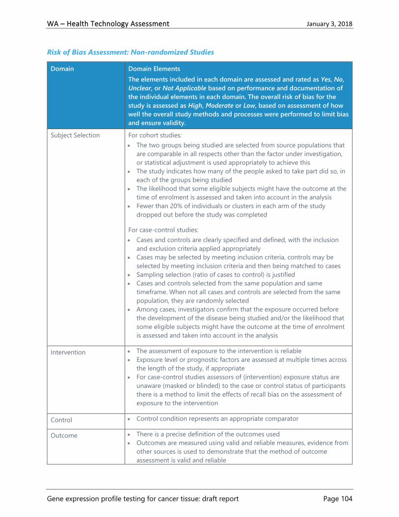
WA – Health Technology Assessment January 3, 2018
Gene expression profile testing for cancer tissue: draft report Page 104
Risk of Bias Assessment: Non-randomized Studies
Domain Domain Elements
The elements included in each domain are assessed and rated as Yes, No,
Unclear, or Not Applicable based on performance and documentation of
the individual elements in each domain. The overall risk of bias for the
study is assessed as High, Moderate or Low, based on assessment of how
well the overall study methods and processes were performed to limit bias
and ensure validity.
Subject Selection For cohort studies:
The two groups being studied are selected from source populations that
are comparable in all respects other than the factor under investigation,
or statistical adjustment is used appropriately to achieve this
The study indicates how many of the people asked to take part did so, in
each of the groups being studied
The likelihood that some eligible subjects might have the outcome at the
time of enrolment is assessed and taken into account in the analysis
Fewer than 20% of individuals or clusters in each arm of the study
dropped out before the study was completed
For case-control studies:
Cases and controls are clearly specified and defined, with the inclusion
and exclusion criteria applied appropriately
Cases may be selected by meeting inclusion criteria, controls may be
selected by meeting inclusion criteria and then being matched to cases
Sampling selection (ratio of cases to control) is justified
Cases and controls selected from the same population and same
timeframe. When not all cases and controls are selected from the same
population, they are randomly selected
Among cases, investigators confirm that the exposure occurred before
the development of the disease being studied and/or the likelihood that
some eligible subjects might have the outcome at the time of enrolment
is assessed and taken into account in the analysis
Intervention The assessment of exposure to the intervention is reliable
Exposure level or prognostic factors are assessed at multiple times across
the length of the study, if appropriate
For case-control studies assessors of (intervention) exposure status are
unaware (masked or blinded) to the case or control status of participants
there is a method to limit the effects of recall bias on the assessment of
exposure to the intervention
Control Control condition represents an appropriate comparator
Outcome There is a precise definition of the outcomes used
Outcomes are measured using valid and reliable measures, evidence from
other sources is used to demonstrate that the method of outcome
assessment is valid and reliable

WA – Health Technology Assessment January 3, 2018
Gene expression profile testing for cancer tissue: draft report Page 105
Domain Domain Elements
The elements included in each domain are assessed and rated as Yes, No,
Unclear, or Not Applicable based on performance and documentation of
the individual elements in each domain. The overall risk of bias for the
study is assessed as High, Moderate or Low, based on assessment of how
well the overall study methods and processes were performed to limit bias
and ensure validity.
Investigators use single outcome measures and do not rely on composite
outcomes, or the outcome of interest can be calculated from the
composite outcome
The study has an appropriate length of follow-up for the outcome
reported and groups are assessed at the same time points
Outcome reporting of entire group or subgroups is not selective
When patient-reported outcomes are used there is a method for
validating the measure
Masked Outcome
Assessment
The assessment of outcome(s) is made blind to exposure status. Where
outcome assessment blinding was not possible, there is recognition that
knowledge of exposure status could have influenced the assessment of
outcome
For case-control study: assessors of exposure status are unaware (masked
or blinded) of the case or control status of participant)
Confounding The main potential confounders are identified and taken into account in
the design and analysis of the study
Statistical Analysis Comparison is made between full participants and those who dropped
out or were lost to follow up, by exposure status
If the groups were not followed for an equal length of time, the analysis
was adjusted for differences in the length of follow-up
All major confounders are adjusted for using multiple variable logistic
regression or other appropriate statistical methods
Confidence intervals (or information with which to calculate them) are
provided
For case-control studies that use matching, conditional analysis is
conducted or matching factors are adjusted for in the analysis
Other Biases (as
appropriate)
List others in table footnote and describe, e.g.,
Sample size adequacy
Interest Disclosure Disclosures of interest are provided for authors/funders/commissioners
of the study
Interests are unlikely to significantly affect study validity
Funding Source There is a description of source(s) of funding
Funding source is unlikely to have a significant impact on study validity

WA – Health Technology Assessment January 3, 2018
Gene expression profile testing for cancer tissue: draft report Page 106
Risk of Bias Assessment: Economic Studies
Domain Domain Elements
The elements included in each domain are assessed and rated as Yes, No,
Unclear, or Not Applicable based on performance and documentation of the
individual elements in each domain. The overall risk of bias for the study is
assessed as High, Moderate or Low, based on assessment of how well the
overall study methods and processes were performed to limit bias and ensure
validity.
Target Population Target population and care setting described
Describe and justify basis for any target population stratification, identify
any a priori identifiable subgroups
If no subgroup analyses performed justify why they were not required
Perspective State and justify the analytic perspective (e.g., societal, payer, etc.)
Time Horizon Describe and justify the time horizon(s) used in the analysis
Discount Rate State and justify the discount rate used for costs and outcomes
Comparators Describe and justify selected comparators
Competing alternatives appropriate and clearly described
Modelling Model structure (e.g., scope, assumptions made) is described and justified
Model diagram provided, if appropriate
Model validation is described (may involve validation of different aspects
such as structure, data, assumptions, coding, etc., as well as different
validation models such as comparison with other models)
Data sources listed and assumptions for use justified
Statistical analyses are described
Effectiveness Estimates of efficacy/effectiveness of interventions are described and
justified
The factors that are likely to have an impact on effectiveness (e.g.,
adherence, diagnostic accuracy, values & preferences) are described and an
explanation of how they were factored into the analysis is included
The strength of evidence for the relationship between the intervention and
outcomes, and any necessary links, is described
Outcomes All relevant outcomes are identified, measured, and valued appropriately
(including harms/adverse events) for each intervention, and the justification
for information/assumptions is given
Any quality of life measures used in modelling are described and their use
justified
Any other outcomes that were considered, but rejected, are described with
the rationale for rejection
Ethical and equity related outcomes are considered and included when
appropriate

WA – Health Technology Assessment January 3, 2018
Gene expression profile testing for cancer tissue: draft report Page 107
Domain Domain Elements
The elements included in each domain are assessed and rated as Yes, No,
Unclear, or Not Applicable based on performance and documentation of the
individual elements in each domain. The overall risk of bias for the study is
assessed as High, Moderate or Low, based on assessment of how well the
overall study methods and processes were performed to limit bias and ensure
validity.
Resource Use/Costs All resources used are identified, valued appropriately, and included in the
analyses
Methods for costing are reporting (e.g., patient level)
Resource quantities and unit costs are both reported
Methods for costing time (e.g., lost time, productivity losses) are
appropriate and a justification is provided if time costs are not considered
Uncertainty Sources of uncertainty in the analyses are identified and justification for
probability distributions used in probabilistic analyses are given
For scenario analyses, the values and assumptions tested are provided and
justified
Results All results are presented in a disaggregated fashion, by component, in
addition to an aggregated manner
All results are presented with undiscounted totals prior to discounting and
aggregation
Natural units are presented along with alternative units (e.g., QALYs)
The components of the incremental cost-effectiveness ratio (ICER) are
shown (e.g., mean costs of each intervention in numerator and mean
outcomes of each intervention in the denominator)
Results of scenario analyses, including variability in factors such as practice
patterns and costs, are reported and described in relation to the reference
(base) case
Interest Disclosure Disclosures of interest are provided for authors/funders/commissioners of
the study
Interests are unlikely to significantly affect study validity
Funding Source There is a description of source(s) of funding
Funding source is unlikely to have a significant impact on study validity

WA – Health Technology Assessment January 3, 2018
Gene expression profile testing for cancer tissue: draft report Page 108
Risk of Bias Assessment: Clinical Practice Guidelines
Domain Domain Elements
Assessment indicates how well the guideline methodology and
development process were performed to limit bias and ensure validity for
elements in domain (each domain rated as Good, Fair, or Poor overall
based on performance and documentation of elements)
Scope and Purpose Objectives specifically described
Health question(s) specifically described
Population (patients, public, etc.) specified
Rigor of Evidence
Development
Systematic literature search
Study selection criteria clearly described
Strengths and limitations of individual studies and overall strength of the
body of evidence assessed
Rigor of
Recommendations
Development
Methods for developing recommendations clearly described
Explicit link between recommendations and supporting evidence and
includes strengths/limitations
Balance of benefits and harms (side effects, risks) considered
External review conducted
Updating procedure specified
Stakeholder
Involvement
Relevant professional groups are represented
Views and preferences of target population sought
Target users defined
Clarity and
Presentation
Recommendations specific, unambiguous
Different management options clearly presented
Key recommendations easily identifiable
Applicability and
Implementation
Provides advice and/or tools on how the recommendations can be put
into practice
Description of facilitators and barriers to application
Potential resource implications considered
Monitoring/audit/review criteria presented
Editorial Independence Views of funding body have not influenced the content of the guideline
Competing interests of members have been recorded, monitored, and
addressed appropriately

WA – Health Technology Assessment January 3, 2018
Gene expression profile testing for cancer tissue: draft report Page 109
Appendix C. Evidence Tables
Abbreviations Used in Evidence Tables
aOR: adjusted odds ratio
AS: active surveillance
BCI: Breast Cancer Index
CI: confidence interval
CT: chemotherapy
DCS: Decisional Conflict Scale
DI: decision impact
ECOG: Eastern Cooperative Oncology Group
ER: estrogen receptor
FACT-G v.4: Functional Assessment of Cancer
Therapy-General, version 4
FFPE: fixed paraffin-embedded
GEP: gene expression profile
HER2: human epidermal growth factor
receptor 2
HR: hazard ratio
HT: hormone treatment
IHC: immunohistochemistry
IMRT: Intensity-modulated radiation therapy
ITT: intention to treat
LN: lymph node
NCDB: National Cancer Data Base
N0: no nodal involvement
N1: 1-3 nodes
N2: 4-9 nodes
N3: > 9 or IC
NR: not reported
OR: odds ratio
pN0 (i-/i+): no axillary lymph node
involvement or isolated tumor cells
pN1mi: micro-metastases
PR: progesterone receptor
PSA: prostate-specific antigen
QALY: quality-adjusted life years
RP: radical prostatectomy
ROR: risk of Reoccurrence
RR: risk ratio
RS: recurrence score
RT: radiotherapy
SD: standard deviation
STAI: State-Trait Anxiety Inventory
STO-3: Stockholm Tamoxifen Trial

WA – Health Technology Assessment January 3, 2018
Gene expression profile testing for cancer tissue: draft report Page 110
Breast Cancer
Table 5. Evidence Table: Breast Cancer Systematic Reviews
Study Citation
Tests
Design
Study Inclusion Criteria
Outcomes
Included Studies
Characteristics Outcomes Reported
Risk of Bias
Comments
Augustovski
201519
Oncotype DX
Design:
Systematic review and meta-
analysis
Study inclusion criteria:
Studies that reported prospectively
data on decisions regarding
chemotherapy treatment before
and after availability of results of
the 21-gene assay (Oncotype DX)
in patients with early breast cancer
Studies of hypothetical decisions
and those with fewer than 80% of
LN-negative (LN- or N0) patients
or those not reporting LN status
were excluded
Outcomes:
Impact of test on clinical decision
making (proportion of patients
whose treatment decision was
altered after the test)
Net chemotherapy change
(difference in the number of
patients who were assigned to
receive chemotherapy before vs.
after the test)
For Decision Impact (DI)
outcome: 15 studies
For Net Chemotherapy
Change (NC) outcome: 15
studies
Characteristics:
Included studies conducted
in U.S., Canada, U.K.,
Australia, France, Germany,
Spain, Mexico, Israel, and
Japan
Total n = 2,229
7 studies had universal
enrollment (lower risk of
bias) of subjects and in 8 it
was selective (higher risk of
bias)
Global pooled DI:
29.52% (95% CI, 26.29 to 32.86%)
I2 = 59.50%
Pooled DI for studies with universal
enrollment
28.97% (95% CI, 26.65 to 31.34%)
I2 = 0.00%
Pooled DI for studies with selective
enrollment
29.43% (95% CI, 22.88 to 36.45%)
I2 = 74.09%
Global pooled NC
12.00% (95% CI, 8.00 to 16.00%)
I2 = 92.00%
Pooled NC for studies with universal
enrollment
9.00% (95% CI, 4.00 to 14.00%)
I2 = 89.00
Pooled NC for studies with selective
enrollment
16.00 (95% CI, 7.00 to 24.00)
Low
No specific protocol described
or registered
No list of excluded studies
Subgroup analysis of studies
with higher risk of bias from
selective enrollment and all
estimates for NC are highly
subject to heterogeneity,
making pooled estimates
suspect for their validity
For the Oncotype DX test
there is a high overlap of
included studies with the Blok
201820 and Scope 201721
reviews
Research was funded by a
grant from the test
manufacturer
No other author financial
relationships with test
manufacturer
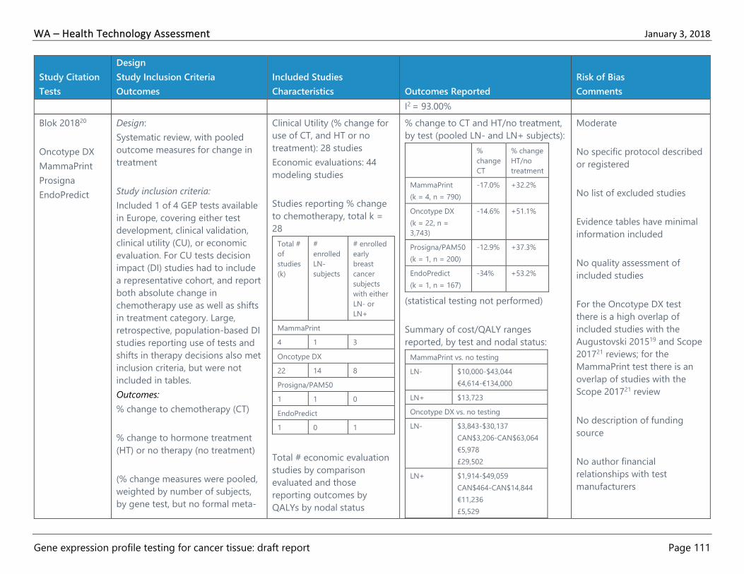
WA – Health Technology Assessment January 3, 2018
Gene expression profile testing for cancer tissue: draft report Page 111
Study Citation
Tests
Design
Study Inclusion Criteria
Outcomes
Included Studies
Characteristics Outcomes Reported
Risk of Bias
Comments
I2 = 93.00%
Blok 201820
Oncotype DX
MammaPrint
Prosigna
EndoPredict
Design:
Systematic review, with pooled
outcome measures for change in
treatment
Study inclusion criteria:
Included 1 of 4 GEP tests available
in Europe, covering either test
development, clinical validation,
clinical utility (CU), or economic
evaluation. For CU tests decision
impact (DI) studies had to include
a representative cohort, and report
both absolute change in
chemotherapy use as well as shifts
in treatment category. Large,
retrospective, population-based DI
studies reporting use of tests and
shifts in therapy decisions also met
inclusion criteria, but were not
included in tables.
Outcomes:
% change to chemotherapy (CT)
% change to hormone treatment
(HT) or no therapy (no treatment)
(% change measures were pooled,
weighted by number of subjects,
by gene test, but no formal meta-
Clinical Utility (% change for
use of CT, and HT or no
treatment): 28 studies
Economic evaluations: 44
modeling studies
Studies reporting % change
to chemotherapy, total k =
28
Total #
of
studies
(k)
#
enrolled
LN-
subjects
# enrolled
early
breast
cancer
subjects
with either
LN- or
LN+
MammaPrint
4 1 3
Oncotype DX
22 14 8
Prosigna/PAM50
1 1 0
EndoPredict
1 0 1
Total # economic evaluation
studies by comparison
evaluated and those
reporting outcomes by
QALYs by nodal status
% change to CT and HT/no treatment,
by test (pooled LN- and LN+ subjects):
%
change
CT
% change
HT/no
treatment
MammaPrint
(k = 4, n = 790)
-17.0% +32.2%
Oncotype DX
(k = 22, n =
3,743)
-14.6% +51.1%
Prosigna/PAM50
(k = 1, n = 200)
-12.9% +37.3%
EndoPredict
(k = 1, n = 167)
-34% +53.2%
(statistical testing not performed)
Summary of cost/QALY ranges
reported, by test and nodal status:
MammaPrint vs. no testing
LN- $10,000-$43,044
€4,614-€134,000
LN+ $13,723
Oncotype DX vs. no testing
LN- $3,843-$30,137
CAN$3,206-CAN$63,064
€5,978
£29,502
LN+ $1,914-$49,059
CAN$464-CAN$14,844
€11,236
£5,529
Moderate
No specific protocol described
or registered
No list of excluded studies
Evidence tables have minimal
information included
No quality assessment of
included studies
For the Oncotype DX test
there is a high overlap of
included studies with the
Augustovski 201519 and Scope
201721 reviews; for the
MammaPrint test there is an
overlap of studies with the
Scope 201721 review
No description of funding
source
No author financial
relationships with test
manufacturers

WA – Health Technology Assessment January 3, 2018
Gene expression profile testing for cancer tissue: draft report Page 112
Study Citation
Tests
Design
Study Inclusion Criteria
Outcomes
Included Studies
Characteristics Outcomes Reported
Risk of Bias
Comments
analysis was conducted and no
confidence intervals were
provided. Pooled % change
outcomes were not calculated by
nodal status group)
Economic evaluation outcomes
were reported in tabular form and
not pooled. Various studies
reported cost/QALY (in $USD,
CAN$, €, or £ depending on study)
cost change (increased, decreased,
savings), or whether the test
strategy was dominant in the
economic model. The only
comparable unit is QALY.
number
of
studies
(k)
LN- LN+ or
mixed
MammaPrint vs. no testing
(usual care, best practice or
Adjuvant! Online tool)
7 4 1
Oncotype DX vs. no testing
(usual care, best practice, or
Adjuvant! Online tool)
32 14 5
EndoPredict vs. no testing
(best practice)
1 0 1
Head-to-head comparisons
(Oncotype DX vs. Mammostrat
and MammaPrint vs.
Oncotype DX)
4 2
(1 each)
0
Oncotype DX vs. Mammostrat
LN- $453,600 (in favor of
Mammostrat)
Oncotype DX vs. MammaPrint
LN- €1,457 (in favor of
MammaPrint)
Scope 201721
Oncotype DX
MammaPrint
Mammostrat
(eligible, but no
studies
identified)
Design:
Systematic review, with narrative
synthesis
Study inclusion criteria:
Studies published in English
including patients with early
invasive breast cancer, tested with
Oncotype DX, MammaPrint, IHC4,
or Mammostrat compared to usual
care, no test, or statistical
Studies reporting change in
treatment recommendations,
k = 34
Oncotype DX: k = 28
MammaPrint: k = 6
(review did not locate studies
for other eligible tests
reporting this outcome)
[note: table 3, pg. 38-41
indicates that some studies
did not have a recorded pre-
test treatment
(Note: outcomes were not pooled or
reported quantitatively due to author
concerns about heterogeneity among
studies.)
Oncotype DX
“…the use of Oncotype DX leads to
changes in treatment
recommendations for between 21%
and 74% of all patients who
underwent Oncotype DX testing.”21(p.
37)
High
No specific protocol described
or registered
Inclusion criteria unclear (and
included studies, for example,
those with hypothetical
decisions, that were excluded
from Medline search results by
this WA HTA report’s authors

WA – Health Technology Assessment January 3, 2018
Gene expression profile testing for cancer tissue: draft report Page 113
Study Citation
Tests
Design
Study Inclusion Criteria
Outcomes
Included Studies
Characteristics Outcomes Reported
Risk of Bias
Comments
prognosis scoring using usual
clinical data only
Outcomes:
The extent to which test results are
used in treatment decisions
recommendation and for
others it was inferred
according to existing
guidelines or prediction
models]
“…change from chemotherapy to no
chemotherapy ranged from 6% to 51%
of all patients tested. However, in one
study more chemotherapy was used
after the introduction of Oncotype
DX.”21(p. 37)
“It was not clear in a large number of
the studies whether these figures
represented actual changes in the
treatments patients received.”21(p. 37)
MammaPrint
“…the use of MammaPrint in addition
to clinicopathological factors led to
changes in treatment
recommendations for between 18%
and 40% of all patients tested, and …
between 2% and 32% of all patients
would be recommended to change
from chemotherapy to no
chemotherapy.”21(p. 37)
“Again, in several of these studies it is
not clear if actual treatment changes
occurred following introduction of the
test.”21(p. 42)
because they did not meet
inclusion criteria)
No list of excluded studies
Evidence tables have minimal
information included
Limited narrative synthesis of
data
Article states that quality of
prognostic studies was
performed, but neither the
tables of those studies or ones
including studies treatment
change outcomes have
description of study quality
included, but some issues
related to methodological
quality of included studies are
described in narrative text
No description of funding
source
None of authors had financial
relationships with test
manufacturers

WA – Health Technology Assessment January 3, 2018
Gene expression profile testing for cancer tissue: draft report Page 114
Table 6. Evidence Table: Breast Cancer Randomized Controlled Trials
Study Citation
Tests
Design
Study Inclusion
Criteria
Outcomes
Included Studies
Characteristics Outcomes Reported
Risk of Bias
Comments
Bear 201717
Oncotype DX
Design:
Prospective study of
one group of
patients (n = 59
after 64 enrolled
and 5 excluded,
with n = 33
randomized)
Intervention:
Random
assignment of
patients (n = 17)
with Oncotype DX
recurrence score
(RS) of 11-25 to
neoadjuvant
hormone therapy
(NHT, Group B)
Comparator:
Random
assignment of
patients (n = 16) to
neoadjuvant
chemotherapy
(NCT, Group C)
(other groups not
randomized and
assigned with RS <
Female patients with HR+ (>
10% tumor staining by
immunohistochemistry),
HER2-negative, invasive
cancers ≥ 2 cm, diagnosed by
core needle biopsy, not
amenable to breast-
conserving surgery (BCS).
Patients had ECOG status of 0
or 1, and had ipsilateral nodal
imaging by U.S. or MRI within
6 weeks of enrollment
LN-negative: n = 24
LN-positive: n = 9
Patient Characteristics:
Median age,
yrs (range)
B: 64 (41,80)
C: 56.5 (30,75)
White
/African-
American
(%)
B: 95/5
C: 79/21
Tumor stage
T1/T2/T3/T4
(%)
B: 0/79/16/5
C: 8/62/31/0
Pre/Post-
menopausal
(%)
B: 16/84
C: 39/61
Clinical complete or partial response (cPR, cCR);
pathologic complete response (pCR) in the breast and
in the breast and nodes; successful breast conserving
surgery (BCS). Assessed by as treated analysis (B: n =
18; C: n = 11)
“After adjusting for effects from confounders, including
age, race, menopausal status, and site, Group B
patients had significantly lower clinical response rate,
compared with Group C (Ordinal regression: p =
.0369)”17(p. 921)
Response Results:
cCR (%) B: 22.2
C: 36.4
cPR (%) B: 27.8
C: 36.4
pCR breast (%) B: 6.0
C: 0
pCR breast +
nodes (%)
B: 0
C: 0
Successful BCS B: 72.2
C: 63.6
High
Of 33 women randomized
(17 to NHT and 16 to NCT), 5
(15%) refused NCT; of these
2 agreed to NHT instead
(crossover C to B)
Analysis was by treatment
received rather than ITT; no
description of the dropouts.
Groups B and C were not
similar at baseline by race,
tumor stage, nodal stage, or
menopausal status; Group C
more likely to be younger,
African-American, T3, and
premenopausal
Exact NHT or NCT regimens
at provider discretion and
not accounted for in analysis
No explanation for the non-
standard cutoffs (11 and 25
rather than 18 and 30, which
are typical thresholds for
low-risk and high-risk RS)

WA – Health Technology Assessment January 3, 2018
Gene expression profile testing for cancer tissue: draft report Page 115
Study Citation
Tests
Design
Study Inclusion
Criteria
Outcomes
Included Studies
Characteristics Outcomes Reported
Risk of Bias
Comments
11 to NHT (Group
A) and with RS ≥ 26
to NCT (Group D),
these data not
reported in table)
Setting:
Pilot feasibility
study within a larger
multicenter trial at 7
U.S. and Canadian
centers
Nodal stage
N0/N1/NX
(%)
B: 74/31/5
C: 69/31/0
Authors’ conclusion that
“patients with low RS results
derive little or no benefit
from adding chemotherapy”
has no basis in the data, as
there were no subjects with
low RS who received
chemotherapy
Funding from Genomic
Health + NIH; one author
employed by Genomic
Health; lead author received
speaking and advisory board
honoraria from Genomic
Health
Cardoso 201618
MammaPrint
(70-gene
signature, 70-
GS and
Adjuvant!
Online
Design:
Randomized, phase
3 study (only
patients with
discordant clinical
and genomic risks
were randomized)
Intervention:
Chemotherapy
based on genomic
risk (determined by
MammaPrint) and
clinical risk
Study Population:
Patients (n = 6,693, ITT n =
2,187): Women 18 to 70 years
of age with early stage,
histologically confirmed
primary invasive breast
cancer (stage T1 or T2 or
operable T3)
LN-negative: n = 1,309
LN-positive: n = 752
Patients with high clinical risk and low genomic risk, no
adjuvant chemotherapy:
Rate of survival without distant metastasis at 5 years:
94.7% (95% CI, 92.5 to 96.2)
ITT population:
High clinical risk, low genomic risk, randomized to
chemotherapy based on clinical risk:
5-year rate of survival without distant metastasis:
95.9% (95% CI, 94.0 to 97.2)
High clinical risk, low genomic risk, randomized to no
chemotherapy based on genomic risk:
Moderate
Change in RNA-extraction
solution for MammaPrint test
caused a temporary shift in
the risk calculation from May
24, 2009 to January 30, 2010,
which led to more patients
receiving chemotherapy
before this was corrected
Financial support from
Novartis, F. Hoffmann-La

WA – Health Technology Assessment January 3, 2018
Gene expression profile testing for cancer tissue: draft report Page 116
Study Citation
Tests
Design
Study Inclusion
Criteria
Outcomes
Included Studies
Characteristics Outcomes Reported
Risk of Bias
Comments
(determined by
Adjuvant! Online)
Comparator:
No chemotherapy
based on genomic
risk (determined by
MammaPrint) and
clinical risk
(determined by
Adjuvant! Online)
Setting: 112
institutions in nine
European countries
Physician n and
characteristics not reported
Patient Characteristics:
Median age,
y (range)
55 (23 to
71)
% white NR
% ER-, PR-,
or both
positive
88.4
% HER2-
positive
9.5
Tumor
grade (%)
Grade 1
Grade 2
Grade 3
Missing
data
21.6
49.1
28.8
0.5
Lymph
node status
(%)
Negative
1 node
2 nodes
3 nodes
≥ 4 nodes
79.0
14.1
4.5
2.3
0.1
5-year rate of survival without distant metastasis:
94.4% (95% CI, 92.3 to 95.9)
Adjusted hazard ratio for distant metastasis or death
(chemotherapy vs. no chemotherapy): 0.78 (95% CI,
0.50 to 1.21)
Low clinical risk, high genomic risk, randomized to
chemotherapy based on genomic risk:
5-year rate of survival without distant metastasis:
95.8% (95% CI, 92.9 to 97.6)
Low clinical risk, high genomic risk, randomized to no
chemotherapy based on clinical risk:
5-year rate of survival without distant metastasis:
95.0% (95% CI, 91.8 to 97.0)
Adjusted hazard ratio for distant metastasis or death
(chemotherapy vs. no chemotherapy): 1.17 (95% CI,
0.59 to 2.28)
“Among all patients at high clinical risk, the use of the
70-gene signature to guide chemotherapy treatment
would lead to a reduction in the use of adjuvant
chemotherapy in 1550 of 3356 patients (46.2%).”18(p. 724)
5-year survival without distant metastasis (clinical-risk
strategy alone vs. genomic-risk strategy alone): 95.0%
vs. 94.7%
Roche, Sanofi-Aventis, Eli
Lilly, and Veridex
2 authors financially affiliated
with test manufacturer

WA – Health Technology Assessment January 3, 2018
Gene expression profile testing for cancer tissue: draft report Page 117
Study Citation
Tests
Design
Study Inclusion
Criteria
Outcomes
Included Studies
Characteristics Outcomes Reported
Risk of Bias
Comments
Subgroup analyses:
LN-negative, 5-year survival without distant metastasis
(chemotherapy vs. no chemotherapy): 95.7% (95% CI,
93.0 to 97.4) vs. 93.2% (95% CI, 90.1 to 95.4)
LN-positive, 5-year survival without distant metastasis
(chemotherapy vs. no chemotherapy): 96.3% (95% CI,
93.1 to 98.1) vs. 95.6% (95% CI, 92.7 to 97.4)
ER-positive, HER2-negative, LN-negative disease, 5-
year survival without distant metastasis (chemotherapy
vs. no chemotherapy): 95.5% (95% CI, 92.5 to 97.3) vs.
93.9% (95% CI, 90.6 to 96.1)
In multivariate analysis, adjusting for chemotherapy
use, clinical risk, and patient and tumor characteristics,
HR for distant metastasis or death with chemotherapy
vs. no chemotherapy in patients with high vs. low
genomic risk using MammaPrint test: HR 2.41 (95% CI,
1.79 to 3.26)

WA – Health Technology Assessment January 3, 2018
Gene expression profile testing for cancer tissue: draft report Page 118
Table 7. Evidence Table: Breast Cancer Observational Studies
Study Citation
Test
Design
Intervention
Comparator
Setting
Study Population
Characteristics Outcomes
Risk of Bias
Comments
Evans 201622
Oncotype DX
Design:
Before-after study,
with single group of
patients
Intervention:
Treatment decision
based on Oncotype
DX test
Comparator:
Treatment decision
based on other
factors without test
Setting:
Women recruited
2011-2015 from 4
sites in Washington,
DC
Study Population:
Females with recently diagnosed
stage 1 or stage 2 breast cancer
and had Oncotype DX testing
ordered by treating clinician; n =
193 who completed pre- and
post-interview before and after
receiving test results
Physician n and characteristics
not reported
Patient Characteristics:
Mean age (y,
SD)
57.14 (9.87)
% white 65.4
Histological
grade (%)
Low
Intermediate
High
Missing/Undefi
ned
23.8
42.0
16.6
17.6
Nodal status
(%)
Positive
Negative
Missing/Undefi
ned
8.3
80.3
11.4
Changes in patient-reported outcomes:
Perception of pros and cons of
chemotherapy both increased (t = 4.27
and t = 8.54, respectively), while
perceived risk of recurrence decreased.
Test did not impact these changes in
any category. Distress did not change
with test (i.e., patients were not
reassured, but not more distressed
either).
Predictors of chemotherapy use:
Recurrence score:
High: 88%
Intermediate: 57%, OR 0.04 (95% CI,
0.01 to 0.27) (vs. high)
Low: 5%, OR 0.01 (95% CI, 0.00 to 0.02)
(vs. high)
Older age: OR 0.89 (0.83 to 0.95)
Pre-test distress: OR 0.47 (95% CI, 0.21
to 1.07)
Pre-test perceived risk: OR 0.47 (95% CI,
0.98 to 1.06)
High
Recruitment limited to clinics in the
Eastern U.S.
Some patients unreachable for pre-test
interview; these patients may have
differed from the women who were
able to be reached to assess for
eligibility and consent to a pre-test
interview
Study funded by grants from the
American Cancer Society, the National
Cancer Institute
Authors declare no conflict of interest

WA – Health Technology Assessment January 3, 2018
Gene expression profile testing for cancer tissue: draft report Page 119
Study Citation
Test
Design
Intervention
Comparator
Setting
Study Population
Characteristics Outcomes
Risk of Bias
Comments
Pre-test chemotherapy pros: OR 1.19
(95% CI, 0.66 to 2.16)
Pre-test chemotherapy cons: OR 0.74
(95% CI, 0.39 to 1.38)
Post-test distress: OR 2.19 (95% CI, 1.05
to 4.57)
Post-test perceived risk: OR 1.01 (95%
CI, 0.98 to 1.03)
Post-test chemotherapy pros: OR 1.83
(95% CI, 0.95 to 3.50)
Post-test chemotherapy cons: OR 0.50
(95% CI, 0.26 to 0.97)
Friese 201723
Li 2017121
Oncotype DX
Design:
Retrospective cohort
study using registry
with prospectively
collected data
Intervention:
Treatment decision
based on Oncotype
DX test
Comparator:
Study Population:
Women (n = 1,527, intervention
n = 778) aged 20 to 79 years,
diagnosed with early-stage (1/2)
breast cancer (ER/PR-positive,
HER2-negative), treated in 2013
to 2014
LN-negative: n = 1,214
LN-positive: n = 303
Missing: n = 10
Chemotherapy recommendations in
overall study population:
Oncologist recommended against
chemotherapy: 47.2%
Chemotherapy decision left to patient:
12.3%
Oncologist recommended
chemotherapy: 40.5%
Oncotype DX Test Group:
Most patients with RS high test results
(scores 31-100) received
Moderate
Results are limited to two geographic
regions in the United States
Measures of communication and
decision making were determined
through patient reports and do not
necessarily represent physician
perspectives

WA – Health Technology Assessment January 3, 2018
Gene expression profile testing for cancer tissue: draft report Page 120
Study Citation
Test
Design
Intervention
Comparator
Setting
Study Population
Characteristics Outcomes
Risk of Bias
Comments
Treatment decision
based on other
factors (no Oncotype
DX test)
Setting:
Los Angeles County
and Georgia
Surveillance,
Epidemiology, and
End Results registries
Physician n and characteristics
not reported
Patient Characteristics:
Mean age (y) at
diagnosis
61.0
% white 56.9
Clinical group: b
LN-negative 1
LN-negative 2
LN-positive
Missing
60.1
19.4
19.8
0.7
Comorbidities (%)
No diagnosis
1 condition
2+ conditions
Missing
72.2
21.5
5.8
0.6
Site (%)
Georgia
Los Angeles County
54.9
45.1
recommendations for chemotherapy
(86.9% to 96.5% across clinical groups).
65.9% to 78.2% of women with LN-
negative disease (majority with low-risk
scores of 0-18) received a
recommendation against chemotherapy.
LN-negative, favorable-risk disease,
recommendation against chemotherapy:
78.2%
LN-negative, favorable-risk disease,
recommendation for chemotherapy:
11.7%
No Test Group:
LN-negative, more favorable-risk
disease, recommendation for
chemotherapy: 23.1%
LN-negative, less favorable-risk disease,
recommendation for chemotherapy:
60.2%
LN-positive, recommendation for
chemotherapy: 83.2%
Chemotherapy Receipt:
Nonresponse bias and missing data are
problematic, but were partially
mitigated by the analyses
Study funded through grants from the
NIH, California Department of Health,
and the CDC
One author reported research funding
from multiple sources including the
test manufacturer

WA – Health Technology Assessment January 3, 2018
Gene expression profile testing for cancer tissue: draft report Page 121
Study Citation
Test
Design
Intervention
Comparator
Setting
Study Population
Characteristics Outcomes
Risk of Bias
Comments
High test score and receipt of
chemotherapy:
LN-negative, more favorable: 87.0%
LN-negative, less favorable: 91.1%
LN-positive: 100%
Low test score and receipt of
chemotherapy:
LN-negative, more favorable: 2.9%
LN-negative, less favorable: 9.5%
LN-positive: 26.6%
Low-risk test vs. no test: OR 0.1 (95% CI,
0.1 to 0.2)
Medium-risk test vs. no test: or 1.4 (95%
CI, 1.1 to 1.7)
High-risk test vs. no test: OR 2.8 (95% CI,
2.0 to 4.0)
Patient Perceptions:
63.9% of patients who received test
found it “very” or “extremely” helpful
65.0% of women with low-risk results
reported the test results shifted their
opinion against chemotherapy
73.1% of women with high-risk results
reported the test results shifted their
opinion toward receiving chemotherapy

WA – Health Technology Assessment January 3, 2018
Gene expression profile testing for cancer tissue: draft report Page 122
Study Citation
Test
Design
Intervention
Comparator
Setting
Study Population
Characteristics Outcomes
Risk of Bias
Comments
Hequet 201734
Prosigna
(PAM50)
Design:
Before-after study, of
a single group of
patients
Intervention:
Treatment
recommendation
after genetic test
results
Comparator:
Treatment
recommendation for
same patient
submitted before
genetic test result
available
Setting:
Multi-center study at
8 hospitals in France
Study Population:
Patients (n = 210):
Postmenopausal women with
ER-positive, HER2-negative, and
LN-negative tumors, stage 1 or
stage 2, diagnosed from March
2015 to January 2016
Physician n and characteristics
not stated
Patient Characteristics:
Mean age 61.9 (no SD)
% PR-
positive
86
TNM stage
(%)
1
2
79
21
Treatment recommendation change after
receiving test results (before vs. after
test):
Overall: 18% (34/194)
From No adjuvant chemotherapy to
adjuvant chemotherapy: 13% (25/194)
From adjuvant chemotherapy to no
adjuvant chemotherapy: 5% (9/194)
# with change in adjuvant
chemotherapy (CT) treatment
recommendation after test result (n =
34):
No CT to
CT
CT to no CT
Low % (n) 0 7
Intermediate
% (n)
10 2
High % (n) 15 0
Change in physician confidence in
treatment recommendation after test
results, % (n):
Increased confidence: 39% (75/192)
No change: 51% (98/192)
Decreased confidence: 11% (21/192)
High
Study reports recommended
treatments rather than actual
treatments
Prior physician experience using test
not reported
Data on 6 month follow-up
questionnaires not fully reported,
including, actual treatment received,
number of physicians who completed
follow-up visit, and DCS measures
Study funded by test manufacturer
4 of 23 authors employed by or funded
by test manufacturer

WA – Health Technology Assessment January 3, 2018
Gene expression profile testing for cancer tissue: draft report Page 123
Study Citation
Test
Design
Intervention
Comparator
Setting
Study Population
Characteristics Outcomes
Risk of Bias
Comments
Patient (n = 171) scores on STAI “state-
anxiety” measure decreased after test
results from a mean of 43.3 (SD 11.6) to
41.5 (12.7), a mean decrease of 1.8
(10.3) (p = .02).
Patient (n = 158) overall scores on DCS
decreased after test results from a mean
of 9.8 (SD 10.6) to 6.2 (7.7), a mean
decrease of 3.5 (10.7) (p < .001)
Patient (n = 151) overall scores on the
functional assessment (FACT-G v.4)
increased after test results from a mean
of 79.4 (13.3) to 80.2 (15.2), a mean
increase of 0.9 (9.5) (p = .264), and at 6
months post-diagnosis there was an
overall decrease to below baseline level
with FACT-G score of 76.77 (n = 161), no
statistical testing reported
Concordance on Prosigna test subtyping
and ROR between central and
replication laboratories:
Tumor subtyping: 94% (kappa = 0.88)
ROR: 91% (kappa = 0.86)
Jasem 201624
Design:
Retrospective cohort
study, using data
Study Population:
Women (n = 143,032,
intervention n = 77,308) with
Use of adjuvant chemotherapy based on
test score in patients who received test:
High

WA – Health Technology Assessment January 3, 2018
Gene expression profile testing for cancer tissue: draft report Page 124
Study Citation
Test
Design
Intervention
Comparator
Setting
Study Population
Characteristics Outcomes
Risk of Bias
Comments
Oncotype DX from the National
Cancer Data Base
(NCDB)
Intervention:
Treatment decision
based on Oncotype
DX test results
Comparator:
Treatment decision
based on other
factors (no test
performed)
Setting:
NCDB registry
(captures about 70%
of newly diagnosed
cancers in the United
States submitted
from about 1,500
hospitals)
ER-positive, LN-negative, early-
stage breast cancer, diagnosed
from 2004 to 2012; analysis for n
= 121,128
Physician n and characteristics
not reported
Patient Characteristics:
Test
(n =
55,404)
No Test
(n =
65,724)
Age (y)
< 45
45-64
65+
57.7
55.5
31.5
42.3
44.5
68.5
%
female
45.8 54.2
%
white
45.9 54.1
%
govern
mental
insura
nce
34.4 65.6
Grade
(%)
1
2
3
Unkno
wn
38.6
49.8
49.8
43.3
61.4
50.2
50.2
56.7
Intermediate test score: aOR 12.00 (95%
CI, 11.38 to 12.65)
High test score: aOR 83.07 (95% CI,
74.25 to 92.94)
Use of adjuvant chemotherapy in
patients with vs. without testing, aOR
(95% CI):
Age, 45-64 vs. < 45 y: 0.45 (0.40 to 0.51)
vs. 0.31 (0.29 to 0.34)
Age, 65+ vs. < 45 y: 0.23 (0.21 to 0.25)
vs. 0.08 (0.07 to 0.09)
Gender, female vs. male: 1.23 (0.92 to
1.65) vs. 0.79 (0.63 to 0.98)
Race, black vs. white: 1.13 (1.03 to 1.23)
vs. 1.29 (1.21 to 1.38)
Insurance, governmental vs. private: 0.88
(0.82 to 0.94) vs. 0.94 (0.88 to 1.04)
Tumor grade, 2 vs. 1: 1.63 (1.54 to 1.74)
vs. 2.45 (2.31 to 2.60)
Tumor grade, 3 vs. 1: 3.06 (2.83 to 3.31)
vs. 9.12 (8.50 to 9.78)
Younger African-American women more
likely to receive adjuvant chemotherapy
despite low RS (aOR, 1.33; 95% CI, 1.16
to 1.54)
Coding errors may have occurred since
the test is new
Test use may be underreported or
erroneously reported
NCDB coverage is lower for elderly
patients, Hispanics, Alaskan natives,
and American Indians, which limits the
generalizability of these findings in
these populations
The study did not include data on
patients tested after 2012 due to a lag
in National Cancer Data Base data;
factors associated with use of the test
may have been different in 2012 due
to wider availability, better accessibility,
and higher adoption rates
Lower representation of uninsured
patients in NCDB compared to the
general population
Study funding not stated
4 of 7 study authors are affiliated with
industry, but none have declared
affiliations with test manufacturer

WA – Health Technology Assessment January 3, 2018
Gene expression profile testing for cancer tissue: draft report Page 125
Study Citation
Test
Design
Intervention
Comparator
Setting
Study Population
Characteristics Outcomes
Risk of Bias
Comments
Jasem 201725
Oncotype DX
Design: Retrospective
cohort using the
analysis of National
Cancer Data Base
(NCDB)
Intervention:
Oncotype DX testing
prior to treatment
Comparator:
No Oncotype DX
testing prior to
treatment
Setting: The NCDB
breast cancer
participant use data
file from 2010 to
2012. The NCDB
includes information
on 70% of newly
diagnosed cancer
cases nationwide
annually and collects
information
submitted from more
than 1500
Commission on
Study Population: (n = 32,425)
Women and men with LN-
positive breast cancer diagnosed
between January 2010 and
December 2012. Patients with
pT2, pT2, pN1 (with 1-3 lymph
nodes involved), HR-positive,
HER2-negative.
Patients with test ordered: n =
10,434
Patients without test ordered: n
= 21,991
LN-positive: n = 32,425
Patient Characteristics:
Test No
Test
Age, years,
%
< 45
45 – 64
≥ 65
22
34
34
78
66
66
% white 33 67
Male
Female
22
32
78
68
Charlson
co-
Factors associated with whether the test
was ordered:
Older patients more likely to get the test
than younger patients: Adjusted odds
ratio (aOR), 1.52, (95% CI, 1.36 to 1.71)
Women more likely to get the test than
men: aOR, 1.65, (95% CI, 1.29 to 2.12)
Patients diagnosed in 2012 were more
likely to get the test than those
diagnosed in 2010: aOR, 1.72 (95% CI,
1.61 to 1.83)
African-American patients were less
likely to get the test than patients of
other races: aOR 0.77 (95% CI, 0.70 to
0.84)
Patients with grade 2 (aOR, 0.81; 95% CI,
0.76 to 0.86) and grade 3 cancer (aOR,
0.47; 95% CI, 0.76 to 0.86) were less
likely to get the test than patients with
grade 1 cancer
Patients with two nodes (aOR, 0.54; 95%
CI, 0.50 to 0.57) and thee nodes (aOR,
0.29; 95% CI, 0.26 to 0.32) involved were
High
Retrospective analysis; potential for
data recording to be inaccurate
Study funding not stated
Authors stated they had no financial
conflicts of interest

WA – Health Technology Assessment January 3, 2018
Gene expression profile testing for cancer tissue: draft report Page 126
Study Citation
Test
Design
Intervention
Comparator
Setting
Study Population
Characteristics Outcomes
Risk of Bias
Comments
Cancer-accredited
facilities
morbidity
score, %
0
≥ 1
32
31
68
69
Insurance,
%
None
Private
Govt.
25
33
32
76
67
68
Year
diagnosis,
%
2010
2011
2012
26
33
37
74
67
63
Grade, %
1
2
3
41
34
21
59
66
79
Tumor size,
%
pT1
pT2
38
25
62
75
# of nodes
involved, %
1
2
3
38
24
14
62
76
86
less likely to get the test than patients
with only one node involved
Difference in patients receiving
chemotherapy based on whether they
received test:
Test ordered: 38% chemotherapy
Test not ordered: 75% chemo
aOR, 0.21 (95% CI, 0.20 to 0.22)
Relationship of RS to chemotherapy
treatment:
Patients with an intermediate RS (aOR,
4.51; 95% CI, 4.51 to 5.01) and high RS
(aOR, 19.79; 95% CI, 15.39 to 92.94)
more likely to receive chemotherapy
than patients with low RS
Kuijer 201632
MammaPrint
Design:
Retrospective cohort
study
Study population:
Female patients (n = 2,043) with
primary breast cancer, older
Administration of chemotherapy in
overall study population (n = 2,043),
linear regression adjusted absolute
High

WA – Health Technology Assessment January 3, 2018
Gene expression profile testing for cancer tissue: draft report Page 127
Study Citation
Test
Design
Intervention
Comparator
Setting
Study Population
Characteristics Outcomes
Risk of Bias
Comments
Intervention:
Treatment
recommendation
based on gene
signature
(determined by
MammaPrint)
Comparator:
Treatment
recommendation
based on
conventional
prognostic
clinicopathological
factors
Setting:
Registry enrolling
patients from
practices in the
Netherlands
than 17 years of age, surgically
treated between November
2011 and October 2013, and had
no prior history of malignancy,
neoadjuvant treatment, or
distant metastasis upon
diagnosis
LN-negative: n = 1,584
LN-positive: n = 459
Physician n and characteristics
not reported
Patient characteristics:
Test
n =
298
No Test
n =
1,745
Mean age (y) at
diagnosis (SD)
55.6
(8.3)
56.2
(8.8)
% white NR NR
Pathological
axillary status
(%):
pN0 (i-/i+)
pN1mi
80
20
77
23
Invasive tumor
grade (%):
Grade 1
Grade 2
14
86
14
86
difference, % (95% CI): -9.5% (95% CI, -
15.7 to -3.3)
Subgroup analysis (group status):
Administration of adjuvant
chemotherapy:
Group A (N0, grade 1, > 2 cm; n = 19): -
24.0% (95% CI, -46.5 to -1.6)
Group B (N0, grade 2, > 1 cm; n = 220):
-5.8% (95% CI, -13.0 to 1.3)
Group C (pN1mi, grade 1/2; n = 59): -
16.0% (95% CI, -28.0 to -4.0)
Subgroup analysis (age):
Administration of adjuvant
chemotherapy:
< 50 years: -26.9% (95% CI, -38.3 to -
15.6)
50 to 59 years: -9.2% (95% CI, -18.7 to
0.3)
60 to 69 years: 3.5% (95% CI, -5.9 to
12.9)
Patient clustering (by hospital or
physician preference) may have
influenced the results (authors tried to
mitigate this by using linear mixed-
effects modeling)
Patient age may have influenced a
physician’s decision to administer
chemotherapy (more aggressive
treatment in younger women); unable
to determine this due to study design
Confounding by indication may be an
issue (although authors attempted to
mitigate this through IV analyses)
Study funded by the Dutch Cancer
Society
Authors reported no financial conflicts
of interest
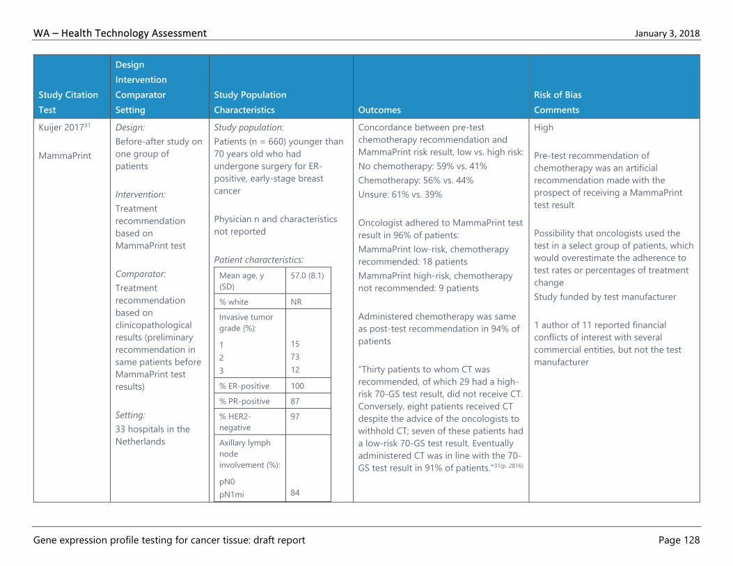
WA – Health Technology Assessment January 3, 2018
Gene expression profile testing for cancer tissue: draft report Page 128
Study Citation
Test
Design
Intervention
Comparator
Setting
Study Population
Characteristics Outcomes
Risk of Bias
Comments
Kuijer 201731
MammaPrint
Design:
Before-after study on
one group of
patients
Intervention:
Treatment
recommendation
based on
MammaPrint test
Comparator:
Treatment
recommendation
based on
clinicopathological
results (preliminary
recommendation in
same patients before
MammaPrint test
results)
Setting:
33 hospitals in the
Netherlands
Study population:
Patients (n = 660) younger than
70 years old who had
undergone surgery for ER-
positive, early-stage breast
cancer
Physician n and characteristics
not reported
Patient characteristics:
Mean age, y
(SD)
57.0 (8.1)
% white NR
Invasive tumor
grade (%):
1
2
3
15
73
12
% ER-positive 100
% PR-positive 87
% HER2-
negative
97
Axillary lymph
node
involvement (%):
pN0
pN1mi
84
Concordance between pre-test
chemotherapy recommendation and
MammaPrint risk result, low vs. high risk:
No chemotherapy: 59% vs. 41%
Chemotherapy: 56% vs. 44%
Unsure: 61% vs. 39%
Oncologist adhered to MammaPrint test
result in 96% of patients:
MammaPrint low-risk, chemotherapy
recommended: 18 patients
MammaPrint high-risk, chemotherapy
not recommended: 9 patients
Administered chemotherapy was same
as post-test recommendation in 94% of
patients
“Thirty patients to whom CT was
recommended, of which 29 had a high-
risk 70-GS test result, did not receive CT.
Conversely, eight patients received CT
despite the advice of the oncologists to
withhold CT; seven of these patients had
a low-risk 70-GS test result. Eventually
administered CT was in line with the 70-
GS test result in 91% of patients.”31(p. 2816)
High
Pre-test recommendation of
chemotherapy was an artificial
recommendation made with the
prospect of receiving a MammaPrint
test result
Possibility that oncologists used the
test in a select group of patients, which
would overestimate the adherence to
test rates or percentages of treatment
change
Study funded by test manufacturer
1 author of 11 reported financial
conflicts of interest with several
commercial entities, but not the test
manufacturer

WA – Health Technology Assessment January 3, 2018
Gene expression profile testing for cancer tissue: draft report Page 129
Study Citation
Test
Design
Intervention
Comparator
Setting
Study Population
Characteristics Outcomes
Risk of Bias
Comments
pN1a
> pN1a
Nx
10
5
1
1
Treatment recommendation change
after MammaPrint test, % of patients:
51% (95% CI, 46% to 56%)
Actually administered treatment (CT)
deviated from preliminary
recommendation in 52% of patients
Subgroup analyses:
Discordance between preliminary
chemotherapy recommendation and
MammaPrint risk results:
Patients with tumors > 2 cm (n = 146):
56%
(k, -0.04; 95% CI, -0.24 to 0.17)
Patients with axillary lymph node
involvement: 62% (k, -0.10; 95% CI, -0.33
to 0.13)
Loncaster
201726
Oncotype DX
Design:
Before-after study of
same group of
patients
Intervention:
Treatment given after
genetic test results
Comparator:
Study Population: (n = 201)
Women with newly diagnosed
breast cancer referred post-
surgery to an oncologist for
chemotherapy by a breast
cancer multi-disciplinary team.
ER+ (quick score ≥ 5/8) and
HER2 0, 1 + or non-amplified;
LN-negative or LN-positive for
post-menopausal women,
PREDICT estimation of overall
Reduction in the number of patients
recommended for chemotherapy after
test results, prior to test, all 201 patients
recommended for chemo.
After test, 63% (127/201) did not receive
chemotherapy
Reduction in the number of LN-positive
women (n = 65) receiving chemotherapy
after test results: (Note: at time NICE
High
The study included 201 patients, 82
from a prospective pilot study of the
genetic test from May to December
2012 and then a retrospective review
of patient files from December 2012 to
March 2015
Authors mislabeled data in Table 2

WA – Health Technology Assessment January 3, 2018
Gene expression profile testing for cancer tissue: draft report Page 130
Study Citation
Test
Design
Intervention
Comparator
Setting
Study Population
Characteristics Outcomes
Risk of Bias
Comments
Treatment
recommendation for
same patient by
multidisciplinary
tumor board
submitted before
genetic test result
available
Setting:
Patients treated for
early breast cancer in
Greater Manchester,
United Kingdom
benefit from chemotherapy >
3%
LN-negative: n = 136
LN-positive: n = 65
Patient Characteristics:
Age, mean
(SD)
55.2 (10)
Tumor grade,
% (n)
1
2
3
1% (2/201)
52% (104/201)
46 %(93/201)
Tumor size,
mm, mean
(SD)
26.1 (14.3)
Node positive,
%
32%
PREDICT
score, mean
(SD)
5.8 (2.4)
RS result,
mean (SD)
20.5 (10.2)
RS result, %
(n)
Low
Intermed.
High
43% (86/201)
44% (89/201)
13% (26/201)
guidelines did not allow for genetic
testing of LN-positive women)
After test, 69% of LN-positive patients
did not receive chemotherapy
compared to 63% overall
% LN-positive women receiving
chemotherapy (CT) by RS score:
CT No CT
Low 8 92
Intermediate 63 37
High 83 17
Estimated overall budget savings from
patients not receiving chemotherapy
(including costs of testing all patients) £
266,427
Estimated budget savings from LN-
positive patients not receiving
chemotherapy (including costs of
testing all patients) £ 110,452
Study designed and funded by test
manufacturer and test manufacturer
revised initial manuscript draft
4 of 8 authors report consulting or
serving on the speaker’s bureau for the
test manufacturer
O’Neill 201727
Design: Study Population: 10% of GEP-tested women did not
initiate therapy
High

WA – Health Technology Assessment January 3, 2018
Gene expression profile testing for cancer tissue: draft report Page 131
Study Citation
Test
Design
Intervention
Comparator
Setting
Study Population
Characteristics Outcomes
Risk of Bias
Comments
Oncotype DX Retrospective cohort
study of claims data
Intervention:
Treatment decision
based on Oncotype
DX test
Comparator:
Treatment decision
based on other
factors (no Oncotype
DX testing)
Setting:
Five state cancer
registries with linked
claims data and GEP
results from 2006 to
2010
Commercially insured women (n
= 5,014) age 24 to 63 years,
newly diagnosed with stage 1 or
2 HR-positive, HER2 non-
positive breast cancer
Physician n and characteristics
not reported
Patient Characteristics:
Age at
diagnosis, y
(%)
24-39
40-49
50-59
60-63
7.2
32.3
41.7
18.8
% white 85.0
Stage (%)
1
2
60.5
39.5
% HER2-
negative
45.5
% ER- and
PR-positive
85.7
Histological
grade (%)
1-2
3
Missing
74.5
21.5
4.0
Initiation of endocrine therapy, low vs.
high RS women: OR 0.40 (95% CI, 0.20
to 0.81)
Predicted probability of endocrine
initiation:
Tested patients vs. untested patients
with chemotherapy initiated within 6
months: 90.4% vs. 87.6%
Tested patients vs. untested patients
without chemotherapy initiated within 6
months: 90.2% vs. 74.3%
16% of test-eligible women did not
initiate endocrine therapy
Receipt of Oncotype DX testing and
likelihood of endocrine therapy
initiation: 10% vs. 19% non-initiation;
OR 2.48 (95% CI, 2.03 to 3.04)
Test status was not significantly related
to discontinuation of endocrine therapy:
OR 0.93 (95% CI, 0.85 to 1.02)
Participants represent a subset of the
commercially insured U.S. population
(from CA, GA, KY, NY, and OH)
The majority of participants were from
urban areas and non-white individuals
were underrepresented in the data set
There is the potential for coding errors
in claims data
High proportion (> 50%) of patients
with unknown HER2 status
The study was unable to account for
variables related to survivorship care
(e.g., rates and severity of side effects
from endocrine therapy), receipt of
care in academic centers vs.
community centers, and other
unmeasured patient-level variables
Study funded by American Cancer
Society and National Cancer Institute
3 authors had affiliations with
commercial entities at the time of the
study, including 2 with the test
manufacturer

WA – Health Technology Assessment January 3, 2018
Gene expression profile testing for cancer tissue: draft report Page 132
Study Citation
Test
Design
Intervention
Comparator
Setting
Study Population
Characteristics Outcomes
Risk of Bias
Comments
Test risk category was not associated
with non-adherence: OR 0.88 (95% CI,
0.76 to 1.02)
Parsons 201628
Oncotype DX
Design: Retrospective
analysis of National
Cancer Data Base
(NCDB) registry in
U.S.
Intervention:
Oncotype DX testing
prior to treatment
Comparator:
No Oncotype DX
testing prior to
treatment
Setting: The NCDB
breast cancer
participant use data
file from 2010 to
2013. The NCDB
includes information
on 70% of newly
diagnosed cancer
cases nationwide
annually and collects
information from
more than 1500
Study population:
(n = 132,651) Women age 18 to
70 years, ER-positive (or
borderline), HER2-negative (or
borderline), early stage breast
cancer (T1b – T3 and N0 or
N1mi, stage 1 or 2) diagnosed
between 2010 and 2013
LN-negative: n = 121,688
LN-positive: n = 8,047
Patients with tubular and
mucinous histology or those
who did not have surgery
excluded; must have record of
test being ordered and whether
chemotherapy was received.
Group A received Oncotype DX
testing (n = 74,778)
Group B did not receive
Oncotype DX testing (n =
54,987)
Association of ordering test with
recommendation for chemotherapy:
Patients who did not receive test more
likely to receive chemotherapy than
those who did: OR, 1.21 (95% CI, 1.175
to 1.249; p < .0001)
Association of RS result with receiving
chemotherapy:
Of the 74,778 patients who received
test, the RS results were:
Low: 60% (44,505/74,778)
Intermediate: 32% (23,920/74,778)
High: 8% (6353/74,778)
Patients with intermediate RS more
likely to receive chemotherapy than low
RS: OR, 12.9 (95% CI, 12.2 to 13.6; p <
.001)
Patients with high RS more likely to
receive chemotherapy than low RS: OR
87.2 (95% CI, 79.6 to 95.6; p < .001)
High
Conflict of interest: “This research was
conducted by employees of the
Gundersen Medical Foundation in
which no sponsorship or financial
conflict of interest exists.”28(p. 325)

WA – Health Technology Assessment January 3, 2018
Gene expression profile testing for cancer tissue: draft report Page 133
Study Citation
Test
Design
Intervention
Comparator
Setting
Study Population
Characteristics Outcomes
Risk of Bias
Comments
Commission on
Cancer-accredited
facilities
Patient Characteristics:
Test No Test
Age,
years, %
< 40
40 – 49
50 – 59
60 – 70
3
21
34
42
3
16
29
51
% white 87 86
Comorbidi
ties
%
0
1
2
86
12
2
85
13
3
Insurance,
%
Private
Medicare
Medicaid
70
21
6
61
24
7
Year
diagnosis,
%
2010
2011
2012
2013
71
15
8
6
25
25
25
24
AJCC
stage, %
1B
1C
2
23
54
22
38
40
20

WA – Health Technology Assessment January 3, 2018
Gene expression profile testing for cancer tissue: draft report Page 134
Study Citation
Test
Design
Intervention
Comparator
Setting
Study Population
Characteristics Outcomes
Risk of Bias
Comments
3 1 2
Node
status, %
pN0
pN0+
pN1mi
90
4
6
90
3
6
Pestalozzi
201737
Oncotype DX
Design:
Before-after study of
same group of
patients
Intervention:
Treatment
recommendation by
multidisciplinary
tumor board after
genetic test results
Comparator:
Treatment
recommendation for
same patient by
multidisciplinary
tumor board
submitted before
genetic test result
available
Setting:
Study Population: (n = 229)
Patients with completely
resected breast cancer with
pathologically confirmed
negative margins, ≥ 10% ER-
positive invasive malignant cells,
HER2-negative, 1-3 positive
lymph nodes, suitable to receive
chemotherapy, WHO
performance status 0 – 1
Patients excluded for bilateral
invasive breast cancer, tumors
state cT4, pT4 or nodes pN ≥ 2,
known metastatic breast cancer,
pregnant women, inability to
give consent
Patients divided into “low risk”
(LR) group (n = 154) (pN0 tumor
and ≤ 1 risk factor) or “non-low
risk” (NLR) group (n = 70) (pN0
tumor plus ≥ 2 risk factors or
pN1a plus ≥ 1 risk factor)
Treatment recommendation change
after receiving test results (before vs.
after test):
Overall: 20% (45/222)
LR group: 15% (23/154; 95% CI, 10 to
22%)
NLR group: 32% (22/68; 95% CI, 22 to
45%)
From No Adjuvant Chemotherapy to
Adjuvant Chemotherapy
recommendation:
LR group: 4% (5/113; 95% CI, 1 to 10%)
NLR group: 16% (3/19; 95% CI, 3 to
40%)
From Adjuvant Chemotherapy to No
Adjuvant Chemotherapy
recommendation:
LR group: 44% (18/41; 95% CI, 28 to
60%)
NLR group: 40% (19/48; 95% CI, 26 to
55%)
High
Study reported on “reason for change
of recommendation” but did not
explain method for determining reason
Manufacturer provided tests free of
charge
Study funded in part by Swiss State
Secretariat for Education, Research,
and Innovation
1 of 20 authors reported consulting
fees from manufacturer

WA – Health Technology Assessment January 3, 2018
Gene expression profile testing for cancer tissue: draft report Page 135
Study Citation
Test
Design
Intervention
Comparator
Setting
Study Population
Characteristics Outcomes
Risk of Bias
Comments
Multi-center study at
18 study sites in
Switzerland
Risk factors: ER-positivity < 50%;
PR-positivity < 50%; grade 3;
tumor size > 5 cm; “extensive
lympho-vascular invasion;” Ki67
> 30%
LN-negative: n = 141
LN-positive: n = 88
Patient Characteristics:
LR NLR
Age,
median
(range)
58
(35–82)
58
(32–
79)
pT stage,
%
T1
T1a
T1b
T1c
T2
T3
Tis
1
1
12
47
36
3
1
3
--
7
48
33
9
--
pN state,
%
pN0
pN1a
76
24
28
72
Tumor
grade
(BRE), %
G1
15
7
Reason for change of recommendation:
LR
(n = 154)
NLR
(n = 67)
% patients
with
decision
change,
15
(23/154)
33
(22/67)
Reason for change
Risk score 100
(23/23)
95
(21/22)
Opinion of
tumor
board
changed
4 (1/23) 5 (1/22)
Patient
preference
13 (3/23) 5 (1/22)
Other 9 (2/23) 5 (1/22)

WA – Health Technology Assessment January 3, 2018
Gene expression profile testing for cancer tissue: draft report Page 136
Study Citation
Test
Design
Intervention
Comparator
Setting
Study Population
Characteristics Outcomes
Risk of Bias
Comments
G2
G3
78
8
39
54
Ray 201630
Oncotype DX
Design: Retrospective
analysis of Kaiser
Permanente’s tumor
registry records
Intervention:
Oncotype DX testing
prior to treatment
Comparator:
No Oncotype DX
testing prior to
treatment
Setting: Patients
treated for breast
cancer by Kaiser
Permanente
Northern California
2005–2012
Study Population: n = 7004,
Women with ER/PR-positive,
HER2-negative, stage 1 and
stage 2 breast cancer with
primary tumors ≥ 0.51 cm with
either no node involvement or
only ≤ 2mm axillary node micro-
metastases
LN-negative: n = NR
LN-positive: n = NR
Intervention group:
n = 1567, received Oncotype DX
test
Control group: n = 5437, did not
receive Oncotype DX test
Patient Characteristics:
< 65 years old: 57%
Characteristic Test No
test
% White 68 72
Tumor size, %
≤ 1 cm
18
33
Patient characteristics associated with
test use:
Overall, 22% of women received test
(1567/7004)
Compared to women aged 50 to < 65
years:
Women aged 40 to < 50 years more
likely to be tested: OR 1.22 (95% CI, 1.04
to 1.44)
Women aged 65 to < 75 years less likely
to be tested: OR 0.42 (95% CI, 0.36 to
0.49)
Women aged 75+ years less likely to be
tested, OR 0.04 (95%, CI 0.02 to 0.06)
Compared to women with tumors > 2.0
cm:
Women with tumors 0.5 cm to ≤ 1.0 cm
less likely to be tested: OR 0.51 (95% CI,
0.42 to 0.61)
Women with tumors > 1.0 to ≤ 2.0 cm
more likely to be tested: OR 1.20 (95%
CI, 1.03 to 1.40)
Moderate
Funded by National Cancer Institute,
National Institutes of Health
Financial conflicts of interest for study
authors not stated

WA – Health Technology Assessment January 3, 2018
Gene expression profile testing for cancer tissue: draft report Page 137
Study Citation
Test
Design
Intervention
Comparator
Setting
Study Population
Characteristics Outcomes
Risk of Bias
Comments
1 – 2 cm
> 2 cm
58
24
45
22
Tumor stage, %
1
2
75
25
74
26
Comorbidity
score, %
Low
Intermediate
High
79
20
1
71
25
3
Initial
treatment, %
Chemotherapy
Radiation
Hormone
therapy
26
53
70
22
49
60
Each $10,000 increase in residential
block-group median income was
associated with increased odds of
testing: OR 1.05 (95% CI, 1.03 to 1.07)
Correlations between RS and
chemotherapy use:
Among patients tested with Oncotype
DX, use of chemotherapy by low,
intermediate, and high risk:
% patients
w/score
% patients
receive
chemo
Low 52 8
Intermediate 39 40
High 9 72
Association of test use with reduction in
chemotherapy use:
Between 2005 and 2012, % of eligible
women receiving test rose from 8% to
25 %
Among women tested, between 2005
and 2012, % of women receiving
chemotherapy dropped from 26% to
22%

WA – Health Technology Assessment January 3, 2018
Gene expression profile testing for cancer tissue: draft report Page 138
Study Citation
Test
Design
Intervention
Comparator
Setting
Study Population
Characteristics Outcomes
Risk of Bias
Comments
“In analyses with individual-level
propensity score matching (n = 2924),
receipt of the 21-gene test testing was
associated with decreased odds of
chemotherapy (OR, 0.74; 95% CI, 0.63 to
0.87) corresponding to a reduction in
the percentage of women receiving
chemotherapy from 32.7% to 26.5%, or
an absolute reduction of 6.2% (95% CI,
5.3% to 13.6%)”30(p. 6)
Sanft 201536
BCI
Design:
Before-after study,
cohort was compared
before and after BCI
test
Intervention:
Treatment
recommendation
based on the BCI test
Comparator:
Treatment
recommendation
prior to BCI test
Setting:
Physicians and
patients from the
Study Population:
Patients (n = 96): Women with a
history of ER- or PR-positive,
stage 1 to 3 breast cancer, who
had completed at least 3.5 years
of adjuvant endocrine therapy
and were eligible for extended
adjuvant endocrine therapy
not reported
Physician n and characteristics
not stated
LN-negative: n = 70
LN-positive: n = 26
Patient Characteristics:
Median age
(range)
60.5 (45-87)
% white 91
Treatment recommendation change
after receiving test results (before vs.
after test):
Overall: 26%
Extended adjuvant therapy: 74% vs. 54%
(OR, 0.14; 95% CI, 0.04 to 0.46)
Post-BCI recommendation for extended
adjuvant treatment, based on pre-BCI
recommendation:
Pre-BCI Post-BCI
Yes: 74% Yes: 69%
No: 31%
No: 26% Yes: 12%
No: 88%
Physician who felt strongly confident in
recommendation (before vs. after test):
8% vs. 24% (OR, 4.75; 95% CI, 1.62 to
13.96)
High
Prior physician experience using test
not reported
Use of OR for estimate of effect when
an outcome that is not rare generally
overestimates the effect size
Study funder not disclosed
3 of 14 authors had a financial
relationship with test manufacturer

WA – Health Technology Assessment January 3, 2018
Gene expression profile testing for cancer tissue: draft report Page 139
Study Citation
Test
Design
Intervention
Comparator
Setting
Study Population
Characteristics Outcomes
Risk of Bias
Comments
Breast Center of Yale
Cancer Center
% ER-, PR-
positive
100, 88
Histological
grade (%)
Grade 1
Grade 2
Grade 3
31
52
17
TNM stage
(%)
Stage 1
Stage 2
Stage 3
60
35
5
Nodal
status at
diagnosis*
(%)
N0
N1
N2
N3
73
23
4
0
Tsai 201733
MammaPrint
(used to further
risk stratify after
use of the 21-
gene (Oncotype
DX) assay)
Design:
Best-after study on
one group of
patients
Intervention:
Treatment
recommendation
based on
Study population:
Patients (n = 840) with LN-
positive or LN-negative breast
cancer and an intermediate 21-
gene assay result at enrollment
LN-negative: n = 744
LN-positive: n = 96
Change in treatment recommendation
based on MammaPrint test results:
Low Risk Patients:
Removed chemotherapy for
MammaPrint low-risk patients who were
initially recommended chemotherapy:
OR 108.00 (95% CI, 18.98 to 4304.77)
High Risk Patients:
Removed chemotherapy for
MammaPrint high-risk patients who
High
Recurrence and survival data were not
collected
Inclusion criteria limited patients to
those with ER-positive, HER2-negative
breast cancer and majority were LN-
negative

WA – Health Technology Assessment January 3, 2018
Gene expression profile testing for cancer tissue: draft report Page 140
Study Citation
Test
Design
Intervention
Comparator
Setting
Study Population
Characteristics Outcomes
Risk of Bias
Comments
MammaPrint test
results
Comparator:
Treatment
recommendation
made prior to
MammaPrint test
(based on 21-gene
assay)
Setting:
U.S. clinical practices
Physician n and characteristics
not reported
Patient characteristics:
Mean age, y
(range)
59 (27-93
% white 87.3
Tumor grade
(%):
T1
T2
75.7
23.6
% ER-positive 99.9
% HER2-
negative
97.7
% LN-
negative
88.6
were initially recommended
chemotherapy: OR 0.01 (95% CI, 0.001
to 0.04)
All Patients:
Removed chemotherapy as a result of
the MammaPrint test when
chemotherapy was initially
recommended (prior to test): OR 0.64
(95% CI, 0.50 to 0.82)
Treatment recommendations changed
after MammaPrint test results received:
Overall: 33.6%
Low-risk result, chemotherapy removed
from treatment: 28.9%
High-risk result; chemotherapy added to
treatment: 36.7%
Recommended endocrine therapy
before vs. after MammaPrint test results:
94.5% vs. 95.6%
Subgroup analyses:
Discordant 21-GA and MammaPrint
Results:
Patients (n = 368) with discordant
results between 21-GA and
MammaPrint tests, treatment regimen
changed: 75.8%
Use of OR for estimate of effect for an
outcome that is not rare generally
overestimates the effect
Study was funded by test manufacturer
who had had input on the design of
the study as well as the conduct,
collection, management of the study,
analysis and interpretation of the data,
and review and approval of the
manuscript
8 of 14 authors reported financial
conflicts of interest, but not with the
manufacturer of MammaPrint test

WA – Health Technology Assessment January 3, 2018
Gene expression profile testing for cancer tissue: draft report Page 141
Study Citation
Test
Design
Intervention
Comparator
Setting
Study Population
Characteristics Outcomes
Risk of Bias
Comments
MammaPrint low-risk patients
recommended no adjuvant
chemotherapy: 90.6%
MammaPrint high-risk patients
recommended adjuvant chemotherapy:
87.8%
LN Status:
LN-positive, change in treatment
decision: 32.1%
LN-negative, change in treatment
decision: 33.7%
Change in chemotherapy treatment
decision due to MammaPrint test result:
HR 1.85 (95% CI, 1.31 to 2.63)
Wuerstlein
201635
Prosigna
(PAM50)
Design: Before-after
study of one group
of patients
Intervention:
Treatment
recommendation
after genetic test
results
Comparator:
Treatment
recommendation for
same patient
Study Population
Patients (n = 201):
Postmenopausal women with
ER-positive, HER2-negative, and
LN-negative tumors, stage 1 or
stage 2
LN-negative: n = 196
LN-positive: n = 1
Physician n and characteristics
not stated
Patient Characteristics
Treatment recommendation change
after receiving test results (before vs.
after test):
Overall: 18% (36/198)
From No Adjuvant Chemotherapy to
Adjuvant Chemotherapy: 11% (22/198)
From Adjuvant Chemotherapy to No
Adjuvant Chemotherapy: 2% (5/198)
Change in Chemotherapy regimen: 5%
(9/198)
Change in physician confidence in
treatment recommendation after test
results % (n):
High
Study reports recommended
treatments rather than actual
treatments
Study funded by test manufacturer
7 of 17 authors employed by or
received funding from test
manufacturers

WA – Health Technology Assessment January 3, 2018
Gene expression profile testing for cancer tissue: draft report Page 142
Study Citation
Test
Design
Intervention
Comparator
Setting
Study Population
Characteristics Outcomes
Risk of Bias
Comments
submitted before
genetic test result
available
Setting: Multi-center
study at 11 sites in
Germany
Median age 64 (range
40 to 81)
%ER-, PR-
positive
99, 87
ECOG status
(%)
0
1
90
10
Tumor
Stage (%)
pT1
pT2
77
23
Increased confidence: 89% (177/198)
No change: 9% (18/198)
Decreased confidence: 2% (3/198)
Change in physician confidence with
patient cancer characteristics (subtype,
ROR) % (n):
Increased confidence: 88% (174/198)
No change: 10% (20/198)
Decreased confidence: 2% (4/198)
Patient (n = 187) scores on STAI “state-
anxiety” measure decreased after test
results from a mean of 40.5 (SD 11.1) to
38.5 (11), (p = .082)
Study did not report patient overall
scores on DCS or FACT-G v.4 in
supplementary materials table
Concordance on Prosigna test subtyping
and ROR between central and
replication laboratories:
Tumor subtyping: 94% (kappa = 0.89)
ROR: 93% (kappa not reported; Pearson
correlation 0.963)
Discordance in intrinsic subtype
classification between Prosigna test

WA – Health Technology Assessment January 3, 2018
Gene expression profile testing for cancer tissue: draft report Page 143
Study Citation
Test
Design
Intervention
Comparator
Setting
Study Population
Characteristics Outcomes
Risk of Bias
Comments
results and local assessments by
treating physician (IHC-based): 28%

WA – Health Technology Assessment January 3, 2018
Gene expression profile testing for cancer tissue: draft report Page 144
Table 8. Evidence Table: Breast Cancer Economic Studies
Citation
Test
Design
Comparators
Population
Analytic Assumptions Main Findings
Risk of Bias
Comments
Gustavsen
201451
BCI
Design:
Decision-analytic
model
Comparators:
BCI at initial
diagnosis and at 5
years post-
diagnosis,
compared to usual
care
Population:
Women with ER+, LN-
breast cancer at initial
diagnosis making decision
regarding adjuvant
chemotherapy, and
women with ER+, LN-
breast cancer recurrence-
free at 5 years post-
diagnosis making decision
regarding duration of
endocrine therapy
Analytic Assumptions:
U.S. commercial payer
perspective, patient flow
models based on 2012
NCCN guidelines; 2
scenario decision tree
representing outcomes
with and without use of
BCI; health outcomes
modeled were recurrence
or no recurrence after 5
years and extended
endocrine treatment or
observation at 5 years
post-diagnosis; BCI cost at
diagnosis $4,950 and at 5
years $3,450
Mean costs per patient (net difference) for payer
with BCI used at diagnosis for adjuvant
chemotherapy planning vs. usual care using
clinicopathologic factors to guide treatment:
$41,634 vs.$45,437 (-$3,803)
Sensitivity analyses ranged from -$4,000 to -$2,800
in favor of BCI testing
Mean costs per patient (net difference) for payer
with BCI used at 5 years post-diagnosis for
extended endocrine treatment planning vs. usual
care using clinicopathologic factors to guide
treatment:
$20,904 vs.$22,708 (-$1,803)
Sensitivity analyses ranged from -$2,300 vs.-$300
High
Discount rate not specified
Statistical methods not well
described, but employs
deterministic decision-analysis
modeling which is generally not
considered best practice
One-way sensitivity analyses does
not account for interrelationships
among both inputs and outcomes
Model parameters based on
questionable assumptions (e.g.,
assumed same decision impact as
Oncotype DX because no such
studies exist for BCI test)
Based on NCCN guideline
recommendations which are now
out of date
Study funded by the marketer of
the test
3 of 7 authors had a financial
relationship with test manufacturer

WA – Health Technology Assessment January 3, 2018
Gene expression profile testing for cancer tissue: draft report Page 145
Citation
Test
Design
Comparators
Population
Analytic Assumptions Main Findings
Risk of Bias
Comments
Hall 201750
Oncotype DX
MammaPrint
Prosigna
Design: Modified
Markov modeling
using methods
guidance from U.K.
NICE
Comparators:
Oncotype DX
testing vs.
alternative gene
testing with
MammaPrint,
Prosigna and IHC4
tests
Population:
Women age 40 or older
with ER-positive, HER2-
negative, clinically high risk
(1-10 LN+ or LN- with
tumor size ≥ 30 mm)
surgically treated early
invasive breast cancer
Analytic Assumptions:
U.K. NHS perspective using
lifetime horizon; inputs
based on OPTIMA
preliminary feasibility
(prelim) trial of standard
treatment vs. Oncotype DX
guided therapy using high
risk cutoff score of > 25;
tissue samples from
OPTIMA prelim trial were
also tested with
MammaPrint, Prosigna and
IHC4 tests; costs reported
in 2012-12 £; Monte Carlo
simulation sensitivity
analyses based on
effectiveness of
chemotherapy, and
survival after recurrence;
test costs £2,580 for
Oncotype, £2.207 for
MammaPrint, £1,672
Base-case Analysis:
Lifetime per patient cost in (no testing), assuming
chemotherapy for all patients:
£13,961 (95% CI, £10,535 to £21,203
Lifetime per patient QALYs in base-case analysis (no
testing), assuming chemotherapy for all patients:
7.69 (95% CI, 5.06 to 9.58)
Mean incremental cost (in £) per person (95% CI):
Oncotype DX: -180 (-4,610 to 4,592)
MammaPrint: 195 (-3,206 to 3.430)
Prosigna Subtype: -281 (-3,553 to 2,774)
Prosigna ROR: -474 (-4,078 to 2,955)
Mean incremental QALYs (95% CI) per person for
testing:
Oncotype DX: 0.2 (-1.07 to 1.4)
MammaPrint: 0.18 (-0.87 to 1.1)
Prosigna Subtype: 0.18 (-0.91 to 1.15)
Prosigna ROR: 0.18 (-0.95 to 1.15)
Probability that test is cost-effective:
Oncotype DX: 0.77
MammaPrint: 0.75
Prosigna Subtype: 0.77
Prosigna ROR: 0.77
Sensitivity Analyses—Constant relative
chemotherapy effect
Probability that test is cost-effective:
Moderate
Effectiveness estimates for
alternative tests assumed from the
degree of discordance between the
test and the Oncotype DX test seen
in the OPTIMA prelim trial because
those tests were not directly used
in the trial
Study funded by U.K. NIHR HTA
Programme
Authors are all in academic
positions, but there is no specific
declaration of interests for each
author

WA – Health Technology Assessment January 3, 2018
Gene expression profile testing for cancer tissue: draft report Page 146
Citation
Test
Design
Comparators
Population
Analytic Assumptions Main Findings
Risk of Bias
Comments
(£1.576-£1,773) for
Prosigna
Oncotype DX: 0.33
MammaPrint: 0.31
Prosigna Subtype: 0.41
Prosigna ROR: 0.35
Sensitivity Analyses—Variable survival after
recurrence
Probability that test is cost-effective:
Oncotype DX: 0.97
MammaPrint: 0.94
Prosigna Subtype: 0.94
Prosigna ROR: 0.94

WA – Health Technology Assessment January 3, 2018
Gene expression profile testing for cancer tissue: draft report Page 147
Prostate Cancer
Table 9. Evidence Table: Prostate Cancer Observational Studies
Study Citation
Test
Design
Intervention
Comparator
Setting
Study Population
Characteristics Outcomes
Risk of Bias
Comments
Albala 201638
Oncotype DX
Design:
Before-after study,
cohort with historical
comparison group
Intervention:
Oncotype DX test
Comparator:
No genomic testing
during historical
period
Setting:
Physicians and
patients from one
large integrated
group practice in
Syracuse, NY with
urologists,
pathologists, and
radiation oncologists,
onsite laboratory and
IMRT facilities.
Patients had to be
insured by the major
Intervention group, n = 80:
NCCN very-low-risk, low-risk or
favorable intermediate-risk
prostate cancer, enrolled July 2,
2014 through March 11, 2016
Comparison group, n = 100,
consecutive eligible low-risk
patients, enrolled working
backward from December 31,
2013 (earliest end date for
selection not given)
Patient characteristics:
Test No
test
Mean age,
years
61.6 64.9
% ≥ 65 years 27 52
% white 100 97.5
% Gleason:
3+3
3+4
66.3
33.8
79
21
Mean PSA
(ng/mL)
5.2 5.5
% NCCN risk
group:
Very low
28.8
35
36.3
25
46
29
Net treatment decision difference
between intervention group and
comparator group [Intervention, n
= 51 vs. control, n = 71,
(difference)]:
Active surveillance: 59% vs. 38%
(+21%)
Radical prostatectomy: 25% vs. 35%
(-10%)
IMRT: 12% vs. 25% (-13%)
Whole-gland ablation, cryoablation,
and other: 4% vs. 1% (-3%)
Physician attitude about test use,
based on 7-point Likert scale (n =
number of physicians filling out
survey not provided, no detailed
data presented):
“In 91% of cases, physicians
indicated increased confidence in
decision making after test use.
Similarly, in 90% of cases,
physicians found the test useful in
clinical practice.” (p.128)
High
Historical comparator group with clinical
and temporal differences that were not
controlled for in analysis
Could not mask physicians to use of test or
treatment decision
Study funder not named, one author
employed by test manufacturer

WA – Health Technology Assessment January 3, 2018
Gene expression profile testing for cancer tissue: draft report Page 148
Study Citation
Test
Design
Intervention
Comparator
Setting
Study Population
Characteristics Outcomes
Risk of Bias
Comments
carrier in area
(Excellus BCBS).
Low
Intermediate
Cost outcomes (aggregate total
costs of care related to prostate
cancer for each group, based on
claims data for index diagnostic
biopsy and for 180 days afterward)
Untested historical group:
$1,007,434
Tested cohort: $890,829
Average difference on per patient
basis: $2,286 less for tested patients
(n = 51)
Cost of test per patient, included in
total aggregate costs: $4,520
Badani 201539
Oncotype DX
Design:
Before-after study of
one group of
patients
Intervention:
Treatment
recommendation
after genetic test
results
Comparator:
Treatment
recommendation for
the same patient
submitted before
Patients (n = 158): Men newly
diagnosed (within past 6
months) with very low, low, or
low-intermediate risk prostate
cancer, 50 years and older with
greater than 10-year life
expectancy
Urologist n and characteristics
not stated
Patient characteristics:
Mean age
(+/- SD)
69.9
(7.26)
% white 76.6
Treatment recommendation change
after receiving test results (before
vs. after test):
Overall: 18%
Active surveillance: 41% vs. 51%
Radical prostatectomy (with and
without LN dissection): 51% vs. 42%
EBRT: 6% vs. 4%
Multimodal: 2% vs. 3%
6 of 64 (9%) recommendations for
active surveillance were changed to
radical prostatectomy
High
Urologists provided with NCCN risk
classification training after baseline
treatment recommendation, which may
have influenced post-test treatment
recommendation if not part of clinical
practice prior to study
Before-after studies do not have an
independent control group
Prior physician experience using test not
reported

WA – Health Technology Assessment January 3, 2018
Gene expression profile testing for cancer tissue: draft report Page 149
Study Citation
Test
Design
Intervention
Comparator
Setting
Study Population
Characteristics Outcomes
Risk of Bias
Comments
genetic test result
available
Setting:
Patients and
urologists from 1
academic and 2
community urology
practices in the U.S.
% Gleason:
3+3
3+4
70.3
29.7
Mean PSA
(ng/mL), (+/-
SD)
5.8, (2.9)
% NCCN risk
group:
Very low
Low
Low-
intermediate
22.2
44.9
32.9
Change in recommended treatment
intensity, # (%):
25 (15.8%) decreased treatment
intensity
14 (8.9%) had increased treatment
intensity
2 (1.4%) equivocal intensity change
59 (37.3%) no change
Physician confidence in treatment
recommendation after test:
84.8% of cases rated as strongly
agreed, agreed, or somewhat
agreed
8.8% of cases rated as disagreed or
somewhat agreed
Physician assessment of test result
usefulness in clinical decision
making:
78.5% of cases rated as extremely
useful, useful, or somewhat useful
15.8% of cases rated as not useful
or somewhat not useful
Up to 6 months could have elapsed
between diagnosis and enrollment in study,
and time between initial diagnosis or
treatment recommendation and availability
of test result and final treatment
recommendation not reported
Actual treatment received and patient
attitudes not reported
Study funded by test manufacturer
7 of 8 authors had a financial relationship
with test manufacturer
Crawford 201440
Prolaris
Design:
Before-after study of
one group of
patients
Physicians (n = 150) from 31 U.S.
states who completed a
voluntary survey when ordering
the test, 134 (89.3%) specialized
in urology
Overall % with change in
recommended therapy after test:
198/305 (64.9%)
High
Prior physician experience using test not
reported

WA – Health Technology Assessment January 3, 2018
Gene expression profile testing for cancer tissue: draft report Page 150
Study Citation
Test
Design
Intervention
Comparator
Setting
Study Population
Characteristics Outcomes
Risk of Bias
Comments
Intervention:
Treatment
recommendation
after test results
(options included RP,
RT, ART, WW, AS,
other, or
combinations)
Comparator:
Treatment
recommendation for
the same patients
before test results
(same categories as
pre-test options)
Planned interim
report of first 331
patients enrolled in
registry, July 19, 2013
to December 9, 2013
Setting:
Registry enrolling
patients from U.S.
based practices
Patients (n = 305), 26 removed
from 331 enrolled in registry
due to “undecided” selected as
treatment either pre- or post-
test
Patient characteristics:
Mean age
(+/- SD)
67.4
(7.43)
% white 76.6
% Gleason:
≤ 6
3+4
4+3
51.4
28.5
11.8
Mean PSA
(ng/mL), (+/-
SD)
57.7,
(28.32)
% AUA risk
group:
Low
Intermediate
High
44.3
42.9
12.8
Recommendation before vs. after
test, # (net % change):
Radical prostatectomy: 105 vs. 53 (-
49.5%)
Radiation: 105 vs. 75 (-28.6%) [note,
abstract reports -29.6%]
Change from recommended
interventional therapy to watchful
waiting/active surveillance, # (net %
change):
61/164 (37.2%)
Change in recommended treatment
intensity, pre- vs. post-test, # (%):
Reduced intensity: 122 (40%)
No change: 107 (35.1%)
Increased intensity: 76 (24.9%)
For 116 patients (38% of subjects),
actual treatment was confirmed
with chart review, # (%):
Concordant with recommendation:
93/116 (80.2%)
Discordance with recommendation:
23/116 (19.8%), 6/23 had decreased
intensity treatment and 17/23 had
increased intensity treatment
Enrollment of patients into registry was
voluntary so sample may represent a group
of patients for whom increased prognostic
information was desired
Other factors contributing to treatment
recommendation or actual treatment
initiated, including patient attitudes, were
not assessed
Decision impact confirmed by chart review
for approximately one-third of the sample
Study funded by test manufacturer
5 of 7 authors declared a financial interest
with the manufacturer
Journal peer reviewers received honoraria
for their reviews

WA – Health Technology Assessment January 3, 2018
Gene expression profile testing for cancer tissue: draft report Page 151
Study Citation
Test
Design
Intervention
Comparator
Setting
Study Population
Characteristics Outcomes
Risk of Bias
Comments
Dall-Era 201541
Oncotype DX
Design:
Before-after study,
cohort with historical
comparison group
Intervention:
Treatment
recommendation
after test results,
collected from
medical records by
physician/staff, tests
ordered soon after
test became available
in May 2013
Comparator:
Patients diagnosed
before test was
developed, from May
2012 to April 2013
Setting:
U.S.-based urologists
(93% in large urology
groups), with a mean
of 20 years of clinical
practice experience,
in CA, CO, GA, NJ,
NY, PA, TX, VA
Physicians (n = 15): Urologists
who ordered at least 4 tests,
soon after the test’s launch
Patients with low- or low-
intermediate risk prostate cancer
whose cases were identified and
enrolled in study by their
urologists (n = 124 in test group,
n = 87 in historical group)
Patient characteristics:
Test Historical
Mean age,
years (+/-
SD)
65.2
(9.9)
64.4
(10.4)
% white 81 75
% Gleason:
≤ 3+3
3+4
84
16
84
16
% PSA 4-10
(ng/mL)
68 72
% NCCN risk
group:
Very Low
Low
Intermediate
42
40
17
39
43
18
Treatment recommended (test
group [n = 114] vs. historical group
[n = 60])
Active surveillance/watchful waiting:
61% vs. 50%
Immediate treatment (total): 39%
vs. 50%
Radical prostatectomy only: 17% vs.
32%
Radiation (including IMRT) only:
12% vs. 10%
Brachytherapy only: 4% vs. 3%
Multimodality therapy: 1% vs. 2%
Other monotherapy: 6% vs. 3%
Treatment received (test group [n =
117] vs. historical group [n = 83])
Active surveillance/watchful waiting:
67% vs. 43%
Immediate treatment (total): 33%
vs. 57%
Radical prostatectomy only: 14% vs.
27%
Radiation (including IMRT) only: 6%
vs. 14%
Brachytherapy only: 4% vs. 6%
Multimodality therapy: 3% vs. 5%
Other monotherapy: 6% vs. 5%
High
Only urologists familiar with the test,
having ordered at least 4 tests over the
May 2013 to February 2014 period, were
invited to participate
Patients were selected by their urologists
for inclusion in the study, with no reporting
of how many subjects were evaluated for
inclusion
Reasons for missing data for recommended
treatment in the test group (10/124, 8%)
and historical group (27/87, 31%) not
reported; lower proportion of missing data
for treatment actually received—6% and
5%, respectively; treatment
recommendation was not documented in
37 charts and for 11 patients the actual
treatment received was not documented
Outcomes abstracted from patient charts
by urologists or their staff rather than
independent entity
Timing of data collection relative to chart
entry of treatment recommended and
actually received not stated--study relied
on retrospective data collection were the
pre-test and post-test recommendation

WA – Health Technology Assessment January 3, 2018
Gene expression profile testing for cancer tissue: draft report Page 152
Study Citation
Test
Design
Intervention
Comparator
Setting
Study Population
Characteristics Outcomes
Risk of Bias
Comments
and/or treatment would have been
abstracted at the same time for the test
group
Patients were not informed or asked to
consent to having their data used in the
study because a commercial IRB deemed
study to be exempt
Study funding source not stated
All authors had a financial relationship with
the test manufacturer
Eure, 201742
Oncotype DX
Design:
Before-after study,
cohort with historical
comparison group;
pre-specified interim
analysis of an
observational study
of test in urology
practices
Intervention:
Treatment
recommendation
after test results
Comparator:
Patients diagnosed
before test was
Physicians (n = unclear, 26
practices enrolled) who
practiced at a center that had
experience in AS and annually
treated at least 25 newly
diagnosed prostate cancer
patients who were NCCN very
low- low, or intermediate risk
Patients (n = 297) with a valid
test result, pre-test physician
treatment recommendations,
and post-test shared decision
recorded, enrolled between July
2014 and September 2015
Historical group (n = 247)
patients diagnosed between
Test result reclassified risk status for
23% of patients in test group
Test group patients with a change
in shared management decision
after test results discussed, #, %:
NCCN low risk group: 31/111 (28%)
Switch from IT to AS: 21/41 (51%)
Switch from AS to IT: 10/70 (14%)
% with shared management
decision for AS (test group after
testing vs. historical group):
Total: 62% vs. 40%
Intermediate risk: 23% vs. 19%
High
Historical group patients just had to meet
entry criteria and were not individually
consented for study, but test group
patients were individually consented for
study, which may bias toward a group more
likely to seek information and have
involvement in treatment decisions--this
could have led to an overestimate of the
influence of the test
Pre-test treatment recommendations
recorded only for the NCCN low risk group,
but the difference in AS recommendation
(62% vs. 40%) suggests that the groups
may not have been clinically similar in ways
that may not be evident
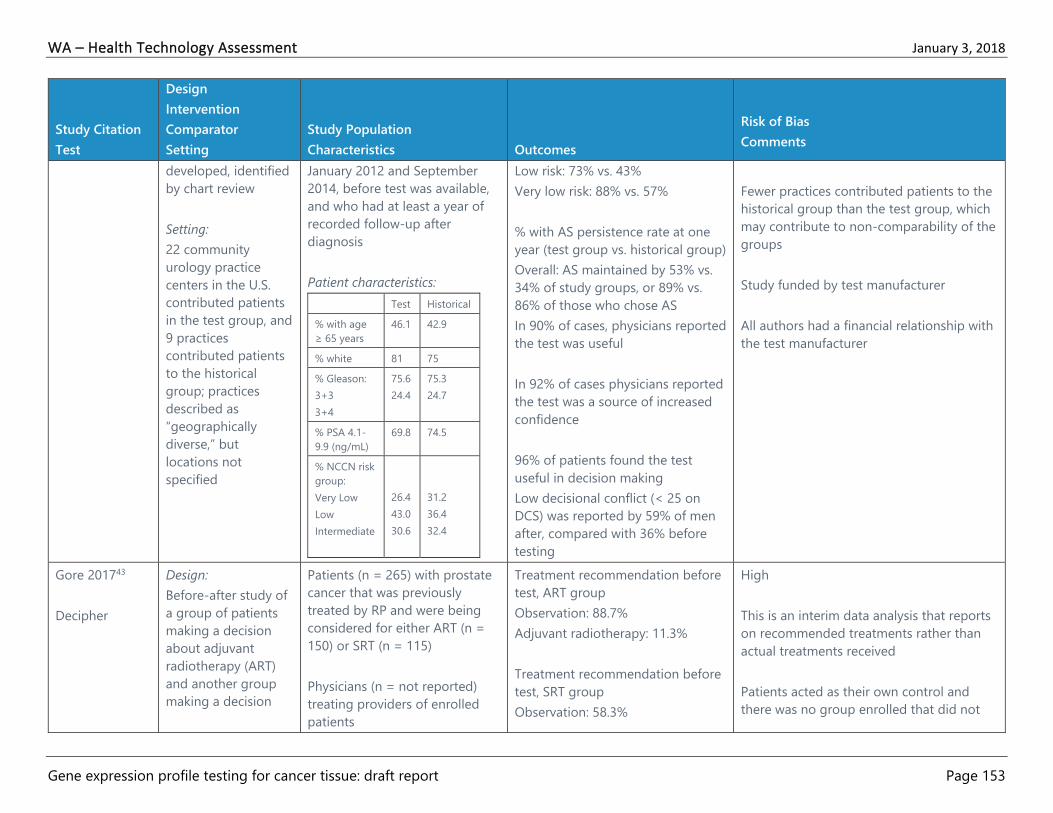
WA – Health Technology Assessment January 3, 2018
Gene expression profile testing for cancer tissue: draft report Page 153
Study Citation
Test
Design
Intervention
Comparator
Setting
Study Population
Characteristics Outcomes
Risk of Bias
Comments
developed, identified
by chart review
Setting:
22 community
urology practice
centers in the U.S.
contributed patients
in the test group, and
9 practices
contributed patients
to the historical
group; practices
described as
“geographically
diverse,” but
locations not
specified
January 2012 and September
2014, before test was available,
and who had at least a year of
recorded follow-up after
diagnosis
Patient characteristics:
Test Historical
% with age
≥ 65 years
46.1 42.9
% white 81 75
% Gleason:
3+3
3+4
75.6
24.4
75.3
24.7
% PSA 4.1-
9.9 (ng/mL)
69.8 74.5
% NCCN risk
group:
Very Low
Low
Intermediate
26.4
43.0
30.6
31.2
36.4
32.4
Low risk: 73% vs. 43%
Very low risk: 88% vs. 57%
% with AS persistence rate at one
year (test group vs. historical group)
Overall: AS maintained by 53% vs.
34% of study groups, or 89% vs.
86% of those who chose AS
In 90% of cases, physicians reported
the test was useful
In 92% of cases physicians reported
the test was a source of increased
confidence
96% of patients found the test
useful in decision making
Low decisional conflict (< 25 on
DCS) was reported by 59% of men
after, compared with 36% before
testing
Fewer practices contributed patients to the
historical group than the test group, which
may contribute to non-comparability of the
groups
Study funded by test manufacturer
All authors had a financial relationship with
the test manufacturer
Gore 201743
Decipher
Design:
Before-after study of
a group of patients
making a decision
about adjuvant
radiotherapy (ART)
and another group
making a decision
Patients (n = 265) with prostate
cancer that was previously
treated by RP and were being
considered for either ART (n =
150) or SRT (n = 115)
Physicians (n = not reported)
treating providers of enrolled
patients
Treatment recommendation before
test, ART group
Observation: 88.7%
Adjuvant radiotherapy: 11.3%
Treatment recommendation before
test, SRT group
Observation: 58.3%
High
This is an interim data analysis that reports
on recommended treatments rather than
actual treatments received
Patients acted as their own control and
there was no group enrolled that did not

WA – Health Technology Assessment January 3, 2018
Gene expression profile testing for cancer tissue: draft report Page 154
Study Citation
Test
Design
Intervention
Comparator
Setting
Study Population
Characteristics Outcomes
Risk of Bias
Comments
about salvage
radiotherapy (SRT)
Intervention:
Treatment
recommendation
after test results, with
recruitment into
study between May
2014 and February
2016
Comparator:
Treatment
recommendation for
the same patients
before test results
Setting:
19 U.S. and one
Canadian community
and academic sites
participating in the
Society of Urologic
Oncology Clinical
Trials Consortium
Patient characteristics:
ART
Group
SRT
Group
Median
age at
RP, years
(range)
64 (46-
76)
63 (48-
75)
% non-
Hispanic
white
84 82.6
%
Gleason:
3+3
3+4
75.6
24.4
75.3
24.7
Median
pre-RP
PSA
(range)
(ng/mL)
6.3 (0.9-
40.5)
6.7
(0.8-
55.5)
%
Gleason
score:
6
3+4
4+3
1.3
52
27.3
7.8
42.6
31.3
Either ART, or SRT and ART: 9.6%
Change after testing
ART arm: 18% (27 patients)
SRT arm: 32% (37 patients)
Multivariable model OR (95% CI) for
either ART or SRT after test result
known, adjusted for age, pre-RP
PSA, extraprostatic extension,
seminal vesicle invasion, surgical
margin, Gleason score, (and for SRT
group, years to biochemical
recurrence or PSA rise)
ART group: aOR 1.48 (1.19 to 1.85)
SRT group: aOR 1.30 (1.03 to 1.65)
Patient median decisional conflict
score (IQR) before and after test,
ART group:
25 (8 to 44) and 19 (2 to 30)
Patient median decisional conflict
score before and after test, SRT
group:
27 (16-41) and 23 (4-30)
Physician median decisional conflict
score (IQR) before and after test,
ART group:
get testing—such a group given an
equivalent amount of time to make a
decision could have similar levels of
decisional conflict and their physicians
might make similar types of treatment
recommendations
Patients in the SRT group had varying
amounts of time since RP, which could
influence treatment recommendations as
well as decisional conflict scores
Use of OR for estimate of effect for an
outcome that is not rare generally
overestimates the effect
Study funded by test manufacturer
7 of 22 authors declared a financial interest
with the manufacturer
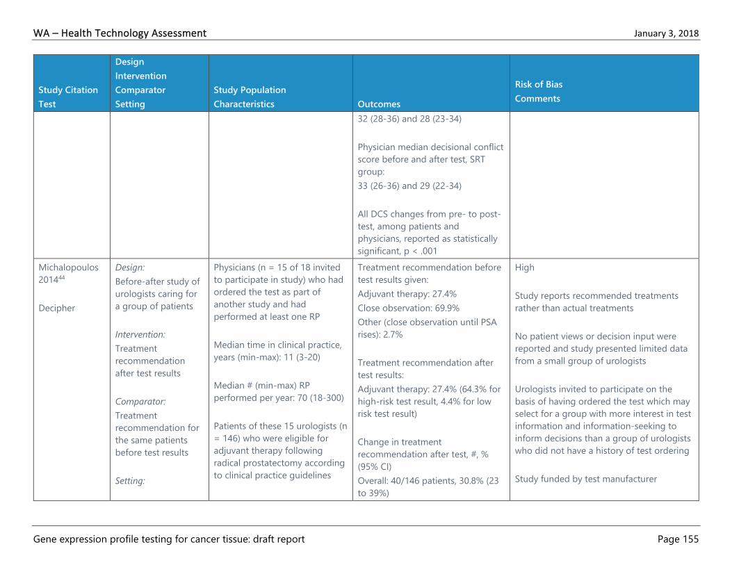
WA – Health Technology Assessment January 3, 2018
Gene expression profile testing for cancer tissue: draft report Page 155
Study Citation
Test
Design
Intervention
Comparator
Setting
Study Population
Characteristics Outcomes
Risk of Bias
Comments
32 (28-36) and 28 (23-34)
Physician median decisional conflict
score before and after test, SRT
group:
33 (26-36) and 29 (22-34)
All DCS changes from pre- to post-
test, among patients and
physicians, reported as statistically
significant, p < .001
Michalopoulos
201444
Decipher
Design:
Before-after study of
urologists caring for
a group of patients
Intervention:
Treatment
recommendation
after test results
Comparator:
Treatment
recommendation for
the same patients
before test results
Setting:
Physicians (n = 15 of 18 invited
to participate in study) who had
ordered the test as part of
another study and had
performed at least one RP
Median time in clinical practice,
years (min-max): 11 (3-20)
Median # (min-max) RP
performed per year: 70 (18-300)
Patients of these 15 urologists (n
= 146) who were eligible for
adjuvant therapy following
radical prostatectomy according
to clinical practice guidelines
Treatment recommendation before
test results given:
Adjuvant therapy: 27.4%
Close observation: 69.9%
Other (close observation until PSA
rises): 2.7%
Treatment recommendation after
test results:
Adjuvant therapy: 27.4% (64.3% for
high-risk test result, 4.4% for low
risk test result)
Change in treatment
recommendation after test, #, %
(95% CI)
Overall: 40/146 patients, 30.8% (23
to 39%)
High
Study reports recommended treatments
rather than actual treatments
No patient views or decision input were
reported and study presented limited data
from a small group of urologists
Urologists invited to participate on the
basis of having ordered the test which may
select for a group with more interest in test
information and information-seeking to
inform decisions than a group of urologists
who did not have a history of test ordering
Study funded by test manufacturer
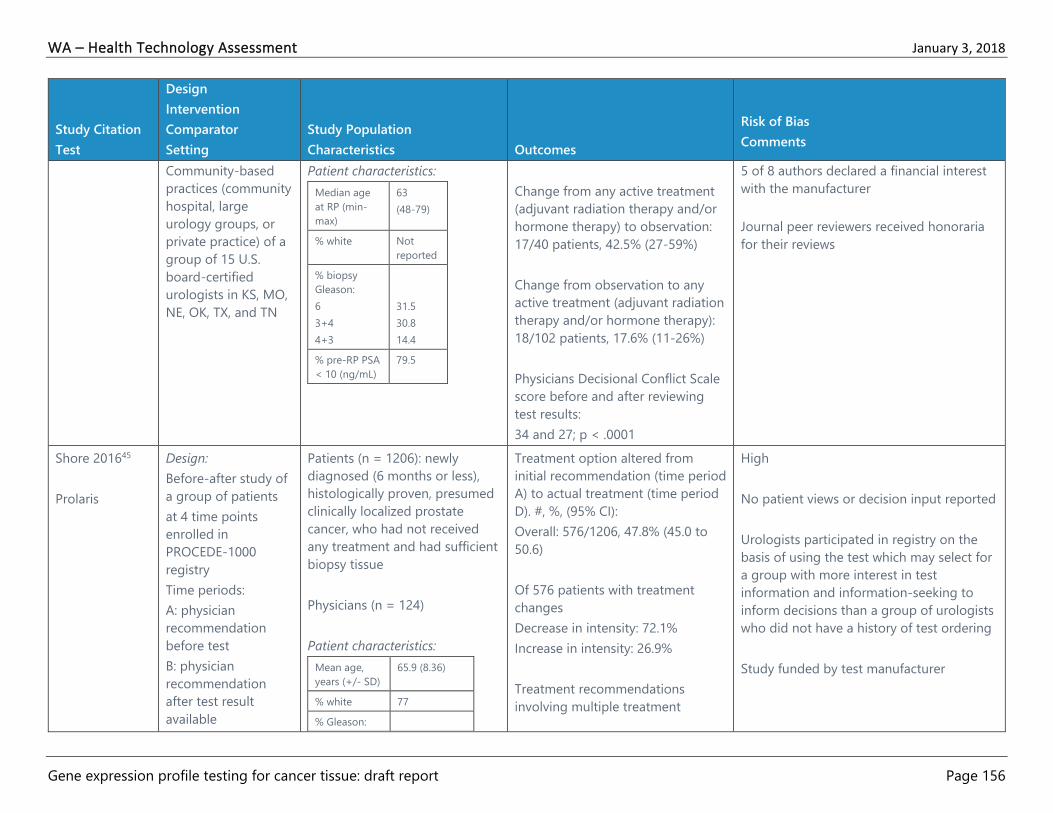
WA – Health Technology Assessment January 3, 2018
Gene expression profile testing for cancer tissue: draft report Page 156
Study Citation
Test
Design
Intervention
Comparator
Setting
Study Population
Characteristics Outcomes
Risk of Bias
Comments
Community-based
practices (community
hospital, large
urology groups, or
private practice) of a
group of 15 U.S.
board-certified
urologists in KS, MO,
NE, OK, TX, and TN
Patient characteristics:
Median age
at RP (min-
max)
63
(48-79)
% white Not
reported
% biopsy
Gleason:
6
3+4
4+3
31.5
30.8
14.4
% pre-RP PSA
< 10 (ng/mL)
79.5
Change from any active treatment
(adjuvant radiation therapy and/or
hormone therapy) to observation:
17/40 patients, 42.5% (27-59%)
Change from observation to any
active treatment (adjuvant radiation
therapy and/or hormone therapy):
18/102 patients, 17.6% (11-26%)
Physicians Decisional Conflict Scale
score before and after reviewing
test results:
34 and 27; p < .0001
5 of 8 authors declared a financial interest
with the manufacturer
Journal peer reviewers received honoraria
for their reviews
Shore 201645
Prolaris
Design:
Before-after study of
a group of patients
at 4 time points
enrolled in
PROCEDE-1000
registry
Time periods:
A: physician
recommendation
before test
B: physician
recommendation
after test result
available
Patients (n = 1206): newly
diagnosed (6 months or less),
histologically proven, presumed
clinically localized prostate
cancer, who had not received
any treatment and had sufficient
biopsy tissue
Physicians (n = 124)
Patient characteristics:
Mean age,
years (+/- SD)
65.9 (8.36)
% white 77
% Gleason:
Treatment option altered from
initial recommendation (time period
A) to actual treatment (time period
D). #, %, (95% CI):
Overall: 576/1206, 47.8% (45.0 to
50.6)
Of 576 patients with treatment
changes
Decrease in intensity: 72.1%
Increase in intensity: 26.9%
Treatment recommendations
involving multiple treatment
High
No patient views or decision input reported
Urologists participated in registry on the
basis of using the test which may select for
a group with more interest in test
information and information-seeking to
inform decisions than a group of urologists
who did not have a history of test ordering
Study funded by test manufacturer

WA – Health Technology Assessment January 3, 2018
Gene expression profile testing for cancer tissue: draft report Page 157
Study Citation
Test
Design
Intervention
Comparator
Setting
Study Population
Characteristics Outcomes
Risk of Bias
Comments
C: treatment
recommendation
after consultation
with patient
D: actual treatment
received, measured
3-6 months after
consultation
Intervention:
Treatment
recommendation and
treatment after test
results (Time periods
B, C, D)
Comparator:
Treatment
recommendation for
the same patients
before test results
(Time period A)
Setting:
U.S. based urology
practices, with study
site selection based
on previous
investigator
experience with the
6
3+4
4+3
47.8
27.9
11.9
Mean pre-
biopsy PSA
(+/- SD)
(ng/mL)
7.8 (8.15)
% AUA risk
group:
Low
Intermediate
High
40.3
42.0
17.7
options, before test vs. actual
treatment: 31.6% vs. 12.9%
Change from noninterventional to
interventional treatments: 101/417
(24.2%)
Change from interventional to
noninterventional treatments:
112/789 (14.2%)
5 of 13 authors declared a financial interest
with the manufacturer

WA – Health Technology Assessment January 3, 2018
Gene expression profile testing for cancer tissue: draft report Page 158
Study Citation
Test
Design
Intervention
Comparator
Setting
Study Population
Characteristics Outcomes
Risk of Bias
Comments
test (no further
details given)

WA – Health Technology Assessment January 3, 2018
Gene expression profile testing for cancer tissue: draft report Page 159
Table 10. Evidence Table: Prostate Cancer Economic Studies
Citation
Test
Design
Comparators
Population
Analytic
Assumptions Main Findings
Risk of Bias
Comments
Abala 201638
Oncotype DX
See observational studies evidence
table for details, one economic
outcome (costs) reported, but no
formal economic analysis provided
Lobo 201754
Decipher
Design:
Cost effectiveness
analysis
Comparators:
Genetic testing-
based care vs.
alternatives of
100% uptake of
adjuvant therapy
or usual care for
outcomes of
QALYs, and
biochemical
recurrence or
metastasis at 5-
and 10-year post
treatment intervals
Population:
Men with prostate
cancer in the post-
prostatectomy
setting
Analytic Assumptions:
Model included
inputs for disease
progression, death,
treatment
complications above
those expected for
RP alone,
probabilities for
adjuvant therapy
delivery or salvage
therapy delivery
across usual care and
genetic test risk
groups, utility values
for side effects and
health states, and
costs
Markov modeling conducted to provide estimates below
Genetic test-based care vs. 100% adjuvant therapy
Mean difference in life years: 0.024 (95% CI, -0.012 to
0.059; p = .187)
Mean difference in QALYs: 0.140 (95% CI, 0.080 to 0.201;
p < .001)
Average per-person cost of care, assuming a test cost of
$4000: $23,823 vs. $31,125
Patients receiving 100% adjuvant therapy had improved
clinical outcomes at 5 and 10 years
Genetic test-based care vs. usual care (mean difference,
95% CI)
Mean difference in life years: 0.023 (95% CI, -0.012 to
0.058; p = .191)
Mean difference in QALYs: 0.066 (95% CI, 0.016 to 0.117;
p = .0098)
Average per-person cost of care, assuming a test cost of
$4000: $23,823 vs. $18,370
Genetic test-based care provided improved clinical
outcomes, including reduced incidence of metastases
ICER: $90,883
Clinical outcomes (%, 95% CI)
High
No analytic perspective stated
Effectiveness estimates for clinical
outcomes based on two non-
comparative before-after studies of
treatment decisions rather than
actual treatment or other clinical
outcomes
Other model inputs generally based
on single studies rather than
systematic reviews of multiple
studies, and sensitivity analysis
ranges were varied by set amounts
(e.g., +/-10%) rather than the
plausible ranges reported across a
comprehensive set of studies or
other data sources
Study funded by test manufacturer
3 of 11 authors had a financial
relationship with test manufacturer

WA – Health Technology Assessment January 3, 2018
Gene expression profile testing for cancer tissue: draft report Page 160
Citation
Test
Design
Comparators
Population
Analytic
Assumptions Main Findings
Risk of Bias
Comments
Genetic test cost
modeled at
$4000/test and
varied from $0-5000
in sensitivity analyses
Discount rate set at
3% and varied from
0-5%
Biochemical recurrence-free at 5 years
Genetic test-based care: 68.9 (68.0 to 69.8)
100% adjuvant therapy: 78.3 (77.5 to 79.2)
Usual care: 64.0 (63.1 to 65.0)
Biochemical recurrence-free at 10 years
Genetic test-based care: 47.0 (46.1 to 48.0)
100% adjuvant therapy: 58.3 (57.4 to 59.3)
Usual care: 42.8 (41.9 to 43.8)
Metastasis at 5 years
Genetic test-based care: 3.46 (3.10 to 3.82)
100% adjuvant therapy: 2.76 (2.44 to 3.08)
Usual care: 4.12 (3.73 to 4.51)
Metastasis at 10 years
Genetic test-based care: 9.48 (8.91 to 10.1)
100% adjuvant therapy: 7.95 (7.42 to 8.48)
Usual care: 10.9 (10.3 to 11.5)
p < .001 for all comparisons
Sensitivity analysis
Results showed that model findings were robust based
on outputs not being sensitive to changes in inputs
Ontario 201755
Prolaris
Design:
Budget impact
analysis
Comparators:
New scenario vs.
reference scenario:
Anticipated clinical
practice with vs.
without the test
Population:
Men with newly
diagnosed, low- or
intermediate-risk,
localized prostate
cancer in Ontario,
Canada
Analytic Assumptions:
Incidence, 8500 cases
per year, 65% of
(Note: all costs and estimates are in 2016 Canadian
Dollars)
Base case:
Estimated costs associated with test itself: +$47.9
million in the first 5 years
Estimated costs associated with additional physician
visits required to interpret the test result: +$0.7 million
Estimated savings due to treatment changes
(increased use of active surveillance and decreased use
of interventional treatment): -$7.3 million
Moderate
Not a cost-effectiveness analysis
per se, but an estimate of budget
impact of test adoption in one
Canadian province (Ontario)
Costs expressed in 2016 Canadian
dollars

WA – Health Technology Assessment January 3, 2018
Gene expression profile testing for cancer tissue: draft report Page 161
Citation
Test
Design
Comparators
Population
Analytic
Assumptions Main Findings
Risk of Bias
Comments
which would occur in
men over 65 years of
age, with 34% in the
low-risk and 46% in
the intermediate-risk
group
Initial treatment
patterns assumed
that for low-risk men,
61% would have
AS/WW, 22% RT,
17% RP, and for
intermediate-risk
men 34% would have
AS/WW, 38% RT, and
28% RP
Publicly funding the CCP test would result in a net
budget impact of +$41.3 million in the first 5 years
Sensitivity analysis:
Net budget impact was most sensitive to assumptions
regarding the uptake of the test and the extent to which
the test altered the distribution of treatment from current
practice.
Three factors had a moderate impact on the results by
affecting the size of the target population:
1. Percentage of low- and intermediate-risk tumors
among all patients newly diagnosed with prostate cancer
2. Proportion of patients on watchful waiting among
those on non-interventional treatment
3. Annual change in prostate cancer incidence
Budget impact was sensitive to the unit cost of the test,
but not to the annual costs of prostate cancer treatments
or the number of extra physician visits associated with the
test, because the cost of the test is much higher than
either the extra physician visits or the savings associated
with treatment change
Study authors state that there was
not sufficient data to support a
primary economic evaluation of the
test for use in men with localized
prostate cancer. “…based on the
results of the clinical evidence
review, the effect of the CCP test on
patient-important clinical outcomes
(e.g., survival or biochemical
recurrence) is currently unknown.
No prospective studies have been
conducted to evaluate these
outcomes.”55(p. 32)
No specific comparator or
competing alternatives aside from
current clinical practice patterns
included in budget impact analysis
Individual level and patient level
costs not modeled
Limited number of sensitivity
analyses considered
Authors are contractors or
employees of Health Quality
Ontario, a governmental agency,
which also funded the report to
support the work of the Ontario
Health Technology Advisory
Committee and make

WA – Health Technology Assessment January 3, 2018
Gene expression profile testing for cancer tissue: draft report Page 162
Citation
Test
Design
Comparators
Population
Analytic
Assumptions Main Findings
Risk of Bias
Comments
recommendations to the Ontario
Ministry of Health about public
funding of services

WA – Health Technology Assessment January 3, 2018
Gene expression profile testing for cancer tissue: draft report Page 163
Colon Cancer
Table 11. Evidence Table: Colon Cancer Observational Studies
Study Citation
Test
Design
Intervention
Comparator
Setting
Study Population
Characteristics Results
Risk of Bias
Comments
Renfro 201747
Srivastava
201448
Oncotype DX
Design:
Before-after study
of one group of
patients and the
physicians
Intervention:
Treatment
recommendations
after test results
Comparator:
Treatment
recommendations
for the same
patient before the
test
Setting:
17 academic and
community sites in
the U.S. that
enrolled patients
between May 2010
and May 2012
Consecutive patients (n = 221)
with stage 2A colon cancer, n
= 141 of whom had resected
T3 mismatch repair-proficient
(MMR-P) tumors and were
included in the primary
analysis
Physicians (n = 105) of study
patients were surveyed
Patient characteristics (n =
141):
Mean age (range),
years
63.2 (27-
87)
% male 58.9
% white 92.2
% adenocarcinoma
tumor type
93.6
% WHO tumor
grade
Low
High
88.7
11.3
Post-test treatment recommendations, by pre-test
recommendation:
Observation (n = 68)
Observation (no change): 79.4%
5-FU monotherapy: 11.8%
5-FU + oxaliplatin: 11.1%
5-FU monotherapy (n = 34)
Observation: 70.6%
5-FU monotherapy (no change): 23.5%
5-FU + oxaliplatin: 5.9%
5-FU + oxaliplatin (n = 39)
Observation: 53.8%
5-FU monotherapy: 5.1%
5-FU + oxaliplatin (no change): 41.0%
Total (n = 141)
Observation: 70.2%
5-FU monotherapy: 12.8%
5-FU + oxaliplatin: 17.0%
Treatment recommendation change, by recurrence
score group:
Low (< 30), n = 100
No change: 54.0%
Increased intensity: 7.0%
Decreased intensity: 39.0%
Intermediate (30-40), n = 33
No change: 60.1%
High
MMR-P status is associated with
higher risk of colon cancer
recurrence
Study did not enroll targeted
number of patients with MMR-P
tumors due to higher number of
MMR-D tumors than expected in
enrollees
Before-after studies do not have
an independent control group
Slightly different n included in
each paper, data primarily
abstracted from Srivastava
2014,48 unless otherwise
indicated, because it was a more
complete reporting of outcomes
Renfro 201747
Study funded by the U.S. NIH
under a Clinical Translational
Science Award Grant K12

WA – Health Technology Assessment January 3, 2018
Gene expression profile testing for cancer tissue: draft report Page 164
Study Citation
Test
Design
Intervention
Comparator
Setting
Study Population
Characteristics Results
Risk of Bias
Comments
Increased intensity: 18.2%
Decreased intensity: 21.2%
High (> 41), n = 7
No change: 57.1%
Increased intensity: 42.8%
Decreased intensity: 0.0%
Total (n = 140)
No change: 55.7%
Increased intensity: 11.4%
Decreased intensity: 32.9%
Mean change (95% CI) in patient Decisional
Conflict Scale score after test (n = 139) (data from
Renfro 201747)
Effective decision: -5.74 (-9.21 to -2.27); p = .001
Informed: -9.32 (-13.52 to -5.12); p < .001
Support: -4.63 (-7.85 to -1.41); p = .005
Uncertainty: -12.13 (-17.91 to -6.35); p < .001
Values clarity: -8.27 (-12.45 to -4.09); p < .001
Total: -7.88 (-11.25 to -4.50); p < .001
Physician survey results, % of patient decisions (n =
150)
More confident in recommendation after ordering
test
Strongly agree: 28.7%
Agree: 55.3%
Neither agree nor disagree: 12.7%
3 of 5 authors had a financial
relationship with test
manufacturer
Srivastava 201448
Study funded by test
manufacturer
3 of 13 authors had a financial
relationship with test
manufacturer

WA – Health Technology Assessment January 3, 2018
Gene expression profile testing for cancer tissue: draft report Page 165
Study Citation
Test
Design
Intervention
Comparator
Setting
Study Population
Characteristics Results
Risk of Bias
Comments
Disagree: 1.3%
Strongly disagree: 2.0%
Test results provided additional clinically relevant
information
Strongly agree: 30.7%
Agree: 55.3%
Neither agree nor disagree: 10.7%
Disagree: 1.3%
Strongly disagree: 2.0%
Test results influenced my treatment decision
Strongly agree: 24.0%
Agree: 25.4%
Neither agree nor disagree: 10.9%
Disagree: 1.4%
Strongly disagree: 2.2%
Patient survey results (n = 138)
Test results influenced treatment decision
Strongly agree: 60.1%
Agree: 25.4%
Neither agree nor disagree: 10.9%
Disagree: 1.4%
Strongly disagree: 2.2%
Brenner 201649
Oncotype DX
Design:
Before-after study
of one group of
patients and the
physicians
All patients (n = 312) with
stage 2 colon cancer who had
testing at 7 institutions
between January 2011 and
May 2012
Treatment received after test, by pre-test
recommendation:
Observation (n = 148):
Observation (no change): 83.1%
5-FU only: 15.5%
High
Preplanned retrospective analysis
from a prospectively designed
registry

WA – Health Technology Assessment January 3, 2018
Gene expression profile testing for cancer tissue: draft report Page 166
Study Citation
Test
Design
Intervention
Comparator
Setting
Study Population
Characteristics Results
Risk of Bias
Comments
Intervention:
Treatment
recommendations
after test results
Comparator:
Treatment
recommendations
for the same
patient before the
test
Setting:
Registry enrolling
patients from 7
institutions in Israel
(n = 269 with MMR-P tumors,
test results analyzed)
39 excluded due to MMR-
deficient tumors
3 excluded due to no pre-
test treatment
recommendation
1 excluded based on
pathology/laboratory
exclusion criteria
Patient characteristics:
Median age (IQR),
years
68 (60-75)
%
adenocarcinoma
83.6
% tumor grade
low
moderate
high
18.2
72.5
7.1
5-FU + oxaliplatin: 1.4%
5-FU only (n = 105):
Observation: 57.1%
5-FU only (no change): 41.9%
5-FU + oxaliplatin: 1.0%
5-FU + oxaliplatin (n = 16)
Observation: 68.8%
5-FU only: 31.3%
5-FU + oxaliplatin (no change): 0%
Total (n = 269):
Observation: 72.1%
5-FU only: 26.8%
5-FU + oxaliplatin: 1.1%
Treatment change by recurrence score group
Low (< 30), n = 157
No change: 62.4%
Increased intensity: 2.6%
Decreased intensity: 35.0%
Intermediate (30-40), n = 85
No change: 69.4%
Increased intensity: 12.9%
Decreased intensity: 17.7%
High (> 41), n = 27
No change: 37.0%
Increased intensity: 40.8%
Decreased intensity: 22.2%
Before-after studies do not have
an independent control group
and changes in treatment
decisions may be due to
additional or other factors—no
analysis for these potential
confounders was undertaken
Study does report actual
treatment received rather than
recommended
Study funded by test
manufacturer’s industrial
representative in Israel
5 of 19 authors had a financial
relationship with test
manufacturer

WA – Health Technology Assessment January 3, 2018
Gene expression profile testing for cancer tissue: draft report Page 167
Study Citation
Test
Design
Intervention
Comparator
Setting
Study Population
Characteristics Results
Risk of Bias
Comments
Overall (n = 269)
No change: 62.1%
Increased intensity: 9.7%
Decreased intensity: 28.3%

WA – Health Technology Assessment January 3, 2018
Gene expression profile testing for cancer tissue: draft report Page 168
Table 12. Evidence Table: Colon Cancer Economic Studies
Citation
Test
Design
Comparators
Population
Analytic Assumptions Main Findings
Risk of Bias
Comments
Alberts 201457
Oncotype DX
Design:
Cost effectiveness
using decision
analysis
Treatment
recommendation
after gene testing.
Treatment
recommendation
before gene
testing
Patients (n = 141) with stage 2, T3, MMR-P
colon cancer in clinical practice who had
undergone surgery and were eligible for
adjuvant chemotherapy
Analytic Assumptions:
Perspective adopted was that of U.S. third
party payer, costs standardized to 2014
U.S. dollars, model employed patient and
clinical decision impact estimates from the
Mayo Clinic Cancer Research Consortium
(MCCRC) study of fluoropyrimidine
monotherapy vs. combination
chemotherapy with oxaliplatin (FOLFOX)
and according to NCCN guidelines with
and without the availability of genetic
testing for patients with stage 2 colon
cancer, test cost was set at $3640 per
patient, economic estimates were
determined for the end outcomes of
recurrence or not recurrence and
subsequent survival or death, sensitivity
analyses were conducted over the plausible
ranges derived from the MCCRC study, the
QUASAR chemotherapy RCT, U.S. incidence
and mortality statistics, CMS provided drug
costs, a 3% discount rate was applied and
varied over 1-5%
Total estimated lifetime
costs with the genetic test
vs. without (U.S. 2014
dollars, difference):
$103,775 vs. $104,767,
-$991
Sensitivity analysis found
76.1% of cost savings and
QALY improvement found
in 96.1% of scenarios with
use of genetic test
At a QALY threshold of
$50,000 the probability of
cost-effectiveness was
95.5%
Moderate
Note: this paper draws decision
impact estimates derived from
the MCCRC study,47,48 which is
abstracted in the accompanying
evidence table for colon cancer
GEP studies
Effectiveness and outcome
estimates based largely on data
from single RCT
Some costs, such as lost time
and productivity, are not
modeled
Study funded by test
manufacturer
2 of 12 authors had a financial
relationship via a contract with
test manufacturer

WA – Health Technology Assessment January 3, 2018
Gene expression profile testing for cancer tissue: draft report Page 169
Appendix D. Risk of Bias Assessments
Breast Cancer
Table 13. Risk of Bias: Breast Cancer Systematic Reviews and Meta-analyses
Citation
Protocol
Described
& Used
Search
Strategy
Study
Selection
Study
Characteristics
Quality
Assessment
of Included
Studies
Data
Extraction
Study
Synthesis
Statistical
Methods
Interest
Disclosure
Funding
Source
Overall Risk
of Bias
Assessment
Augustovski
201519
Unclear Yes Yes Yes Yes Yes Yes Yes Yes No Low
Blok 201820 Unclear Yes Yes Unclear No Yes Yes Yes Yes Unclear Moderate
Scope 201721 Unclear Yes Unclear Unclear Unclear Yes Yes NA Yes Unclear High
Table 14. Risk of Bias: Breast Cancer Randomized Controlled Trial
Citation/
Test
Randomization
and Allocation
Concealment Intervention Outcomes
Masking of
Investigators
and
Participants
Masking of
Outcome
Assessors
Intention
to Treat
Analysis
Statistical
Analysis
Interest
Disclosure
Funding
Source
Overall Risk
of Bias
Assessment
Bear 201717
Oncotype DX
Yes Yes Yes No No No Yes Yes No High
Cardoso
201618
MammaPrint
Unclear Yes Yes No No Yes No No No Moderate

WA – Health Technology Assessment January 3, 2018
Gene expression profile testing for cancer tissue: draft report Page 170
Table 15. Risk of Bias: Breast Cancer Observational Studies
Citation/
Test
Subject
Selection Intervention Control Outcome
Masked
Outcome
Assessment Confounding
Statistical
Analysis
Interest
Disclosure
Funding
Source
Overall Risk
of Bias
Assessment
Evans 201622
Oncotype DX
No Yes No Unclear No Unclear Yes Yes Yes High
Friese 201723
Oncotype DX
Unclear Yes Yes Yes No Yes Yes No Yes Moderate
Hequet 201734
Prosigna
Yes Yes No No No No Yes No No High
Jasem 201624
Oncotype DX
Unclear Yes No Yes No Yes Yes No Yes High
Jasem 201725
Oncotype DX
Yes No No Yes No Yes Yes Yes Unclear High
Kuijer 201632
MammaPrint
Unclear Yes Yes Yes No No Yes Yes Yes High
Kuijer 201731
MammaPrint
Unclear Yes No Yes No No No No No High
Loncaster 201726
Oncotype DX
No Yes No Yes No No Yes No No High
O’Neill 201727
Oncotype DX
Unclear Yes Yes Yes No Yes Yes No Yes High
Parsons 201628
Oncotype DX
Yes Yes No Yes No No No Yes Yes High
Pestalozzi 201737
Oncotype DX
Yes Yes No Yes No No Yes Yes Yes High
Ray 201630
Oncotype DX
Yes Yes No Yes No Yes Unclear No Yes Moderate
Sanft 201536
BCI
Unclear No Yes Yes No No No No Unclear High
Tsai 201733
MammaPrint
Unclear Yes Yes Yes No No No No No High

WA – Health Technology Assessment January 3, 2018
Gene expression profile testing for cancer tissue: draft report Page 171
Citation/
Test
Subject
Selection Intervention Control Outcome
Masked
Outcome
Assessment Confounding
Statistical
Analysis
Interest
Disclosure
Funding
Source
Overall Risk
of Bias
Assessment
Wuerstlein
201635
Prosigna
Yes Yes No Yes No No No No No High
Table 16. Risk of Bias: Breast Cancer Economic Studies
Part 1
Citation Target
Population
Perspective Time Horizon Discount Rate Comparators Modeling Effectiveness
Gustavsen 201451 Yes Yes Yes No Yes No No
Hall 201750 Yes Yes Yes Yes Yes Yes Unclear
Part 2
Citation Outcomes
Resource
Use/Costs Uncertainty Results
Interest
Disclosure Funding Source
Overall Risk of
Bias Assessment
Gustavsen 2014 No Yes Unclear No No No High
Hall 2017 Yes Yes Yes Yes Unclear Yes Moderate

WA – Health Technology Assessment January 3, 2018
Gene expression profile testing for cancer tissue: draft report Page 172
Table 17. Risk of Bias: Breast Cancer Guidelines
Citation
Scope &
Purpose
Rigor of
Evidence
Development
Rigor of
Recommendations
Development
Stakeholder
Involvement
Clarity &
Presentation
Applicability &
Implementation
Editorial
Independence
Overall Assessment
Comments
Duffy 201763 Good Poor Fair Poor Good Fair Poor Poor
Harris 201658 Good Good Good Good Good Good Fair Good
Krop 201759 Good Good Good Good Good Good Fair Good
NCCN 201760 Fair Fair Good Good Fair Fair Fair Fair
NICE 201361 Good Good Good Good Good Good Fair Good
Senkus 201562 Fair Poor Fair Poor Good Fair Poor Poor
Prostate Cancer
Table 18. Risk of Bias: Prostate Cancer Observational Studies
Citation/
Test
Subject
Selection Intervention Control Outcome
Masked
Outcome
Assessment Confounding
Statistical
Analysis
Interest
Disclosure
Funding
Source
Overall Risk
of Bias
Assessment
Albala 201638
Oncotype DX
Yes Yes No
Yes
No No N/A No No High
Badani 201539
Oncotype DX
No
Yes Yes Yes No No N/A No No High
Crawford 201440
Prolaris
No Yes Yes No No No N/A No No High
Dall’Era, 201541
Oncotype DX
No
Yes Yes Unclear
Unclear
No N/A No No High
Eure 201742
Oncotype DX
Yes Yes No
Unclear
No No N/A No No High

WA – Health Technology Assessment January 3, 2018
Gene expression profile testing for cancer tissue: draft report Page 173
Citation/
Test
Subject
Selection Intervention Control Outcome
Masked
Outcome
Assessment Confounding
Statistical
Analysis
Interest
Disclosure
Funding
Source
Overall Risk
of Bias
Assessment
Gore 201743
Decipher
Yes Yes Yes Unclear
No No N/A No No High
Michalopoulos
201444
Decipher
No
Yes Yes No No No N/A No No High
Shore 201645
Prolaris
No
Yes Yes Yes No No N/A No No High
Table 19. Risk of Bias: Prostate Cancer Economic Studies
Part 1
Citation
Target
Population Perspective Time Horizon Discount Rate Comparators Modeling Effectiveness
Lobo 201754 Yes No Yes Yes Yes Yes No
Ontario 201755 Yes Yes Yes N/A Unclear Yes Yes
Part 2
Citation Outcomes
Resource
Use/Costs Uncertainty Results
Interest
Disclosure Funding Source
Overall Risk of
Bias Assessment
Lobo 2017 Yes Yes Yes Yes No No High
Ontario 2017 Yes No Yes Yes Yes Yes Moderate

WA – Health Technology Assessment January 3, 2018
Gene expression profile testing for cancer tissue: draft report Page 174
Table 20. Risk of Bias: Prostate Cancer Guidelines
Citation
Scope &
Purpose
Rigor of
Evidence
Development
Rigor of
Recommendations
Development
Stakeholder
Involvement
Clarity &
Presentation
Applicability &
Implementation
Editorial
Independence
Overall Assessment
Comments
NCCN 201764 Fair Fair Good Good Fair Fair Fair Fair
Sanda 201765 Good Good Good Good Good Good Fair Good
Colon Cancer
Table 21. Risk of Bias: Colon Cancer Observational Studies
Citation/
Test
Subject
Selection Intervention Control Outcome
Masked
Outcome
Assessment Confounding
Statistical
Analysis
Interest
Disclosure
Funding
Source
Overall Risk
of Bias
Assessment
Brenner
201649
Oncotype DX
Yes
Yes Yes Yes
No No Yes No No High
Renfro
201747
Srivastava
201448
Oncotype DX
Yes
Yes Yes No
No No No No No High

WA – Health Technology Assessment January 3, 2018
Gene expression profile testing for cancer tissue: draft report Page 175
Table 22. Risk of Bias: Colon Cancer Economic Studies
Part 1
Citation Target Population Perspective Time Horizon Discount Rate Comparators Modeling Effectiveness
Alberts 201457 Yes Yes Yes Yes Yes Yes Yes
Part 2
Citation Outcomes
Resource
Use/Costs Uncertainty Results
Interest
Disclosure Funding Source
Overall Risk of Bias
Assessment
Alberts 2014 No No Yes Yes Yes No Moderate
Table 23. Risk of Bias: Colon Cancer Guidelines
Citation
Scope &
Purpose
Rigor of
Evidence
Development
Rigor of
Recommendations
Development
Stakeholder
Involvement
Clarity &
Presentation
Applicability &
Implementation
Editorial
Independence
Overall Assessment
Comments
NCCN 201767 Fair Fair Good Good Fair Fair Fair Fair
Sepulveda 201766 Good Good Good Fair Good Good Fair Good
Van Cutsem 201668 Fair Poor Poor Fair Fair Fair Poor Poor
Multiple Myeloma
Table 24. Risk of Bias: Multiple Myeloma Guidelines
Citation
Scope &
Purpose
Rigor of
Evidence
Development
Rigor of
Recommendations
Development
Stakeholder
Involvement
Clarity &
Presentation
Applicability &
Implementation
Editorial
Independence
Overall Assessment
Comments
Moreau 201770 Fair Poor Fair Poor Fair Fair Poor Fair
NCCN 201769 Fair Poor Good Fair Fair Fair Fair Fair

WA – Health Technology Assessment January 3, 2018
Gene expression profile testing for cancer tissue: draft report Page 176
Appendix E. GRADE Profile Ratings
Table 25. GRADE Quality of Evidence for Breast Cancer
Number of Studies,
by Test and Study
Design Risk of Bias Inconsistency Indirectness Imprecision
Publication
Bias
Other
Factors* Comments
Overall Quality
of Evidence
Rating
Outcome: Clinical Utility–mortality or morbidity
Oncotype DX
(1 RCT)
MammaPrint
(1 RCT)
High for
Oncotype DX;
Moderate for
MammaPrint
Not evaluated
(1 study for
each test)
Indirectness
serious for
Oncotype DX
and not
serious for
MammaPrint
NA (only
one study
for each
test)
Not
evaluated
Oncotype DX RCT used
clinical response as an
surrogate/intermediate
outcome measure;
MammaPrint RCT
reported survival
Oncotype DX
Very low
●◌◌◌
MammaPrint
Moderate
●●●◌
Outcome: Clinical Utility–patient management decisions
Oncotype DX
(3 SRs with largely
overlapping primary
studies, total k = 38;
and k = 10 additional
studies, 1 RCT and 9
observational studies)
MammaPrint
(2 SRs with
overlapping primary
studies, total k = 7; 4
additional studies, 1
RCT and 3
observational studies)
Prosigna
(1 SR with k = 1
study, and 2
Moderate to
High for
Oncotype DX;
Moderate to
High for
MammaPrint;
High for all
other tests
Results overall
consistent for
these
outcomes
Minor
indirectness;
One meta-
analysis for
Oncotype DX
found high I2
for some
comparisons
Precision
related to
size of
study, as
expected,
but not
serious
Evaluated
only for
Oncotype DX
in one meta-
analysis and
was not
detected
Oncotype DX
Moderate
●●●◌
MammaPrint
Low
●●◌◌
Prosigna,
Endopredict,
BCI, and
Mammostrat
Very low
●◌◌◌

WA – Health Technology Assessment January 3, 2018
Gene expression profile testing for cancer tissue: draft report Page 177
Number of Studies,
by Test and Study
Design Risk of Bias Inconsistency Indirectness Imprecision
Publication
Bias
Other
Factors* Comments
Overall Quality
of Evidence
Rating
additional
observational studies)
BCI
(0 SRs and 1
additional
observational study)
Endopredict
(1 SR with k = 1
study, and 0
additional studies)
BCI
(0 SRs and 1
additional
observational study)
Mammostrat
(1 SR with k = 1
study; and 0
additional studies)
Outcome: Clinical Utility–quality of life
Oncotype DX
(2 additional
observational studies)
Prosigna
(2 additional
observational studies)
BCI
(1 additional
observational study)
High Results
generally
consistent
across studies
that reported
the outcomes
Serious Not
evaluated
Not
evaluated
No studies reported on
true QoL measures; but
some studies did report
on pre- and post-test
levels of decisional
conflict, anxiety, and
distress
Oncotype DX,
Prosigna, and
BCI
Very low
●◌◌◌
Other tests
Not applicable
(no eligible
studies)
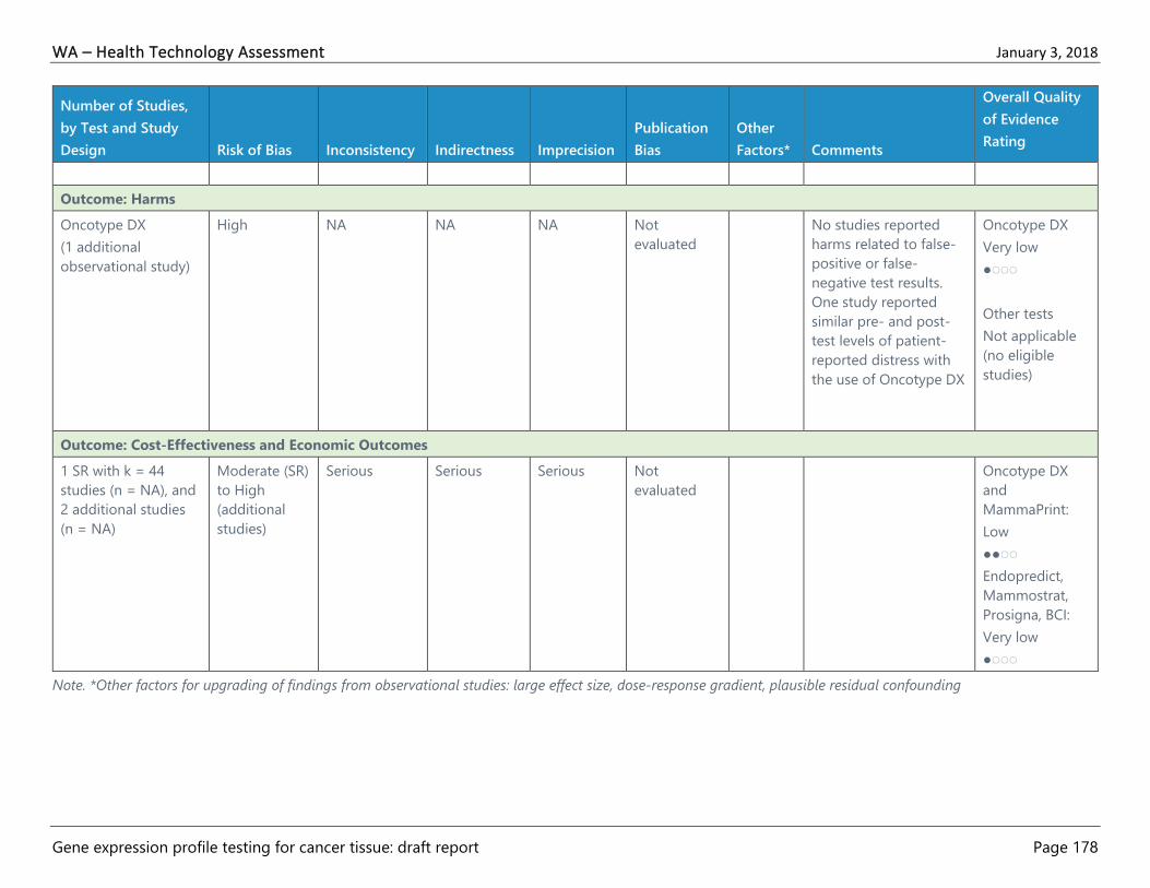
WA – Health Technology Assessment January 3, 2018
Gene expression profile testing for cancer tissue: draft report Page 178
Number of Studies,
by Test and Study
Design Risk of Bias Inconsistency Indirectness Imprecision
Publication
Bias
Other
Factors* Comments
Overall Quality
of Evidence
Rating
Outcome: Harms
Oncotype DX
(1 additional
observational study)
High NA NA NA Not
evaluated
No studies reported
harms related to false-
positive or false-
negative test results.
One study reported
similar pre- and post-
test levels of patient-
reported distress with
the use of Oncotype DX
Oncotype DX
Very low
●◌◌◌
Other tests
Not applicable
(no eligible
studies)
Outcome: Cost-Effectiveness and Economic Outcomes
1 SR with k = 44
studies (n = NA), and
2 additional studies
(n = NA)
Moderate (SR)
to High
(additional
studies)
Serious Serious Serious Not
evaluated
Oncotype DX
and
MammaPrint:
Low
●●◌◌
Endopredict,
Mammostrat,
Prosigna, BCI:
Very low
●◌◌◌
Note. *Other factors for upgrading of findings from observational studies: large effect size, dose-response gradient, plausible residual confounding

WA – Health Technology Assessment January 3, 2018
Gene expression profile testing for cancer tissue: draft report Page 179
Table 26. GRADE Quality of Evidence for Prostate Cancer
Number of
Studies (k), and
Subjects (n),
by Test and
Study Design
Risk
of
Bias
Inconsistency Indirectness Imprecision Publication
Bias
Other
Factors*
Comments Overall Quality of
Evidence Rating
Outcome: Clinical Utility–mortality or morbidity
0 No studies reported
outcome
Not applicable (no
eligible studies)
Outcome: Clinical Utility–patient management decisions
Oncotype DX
k = 4
n = 659 tested,
1093 total, and
15 physicians
Prolaris
k = 1
n = 1,206
Decipher
k = 2
n = 411, and 15
physicians
(all studies are
before-after
designs)
High Results
generally
consistent
across studies
Some studies
did not
report actual
treatment
received
Not
evaluated
Not
evaluated
3 studies on Oncotype DX
used historical comparator
group and 4 used same
patient comparator (1 on
Oncotype DX, 1 on Prolaris,
and 2 on Decipher) and
were consistent in finding
decreased treatment
intensity after testing
Oncotype DX,
Prolaris, and Decipher
Very low
●◌◌◌
Outcome: Clinical Utility–quality of life
0 Not applicable (no
eligible studies)

WA – Health Technology Assessment January 3, 2018
Gene expression profile testing for cancer tissue: draft report Page 180
Number of
Studies (k), and
Subjects (n),
by Test and
Study Design
Risk
of
Bias
Inconsistency Indirectness Imprecision Publication
Bias
Other
Factors*
Comments Overall Quality of
Evidence Rating
Outcome: Harms
0 No studies reported
outcome
Not applicable (no
eligible studies)
Outcome: Cost-Effectiveness and Economic Outcomes
Oncotype DX
k = 3
n = 80 tested in 1
study, and
modeling in 2
studies
High Not serious Serious
(modeling
designs and
non-U.S.
setting)
Not
evaluated
Not
evaluated
No comparison across
studies possible given that 1
study reported costs only, 1
was a budget impact
analysis, and 1 was a cost-
effectiveness analysis,
however there was
consistency about test
increasing costs in the 2
modeling studies
There is some indirectness
due to the modeling study
with moderate risk of bias
being done from a
Canadian perspective
Oncotype DX
Very low
●◌◌◌
Prolaris and Decipher
Not applicable (no
eligible studies)
Note. *Other factors for upgrading of findings from observational studies: large effect size, dose-response gradient, plausible residual confounding

WA – Health Technology Assessment January 3, 2018
Gene expression profile testing for cancer tissue: draft report Page 181
Table 27. GRADE Quality of Evidence for Colon Cancer
Number of
Studies
(subjects),
by Test, and
Study Design
Risk
of
Bias Inconsistency Indirectness Imprecision
Publication
Bias
Other
Factors* Comments
Overall Quality of
Evidence Rating
Outcome: Clinical Utility–mortality or morbidity
0 No studies reported outcome Not applicable (no
eligible studies)
Outcome: Clinical Utility–patient management decisions
Oncotype DX
k = 2
n = 410
(both studies
are before-
after designs)
High Not serious Not serious Both studies used same patient
comparator in before-after analyses
and were consistent in finding a
decrease in recommended or actual
treatment intensity after testing with
larger study reporting actual
treatment
Oncotype DX
Very low
●◌◌◌
ColoPrint
Not applicable (no
eligible studies)
Outcome: Clinical Utility–quality of life
Oncotype DX
k = 1
n = 139
(before-after
design)
High Not
evaluated (1
study)
Serious Not
evaluated
Not
evaluated
1 study reported patient decisional
conflict which may relate to quality
of life, but is an incomplete and
indirect measure
Oncotype DX
Very low
●◌◌◌
ColoPrint
Not applicable (no
eligible studies)
Outcome: Harms
0 No studies reported outcome Not applicable (no
eligible studies)

WA – Health Technology Assessment January 3, 2018
Gene expression profile testing for cancer tissue: draft report Page 182
Number of
Studies
(subjects),
by Test, and
Study Design
Risk
of
Bias Inconsistency Indirectness Imprecision
Publication
Bias
Other
Factors* Comments
Overall Quality of
Evidence Rating
Outcome: Cost-Effectiveness and Economic Outcomes
Oncotype DX
k = 1
(decision
analysis
modeling
study)
n = NA
Mode
rate
Serious Serious Not
evaluated
Not
evaluated
Underlying evidence used in
modeling is scant and sensitivity
analysis showed decreased cost-
effectiveness approximately 25% of
the time raising questions about
consistency. Modeling limited with
few outcomes raising questions
regarding applicability to current
practice.
Oncotype DX
Very low
●◌◌◌
ColoPrint
Not applicable (no
eligible studies)
Note. *Other factors for upgrading of findings from observational studies: large effect size, dose-response gradient, plausible residual confounding

WA – Health Technology Assessment January 3, 2018
Gene expression profile testing for cancer tissue: draft report Page 183
Table 28. GRADE Quality of Evidence for Multiple Myeloma
Number
of Studies
(subjects),
by Study
Design
Risk of
Bias Inconsistency Indirectness Imprecision
Publication
Bias
Other
Factors*
Comments
Overall Quality of
Evidence Rating
Outcome: Clinical Utility–mortality or morbidity
0 No studies reported outcome Not applicable (no
eligible studies)
Outcome: Clinical Utility–patient management decisions
0 No studies reported outcome Not applicable (no
eligible studies)
Outcome: Clinical Utility–quality of life
0 No studies reported outcome Not applicable (no
eligible studies)
Outcome: Harms
0 No studies reported outcome Not applicable (no
eligible studies)
Outcome: Cost-Effectiveness and Economic Outcomes
0 No studies reported outcome Not applicable (no
eligible studies)
Note. *Other factors for upgrading of findings from observational studies: large effect size, dose-response gradient, plausible residual confounding
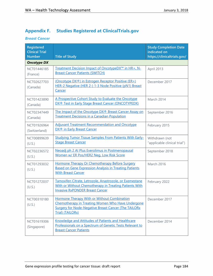
WA – Health Technology Assessment January 3, 2018
Gene expression profile testing for cancer tissue: draft report Page 184
Appendix F. Studies Registered at ClinicalTrials.gov
Breast Cancer
Registered
Clinical Trial
Number Title of Study
Study Completion Date
indicated on
https://clinicaltrials.gov/
Oncotype DX
NCT01446185
(France)
Treatment Decision Impact of OncotypeDX™ in HR+, N-
Breast Cancer Patients (SWITCH) April 2013
NCT02627703
(Canada)
(Oncotype DX®) in Estrogen Receptor Positive (ER+)
HER-2 Negative (HER 2-) 1-3 Node Positive (pN1) Breast
Cancer
December 2017
NCT01423890
(Canada)
A Prospective Cohort Study to Evaluate the Oncotype
DX® Test in Early Stage Breast Cancer (ONCOTYPEDX) March 2014
NCT02347449
(Canada)
The Impact of the Oncotype DX® Breast Cancer Assay on
Treatment Decisions in a Canadian Population September 2016
NCT01926964
(Switzerland)
Adjuvant Treatment Recommendation and Oncotype
DX® in Early Breast Cancer February 2015
NCT00899639
(U.S.)
Studying Tumor Tissue Samples From Patients With Early-
Stage Breast Cancer Withdrawn (not
“applicable clinical trial”)
NCT02236572
(U.S.)
Neoadj ph 2 AI Plus Everolimus in Postmenopausal
Women w/ ER Pos/HER2 Neg, Low Risk Score September 2018
NCT01293032
(U.S.)
Hormone Therapy Or Chemotherapy Before Surgery
Based on Gene Expression Analysis in Treating Patients
With Breast Cancer
March 2016
NCT01272037
(U.S.)
Tamoxifen Citrate, Letrozole, Anastrozole, or Exemestane
With or Without Chemotherapy in Treating Patients With
Invasive RxPONDER Breast Cancer
February 2022
NCT00310180
(U.S.)
Hormone Therapy With or Without Combination
Chemotherapy in Treating Women Who Have Undergone
Surgery for Node-Negative Breast Cancer (The TAILORx
Trial) (TAILORx)
December 2017
NCT01619306
(Singapore)
Knowledge and Attitudes of Patients and Healthcare
Professionals on a Spectrum of Genetic Tests Relevant to
Breast Cancer Patients
December 2014
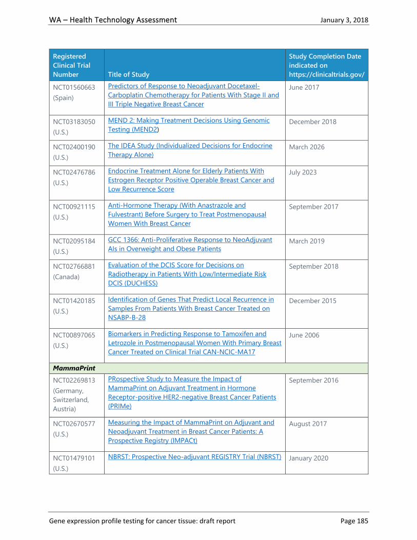
WA – Health Technology Assessment January 3, 2018
Gene expression profile testing for cancer tissue: draft report Page 185
Registered
Clinical Trial
Number Title of Study
Study Completion Date
indicated on
https://clinicaltrials.gov/
NCT01560663
(Spain)
Predictors of Response to Neoadjuvant Docetaxel-
Carboplatin Chemotherapy for Patients With Stage II and
III Triple Negative Breast Cancer
June 2017
NCT03183050
(U.S.)
MEND 2: Making Treatment Decisions Using Genomic
Testing (MEND2) December 2018
NCT02400190
(U.S.)
The IDEA Study (Individualized Decisions for Endocrine
Therapy Alone) March 2026
NCT02476786
(U.S.)
Endocrine Treatment Alone for Elderly Patients With
Estrogen Receptor Positive Operable Breast Cancer and
Low Recurrence Score
July 2023
NCT00921115
(U.S.)
Anti-Hormone Therapy (With Anastrazole and
Fulvestrant) Before Surgery to Treat Postmenopausal
Women With Breast Cancer
September 2017
NCT02095184
(U.S.)
GCC 1366: Anti-Proliferative Response to NeoAdjuvant
AIs in Overweight and Obese Patients March 2019
NCT02766881
(Canada)
Evaluation of the DCIS Score for Decisions on
Radiotherapy in Patients With Low/Intermediate Risk
DCIS (DUCHESS)
September 2018
NCT01420185
(U.S.)
Identification of Genes That Predict Local Recurrence in
Samples From Patients With Breast Cancer Treated on
NSABP-B-28
December 2015
NCT00897065
(U.S.)
Biomarkers in Predicting Response to Tamoxifen and
Letrozole in Postmenopausal Women With Primary Breast
Cancer Treated on Clinical Trial CAN-NCIC-MA17
June 2006
MammaPrint
NCT02269813
(Germany,
Switzerland,
Austria)
PRospective Study to Measure the Impact of
MammaPrint on Adjuvant Treatment in Hormone
Receptor-positive HER2-negative Breast Cancer Patients
(PRIMe)
September 2016
NCT02670577
(U.S.)
Measuring the Impact of MammaPrint on Adjuvant and
Neoadjuvant Treatment in Breast Cancer Patients: A
Prospective Registry (IMPACt)
August 2017
NCT01479101
(U.S.)
NBRST: Prospective Neo-adjuvant REGISTRY Trial (NBRST) January 2020

WA – Health Technology Assessment January 3, 2018
Gene expression profile testing for cancer tissue: draft report Page 186
Registered
Clinical Trial
Number Title of Study
Study Completion Date
indicated on
https://clinicaltrials.gov/
NCT03053193
(U.S.)
MammaPrint, BluePrint, and Full-genome Data Linked
With Clinical Data to Evaluate New Gene EXpression
Profiles (FLEX)
December 2027
NCT02669745
(U.S.)
The Study of Molecular Risk Panels (70-gene Assay) in
Chinese Breast Cancer Patients November 2017
NCT01501487
(U.S.)
MINT I Multi- Institutional Neo-adjuvant Therapy
MammaPrint Project I (MINT) June 2017
NCT00904566
(Japan)
Validation of 70-gene MammaPrint Profile in Japanese
Population January 2009
NCT01617954
(U.S.)
PRospective Study Of MammaPrint in Patients With an
Intermediate Recurrence Score (PROMIS) December 2015
NCT02209857
(Netherlands)
The Symphony Triple A Study: Using Symphony in
Treatment Decisions Concerning Adjuvant Systemic
Therapy (Symphony)
May 2016
NCT03080428
(N/A)
Assessment of Genomic Test Impact on Shared Decision
of Adjuvant Chemotherapy in ER-positive, Her2-negative
Early Breast Cancer (OPTIGEN)
May 2023 Withdrawn
due to lack of funding
NCT02689921
(U.S.)
NEOADjuvant Aromatase Inhibitor and
Pertuzumab/Trastuzumab for Women With Breast Cancer
(NEOADAPT)
April 2018
NCT00433589
(Netherlands)
Genetic Testing or Clinical Assessment in Determining the
Need for Chemotherapy in Women With Breast Cancer
That Involves No More Than 3 Lymph Nodes (MINDACT)
March 2020
EndoPredict
NCT02773004
(France)
Prospective Study Assessing EndoPredict® Genomic Test
Impact on Shared Decision of Adjuvant Chemotherapy in
Patients With ER-positive, Her2-negative Early Breast
Cancer (ADENDOM)
December 2017
NCT03080428
(N/A)
Assessment of Genomic Test Impact on Shared Decision
of Adjuvant Chemotherapy in ER-positive, Her2-negative
Early Breast Cancer (OPTIGEN)
May 2023 withdrawn
due to lack of funding
NCT01805271
(France)
Safety Study of Adding Everolimus to Adjuvant Hormone
Therapy in Women With High Risk of Relapse, ER+ and
HER2- Primary Breast Cancer, Free of Disease After
June 2031

WA – Health Technology Assessment January 3, 2018
Gene expression profile testing for cancer tissue: draft report Page 187
Registered
Clinical Trial
Number Title of Study
Study Completion Date
indicated on
https://clinicaltrials.gov/
Receiving at Least One Year of Adjuvant Hormone
Therapy
Prosigna (PAM50)
NCT01899079
(Spain)
A Prospective Observational Study of Clinical Outcomes
for the NanoString® Technologies Prosigna Gene
Signature Assay
July 2014
NCT02653755
(U.S.)
The PRECISION Trial (Profiling Early Breast Cancer for
Radiotherapy Omission): A Phase II Study of Breast-
Conserving Surgery Without Adjuvant Radiotherapy for
Favorable-Risk Breast Cancer
June 2023
NCT03197805
(France)
Assessment of the Impact of RNA Genomic Profile on
Treatment Decision-making in HER2 Equivocal Breast
Cancer Patients (EQUIVOK)
April 2019
NCT02889874
(Australia, New
Zealand)
EXamining PErsonalised Radiation Therapy for Low-risk
Early Breast Cancer (EXPERT) December 2023
NCT01560663
(Spain)
Predictors of Response to Neoadjuvant Docetaxel-
Carboplatin Chemotherapy for Patients With Stage II and
III Triple Negative Breast Cancer
June 2017
NCT00991263
(U.S.)
Study of Tissue Samples From Women Treated With
Paclitaxel for Breast Cancer on Clinical Trial CALGB-9344
or CALGB-9741
February 2012
Breast Cancer Index (BCI)
NCT02057029
(U.S.)
Assessment of the Decision-making Impact of the Breast
Cancer Index in Recommending Extended Adjuvant
Endocrine Therapy for Patients With Early Stage ER-
positive Breast Cancer
June 2015
Mammostrat
NCT02067416
(U.S.)
PREDATOR: Neoadjuvant Gene Prediction for Breast
Cancer June 2014 Terminated
when funding agent
withdrew funding
Note. N/A = not available

WA – Health Technology Assessment January 3, 2018
Gene expression profile testing for cancer tissue: draft report Page 188
Prostate Cancer
Registered
Clinical Trial
Number Title of Study
Study Completion Date
indicated on
https://clinicaltrials.gov/
Decipher
NCT02723734
(U.S.)
Validation Study on the Impact of Decipher® Testing -
VANDAAM Study (VANDAAM) May 2021
NCT02080689
(U.S.)
Prospective Clinical Utility Study to Assess the Impact
of Decipher on Treatment Decisions After Surgery
(PRO-IMPACT)
February 2017
NCT02034812
(U.S.)
Decision Impact Study to Measure the Influence of
DECIPHER on Treatment Recommendations for
Radiation Oncologists (DECIDE3)
July 2014
NCT02020876
(U.S.)
Decision Impact Study to Measure the Influence of
DECIPHER on Treatment Recommendations (DECIDE) December 2013
NCT02034825
(U.S.)
Multi-site Decision Impact Study for Decipher (ASSESS-
D) January 2015
NCT03237026
(Taiwan)
Decipher the Biology of Lethal Prostate Cancer—Using
Urine Metabolomics Profiling to Search for Predictive
and Prognostic Markers for Treatment Outcomes in
Prostate Cancer Patients
July 2022
NCT03237702
(Taiwan)
Decipher Lethal Prostate Cancer Biology - Urine
Metabolomics April 2020
NCT02609269
(U.S.)
Decipher Genomics Resource Information Database
(GRID®) December 2020
NCT02783950
(U.S.)
Genomics in Michigan Impacting Observation or
Radiation (G-MINOR) November 2021
NCT02721979
(U.S.)
ARN-509 in Treating Patients With Prostate Cancer
Who Are in Active Surveillance July 2020
Prolaris
NCT03290508
(U.S.)
Long-term Study to Evaluate and Clinical Outcomes in
Patients With Favorable Intermediate Risk Localized
Prostate Cancer
September 2027
NCT03152448
(U.S. Veterans
Affairs Health
System)
Prospective Prolaris Value and Efficacy (P-PROVE) January 2024

WA – Health Technology Assessment January 3, 2018
Gene expression profile testing for cancer tissue: draft report Page 189
Registered
Clinical Trial
Number Title of Study
Study Completion Date
indicated on
https://clinicaltrials.gov/
NCT02209584
(U.S.)
Open Registry Measuring Impact of Genomic Testing
on Treatment Decision After Biopsy in Newly
Diagnosed Prostate Cancer Patients (PROCEDE-2000)
December 2015
NCT02454595
(U.S.)
Registry to Measure the Impact of Adding Genomic
Testing July 2017 Terminated due
to lack of enrollment
NCT01954004
(U.S.)
Open Registry Measuring Impact of Genomic Testing
on Treatment of Prostate Cancer Patients September 2014
Oncotype DX prostate
NCT02668276
(U.S.)
Engaging Newly Diagnosed Men About Cancer
Treatment Options (ENACT) August 2022
Colon Cancer
Multiple Myeloma
Registered
Clinical Trial
Number Title of Study
Study Completion Date
indicated on
https://clinicaltrials.gov/
ColoPrint
NCT00903565
(U.S.)
A Prospective Study for the Assessment of Recurrence Risk
in Stage II Colon Cancer Patients Using ColoPrint (PARSC) December 2019
Oncotype DX Colon
NCT01479894
(U.S.)
Genetic Analysis to Evaluate the Racial Difference in the
Outcome of Patients With Colon Cancer January 2015
Registered
Clinical Trial
Number Title of Study
Study Completion Date
indicated on
https://clinicaltrials.gov/
My Prognostic Risk Signature MyPRS
NCT01313897
(U.S.)
UARK 2010-35, A Study of Expanded Natural Killer Cell
Therapy for Multiple Myeloma (NK2010-35) October 2016
SKY92-EMC92
NCT02911571
(U.S.)
PRospective Multiple Myeloma Impact Study (PROMMIS) December 2024

WA – Health Technology Assessment January 3, 2018
Gene expression profile testing for cancer tissue: draft report Page 190
Appendix G. Detailed Clinical Practice Guidelines
American Society of Clinical Oncology Guidelines for Breast Cancer58,59
Oncotype DX
If a patient has ER/PR-positive, HER2-negative (LN-negative) breast cancer, the clinician
can use Oncotype DX to guide decisions on adjuvant systemic chemotherapy.
If a patient has ER/PR-positive, HER2-negative (LN-positive) breast cancer, the clinician
should not use Oncotype DX to guide decisions on adjuvant systemic chemotherapy.
If a patient has HER2-positive breast cancer or triple-negative breast cancer, the clinician
should not use Oncotype DX to guide decisions on adjuvant systemic therapy.
MammaPrint
If a patient has ER/PR-positive, HER2-negative, LN-negative breast cancer, the
MammaPrint assay may be used in those with high clinical risk per MINDACT
categorization to inform decisions on withholding adjuvant systemic chemotherapy
because of its ability to identify a good-prognosis population with potentially limited
chemotherapy benefit.
If a patient has ER/PR-positive, HER2-negative, LN-negative breast cancer, the
MammaPrint assay should not be used in those with low clinical risk per MINDACT
categorization to inform decisions on withholding adjuvant systemic chemotherapy,
because women in the low clinical risk category had excellent outcomes and did not
appear to benefit from chemotherapy even with a genomic high-risk cancer.
If a patient has ER/PR-positive, HER2-negative, LN-positive, breast cancer, the
MammaPrint assay may be used in patients with one to three positive nodes and at high
clinical risk per MINDACT categorization to inform decisions on withholding adjuvant
systemic chemotherapy because of its ability to identify a good-prognosis population
with potentially limited chemotherapy benefit. However, such patients should be
informed that a benefit of chemotherapy cannot be excluded, particularly in patients
with greater than one involved lymph node.
If a patient has ER/PR-positive, HER2-negative, LN-positive, breast cancer, the
MammaPrint assay should not be used in patients with one to three positive nodes and
at low clinical risk per MINDACT categorization to inform decisions on withholding
adjuvant systemic chemotherapy. There are insufficient data on the clinical utility of
MammaPrint in this specific patient population.
If a patient has HER2-positive breast cancer, the clinician should not use the
MammaPrint assay to guide decisions on adjuvant systemic therapy. Additional studies
are required to address the role of MammaPrint in patients with this tumor subtype who
are also receiving HER2-targeted therapy.
If a patient has ER/PR-negative and HER2-negative (triple negative) breast cancer, the
clinician should not use the MammaPrint assay to guide decisions on adjuvant systemic
chemotherapy.

WA – Health Technology Assessment January 3, 2018
Gene expression profile testing for cancer tissue: draft report Page 191
EndoPredict
If a patient has ER/PR-positive, HER2-negative (LN-negative) breast cancer, the clinician
can use EndoPredict to guide decisions on adjuvant systemic chemotherapy.
If a patient has ER/PR-positive, HER2-negative (LN-positive) breast cancer, the clinician
should not use EndoPredict to guide decisions on adjuvant systemic chemotherapy.
If a patient has HER2-positive breast cancer or triple-negative breast cancer, the clinician
should not use EndoPredict to guide decisions on adjuvant systemic therapy.
Prosigna (PAM50 risk of recurrence score)
If a patient has ER/PR-positive, HER2-negative (LN-negative) breast cancer, the clinician
can use the Prosigna score, in conjunction with other clinicopathologic variables, to
guide decisions on adjuvant systemic therapy.
If a patient has ER/PR-positive, HER2-negative (LN-positive) breast cancer, the clinician
should not use Prosigna to guide decisions on adjuvant systemic therapy.
If a patient has HER2-positive breast cancer or triple-negative breast cancer, the clinician
should not use Prosigna to guide decisions on adjuvant systemic therapy.
Breast Cancer Index
If a patient has ER/PR-positive, HER2-negative (LN-negative) breast cancer, the clinician
can use BCI to guide decisions on adjuvant systemic therapy.
If a patient has ER/PR-positive, HER2-negative (LN-positive) breast cancer, the clinician
should not use BCI to guide decisions on adjuvant systemic therapy.
If a patient has HER2-positive breast cancer or triple-negative breast cancer, the clinician
should not use BCI to guide decisions on adjuvant systemic therapy.
Mammostrat
If a patient has ER/PR-positive, HER2-negative (LN-positive or LN-negative) breast
cancer, the clinician should not use the five-protein assay to guide decisions on adjuvant
systemic therapy.
If a patient has HER2-positive breast cancer or triple-negative breast cancer, the clinician
should not use the five-protein assay (Mammostrat) to guide decisions on adjuvant
systemic therapy.
European Group on Tumor Markers Guidelines on the Use of Biomarkers in Breast Cancer63
Oncotype DX
Oncotype DX may provide added value to established factors for determining prognosis
and aiding decision-making with respect to administration of adjuvant chemotherapy in
newly diagnosed breast cancer patients with lymph LN-negative invasive disease that is
ER-positive but HER2-negative (Level of evidence [LOE], IB; strength of recommendation
[SOR], A). In addition, Oncotype DX may be considered for identifying HER2-negative,
ER-positive patients with 1 to 3 involved lymph nodes for treatment with adjuvant
chemotherapy (LOE, IB; SOR, A).

WA – Health Technology Assessment January 3, 2018
Gene expression profile testing for cancer tissue: draft report Page 192
Before performing the test, any biopsy cavity in the cancer specimen should be removed
by manual dissection.
MammaPrint
MammaPrint may be used for determining prognosis and guiding decision-making with
respect to the administration of adjuvant chemotherapy in patients with newly
diagnosed invasive breast cancer that is LN-negative or LN-positive (1 to 3 metastatic
nodes). Patients at high risk based on clinical and pathological criteria but at low risk
based on MammaPrint may be the candidates for avoiding having to receive adjuvant
chemotherapy (LOE, IA; SOR, A).
EndoPredict
In combination with established clinical and pathological factors, EndoPredict may be
used for predicting outcome and aiding adjuvant therapy decision-making in hormone
receptor-positive, HER2-negative patients that are either LN-negative or LN-positive (1
to 3 metastatic nodes). (LOE, IB; SOR, A).
Prosigna
In combination with established clinical and pathological factors, Prosigna may be used
for predicting outcome and aiding adjuvant therapy decision-making in hormone
receptor-positive, HER2-negative patients that are either lymph LN-negative or lymph
LN-positive (1 to 3 metastatic nodes). (LOE IB; SOR, A).
BCI
In combination with established clinical and pathological factors, BCI may be used for
predicting outcome and aiding adjuvant therapy decision-making in lymph LN-negative,
hormone receptor-positive and HER2-negative patients. (LOE, IB; SOR, A).

WA – Health Technology Assessment January 3, 2018
Gene expression profile testing for cancer tissue: draft report Page 193
Appendix H. See Attachment for Excluded Studies