general pathology cellular and organ pathology disorders of glycogen degradation. pathology of...
TRANSCRIPT
General Pathology Cellular and Organ Pathology
Disorders of Glycogen Degradation.
Pathology of Calcification.
Jaroslava Dušková
Inst. Pathol. ,1st Med. Faculty, Charles Univ. Prague

Glycogen linear and branched polymer cca 60 000 - D-glucose
molecules– monoparticles (beta) - muscle– complex particles (alpha) -
hepatocyte

EnzymesInvolved in Glycogen Metabolism
g.-synthase - brancher phophorylase kinase - debrancher g-6-phosphatase -glucosidase

Storage DiseasesDef.:
inborn errors of metabolism (mostly single gene abnormality) leading to an enzyme defect with subsequent accumulation of the substrate (& lack of
the product) in tissues or organs „thesaurismoses“

E defect - gl-6 - phosphatase
Organ damage - liver, kidney
Glycogenosis I – von Gierke

E -defect - alfa1, 4 - glycosidase Organ damage - heart
Glycogenosis II – Pompe

Glycogen Storage Diseases -1.
Disease E- def Severity of Disease
Tissues Involved
von Gierke
Glucose-6
phosphatase
Severe Liver,kidney, gut
Pompe 1,4 glucosidase
Lethal Heart (+systemic)
Cori Amylo-1,6 Glucosidase (debrancher)
Lethal Liver (+systemic)

Glycogen Storage Diseases -2.
Disease E- def Severity of Disease
Tissues Involved
Andersen Amylo-1,4-1,6 transglucosidase (brancher)
Lethal Liver (+systemic)
Mc Ardle Muscle phosphorylase
Mild Skeletal muscle
Hers Liver
phosphorylaseMild Liver

Clear Intracellular Vacuoles & adjunct techniques
accumulations of water neg. lipides SUDAN, OIL RED polysaccharides PAS, A-PAS

Glycogen water soluble
easily lost in long lasting water based fixative solutions
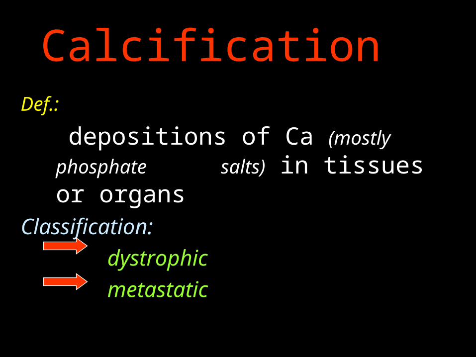
CalcificationDef.:
depositions of Ca (mostly phosphate
salts) in tissues or organs Classification:
dystrophic
metastatic

Matrix vesicles - osteoblasts- nidus calcification
Non collagen proteins - osteopontin,
osteonektin, osteokalcin, Gla protein, sialoprotein
Alkalic phosphatase Phospholipids Collagen I Hydroxyapatite
Calcification - physiology

Calcification dystrophic metastatic
Calcinosis localized generalized
Chondrocalcinosis - pseudogout
Pathology Conditions with Calcium Deposits

Basophilic
Von Kossa - Ag impregnation
Alizarine red +
Tetracyclin fluorescence
Polarized light birefringence
Calcification - Microscopy

Calcification
Dystrophic Serum Ca level: normal
Tissues/Organs status
dystrophic changes
(necrosis, scar…, low
metab. turnover)
MetastaticSerum Ca level:
Tissues/Organs status
normal, local alcalisation
(acid secretion - urine,
stomach juice, sweat…)

Ca in mitochondria and GER
in cytoplasm bound to proteins
Released after cell damage
Activaton of protein kinases
Activaton of phospholipid
degradation and loss
Activaton of proteases
Cytoskeletal disassembly
Membrane damage
Phosphorylation of protein
andchromatin
phragmentation

Necrosis or degeneration
of tissue
Release of enzymes
Breakdown of organic
phophatesAlteration of pH
Increased deposition of calcium
Dystrophic Calcification

Calcification
Dystrophic Serum Ca level: normal
Tissues/Organs status
dystrophic changes
(necrosis, scar…, low
metab. turnover)
MetastaticSerum Ca level:
Tissues/Organs status
normal, local alcalisation
(acid secretion - urine,
stomach juice, sweat…)

Ca phophate Ca3(PO4)2
Ca diphosphate (Ca2P2O7)
Hydroxyapatite (Ca5 (PO4)3.OH
Ca Salts in the Calcified Foci

Calcification dystrophic metastatic
Calcinosis localized generalized
Chondrocalcinosis - pseudogout
Pathology Conditions with Calcium Deposits
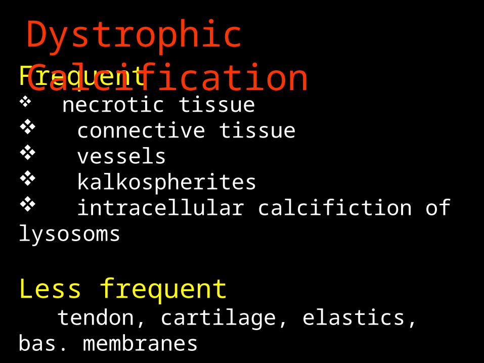
Frequent necrotic tissue connective tissue vessels kalkospherites intracellular calcifiction of lysosoms
Less frequent tendon, cartilage, elastics, bas. membranes
Dystrophic Calcification

Forms - localised generalised
Localisation – connective tissue,
muscles
Calcinosis

pyrophophate and hydroxyapatite deposits
localisation – synovial membrane, cortilage, bone
Chondrocalcinosiscrystal deposit disease (pseudogout)

Calcium pyrophosphate deposition disease(Chondrocalcinosis, pseudogout)
Clinic: may simulate different diseasesArthroscopy - chalky white depositsTophaceus deposits with crystalline aggregatesCrystals stain with von Kossa techniqueForeign body type multinucleated giant cell reactionDeposits are in synovium, cartilage, bone