genomicsequencingprotocol dna - pnas · f--.il. 31. proc. natl. acad. sci. usa89(1992) 1829 ......
TRANSCRIPT

Proc. Nati. Acad. Sci. USAVol. 89, pp. 1827-1831, March 1992Genetics
A genomic sequencing protocol that yields a positive display of5-methylcytosine residues in individual DNA strands
(genomic sequencing/DNA methylation/bisulfite modiflication/PCR/kininogen gene)
MARIANNE FROMMER*t, LOUISE E. MCDONALD*t, DOUGLAS S. MILLAR*, CHRISTINA M. COLLISt,FUJIKO WATTt, GEOFFREY W. GRIGGt, PETER L. MOLLOYt, AND CHERYL L. PAUL**The Kanematsu Laboratories, Royal Prince Alfred Hospital, Missenden Road, Camperdown, Sydney, NSW 2050, Australia; and tDivision of BiomolecularEngineering, Laboratory for Molecular Biology, Commonwealth Scientific and Industrial Research Organization, 103 Delhi Road, North Ryde, Sydney, NSW2113, Australia
Communicated by W. J. Peacock, October 10, 1991 (received for review July 25, 1991)
ABSTRACT The modulation of DNA-protein interactionsby methylation of protein-binding sites in DNA and the occur-rence in genomic imprinting, X chromosome inactivation, andfragile X syndrome of different methylation patterns in DNA ofdifferent chromosomal origin have underlined the need toestablish methylation patterns in individual strands of partic-ular genomic sequences. We report a genomic sequencingmethod that provides positive identification of 5-methylcy-tosine residues and yields strand-specific sequences of individ-ual molecules in genomic DNA. The method utilizes bisulfite-induced modification of genomic DNA, under conditionswhereby cytosine is converted to uracil, but 5-methylcytosineremains nonreactive. The sequence under investigation is thenamplified by PCR with two sets of strand-specific primers toyield a pair of fragments, one from each strand, in which alluracil and thymine residues have been amplified as thymineand only 5-methylcytosine residues have been amplified ascytosine. The PCR products can be sequenced directly toprovide a strand-specific average sequence for the populationof molecules or can be cloned and sequenced to providemethylation maps of single DNA molecules. We tested themethod by derming the methylation status within single DNAstrands of two closely spaced CpG dinucleotides in the pro-moter of the human kininogen gene. During the analysis, weencountered in sperm DNA an unusual methylation pattern,which suggests that the high methylation level of single-copysequences in sperm may be locally modulated by binding ofprotein factors in germ-line cells.
Cytosine methylation has long been recognized as an impor-tant factor in the silencing of genes in mammalian cells.Recent studies have shown that cytosine methylation atsingle CpG dinucleotides within the recognition sites of anumber of transcription factors is sufficient to block bindingof the factors to DNA (1-6) and to inhibit transcription (3-5).Therefore, to determine the role of cytosine methylation inspecific regulatory mechanisms in vivo, it has become im-portant to know the methylation status of individual CpGdinucleotides in genomic DNA. Genomic sequencing proto-cols, which have been developed to ascertain the methylationstatus of selected regions within genes, utilize the Maxam andGilbert chemical cleavage reactions carried out on genomicDNA (7) with various additional procedures to enhance thesignal from the sequence under investigation (8, 9). Theseprotocols are versatile in that they can be adapted foridentification of protein-binding sites on genomic DNA invivo (8, 10) but have two major drawbacks with respect to theidentification of 5-methylcytosine residues. First, 5-methyl-cytosine is identified by the lack of a band in all tracks of a
sequencing gel; any background cleavage ladder or closespacing of bands can result in difficulties of interpretation.Second, the sequence obtained represents a population av-erage for many DNA molecules, so that the protocols cannotbe adapted for sequencing small or mixed DNA samples. Toaddress these problems, we have developed a genomic se-quencing method that provides positive identification andlocalization of 5-methylcytosine in genomic DNA. Themethod is based on sodium bisulfite-mediated conversion ofcytosine to uracil in single-stranded DNA (11-15), followedby PCR amplification of the resultant modified DNA. Exactmethylation maps of single DNA strands from individualgenomic DNA molecules can readily be established, wherethe position of each 5-methylcytosine is given by a clearpositive band on a sequencing gel.
MATERIALS AND METHODSDNA. Human DNA from HeLa cells, placenta, liver, and
sperm was prepared by standard procedures (16, 17). Aplasmid, phKG2.0, containing the promoter and first exon ofthe human kininogen gene was constructed by subcloninginto Bluescribe pBS+ (Stratagene) a 1.9-kilobase (kb) frag-ment (Sca I/EcoRI) from the plasmid phKG5.2, a kind gift ofN. Kitamura (18). To avoid possible PCR artefacts arisingfrom plasmid contamination of human genomic DNA sam-ples, the only cloned kininogen DNA permitted into thegenomic sequencing laboratory had been previously methyl-ated with Alu I methylase and tested for complete methyl-ation by comparing the relative amounts of linear, relaxed,and supercoiled DNA before and after extensive Alu Idigestion.Sodium Bisulfite Modification. The bisulfite reaction was
carried out on 10 jig ofDNA, consisting of plasmid DNA witha known methylation pattern, or of human genomic DNAcontaining added plasmid DNA at single-copy frequency, orof 2 ,ug ofhuman genomicDNA plus 8 ,.ug ofcarrier Bluescriptplasmid. DNA was linearized with the restriction enzymeEcoRI or sheared through a fine needle, alkali denatured,neutralized, and precipitated. Denatured DNA was incu-bated in a total vol of 1.2 ml with freshly prepared 3.1 Msodium bisulfite/0.5 mM hydroquinone, pH 5.0, at 50°C for16 or 40 hr under mineral oil, followed by successive dialysesat 4°C in large volumes of (i) 5 mM sodium acetate/0.5 mMhydroquinone, pH 5.2, (ii) 0.5 mM sodium acetate (pH 5.2),and (iii) deionized water, to remove unreacted bisulfite. Thedialyzed solution was dried under vacuum and resuspendedin 100 ,ul of 10 mM Tris'HCI/0.1 mM EDTA, pH 7.5. NaOHwas added to a final concentration of 0.3 M at room temper-ature for 10 min, followed by ammonium acetate (pH 7) to a
tPresent address: Centre for Molecular Biology and Biotechnology,University ofQueensland, St. Lucia, Brisbane, Qld 4072, Australia.
1827
The publication costs of this article were defrayed in part by page chargepayment. This article must therefore be hereby marked "advertisement"in accordance with 18 U.S.C. §1734 solely to indicate this fact.

Proc. Natl. Acad. Sci. USA 89 (1992)
final concentration of 3 M. The bisulfite-reacted DNA wasprecipitated, resuspended in 100 gl of 10 mM Tris HCI/0.1mM EDTA, pH 7.5, and stored at -20TC for up to 2 months.PCR Primers. Primers for Bluescript plasmid DNA were as
follows: For the upper strand,
pBl al: 5'-AACACTGCAGCCAACTTACTTCTAACAAC-3';pBl a2: 5'-GTTTTAGAATTCTTAGTAATAAATTAGTTA-3'.
For the lower strand,
pBl bl: 5'-CTCCGAATTCATCAACAATAAACCAACCA-3';pBl b2: 5'-ATAATACTGCAGTTAA1TTAF1TfT-ilGATTA-3'.
These were used to amplify a 320-base-pair (bp) segment ofbisulfite-reacted pBluescript KS+, from positions 2125-2443(Stratagene). Primers for the kininogen promoter region inbisulfite-reacted human DNA were as listed in Fig. 1B.PCR Amplification, Cloning, and Sequencing. The se-
quence ofinterest in the bisulfite-reacted DNA was amplifiedby PCR in two separate reaction mixtures, each containingone pair of strand-specific primers. Each amplification reac-tion was normally carried out on 1-5 ,ul of DNA for 25-30cycles using standard conditions, with denaturation at 94TC,annealing at 50TC-55TC, and extension at 72TC. The amplifiedfragments were digested with the appropriate restrictionenzymes, for which a site had been included in each strand-specific primer. The amplified fragments were gel purifiedand cloned into M13mp19. Individual clones were sequencedby the dideoxynucleotide chain-termination method.
A 5 ,-380 , 420
455AACGAGT
".46O 500
RESULTS
Positive Identification of 5-Methylcytosine Following Treat-ment with Sodium Bisulfite. It has been demonstrated that, insingle-stranded DNA, sodium bisulfite preferentially deam-inates cytosine residues to uracil, compared with a very slowrate of deamination of 5-methylcytosine to thymine (13-15).To use this difference in bisulfite reactivity for genomicsequencing of5-methylcytosine residues, total genomic DNAis fully denatured and treated with sodium bisulfite underconditions such that cytosine is converted stoichiometricallyto uracil, but 5-methylcytosine remains nonreactive. Wehave used either mechanically sheared or restriction enzyme-cleaved DNA, which is then denatured by alkali prior totreatment with bisulfite. The second part of the procedureinvolves PCR amplification of any region of interest in thebisulfite-reacted DNA to yield a fragment in which all uracil(formerly cytosine) and thymine residues have been ampli-fied as thymine and only 5-methylcytosine residues havebeen amplified as cytosine. Because the bisulfite reactionyields products in which opposite DNA strands are no longercomplementary, it is possible to design two pairs of PCRprimers such that each pair is specific for only one of thebisulfite-reacted DNA strands. The design of primers used tosequence the promoter region of the human kininogen geneis shown in Fig. 1. It is shown that the primers for each strandwill differ in every position where there is a C or G in theoriginal sequence.
EcoRil MLTFCCTGG CCACGTGX
I I
540 5i0
m-tpzlre,nv,,__
TATAn,A420
Human genomnic DNA (a)from kininogen genepromoter region (b)
BModified (a) stad
Amplified (al) strandmissing G except whenopposite 5mC
Amplified (a2) strandmissing C except at 5mC
Modified (b) strand
Amplified (hi) strandmissing G except whenopposite 5mC
Amplified (b2) strandmissing C except at 5mC
3'---ACTGGAAGTTTTACCGAAACGTTTTCGTGTCC--------GGTG= -----I-- AAACzscTATTGGG~TCGTCTGGTAATTGGAACC--- b'
~Deaturation| t Sodium bisufite modification
m5 '---TGAUUTTUAAAAATGGUTTTG UA AGA G--------UUACGTGA-----TGTTTGGATAAUUUAGuAGAGUUATTTAAuTTTGG--3 '
I Strand-specific PCR with pnmers hKG al/a2
3' AAzgaa=TACCAAAACATTTTCAATATcc- AATGCACT---- -- ACCTTTATATCT TcZ C 5Primer hKG al Xba
5 ' TT=Tc&gAATGGTTTTGTAAAAGTTATAGG -------TTACGTGA------------TGGATAATTTAGTAGAGTTASScIgaTTTTGG 3'Xbal PrimerhKG a2
3 ---AUTGGAAGTTTTTAUUGAAAUGTTTTUAGTGTWU--------GGTGCT------- -AuAAATsTATGGGTUGTUTUGGTAAATTGGAAAUU---5'
I Strand-specific PCR with primers hKG bl/b2
TAACCTLh"&TATAACTTTACAAAAATCAC ----------CCACGTAA------- TATTTAAATAACCCAACAAAACCATCagaCTXa Prm_ MKCbl
3'
3.
ATTGGAAGaTZQATTGAAATGTTTTTAGTG-----------GGTGCATT------- IATAAATTrTGGGTTGTTGGTAg=CtGA 5 PIPrimerhKG b2 Xbal
FIG. 1. Amplification of a sequence in the promoter region of the human kininogen gene after bisulfite treatment of genomic DNA. (A)Kininogen promoter region before bisulfite treatment. Numbers below the line represent base pairs, corresponding to EMBL and GenBank database entries HSKIN01 and HUMKINO1. Locations are shown for the TATA box, the two CpG dinucleotides, and one CpTpG trinucleotide,which forms part of an EcoRIl site. The most 5' transcription start site is marked with an arrow. Nucleotide sequences used to designstrand-specific primers are given for both the upper (a) and lower (b) strands of genomic DNA. The sequence of a hypothetically methylatedMLTF/USF site is shown between the primer sites. (B) Strand-specific primers for the bisulfite-reacted DNA. Sequences in the fully reacted(a) and (b) strands used to design the two pairs of strand-specific primers are shown. Sequences of the MLTF/USF site show that the locationof 5-methylcytosine residues in the original genomic DNA are given by the location ofany cytosine residues remaining on one strand and guanineresidues remaining on the complementary strand of each PCR fragment.
51---TGACCTTCAAAAATcGcTTrGcAAAAGTcAcAGG-------CCAUGTGA--------TUTTTUUATAAQUUA--y_-mrr-marr~z r azr-rxar-rr&wrrTT-X --Ic e AarrsAA oor ^~~x
1828 Genetics: Frommer et al.
F--.Il . 3 1
Proc. Natl. Acad. Sci. USA 89 (1992) 1829
The procedure yields a sequence and methylation patternspecific for each strand of the original genomic DNA. If thePCR products from the bisulfite-treated DNA are cloned andindividual clones are sequenced, the sequences will providemethylation maps of single DNA strands from individualDNA molecules in the original genomic DNA sample. Alter-natively, the PCR products can be directly sequenced toobtain a population average for each strand. In both in-stances, the position of each 5-methylcytosine will be givenby a positive band on a sequencing gel.
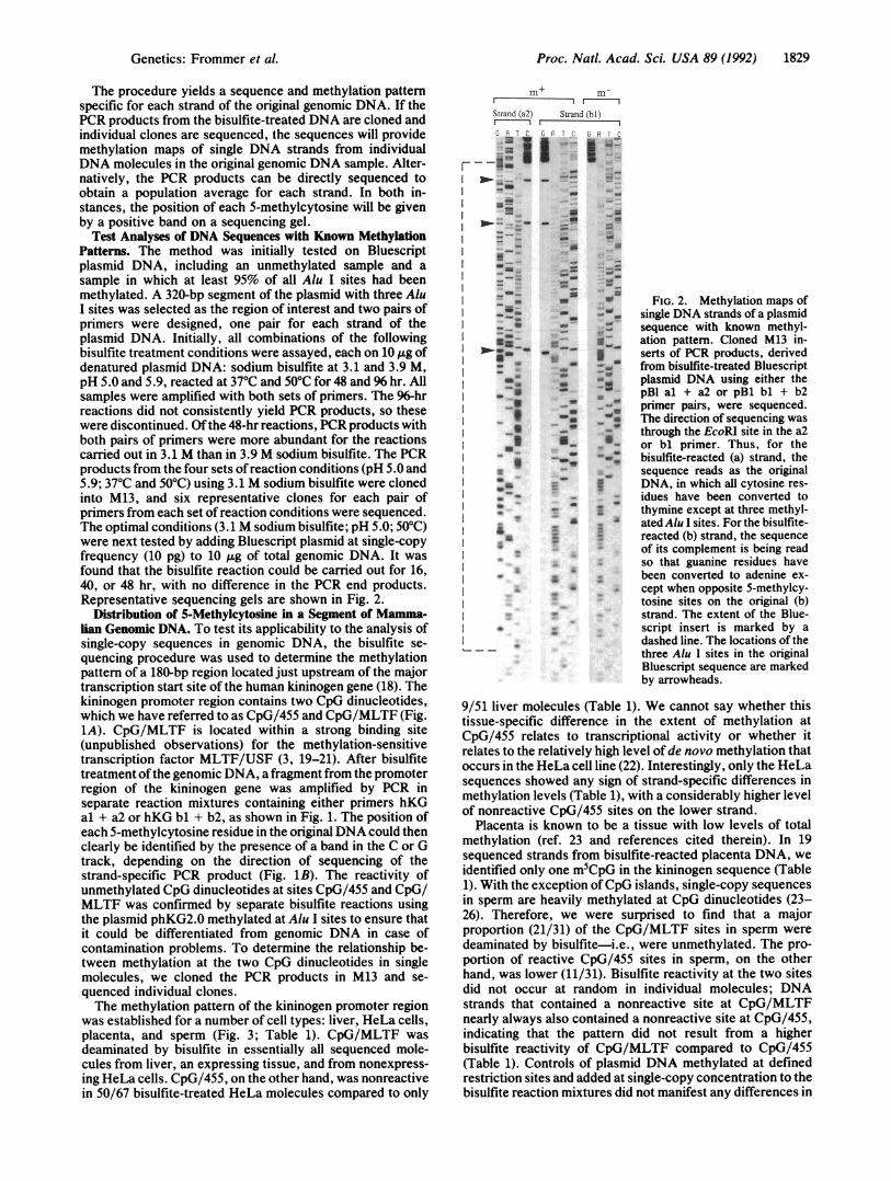
Test Analyses of DNA Sequences with Known MethylationPatterns. The method was initially tested on Bluescriptplasmid DNA, including an unmethylated sample and asample in which at least 95% of all Alu I sites had beenmethylated. A 320-bp segment of the plasmid with three AluI sites was selected as the region of interest and two pairs ofprimers were designed, one pair for each strand of theplasmid DNA. Initially, all combinations of the followingbisulfite treatment conditions were assayed, each on 10 Ag ofdenatured plasmid DNA: sodium bisulfite at 3.1 and 3.9 M,pH 5.0 and 5.9, reacted at 370C and 50'C for 48 and 96 hr. Allsamples were amplified with both sets of primers. The 96-hrreactions did not consistently yield PCR products, so thesewere discontinued. Ofthe 48-hr reactions, PCR products withboth pairs of primers were more abundant for the reactionscarried out in 3.1 M than in 3.9 M sodium bisutfite. The PCRproducts from the four sets ofreaction conditions (pH 5.0 and5.9; 370C and 500C) using 3.1 M sodium bisulfite were clonedinto M13, and six representative clones for each pair ofprimers from each set of reaction conditions were sequenced.The optimal conditions (3.1 M sodium bisulfite; pH 5.0; 500C)were next tested by adding Bluescript plasmid at single-copyfrequency (10 pg) to 10 ,ug of total genomic DNA. It wasfound that the bisulfite reaction could be carried out for 16,40, or 48 hr, with no difference in the PCR end products.Representative sequencing gels are shown in Fig. 2.
Distribution of 5-Methylcytosine in a Segment of Mamma-lian Genomic DNA. To test its applicability to the analysis ofsingle-copy sequences in genomic DNA, the bisulfite se-quencing procedure was used to determine the methylationpattern of a 180-bp region located just upstream of the majortranscription start site of the human kininogen gene (18). Thekininogen promoter region contains two CpG dinucleotides,which we have referred to as CpG/455 and CpG/MLTF (Fig.1A). CpG/MLTF is located within a strong binding site(unpublished observations) for the methylation-sensitivetranscription factor MLTF/USF (3, 19-21). After bisulfitetreatment ofthe genomic DNA, a fragment from the promoterregion of the kininogen gene was amplified by PCR inseparate reaction mixtures containing either primers hKGal + a2 or hKG bl + b2, as shown in Fig. 1. The position ofeach 5-methylcytosine residue in the original DNA could thenclearly be identified by the presence of a band in the C or Gtrack, depending on the direction of sequencing of thestrand-specific PCR product (Fig. 1B). The reactivity ofunmethylated CpG dinucleotides at sites CpG/455 and CpG/MLTF was confirmed by separate bisulfite reactions usingthe plasmid phKG2.0 methylated at Alu I sites to ensure thatit could be differentiated from genomic DNA in case ofcontamination problems. To determine the relationship be-tween methylation at the two CpG dinucleotides in singlemolecules, we cloned the PCR products in M13 and se-quenced individual clones.The methylation pattern of the kininogen promoter region
was established for a number of cell types: liver, HeLa cells,placenta, and sperm (Fig. 3; Table 1). CpG/MLTF wasdeaminated by bisulfite in essentially all sequenced mole-cules from liver, an expressing tissue, and from nonexpress-ing HeLa cells. CpG/455, on the other hand, was nonreactivein 50/67 bisulfite-treated HeLa molecules compared to only
m+ m-S I )Stgrand (a2 ) Strand (bl1)
~ I-
"---
-
I .-. zu
_-_
II
AS
ef T C CF TC
- SE
-- .-
--
a_- -Z
_ 1-
_ _
_ - _
* S
SW
-7 1
__ _
FIG. 2. Methylation maps ofsingle DNA strands of a plasmidsequence with known methyl-ation pattern. Cloned M13 in-serts of PCR products, derivedfrom bisulfite-treated Bluescriptplasmid DNA using either thepBl al + a2 or pBi bi + b2primer pairs, were sequenced.The direction of sequencing wasthrough the EcoRI site in the a2or bl primer. Thus, for thebisulfite-reacted (a) strand, thesequence reads as the originalDNA, in which all cytosine res-idues have been converted tothymine except at three methyl-ated Alu I sites. For the bisulfite-reacted (b) strand, the sequenceof its complement is being readso that guanine residues havebeen converted to adenine ex-cept when opposite 5-methylcy-tosine sites on the original (b)strand. The extent of the Blue-script insert is marked by adashed line. The locations of thethree Alu I sites in the originalBluescript sequence are markedby arrowheads.
9/51 liver molecules (Table 1). We cannot say whether thistissue-specific difference in the extent of methylation atCpG/455 relates to transcriptional activity or whether itrelates to the relatively high level of de novo methylation thatoccurs in the HeLa cell line (22). Interestingly, only the HeLasequences showed any sign of strand-specific differences inmethylation levels (Table 1), with a considerably higher levelof nonreactive CpG/455 sites on the lower strand.
Placenta is known to be a tissue with low levels of totalmethylation (ref. 23 and references cited therein). In 19sequenced strands from bisulfite-reacted placenta DNA, weidentified only one m5CpG in the kininogen sequence (Table1). With the exception ofCpG islands, single-copy sequencesin sperm are heavily methylated at CpG dinucleotides (23-26). Therefore, we were surprised to find that a majorproportion (21/31) of the CpG/MLTF sites in sperm weredeaminated by bisulfite-i.e., were unmethylated. The pro-portion of reactive CpG/455 sites in sperm, on the otherhand, was lower (11/31). Bisulfite reactivity at the two sitesdid not occur at random in individual molecules; DNAstrands that contained a nonreactive site at CpG/MLTFnearly always also contained a nonreactive site at CpG/455,indicating that the pattern did not result from a higherbisulfite reactivity of CpG/MLTF compared to CpG/455(Table 1). Controls of plasmid DNA methylated at definedrestriction sites and added at single-copy concentration to thebisulfite reaction mixtures did not manifest any differences in
Genetics: Frommer et al.

Proc. Natl. Acad. Sci. USA 89 (1992)
s I. itl El
F IA. ,a I.ww 4 .I,-.~~~
_., Alo
":.b3 w .S
S r-a i_ '
B llt t
F-
CpG 10.,Ml.,TF
CpG 10-455
'I,.
Z
..
I:r.
SI, ru 'IA
SS..
As
" Are_
A*$1!1
:::$ __
..... tlb-
.,. A_
dab_ "":. ,,,
se
A.AS ,,.
21,24_ ,£
usas,-h0.. __
... A,
WSEr
30|1st ....... ;0,|.so
SsrS:eF
e:: _
. .t
S....
He
:
_He
tiZ: I'for
or
:^-
A:pi
S:s
*''an
*3g
t::
IS
..
a,
FIG. 3. Characteristic strand-specific sequences from the kinin-ogen promoter region. (A) Strands (b2) and (al) sequenced in thedirection primer-CpG/MLTF-CpG/455-primer. (B) Strands (a2)and (bl) sequenced in the direction primer-CpG/455-CpG/MLTF-primer. The genomic DNAs used for the bisulfite reactions and theDNA strands sequenced are shown above each set of sequencingproducts. The extent of the kininogen gene insert is marked by adashed line. The location on the sequencing gels of CpG/455 andCpG/MLTF and the location of the CpNpG trinucleotide within anEcoRIl site are marked by arrowheads.
reactivity of particular 5-methylcytosine residues. We con-clude that the MLTF/USF binding site in sperm DNA ismethylated less frequently than a CpG dinucleotide located100 bp further upstream.
Digestion with the methylation-sensitive restriction en-zyme Pml I (CACGTG), which recognizes the core of theMLTF/USF binding site and does not digest either a fullymethylated or a hemimethylated site, has confirmed thisresult. HeLa, placenta, and liver genomic DNA showed totaldigestion at the kininogen Pml I site on Southern blots,
whereas sperm DNA was partially digested to o50%. Meth-ylation at CpG/455 could not be tested by restriction analy-SiS.We found no methylation at any non-CpG site inDNA from
HeLa cells, liver, or placenta. However, in a total of 31sequenced molecules from sperm DNA, we found 3 mole-cules that had not become deaminated by bisulfite at a singleCpTpG/CpApG, which was located within an EcoRIl site(Figs. 1A and 3). We do not believe that this rare CpTpG/CpApG methylation site in sperm represents a plasmid con-taminant. The three nonreactive sites occurred in two sepa-rate amplification reactions. One sequence was an (a) strand,and two were (b) strands with different methylation patternsat CpG/455 and CpG/MLTF (m'-m- and m--m-). None ofthe three apparently EcoRII-methylated molecules fromsperm was also methylated at an Alu I site within the 180-bpsequenced region.
DISCUSSIONUsing both in vitro methylated plasmid DNA and mammaliangenomic DNA, we have demonstrated the feasibility ofidentifying specific patterns of cytosine methylation by usingthe discrimination in deamination reactivity of cytosine and5-methylcytosine provided by bisulfite. We have found noconsistent lack of reactivity of any unmethylated cytosineresidue in the test substrates. Therefore, false positives oroverestimation of the extent of a partially methylated se-quence does not present a problem in the analysis. It shouldbe possible to use the bisulfite method to obtain a goodestimate of exact methylation levels. A small proportion of5-methylcytosine residues in single-stranded DNA may bedeaminated to thymine by the bisulfite treatment (15), so thatthe exact proportion of5-methylcytosine in the original DNAmay be slightly greater than the measured proportion ofnonreactive sites after bisulfite treatment and PCR. We havefound no difference between the reactivities of the variousmethylated sites analyzed in plasmids of known methylationpattern. By carrying out concomitant bisulfite reactions withmethylated cloned DNA including the region of interest andincorporating a methylated plasmid control into each ge-nomic bisulfite reaction, the extent of 5-methylcytosine re-activity can be accurately determined for each sequencingexperiment. For example, based on Alu 1-methylated plasmidcontrols, we can estimate that 35/42 nonreactive HeLa (b)strand sequences (Table 1) indicate a level of methylation ofCpG/455 in the (b) strand of close to 100%, and that thereactivity values of the other sites could be increased pro-portionately to yield exact methylation level.Major advantages of the bisulfite method are (i) the posi-
tive display of 5-methylcytosine and (ii) the capacity togenerate sequence data for individual DNA strands fromsingleDNA molecules. In methods dependent on Maxam andGilbert sequencing reactions, each cleaved DNA strandcontributes to one band in the sequencing gel. By contrast,the methylation status of every cytosine in a single bisulfite-reacted molecule can be read in the cloned product. This canbe of great advantage, for example, when analyzing thesuccessive demethylation of sites along a DNA strand as inthe chicken vitellogenin gene (27) and the E2A promoter ofintegrated adenovirus (28) or when the DNA being analyzedcomes from a mixed population of cells. The data can berelated to the parental origin of the DNA of interest (25,29-32), the direction of transcription, or the binding charac-teristics of specific protein factors.The minimum quantity of genomic DNA, analyzed in the
experiments described here, was 2 pg in a total reactionmixture of 1.2 ml. This is already 2- to 5-fold less than thequantity used in Maxam and Gilbert-based procedures.Smaller quantities of genomic DNA mixed with plasmid
i N .- __ -___
C:pG 10-.4 5 5
1830 Genetics: Frommer et al.
.Al.
Proc. Natl. Acad. Sci. USA 89 (1992) 1831
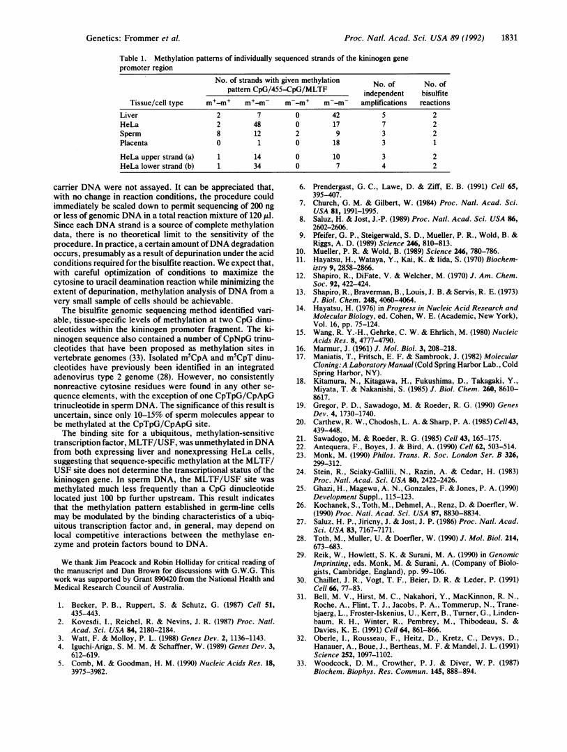
Table 1. Methylation patterns of individually sequenced strands of the kininogen genepromoter region
No. of strands with given methylationpattern CpG/455-CpG/MLTF independent bisulfite
Tissue/cell type m+-m+ m+-m- m--m+ m--m- amplifications reactions
Liver 2 7 0 42 5 2HeLa 2 48 0 17 7 2Sperm 8 12 2 9 3 2Placenta 0 1 0 18 3 1
HeLa upper strand (a) 1 14 0 10 3 2HeLa lower strand (b) 1 34 0 7 4 2
carrier DNA were not assayed. It can be appreciated that,with no change in reaction conditions, the procedure couldimmediately be scaled down to permit sequencing of 200 ngor less of genomic DNA in a total reaction mixture of 120 1.d.Since each DNA strand is a source of complete methylationdata, there is no theoretical limit to the sensitivity of theprocedure. In practice, a certain amount ofDNA degradationoccurs, presumably as a result of depurination under the acidconditions required for the bisulfite reaction. We expect that,with careful optimization of conditions to maximize thecytosine to uracil deamination reaction while minimizing theextent of depurination, methylation analysis ofDNA from avery small sample of cells should be achievable.The bisulfite genomic sequencing method identified vari-
able, tissue-specific levels of methylation at two CpG dinu-cleotides within the kininogen promoter fragment. The ki-ninogen sequence also contained a number of CpNpG trinu-cleotides that have been proposed as methylation sites invertebrate genomes (33). Isolated m5CpA and m5CpT dinu-cleotides have previously been identified in an integratedadenovirus type 2 genome (28). However, no consistentlynonreactive cytosine residues were found in any other se-quence elements, with the exception of one CpTpG/CpApGtrinucleotide in sperm DNA. The significance of this result isuncertain, since only 10-15% of sperm molecules appear tobe methylated at the CpTpG/CpApG site.The binding site for a ubiquitous, methylation-sensitive
transcription factor, MLTF/USF, was unmethylated in DNAfrom both expressing liver and nonexpressing HeLa cells,suggesting that sequence-specific methylation at the MLTF/USF site does not determine the transcriptional status of thekininogen gene. In sperm DNA, the MLTF/USF site wasmethylated much less frequently than a CpG dinucleotidelocated just 100 bp further upstream. This result indicatesthat the methylation pattern established in germ-line cellsmay be modulated by the binding characteristics of a ubiq-uitous transcription factor and, in general, may depend onlocal competitive interactions between the methylase en-zyme and protein factors bound to DNA.
We thank Jim Peacock and Robin Holliday for critical reading ofthe manuscript and Dan Brown for discussions with G.W.G. Thiswork was supported by Grant 890420 from the National Health andMedical Research Council of Australia.
1. Becker, P. B., Ruppert, S. & Schutz, G. (1987) Cell 51,435-443.
2. Kovesdi, I., Reichel, R. & Nevins, J. R. (1987) Proc. Nail.Acad. Sci. USA 84, 2180-2184.
3. Watt, F. & Molloy, P. L. (1988) Genes Dev. 2, 1136-1143.4. Iguchi-Ariga, S. M. M. & Schaffner, W. (1989) Genes Dev. 3,
612-619.5. Comb, M. & Goodman, H. M. (1990) Nucleic Acids Res. 18,
3975-3982.
6. Prendergast, G. C., Lawe, D. & Ziff, E. B. (1991) Cell 65,395-407.
7. Church, G. M. & Gilbert, W. (1984) Proc. Natl. Acad. Sci.USA 81, 1991-1995.
8. Saluz, H. & Jost, J.-P. (1989) Proc. Natl. Acad. Sci. USA 86,2602-2606.
9. Pfeifer, G. P., Steigerwald, S. D., Mueller, P. R., Wold, B. &Riggs, A. D. (1989) Science 246, 810-813.
10. Mueller, P. R. & Wold, B. (1989) Science 246, 780-786.11. Hayatsu, H., Wataya, Y., Kai, K. & Iida, S. (1970) Biochem-
istry 9, 2858-2866.12. Shapiro, R., DiFate, V. & Welcher, M. (1970) J. Am. Chem.
Soc. 92, 422-424.13. Shapiro, R., Braverman, B., Louis, J. B. & Servis, R. E. (1973)
J. Biol. Chem. 248, 4060-4064.14. Hayatsu, H. (1976) in Progress in Nucleic Acid Research and
Molecular Biology, ed. Cohen, W. E. (Academic, New York),Vol. 16, pp. 75-124.
15. Wang, R. Y.-H., Gehrke, C. W. & Ehrlich, M. (1980) NucleicAcids Res. 8, 4777-4790.
16. Marmur, J. (1%1) J. Mol. Biol. 3, 208-218.17. Maniatis, T., Fritsch, E. F. & Sambrook, J. (1982) Molecular
Cloning:A Laboratory Manual (Cold Spring Harbor Lab., ColdSpring Harbor, NY).
18. Kitamura, N., Kitagawa, H., Fukushima, D., Takagaki, Y.,Miyata, T. & Nakanishi, S. (1985) J. Biol. Chem. 260, 8610-8617.
19. Gregor, P. D., Sawadogo, M. & Roeder, R. G. (1990) GenesDev. 4, 1730-1740.
20. Carthew, R. W., Chodosh, L. A. & Sharp, P. A. (1985) Cell 43,439-448.
21. Sawadogo, M. & Roeder, R. G. (1985) Cell 43, 165-175.22. Antequera, F., Boyes, J. & Bird, A. (1990) Cell 62, 503-514.23. Monk, M. (1990) Philos. Trans. R. Soc. London Ser. B 326,
299-312.24. Stein, R., Sciaky-Gallili, N., Razin, A. & Cedar, H. (1983)
Proc. Natl. Acad. Sci. USA 80, 2422-2426.25. Ghazi, H., Magewu, A. N., Gonzales, F. & Jones, P. A. (1990)
Development Suppl., 115-123.26. Kochanek, S., Toth, M., Dehmel, A., Renz, D. & Doerfler, W.
(1990) Proc. Natl. Acad. Sci. USA 87, 8830-8834.27. Saluz, H. P., Jiricny, J. & Jost, J. P. (1986) Proc. Natl. Acad.
Sci. USA 83, 7167-7171.28. Toth, M., Muller, U. & Doerfler, W. (1990) J. Mol. Biol. 214,
673-683.29. Reik, W., Howlett, S. K. & Surani, M. A. (1990) in Genomic
Imprinting, eds. Monk, M. & Surani, A. (Company of Biolo-gists, Cambridge, England), pp. 99-106.
30. Chaillet, J. R., Vogt, T. F., Beier, D. R. & Leder, P. (1991)Cell 66, 77-83.
31. Bell, M. V., Hirst, M. C., Nakahori, Y., MacKinnon, R. N.,Roche, A., Flint, T. J., Jacobs, P. A., Tommerup, N., Trane-bjaerg, L., Froster-Iskenius, U., Kerr, B., Turner, G., Linden-baum, R. H., Winter, R., Pembrey, M., Thibodeau, S. &Davies, K. E. (1991) Cell 64, 861-866.
32. Oberle, I., Rousseau, F., Heitz, D., Kretz, C., Devys, D.,Hanauer, A., Boue, J., Bertheas, M. F. & Mandel, J. L. (1991)Science 252, 1097-1102.
33. Woodcock, D. M., Crowther, P. J. & Diver, W. P. (1987)Biochem. Biophys. Res. Commun. 145, 888-894.
Genetics: Frommer et al.