geographic variation in the pes of the salamander hynobius
TRANSCRIPT
ZOOLOGICAL SCIENCE 10: 1017-1027 (1993) © 1993 Zoological Society of Japan
Geographic Variation in the Pes of the Salamander Hynobius lichenatus: A Comparison with Tetradactyl Hynobius hidamontanus
and Pentadactyl Hynobius nigrescens
M A S A T O H A S U M I a n d H I S A A K I I W A S A W A
Biological Institute, Faculty of Science, Niigata University, Niigata 950-21, Japan
ABSTRACT—Variation in the pes was examined radiographically in 858 feet of three species of the genus Hynobius. The fifth toe of H. lichenatus was sometimes missing or barely expressed, caused by the absence of the fifth tarsale or by the fusion of the fourth and fifth tarsalia. Although the phalangeal formula was stable in H. hidamontanus (22320) and H. nigrescens (22332), it was highly variable in H. lichenatus (22332 and 31 other types were observed). In H. hidamontanus and some populations of H. lichenatus, there were unusual lots of feet with unossified first centrale and tibiale. Moreover, the unossification of the fifth tarsale, not influencing the occurrence of the fifth toe, was found in one population which corresponded to the southern limit of distribution of H. lichenatus. A postminimus ( = extra ankle bone), occurring in some primitive tetrapods, appeared sporadically on the postaxial side in the tarsus of H. lichenatus and H. nigrescens, but not of H. hidamontanus. The complete absence of both the fifth toe and the postminimus, and the presence of only two centralia, due to the fusion of the second and third centralia, suggest that H. hidamontanus is one of the most derived groups among the Hynobius species.
INTRODUCTION
Foot morphology has traditionally provided the systematic characters used in taxonomy [2]. The absence of the fifth toe occurs sporadically in genera of different groups of urodeles [15]. Noble [32] mentioned that the presence or absence of the fifth toe was considered a generic character in families except in Hynobiidae. Hynobius is a genus of this family that includes the most primitive living salamanders [10]. Hynobius lichenatus is broadly distributed in northeastern Japan, and its external morphology markedly varies in its geographic range [18−20, 38]. The fifth toe of this species is sometimes missing or barely expressed [7, 12, 19, 26, 29, 31, 38, 39] as well as that of H. kimurae [38].
In H. lichenatus, Maruyama [29] noted variation in the pes in one population, and Aoki [7] reported the absence of the fifth tarsale in one specimen with the rudimentary fifth toe. However, detailed
data are not available on intra- and interspecific variation in the pes of Hynobius species. We report here geographic variation in the pes of H. lichenatus, in comparison with that of the tetradactyl H. hidamontanus or the pentadactyl H. nigrescens, and discuss the cause of fifth toe missing and phylogenetic relationships of these taxa to other members of the family Hynobiidae in considering primitive and derived characters.
MATERIALS AND METHODS
283 male and 49 female adults of Hynobius lichenatus Boulenger were collected at random from 19 oviposition sites in northeastern Honshu, the mainland of Japan, during the breeding seasons of 1983−1985. For comparison, 18 adult males of H. hidamontanus Matsui were collected from one population in 1989, and 48 male and 31 female adults of H. nigrescens Stejneger, from two populations in 1984, 1990, and 1993. The sample sites are shown in Figure 1 and the Appendix. Sample sites 16−19 nearly correspond to the southern limit of distribution for H. lichenatus.
Accepted August 17, 1993 Received July 19, 1993

1018 M. HASUMI AND H. IWASAWA
FIG. 1. Map of northeastern Honshu, the mainland of Japan, showing sample sites of Hynobius lichenatus (1-19), H. hidamontanus (20), and H. nigrescens (21, 22). See the Appendix.
As soon as possible after collection, the animals were anesthetized with 0.01% p-aminobenzoic acid ethyl ester aq. and fixed in 10% formalin. The right and left feet of each animal were cut off at the groin, marked with string, and preserved in 70% ethanol. Radiographs of these 858 feet were taken with SOFRON equipment (Type SRO-M50, Soken Co., Ltd., Tokyo). Voucher specimens are currently stored in the Zoological Specimen Room, Biological Institute, Faculty of Science, Niigata University, Japan.
The composition of the pes of H. lichenatus is depicted in Figure 2. In the pes the bony shape and size were disregarded when investigating phalangeal formulae and numbers of tarsals. When there was a clear boundary between two bones fused (e.g., between two phalanges, phalanx and metatarsal, two tarsalia, tarsale and centrale, tar-sale and fibulare, two centralia, first centrale and tibiale, or fibulare and intermedium), each bone
FIG. 2. Diagram showing the composition of the pes (left, dorsal view) of Hynobius lichenatus. I, 1st toe; II, 2nd toe; III, 3rd toe; IV, 4th toe; V, 5th toe; c, centrale; fi, fibula; fib, fibulare; i, intermedium; m, metatarsal; o, outline; p, phalanges; pm, postmini-mus; t, tarsale; ti, tibia; tib, tibiale.
was counted separately. Unossified cartilage cannot be detected radiographically [18]: this cartilage is faintly visible in X-ray photographs, but occasionally invisible. Unossified tarsal cartilages were included in the number of tarsal bones, presuming empty elements of the tarsus ( = ankle region) except for a postminimus element because of its position on the postaxial side. Data for both sexes were combined because there was little difference between them. Regarding the phalangeal formula and the number of tarsal elements, the frequency of right-left asymmetry was examined in each population.
RESULTS
Phalangeal formulae In H. lichenatus, although the phalangeal formu
lae were highly variable with 32 types, many feet had a phalangeal formula of 22332 for the number

1019 Pes Variation in Salamanders
of phalanges from the first toe to the last one (Table 1 and Fig. 3A). However, the relative frequency of specimens with the other phalangeal formulae was much higher in some localities. Especially in sample site 6, a phalangeal formula of 22330 (13/36, 36.1%) or 22331 (11/36, 30.6%) was generally seen. Among feet with a phalangeal formula of 22332, the rates of specimens consisted of phalanges shrunken and nearly fused were 0/4, 2/15, 2/2, 3/8, 5/14, 4/4, 16/62, 6/25, 6/9, 6/9, 2/ 7, 0/22, 2/14, 17/93, 8/27, 4/16, 5/13, 2/15, and 18/93, respectively from sample sites 1-19.
The phalangeal formulae could not be applied to the following four specimens: (1) in one right foot in sample site 2, the rudimentary fifth toe had three very tiny bones close to the fourth metatarsal, the third and fourth tarsalia were fused into a much larger element which supported both the third and fourth metatarsals, and the fifth tarsale was absent; (2) in one right foot in sample site 5, the deformed second toe had a branched large phalanx which rested upon the enlarged second metatarsal, the rudimentary fifth toe consisted only of the very thin fifth metatarsal, the enlarged tarsale 1 + 2 and the first centrale were nearly fused, and the fifth tarsale and fibulare appeared to be fused though their boundary was clear (Fig. 3B); (3) in one right foot in sample site 8, the underdeveloped fifth toe had a single phalanx resting upon the slender metatarsal which branched from the fourth metatarsal, the fifth tarsale was lacking, and the first centrale and tibiale were fused; and (4) in one right foot in sample site 15, the rudimentary fifth toe had the same phalanx as that in sample site 8 mentioned above, the fourth metatarsal bore two phalanges, and the fourth and fifth tarsalia were fused.
In sample site 14, two feet were false tetradacty-ly: one right foot had a phalangeal formula of 22302 and no fourth metatarsal, but the fourth tarsale was present (Fig. 3C); and the other right foot had a phalangeal formula of 22022 and a tiny third metatarsal, assuming a triangle-like appearance. In sample site 18, one right foot with the underdeveloped fifth toe was false pentadactyly and had a phalangeal formula of 22320 and no fifth metatarsal, although the fifth tarsale was normal (Fig. 3D).
In H. hidamontanus, 34 of 36 feet (94.4%) had a phalangeal formula of 22320; and in H. nigrescens, 148 of 158 feet (93.7%), a phalangeal formula of 22332 (Table 1 and Fig. 4). The rates of different phalangeal formulae on right and left sides (right-left asymmetry) were 0/2, 3/11, 0/3, 5/12, 4/15, 8/18, 7/38, 13/25, 1/5, 4/7, 3/5, 5/15, 3/13, 14/ 54, 7/18, 4/10, 3/9, 6/12, 17/60, 2/18, 8/59, and 2/20, respectively from sample sites 1-22.
Number of tarsals
In H. lichenatus, the number of tarsals was variable, ranging from 6 to 12 (Table 2); and the tarsus was composed of 10 tarsal elements in many populations (Fig. 3A), but eight or nine elements in some. In the tarsus, there were four tarsalia which consisted of the enlarged tarsale 1+2 and the third, fourth, and fifth tarsalia in nature; however, three tarsalia were also seen, due mainly to the fusion of the fourth and fifth tarsalia or to the absence of the fifth tarsale. The specimens having three tarsalia were more numerous in some populations (e.g., sample site 6, 22/36, 61.1%; sample site 8, 27/50, 54.0%; sample site 17, 13/18, 72.2%). The number of centralia was three in general in the tarsus, but two centralia, due to the fusion of the second and third centralia, were normally seen (e.g., sample site 4, 15/24, 62.5%; sample site 5, 21/30, 70.0%; sample site 6, 18/36, 50.0%; sample site 8, 32/50, 64.0%), to some extent independently of the occurrence of the three tarsalia. For example, in sample site 17, 17 of 18 feet (94.4%) had three centralia despite the three tarsalia in 13 feet. The smaller number of tarsal elements partially coincided with the smaller number of phalanges, especially the fifth two phalanges. A unique right foot having six tarsal bones which appeared in sample site 6 was composed of tarsale 1 + 2, the fused third and fourth tarsalia which supported both the third and fourth metatarsals, the fused first centrale and tibiale, the fused second and third centralia, the fibulare, and the intermedium, and had a phalangeal formula of 12320 (Fig. 3E). One right and three left feet contained 12 tarsal bones, due to having four centralia and the postminimus in sample sites 7 and 12, and five centralia (Fig. 3F) in sample sites 14 and 16.

1020 M. HASUMI AND H. IWASAWA
TABLE 1. Intra- and interspecific variation in the phalangeal formula in the foot of Hynobius lichenatus (sample sites 1-19), H. hidamontanus (20), and H. nigrescens (21, 22)
Phalangeal formulae
00032 02221 02331 12210 12220 12320 12322 12330 12331 12332 20031 20332 21322 21330 21331 21332 22022 22132 22211 22220 22222 22230 22232 22302 22311 22320 22321 22322 22330 22331 22332 22343 22431 23331 Extra
1
4
2
1
1 1 3
15
1
3
4 2
4
1
1
4 10 8
5
1
7 7
14
1
6
3
5
13 11 4
7
1
1 1
1
1 1 8
62
8
1
1
2
2 17 25
1
1
9
1 9
Sample site
10
1
1
3 9
11
1
1
1 7
12
1
1
1
1 4
22
13
2
1
3 6
14
14
1 1
1 1
1 1
2
3 1 3
93
15
1
1
2
1
1
2 27
1
16
1
1
2
16
17
1
1
2 13
1
18
1
1
2
1
4 15
19
1
1 1
3
1
1
1
2
2 3
11 93
20
1
34
1
21
1
1
2
2
1 110
1
22
1
1
38
Sample size 4 22 6 24 30 36 76 50 10 14 10 30 26 108 36 20 18 24 120 36 118 40
Figures indicate the number of specimens.
FIG. 3. X-ray photographs of the pes of Hynobius lichenatus. Dorsal view of the "left" pes is shown for the convenience of the readers. The original right pes is expressed by asterisks superscribed the alphabetical numbers. Scale bar represents 5 mm. (A): Standard. (B*): Anomaly (see text). (C*): False tetradactyly without the fourth toe. (D*): False pentadactyly (arrows) without the fifth phalanges and metatarsal, and with the fifth tarsale. (E*): Tetradactyly with six tarsal elements. (F): Pentadactyly with five centralia, appearing in the primitive tetrapods. (G): Tetradactyly with the same composition as that of H. hidamontanus (see Fig. 4A). (H*): Tetradactyly with the fused third and fourth tarsalia. (I*): Tetradactyly with 11 tarsals including very tiny two. (J): Tetradactyly with the fifth tarsale and the unossified first centrale and tibiale. (K*): Pentadactyly with

1021 Pes Variation in Salamanders
the rudimentary fifth toe despite its normal phalanges and metatarsal. (L*): Pentadactyly with the rudimentary fifth phalanges and metatarsal. (M*): Pentadactyly with the unossified tibiale. (N): Pentadactyly with the unossified first centrale and tibiale. (O*): Pentadactyly with the unossified fifth tarsale, first centrale, and tibiale. (P*): Pentadactyly with the postminimus (arrow), also appearing in the primitive tetrapods.
1022 M. HASUMI AND H. IWASAWA
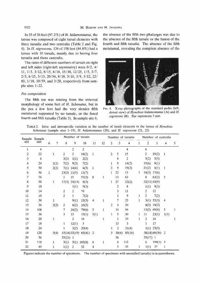
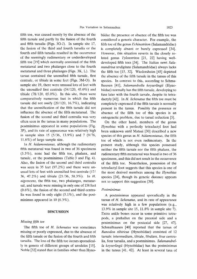
In 35 of 36 feet (97.2%) of H. hidamontanus, the tarsus was composed of eight tarsal elements with three tarsalia and two centralia (Table 2 and Fig. 4). In H. nigrescens, 134 of 158 feet (84.8%) had a tarsus with 10 tarsals, mainly due to having four tarsalia and three centralia.
The rates of different numbers of tarsals on right and left sides (right-left asymmetry) were 0/2, 4/ 11, 1/3, 3/12, 8/15, 8/18, 15/38, 12/25, 1/5, 3/7, 2/5, 6/15, 3/13, 20/54, 8/18, 3/10, 3/9, 3/12, 22/ 60, 1/18, 10/59, and 3/20, respectively from sample sites 1-22.
Pes composition The fifth toe was missing from the external
morphology of some feet of H. lichenatus, but in the pes a few feet had the very slender fifth metatarsal supported by no tarsale, or the fused fourth and fifth tarsalia (Table 3). In sample site 6,
the absence of the fifth two phalanges was due to the absence of the fifth tarsale or the fusion of the fourth and fifth tarsalia. The absence of the fifth metatarsal, revealing the complete absence of the
FIG. 4. X-ray photographs of the standard pedes (left, dorsal view) of Hynobius hidamontanus (A) and H. nigrescens (B). Bar represents 5 mm.
TABLE 2. Intra- and interspecific variation in the number of tarsal elements in the tarsus of Hynobius lichenatus (sample sites 1−19), H. hidamontanus (20), and H. nigrescens (21, 22)
Sample site
1 2 3 4 5 6 7 8 9
10 11 12 13 14 15 16 17 18 19 20 21 22
Sample . size
4 22
6 24 30 36 76 50 10 14 10 30 26
108 36 20 18 24
120 36
118 40
6
1
7
1
1(1) 2(2)
1
1 2(2)
1
5(4)
1 1
Number of tarsals
8
2 3(3) 7(2) 7(1)
13(3) 1
17(3)
2 2
2 7 3
1 1
15(14) 35(21)
3(1) 1(1)
9
2 2
1(1) 9(3)
14(4) 11(3) 15 24(14)
1(1) 2 1 9(1) 6(2)
16(2) 13 2
12(1) 3(2)
33(19) 1
5(1) 2
10
2 16(2) 2(2) 7(2) 6(3)
11(7) 51(2)
8(3) 9(3)
10 7(2)
15(3) 16(2) 79(6)
15(1) 16 5
20(4) 65(41)
102(8) 32
11
1
1
8
4
5
5(1)
2
6 4
12
1
1
1
1
1
Number of tarsalia
2
2
1 2 1
1
2
1
1 5
3
5 4 9 9
22 13 27
2 3 1 7 4
14 5 1
13 2
30(4) 36
6 5
4
4 15 2
14(2) 19(2) 13 63 22(2)
8 11 9
23 20 94 30 19 5
21(4) 85(16)
112 35
1
1
1 1
1 1
1
Number of centralia
2
4 2 3(2)
15(6) 21(2) 18(3) 8
32(11)
1(1) 2 2 3(1) 8(2)
13(2) 11 2 1
1(1) 38(18) 35(17)
6 1(1)
3
19(2) 3(3) 9(1) 8(1)
17(6) 63(2) 18(9) 9(3)
12 7(2)
22(3) 18(2) 89(6) 23(1) 16 17 23(5) 80(36)
1 109(1) 37
4
1
1
5
4
5
1(1)
2
3 1
5
1
1
Figures indicate the number of specimens. The number of specimens with unossified tarsal(s) is in parentheses.
1023 Pes Variation in Salamanders
fifth toe, was caused mostly by the absence of the fifth tarsale and partly by the fusion of the fourth and fifth tarsalia (Figs. 3G-J). In sample site 17, the fusion of the third and fourth tarsalia or the fourth and fifth tarsalia resulted in the occurrence of the seemingly rudimentary or underdeveloped fifth toe [19] which normally consisted of the fifth metatarsal and two phalanges close to the fourth metatarsal and three phalanges (Figs. 3K, L). The tarsus contained the unossified fifth tarsale, first centrale, or tibiale in some feet (Figs. 3M-0). In sample site 19, there were unusual lots of feet with the unossified first centrale (54/120, 45.0%) and tibiale (78/120, 65.0%). In this site, there were comparatively numerous feet in which the fifth tarsale did not ossify (20/120, 16.7%), indicating that the unossification of the fifth tarsale did not influence the absence of the fifth metatarsal. The fusion of the second and third centralia was very often seen in the tarsus in many populations. The postminimus appeared in some populations (Fig. 3P), and its rate of appearance was relatively high in sample sites 15 (5/36, 13.9%) and 7 (9/76, 11.8%) of large sample size.
In H. hidamontanus, although the rudimentary fifth metatarsal was found in two of 36 specimens (5.5%), none had the fifth toe, phalanx, and tarsale, or the postminimus (Table 3 and Fig. 4). Also, the fusion of the second and third centralia was seen in 35 feet (97.2%) and there were unusual lots of feet with unossified first centrale (17/ 36, 47.2%) and tibiale (21/36, 58.3%). In H. nigrescens, the fifth toe, two phalanges, metatarsal, and tarsale were missing in only one of 158 feet (0.6%), the fusion of the second and third centralia was found in only eight (5.1%), and the postminimus appeared in 10 (6.3%).
DISCUSSION
Missing fifth toe The fifth toe of H. lichenatus was sometimes
missing or poorly expressed, due to the absence of the fifth tarsale or the fusion of the fourth and fifth tarsalia. The loss of the fifth toe occurs sporadically in genera of different groups of urodeles [15]. Noble [32] stated that in families other than Hyno-
biidae the presence or absence of the fifth toe was considered a generic character. For example, the fifth toe of the genus Echinotriton (Salamandridae) is completely absent or barely expressed [34]. However, this situation reverts in the closely related genus Tylototriton [21, 22] having well-developed fifth toes [34]. The Italian newt Sala-mandrina terdigitata (Salamandridae) always lacks the fifth toe [15, 32]. Wiedersheim [45] depicted the absence of the fifth tarsale in the tarsus of this species. In contrast to this, according to Schmalhausen [41], Salamandrella keyserlingii (Hyno-biidae) normally has the fifth tarsale, developing to fuse later with the fourth tarsale, despite its tetra-dactyly [42]. In H. lichenatus the fifth toe must be completely expressed if the fifth tarsale is normally present in the tarsus. Possibly the presence or absence of the fifth toe of this species is an ontogenetic problem, due to tarsal reduction [5].
On the other hand, members of the genus Hynobius with a perfectly tetradactyl foot had been unknown until Matsui [30] described a new species of this genus as H. hidamontanus, the fifth toe of which is not even rudimentary. In the present study, although this species possessed neither the fifth tarsale nor the fifth phalanx, the rudimentary fifth metatarsal was found in two of 36 specimens, and this did not result in the occurrence of the fifth toe. Nonetheless, possession of the tetradactyl foot suggests that this species is one of the most derived members among the Hynobius species [24], though its genetic distance appears not to support this suggestion [30].
Postminimus
A postminimus appeared sporadically in the tarsus of H. lichenatus, and its rate of appearance was relatively high in a few populations (e.g., 13.9% in sample site 15, 11.8% in sample site 7). Extra ankle bones occur in some primitive tetra-pods, a prehallux on the preaxial side and a postminimus on the postaxial side [27, 47]. Schmalhausen [40] reported that the tarsus of Ranodon sibiricus (Hynobiidae) consisted of 12 tarsals: intermedium, tibiale, fibulare, four centralia, four tarsalia, and a postminimus. Salamandrella keyserlingii (Hynobiidae) has the postminimus in the tarsus [41, 42]. At least in several taxa of

1024 M. HASUMI AND H. IWASAWA
TABLE 3. Intra- and interspecific variation in the condition of the pes of Hynobius lichenatus
Sample site
1 2 3 4 5 6 7 8 9
10 11 12 13 14 15 16 17 18 19 20 21 22
Sample size
4 22
6 24 30 36 76 50 10 14 10 30 26
108 36 20 18 24
120 36
118 40
Absence of toe 5
0 0 0 4 3 9 0 3 0 0 0 1 4 4 2 0 0 0 5
36 0 1
Absence of phalanges 5
0 2 0 5 7
21 2 4 0 0 0 2 6 3 2 1 0 1 6
36 0 1
Absence of metatarsal 5
0 0 0 3 2 7 0 3 0 0 0 1 2 2 2 0 0 1 4
34 0 1
Absence of tarsale 5
0 1 0 5 2 6 0 3 0 0 0 1 3 3 2 0 0 0 3
36 0 1
Unossified tarsale 5
0 0 0 2 2 0 0 2 0 0 0 0 0 0 0 0 0 4
20 0 0 0
Figures indicate the number of specimens.
hynobiids the postminimus certainly occurs, indicating that this family remains primitive among living salamanders. The absence of the postminimus from the samples examined in H. hidamonta-nus prompts us to reconsider its phylogenetic relationship to other members of this family.
Unossified tarsal cartilages
The tarsus of H. lichenatus normally consisted of well-ossified tarsal elements, but occasionally possessed a few unossified tarsal cartilages, restricted almost to the first centrale and the tibiale. These cartilages were relatively numerous in some populations (e.g., the unossified tibiale more than 30% in sample sites 3, 4, 5, 6, 8, and 9), in which the snout-vent length (SVL) was much smaller except in sample site 6, and the fifth toe was barely expressed except in sample site 9 [19]. In sample site 19, however, there were unusual lots of feet with the unossified tibiale (65.0%) even though the
specimens had a greater SVL and a more developed fifth toe [19]. Moreover, 45.0% of the first centralia and 16.7% of the fifth tarsalia did not ossify in this population: the unossification of the fifth tarsale was very rare in the other populations. Although Nussbaum [33] stated concerning Dicamptodon ensatus (Dicamptodontidae) that the ossification of tarsal cartilages was the result of aging rather than of metamorphosis, Necturus (Proteidae) species has the tarsus with six cartilaginous tarsals throughout its aquatic life [16, 23], and Alberch [1, 2] and Alberch and Alberch [4] depicted cartilaginous tarsals in the tarsus of the genus Bolitoglossa (Plethodontidae) throughout its terrestrial life. It is not known when ossification of the tarsals occurs in H. lichenatus and whether all tarsal elements ossify simultaneously.
Francis [14] reported concerning Salamandra salamandra (Salamandridae) that the first centrale and the tibiale normally remained cartilaginous.

1025 Pes Variation in Salamanders
(sample sites 1−19), H. hidamontanus (20), and H. nigrescens (21, 22)
Tarsalia 4 + 5
0 6 4 5 9
16 13 25
2 3 1 6 3
11 5 1
11 3
25 0 6 4
Unossified centrale 1
0 2 5 7 3 9 2
20 4 0 2 4 4 8 2 0 0 6
54 17
1 1
Unossified tibiale
0 2 6 8
10 13 2
20 4 0 2 4 4 8 2 0 1 6
78 21 10
1
Centrale 1 + tibiale
0 0 0 0 0 1 0 0 0 0 1 1 0 0 1 1 0 0 0 0 1 1
Centralia 2 + 3
4 2 3
15 21 19 8
32 1 2 3 4 8
13 12 3 1 1
38 35 6 2
Fibulare + intermedium
0 0 0 0 0 0 0 1 0 2 0 0 0 2 0 0 0 0 0 0 0 0
Presence of postminimus
2 2 0 0 0 2 9 1 2 1 0 2 0 0 5 0 0 0 0 0 6 4
Hilton [23] observed the unossified first centrale and tibiale in the tarsus of Taricha granulosa (Salamandridae). It is therefore likely that the unossification of only these two elements normally occurs in adult salamanders of several species, though probably the tarsus with the unossified fifth tarsale which bears the fifth metatarsal is not normally present except in specimens of sample site 19 of H. lichenatus. In this site, it was interesting that there were unusual lots of feet with unossified first centrale and tibiale, similar to those of H. hidamontanus. Also, the skull shape of all specimens in this site differs articulately from that in the others and the maxilla-maxilla length/snout-condyle length is the greatest of all populations examined (Hasumi, personal observation). This population, which corresponds to the southern limit of distribution of H. lichenatus, appears out of intraspecific variation in the osteological characters of this species, and therefore its validity as H. lichenatus must be reconsidered. The presence of
several cryptic taxa has already been suggested in this species [30]. However, the present results suggest that interpopulation variation in the pes of this species is an ontogenetic problem except in sample site 19.
Terminology
A tarsal element, which rests upon the tibiale and often provides partial support for the first metatarsal, is generally called the first centrale [28, 34, 37, 41, 42]. The same element is also called the cartilago prehallucis [14, 25, 29], the tarsale pre-hallucis [27], the naviculare [9], or the mediale [2, 40]. According to Schmalhausen [41] and de Saint-Aubain [9], however, the prehallucis rests upon the first centrale. On the other hand, Wiedersheim [45], Osawa [35], and Branch [8] regarded the first centrale as the first tarsale. We considered here the possibility of the separation of the tarsale 1 + 2 in the tarsus of Andrias japonicus (Cryptobranchidae), but confirmed in its skeletal
1026 M. HASUMI AND H. IWASAWA
specimen that Osawa's [35] description was erroneous .
A tarsal e lement which bears a metatarsal is generally designated as the tarsale [12, 15, 32, 34, 40, 42, 44, 47]. The distal tarsal [ 1 - 3 , 6, 9, 13, 17, 28, 35, 37, 41 , 43, 46] and the basal tarsal [14] are normally employed for this element . Duel lman and Trueb [11] erroneously used for it the term "tarsal" , any bone lying between the tibia a n d / o r fibula and the metatarsals [36].
The postminimus [27, 4 1 , 47] is also called the posttarsale [42]. Except those ment ioned above, the terminology for the pes does not seem to be very confused, to the best of our knowledge.
ACKNOWLEDGMENTS
We thank Professor N. Nara of Hirosaki University for giving us information on four populations of H. lichenatus in the Aomori district. The senior author is indebted to Mr. M. Kakegawa of Honjo Senior High School and to Mr. F. Kishi of the Shirouma Association of Naturalists (Shirouma-shizennokai) for their help in collecting specimens of H. hidamontanus. Handling of this species is regulated by the Government of Hakuba-mura, and the collection was made under the permission authorized by this Government. We are also grateful to Professor K. Kobayashi of Nippon Dental University for the use of SOFRON equipment.
REFERENCES
1 Alberch P (1980) Ontogenesis and morphological diversification. Amer Zool 20: 653-667
2 Alberch P (1981) Convergence and parallelism in foot morphology in the neotropical salamander genus Bolitoglossa. I. Function. Evolution 35: 84-100
3 Alberch P (1983) Morphological variation in the neotropical salamander genus Bolitoglossa. Evolution 37: 906-919
4 Alberch P, Alberch J (1981) Heterochronic mechanisms of morphological diversification and evolutionary change in the neotropical salamander, Bolitoglossa occidentalis (Amphibia: Plethodonti-dae). J Morphol 167: 249-264
5 Alberch P, Gale EA (1983) Size dependence during the development of the amphibian foot. Colchicine-induced digital loss and reduction. J Embryol exp Morphol 76: 177-197
6 Alberch P, Gale EA (1985) A developmental analysis of an evolutionary trend: Digital reduction in amphibians. Evolution 39: 8-23
7 Aoki R (1977) On the fifth toe of the genus Hynobius. Nippon Herpetol J 9: 13-14 (in Japanese)
8 Branch HE (1935) A Laboratory Manual of Cryp-tobranchus alleganiensis Daudin. Vantage Press, New York, 2nd ed
9 de Saint-Aubain ML (1981) Amphibian limb ontogeny and its bearing on the phylogeny of the group. Z zool Syst Evolut-forsch 19: 175-194
10 Dowling HG, Duellman WE (1973) Systematic Herpetology: A Synopsis of Families and Higher Categories. HISS Publications, New York
11 Duellman WE, Trueb L (1986) Biology of Amphibians. McGraw-Hill, New York
12 Dunn ER (1927) On the loss of the fifth toe in certain salamanders. Science 66: 509
13 Fabrezi M (1993) The anuran tarsus. Alytes 11: 47-63
14 Francis ETB (1934) The Anatomy of the Salamander. Oxford University Press, London
15 Gadow H (1901) Amphibia and Reptiles. Macmil-lan, London
16 Gilbert SG (1973) Pictorial Anatomy of the Nectur-us. University of Washington Press, Seattle
17 Hanken J, Dinsmore CE (1986) Geographic variation in the limb skeleton of the red-backed salamander, Plethodon cinereus. J Herpetol 20: 97-101
18 Hasumi M, Iwasawa H (1987) Geographic variation in the tail of the Japanese salamander, Hynobius lichenatus, with special reference to taxonomic bearing. Zool Sci 4: 159-166
19 Hasumi M, Iwasawa H (1987) Geographic variation in morphological characters of the Japanese salamander, Hynobius lichenatus. Sci Rep Niigata Univ Ser D (Biol) 24: 15-30
20 Hasumi M, Iwasawa H (1988) Geographic variation in the number of teeth of the Japanese salamander, Hynobius lichenatus. Sci Rep Niigata Univ Ser D (Biol) 25: 1-17
21 Hayashi T, Matsui M (1989) Preliminary study of phylogeny in the family Salamandridae: Allozyme data. In "Current Herpetology in East Asia" Ed by M Matsui, T Hikida, RC Goris, Herpetological Society of Japan, Kyoto, pp 157-167
22 Hayashi T, Matsui M, Utsunomiya T, Tanaka S, Ota H (1992) Allozyme variation in the newt Tylototriton andersoni from three islands of Ryukyu Archipelago. Herpetologica 48: 178-184
23 Hilton WA (1948) The carpus and tarsus of salamanders. J Entomol Zool 40: 1-13
24 Holder N (1983) Developmental constraints and the evolution of vertebrate digit patterns. J theor Biol 104: 451-471
25 Ichikawa M (1943) Variation in the carpus and tarsus of the Japanese red-bellied newt, Cynops pyrrhogaster. Zool Mag 55: 331-334 (in Japanese)

1027 Pes Variation in Salamanders
26 Inukai T, Makino S (1933) On a local variation of Hynobius lichenatus Boulenger found in Akita-ken and Yamagata-ken. Rep Jpn Assoc Ad Sci 8: 454-457 (in Japanese)
27 Jarvik E (1980) Basic Structure and Evolution of Vertebrates Vol 2. Academic Press, London
28 Kent GC (1983) Comparative Anatomy of the Vertebrates. Mosby, St Louis, 5th ed
29 Maruyama T (1977) Digital variation in the hind legs of the salamander Hynobius lichenatus. Jpn J Herpetol 7: 10-14 (in Japanese with English abstract)
30 Matsui M (1987) Isozyme variation in salamanders of the nebulosus-lichenatus complex of the genus Hynobius from eastern Honshu, Japan, with a description of a new species. Jpn J Herpetol 12: 50-64
31 Nakamura K, Uéno S-I (1963) Japanese Reptiles and Amphibians in Colour. Hoiku-sha, Osaka (in Japanese)
32 Noble GK (1931) The Biology of the Amphibia. McGraw-Hill, New York
33 Nussbaum RA (1976) Geographic variation and systematics of salamanders of the genus Dicamp-todon Strauch (Ambystomatidae). Misc Publ Mus Zool Univ Michigan 149: 1-94
34 Nussbaum RA, Brodie ED Jr (1982) Partitioning of the salamandrid genus Tylototriton Anderson (Amphibia: Caudata) with a description of a new genus. Herpetologica 38: 320-332
35 Osawa G (1902) Beiträge zur Anatomie des japanischen Riesensalamanders. Mitt Med Fac Univ Tokio 5: 1-207
36 Peters JA (1964) Dictionary of Herpetology. Hafner Publishing, New York
37 Romer AS, Parsons TS (1977) The Vertebrate Body. Saunders, Philadelphia, 5th ed
38 Sato I (1943) Monograph of Japanese Tailed Bat-rachians. Nippon Shuppan-sha, Osaka (in Japanese)
39 Sawano J (1947) Normal Stages of Development of the Japanese Salamander Hynobius lichenatus. Grus-bunko, Sapporo (in Japanese)
40 Schmalhausen II (1968) The Origin of Terrestrial Vertebrates. Academic Press, New York
41 Schmalhausen JJ (1910) Die Entwicklung des Extremitätenskelettes von Salamandrella kayserlingii. Anat Anz 37: 431-446
42 Starck D (1979) Vergleichende Anatomie der Wirbeltiere 2. Springer-Verlag, Berlin
43 Wake DB, Özeti N (1969) Evolutionary relationships in the family Salamandridae. Copeia 1969: 124-137
44 Webster D, Webster M (1974) Comparative Vertebrate Morphology. Academic Press, New York
45 Wiedersheim R (1875) Salamandrina perspicillata und Geotriton fuscus. Versuch einer vergleichenden Anatomie der Salamandrinen. Sordo-Muti, Genua
46 Worthington RD (1974) High incidence of anomalies in a natural population of spotted salamanders, Ambystoma maculatum. Herpetologica 30: 216-220
47 Zug G (1979) The endoskeleton: The comparative anatomy of the girdles, the sternum, and the paired appendages. In "Hyman's Comparative Vertebrate Anatomy" Ed by MH Wake, University of Chicago Press, Chicago, 3rd ed, pp 238-264
APPENDIX
Sample sites
Hynobius lichenatus: (1) Maedanome, Goshogawara-shi, Aomori Prefecture; (2) Kudoji, Hirosaki-shi, Aomori Pref.; (3) Mt. Ajara-yama, Ohwani-machi, Aomori Pref.; (4) Mase-keikoku, Hachimori-machi, Akita Pref.; (5) Natsuzaka, Takko-machi, Aomori Pref.; (6) Kawamata, Tamayama-mura, Iwate Pref.; (7) Hiru-kawa, Ohmagari-shi, Akita Pref.; (8) Idosawa, Ichi-noseki-shi, Iwate Pref.; (9) Yamanome, Ichinoseki-shi, Iwate Pref.; (10) Iragawa, Atsumi-machi, Yamagata Pref.; (11) Hataya, Yamanobe-machi, Yamagata Pref.; (12) Mt. Ninoji-dake, Shibata-shi, Niigata Pref.; (13) Hibara, Kitashiobara-mura, Fukushima Pref.; (14) Yuta-gami, Tagami-machi, Niigata Pref.; (15) Kamijo, Kamo-shi, Niigata Pref.; (16) Tanne, Kashiwazaki-shi, Niigata Pref.; (17) Mt. Atema-yama, Tokamachi-shi, Niigata Pref.; (18) Okushiobara, Shiobara-machi, Tochigi Pref.; and (19) Fujiwara, Minakami-machi, Gunma Pref.
H. hidamontanus: (20) Ochikura, Hakuba-mura, Nagano Pref.
H. nigrescens: (21) Mt. Tenjin-yama, Iwamuro-mura, Niigata Pref. and (22) Maikomidaira, Oumi-machi, Niigata Pref.