gestational trophoblastic disease (gtd)
DESCRIPTION
A presentation made to the 5th year class of Bachelor of Medicine and Bachelor of Surgery at Makerere University College of Health Sciences.TRANSCRIPT

Gestational Trophoblastic Disease
Juvenal Nkeramahame, MBChB; Deogratias Migadde, MBChB; Jane Okutiru, MBChB.
IntroductionDefn. GTD is a group of disorders including pre-malignant conditions of complete & partial hydatidiform moles through to the malignant invasive mole, choriocarcinoma & placental site trophoblastic tumour/epithelioid trophoblastic tumour. RCOG, ESMO 2014
GTD refers to pregnancy-related trophoblastic proliferative abnormalities (Williams Obst)
Malignant forms of GTD a.k.a GTN or postmolar GTN and usu.followhydatidiform mole

Classifn (FIGO)
• Hydatidiform molar (molar preg)-complete or partial
• Invasive mole ( chorioadenoma destruens)
• Choriocarcinoma
• Placental-site trophoblastic tumor( PSTT)
• Epithelioid trophoblastic tumor(ETT)
HYDATIDIFORM MOLE
• Refers to abnormal trophoblastic proliferation & edema of chorionic villi stroma
• Rapidly proliferating trophoblasts secrete large amts of hCG.
• Absence or presence of fetal or embryonic tissue used to classifty into complete or partial HM.
• Theca-lutein cysts in ovaries are commonly assoc with HM. Result from hyperstimulation of lutein elements by hCG

Complete HM
• Complete HM results from 1 or 2 sperms fertilizing a ‘blank’ ovum-without DNA.
• Hence no embryo is formed.
• 85% have diploid karyotype ie 46, XX, both xsomes of paternal origin (androgenesis)

Partial HM
• PHM results from 2 sperms fertilizing a normal ovum.
• karyotype is triploid in 85% cases ie 69, XXX, 69, XXY, 69 XYY.
• Some fetal tissue is present but w/ multiple mal4mns
• The fetus is nonviable

R/factors for HM
• Age <20yrs and >35yrs (Williams Obs, 24th ed.)
• Previous molar pregnancy
Clinical presentn
• Exaggerated signs of pregnancy
• Hyperemesis gravidarum
• Uterine bleeding- spotting, profuse bleeding
Anemia-iron def.
Signs Hemorrhagic shock
• FH greater than for gest. age
• No fetal activity- no FHR
• Gest. HTN ( pre-eclampsia/eclampsia b4 20 wks)
• Thyrotoxicosis-thyrotropin like effect of hCG
• Embolic pheno.
Diagnostic features HM
• Continuous or intermittent brown or bloody pv d/c evident around 12woa
• Uterine enlargement out of propn to gest. age
• Absence of fetal parts & fetal heart activity
• Xtic app on Abd. USS. honey-comb app
• Serum ᵝhCG higher than expected
• Pre-clampsia/eclampsia b4 20 wks(24wks)
• Hyperemesis gravidarum

Management of HM
• Immediate evacuation
• Suction and curettage is treatment of choice
• D&C
• Subsqnt evaluatn for persistent troph prolifn or malignant change( serum ᵝhCG
Follow-up
• Prevent preg for a minimum of 6mo.-contraception
• Monitor serum hCG atleast every 2wks

Gestational Trophoblastic Neoplasia (GTN)
• aka malignant GTD. –invasive mole, CC, PSTT, ETT
• May follow molar preg, normal preg or abortive preg including ectopic p.
• Usu. diagnosed on persistently raised serum ᵝhCG after HM
• GTN common after CHM (20%) but may also follow PHM (0.5%)
• Commonest site of mets is lung

Choriocarcinoma (CC)
• Extremely malignant
• Rapidly grows invading myometrium and blood vessels causing h’geand necrosis
• Mets dev early- blood-borne mostly to lungs and vagina
• Ovarian theca-lutein cysts involved in abt 1/3 of cases

GTN cont’d• Invasive mole
Excessive growth of trophoblasts penetrating myometr, may reach parametrium amd peritoneal cavity
Less mets than CC
• PSTT
Gtn dev at implantation site.
Composed of cytotrophoblasts, many prolactin-secreting cells and few gonadotroph cells. Hence Hcg level may be normal
GTN cont’d
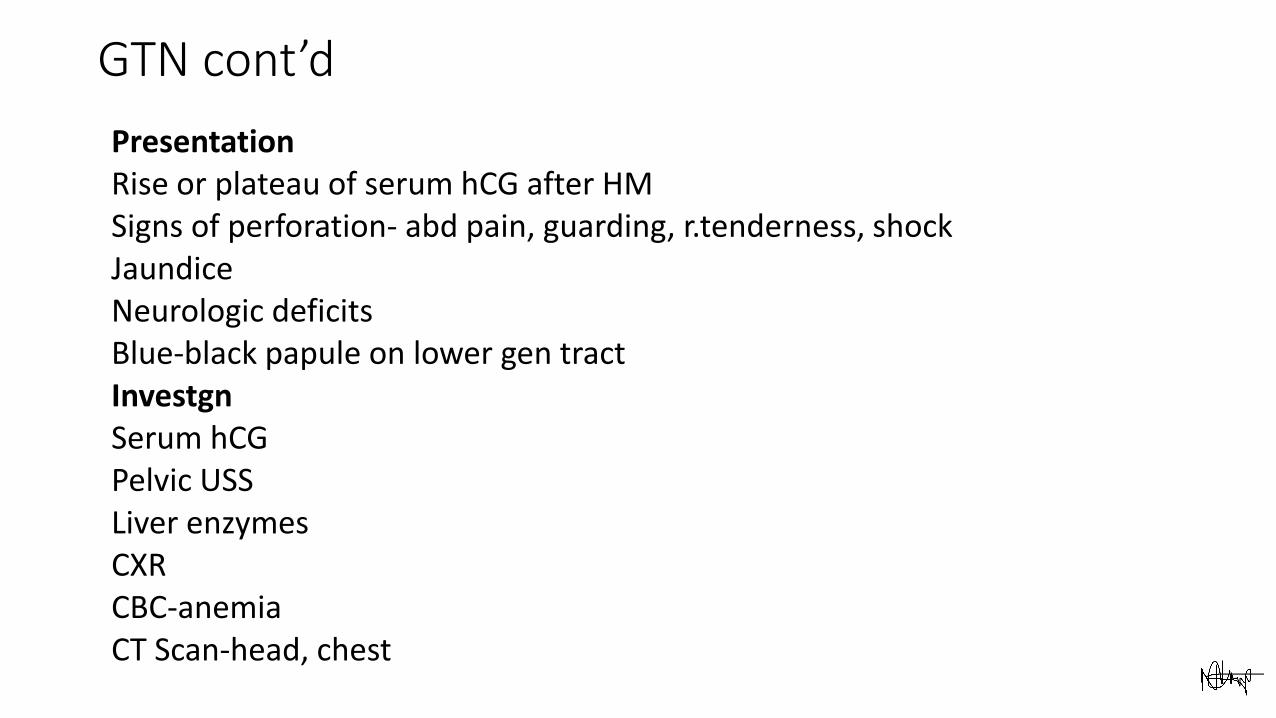
PresentationRise or plateau of serum hCG after HMSigns of perforation- abd pain, guarding, r.tenderness, shockJaundiceNeurologic deficitsBlue-black papule on lower gen tractInvestgnSerum hCGPelvic USSLiver enzymesCXRCBC-anemiaCT Scan-head, chest

WHO/ FIGO Prognostic scoring indexFIGO Scoring 0 1 2 4
Age (years) <40 >40 - -
Antecedent pregnancy Mole Abortion Term
Interval months from end of index preg to treatment
<4 4-<7 7-<13 >13
Pretreatment serum Hcg(iu/l)
<1,000 1,000-<10,000 10,000-<100,000 >100,000
Largest tumor size(+uterus)
<3 3-<5 5+ -
Site of metastases Lung Spleen, kidney GI Brain, liver
Number of metastases - 1-4 5-8 8+

Treatment for GTN
• Low-risk(score <6). Single-agent chemo
IM methotrexate alternating with folinic acid for 1 wk
Monitor Hcg
• High-risk(score >7)
EMA-CO regimen
EMA-CE regimen
After normal Hcg, continue for 6weeks (RCOG 2010)
REFERENCES
Royal College of Obstetricians and Gynecologists. The management of Gestational Trophoblastic Disease. 3rd ed. 2010
FG. Cunningham, et al. Williams Obstetrics. 22nd ed.
M.J Secki, N.J Sebire et al. Gestational Trophoblastic Disease: ESMO Clinical guidelines for diagnosis, treatment and follow-up. Ann Onc2013