gjk thesis chap1
DESCRIPTION
Thesis very usefulTRANSCRIPT
1
Gary J. Kapral PhD Thesis, December 2013
1. Introduction
1.1 Where we are coming from: a brief history of RNA
Ribonucleic Acid is one of the most versatile biological polymers on earth, both
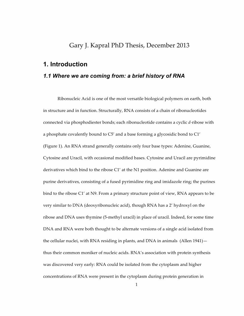
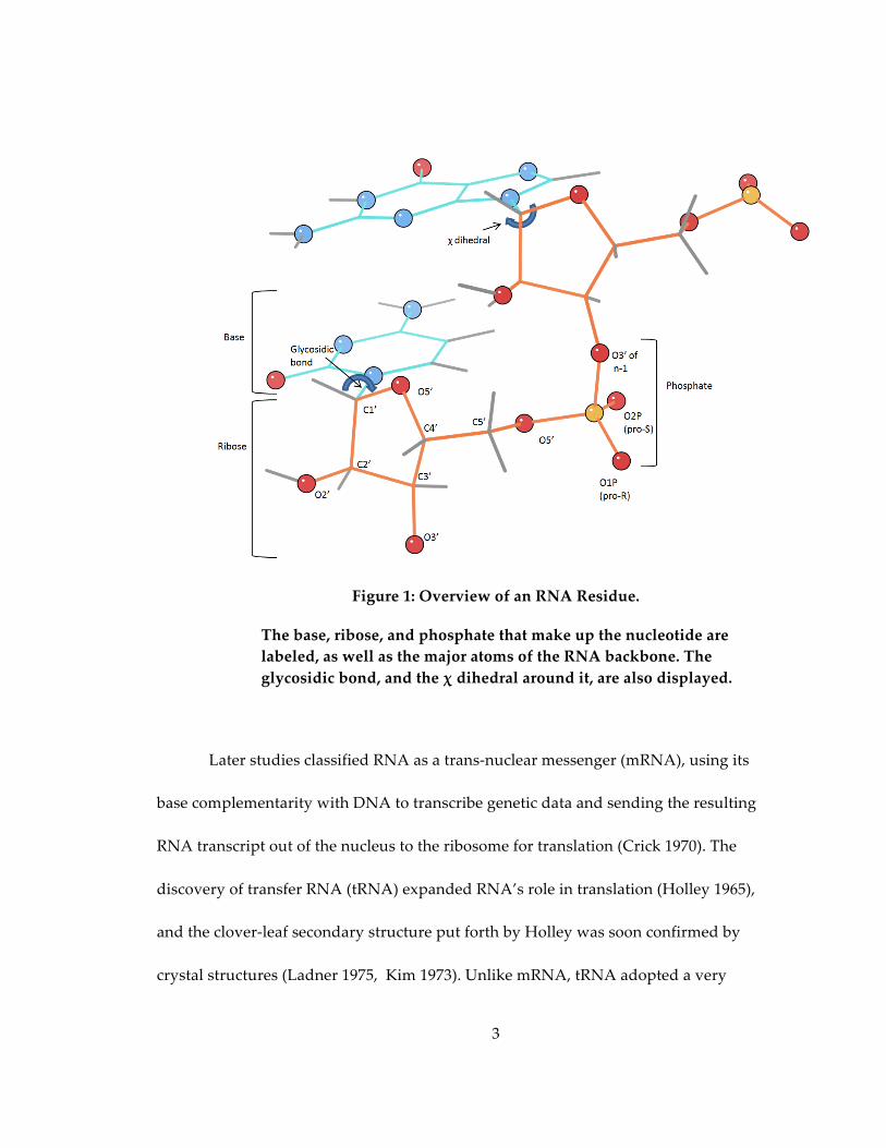
in structure and in function. Structurally, RNA consists of a chain of ribonucleotides
connected via phosphodiester bonds; each ribonucleotide contains a cyclic d-‐‑ribose with
a phosphate covalently bound to C5’ and a base forming a glycosidic bond to C1’
(Figure 1). An RNA strand generally contains only four base types: Adenine, Guanine,
Cytosine and Uracil, with occasional modified bases. Cytosine and Uracil are pyrimidine
derivatives which bind to the ribose C1’ at the N1 position. Adenine and Guanine are
purine derivatives, consisting of a fused pyrimidine ring and imidazole ring; the purines
bind to the ribose C1’ at N9. From a primary structure point of view, RNA appears to be
very similar to DNA (deoxyribonucleic acid), though RNA has a 2’ hydroxyl on the
ribose and DNA uses thymine (5-‐‑methyl uracil) in place of uracil. Indeed, for some time
DNA and RNA were both thought to be alternate versions of a single acid isolated from
the cellular nuclei, with RNA residing in plants, and DNA in animals (Allen 1941)—
thus their common moniker of nucleic acids. RNA’s association with protein synthesis
was discovered very early: RNA could be isolated from the cytoplasm and higher
concentrations of RNA were present in the cytoplasm during protein generation in
2
rapidly growing cells (Caspersson 1939). But in what was to become a rather
commonplace occurrence, study of RNA was overshadowed for over a decade while
DNA took the spotlight: the Avery-‐‑MacLeod-‐‑McCarty experiment showed that DNA
was responsible for passing on genetic information (Avery 1944), Chargaff established
the ratio of AT and GC pairs (Chargaff 1952), Hershey and Chase confirmed DNA as the
vehicle for inheritance (Hershey and Chase 1952), and Watson and Crick solved the first
structure of DNA (Watson and Crick 1953). Finally, in 1955, research once again began
to focus on RNA, as Goldstein and Plaut showed that the RNA obtained from the
cytoplasm had been synthesized in the nucleus (Goldstein and Plaut 1955). Based on this
work, the Central Dogma of Molecular Biology was put forth by Crick (Crick 1958):
DNA is transcribed to RNA through complementary pairing, which then is translated to
protein (and never the reverse); RNA’s nebulous role as a intermediary between DNA
and protein was established.
3
Figure 1: Overview of an RNA Residue.
The base, ribose, and phosphate that make up the nucleotide are labeled, as well as the major atoms of the RNA backbone. The glycosidic bond, and the χ dihedral around it, are also displayed.
Later studies classified RNA as a trans-‐‑nuclear messenger (mRNA), using its
base complementarity with DNA to transcribe genetic data and sending the resulting
RNA transcript out of the nucleus to the ribosome for translation (Crick 1970). The
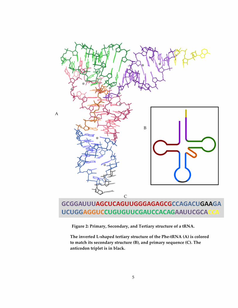
discovery of transfer RNA (tRNA) expanded RNA’s role in translation (Holley 1965),
and the clover-‐‑leaf secondary structure put forth by Holley was soon confirmed by
crystal structures (Ladner 1975, Kim 1973). Unlike mRNA, tRNA adopted a very
4
specific L-‐‑shaped tertiary structure (Figure 2). This structure provided the answer for
how the 3-‐‑nucleotide codon of mRNA (Nirenberg 1965) was translated to a single amino
acid: tRNAs—each of which has a particular amino acid bound to the CCA stem—attach
to mRNA via an anticodon found at the base of the tRNA structure, which is
complementary to the correct mRNA codon. A third type of RNA was discovered in
1956 when George Palade discovered that the microsome organelle was RNA-‐‑rich,
containing slightly more RNA than protein (Palade and Siekevitz 1956). By 1960, it was
shown that the microsomes, renamed ribosomes, consisted of two subunits, each
consisting of RNA and protein (Tissieres and Watson 1958), and were responsible for
protein synthesis (Siekevitz and Palade 1958). The conflux of mRNA, tRNA, and
ribosomal RNA (rRNA) in translation was shown by modification of the 16S ribosomal
subunit (Noller and Chaires 1972). It was also discovered that the sequence of the 16S
subunit was highly conserved among species; Carl Woese used this information to
propose a new phylogenetic taxonomy based around 3 domains: Eukaryotes, Bacteria,
and a new group called Archaea (Woese and Fox 1977).
5
Figure 2: Primary, Secondary, and Tertiary structure of a tRNA.
The inverted L-‐‑shaped tertiary structure of the Phe-‐‑tRNA (A) is colored to match its secondary structure (B), and primary sequence (C). The anticodon triplet is in black.
A
B
C

6
In addition to RNA’s important roles in gene transcription and translation, it can
also act as a catalytic agent. In 1967, Carl Woese suggested that the 2’ hydroxyl could
serve as a site for catalysis (Woese 1967). He focused on RNA’s ability to store genetic
information and putative catalytic ability as the basis for early life, an idea formalized
and expanded on by Walter Gilbert in his Nature paper entitled The RNA World (Gilbert
1986). Thomas Cech and Sidney Altman discovered evidence of such catalytic
“ribozymes” (Zaug 1986, Guerrier-‐‑Takada 1983) in Tetrahymena transcript self-‐‑splicing
and RNase P catalysis. It has since come to light that RNA can catalyze the formation of
RNA; a particularly good example is two artificially designed ribozymes that catalyze
each other’s formation indefinitely so long as resources are available (Lincoln 2009).
Ribozymes, tRNA, and rRNA belong to a group of RNAs called noncoding
RNAs (ncRNAs) because, unlike mRNA, they do not code for amino acids. A number of
uses have been found for ncRNA, particularly in gene splicing and regulation. Small,
low-‐‑molecular weight RNAs found in the nucleus (snRNA) engage in complex
interactions with particular proteins to form small nuclear ribonucleoproteins (snRNPs)
(Lerner 1979, Lerner 1980). These snRNPs have very prominent roles in pre-‐‑mRNA
processing, particularly in removing introns from the post-‐‑transcriptional pre-‐‑mRNA, as
part of a large RNA-‐‑protein complex called the spliceosome (Jurica 2003). Some introns
bypass the need for a spliceosome: the group I and group II introns can self-‐‑catalyze,
causing their own cleavage out of the transcript (Stahley 2005, Toor 2008).

7
RNA’s role in gene regulation was expanded through the processes of SELEX
and in vitro selection, which showed that particular mRNA transcripts could be
engineered to form ligands, called aptamers, that bound to a particular target (Tuerk
1990; Ellington 1990). This set off a search for natural aptamer occurrences, resulting in
the description of riboswitches (Nahvi 2002)—RNA sequences that bound directly to
small metabolites, such as free Guanine or thiamine pyrophosphate (TPP), forming a
stable tertiary structure that prevented transcription of that region. Many riboswitches
affect downstream translation of nearby genes, turning them on and off according to the
presence of the riboswitch’s target metabolite (Nahvi 2002). Some do so directly by
folding to sequester the Shine-‐‑Dalgarno sequence, thus inhibiting translation (Winkler
2002).
A third, novel method of gene regulation was determined by Andrew Fire and
Craig Mello (Fire 1998). Dubbed RNA interference, this method of regulation depends
on short RNAs known as micro RNA (miRNA) and small interfering RNA (siRNA).
Both begin as rather long double-‐‑stranded RNA (dsRNA) chains which are then
converted to 21-‐‑nt dsRNA by the appropriately named enzyme Dicer (Bernstein 2001).
The dsRNA is unwound into 2 single strands, one of which is degraded while the other
binds to the RNA-‐‑induced silencing complex (RISC), a multi-‐‑protein complex with
RNase activity. RISC uses this bound miRNA or siRNA as a guide to bind with the
target mRNA transcripts via base complementarity. The part of RISC with RNase
8
activity, the Argonaute protein, then makes an endonucleolytic cut in the mRNA strand,
cleaving the strand and preventing further expression. The main difference in siRNA
and miRNA lies in how they accomplish their regulatory tasks. The siRNA exhibits
perfect base pairing with its target and causes cleavage with its target in almost 100% of
cases. On the other hand, miRNA is more lenient in where it binds to the mRNA,
allowing some non-‐‑Watson-‐‑Crick base pairing. The miRNA-‐‑RISC complex still causes
translation repression, but rather than degrading the target, the mRNA is moved to P-‐‑
bodies (processing bodies) where other proteins determine whether the mRNA is
degraded or stored until needed.
These myriad roles of RNA in controlling gene expression show how our
understanding of RNA function has shifted in the past hundred years of study, from
that of an intermediate, less efficient version of DNA to an integral part in cellular
vitality. RNA has been shown to be a crucial factor in each step of protein synthesis,
from providing the mRNA transcript to post-‐‑transcriptional regulation to peptide
synthesis. Despite this, our understanding of RNA structure is woefully inadequate.
RNA is difficult to crystallize, a fact borne out by the low number of RNA-‐‑containing
structures in the Protein Data Bank (PDB): as of March, 2013, only 2522 RNA structures
are in the PDB vs. 86,600 protein structures and 4177 DNA structures (Berman 2000).
Thus RNA accounts for barely over a third of the total deposited nucleic acid structures,
which has a second more sinister effect: because they differ by a single backbone atom
9
and a single methyl group on a single base, many model building and prediction
software packages treat both nucleic acids the same. This works poorly, because DNA
has evolved for fidelity of information storage in double-‐‑helix form while RNA adopts
very complex and diverse tertiary structures and functions, so that their properties are
actually quite different.
In order to facilitate RNA study and capitalize on new results, structural
biologists need a better way of looking at RNA structural interactions, and a
nomenclature/classification system which reflects the uniqueness of RNA’s backbone
structure that strongly influences its ability to act as a genetic regulator and a catalyst.
The development of a common classification system and tools with which to analyze the
existing data and improve the sparse number of available models will go a long way to
giving RNA biologists the insight into RNA structure they need to describe better
experiments and determine new RNA structures. Because the amount of data available
for study has recently reached the critical mass necessary to make an undertaking such
as this feasible, the laying of the foundation for the new investigations of RNA backbone
structure and interactions can now begin.
1.2 The chemical and structural importance of RNA backbone
The first steps to understanding how RNA performs such a wide variety of vital
functions is to understand the structure. Given its important role, it is somewhat
surprising, and disappointing, that the number of RNA structures deposited in the
10
Nucleic Acid Database (NDB; Berman 1992) and PDB was very low at the turn of the
millennium (236 RNA vs. 939 DNA and 9812 proteins) . A part of this was simply due to
lack of information—RNA primarily adopted A-‐‑form helix, which was structurally
similar to A-‐‑form DNA; but when it did not, there were few methods of recourse to
determine its structure. Even the refinement parameters used in X-‐‑PLOR and CNS
(Parkinson 1996, Brunger 1998) were largely generalized to both nucleic acids, and
tested almost exclusively on DNA structures. Thus, many early models of RNA
structures resembled their DNA counterparts with only the very few high resolution
structures working well to illustrate non-‐‑A-‐‑form RNA structure. Many early structures
show clear distinction between the standardization and quality of A-‐‑form helix versus
the intervening RNA “loop” regions that connect them (Duarte and Pyle 1998; Ferre-‐‑
D’Amare 1998).
Fortunately, the publication of the ribosome structure in 2000, proving
structurally that the ribosome is really a ribozyme (Ban 2000; Nissen 2000), set off an
explosion of RNA structural studies that continues to this day. The revitalized focus on
structure helped prove the existence of riboswitches (Winkler and Batey 2005), and
aptamer structures have been used to further medical advances in viral transcription
inhibition (Eulberg and Klussman 2003). The structure of the RNA Induced Silencing
Complex (RISC) has been solved, giving us more insight into RNAi and the siRNA and
miRNA that drive the process and keep the body safe from invading RNA-‐‑based
11
pathogens (Fire 1998; Tuschl 2001). Improved structures of the ribosome have elucidated
translation even further, and led to a myriad of drugs designed to inhibit translation in
pathogenic species(Matt 2012; Borovinskaya 2007; Bulkley 2010).
Traditionally, much of the study surrounding RNA has focused on the bases. The
reasons for this are twofold. Chemically, they are variable pendant groups in a polymer
of ribose-‐‑phosphodiester linkages, and with their strong tendency to form
complementary base-‐‑pairs, changing the sequence of pendant groups is an obvious first
step if one wishes to alter the polymer structure. Secondly, their positions are easy to
determine using both X-‐‑ray crystallography and NMR spectroscopy. Base pairing and
base stacking interactions stabilize the overall RNA structure, allowing them to be easily
seen through NMR, and their electron-‐‑rich pseudo-‐‑aromatic properties make them easy
to identify in the electron density. In addition, base positions can easily be inferred on
the bases of Watson-‐‑Crick complementarity. Overall, the base sequence is easily
changed, and the results of those changes are fairly easy to determine.
Yet much of the recent research, particularly concerning catalysis, has focused on
the properties of the 2’ hydroxyl of the ribose. The 2’ hydroxyl has an extreme effect on
RNA structure. From a purely steric point of view, it prevents RNA from adopting the
low-‐‑energy B-‐‑form helix common to most DNA structures; most RNA is in A-‐‑form helix
(Murray 2003) which exposes the bases and the 2’ hydroxyl to solvent. The 2’ hydroxyl is
responsible for self-‐‑splicing, as in the case of the Group I and Group II introns (Cate
12
1996; Toor 2008). It also can act as a proton shuttle, as in the peptidyl transferase site of
the ribosome, catalyzing the binding of the amino-‐‑acyl amino from A-‐‑site tRNA to the
amino-‐‑acyl carbonyl carbon of the P-‐‑site tRNA, thus lengthening the nascent peptide
(Ban 2000). The 2’OH is also the center location of a wide variety of backbone
modifications, particularly methylation (Kiss 2001), and can participate in hydrogen
bond interactions (Bolton and Kearns 1978) or even ligand binding (Greenbaum 2001).
With so many important processes dependent on the 2’OH, and others
dependent on the negatively charged phosphate, the RNA backbone is not to be ignored.
The backbone of any given nucleotide has 12 torsion angles (including the χ angle
around the glycosidic bond) and 19 atoms, compared to 4 torsions (including χ1) and 7
atoms in an average amino acid backbone (Figure 1). While the huge number of torsions
gives the RNA backbone the flexibility needed to effectively catalyze reactions, they
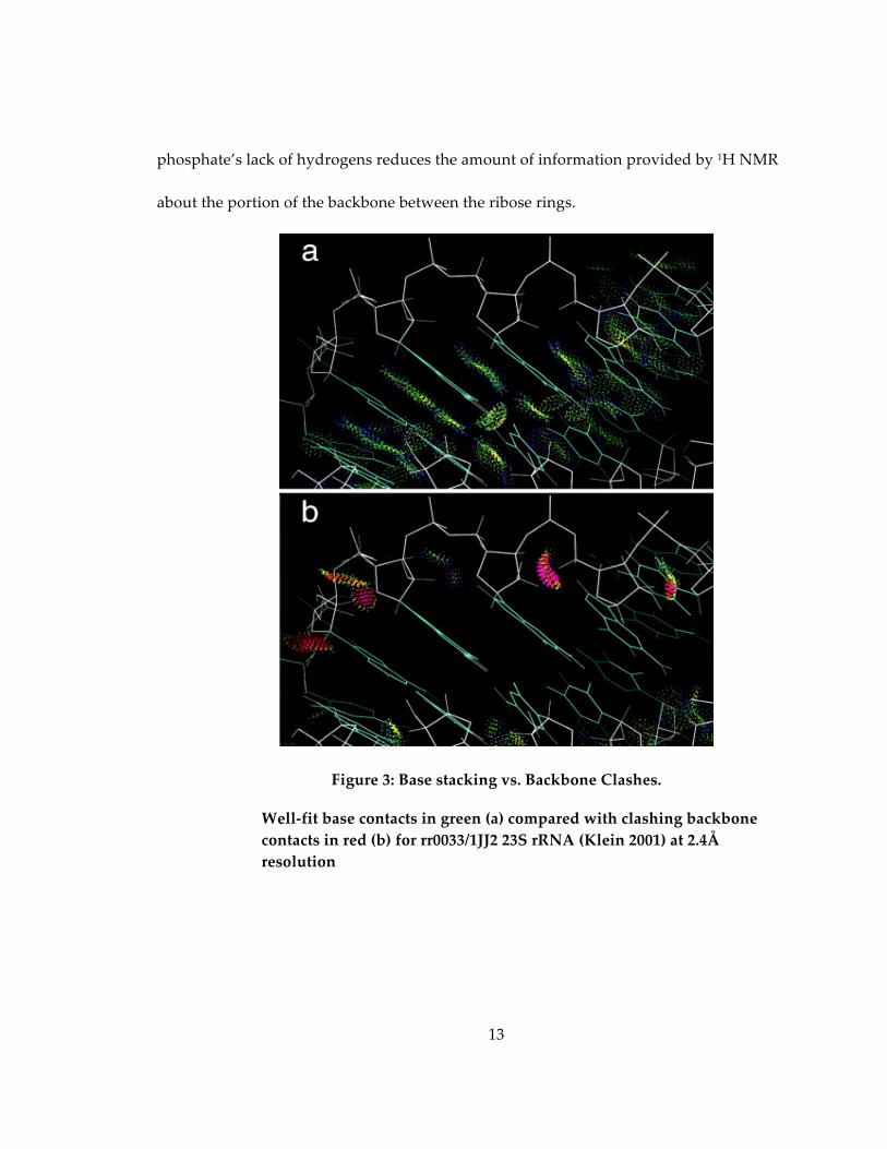
wreak havoc on current structure building programs. As a result (Figure 3), errors along
the backbone are frequent at the resolutions commonly attainable (2Å-‐‑3Å). The reason
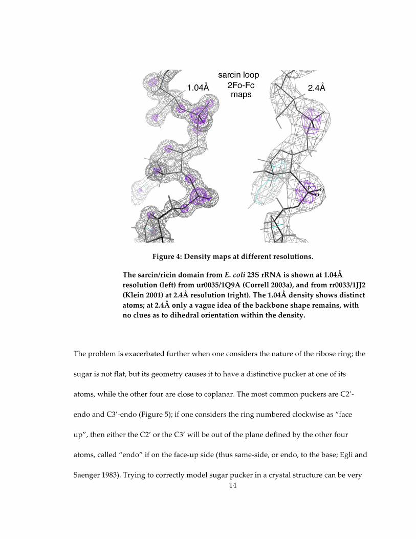
for this difficulty in determining backbone positions is evident from a look at the
electron density (Figure 4): even at good resolutions (2.4Å), there is simply not enough
detail to accurately place C5’ or the sugar—even the phosphate oxygen positions are
unclear. The problem is exacerbated because the crystallographically invisible but
sterically important hydrogen atoms (Word, 1999a) are not modeled, while the
13
phosphate’s lack of hydrogens reduces the amount of information provided by 1H NMR
about the portion of the backbone between the ribose rings.
Figure 3: Base stacking vs. Backbone Clashes.
Well-‐‑fit base contacts in green (a) compared with clashing backbone contacts in red (b) for rr0033/1JJ2 23S rRNA (Klein 2001) at 2.4Å resolution
14
Figure 4: Density maps at different resolutions.
The sarcin/ricin domain from E. coli 23S rRNA is shown at 1.04Å resolution (left) from ur0035/1Q9A (Correll 2003a), and from rr0033/1JJ2 (Klein 2001) at 2.4Å resolution (right). The 1.04Å density shows distinct atoms; at 2.4Å only a vague idea of the backbone shape remains, with no clues as to dihedral orientation within the density.
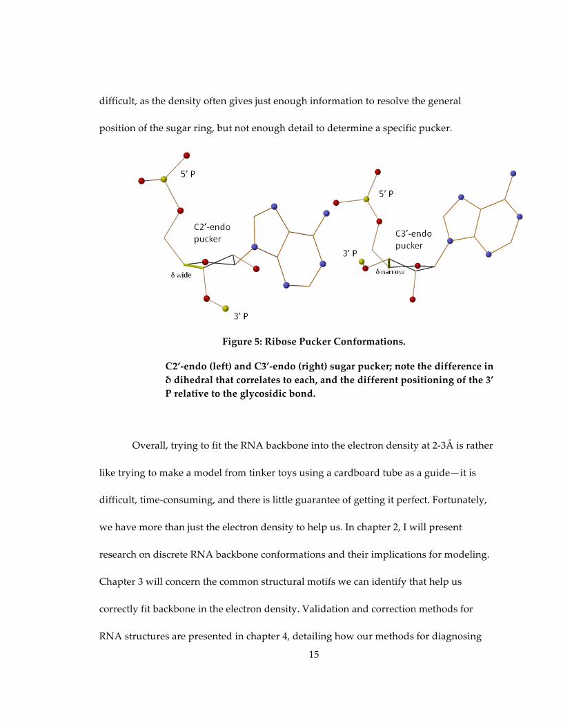
The problem is exacerbated further when one considers the nature of the ribose ring; the
sugar is not flat, but its geometry causes it to have a distinctive pucker at one of its
atoms, while the other four are close to coplanar. The most common puckers are C2’-‐‑
endo and C3’-‐‑endo (Figure 5); if one considers the ring numbered clockwise as “face
up”, then either the C2’ or the C3’ will be out of the plane defined by the other four
atoms, called “endo” if on the face-‐‑up side (thus same-‐‑side, or endo, to the base; Egli and
Saenger 1983). Trying to correctly model sugar pucker in a crystal structure can be very
15
difficult, as the density often gives just enough information to resolve the general
position of the sugar ring, but not enough detail to determine a specific pucker.
Figure 5: Ribose Pucker Conformations.
C2’-‐‑endo (left) and C3’-‐‑endo (right) sugar pucker; note the difference in δ dihedral that correlates to each, and the different positioning of the 3’ P relative to the glycosidic bond.
Overall, trying to fit the RNA backbone into the electron density at 2-‐‑3Å is rather
like trying to make a model from tinker toys using a cardboard tube as a guide—it is
difficult, time-‐‑consuming, and there is little guarantee of getting it perfect. Fortunately,
we have more than just the electron density to help us. In chapter 2, I will present
research on discrete RNA backbone conformations and their implications for modeling.
Chapter 3 will concern the common structural motifs we can identify that help us
correctly fit backbone in the electron density. Validation and correction methods for
RNA structures are presented in chapter 4, detailing how our methods for diagnosing
16
problem areas of RNA backbone structure lead to better X-‐‑ray and NMR structures and
have been incorporated into the PHENIX refinement package. The final results of these
validation and correction methods will be presented in chapter 5, as we take on
corrections of several RNA structures, including one of the largest RNA structures to
date—two 70S ribosomes in one asymmetric unit.
References * Allen, F W. "ʺThe Biochemistry of the Nucleic Acids, Purines, and Pyrimidines."ʺ Annual
Review of Biochemistry 10, no. 1 (1941): 221-‐‑244. * Avery, Oswald T., Colin M. MacLeod and Maclyn McCarty. "ʺStudies on the Chemical
Nature of the Substance Inducing Transformation of Pneumococcal Types: Induction of Transformation by a Desoxyribonucleic Acid Fraction Isolated from Pneumococcus Type Iii."ʺ The Journal of Experimental Medicine 79, no. 2 (1944): 137-‐‑158.
* Ban, Nenad, Poul Nissen, Jeffrey Hansen, Peter B. Moore and Thomas A. Steitz. "ʺThe
Complete Atomic Structure of the Large Ribosomal Subunit at 2.4 Å Resolution."ʺ Science 289, no. 5481 (2000): 905-‐‑920.
* Berman, H. M., W. K. Olson, D. L. Beveridge, J. Westbrook, A. Gelbin, T. Demeny, S. H.
Hsieh, A. R. Srinivasan and B. Schneider. "ʺThe Nucleic Acid Database. A Comprehensive Relational Database of Three-‐‑Dimensional Structures of Nucleic Acids."ʺ Biophysical Journal 63, no. 3 (1992): 751-‐‑759.
* Berman, Helen M., John Westbrook, Zukang Feng, Gary Gilliland, T. N. Bhat, Helge
Weissig, Ilya N. Shindyalov and Philip E. Bourne. "ʺThe Protein Data Bank."ʺ Nucleic Acids Research 28, no. 1 (2000): 235-‐‑242.
* Bernstein, Emily, Amy A. Caudy, Scott M. Hammond and Gregory J. Hannon. "ʺRole
for a Bidentate Ribonuclease in the Initiation Step of Rna Interference."ʺ Nature 409, no. 6818 (2001): 363-‐‑366.
17
* Bolton, Philip H. and David R. Kearns. "ʺHydrogen Bonding Interactions of Polyamines
with the 2′ Oh of Rna."ʺ Nucleic Acids Research 5, no. 4 (1978): 1315-‐‑1324. * Borovinskaya, Maria A., Raj D. Pai, Wen Zhang, Barbara S. Schuwirth, James M.
Holton, Go Hirokawa, Hideko Kaji, Akira Kaji and Jamie H. Doudna Cate. "ʺStructural Basis for Aminoglycoside Inhibition of Bacterial Ribosome Recycling."ʺ Nat Struct Mol Biol 14, no. 8 (2007): 727-‐‑732.
* Brunger, Axel T., Paul D. Adams, G. Marius Clore, Warren L. DeLano, Piet Gros, Ralf
W. Grosse-‐‑Kunstleve, Jian-‐‑Sheng Jiang, John Kuszewski, Michael Nilges, Navraj S. Pannu, Randy J. Read, Luke M. Rice, Thomas Simonson and Gregory L. Warren. "ʺCrystallography & Nmr System: A New Software Suite for Macromolecular Structure Determination."ʺ Acta Crystallographica Section D 54, no. 5 (1998): 905-‐‑921.
* Bulkley, David, C. Axel Innis, Gregor Blaha and Thomas A. Steitz. "ʺRevisiting the
Structures of Several Antibiotics Bound to the Bacterial Ribosome."ʺ Proceedings of the National Academy of Sciences 107, no. 40 (2010): 17158-‐‑17163.
* Caspersson, T. and J. Schultz. "ʺPentose Nucleotides in the Cytoplasm of Growing
Tissues."ʺ Nature 143, (1939): 602-‐‑603. * Cate, J. H., A. R. Gooding, E. Podell, K. Zhou, B. L. Golden, A. A. Szewczak, C. E.
Kundrot, T. R. Cech and J. A. Doudna. "ʺRna Tertiary Structure Mediation by Adenosine Platforms."ʺ Science (New York, N.Y.) 273, no. 5282 (1996): 1696-‐‑1699.
* Chargaff, E., R. Lipshitz and C. Green. "ʺComposition of the Desoxypentose Nucleic
Acids of Four Gera of Sea-‐‑Urchin."ʺ J. Biol. Chem. 195, (1952): 155-‐‑160. * Crick, F. H. C. "ʺIdeas on Protein Synthesis."ʺ In Symposia of the Society for Experimental
Biology Symposium XII: The Biological Replication of Macromolecules, 138-‐‑163. University College London: Cambridge University Press, 1958.
* Crick, Francis. "ʺCentral Dogma of Molecular Biology."ʺ Nature 227, no. 5258 (1970): 561-‐‑
563. * Duarte, Carlos M. and Anna Marie Pyle. "ʺStepping through an Rna Structure: A Novel
Approach to Conformational Analysis."ʺ Journal of Molecular Biology 284, no. 5 (1998): 1465-‐‑1478.
18
* Ellington, Andrew D. and Jack W. Szostak. "ʺIn Vitro Selection of Rna Molecules That
Bind Specific Ligands."ʺ Nature 346, no. 6287 (1990): 818-‐‑822. * Eulberg, Dirk and Sven Klussmann. "ʺSpiegelmers: Biostable Aptamers."ʺ ChemBioChem
4, no. 10 (2003): 979-‐‑983. * Fire, Andrew, SiQun Xu, Mary K. Montgomery, Steven A. Kostas, Samuel E. Driver
and Craig C. Mello. "ʺPotent and Specific Genetic Interference by Double-‐‑Stranded Rna in Caenorhabditis Elegans."ʺ Nature 391, no. 6669 (1998): 806-‐‑811.
* Gilbert, Walter. "ʺOrigin of Life: The RNA World."ʺ Nature 319, no. 6055 (1986): 618-‐‑618. * Goldstein, L. and W. Plaut. "ʺDirect Evidence for Nuclear Synthesis of Cytoplasmic
Ribose Nucleic Acid."ʺ PNAS 41, no. 11 (1955): 874-‐‑880. * Greenbaum, Nancy L., Claudius Mundoma and Dean R. Peterman. "ʺProbing of Metal-‐‑
Binding Domains of Rna Hairpin Loops by Laser-‐‑Induced Lanthanide(Iii) Luminescence†."ʺ Biochemistry 40, no. 4 (2001): 1124-‐‑1134.
* Guerrier-‐‑Takada, Cecilia, Katheleen Gardiner, Terry Marsh, Norman Pace and Sidney
Altman. "ʺThe Rna Moiety of Ribonuclease P Is the Catalytic Subunit of the Enzyme."ʺ Cell 35, no. 3, Part 2 (1983): 849-‐‑857.
* Holley, R. W., G. A. Everett, J. T. Madison and A. Zamir. "ʺNucleotide Sequences in the Yeast Alanine Transfer Ribonucleic Acid."ʺ J Biol Chem 240, (1965): 2122-‐‑8.
* Jurica, Melissa S. and Melissa J. Moore. "ʺPre-‐‑Mrna Splicing: Awash in a Sea of
Proteins."ʺ Molecular Cell 12, no. 1 (2003): 5-‐‑14. * Kim, S. H., G. J. Quigley, F. L. Suddath, A. McPherson, D. Sneden, J. J. Kim, J. Weinzierl
and Alexander Rich. "ʺThree-‐‑Dimensional Structure of Yeast Phenylalanine Transfer Rna: Folding of the Polynucleotide Chain."ʺ Science 179, no. 4070 (1973): 285-‐‑288.
* Kiss, Tamas. "ʺSmall Nucleolar Rna-‐‑Guided Post-‐‑Transcriptional Modification of
Cellular Rnas."ʺ EMBO J 20, no. 14 (2001): 3617-‐‑3622. * Ladner, J. E., A. Jack, J. D. Robertus, R. S. Brown, D. Rhodes, B. F. Clark and A. Klug.
"ʺStructure of Yeast Phenylalanine Transfer Rna at 2.5 a Resolution."ʺ Proc Natl Acad Sci U S A 72, no. 11 (1975): 4414-‐‑8.
19
* Lerner, Michael R., John A. Boyle, Stephen M. Mount, Sandra L. Wolin and Joan A.
Steitz. "ʺAre Snrnps Involved in Splicing?"ʺ Nature 283, no. 5743 (1980): 220-‐‑224. * Lerner, Michael Rush and Joan Argetsinger Steitz. "ʺAntibodies to Small Nuclear Rnas
Complexed with Proteins Are Produced by Patients with Systemic Lupus Erythematosus."ʺ Proceedings of the National Academy of Sciences 76, no. 11 (1979): 5495-‐‑5499.
* Lincoln, Tracey A. and Gerald F. Joyce. "ʺSelf-‐‑Sustained Replication of an Rna Enzyme."ʺ
Science 323, no. 5918 (2009): 1229-‐‑1232. * Matt, Tanja, Chyan Leong Ng, Kathrin Lang, Su-‐‑Hua Sha, Rashid Akbergenov, Dmitri
Shcherbakov, Martin Meyer, Stefan Duscha, Jing Xie, Srinivas R. Dubbaka, Déborah Perez-‐‑Fernandez, Andrea Vasella, V. Ramakrishnan, Jochen Schacht and Erik C. Böttger. "ʺDissociation of Antibacterial Activity and Aminoglycoside Ototoxicity in the 4-‐‑Monosubstituted 2-‐‑Deoxystreptamine Apramycin."ʺ Proceedings of the National Academy of Sciences 109, no. 27 (2012): 10984-‐‑10989.
* Murray, L. J. W., W. B. Arendall Iii, D. C. Richardson and J. S. Richardson. "ʺRna
Backbone Is Rotameric."ʺ Proceedings of the National Academy of Sciences 100, no. 24 (2003): 13904-‐‑13909.
* Nahvi, Ali, Narasimhan Sudarsan, Margaret S. Ebert, Xiang Zou, Kenneth L. Brown
and Ronald R. Breaker. "ʺGenetic Control by a Metabolite Binding Mrna."ʺ Chemistry & Biology 9, no. 9 (2002): 1043-‐‑1049.
* Nirenberg, M, P Leder, M Bernfield, R Brimacombe, J Trupin, F Rottman and C O'ʹNeal.
"ʺRna Codewords and Protein Synthesis, Vii. On the General Nature of the Rna Code."ʺ Proceedings of the National Academy of Sciences 53, no. 5 (1965): 1161-‐‑1168.
* Noller, H. F. and J. B. Chaires. "ʺFunctional Modification of 16s Ribosomal Rna by
Kethoxal."ʺ Proc Natl Acad Sci U S A 69, no. 11 (1972): 3115-‐‑8. * Palade, G. E. and P. Siekevitz. "ʺLiver Microsomes; an Integrated Morphological and
Biochemical Study."ʺ J Biophys Biochem Cytol 2, no. 2 (1956): 171-‐‑200. * Parkinson, G., J. Vojtechovsky, L. Clowney, A. T. Brünger and H. M. Berman. "ʺNew
Parameters for the Refinement of Nucleic Acid-‐‑Containing Structures."ʺ Acta Crystallographica. Section D, Biological Crystallography 52, no. Pt 1 (1996): 57-‐‑64.

20
* Siekevitz, Philip and George E. Palade. "ʺA Cytochemical Study on the Pancreas of the
Guinea Pig: Ii. Functional Variations in the Enzymatic Activity of Microsomes."ʺ The Journal of Biophysical and Biochemical Cytology 4, no. 3 (1958): 309-‐‑318.
* Stahley, Mary R. and Scott A. Strobel. "ʺStructural Evidence for a Two-‐‑Metal-‐‑Ion
Mechanism of Group I Intron Splicing."ʺ Science 309, no. 5740 (2005): 1587-‐‑1590. * Tissieres, A. and J. D. Watson. "ʺRibonucleoprotein Particles from Escherichia Coli."ʺ
Nature 182, no. 4638 (1958): 778-‐‑80. * Toor, Navtej, Kevin S. Keating, Sean D. Taylor and Anna Marie Pyle. "ʺCrystal Structure
of a Self-‐‑Spliced Group Ii Intron."ʺ Science 320, no. 5872 (2008): 77-‐‑82. * Tuerk, C. and L. Gold. "ʺSystematic Evolution of Ligands by Exponential Enrichment:
Rna Ligands to Bacteriophage T4 DNA Polymerase."ʺ Science 249, no. 4968 (1990): 505-‐‑10.
* Tuschl, Thomas. "ʺExpanding Small Rna Interference."ʺ Nat Biotech 20, no. 5 (2002): 446-‐‑
448. * Watson, J. D. and F. H. C. Crick. "ʺMolecular Structure of Nucleic Acids: A Structure for
Deoxyribose Nucleic Acid."ʺ Nature 171, no. 4356 (1953): 737-‐‑738. * Winkler, Wade C. and Ronald R. Breaker. "ʺRegulation of Bacterial Gene Expression by
Riboswitches."ʺ Annual Review of Microbiology 59, no. 1 (2005): 487-‐‑517. * Winkler, Wade, Ali Nahvi and Ronald R. Breaker. "ʺThiamine Derivatives Bind
Messenger Rnas Directly to Regulate Bacterial Gene Expression."ʺ Nature 419, no. 6910 (2002): 952-‐‑956.
* Woese, C. R. and G. E. Fox. "ʺPhylogenetic Structure of the Prokaryotic Domain: The
Primary Kingdoms."ʺ Proc Natl Acad Sci U S A 74, no. 11 (1977): 5088-‐‑90. * Woese, Carl R. The Genetic Code : The Molecular Basis for Genetic Expression. New York:
Harper & Row, 1967. * Word, J. Michael, Simon C. Lovell, Thomas H. LaBean, Hope C. Taylor, Michael E.
Zalis, Brent K. Presley, Jane S. Richardson and David C. Richardson. "ʺVisualizing and Quantifying Molecular Goodness-‐‑of-‐‑Fit: Small-‐‑Probe Contact Dots with

21
Explicit Hydrogen Atoms."ʺ Journal of Molecular Biology 285, no. 4 (1999): 1711-‐‑1733.
* Zaug, A. J. and T. R. Cech. "ʺThe Intervening Sequence Rna of Tetrahymena Is an
Enzyme."ʺ Science 231, no. 4737 (1986): 470-‐‑5.