glomerulonephritis nurse teaching jan 2017
TRANSCRIPT

GLOMERULONEPHRITISAMBER Z JAFFERIEMERGENCY DEPARTMENT SIH
2
PLANGeneral over view
Little revision anatomy

3
GLOMERULONEPHRITISNephros – kidney
-itis – inflammation of
Glomus – small round ball or knot
Pathos – suffering or disease
-osis – diseased condition
Glomerulonephritis – inflammation of the glomeruli
Glomerulopathy – disease of the glomeruli

4
Light micrograph of a normal glomerulus. There are only 1 or 2 cells per capillary tuft, the capillary lumens are open, the thickness of the glomerular capillary wall (long arrow) is similar to that of the tubular basement membranes (short arrow), and the mesangial cells and mesangial matrix are located in the central or stalk regions of the tuft (arrows). Courtesy of Helmut G Rennke.
5
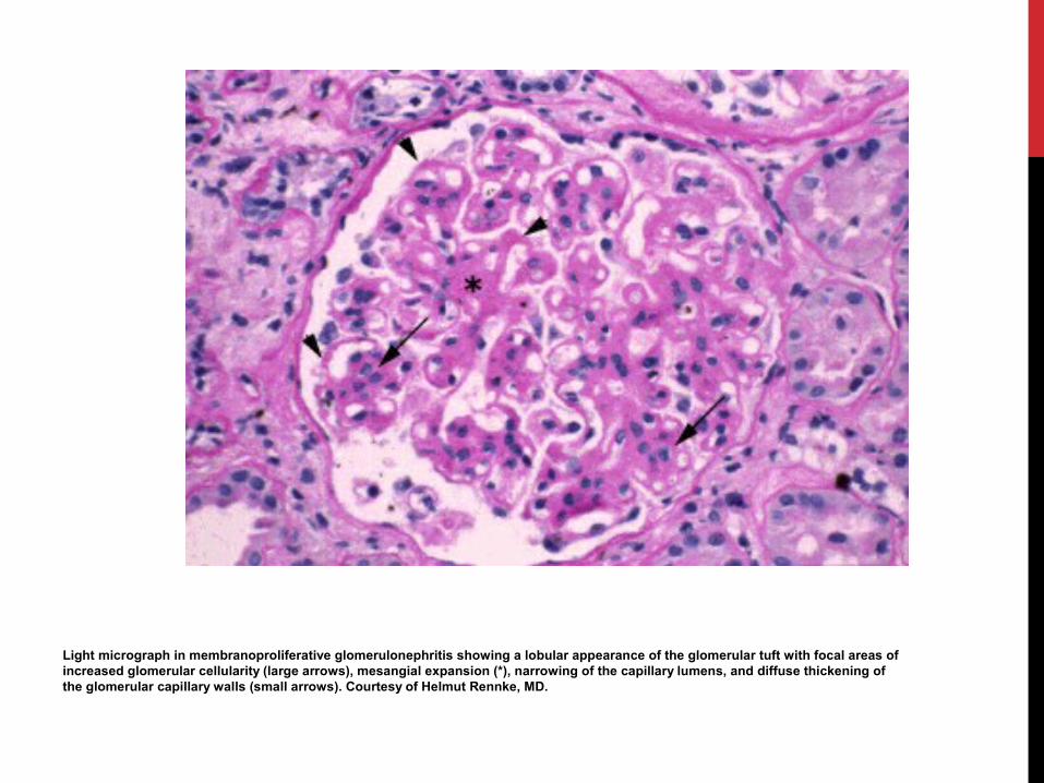
Light micrograph in membranoproliferative glomerulonephritis showing a lobular appearance of the glomerular tuft with focal areas of increased glomerular cellularity (large arrows), mesangial expansion (*), narrowing of the capillary lumens, and diffuse thickening of the glomerular capillary walls (small arrows). Courtesy of Helmut Rennke, MD.
6
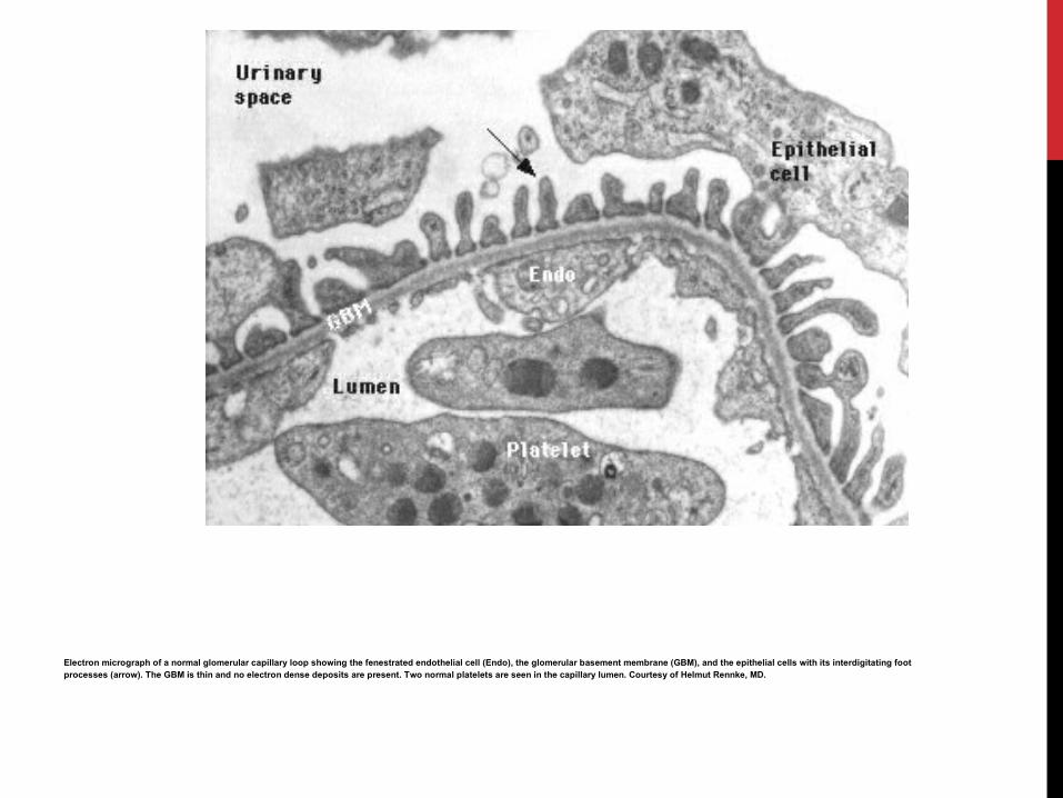
Electron micrograph of a normal glomerular capillary loop showing the fenestrated endothelial cell (Endo), the glomerular basement membrane (GBM), and the epithelial cells with its interdigitating foot processes (arrow). The GBM is thin and no electron dense deposits are present. Two normal platelets are seen in the capillary lumen. Courtesy of Helmut Rennke, MD.

7
Electron micrograph in dense deposit disease (DDD) showing dense, ribbon-like appearance of subendothelial and intramembranous material (arrow) and narrowing of the capillary lumen due to proliferation of cells (double arrow). Courtesy of Helmut Rennke, MD.

8
GLOMERULAR DISEASEPrimary – confined to the kidney
Secondary – due to a systemic disease

9
GLOMERULAR INJURYImpairment of selective filtering properties of the kidney leading to a decreased GFR
Molecules normally not filtered such as constituents of the blood, pass into the urine and are excreted

10 MD consult

11
Anatomy of Kidney
Note the positions of
Glomerulus
Loop of Henle
PCT, DCT, CT
Cortex, Medulla, Pelvis.
MD consult
12
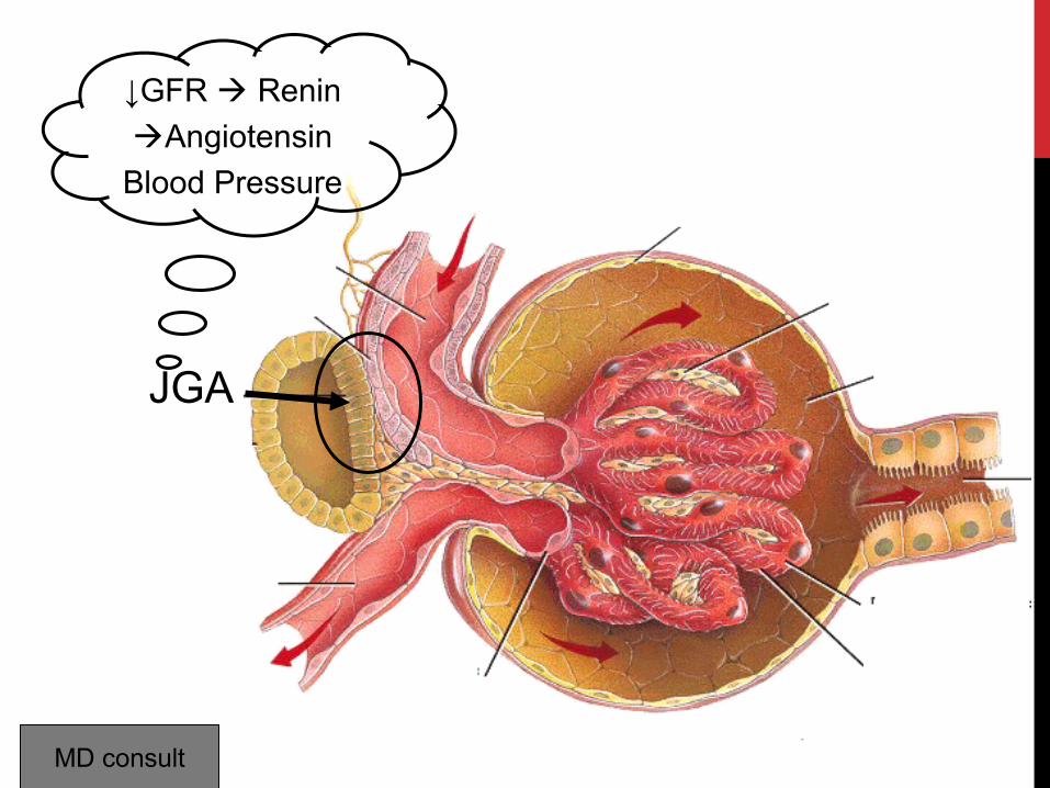
JGA
↓GFR Renin
Angiotensin
Blood Pressure
MD consult

ULTRASTRUCTURE – GLOM. CAPILLARY

14
POSSIBLE CLINICAL MANIFESTATIONSProteinuria – asymptomatic
Haematuria – asymptomatic
Hypertension
Nephrotic syndrome
Nephritic syndrome
Acute renal failure
Rapidly progressive renal failure
End stage renal failure

15
GLOMERULONEPHRITIS
Presence of glomerular disease as opposed to tubulointersititial or vascular disease is suspected from history
Haematuria (especially dysmorphic red cells)
Red cell casts
Lipiduria (glomerular permeability must be increased to allow the filtration of large lipoproteins)
Proteinuria (may be in nephrotic range of >3.5 g/24hours)

16
Phase contrast microscopy showing dysmorphic red cells in a patient with glomerular bleeding. Acanthocytes can be recognized as ring forms with vesicle-shaped protrusions (arrows). Courtesy of Hans Köhler, MD.

17
DIAGNOSISLook for clues
• History• Haematuria• Proteinuria• Azotemia
• Azote – nitrogen • A – without • Zoe – life • “The gas does not support life”
(French chemists Gayton de Morveau (1737-1816) and Antoine Lavoisier (1743-1794) )
McCarthy ET, November 2008

18
DIAGNOSISCan be difficult to distinguish between Glomerular disease and tubulo-interstitial disease
Tubular disease does not directly increase protein excretion but nephron loss due to the disease can have the same end result

19
CLINICAL PATTERNSPatients age and characteristics of the urine sediment can allow narrowing of the differential diagnosis options prior to biopsy
‘URINE IS THE LIQUID BIOPSY OF THE KIDNEY’ Walter Piering MD – Prof Med Wisconsin

20
URINARY PATTERNS3 different patterns
• Focal nephritic• Diffuse nephritic• Nephrotic

21
URINARY PATTERNSFocal nephritic
• Associated with inflammatory to less than half of the glomeruli on light microscopy
• Red cells – often dysmorphic• Occasional red cell casts• Mild proteinuria (<1.5g/day)

22
URINARY PATTERNSDiffuse nephritic
• Damage to all or almost all of the glomeruli• Similar to focal disease but may also have heavy
proteinuria (even nephrotic range), oedema, hypertension and/or renal insufficiency
• - ‘full house’ urinary sediment – red cells, white cells, red cell casts, white cell casts, hyaline casts

23
URINARY PATTERNSNephrotic
• Heavy proteinuria• Lipiduria – refractile fat bodies that look like a maltese
cross under polarised light• Few cells• Few casts – but those present are hyaline and granular

24
NON SPECIFIC NATURE OF HISTOLOGIC PATTERNSMembranous GN – usually Immune complex disease (infective endocarditis, SLE, Hepatitis C)
Membranous nephropathy – drugs (gold, penicillamine), SLE, Hepatitis B, malignancy
Focal glomerulosclerosis can be primary ( minimal change), or secondary (intraglomerular hypertension, or healing previous glomerular injury)

25
PATTERN DIAGNOSISFOCAL GN
<15 years – mild post infectious GN, IgA nephropathy, thin basement membrane disease, hereditary nephritis, Henoch Schonlein Purpura, mesangial proliferative GN
15-40 years – IgA nephropathy, thin basement membrane disease, lupus hereditary nephritis, mesangial proliferative GN
>40 years – IgA nephropathy
26
PATTERN DIAGNOSISDIFFUSE GNPost infectious GN, lupus GN, membranoproliferative GN, mixed cryoglobulinaemia
Often associated with decreased complement
Classic findings • PSGN (anti strep antibodies)• Lupus nephritis (ANA)• Anti-GBM disease (anti GBM Abs) • Mixed cryoglobulinaemia (circulating cryoglobulins)• Wegener's granulomatosis (anti neutrophil cytoplasmic
abs)

27
PATTERN DIAGNOSISDIFFUSE GN<15 years – Post infectious GN, membranoproliferative GN
15-40 years – Post infectious GN, rapidly progressive GN, fibrillary GN, membranoproliferative GN
>40years – rapidly progressive GN, vasculitis, fibrillary glomerulonephritis

28
PATTERN DIAGNOSISNEPHROTIC SYNDROME<15 years – minimal change disease, focal glomerulosclerosis, mesangial proliferative GN
15-40 years – focal glomerulosclerosis, minimal change disease, membranous nephropathy including lupus, diabetic nephropathy, preeclampsia, post infectious GN
>40 years – focal glomerulosclerosis, membranous nephropathy, diabetic nephropathy, minimal change disease, IgA nephropathy, primary amyloidosis or related disorder – light chain deposition disease (up to 20% of pts over 60), benign nephrosclerosis, post infectious GN

29
GENERAL WORKUP ? GLOMERULAR DISEASEHistory
• Family history kidney disease and hearing trouble (Alport’s syndrome)
• Medications that can damage the kidney (NSAID’s, ACEI, penicillamine, gold, mercury in some skin lightening creams)
• Recent throat infection - ? Strep- PSGN or viral – Wegener's granulomatosis, IgA
• Cancer – solid tumours, Hodgkin’s (minimal change) or non Hodgkin’s (MPGN)
30
GENERAL WORKUP ? GLOMERULAR DISEASEPhysical Examination
• Inspection – appearance, colour, pitting oedema, xanthelasma, alopecia, facial rash, purpura, clubbing, livedo reticularis
• Palpation – pulse, hepatomegaly, palpable kidneys, splenomegaly, palapable bladder
• Percussion – hepatomegaly, splenomegaly• Auscultation – renal artery bruits, other bruits, cardiac
lesions, hypertension,

31
GENERAL WORKUP ? GLOMERULAR DISEASELaboratory work
• UECB• LFT• BSL• FBC• Urine microscopy and culture• ACR• Serum and urine protein electrophoresis• Renal ultrasound

32
GENERAL WORKUP ? GLOMERULAR DISEASELaboratory work
• Specific serology• For a nephrotic type picture
• HIV, HCV, HBV, ANA, serum cryoglobulins, anti DNA ab, complements
• For a nephritic type picture• Blood cultures, ASOT, AntiDNAse B, ANA, Anti DNA ab,
anti GBM ab, anti neutrophil cytoplasmic ab, complements

33
Pre urinalysis

34
IGA NEPHROPATHY – BUERGER’S DISEASEMost common cause of GN in Asia but uncommon in Sth America or Africa
15-40% of all biopsy proven GN
Male > Females
2nd-3rd decade
Most commonly asymptomatic with serendipitous finding of haematuria and mild proteinuria
Another classic presentation is macroscopic haematuria in conjunction with a viral infection
Renal function is usually normal but occasionally a patient will present with acute renal failure due to acute tubular necrosis secondary to the gross haematuria
Biopsy – mild to moderate mesangial cell proliferation, IgA deposits in the mesangium on immunofluorescence, often with C3 deposition also

35
IGA NEPHROPATHY – BUERGER’S DISEASESlowly progressive
By 20 years, 50% have end stage kidney disease
Worse prognosis if >1g/day proteinuria, hypertension, increased creatinine of glomerular fibrosis at biopsy, on presentation

36
IGA NEPHROPATHY – BUERGER’S DISEASEManagement
• Aggressive control of blood pressure and proteinuria with ACEI’s or AR2B’s
• Corticosteroids +/- azathiprine – varied schools of thought• However if rapidly progressive GN with crescent
deposition treatment should be aggressive with high dose steroids and cyclophosphamide
• Consult the Nephrologist

37
RAPIDLY PROGRESSIVE GN (PRGN)Medical emergency
‘full house’ nephritic urinary sediment
Immediate hospitalisation and biopsy
Crescentic GN – proliferation of cells outside the glomerulus, but within Bowman’s space
If IgG present in linear stain along the basement membrane – consistent with anti glomerular basement membrane antibiodies (AGBM ab’s) which is a marker of Goodpasture’s syndrome
Presence of a linear pattern or complement in a granular pattern on the capillary wall suggests an immune complex associated disease such as lupus, IgA nephropathy of PSGN
Absence of immune deposition suggests a vasculitic process such as Wegener’s granulomatosis or microscopic polyangiitis
38
RPGN – EG GOODPASTURE’SAutoimmune
Commonly 2nd-3rd decade and second peak in 60+ age group
Some present with renal involvement (Goodpasture’s disease)
Some present with pulmonary haemorrhage and nephritis (Goodpasture’s syndrome)
Rarely some present with only pulmonary involvement


40
GOODPASTURES

41
RPGN – EG GOODPASTURE’SClassic – haemoptysis after upper respiratory infection and have nephritic urinary sediment
History of smoking or hydrocarbon exposure is common
CXR – pulmonary haemorrhage
Lab- iron deficiency anaemia and renal dysfunction, circulating anti-GBM antibodies
Kidney biopsy crescentic GN with linear staining IgG and C3 along the glomerular basement membrane

42
RPGN – EG GOODPASTURE’STreatment
• High dose IV steroids (methyl pred 500mg daily for 3 days) followed by oral prednisolone and cyclophosphamide
• Plasma exchange every other day until anti-GBM Ab titire is negative
• Px guarded (if present with oliguria and elevated creatinine, or severe scarring – unlikely to recover renal function)

43
NEPHROTIC SYNDROMECan be due to systemic or local renal disease
Diabetic nephropathy most common cause
Other common causes include amyloidosis (often secondary to multiple myeloma), light chain deposition disease, minimal change disease, focal segmental glomerulosclerosis, membranous nephropathy, membranoproliferative glomerulonephritis, fibrillary glomerulonephritis

44
NEPHROTIC SYNDROME EG MINIMAL CHANGE DISEASEOther name lipoid nephrosis or nil disease
Most common cause of nephrotic syndrome in kids 2-12 years but also in adults
Onset often acute and precipitant my be beesting, viral infection, allergy or immunization
Association with Hodgkin’s lymphoma and other T cell malignancies

45
NEPHROTIC SYNDROME EG MINIMAL CHANGE DISEASE
Clinical – dramatic weight gain, pitting oedema, normal blood pressure. Urine – proteinuria, hyaline casts, oval fat bodies. Usually no red cells. Normal renal function but sometimes failure secondary to severe hypoalbuminaemia or prerenal azotemia leading to volume contraction.
Children don’t need biopsy unless hypertensive or other complications
If biopsy done, EM fusion of podocytes (foot processes of glomerular visceral epithelial cells)

46
NEPHROTIC SYNDROME EG MINIMAL CHANGE DISEASE
Treatment
• Oral corticosteroids – prednisolone 2mg/kg/day• Cyclophosphamide if relapsing diseas

47
NEPHROTIC SYNDROME EG FSGNMost common cause of nephrotic syndrome in young adults
Classic nephrotic syndrome and a small amount of blood in the urine
Can occur in minimal change disease which becomes resistant to prednisolone
Can be secondary heroin use
Can be secondary to HIV infection
Associated with other diseases (morbid obesity, persistent reflux nephropathy, sickle cell disease , cyanotic congenital heart disease)

48
NEPHROTIC SYNDROME EG FSGNDiagnosis – biopsy – light microscopic pattern of segmental or total sclerosis of glomerular tufts
Treatment – prednisolone 1mg/kg/day often for 6-8 months
Complete remission only in 50%
ACEI
Poor prognosticators – tubulointerstitial disease, increased creatinine, marked proteinuria

49
NEPHROTIC SYNDROME EG MEMBRANOUS NEPHROPATHYMost common cause of nephrotic syndrome in 40-60 yo’s
Usually frank nephrotic syndrome, low grade microhaematuria, relatively preserved renal function
Some people asymptomatic
Others can lose 10-20g of protein a day and be quite sick
Associated with certain medications eg penicillamine, gold, captopril, NSAID’s, certain viral infections eg Hep B and HCV and malignancies

50
NEPHROTIC SYNDROME EG MEMBRANOUS NEPHROPATHYDiagnosis is on kidney biopsy; glomeruli appear normocellular with thickening of the GBM, immune deposits on outer side of GBM
Mx – rule out secondary causes
Mx – supportive – ACEI/AR2B for proteinuria, statins for hypercholesterolaemia, prophylactic warfarin (if very low albumin markedly increased risk of venous thrombosis)
Prednisolone may be used
Some may not progress over 10 years, but marked proteinuria and increased creatinine – 40 % progress to ESKD

51
NEPHROTIC SYNDROME EG MEMBRANOPROLIFERATIVE GLOMERULONEPHRITISIdiopathic if between 10-30 year
Between 35-60 years usually secondary to Hepatitis C
Clinical- hypertension, mild nephrotic syndrome, microhaematuria, relatively preserved renal function
Pts with HCV may have circulating cryoglobulins including triad of weakness, arthralgias and palpable purpura
In kids 2 forms
• MPGN 1 – circulating immune complexes passively trapped in glomeruli
• MPGN 2 – circulating IgG (nephritic factor) that activates complement via the alternative pathway

52
NEPHROTIC SYNDROME EG MEMBRANOPROLIFERATIVE GLOMERULONEPHRITIS
Diagnosis – serum complement (depressed), hepatitis serologies, biopsy – glomeruli are hypercellular, often lobular in appearance – more detailed changes.
Treatment – manage hypertension, ACEI/AR2B, salt restriction, diuretics, treat HCV with interferon
50% progress to ESKD
Tends to recur in a kidney transplant
53
NEPHROTIC SYNDROME EG FIBRILLARY GLOMERULONEPHRITIS
Recently recognised – 40-60 years
Similar to MPGN but serum complement normal and microscopy of biopsy demonstrates fibrilllar deposist in the mesangium.
Prognosis guarded

54
Diseases PSGN IgA Nephropathy MPGN RPGN
Age and Sex All ages, mean 7 years, 2:1 male
2:1 male, 15-35 yrs 6:1 male, 15-30 yrs Mean 51yrs, 2:1 male
Clinical Manifestations 90% 50% 90% 90%
Acute nephritic syndrome
Occasionally 50% Rare rare
Asymptomatic haematuria
10-20% Rare Rare 10-20%
Nephrotic syndrome 70% 30-50% Rare 25%
Hypertension 50% Rare 50% 60%
Acute renal failure Latent 1-3 weeks Follows viral infection Pul haemorrhage, iron def
none
Lab findings ASOT IgA +anti GBM membrane + ANCA
Positive streptozyme IgA in dermal caps
C3-C9 N C1 and C4
Immunogenetics HLA B12
Light microscopy Diffuse proliferation Focal proliferation Focal- diffuse crescentic Crescentic GN
Immunoflourescence Granular IgG and C3 Diffuse mesangial IgA Linear IgG and C3 No immune complexes
Electron microscopy Subepithelial humps Mesangial deposits No deposits No deposits
Prognosis 95% cure5% progress
Slow progression in 25-50 years
75% stabilise or improve if treated early
75% stabilise or improve if treated early
Treatment Supportive None established Plasman exchange, cyclosphosphamide, steroids
Pulsed steroid therpy

55
CONCLUSIONSTake a history
Do a urine test
If haematuria and proteinuria - ?GN
Exclude secondary causes
Biosy is the definitive way to diagnose but some hints from history and

56
ACKNOWLEDGEMENTSHandbook of nephrology…..Wilcox et al
Up to date
MD Consult