granulomatous-like immune reaction and hepatic fibrosis induced
TRANSCRIPT

www.landesbioscience.com Virulence 123
Virulence 1:3, 123-129; May/June 2010; © 2010 Landes Bioscience
ReseaRch papeR ReseaRch papeRVirulence 1:3, 123-129; May/June 2010; © 2010 Landes Bioscience
*Correspondence to: Mónica Catarina Botelho; Email: [email protected]; [email protected]: 10/27/09; Revised: 01/27/10; Accepted: 01/28/10Previously published online: www.landesbioscience.com/journals/virulence/article/11348
Introduction
Human schistosomes currently infect more than 200 mil-lion people in 76 countries worldwide in the endemic areas of Africa, the Caribbean, Central America, South America, south- eastern Asia and the Middle East. Prevalence is thought to be rising mainly due to increasing travelers from the US and Europe to these endemic regions for business or leisure. Wars are also known to increase the impact of schistosomiasis as demonstrated by a case recently published by our group.1
Of the three major human schistosome species, S. haemato-bium, causing urinary schistosomiasis, is the most prevalent spe-cies in sub-Saharan Africa where it is responsible for a substantial amount of schistosome-associated pathology.2
The histopathologic hallmark of an infection with schisto-somes is periovular granuloma formation in the primary target organs, e.g., the liver. As the infection ages, important hepatic fibrosis develops, which in murine schistosomiasis is mainly egg granuloma-associated. Schistosomal granuloma formation is generally considered to be the result of a delayed-type hypersen-sitivity response generated by the host toward antigens secreted by tissue-deposited parasite eggs.3-6 Schistosomal egg granuloma formation and ensuing hepatic fibrosis are intriguing pathophysi-ological processes demonstrating the complex interactions that
Granulomatous-like immune reaction and hepatic fibrosis induced by
Schistosoma haematobium immature wormsMonica C. Botelho,1,2,* Paula A. Oliveira,3 Paulo Vieira,1 Maria de Lurdes Delgado,1 Ligia Lourenço,3 Carlos Lopes,4,5
Jose C. Machado2,6 and Jose M. Correia da Costa1
1CIBP—Centre for Parasite Immunology and Biology; National Institute of Health; Porto, Portugal; 2IPATIMUP—Institute of Molecular Pathology and Immunology of Porto University; Porto, Portugal; 3Department of Veterinary Sciences; CECAV; University of Trás-os-Montes and Alto Douro; Vila Real, Portugal; 4Department of Molecular Biology
and Immunology; ICBAS; Porto University; Porto, Portugal; 5Department of Pathology; Portuguese Institute of Oncology; Porto, Portugal; 6FMUP—Faculty of Medicine of Porto University; Porto, Portugal
Key words: Schistosoma haematobium, adult worms, liver lesions, liver fibrosis
exist between the host and the parasite. Parasite stage-specific factors as well as host organ-specific modalities and the genetic background have been identified or implicated in the pathogen-esis of schistosomiasis.6 However, experimental evidence previ-ously obtained in the murine model of schistosomiasis mansoni also points toward the involvement of adult S. mansoni worms and their secreted antigens in the modulation of the egg antigen-induced liver granuloma and in the fibrotic response.6-12
In the present work, while studying schistosomiasis-associated inflammation, interestingly we observed unexpected hepatic lesions induced by S. haematobium immature male worms in the golden hamster. Here we characterized these lesions and the nature of the local immune response by examining the hepatic inflammatory infiltrate. We demonstrate the induction of hepatic fibrosis and hepa-titis, characteristic of egg granulomas, induced by male adult worms of S. haematobium alone. This study provides further experimental evidence for the role that schistosome worms, and their derived anti-gens, may play in the pathology of the infection and modulation of chronic inflammation in the murine model of schistosomiasis.
Results
Animals’ general aspect. During the experimental work no animal died and all animals exhibited normal cage activity.
Golden hamsters were inoculated with Schistosoma haematobium cercariae to examine histological lesions at different time points over an 18 month period of infection. Hamsters were sacrificed 26 weeks and 82 weeks after inoculation. The parasite was found in the blood and in the liver of infected animals as was expected, but we found exclusively male worms, no female worms nor eggs. Interestingly we observed unexpected hepatic lesions induced by S. haematobium immature male worms alone in the golden hamster, characteristic of schistosome eggs. Samples from liver, kidneys, lungs, bladder and gastrointestinal tract were collected during necropsy to evaluate injuries induced by S. haematobium. Notably we observed hepatitis in the liver of infected hamsters, no lesions were found in other organs. We also found liv-er fibrosis in infected hamsters. This study provides further experimental evidence for the role that schistosome worms, and their derived antigens, may play in the pathology of the infection and modulation of liver chronic inflammation in the murine model of schistosomiasis.

124 Virulence Volume 1 Issue 3
Microscopic lesions. No microscopic changes were seen in the spleen, lung, kidney, urinary bladder and gastrointestinal tract in samples obtained from all animals.
No liver histopathological changes were observed in ham-sters from control groups. The liver histopathological findings observed in all groups are given in Table 2. Liver histopathology of animals infected with S. haematobium was characterized by dilated portal vein and marked periportal fibrosis (Fig. 3B and D). The number of liver flukes within dilated portal vein varies markedly and we did not observed any egg. Figure 1B shows a transverse section through a flat trematode worm within a dilated portal vein. Intestinal canal lined by simple columnar epithelium and viteline glands are seen.
Immunological worm reaction. Periworm granuloma-like for-mation occurred in all infected animals. The worms were lodged in portal veins of the liver (Fig. 1B). Granulomas of schistosome-infected hamsters are composed of numerous macrophages, eosinophilic granulocytes, lymphocytes and fibroblasts. Similar dominant cell types were characterizing the granulomatous-like reaction around the worms, thus closely resembling the hepatic granulomas induced by schistosome eggs although without the concentric layers (Fig. 1B–D).
The weight of the animals remained normal throughout the experiment.
Liver and fecal analysis, urinalysis, total adult worm burden and antibody production. Because Vuong et al.13 reported that a mice Mus musculus infected with S. haematobium presented a squamous cell carcinoma of the urinary bladder, suggesting that the eggs from S. haematobium must cross the urothelium, while hamsters Mesocricetus displayed diverse lesions in digestive and genital tracts, liver and lungs, we searched the urine content, as well as the fecal content, for eggs to assert the single sex infection with males. All animals had their liver homogenate, feces and urine sediment negative for the presence of eggs as expected. In a time course of infections with S. haematobium, worm recovery was similar and remained constant in all groups. All worms were male. The antibody production was analyzed by IHA and found positive in all infected animals (Table 1).
Gross lesions. The livers from control groups showed no gross lesions on either the diaphragmatic or the visceral surface. Hamsters from groups 2 and 4 showed several tortuous whitish tracts. No macroscopic changes were observed in other organs. The weight of the organ remained normal throughout the experiment.
Table 1. Challenge worm recoveries and antibody production analyzed by IHA
Group HamsterWeeks between challenge
and perfusionNº challenge
cercariaeNº worm
recovered% recovery IHA titre
Group1 (Control)1 26 - - - -
2 26 - - - -
Group 2 (Infected)
3 26 90 23 25.56% 1:160
4 26 100 26 26.00% 1:160
5 26 150 41 27.33% 1:320
Group 3 (Control)6 82 - - - -
7 82 - - - -
Group 4 (Infected)
8 82 90 24 26.67% 1:160
9 82 100 28 28.00% 1:160
10 82 95 25 26.32% 1:160
Table 2. Classification of histopathological data observed in liver sections obtained from hamsters infected with Schistosoma haematobium
Group Hamster P PH F LPI BP E MT R
Group1 (Control)1 - - - - - - - -
2 - - - - - - - -
Group 2 (Infected)
3 + ++ +++ ++ ++ ++ ++ ++
4 + ++ ++ ++ +++ +++ ++ ++
5 + +++ +++ +++ +++ +++ +++ +++
Group 3 (Control)6 - - - - - - - -
7 - - - - - - - -
Group 4 (Infected)
8 + ++ ++ ++ ++ ++ + +
9 + +++ +++ +++ ++ ++ +++ +++
10 + ++ +++ +++ ++ ++ +++ +++
P, S. haematobium in histological section; PH, fibrous perihepatitis; F, lymphoid follicles; LPI, infiltration of lymphocytes and plasma cells; BP, bilharzial pigment; E, infiltration of eosinophils; MT, Masson Trichrome stain; R, Reticulin stain; -, absent; +, mild; ++, moderate; +++, severe.

www.landesbioscience.com Virulence 125
bridging and fibrous septa, connecting portal areas to each other’s and lobule centers, hepatocytes were separated by blue colored collagen sheath. The aminopeptide of type III procollagen is the most widely used parameter. The reticulin technique was used to identify procollagen type III. In Figure 3C we may observe the normal trabecular pattern in the hamster liver sections stained for reticulin, which outlines the walls of sinusoids. Plates of hepa-tocytes in the normal adult liver are mostly one cell thick. In animals infected with S. haematobium we observe thin fibrous septa which tend to from bridges between damaged centrolobu-lar zones (Fig. 3D).
Discussion
This study produced a very surprising finding: the consistent development of granulomatous-like immune reaction and fibrosis caused by adult worms of S. haematobium, characteristic of schis-tosome eggs. This detailed morphological study of hepatic lesions that occur in the hamster model of schistosomiasis is a contri-bution to understanding the pathogenesis of hepatic schistoso-miasis. In the present work histopathological and histochemical studies were performed on the different organs. Histopathology
Macrophages, fibroblasts and lymphocytes, and eosinophils were found to infiltrate along the outer layer of the hepatic induced granuloma-like. These histopathology appearances represent an early advanced feature of granuloma formation. Cellular infiltration was very apparent in the livers of infected animals. A slight to moderate inflammation was seen both within centrolobular vein and within portal areas (lymphocytes, plasma cells and macrophages with abundant bilharzial pigment) (Fig. 1C). Lymphoid follicles were often present in portal areas. These lesions were more severe in hamsters from group 4 than in those of group 2. Diffuse infiltration of eosinophils was often observed in areas of hepatic parenchyma and portal spaces (Fig. 1D).
The granules observed in the interior of macrophages were negative for hemosiderosis, bile granules or other type of infec-tion as observed by the negative staining of PAS, Prussian Blue, Fouchet and Ziehl-Nielsen (Fig. 2).
Hepatic fibrosis. Schistosomiasis lead to a pathobiochemical reaction termed liver fibrosis. Thus, histological examination of a liver biopsy is essential for a diagnosis of liver fibrosis. All infected animals showed well-developed fibrosis. Figure 3A represents normal liver Masson Trichrome stain. The liver section showed in Figure 3B represents liver fibrosis, with portal-portal fibrous
Figure 1. Histopathology of parasitic liver. (A) Control; (B) Liver section of Schistosoma haematobium-infected hamster, H&E, 400X; (C) Macrophages with abundant bilharzial pigment, H&E, 200X; (D) Eosinophiles infiltrate, Wright, 1000X.

126 Virulence Volume 1 Issue 3
intra-epithelial neoplasia, in the bladders of CD-1 mice instilled with Sh.17 Although we do not yet fully understand the host-par-asite interactions of S. haematobium adult worms, or the immu-nological mechanism in associated fibrosis, the present study revealed intriguing novel aspects. There is no report in the litera-ture that associates S. haematobium adult worms to hepatic fibro-sis. But in schistosomiasis mansoni Baki et al.18 reported hepatic fibrosis and histopathological lesions in mice experimentally infected with male Schistosoma mansoni. These authors revealed that, from the 25th week post-infection, a diffuse fibrosis affected the main branches of the portal vascular system following the host inflammatory reaction, associated with the proliferation of myofibroblasts in situ.19 An increase of fibrotic deposit occurred during chronic unisexual infection suggesting that antigenic sub-stances secreted by adult schistosomes, in the absence of any eggs, might initiate periportal and perisinusoidal fibrous reaction, con-firming the results in the present paper.19 It is generally accepted that the main lesions in established and chronic infection are due not to the adult worms but to eggs that are trapped in the tis-sues. The eggs secrete proteolytic enzymes that provoke typical eosinophilic inflammatory and granulomatous reactions, which are progressively replaced by fibrotic deposits.20 It is intriguing that the worms caused the alterations described above in liver
of liver sections from all infected hamsters revealed the pres-ence of S. haematobium adult worms and fibrous perihepatitis. In addition, we recorded infiltration of eosinophils, plasma cells, macrophages with bilharzial pigment and fibroblasts. These features are the characterization of granulomas. Because these features, fibrosis and multinuclear cell formation are both hall-marks of granuloma development.14 Our experimental single sex infection model revealed that adult worms had striking effects of granuloma-like development characterized by cellularity and/or cell composition (monocytes/macrophages, neutrophils and eosinophils) and fibrosis. In this experimental work, we did not observe S. haematobium eggs, this is explained by the inexistence of females during the experimental infection.
We have previously demonstrated that S. haematobium worm extract (Sh) has the potential to induce tumor development in a xenograft model, in which Sh-treated Chinese Hamster Ovary (CHO) cells formed tumors with similar phenotypes in all inoc-ulated nude mice.15 We have also demonstrated that Sh is likely to participate in a number of carcinogenesis mediated processes, such as increased cell proliferation and loss of p27, decreased apoptosis and increased expression of Bcl-2, and increased migration and invasion, all of which are processes needed for cancer cell survival,16 as well as inducing dysplasia, a low grade
Figure 2. Macrophage granules. (A) Prussian blue, 400X; (B) Fouchet, 400X; (C) Ziehl-Nielsen, 400X; (D) PAS, 400X.

www.landesbioscience.com Virulence 127
explanation it is known that established worms elicit an immune response which prevents continuous further accumulation of worms while themselves remaining invulnerable to immunologi-cal attack.24 In accordance we found positive immune reaction detected by IHA in all infected animals.
In contrast, Moloney and collaborators,25 demonstrated that schistosome female worms from single sex infections induced no overt pathology in the host. Male worm burdens from these single sex infections induced distended hepatic portal veins and marked deposition of pigment in the livers of infected mice but induced neither hepatic lesions nor immune cell infiltrate. On the other hand, in agreement with our work, Jacobs et al.6 demonstrated the positive modulation of hepatic S. mansoni egg antigen-induced granuloma formation and periparticulate fibrosis by living adult S. haematobium worms, resulting in more severe liver pathology. These researchers demonstrated the complex interactions that exist between parasite and host. Once more, the implication of the schistosome worm as a primum movens in the genesis and modula-tion of fibrosis has been demonstrated and therefore Schistosome worms deserve our full attention in the unraveling of the mecha-nisms underlying schistosome infection and pathology.6
tissue similar to the ones described in egg granuloma-associated fibrosis. The most studied feature in the pathology of schisto-somiasis is currently the immunogenicity of the egg antigens. Schistosome eggs express various glycosylated proteins and lipids that are able to induce humoral and cellular immune reactions.21 Nevertheless, schistosome worms and eggs antigens share com-mon molecules.21,22
We offer one possible explanation for the worm-induced gran-uloma-like and fibrosis in our model. In the course of an infection, it is established that the immune response progresses through at least three phases. In the first 3–5 weeks, during which the host is exposed to migrating immature parasites, the dominant response is T helper 1 (Th1)-like. As the parasites mature, mate and begin to produce eggs at weeks 5–6, the response alters markedly; the Th1 component decreases and this is associated with the emer-gence of a strong Th2 response. Immature dendritic cells (DCs) can acquire schistosome egg antigens and induce Th2 responses, but the process by which this occurs is unclear. Primarily, this response is induced by egg antigens.23 It is possible that in the absence of eggs DCs acquire schistosome worm antigens instead and induce also a Th2 response. In accordance with our
Figure 3. Liver fibrosis (A) Hamster liver section with normal Masson Trichrome stain, Trichrome stain, 200X; (B) Portal-portal fibrous bridging and fibrous septa, connecting portal areas to each others and lobule centers, Masson’s trichrome stain was used. Collagen fibres show blue staining. Magnification 400X. (C) Normal reticulin pattern, reticulin stain, 100X; (D) black lines between hepatocytes, hamster with hepatic fibrosis, reticulin stain, 200X.
128 Virulence Volume 1 Issue 3
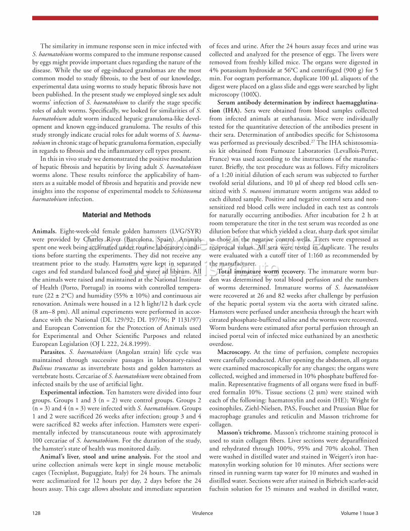
of feces and urine. After the 24 hours assay feces and urine was collected and analyzed for the presence of eggs. The livers were removed from freshly killed mice. The organs were digested in 4% potassium hydroxide at 56°C and centrifuged (900 g) for 5 min. For oogram performance, duplicate 100 µL aliquots of the digest were placed on a glass slide and eggs were searched by light microscopy (100X).
Serum antibody determination by indirect haemagglutina-tion (IHA). Sera were obtained from blood samples collected from infected animals at euthanasia. Mice were individually tested for the quantitative detection of the antibodies present in their sera. Determination of antibodies specific for Schistosoma was performed as previously described.27 The IHA schistosomia-sis kit obtained from Fumouze Laboratories (Levallois-Perret, France) was used according to the instructions of the manufac-turer. Briefly, the test procedure was as follows. Fifty microliters of a 1:20 initial dilution of each serum was subjected to further twofold serial dilutions, and 10 µl of sheep red blood cells sen-sitized with S. mansoni immature worm antigens was added to each diluted sample. Positive and negative control sera and non-sensitized red blood cells were included in each test as controls for naturally occurring antibodies. After incubation for 2 h at room temperature the titer in the test serum was recorded as one dilution before that which yielded a clear, sharp dark spot similar to those in the negative control wells. Titers were expressed as reciprocal values. All sera were tested in duplicate. The results were evaluated with a cutoff titer of 1:160 as recommended by the manufacturer.
Total immature worm recovery. The immature worm bur-den was determined by total blood perfusion and the numbers of worms determined. Immature worms of S. haematobium were recovered at 26 and 82 weeks after challenge by perfusion of the hepatic portal system via the aorta with citrated saline. Hamsters were perfused under anesthesia through the heart with citrated phosphate-buffered saline and the worms were recovered. Worm burdens were estimated after portal perfusion through an incised portal vein of infected mice euthanized by an anesthetic overdose.
Macroscopy. At the time of perfusion, complete necropsies were carefully conducted. After opening the abdomen, all organs were examined macroscopically for any changes; the organs were collected, weighed and immersed in 10% phosphate buffered for-malin. Representative fragments of all organs were fixed in buff-ered formalin 10%. Tissue sections (2 µm) were stained with each of the following: haematoxylin and eosin (HE); Wright for eosinophiles, Ziehl-Nielsen, PAS, Fouchet and Prussian Blue for macrophage granules and reticulin and Masson trichrome for collagen.
Masson’s trichrome. Masson’s trichrome staining protocol is used to stain collagen fibers. Liver sections were deparaffinized and rehydrated through 100%, 95% and 70% alcohol. Then were washed in distilled water and stained in Weigert’s iron hae-matoxylin working solution for 10 minutes. After sections were rinsed in running warm tap water for 10 minutes and washed in distilled water. Sections were after stained in Biebrich scarlet-acid fuchsin solution for 15 minutes and washed in distilled water,
The similarity in immune response seen in mice infected with S. haematobium worms compared to the immune response caused by eggs might provide important clues regarding the nature of the disease. While the use of egg-induced granulomas are the most common model to study fibrosis, to the best of our knowledge, experimental data using worms to study hepatic fibrosis have not been published. In the present study we employed single sex adult worms’ infection of S. haematobium to clarify the stage specific roles of adult worms. Specifically, we looked for similarities of S. haematobium adult worm induced hepatic granuloma-like devel-opment and known egg-induced granuloma. The results of this study strongly indicate crucial roles for adult worms of S. haema-tobium in chronic stage of hepatic granuloma formation, especially in regards to fibrosis and the inflammatory cell types present.
In this in vivo study we demonstrated the positive modulation of hepatic fibrosis and hepatitis by living adult S. haematobium worms alone. These results reinforce the applicability of ham-sters as a suitable model of fibrosis and hepatitis and provide new insights into the response of experimental models to Schistosoma haematobium infection.
Material and Methods
Animals. Eight-week-old female golden hamsters (LVG/SYR) were provided by Charles River (Barcelona, Spain). Animals spent one week being acclimated under routine laboratory condi-tions before starting the experiments. They did not receive any treatment prior to the study. Hamsters were kept in separated cages and fed standard balanced food and water ad libitum. All the animals were raised and maintained at the National Institute of Health (Porto, Portugal) in rooms with controlled tempera-ture (22 ± 2°C) and humidity (55% ± 10%) and continuous air renovation. Animals were housed in a 12 h light/12 h dark cycle (8 am–8 pm). All animal experiments were performed in accor-dance with the National (DL 129/92; DL 197/96; P 1131/97) and European Convention for the Protection of Animals used for Experimental and Other Scientific Purposes and related European Legislation (OJ L 222, 24.8.1999).
Parasites. S. haematobium (Angolan strain) life cycle was maintained through successive passages in laboratory-raised Bulinus truncatus as invertebrate hosts and golden hamsters as vertebrate hosts. Cercariae of S. haematobium were obtained from infected snails by the use of artificial light.
Experimental infection. Ten hamsters were divided into four groups. Groups 1 and 3 (n = 2) were control groups. Groups 2 (n = 3) and 4 (n = 3) were infected with S. haematobium. Groups 1 and 2 were sacrificed 26 weeks after infection; group 3 and 4 were sacrificed 82 weeks after infection. Hamsters were experi-mentally infected by transcutaneous route with approximately 100 cercariae of S. haematobium. For the duration of the study, the hamster’s state of health was monitored daily.
Animal’s liver, stool and urine analysis. For the stool and urine collection animals were kept in single mouse metabolic cages (Tecniplast, Buguggiate, Italy) for 24 hours. The animals were acclimatized for 12 hours per day, 2 days before the 24 hours assay. This cage allows absolute and immediate separation

www.landesbioscience.com Virulence 129
toned in 0.2% gold chloride for 2 minutes and rinsed in dis-tilled water, fixed with 2% aqueous sodium thiosulphate (hypo) for 2 minutes and washed in tap water for 2 minutes. Sections were counterstained with neutral red for 2 minutes, dehydrated, cleared and mounted.
Histological analysis. A histological study was conducted on H&E-stained tissue sections to evaluate the following parame-ters: presence of S. haematobium immature worms in histological section; fibrous perihepatitis, lymphoid follicles, infiltration of lymphocytes and plasma cells; and the presence of bilharzial pig-ment. Wright-stained tissue sections were used to evaluate infil-tration of eosinophils. Masson Trichrome and Reticulin stains were evaluated. To assess the severity of hepatic lesions, two researchers evaluated tissue sections independently, as follows: -, absent; +, mild; ++, moderate; +++, severe.
Acknowledgements
We thank Dr. Fernanda Seixas Travassos and Dr. Adelina Gama for many helpful discussions.
differentiated in phosphomolybdic-phosphotungstic acid solu-tion for 15 minutes. Sections were then transferred directly (with-out rinse) to aniline blue solution and stained for 5–10 minutes. Rinsed briefly in distilled water and differentiated in 1% ace-tic acid solution for 2–5 minutes. Later were washed in distilled water and dehydrated through 95% ethyl alcohol, absolute ethyl alcohol and clear in xylene. Finally were mounted with resinous mounting medium.
Reticulin. The reticulin technique was based on the follow-ing technique: liver sections were deparaffinized in xylene then took through alcohols to water. Sections were oxidized in acidi-fied potassium permanganate during 3 minutes, rinsed in dis-tilled water and decolorized with 2% oxalic acid for 1 minute. Sections were rinsed again in distilled water, placed in mordant in 4% iron aluminium for 10 minutes, and rinsed in distilled water. Sections were impregnated in ammoniacal silver solution for 11 seconds and quickly rinsed in distilled water. Sections were immediately reduced with 10% aqueous formalin for 2 minutes and washed in running tap water for 2 minutes. Sections were
References1. Vieira P, Miranda HP, Cerqueira M, Delgado ML,
Coelho H, Antunes D, et al. Latent schistosomiasis in Portuguese soldiers. Mil Med 2007; 172:144-6.
2. Mutapi F, Winborn G, Midzi N, Taylor M, Mduluza T, Maizels RM. Cytokine responses to Schistosoma haematobium in a Zimbabwean population: contrasting profiles for IFNgamma, IL-4, IL-5 and IL-10 with age. BMC Infect Dis 2007; 7:139.
3. Jacobs W, Deelder A, Bogers J, Van de Vijver K, Van Marck E. Schistosomal granuloma modulation III. Schistosma haematobium worms accelerate S. mansoni soluble egg antigen-induced hepatic granuloma forma-tion in vivo. Parasitol Res 1999; 85:905-9.
4. Boros DL, Warren KS. Delayed hypersensitivity-type granuloma formation and dermal reaction induced and elicited by a soluble factor isolated from Schistosoma mansoni eggs. J Exp Med 1970; 132:488-507.
5. Boros DL. Immunopathology of Schistosoma mansoni infection. Clin Microbiol Rev 1989; 2:250-69.
6. Grzych JM, Pearce E, Cheever A, Caulada ZA, Caspar P, Heiny S, et al. Egg deposition is the major stimulus for the production of Th2 cytokines in murine schisto-somiasis mansoni. Immunol 1991; 146:1322-7.
7. Van Marck EA, Kestens L, Stocker S, Grimaud JA, Gigase PL, Deelder AM. Fibrosis around schistosomal egg antigen-coated beads in the liver of mice. Contrib Microbiol Immunol 1983; 7:251-9.
8. Cheever AW, Lewis FA, Wynn TA. Schistosoma man-soni: unisexual infections sensitized mice for granuloma formation around intravenously injected eggs. Parasitol Res 1997; 83:57-9.
9. Leptak CL, McKerrow JH. Schistosome egg granulo-mas and hepatic expression of TNFalpha are dependent on immune priming during parasite maturation. J Immunol 1997; 158:301-7.
10. Jacobs W, Bogers J, Deelder A, Wéry M, Van Marck E. Adult Schist osoma mansoni worms positively modulate soluble egg antigen-induced inflammatory hepatic granuloma formation in vivo. Stereological analysis and immunophenotyping of extracellular matrix proteins, adhesion molecules and chemokines. Am J Pathol 1997; 150:2033-45.
11. Jacobs W, Van de Vijver K, Deelder A, Van Marck E. Morphometrical and immunopathological dissection of the hepatic Schistosoma haematobium granuloma in the murine host. Parasite 1998; 5:299-306.
12. Jacobs W, Van Marck E. Adhesion and co-stimulatory molecules in the pathogenesis of hepatic and intestinal schistosomiasis mansoni. Mem Inst Oswaldo Cruz 1998; 93:523-9.
13. Vuong PN, Bayssade-Dufour C, Albaret JL, Farhati K. Histopathological observations in new and classic mod-els of experimental Schistosoma haematobium infections. Trop Med Int Health 1996; 1:348-58.
14. Sulahian A, Garin YJ, Izri A, Verret C, Delaunay P, van Gool T, et al Development and evaluation of a western blot kit for diagnosis of schistosomiasis. Clin Diagn Lab Immunol 2005; 12:548-51.
15. Hirata M, Hirata K, Kage M, Zhang M, Hara T, Fukuma T. Effect of nitric oxide synthase inhibition on Schistosoma japonicum egg-induced granuloma for-mation in the mouse liver. Parasite Immunol 2001; 23:281-9.
16. Botelho M, Oliveira P, Gomes J, Gartner F, Lopes C, Correia da Costa JM, et al. Tumourigenic effect of Schistosoma haematobium total antigen in mammalian cells. Int J Exp Path 2009; 90:448-53.
17. Botelho M, Ferreira AC, Oliveira MJ, Domingues A, Machado JC, Correia da Costa JM. Schistosoma hae-matobium total antigen induces increased proliferation, migration and invasion, and decreases apoptosis of nor-mal epithelial cells. Int J Parasitol 2009; 39:1083-91.
18. Botelho MC, Oliveira PA, Lopes C, Correia da Costa JM, Machado JC. Urothelial dysplasia and inflamma-tion induced by Schistosoma haematobium total antigen instillation in mice normal urothelium. Urol Oncol 2009; In press.
19. Baki CA, Guerret S, Grimaud JA, Chevallier M. Liver fibrosis in unisexual murine Schistosomiasis: quantita-tive study and morphological changes in mice with chronic infection. Cell Mol Biol (Noisy-le-grand) 1998; 44:627-33.
20. Gryseels B, Polman K, Clerinx J, Kestens L. Human schistosomiasis. Lancet 2006; 368:1106-18.
21. Van de Vijver KK, Colpaert CG, Jacobs W, Kuypers K, Hokke CH, Deelder AM, et al. The host’s genetic background determines the extent of angiogenesis induced by schistosome egg antigens. Acta Trop 2006; 99:243-51.
22. Soliman K, Abou-El Dobal S, Marei N. Effect of carnosine administration on the immune response of rabbit to Schistosoma mansoni antigens. J Egypt Soc Parasitol 2003; 33:663-78.
23. Faria-Pinto P, Rezende-Soares FA, Molica AM, Montesano MA, Marques MJ, Rocha MO, et al. Mapping of the conserved antigenic domains shared between potato apyrase and parasite ATP diphospho-hydrolases: potential application in human parasitic diseases. Parasitology 2008; 135:943-53.
24. Pearce EJ, MacDonald AS. The immunobiology of schistosomiasis. Nat Rev Immunol 2002; 2:499-511.
25. Agnew AM, Murare HM, Doenhoff MJ. Immune attrition of adult schistosomes. Parasite Immunol 1993; 15:261-71.
26. Moloney NA, Hinchcliffe P, Webbe G. The ability of single sex infections of Schistosoma japonicum to induce resistance to reinfection in mice. J Helminthol 1986; 60:250-4.
27. Van Gool T, Vetter H, Vervoort T, Doenhoff MJ, Wetsteyn J, Overbosch D. Serodiagnosis of imported schistosomiasis by a combination of a commercial indi-rect hemagglutination test with Schistosoma mansoni adult worm antigens and an enzyme-linked immu-nosorbent assay with S. mansoni egg antigens. J Clin Microbiol 2002; 40:3432-7.