growth and characterization of uric acid crystals.pdfphysics annamalai university and edx was...
TRANSCRIPT

International Journal of Science and Research (IJSR) ISSN (Online): 2319-7064
Impact Factor (2012): 3.358
Volume 3 Issue 8, August 2014 www.ijsr.net
Licensed Under Creative Commons Attribution CC BY
Growth and Characterization of Uric Acid Crystals
G. Vasuki1, R. Selvaraju2
1, 2Department of Engineering Physics, Annamalai University, Annamalainagar-608 002, TamilNadu, India
Abstract: Kidney stone (renal calculi) and urinary tract infection is a one of the major disease affecting human being since from ancient period. Urinary tract infection occurs in kidney, urinary bladder and urethra. Crystals of uric acid as well as its sodium salts have been found in the renal tract and urinary bladder. Uric acid is also often found as a constituent of urinary calculi and has a high incidence in nephrolithiasis resulting in most affected kidney function. Uric acid crystals have been grown in single diffusion gel technique. The growth of crystal is studied by FT-IR, FT-Raman, Powder XRD and SEM with EDX. Many medicinal plants (herbals) are available for dilutic urinary calculi particularly Tribulus terrestris and citrus limon. To verify the inhibitive effect, aqueous extracts of herbals was added along with the supernatant solutions. The growth was measured and compared, with and without the aqueous extracts. Inhibition of Uric acid crystal growth was observed in the herbal extracts. Keywords: Gel growth, uric acid crystal, FT-IR, FT-Raman, SEM with EDX 1. Introduction A crystal deposition process is essentially crystallization from solutions. The gel method of crystal growth using gel as a model system for studying crystal deposition, because their viscous nature provides simulation of synovial fluid, cartilage and other biological fluids which are viscous in nature. The increasing incidence of crystal deposition diseases such as urinary stones, kidney stones, gallstones, gout, etc in people of all ages affecting a considerable number of the total population is a major social and economic problem, considering the number of days lost from work and cost of hospitalization [1, 2].This has resulted in an extensive research on crystal deposition disease. A crystal deposition disease may be defined micro crystals and in which the crystals are contributing to tissue damage [3]. Gout is the best known disease of this type. An increase of uric acid (2, 6, 8- trioxy purine, C5H4N403) levels in blood leads to the deposition of uric acid in joints which result in gout causing pain and swelling. Uric acid is found either in pure or in mixed stones associated with sodium and ammonium urates, calcium oxalates, struvite and Hydroxyapatite. In addition, it was suggested that uric acid crystals could act as a support of the heterogeneous nucleation for other crystalline species. Hence studies on nucleation and growth of uric acid play a vital role in medicine and pharmaceutical research Uric acid is known to crystallize in two distinct forms. One is anhydrous and the other is dehydrated. Recently, there has been an increasing interest in growing crystals using gels as a model system for studying crystal deposition diseases in human beings [4-8].Since silica gel acts as an inert medium during the crystallization process, it acts as an ideal medium for studying the crystallization of uric acid in vitro. Uric acid is insoluble in alcohol, ether and most common organic solvents and owing to its extremely low solubility in water, it would be difficult to prepare a pure specimen because of the bulk of the solvent needed. As the uric acid has a very poor aqueous and organic solubility, it is very difficult to crystallize them in an aqueous medium like silica gel. Here we report the growth of uric acid crystals in silica gel using single tube diffusion techniques.
This technique also produces single crystals of uric acid fairly easily without the use bulk solvents. The experiments were conducted at tropical room temperature. 2. Materials and Methods Sodium meta silicate, (Na2Sio3.9H2O), uric acid and sodium hydroxide was used for preparing the gel and glacial acetic acid was used for adjusting the pH value. All the reagents used in this experiment are of analar grade.Crystallizations were conducted in the gel densities between 1.03 and 1.06 g/cc and pH value from4 to 6.5.Optimum values of gel density and pH values for the crystallization were found to be 1.03 g/cc and 6 respectively. Dilute hydrochloric acid was placed over the set gel. The test tube was capped with air tight stopples and kept undisturbed. In the single test tube diffusion method, platy, transparent, single crystals of uric acid were found uniformly throughout the gel medium in about 24hrs,which are shown in fig.1. Here, the silica hydro-gel was chosen because it remains stable and does not react with present or with the product crystals grown. The growth was completed within a period of 21 days and harvested the crystals. Harvested crystals as shown in fig.2.
Figure 1: Uric acid crystals grown in gel
Paper ID: 02015368 696
International Journal of Science and Research (IJSR) ISSN (Online): 2319-7064
Impact Factor (2012): 3.358
Volume 3 Issue 8, August 2014 www.ijsr.net
Licensed Under Creative Commons Attribution CC BY
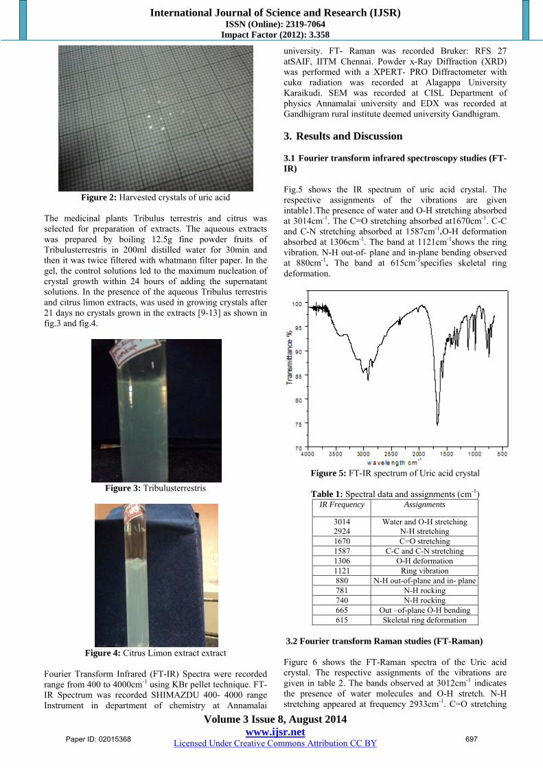
Figure 2: Harvested crystals of uric acid
The medicinal plants Tribulus terrestris and citrus was selected for preparation of extracts. The aqueous extracts was prepared by boiling 12.5g fine powder fruits of Tribulusterrestris in 200ml distilled water for 30min and then it was twice filtered with whatmann filter paper. In the gel, the control solutions led to the maximum nucleation of crystal growth within 24 hours of adding the supernatant solutions. In the presence of the aqueous Tribulus terrestris and citrus limon extracts, was used in growing crystals after 21 days no crystals grown in the extracts [9-13] as shown in fig.3 and fig.4.
Figure 3: Tribulusterrestris
Figure 4: Citrus Limon extract extract
Fourier Transform Infrared (FT-IR) Spectra were recorded range from 400 to 4000cm-1 using KBr pellet technique. FT-IR Spectrum was recorded SHIMAZDU 400- 4000 range Instrument in department of chemistry at Annamalai
university. FT- Raman was recorded Bruker: RFS 27 atSAIF, IITM Chennai. Powder x-Ray Diffraction (XRD) was performed with a XPERT- PRO Diffractometer with cukα radiation was recorded at Alagappa University Karaikudi. SEM was recorded at CISL Department of physics Annamalai university and EDX was recorded at Gandhigram rural institute deemed university Gandhigram. 3. Results and Discussion 3.1 Fourier transform infrared spectroscopy studies (FT-IR) Fig.5 shows the IR spectrum of uric acid crystal. The respective assignments of the vibrations are given intable1.The presence of water and O-H stretching absorbed at 3014cm-1. The C=O stretching absorbed at1670cm-1. C-C and C-N stretching absorbed at 1587cm-1.O-H deformation absorbed at 1306cm-1. The band at 1121cm-1shows the ring vibration. N-H out-of- plane and in-plane bending observed at 880cm-1. The band at 615cm-1specifies skeletal ring deformation.
Figure 5: FT-IR spectrum of Uric acid crystal
Table 1: Spectral data and assignments (cm-1)
IR Frequency Assignments
3014 2924
Water and O-H stretching N-H stretching
1670 C=O stretching1587 C-C and C-N stretching1306 O-H deformation1121 Ring vibration 880 N-H out-of-plane and in- plane781 N-H rocking 740 N-H rocking 665 Out –of-plane O-H bending615 Skeletal ring deformation
3.2 Fourier transform Raman studies (FT-Raman) Figure 6 shows the FT-Raman spectra of the Uric acid crystal. The respective assignments of the vibrations are given in table 2. The bands observed at 3012cm-1 indicates the presence of water molecules and O-H stretch. N-H stretching appeared at frequency 2933cm-1. C=O stretching
Paper ID: 02015368 697

International Journal of Science and Research (IJSR) ISSN (Online): 2319-7064
Impact Factor (2012): 3.358
Volume 3 Issue 8, August 2014 www.ijsr.net
Licensed Under Creative Commons Attribution CC BY
observed at frequency 1681 cm-1and 1648cm-1. C-C and C-N stretching observed at frequency 1592cm-1. O-H deformation appeared at frequency 1354cm-1. C-N stretching appeared at frequency 1285cm-1. Ring vibration observed at frequency 1119cm-1. N-H out-of-plane and in-plane bendings observed at frequency881cm-1.The peak appeared at frequency 782cm-1 representing N-H rocking. N-H-in-plane stretching observed at frequency 704cm-1. The peak appeared at frequency 655cm-1 representing N-H-in-planeO-H bending. Skeletal ring deformation observed at frequency 624cm-1.
Figure 6: FT-Raman spectrum of Uric acid crystal
Table 2: FT-Raman frequencies (in cm-1) with tentative
assignment for Uric acid crystal Frequencies(cm-1) Tentative Assignments
3012 Water molecules and O-Hstretching 2933 N-H stretching 1681 C=O stretching 1648 C=O stretching
1592 C-C and C-N stretching 1354 O-H deformation 1285 C-N stretching 1119 Ring vibration 881 N-H out-of-plane and in-plane bendings 782 N-H rocking 704 N-H-in-plane stretching 655 Out-of-plane O-H bending 624 Skeletal ring deformation
3.3 X-ray diffraction (XRD) Fig.7 shows powder XRD pattern and table 3 gives the data of uric acid crystal. The lattice parameters of uric acid crystal is a=13.12Ǻ b=7.403Ǻ c=6.208Ǻ β= 90.5˚.From this data the uric acid crystal system is monoclinic [JCPDS NO 21-1959]. The samples are highly crystalline nature.
Figure 7: XRD analysis of Uric acid crystal
Table 3: Powder XRD data of uric acid crystal Pos.[º2Th.] Height[cts] FWHM Left[º2Th.] d-spacing[Å] Rel.Int.[%] Mached by 13.3369(6) 2404(41) 0.088(2) 6.63341 100.00 37-1615(JCPDS) 27.026(3) 836(51) 0.08(1) 3.29655 34.79 78-0188 (JCPDS) 28.23(2) 35(8) 0.26(7) 3.15840 1.45 21-1960 (JCPDS)
28.692(4) 167(17) 0.08(1) 3.10887 6.96 82-0821(JCPDS) 41.120 (30 234(16) 0.12(1) 2.19341 9.74 40-0995(JCPDS)
3.4 SEM with EDX The investigations of surface morphology of crystal by using SEM is as shown in fig8(a),fig 8(b) .The SEM was taken at magnification values100X.The magnification of SEM was about 250 times. SEM acceleration voltage is 20,000 volts and kept the sample in highly vacuumed. The micrograps show that the samples have rectangular bar shaped in fig 8(a) and step like growth in fig 8(b). The surface of the grown uric acid crystal appeared clean and free major defect. The elemental position of the sample is identified using energy dispersive X-ray analysis. The EDX spectrum of uric acid is shown in fig9.The highest peak of C and N shows that the more concentrated the element present in the spectrum. C present in the value 0.3Kev and N present in the value 0.4Kev. The atomic percentage of C/N is 39:36.
Figure 8: (a)
Paper ID: 02015368 698

International Journal of Science and Research (IJSR) ISSN (Online): 2319-7064
Impact Factor (2012): 3.358
Volume 3 Issue 8, August 2014 www.ijsr.net
Licensed Under Creative Commons Attribution CC BY
Figure 8: (b) SEM image of uric acid crystal
Figure 9: EDX Spectrum of Uric acid crystal
4. Conclusion Uric acid crystal were grown using gel methods and were characterized by FT-IR, FT-Raman, Powder XRD and SEM with EDX . FT-IR, FT-Raman and powder XRD techniques confirmed the functional groups and crystalline phases of the uric acid crystals. SEM with EDX confirmed the surface morphology and their composition. This study to trial the growth uric acid crystals using Tribulus terrestris and citrus limon. The aqueous extracts of Tribulus terrestris and citrus limon inhibiting the growth of uric acid crystal. 5. Future Scope The crystal deposition diseases such as kidney stone, urinary stone, gallbladder stone occurs in human beings. In take herbal extracts to prevent the formation of urinary stones, kidney stone, gallbladder stone in future. References
[1] W.G.Robertson, M.Peacock and
A.Hodgkinson.J.ChronicDiseases 32 (1979) 469. [2] W.G.Robertson,D.S.Scurr and C.M.Bridge.J.Crystal
Growth 53(1981) 182 [3] P.Dieppe and P.calvert, Crystals and Joint Disease
(Chapman and Hall, London,1983)
[4] S.N.Kalkura and S.Devanarayanan,J.Crystal Growth 83 (1987) 446.
[5] S.N.Kalkura and S.Devanarayanan, J.Crystal Growth 94 (1989) 810.
[6] S.N.Kalkura and S.Devanarayanan, J.Crystal Growth 110 (1991) 265.
[7] J.S.Shah,in: Proc.2nd National Seminar on Crystal Growth, Anna University, Madras, August 1984.
[8] Y.M.F.Marickar and P. koshy, Scanning Microscopy 1 (1987) 571.
[9] Kalkura, S.N., E.K. Girija, M. Kanakavel and P.Ramasamy, 1995. In vitro crystallization of Spherulites of monosodium urate monohydrate.J.Mater.Sci.Med., 6: 577-580.DOI:10.1007/BF00121281
[10] Prasobh GR, Rev issue3, 2011.ikumar KG Asian Journal of Pharmaceutical and Clinical Research Vol.4, issue3,2011.
[11] V.S. Joshi, B.B Parekh, M.J.Joshi, A.B.Vaidya Journal of Crystal Growth 275(2005)e 1403-e1408.
[12] Vimal S Joshi & Mihir J Joshi Indian Journal of pure & Applied Physics Vol.41, March 2003.pp.183-192.
[13] K.Manjula, K.Pazanichamy, S.Kumaran,T.Eevera, C.DaleKeefe, and K. Rajendran International Journal of Pharmacy and Pharmaceutical Sciences Vo14,Suppl,2012.
Author Profile G. Vasuki doing Phd in department of Physics Annamalai University, Chidambaram, Tamilnadu, India Dr. R. Selvaraju working as a Associate Professor Annamalai University, Chidambaram, Tamilnadu, India.
Paper ID: 02015368 699