guide, fob diagnosis,
TRANSCRIPT
GUIDE, FOB DIAGNOSIS,
TBEATMENT AND CONTROL
O.F DENGUE HAEMORRIIAGIC FEVBK
(S eoand EditioD)
•
'I'ECHNICAL ADVISORY COJIMlTTEE OH DENGUE HAEIIORRHAGIC FEvER
for tb. SOUTH EAliT ASIAN AND WESTERN PACIFIC REGlONS
WOIU.D HEALTH ORGANJZATlON 1980

GUIDE FOR DIAGNOSIS, TREATMENT, AND CONTROL
OF DENGUE HAEMORRHAGIC FEVER
(Second Edition)
FOREWORD
A first edition of this guide was published in 1975 by the WHO Scuth-east
Asian and Western Pacific Regional Offices as "Technical Guides for Diagnosis,
Treatment, Surveillance, Prevention and Control of Deng ~'. 2 Haemorrhagic Fever".
It was prepared by a Technical Advisory Committee which met in Manila in 1974
(Annex 1) and in Bangkok in 1975 (Annex 2). In the face of extensive
epidemics which developed in Burma, Indonesia, Thailand and other countries
of the two WHO Regions from 1972 onwards, it was felt that such a guide could
help clinicians and public·health officers facing a feared disease for the
first time.
Since then, dengue haemorrhagic fever has remained endemic with variable
epidemic activity in the two Regions. An important factor has been its
extension to rural areas, causing a primary health care problem as regards
prompt recognition of the disease.
This second edition aims to present the diagnosis, treatment and control
features of DHF in the simplest possible way in view of the problems that exist
for primary health care serv~ces. At the same time it is desirable that
clinicians and public health officers be able to find in it all the technical
data they want. The challenge for the Technical Advisory Committee which met
in Hanila in 1978 (Annex 3) was to find a convenient compromise for this
second edition. It will be noted that the Committee made little change to
the criteria for diagnosis and recommended treatment, which are very similar
to the 1975 edition.
WHO Headquarters, Geneva WHO Regional Office for South-east Asia
WHO Regional Office for the Western Pacific
WB.Oi ';Ji.Tp(u i L '."'·'' ".<·.,. -t
l\tla.L'"' ·,.,u·rncc'

CONTENTS
1. General considerations
2. Clinical diagnosis
3. Treatment
4. Laboratory diagnosis
5. Vector surveillance and control
6. Epidemiological surveillance, control and prevention
7. Primary health care
8. Further readings
1. GENERAL CONSIDERATIONS
1.1 Spreading outbreaks in South~east Asian and Western Pacific Regions
1.2 Other areas at risk 1.3 Characteristics of outbreaks 1.4 Transmission chain of dengue 1.5 Pathogenesis of dengue haemorrhagic fever
Dengue haemorrhagic fever (DHF) can be defined as an acute febrile illness caused by four s·erotypes of dengue virus and characterized clini.,.. cally by haemorrhagic phenomena and a tendency to develop a shock syndrome (dengue shock syndrome - DSS) which may be fatal. Thrombocytopenia with concurrent haemoconcentration are constant findings.
1.1 Spreading outbreaks in South-east Asian and Western Pacific Regions
The classical form of dengue is an acute febrile disease with headaches and joint and muscular pains. It has been known for more than a century ~n the tropical areas of South-east Asia and the Western Pacific Region. However, dengue haemorrhagic fever outbreaks were recognized as a new disease first in the Philippines in 1953 (Philippine haemorrhagic fever) and in Thailand in 1958 (Thai haemorrhagic fever). These outbreaks caused some panic in the populations because of their novelty and the mystery of the causative agent until dengue viruses types 2, 3 and 4 were isolated ~n the Philippines in 1956 and dengue type 1, in addition, in Thailand.
Between 1953 and 1964, DHF was described in the Philippines, Thailand, Viet Nam, Malaysia, Singapore, and Calcutta in India. After a period when DHF was endemo-epidemic in Thailand and the Philippines, there was a considerable increase in reported dengue infections in the years 1971-1978 in various countries of the South-east Asian and Western Pacific Regions. DHF appeared in Indonesia and Burma. During the 1975-1978 period a total of 17,251 hospitalizations (772 deaths) was reported in Burma, 21,818 hospitalizations (916 deaths) in Indonesia, and 71,312 hospitalizations (l ,676 deaths) in Thailand. In the Western Pacific Region, DHF reports were received during the 1975-1978 period from Malaysia (1,288 cases), the Philippines (836 cases), Singapore (533 cases, including dengue fever), and the Socialist Republic of VietNam (66,372 cases for 1976 and 1977 only).
Dengue haemorrhagic fever is an increasing public health problem in most of the countries of the tropical areas of the Western Pacific and Southeast Asian Regions. This disease is among the ten leading causes of hospitalization and death in children in at least eight Asian countries in the tropics.
1.2 Other areas at risk
Other countries may be considered to be at risk of nHF outbreaks. Dengue has also been prevalent in tropical areas of Africa and the Americas and appeared episodically in the temperate regions of North America, southern Africa, and the Mediterranean region of Europe. There are indications in the literature that cases similar to DSS occurred during some earlier outbreaks.

- 2 -
India has serological and virological evidence of dengue infections with the four serotypes, mainly in the south-east of the country, but no DHF outbreaks have been reported since those of 1963-1965 in Calcutta. Sri Lanka has dengue activity but only sporadic DHF/DSS cases have been reported. A large number of islands in the Western Pacific Region were involved in a dengue type 1 pandemic in 1974, while outbreaks caused by serotypes 2 and 3 occurred during previous years. A somewhat similar situation is developing in the Caribbean islands of the Americas, where a pandemic caused by dengue virus type 1 occurred in 1977 following outbreaks caused by types 2 and 3. In these settings, only a few DHF cases were reported heretofore.
1.3 Characteristics of outbreaks
Outbreaks seem to have appeared suddenly in the Philippines and in Thailand. However, retrospective studies have shown that they were very probably preceded by a period when cases occurred but were not recognized. In Thailand, outbreaks occurred first in Bangkok, with a pattern of epidemic activity at two-year cycles which changed later to irregular epidemic cycles. DHF then became endemic in many large cities of Thailand and thereafter it spread gradually to smaller cities and towns during epidemic occurrences. A similar pattern has been observed in Indonesia and Burma.
A seasonal pattern of incidence, coincident with the rainy season, can be observed in some countries.
In South-east Asia, DHF has been observed mainly in children. In Bangkok, where, since 1968, there has been a trend towards reduced attack rates (constant hospital admissions with an increasing population), the modal age of hospitalized children has risen to 6-7 years but throughout the rest of Thailand the modal age is still 4-6 years. DHF is seen more frequently in females than in males.
The attack rate of DHF is difficult to evaluate as the number of dengue virus infections is most of the time unknown. A retrospective evaluation of the impact of DHF during the outbreak in Bangkok-Thonburi in May-November 1962 indicated that in a population of 870,000 children under 15 years of age there were an estimated 150,000 to 200,000 minor illnesses caused by dengue or chikungunya viruses; 4,187 were hospitalized and diagnosed as having DHF and 4,000 additional cases were treated in private clinics or at home. The incidence of shock was about one third of hospitalized DHF cases.
1.4 Transmission chain of dengue
Dengue virus infection is transmitted to man through mosquito bites and 1s therefore ranked among arbovirus (arthropod-borne) diseases. Man is the reservoir of virus, and studies in Malay.sia have~hown that the monkey is a natural reservoir (Fig. 1).

- 3 -
Fig. 1 Transmission of dengue virus
(Monkey)-,
(Man\ Mos.qui~o Mosquito
~Man) Three elements have to be considered: the virus, the vector, and the
host.
The virus
The four serotypes of dengue virus - DEN-1, DEN-2, DEN-3 and DEN-4 -
are antigenically very close to each other but they are different enough to
elicit only partial cross-protection after infection by one of them. After
an incubation period of 4-6 days (minimum 3, maximum 10), the virus is
present in the blood of patients during the acute phase of the disease.
This constitutes a reservoir of virus readily accessible for transmission
of the disease by mosquitoes.
The vector
Aedes aegypti is the most efficient amongst mosquito vectors because
of its domestic habitat. The female mosquito bites man during the day.
After feeding on a person whose blood contains the virus, the female
Ae. aegypti can transmit dengue either after an incubation period of 8-10
days, during which the virus multiplies in its salivary glands, or even
immediately, by a change of host when its blood meal is interrupted.
Dengue outbreaks have also been attributed to Ae. albopictus, Ae. polynes
iensis, and several species of the Ae. scutell~is complex. These species
have their particular geographical distribution and in general are less
efficient vectors than Ae. aegypti.
The host
In man, each of the four types of dengue viruses can cause
e ither classical dengue or dengue haemorrhagic fever, and it is not known
whether one virus type is more pathogenic than another. The acute phase of
infection by dengue viruses, which lasts about 5-7 days, is followed by an
immune response. At the first attack, neutralizing antibodies are produced
predominantly against the virus type inoculated by the mosquito vector
(primary dengue). The first attack gives only temporary and partial
protection against the other three types and secondary or sequential
infections are possible after a rather short period of time.

- 4 -
1.5 Pathogenesis of DHF/DSS
Two main pathophysiologic changes occur in DHF/DSS. One is an increased vascular permeability, giving rise to loss of plasma from _the vascular compartment. This results in haemoconcentration, low pulse pressure and other signs of shock if and when plasma loss is critical. The second change is a disorder in haemostasis which involves all three major factors, namely vascular changes, thrombocytopenia and coagulopathy.
A constant finding in DHF/DSS which may be involved in the production of increased vascular permeability is activation of the complement system with profound depression of C3 and CS levels. Immune complexes have been described in DHF cases associated with secondary dengue infection, which may initiate the complement activation.
One factor which may contribute to DHF/DSS is the enhancement of virus multiplication in monocytes by heterotypic antibodies. This may trigger the production by monocytes of chemical mediators of vascular permeability, activation of complement and tissue thromboplastin which initiates intravascular blood coagulation.
Research has been undertaken to understand better the pathogenic mechanisms of DHF/DSS.

- 5 -
2. CLINICAL DIAGNOSIS
2.1 Classical dengue fever 2.2 Dengue haemorrhagic fever 2.3 Criteria for clinical diagnosis of dengue
haemorrhagic fever (DHF)/dengue shock syndrome (DSS)
2.4 Grading the severity of DHF 2.5 Differential diagnosis of DHF/DSS 2.6 Record sheet
2.1 Classical dengue fever
The clinical features of dengue fever frequently depend on the age of
the patient. Infants and young children may have an undifferentiated
febrile disease with maculopapular rash. Older children and adults may have
either mild febrile syndromes or the classical incapacitating disease with
abrupt onset and high fever, severe headache, muscular and joint pains and
rash. The case fatality rate is exceedingly low.
Many epidemics of classical dengue fever are accompanied by varying
frequency of bleeding complications such as petechial haemorrhage, epistaxis,
gingival bleeding, gastro-intestinal bleeding and haematuria.
Classical dengue fever has to be differentiated from dengue haemorrhagic
fever and chikungunya fever (see Table 1).
2.2 Dengue haemorrhagic fever
Typical cases of DHF, as commonly seen in Asian countries, are character
ized by four major clinical manifestations: high fever, haemorrhagic
phenomena, hepatomegaly and often circulatory failure. Moderate to marked
thrombocytopenia with concurrent haemoconcentration is a distinctive clinical
laboratory finding which differentiates DHF from dengue fever, including
DF with haemorrhagic manifestations.
a) Dengue haemorrhagic fever without shock (DHF)
The illness commonly begins with a sudden rise of temperature which is
accompanied by facial flush and other non-specific constitutional symptoms
such as: anorexia, vomiting, headache and muscle or joint pains. Some
patients complain of sore throat and an injected pharynx may be found on
examination. Epigastric discomfort, tenderness at the right costal margin
and generalized abdominal pain are common. The temperature is typically
high and continues so, persisting for 2-7 days, and then falls to normal
or subnormal by lysis. Occasionally, the temperature may be as high as
40-41°C and febrile convulsion may 01:cur.
The most common haemorrhagic phenomenon is skin haemorrhage: a positive
tourniquet test, easy bruisability and bleeding at venepuncture sites are
present in most cases. Fine petechiae scattered on extremities, axillae,
face, and soft palate may be seen during the early febrile phase. A confluent
petechial rash with characteristic coin-sized areas of normal skin is some
times seen in convalescence after temperature has been normal for 2-3 days.

- 6 -
Maculart maculo-papular or a rubella-type rash may be observed early or late in the disease. Epistaxist gum bleeding are less common. Gastrointestinal haemorrhage is infrequent and more usually follows a period of uncontrolled shock.
The liver is usually palpable early in the febrile phase. The size of the liver varies from just palpable to 2-4 em below the costal margin. Liver size is not correlated with disease severity. The liver may be tender but jaundice is not usually observed even in patients with a bigt tender liver.
In mild or moderate cases , a f ter fever subsides, all signs and symptoms abate. Lysis of fever may be accompan ied by profuse sweating and mild changes in pul se r ate and b l ood pressure, together with coolness of extremities and skin congest i on. Thes e changes reflect mild and transient circulatory dis turb ances . Patients usually recover spontaneously or after fluid and electro l yte t herapy.
b) Dengue shock syndrome (DSS)
In severe cases, following fever of a few days' duration the patient's condition suddenly deteriorates. Accompanying or shortly after the fall in temperature from the 3rd to 7th day of the ,.dis.ease.., there are signs .of , circulatory failure: the skin becomes cool, blotchy and congested, ci r cumoral cyanos is is fr equently observed and the pulse becomes rapid. Altho ugh some patients may appear lethargic, they become restless and then rap i dl y go into a critical stage of shock. Abdominal pain is a frequent compl aint shor tly before the onset of shock.
Shock i s char acterized by a rapid and weak pulse with narrowing of the pul s e pressure (20 mm Hg or less, regardless of the pressure levels, e.g. 100/90) o r hypotension, with cold, clammy skin and restlessness. Patients in shock are i n danger of dying i£ appropriate treatment is not promptly given. Pa t i ents may pass into a stage of profound shock, blood pressure and pul se becoming impe rceptible. The duration of shock is short; the patient may die wi thin 12- 24 hour s or recover rapidly following appropriate antishock therapy. Uncorrected shock may give rise to a more complicated course with metabolic acidosis, severe gastro-intestinal bleeding and a poor prognosis.
Convalescence 1n DHF with or without shock is short and uneventful. Even in cases with profound shock, when shock is overcome, the patient rapidly recovers within 2-3 days. Return of appetite is a good prognostic sign. A common finding in convalescence is bradycardia or sinus arhythmia; absolute bradycardia is seen occasionally.
Clinical laboratory findings
Thrombocytopenia and haemoconcentration are constant findings. A platelet count of below lOO,OOO/mm3 is usually found between the third and eighth days. Haemoconcentration - an evidence of plasma leakage - is always present, even in non-shock cases, but to a lesser degree than in shock cases.

- 7 -
Other common findings are hypoproteinemia, hyponatremia, mildly elevated serum transaminase (SGOT) and blood urea nitrogen levels. Metabolic acidosis may be found in cases with prolonged shock.
The white blood cells count is variable, ranging from leukopenia to
mild leukocytosis. Lymphocytosis with atypical lymphocytes is a common finding. A transient mild albuminuria is sometimes observed. Occult blood is often found in stool. About one third of shock cases have a prolonged prothrombin time (PT) and about half of these patients exhibit prolonged partial thromboplastin time (PTT). Assays of coagulation factors show reductions of factors II, V, VII, IX and XII in severe cases. Decreases 1n fibrinogen levels are also correlated with disease severity.
2.3 Criteria for clinical diagnosis of DHF/DSS
The following criteria have been selected for the clinical diagnosis of DHF and in 90 per cent of cases dengue infection has been confirmed by
etiological diagnosis in the laboratory. Their use will avoid an overdiagnosis of the disease.
1
Clinical
a) Fever - acute onset, high, continuous and lasting 2-7 days. b) Haemorrhagic manifestations including at least a positive
tourniquet testl and any of the following: - petechiae, purpura, ecchymosis - epistaxis, gum bleeding - haematemesis and/or melena.
c) Enlargement of liver (observed at some stage of illness in 90-96 per cent of Thai children and in 60 per cent of adults).
d) Shock - manifested by rapid and weak pulse with narrowing of pulse pressure (20 mm Hg or less) or hypotension, with the presence of cold, clammy skin and restlessness.
Laboratory
a) Thrombocytopenia (lOO,OOO/mm3 or less)} b) Haemoconcentration: haematocrit (Hct) increased by 20 per cent
or more.
Standard method using a blood pressure cuff (Wintrobe 1967) is recommended.
It should be noted that in DHF patients, the test usually gives a definitely
positive result, i.e. more than 20 petechiae/inchz. The test may be negative
or mildly positive during the phase of profound shock. It usually becomes positive and even strongly positive if looked for when recovery from shock has occurred.
2 Direct count using phase contrast microscope (normal 200,000-500,000/mrn3).
In practice, for outpatients a qualitative count from peripheral blood smear is acceptable. In normal persons, 4-10 platelets/oil field (an average of 10
oil field readings recommended) indicates an adequate platelets level. An average of, or below, 2-3/0.F. is considerably low (approximately~lOO,OOO).

- 8 -
The presence of the first two clinical criteria plus thrombocytopenia and haemoconcentration is sufficient to establish a clinical diagnosis of DHF.
When shock occurs with high Hct levels (except in patients with severe bleeding) and marked thrombocytopenia, the diagnosis of DHF/DSS is highly likely.
2.4 Grading the severity of DHF
The spectrum of DHF is classified according to disease severity into four grades:
C/) C/)
~
Grade I - Fever accompanied by non-specific constitutional symptoms: the only haemorrhagic manifestation is a positive tourniquet test.
Grade II - The additional manifestation to those of Grade I is spontaneous bleeding - skin and/or other haemorrhages.
Grade III - Circulatory failure manifested by rapid and weak pulse, narrowing of pulse pressure (20 mm Hg or less) or hypotension, with the presence of cold clammy skin and restlessness.
Grade IV - Profound shock with undetectable blood pressure and pulse.
The presence of thrombocytopenia with concurrent haemoconcentration will differentiate Grade I and II DHF from classical dengue fever.
Grading the severity of the disease has been found clinically and epidemiologically useful in DHF epidemics in children in South-east Asia and the Western Pacific Region. However, it may not be applicable to dengue in adults.
2.5 Differential diagnosis of DHF/DSS
Early in the febrile phase, the differential wide spectrum of viral and bacterial infections. difficult to differentiate from dengue clinically
diagnosis includes a Chikungunya fever may be (see Tables 2 and 3).
By the third or fourth day, usually before shock occurs, the signs and symptoms essential for diagnosis can be observed and should cause no difficulty in diagnosis under epidemic conditions. When shock develops with other manifestations, the clinical diagnosis of DHF can be made with more confidence. Endotoxic shock from bacterial infection, or meningococcemia, may resemble DSS.
The presence of marked thrombocytopenia with concurrent haemoconcentration differentiates DHF/DSS from other diseases.
2. 6 Record sheet
A model record sheet for DHF ~s presented in Table 4.

- 9 -
3. TREATMENT
3.1 General considerations
3.2 Dengue haemorrhagic fever without shock
3.3 Dengue shock syndrome
3.1 General considerations
The major pathophysiological abnormality seen in DHF/DSS is an acute
increase in vascular permeability that leads to leakage of plasma. Plasma
volume studies revealed a reduction of more than 20 per cent in severe
cases. Supporting evidence of plasma leakage includes: serious effusion
found at postmortem, pleural effusion on X-ray, haemoconcentration and
hypoproteinemia. In severe cases, the onset of shock is acute, haematocrit
rises sharply as plasma escapes through the endothelium. Hypovolemic shock,
as a consequence of critical level of plasma loss, leads to tissue anoxia,
metabolic acidosis and death if uncorrected.
Haemostatic changes in DHF involve all three factors: vascular,
thrombocytopenia, and coagulopathy. All patients demonstrate an increase
in capillary fragility (positive tourniquet test) and moderate to marked
thrombocytopenia. Almost 80 per cent of patients with DSS and 17 per cent
of non-shock cases have an abnormal coagulogram suggesting disseminated
intravascular coagulation (DIC), as evidenced by concomittant thrombo
cytopenia, increased fibrinogen degradation product (FDP) and either
prolonged PTT or decreased fibrinogen level. Generally, most clotting
defects result in no clinical haemorrhage. However, in cases of prolonged
uncontrolled shock, DIC may cause important clinical bleeding and may play
an important part in the development of lethal shock. About a third of
shock cases, mostly those with refractory shock, present bleeding, mostly
from gastro-intestinal tract; DIC is implicated as a cause.
In most cases, early and effective replacement of lost plasma with
plasma, plasma expander and/or fluid and electrolyte solution results in
a favourable outcome. The acute onset of shock, and the rapid, often
dramatic,clinical recovery, together with the fact that no destructive or
inflammatory vascular lesions are observed, suggests transient functional
vascular changes, possibly due to a short-acting pharmacological mediator.
As in other virus infections, there is no specific anti-viral treatment
but in DHF/DSS symptomatic and supportive measures are effective.
Management directed at the correction of the major pathophysiologic changes
is essential. With adequate fluid administration, DSS is rapidly reversible.
Adequate rapid replacement will also prevent clinical DIC. The prognosis of the
disease depends upon the early recognition of cases and the monitoring of
pre-shock conditions.
The constant finding that a drop in the platelet count usually precedes
the haematocrit rise which heralds the onset of plasma leakage is of great
diagnostic and prognostic value. Serial determinations of platelet and
haematocrit levels are essential for early recognition and prevention of
shock.

- 10 -
Generally, it is not necessary to hospitalize all suspected cases, or cases, of DHF, since shock may develop in only about a third of patients. Mild and moderate cases can be treated as outpatients. For the purpose of early recognition of shock, parents should be advised to bring the patient back for platelet and haematocrit follow-up, and/or if there are any signs of clinical deterioration, warning signs of shock, e.g., restlessness Rnd/or lethargy, acute abdominal pain, cold extremities, skin congestion, or oliguria, usually on or after the third day of illness.
3.2 Dengue haemorrhagic fever without shock
a) Thirst and dehydration result from high fever, anorexia and vomiting, thus fluid intake by mouth should be ample as tolerated. Electrolyte and dextrose solution (as used in diarrhoeal disease)l and/or fruit juice is preferable to plain water.
b) During the febrile phase there 1s a risk of convulsion and antipyretic drugs may be indicated in patients with hyperpyrexia. Salicylates should be avoided since they are known to cause bleeding and acidosis. Acetaminophen is preferable at the following doses:
under 1 - 3 3 - 6 6 -12
1 year years years years
60 mg/dose 60-120 mg/dose
120 mg/dose 240 mg/dose
c) Closely observe patients for early signs of shock. The critical period is the transition from the febrile to afebrile phase which usually occurs after the third day. Haematocrit determinations are an essential guide in therapy since they reflect the degree of plasma leakage and the need for intravenous fluid. Haemoconcentration usually precedes blood pressure and pulse changes. Haematocrit should be determined daily from the third day on until the temperature becomes normal for 1 or 2 days. If haematocrit is not available, haemoglobin determination may be a less sensitive alternative.
d) Parenteral fluid therapy can be done in an O.P.D. rehydration unit in mild or moderate cases when vomiting produces or threatens dehydration or acidosis or when there are signs of haemoconcentration. The volume of fluid and its composition are similar to the fluids used in the treatment of diarrhoea with moderate dehydration. The following schedule is recommended as a guideline:
1 If WHO Oral Rehydration Solution (ORS - 90 mEg Na per liter) is to be
used in children under 2 years of age, additional fruit juice should be given in proportion of one fruit juice (or plain water) for every two ORS volumes. The Oral Rehydration Solution consists of:
Sodium chloride (table salt) Sodium bicarbonate (baking soda) Potassium chloride Glucose
3.5 g 2.5 g 1.5 g
20.0 g
dissolved 1n one liter of potable water.

- 11 -
Intravenous fluid infusion for moderate dehydration (from Ped. Clinic of
North America, Nov. 1964, 11: 1093; 1n ml per pound body weight,with
some modification):
I I
Weight on admission i I < 15 lbs 16-25 lbs 26-40 lbs >40 lbs I
First day 100 75 60 40 ' ' Second day 75 60 40 40 I
Third day 60 40 40 40 I j
or 1n ml per kg body weight:
i Weight admission I on
7 kg I
7-11 kg I 12-18 kg >18 kg <. I i
First day 220 165 132 88
Second day 165 132 88 88
Third day 132 88 88 88
The fluids shall consist of the following:
1/2 to 1/3 of the total fluid as physiologic saline solution (PSS)
1/2 to 2/3 remaining as 5 per cent glucose in water
For acidosis: · 1/4 of the total fluids shall be sixth-molar
sodium bicarbonate Solution for fluid therapy in DHF:
5 per cent glucose PSS, 5 per cent
glucose 1/2 LR, 5 per cent glucose
Lactated Ringer's glucose 1/2 PSS, 5 in 1/3 PSS.
(LR), per cent
Fluids as listed are calculated to be given over a 24-hour period.
If the child seems severely dehydrated, half of the calculated fluid is
given in the first eight hours and the second half in the next 16 hours.
During rapid administration of fluids it is especially important to watch
for signs of cardiac failure.
Written orders should be explicit as to the type of solution and the
rate of administration. A rough estimate of flow may be derived from the
formula: cc/hr = drops/min. x 3
e) Patients should be hospitalized and treated immediately when there
are any of the following signs and symptoms of shock:
- restlessness/lethargy
cold extremities and circumoral cyanosis
- rapid and feeble pulse
- narrowing of pulse pressure (20 mm Hg or less) or hypotension
- sudden rise of haematocrit or continuously elevated haematocrit
despite administration of I.~. fluid (as indicated in d) above).

- 12 -
3.3 Dengue shock syndrome
Shock is a medical emergency. An immediate administration of intravenous fluid to expand plasma volume is most essential. Children may go into and out of shock for a 48-hour period. Close observation 24 hours a day is imperative.
a) Immediate replacement of plasma loss
Start initial fluid therapy with lactated Ringer's or isotonic saline solution 20 ml/kg I.V. Run fluid as rapidly as possible. Positive pressure may be necessary. In case of continued or profound shock, plasma or plasma expander (Dextran, medium sized M.W. in NSS) is given following initial fluid at a rate of 10-20 ml/kg/hr. In most cases not more than 20-30 ml/kg of plasma (Dextran 10-15 ml/kg) is needed. Fluid administration is continued at this constant rate (10-20 ml/kg/hr) until improvement in vital signs is apparent.
b) Continual replacement of further plasma loss to maintain c i rculating volume
Intravenous fluid (5 per cent D 1/2 lactated Ringer's or 1/2 NSS) is continued even when there is a definite improvement in vital signs and a declining Hct. The rate of fluid replacement should be slowed down to 10 ml/kg/hr and adjusted thereafter to the rate of plasma loss, which may continue for 24 or 48 hours. Microhaematocrit determination is a simple and reliable index for estimating plasma leakage. Monitoring of central venous pressure may be necessary in management of severe cases of shock that are not easily reversible.
Intravenous fluids should be discontinued when the Hct level drops to around 40 per cent and the patient gains appetite. A good urine flow indicates sufficient circulating volume. In general, there is no need for fluid therapy beyond 48 hours after shock terminates. Re-absorption of extravasated plasma takes place (manifested by a further drop in Hct after intravenous fluid is stopped) and may cause hypervolemia, pulmonary oedema or heart failure if more fluid is given. It is extremely important that a drop in Hct at this stage is not taken as a sign of internal haemorrhage. Strong pulse and blood pressure (with wide pulse pressure) and diuresis are good vital signs found at this reabsorption phase. They rule out the likelihood of gastro-intestinal haemorrhage, which is found mostly at the shock stage.
c) Other electrolyte and metabolic disturbances may need specific correction
Commonly, hyponatremia and, occasionally, metabolic acidosis occur. Electrolytes and blood gases should be determined periodically in severely ill patients and patients who do not seem to respond as promptly as expected. This will provide an estimate of the amount of electrolyte (sodium) deficit and help determine the presence and degree of acidosis. The latter, in particular, if uncorrected may lead to disseminated intravascular clotting, causing a more complicated course. The use of heparin

- 13 -
may be indicated in some of these cases but extreme caution should be
exercised in using heparin. In general, early volume replacement and
early correction of acidosis with sodium bicarbonate result in a
favourable outcome with no need for heparin.
d) Sedatives
A sedative therapy is needed in some cases to restrain an agitated
child. Hepatotoxic drugs should be avoided. Chloral hydrate orally or
rectally is highly recommended. In cases without lung complication,
paraldehyde I.M. may be used.
Dosage: chloral hydrate 12.5-50 mg/kg as single hypnotic dose not over 1 Gm.
paraldehyde 0.1 ml/kg I.M. not more than 10 ml.
e) Oxygen therapy
Oxygen therapy should be given to all patients in shock. The oxygen
mask or tent may increase apprehension.
f) Blood transfusion
Blood transfusion is indicated in cases with significant clinical
bleeding, most often haematemesis and melena. Fresh whole blood is
preferable and blood should be given only in a volume up to a normal
red blood cell mass. Fresh frozen plasma and/or concentrated platelets
may be indicated in some cases when consumptive coagulopathy causes
massive bleeding.
A persisting shock, despite an adequate fluid administration, and a
decline in Ret level indicate significant clinical bleeding which requires
prompt treatment. DIC is usual in severe shock and it may play an
important part in the development of massive bleeding and lethal shock.
Coagulogram (PT and PTT) should be studied in all shock cases to document
the onset and severity of DIC, which determine the prognosis.
Blood grouping and matching should be carried out as a routine
precaution for every patient in shock.
g) Monitoring of antishock therapy
Frequent recording of vital signs and Ret determination are important
in evaluating results of treatment. If patients show any sign announcing
a secondary shock, vigorous antishock therapy should be instituted promptly.
Patients should be under constant and careful observation until there is a
reasonable certainty that danger has passed. In practice:
(i) The pulse, the B.P., the respiration and the temperature should be taken every 15-30 minutes or more often, until shock is overcome.
- 14 -
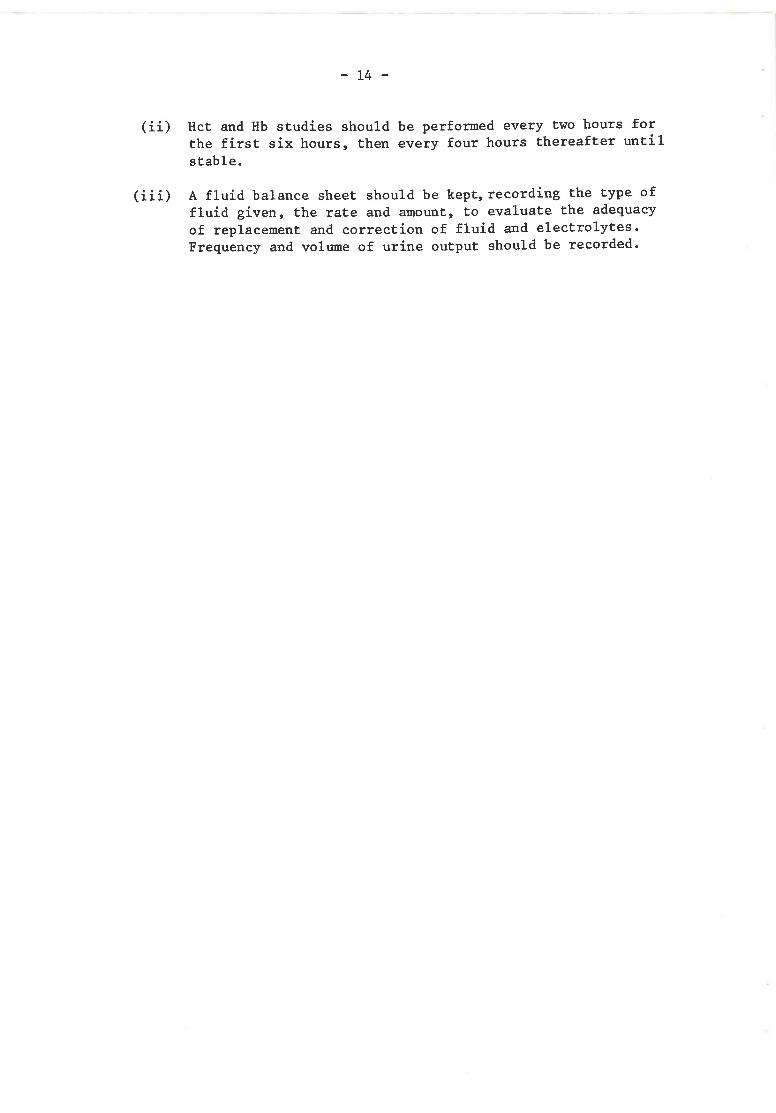
(ii) Hct and Hb studies should be performed every two hours for the first six hours, then every four hours thereafter until stable.
(iii) A fluid balance sheet should be kept,recording the type of fluid given, the rate and amount, to evaluate the adequacy
of replacement and correction of fluid and electrolytes. Frequency and volume of urine output should be recorded.

- 15 -
4. LABORATORY DIAGNOSIS
4.1 Serological tests 4.2 Isolation of virus
Serological tests offer the simplest and most rapid method of confirming a clinical diagnosis of dengue infection. They are also utilized for epidemiological investigations. However, their interpretation may often be difficult. Isolation of virus is the most reliable method although presently available techniques are still complicated and require time.
4.1 Serological tests
a) Collection of specimens
Physicians should be informed of the appropriate conditions for collecting specimens:
blood samples should be drawn from suspect DHF cases on hospital admission or attendance at clinic or as soon as possible thereafter; and on discharge, or shortly before, and, if possible, 14-21 days after disease onset;
an abbreviated case history should accompany specimens, to include, at a m~n~mum: name, address, age, sex, date of onset of illness, date of hospitalization, and brief clinical findings.
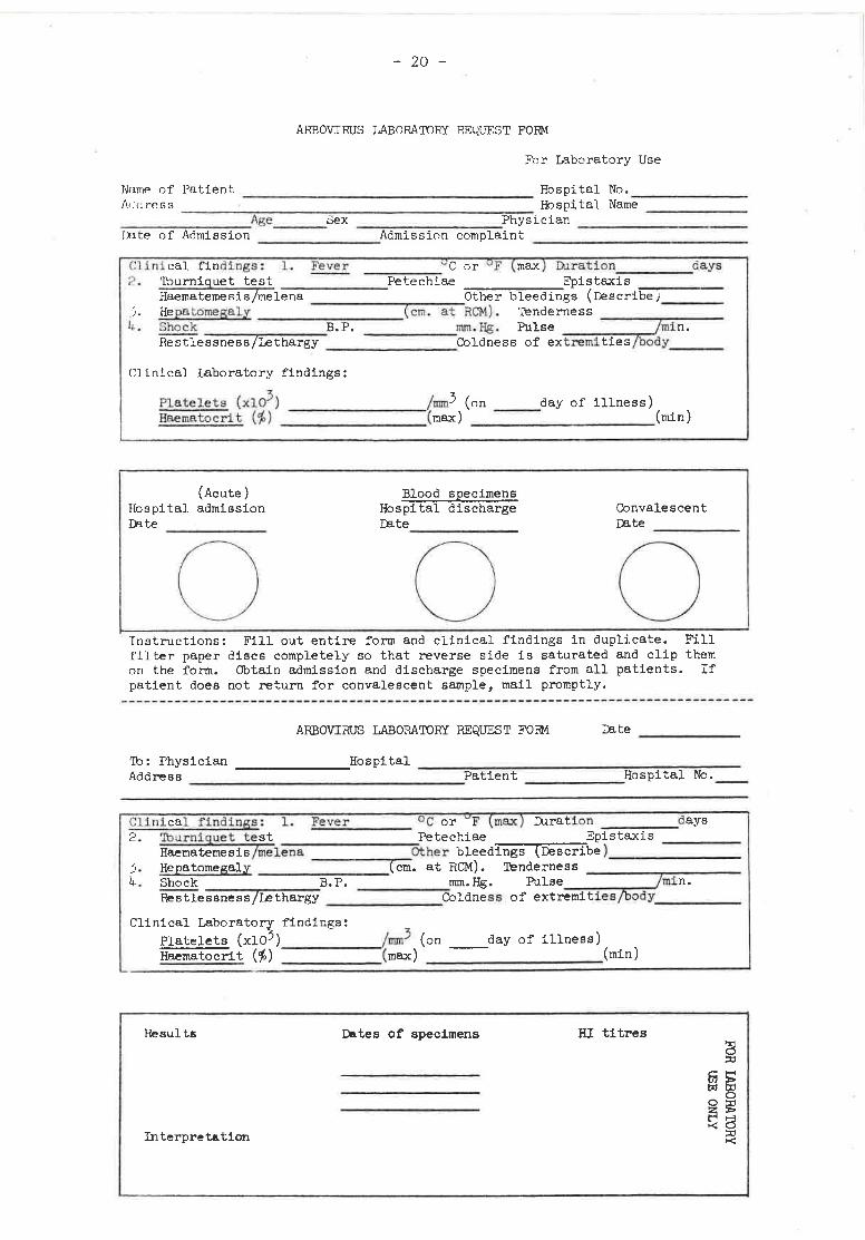
A model of a suitable request form for arbovirus laboratory examination is given on page 20.
Two methods can be utilized for collecting specimens:
(i) Blood collection in tubes or vials
2 to 5 ml of venous blood are collected aseptically, preferably in a heparinized container (if blood is drawn soon after onset it can also be used for isolation of the virus).
Use adhesive tape and pencil, indelible ink, or typewriter to label the container. Name of patient, identification number and date of collection must be indicated.
Tubes or vials with screw caps are preferable. Fix the cap with adhesive tape, wax or other sealing material to prevent leakage during transport.
Ship to laboratory on wet ice as soon as possible. If shipment is delayed, keep in a refrigerator at +4° to +10°C but do not freeze as haemolysis would be an inconvenience for complement fixation.

- 16 -
(ii) Blood collection on filter paper
With a pencil, write patient's initials or number on two or three discs or strips of standardized absorptive paper. 1
Collect fingertip blood (or venous blood in a ~~e) onto
filter paper and saturate through to reverse side.
Discs or strips are allowed to dry protected from flies.
After dessication, place in plastic bagsandstaple to the laboratory examination request form. Store without refrigeration.
Elution of filter paper blood
Elute the disc at 4°C overnight in 1 ml of 12.5 per cent kaolin-borate saline, pH 9.0, in a 12x75 test tube.
In the morning, place the tube at room temperature for 20 minutes, shaking periodically.
Centrifuge for 30 minutes at 2,000 rpm.
For HI tests using goose erythrocytes, without removing kaolin add 0.05 ml 50 per cent goose cells to the tube, shake, without disturbing the pellet, and incubate at 37°C for 30 minutes.
Add 1 ml borate saline, pH 9.0, to the tube.
Centrifuge at 2,000 rpm for 10 minutes and decant supernatant.
This is approximately equivalent to a 1:30 serum dilution.
Each laboratory should standardize the correlation between results with
venous blood and the filter paper disc technique in the same individuals.
b) Haemagglutination inhibition test (HIT)
Haemagglutination inhibition has been the serological test most widely
used. The recommended technique is that of Clarke and Casals adapted to
microtitre equipment and which is described in most manuals.
1
Sera should be extracted with kaolin or with acetone to remove non-specific inhibitors and then absorbed with goose RBC to
remove non-specific agglutinins.
e.g. Whatman No. 3 filter paper, discs of 1/2" or 12.7 mm diameter.

- 17 -
The mode of preparation of haemagglutinating antigen described by
Clarke and Casals is the most satisfactory but its extraction with
acetone is laborious and expensive. A crude 20 per cent suckling
mouse brain suspension in borate saline at pH 6.4 from which gross
debris has been removed by 30 minutes of centrifugation at 3,000 rpm
is stable for 4-6 months and has proved satisfactory.
Paired (acute and convalescent) sera should be tested in the same
run initially using 4 - 8 haemagglutinating units of a single
broadly reactive antigen (usually dengue 1 or 2). If the paired
sera have no antibody or do not show a significant antibody rise,
both specimens may be retested against all four dengue types.
Where chikungunya virus is known to be endemic, all paired sera
should be tes ted agai nst thi s antigen . Where chikungunya virus
is not known to be pres ent, - oniy a sma1Cproporfiori ofthe- sera- - -
need to be test eo aga-~nst:-tbi-sanHgen-:- - ---- --- ·· - - ------------ --- - ·
Known positive and negative sera should be included in each test to
standardize results and maintain quality control. Standard
reference sera are available from WHO and should be included in the
test periodically for quality control. 1
c) Other serological tests
(i) Complement fixation test (CFT)
The CFT may also be used in serological diagnosis wherever
facilities for this test exist. Blood taken on filter paper is unsuitable
because it is lysed.
The CFT is useful since only anti-dengue IgG fixes complement with
dengue antigens. The presence of CF antibody in early convalescent sera
signifies secondary type immune response.
(ii) Neutralization tests
A variety of neutralization tests has been developed to measure
dengue antibody. The 50 per cent endpoint plaque reduction neutralization
test (PRNT) in LLC-MK2 cells has been widely used and is well described in
the literature. Following primary dengue infections, relatively monotypic
neutralizing antibodies are detected during early convalescence.
Following secondary dengue infections, high titred neutralizing antibody is
produced to two, and, most frequently, all four dengue types.
d) Interpretation of results
Identification of infecting virus by serological tests is often
difficult in persons who have had a previous infection with other viruses
of the same serological group as dengue viruses, formerly called Group B
1 Upon request to the Virus Diseases Unit, WHO, Geneva. A limited supply
of inactivated dengue type 2, chikungunya and Japanese B encephalitis
antigen is also available.

- 18 -
arboviruses and now called Flaviviruses. High titres of antibodies can
be found to antigens from several viruses in the group. In instances when
sequential infection consists of a non-dengue flavivirus and a dengue virus,
lilt• lgM serum fraction may contain antibody specific to the second infecting
virus.
(i) Primary antibody response
The primary antibody response to dengue infection is characterized
by slow evolution of HI antibody which is often relatively monotypic,
absence of CF antibody until at least two weeks after onset of illness, and
monospecific neutralizing antibody. Definitive characterization of primary
response is established by demonstrating rising titres of anti-dengue IgM.
In practice, dengue HI antibody titre is generally less than 1:20
1n serum obtained before the fourth day after onset of illness. There is a
fourfold or greater increase in titre in convalescent specimens (1-4 weeks
after onset), with antibody titre not greater than 1:1280.
(ii) Secondary antibody response
The secondary antibody response is characterized by a rapid
evolution of HI and CF antibody. All antibodies are broadly reactive.
Definitive characterization of a secondary response is established by
demonstrating rising titres of anti-dengue IgG.
Evidence of recent infection. In practice, HI antibody to
dengue antigen(s) is less than 1:20 in serum obtained before the fifth day
of illness with response equal to or higher than 1:2560 in convalescent
serum, or HI antibody at least 1:20 in serum obtained before the fifth day
after onset of illness, with rise to ~1:2560 in convalescent serum.
Presumptive recent infection. HI antibody 1:1280 or greater
1n acute specimen without fourfold or greater antibody rise in convalescent
specimen.
(iii) The interpretation of dengue antibodies may be summarized
as follows : 1
1
First specimen
Before 4th day < 1:20
Before 5th day < 1:20 ~ 1:20
Before 7th day ~1:1280
Second specimen
After 1-4 weeks
~ 4x and less than 1:2560
~ 1:2560 "
< 4x
Interpretation
Primary response
Secondary response
Presumptive recent secondary response
These criteria were developed from an extensive experience with virologi-
cally and immunologically studied patients with dengue infections at the U.S.
Army Bangkok Laboratory. If the sensitivity of the HI test system in (continued foot of page 19)

- 19 -
To interpret high-fixed titres, laboratories should establish
baseline titres for the local population taken at , a point of little or no
dengue transmission. Titres more than twice the standard deviation of the
geometric mean may be presumed to indicate recent dengue infection.
4.2 Isolation of virus
The flow chart in Table 5 shows several methods that can be used for
isolation of dengue viruses. Material for isolation should be kept at
+4° to +8°C for a short period of time or frozen for a longer period of
storage. In the latter case it should be maintained without risk of thawing.
Wherever possible, original materials (viraemic serum or infected mosquito
pools) as well as laboratory passage strains should be preserved for future
study. 1
individual laboratories is standardized to that of WHO Reference Laboratories,
the use of the recommended criteria should correctly classify the majority of
dengue infections.
1 WHO should be contacted concerning facilities for the storage of dengue
viruses.
- 20 -
ARBOVIRUS LABORATORY REQUEST FORM
For Laboratory Use
Name of Patient ------------------- Hospital No.---------1\c!d ress Hospital Name ~~-~~--~--Age ______ Sex --~---------Physician Inte of Admission Admission complaint -----------------------
Cli ni cal findings: l. Fever " C or Duration days ~ - 'Iburniquet test Petechiae Epistaxis .,......~----
Haematemesis/mel_e_n_a_________ Other bleedings (Describe; _____ _ -). He pa tomegaLy (cr.l. at RCM) . Tenderness-------....-------4 . Shock B.P. mm . Hg. Pulse /min.
Restlessness/Lethargy Coldness of extremities/cody _____ _
CJinical Laboratory findings:
Platelets (xlo3) Haematocri t (i)
/mm3 (on ___ day of illness) -------(max) __________ (min)
(Acute) Blood s12ecimens Hospital admission Hospital discharge Convalescent Late r.e.te r.e.te
0 0 0 Instructions: Fill out entire form and clinical findings in duplicate. Fill filter paper discs completely so that reverse side is saturated and clip them on the form. Obtain admission and discharge specimens from all patients. If patient does not return for convalescent sample, mail promptly.
ARBOVIRUS LABORATORY REQUEST FOIM I'.e.te ------
'Ib: Physician -----------Hospital --------------------~~~~~~----Address ---------------------------Patient __________ Hospital NO.
Cl inical findings : l. Fever furation days 2. 'Iburniquet test Epistaxis --------
Haematemesis/melena Other bleed.~i-n_g_s~(~De--s_c_r~ibe) ___________ _ ). He12atomegaly --------... (-em-. at RCM). Tenderness - ------------4. Shock B.P. mm.Hg. Pulse /min.
Restlessness/Lethargy Coldness of extremities/body ______ _
Clinical Laborator~ findings: Platelets (xlO ) _______ /mm3 (on __ day of illness) Haernatocrit (1>) (max) (min)
Results Dates of specimens HI titres ~ !l:l
a; ~ 1;:1:1 0
@ ~ ~ ~
!l:l >< Interpretation

- 21 -
5. VECTOR SURVEILLANCE AND CONTROL
5.1 Surveillance 5.2 Long-term control 5.3 Emergency control 5.4 Training
In most instances the most important, and often the only, vector of
dengue haemorrhagic fever is Aedes aegypti. This vector should thus be
the main target of surveillance and control activities wherever it occurs.
Other vectors should only be considered where there is good evidence that
they play an epidemiologically significant role in the transmission of the
disease.
5.1 Surveillance
A number of indices have been described and are currently used to
monitor Ae. aegypti populations in terms of dengue transmission. The most
significant of these indices are tho se related to the abundance of adult
mosquitos expressed either as landing rates or as indoor resting dens i ty.
Whenever adult collections cannot be routinely made to ensure·an acceptable
degree of monitoring, ovi traps can be used as a complementary method.
Should the house index1 and/or Breteau index2 be used, the definition of a
house should be one unit of accommodation irrespective of the number of
people the rein .
Ae. aegypti has a short flight range and a very large number of catching
stations would be required to provide an accurate monitoring of the areas at
risk. As this cannot usually be done, the best alternative is to concentrate
the monitoring on high risk areas as determined by past experience and/or
environmental conditions. Special attention should also be given to areas
where control activities are carried out to evaluate their effectiveness
and develop remedial measures as appropriate.
In a crowded area, many people living within the short flight range of
the vector from its breeding source could be exposed to transmission even
if the house index is low. Distances between houses may be of epidemiological
significance especially in areas with single-storey dwellings. In multi
storey dwellings the population per unit area could be higher than in slum
areas. Thus survey data for single-storey and multi-storey dwellings should
be kept separate.
Emergency control measures are based on insecticide applications and it
is essential to monitor periodically vector susceptibility to the insecticide
most widely used for control operations, i.e. temephos (Abate) and malathion.
Eggs can be sent to appropriate WHO collaborating laboratories for
susceptibility testing when local facilities are not available.
1 Percentage of houses positive for larvae.
2 Number of positive containers per 100 houses.

- 22 -
In areas where Ae. aegypti is absent or very scarce and dengue outbreaks
occur, a special effort should be made to identify the local vector(s) and
develop vector surveillance and control methods accordingly.
5.2 Long-term control
Long-term control should be based on health education and community
part1c1pation, supported by legislation and law enforcement wherever socio
economic conditions permit. This should be facilitated by provision of an
adequate water supply to the communities concerned. Larvicides should be
considered as a complementary measure, and temephos 1% sand granules can
be applied at a target dose of 1 ppm (e.g. 10 g material to 100 litres of
water), especially in high risk localities before periods of anticipated
outbreaks.
The community should part1c1pate by undertaking the disposal
of all water containers which are no longer used (e.g. old tyres, empty tins
and bottles, broken jars, etc.) and by routinely changing the water in
flower vases once a week. Water storage containers which can be moved
should be turned upside down before refilling with water.
Water jars and drums which cannot be disposed of should be adequately
covered to prevent egg laying by Ae. aegypti, or cleaned and scrubbed weekly.
When this is not possible due to their shape or size, Aedes larvae should be
eliminated by transferring the water from one container to another by
filtering it through a cloth.
In certain areas, where vectors other than Ae. aegypti are present,
coconut shells and husks can be buried or burned-,-tree holes can be filled
with sand or cement, leaf axils can be punctured and the tops of bamboo
fences altered to prevent accumulation of water and mosquito breeding.
Large water tanks with taps in coastal areas without a piped water supply
should be covered so as to allow rainwater to enter, but not egg-laying
mosquitos.
Since DHF is an ever present threat, health education should be built
in progressively, beginning at schools and continuing throughout life,
based on simple but accurate information and using all available media
(school books and lectures, newspapers, radio, pamphlets, etc.). While
health education should aim to cover the whole population, more specific
information efforts should be directed towards key components of the
community such as public health service staff and teachers. This health
education should cover vector-borne diseases in general with special
emphasis on DHF wherever this constitutes a major problem.
5.3 Emergency control
a) To be effective, operations must begin when the first few cases
are detected or when there are sound reasons to anticipate an outbreak.
b) The size of area (or areas) to be treated should be determined
through epidemiological and entomological information. If cases are
scattered, adulticide space spraying should be implemented within a minimum
distance of 100 metres radius from houses having cases.

- 23 -
c) Two adulticidal treatments should be made at 10-day intervals
if resources are available or if they can be obtained. Vehicle-mounted
or portable ULV aerosol generators or mist blowers can be used to apply
a suitable insecticide (e.g. technical malathion or fenitrothion) at
above one-half liter per hectare.
d) It is suggested that moderate s1ze cities have at least one
vehicle-mounted aerosol generator, five mist blowers, 10 swing fogs and
1,000 liters of ULV insecticides in order to be prepared to carry out
rapidly adulticidal operations over a 20-kilometer-square area. With
limited funds, such equipment and insecticides can be stockpiled 1n one
city for rapid transportation to other areas when required.
e) Priority areas for vector control are those having a concentration
of cases and/or a high vector density. Special attention should be focused
on areas where people congregate, e.g. hospitals and schools.
f) If necessary, ULV spraying by local air force C47 aircraft or
smaller agricultural type planes and helicopters can be explored.
g) Hospital rooms with DHF patients should be mosquito-proof.
5.4 Training
Medical officers and personnel who see DHF cases should receive short
training courses on vector biology and ecology so that they can inform the
community of the measures they can take to reduce vector breeding and
biting.
Due to the expense and shortage of trained manpower to maintain
special Aedes control teams, health inspectors and related personnel should
be given national training courses on Aedes surveillance and control to
enable them to participate in control operations.

- 24 -
6. EPIDEMIOLOGICAL SURVEILLANCE, CONTROL, AND PREVENTION
6.1 Factors enhancing the risk of occurrence of dengue haemorrhagic fever outbreaks
6.2 Surveillance of dengue haemorrhagic fever 6.3 Control of outbreaks 6.4 Prevention of dengue haemorrhagic fever 6.5 Exchange of information
The objectives of a surveillance programme for DHF are: (1) early
detection of outbreaks, thus permitting a prompt application of control
measures, and (2) monitoring all the important factors that favour an
outbreak of the disease. To accomplish these, impro~ed diagnosis and reporting, use of reliable diagnostic criteria, as well as epidemiological
and entomological surveillance are needed.
6.1 Factors enhancing the risk of occurrence of dengue haemorrhagic fever outbreaks
The occurrence of DHF outbreaks is obviously linked to the density of
mosquito vectors and particularly that of Aedes aegypti. The precise
population density of Ae. aegypti which will sustain dengue virus epidemically
or endemically in a community has not yet been determined successfully.
However, a small number of active biting female mosquitos can easily infect
an entire household. The potential for virus transmission is also enhanced
by the density of human populations. Uncontrolled urbanization in tropical
areas has resulted in the proliferation of Ae. aegypti in densely populated
areas.
Dengue viruses move with people, particularly children. Aedes aegypti
has a short flight range and is less important as a means of transportation
of viruses within cities than man. Schools may be important sites of
dissemination of dengue viruses. Aedes aegypti bite during the day, and
school children bitten by infected mosquitos may take the virus home to
other parts of the city. A second institution which can spread dengue
viruses is the hospital. Visitors and patients with other diseases may be
bitten by infected Aedes aegypti in hospitals.
Transportation of dengue viruses to smaller ·to~s and rural areas is
possible from large cities where the disease is epidemic or endemic. The
factors of dengue transmission and maintenance in smaller towns are not well I
understood.
In most places there seems to be a distinct seasonal pattern in DHF
outbreaks. In subtropical regions north and south of the equator, monsoon
weather patterns prevail. It has been observed in Bangkok, for example~
that several months following the cessation of the rains, usually during
cool dry weather, DHF hospitalization rates decline. The seasonal decline
in dengue transmission is not well understood. It may be related to a
decrease in biting activity, a decrease in longevity of female mosquitos,
or both, and possibly to a small decrease in vector population. Whatever
the cause, the phenomenon is real. During seasonal lows, virus transmission
is most likely to occur only in areas of high transmission potential.

- 25 -
Typicatly, DHF cases have been associated with secondary infections and, occasionally in infants and a few children, with primary dengue infections. In Asia, DHF cases are seen in areas where a high density of Ae. aegypti and the presence of different types of dengue viruses cause multiple infections in children, as evidenced by a typical pattern of secondary (anamnestic) serological response. The hypothesis that DHF is associated with a secondary infection has been put forward from clinical and seroepidemiological studies in Thailand.
6.2 Surveillance of dengue haemorrhagic fever
Surveillance 1s indicated in all endemic areas as well as "receptive areas", defined as areas where Ae. aegypti is known to be present. Undoubtedly, the mere presence of this mosquito is not sufficient in itself to create potential conditions for a DHF outbreak; the biting rate, longevity and population density of the vector must be crucial factors. However, until the density level above which the situation is ready for mass transmission of dengue virus is known, the presence alone of Ae. aegypti is sufficient to declare an area as "receptive".
It is important to understand that, as a general rule, a great amount of silent dengue infection has preceded DHF epidemics. It has been estimated that between 150 and 200 mild or silent dengue infections occur for each case of shock seen in hospital. An understanding of this iceberg phenomenon is essential to the planning of control and prevention programmes of DHF.
Most dengue infections of young children are mild and difficult to distinguish from a common cold or other seasonal fevers. Classical dengue fever is most commonly seen in adults. In areas where dengue viruses are endemic, resident adults are generally immune. The mild nature of the vast majority of dengue infections implies that in endemic areas a fairly high level of dengue transmission may go unnoticed.
Implementation of surveillance is based on the following action:
a) Recognition of cases
Standard criteria for diagnosis should be followed, including clinical laboratory diagnosis (see 2.3, page 7).
b) Confirmation of cases
An important function of a surveillance system is to identify quickly the etiology of presumptive DHF. This should be done by submitting paired sera to a laboratory. For virus isolation, see 4.2, page 19.
c) Prospective surveys
Serological surveys may be used to detect dengue infection when this is relatively silent. They may also give an indication of the presence of specific dengue serotypes. Surveys of patients with fevers of unknown origin and suspect haemorrhagic fever cases should be undertaken, including
examination of paired sera and attempts to isolate the virus.

- 26 -
d) Outbreak investigation
The geographical area affected should be defined rapidly to determine the extent of the insecticide spraying required.
e) Reporting of cases
Cases of DHF should be reported specifying whether with or without shock. Cases and deaths should be reported weekly by geographical area by national health authorities to the Communicable Diseases Adviser in the WHO South-east Asian (New Delhi) and Western Pacific (Manila) Regional Offices. Confirmed and unconfirmed DHF cases by age and sex should be reported annually.
6.3 Control of outbreaks
Two main operations to be carried out simultaneously are mosquito control and treatment of patients in hospitals.
a) Emergency mosquito control
Operations for emergency mosquito control should be carried out as indicated in 5.3, page 22. The objective is to eliminate infected mosquitos and to break the transmission cycle by holding mosquito populations at extremely low levels during the time necessary for viraemic subjects to recover. Complete control of an epidemic may not be feasible if all adult Ae. aegypti cannot be destroyed in the entire affected area. However, a sustained reduction of mosquito populations in selected areas will inevitably result Ln a smaller number of cases.
b) Organization of hospitals during epidemics
Triage. During epidemics, outpatient and inpatient facilities may be overwhelmed and medical care staff can rapidly become exhausted. Under these conditions it is essential that only children requiring hospital care be admitted. A fever and positive tourniquet test are sufficient to produce suspicion of DHF; when possible, a microhaematocrit and platelet count should be done in the Outpatient Department. Patients with thrombocytopenia and elevated haematocrit should be sent to a rehydration ward or, on suspicion of circulatory failure, admitted to hospital. If a patient lives a long distance from hospital and nearby accommodation is not available it may be necessary to admit him for observation. Parents should be carefully instructed to return children promptly to hospital should any danger sign be observed. If properly instructed, triage can be done by paramedical workers. Competent laboratory assistance is essential. Without a laboratory, patients must be evaluated by physical examination. Cool extremities, skin congestion, circumoral cyanosis or a rapid pulse should result in hospitalization. If possible, cases should be hospitalized
for observation or cautioned to remain near hospital until two days after fever terminates.
Intensive care. Patients with a similar degree of severity of illness should be grouped together. Those with shock require intensive 24-hour nursing and physician care. Paramedical workers or parents can assist in oral fluid therapy or in surveillance of the rate of intravenous fluid administration and general status of the patient.

- 27 -
Microhaematocrit determinations are essential in monitoring need for
and success of therapy. IT IS RECOMMENDED THAT ALL INSTITUTES PROVIDING
CARE FOR DHF PATIENTS HAVE MICROHAEMATOCRIT EQUIPMENT AVAILABLE.
c) Other measures
Coordination of DHF emergency control measures can best be implemented
through the establishment of a DHF committee including administrators,
epidemiologists, clinicians, entomologists and virus laboratory represent
atives.
6.4 Prevention of dengue haemorrhagic fever
Prevention of DHF outbreaks in endemic areas is based on surveillance
of virus circulation (see 6.2), together with mosquito surveillance (5.1)
and anti-mosquito long-term control measures (5.2). Contingency planning
should estimate populations at risk and identify the location of equipment,
supplies and personnel which may be required.
Research is being carried out to develop a live tetravalent vaccine
against dengue virus serotypes D1 to D4. Although good progress can be
reported, it will be several years before an acceptable vaccine for mass
use can be available.
6.5 Exchange of information
Rapid exchange of information 1s essential. Narrative epidemiological
reports, results of clinical studies, dengue virus isolation by source and
date, entomological studies, surveys for Aedes vectors, control measures
planned or accomplished, new developments in insecticides and spray equip
ment and other pertinent information will be published in the Dengue
Newsletter for the South-east Asian and Western Pacific Regions. Contribu
tions or requests for copies may be sent to the Project Leader, WHO/VRCRU,
P.B. 302, Jakarta, Indonesia. Copies of the Newsletter are available to all
interested persons.

- 28 -
7. PRIMARY HEALTH CARE
7.1 Recognition of dengue haemorrhagic fever cases
7.2 Management of cases 7.3 Collection of specimens for laboratory examination
7.4 Mosquito control
As has been mentioned ~arlier, DHF tends to spread from large cities
to smaller ones and to villages infested by vector mosquitos, mainly
Aedes aegypti. The case fatality rate of DHF can be considerably decreased
if the appropriate replacement fluid therapy is given early in the course
of the disease. Referral to a well equipped hospital is not always possible.
DHF is therefore a problem for primary health care in endemic rural areas.
Conditions for diagnosis, treatment and control of outbreaks are somewhat
different from what have been set out heretofore. A primary health care
worker, specially trained for this purpose, could be very helpful in the
control of DHF.
7.1 Recognition of DHF cases
An outbreak of DHF should be suspected when:
several children are affected with febrile diseases characterized
by high continuous fever of 2-7 days duration with no response to
malaria treatment;
unexplained deaths occur \vith or without haemorrhage;
patients have petechiae (red spots on the skin) or bleeding from
the nose or the gums, or haematemesis or melena;
when temperature drops, the skin becomes clammy, extremities are
cold and sweaty, and children become drowsy or restless.
7.2 Management of cases
High fever should be treated by sponging. Aspirin (sali cylates)
should not be given.
Oral rehydration must be attempted in early stage of fever with a sugar
and salt solution used for diarrhoeal disease (see footnote page 10) and
repeated small quantities should be given.
If the body temperature drops and cold extrem~t~es occur, with restless
ness, refer promptly to health centre for intravenous fluid administration.
If referral is not possible, enforce oral rehydration until the child urinates
and the skin becomes warm.
7.3 Collection of specimens for laboratory examination
Proof that the outbreak ~s caused by dengue virus must be obtained as
soon as the first suspect cases have been recognized.
Blood films should be taken and sent to the health centre for staining
and examination for malaria parasites and platelet counting.
- 29 -
As indicated in 4.1, blood specimens should be collected on filter
paper discs or strips and sent with clinical data to a specialized laboratory.
Recognition of cases and appropriate collection of specimens can be
facilitated if a member of the community has been designated as "health
communicator" to provide liaison between the community and the health care
group.
7.4 Mosquito control
The disease is transmitted by mosquitos. Local mosquitos should be
identified, as their breeding sites may differ according to species. If this
is not possible it may be assumed that the vector is Aedes aegypti, a mosquito
that bites during the day, rests in houses, and lays eggs in nearby water I
collectors, such as water jars in the house or the yard, old tyres, pots,
discarded bottles and cans, shells, etc.
At the village level, community participation in the control of DHF,
particularly in source reduction of Aedes mosquitos as described in 5.2,
seems to be a feasible and realistic activity which can be carried out through
the primary health care worker.

- 30 -
References
Bharnarapravati, N., Tuchinda, P., Boonyapaknavik, V.
haernorrhagic fever: a study of 100 autopsy cases.
Medicine and Parasitology,~ (4): 500-510 (1967)
Pathology of Thailand Annals of Tropical
Cohen, S.N., and Halstead, S.B. Shock associated with dengue infection.
J. Pediat., ~: 448-456 (1966)
Conference on Dengue Haemorrhagic Fever: New Developments and Future Research,
Singapore, 24-28 October 1977. Asian J. Inf. Dis. jL (1-2), (1978)
Halstead, S.B. Immunological Parameters of Togavirus Infection. In Schlesinger,
R.W. ed. Togaviruses, Academic Press, New York, 1980
Halstead, S.B. for Research.
Dengue Haemorrhagic Fever -- A Public Health Problem and A Field
Bulletin of the Wor ld Health Organization,~ (1): 1-21 (1980)
Halstead, S.B., Nimmannitya, S.,and Cohen, S.N. Observations related to
pathogenesis of dengue haemorrhagic fever. Yale J. Biol. Med., ~ 311-
328 (1969)
Halstead, S.B., Nimmannitya, S., Yamarat, C., and Russell, P.K. Haemorrhagic
fever in Thailand. Newer knowledge regarding etiology. Jap. J. med. Sci.
Biol., 20, Supplement, 96-103 (1967)
Halstead, S.B. Mosquito-borne Haemorrhagic Fevers of South and South-East Asia.
Bulletin of the World Health Organization,~ (1): 3-15 (1966)
Mosquito-borne Haemorrhagic Fevers of South-East Asia and the Western Pacific.
Memoranda. Bulletin of the World Health Organization, ~(1): 17-103 (1966)
Nimmannitya, S., Halstead, S.B., Cohen, S.N., and Margiotta, M.R. Dengue and
chikungunya virus infection in man in Thailand, 1962-1964. Amer. J. trop.
Med. Hyg., 18: 954-971 (1969)
Nimrnannitya, S., and Mansuwan, P. Comparative clinical and laboratory findings
in confirmed dengue and chikungunya infection. Bulletin of the World Health
Organization, 35: 42-43 (1966)
Pathogenetic mechanisms in dengue haemorrhagic fever: Report of an international
collaborative study. Bulletin of the World Health Organization,~: 117-
133 (1973)
Phurnara, T., and Na-Nakorn, S. Hemostasis in Thai haemorrhagic fever.
Bulletin of the World Health Organization, 35~ 45-46 (1966)
Schlesinger, R.W. Dengue Viruses. Virology Monographs, Vol. 16. Springer
Verlag, New York, Vienna, 1977
Suwanik, R., et al. Plasma volume and other fluid space studies in Thai
haemorrhagic fever. J. Med. Ass. Thail~, 50: 48-66 (1967)
Weiss, H.J., and Halstead, S.B. Studies of hemostasis in Thai haemorrhagic
fever. J. Pediat., 66: 918-926 (1965)

TABLE 1
Observed frequency of findings in classical Dengue fever in adults and Dengue and Chikungunya infections in Thai children diagnosed as haemorrhagic fever
Manifestations ~lassical Dengue1 Chikungunya p.n adults Fever
b'ever ++++ ++++
Positive tourniquet test ++ +++
i>etechiae or ecchymosis + ++
Confluent petechial rash 0 0
Hepatomegaly 0 +++
Maculopapular rash ++ ++
Myalgia/arthralgia +++ ++
• Lymphadenopathy ++ ++
Leucopenia ++++ ++++
~hrombocytopenia ++ +
Shock 0 0
Gastrointestinal bleedings 0 o·
1Data from Halstead et al. Mainly Caucasian adults.
Frequency of observation - + ++
+++ ++++
l -26 51 -76 -
25% 50% 75%
100%
Dengue ( D!-il<')
++++
++++
++
+
++++
+
+
++
++
++++
++
+
I '
!
' ' ;
I
I
' I
I i I I
; ' I
i
i
l
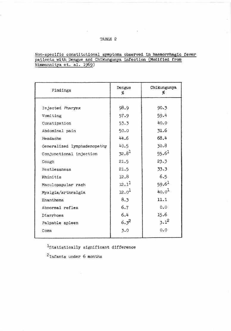
TABlE 2
Non-specific constitutional symptoms observed in haemorrhagia fever patients with Dengue and Chikungunya infection (Modified from Nimmannitya et. al. 1969)
Findings Dengue
%
Injected Pharynx 98.9
Vomiting 57.9 Constipation 53.3 Abdominal pain 50.0
Headache 44.6
Generalized lymphadenopathy 40.5
Conjunctional injection 32.81
Cough 21.5
Restlessness 21.5
Rhinitis 12.8
Maculopapular rash 12.11
Myalgia/arthralgia 12.01
Enanthema 8.3
Abnormal reflex 6.7
Diarrhoea 6.4
Palpable spleen 6.32
Coma 3.0
1statistically significant difference
2rnfants under 6 months
Chikungunya %
90.3
59.4 40.0
31.6
68.4
30.8
55.61
23.3
33.3
6.5
59.61
4o.o1
11.1
o.o
15.6
3.12
o.o

TABlE 3
Major manifestations used as criteria for diagnosis of Dengue haemorrhagic fever - V .S. Chilrungunya infection (Modified from Nimmannitya et. a1. 1969)
Manifestations Dengue Chilrungunya % %
Rever - duration 2 - 4 days 23.6 62.5
5 - 7 days 59.0 31.2
) 7 days 17.4 6.3
Haemorrhagic manifestations.
positive tourniquet test 83.9 77.4
Petechiae scattered 46.5 31.3
Petechial rash ( confl. ) 10.1 0.0
Epistaxis 18.9 12.5
Gum bleeding 1.5 o.o
Melena/hematemesis 11.8 o.o
Hepatomegaly 90.0 75.0
Shock 35.2 o.o
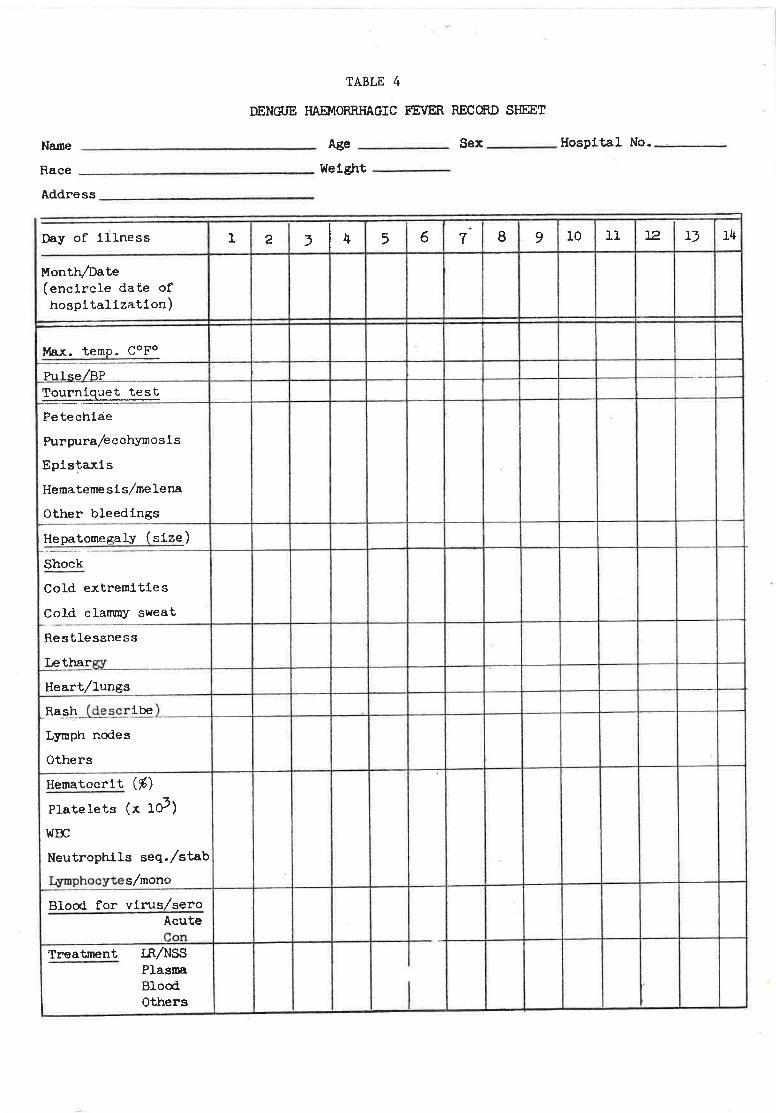
TABLE 4
DENGUE HAEMORRHAGIC FEVER RECORD SHEET
Name Age-----
Race --------------- Weight -----Address ________________ _
Day of iilness 1 2 3 4 5 6
Month/Date (encircle date of hospitalization)
Max. teme. copo
Pulse/BP Tourniguet test ~·
Petechiae
Purpura/ecchymosis
Epis~axis
Hematemesis/melena
Other bleedings
Hepatome s;aly ~size)
Shock ----Cold extremities
Cold clammy sweat f- ·
Restlessness
Lethargy
Heart/lungs
t-Ra_l:!~(descr.ibe)
Lymph nodes
Others
Hematocrit (%)
Platelets (x lo3)
woc Neutrophils seq./stab
Lymphocytes/mono
Blood for virusLsero Acute Con
Treatment IR/NSS Plasma Blood I Others
Sex ____ Hospital No.__,_. __ _
. 7 8 9 10 11 12 13 14

SOURCE
Living patient
Autopsy
Vector mosquitoes
Inoculation
Observation
Passage material
Identification
TABLE 5. FLO\.: CHART FOR DENGUE VIRUS ISOLATIONS
~1ATERIAL FOR INOCULATION PREPARATION
Blood, serum, plasma leucocytes Clot homogenized, serum or plasma undiluted and 1:5 or 1:10.
Various tissues
0 + Mosquitoes
Tissue homogenized as 10-20% W/V suspension in buffer with 1% protein stabilizer and antibiotics. Centrifuge at 3000 rpm 15 minutes. Use undiluted and 1:10.
Homogenized as approximately 10% W/V suspension.
INOCULATION SYSTEMS
A --------------~ 0.2-1.0 pl intrathoracic to mosquitoes Aedes aegypti ~ Ae. albopictus ~
0 Toxorhynchites d' or +
J-(a) Presence of antigens in salivary glands by immunofluorescence (IF) (b) Use of mosquito tissues as CF antigen in typing tests
~ Further passage usually unnecessary unless strain establishment required
I
B
Various cell cultures, e.g. BSCl, vero, LLC-MK2, Ae. albopictus
(a) CPE in cells (b) Plaque formation (c) IF or PAP staining of
infected foci
(a) Cells and/or supernatant (b) Plug of agar + plaque
'V
c 0.01-0.02 ml I.e. to suckling m1.ce < 48 hrs old
\~
(a) Presence of antigen 1.n brain by IF on day 7 (b) Abnormal behaviour (c) Presence of antibody in serum 3-4 weeks after inoculation
.J,; Brain as 10% W/V suspension
I Passage in same system and in other systems
Plaque neutralization test using monotypic monkey serum as reference method.
if
Memb~rs
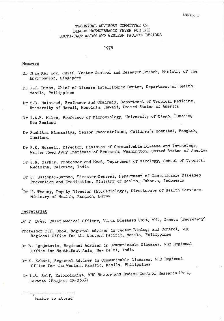
TECHNICAL ADVISORY COMMITTEE ON DENGUE HAEMORRHAGIC FEVER FOR THE
SOUTH-EAST ASIAN AND WESTERN PACIFIC REGIONS
1974
ANNEX I
Dr Chan Kai Lok, Chief, Vector Control and Research Branch, Ministry of the
Environment, Singapore
Dr J.J. Dizon, Chief of Disease Intelligence Center, Department of Health,
Manila, Philippines
Dr S.B. Halstead, Professor and Chairman, Department of Tropical Medicine,
University of Hawaii, Honolulu, Hawaii, United States of America
Dr J. A.R. Miles, Professor of Microbiology, University of Otago, Dunedip,
New Zealand
Dr Suchitra Nimmanitya, Senior Paediatrician, Children's Hospital, Bangkok,
Thailand
Dr P.K. Russell, Director, Division of Communicable Disease and Immunology,
Walter Reed Army Institute of Research, Washington, United States of America
Dr J.K. Sarkar, Professor and Head, Department of Virology, School of Tropical
Medicine, Calcutta, India
Dr J. Sulianti-Saroso, Director-General, Department of Communicable Diseases
Prevention and Eradication, Ministry of Health, Jakarta, Indonesia
Dr U. Thaung, Deputy Director (Epidemiology), Directorate of Health Services,
Ministry of Health, Rangoon, Burma
Secretariat
Dr P. Bres, Chief Medical Officer, Virus Diseases Unit, WHO, Geneva (Secretary)
Professor C.Y. Chow, Regional Adviser in Vector Biology and Control, WHO
Regional Office for the Western Pacific, Manila, Philippines
Dr B. Ignjatovic, Regional Adviser in Communicable Diseases, WHO Regional
Office for South~East Asia, New Delhi, India
Dr K. Kobari, Regional Adviser in Communicable Diseases, HHO Regional
Office for the Western Pacific, Manila, Philippines
Dr L.S. Self, Entomologist, WHO Vector and Rodent Control Research Unit,
Jakarta (Project. IR-03o6)
.,...
Unable to attend
Members
TECHNICAL ADVISORY COMMITTEE ON DENGUE HAEMORRHAGIC FEVER FOR 'mE
SOUTH-EAST ASIAN AND WESTERN PACIFIC REGIONS
1975
ANNEX II
Dr Chan Kai Lok, Chief, Vector Cont~ol and Research Branch, Ministry of the Environment, Singapore
t-r Y.C. Chan, Department of Microbiology, Singapore University, Singapore
Dr J.J. Dizon, Chief of Disease Intelligence Center, Department of Health, Manila, Philippines
Dr N.P. Gupta, Director, WHO Collaborating Centre for Arbovirus Reference ~nd Research, Virus Research Centre, Poona, India
Dr S.B. Halstead, Professor and Chairman, Department of Tropical Medicine, University of Hawaii, Honolulu, Hawaii, United States of America
Dr J.A.R. Miles, Professor of Microbiology, University of Otago, Dunedin, New Zealand
Dr Suchitra Nimmanitya, Senior Paediatrician, Children's Hospital, Bangkok, Thailand
Dr P.K. Russell, Director, Division of Communicable Disease and Immunology, Walter Reed Army Institute of Research, Washington, United States of Am~·r r~ a
D~ J.K. Sarkar, Professor and Head, Department of Virology, School of Tropical Medicine, Calcutta, India
Sr J. Sulianti-Saroso, Director-General, Department of Communicable Diseas~s Prevention and Eradication, Ministry of Health, Jakarta, Indon~sia
Dr U. Thaung, Deputy Director (Epidemiology), Directorate of Hea:!.th Ser'; · ·es,
Minis~ry of Health, Rangoon, Burma
Seoret..ariat
Dr P. Bres, Chief Medical Officer, Virus Diseases Unit, WHO, Geneva (Secretary)
Professor C.Y. Chow, Regional Adviser in Vector Biology and Control, WHO Regional Office for the Western Pacific, Manila, Philippines
Dr B. Ignjatovic, Regional Adviser in Communicable Diseases, WHO Regional Office for South-East Asia, New Delhi, India
Dr T. Okuno, Regional Adviser in Communicable Diseases, WHO Regional Office
for the Western Pacific, Manila, Philippines
Dr C.P. Pant, Project Leader, WHO Vector and Rodent Control Research Unit, Jakarta (Project IR-0306)
Dr M.J. Simons, Head, WHO Immunology -'ln.d Research Training Centre, Universit.y
of Singapore, Singapore 3

Uembers
TECHNICAL ADVISORY COMMITTEE ON
DENGUE HAEMORRHAGIC FEVER
1978
ANNEX III
Dr U Thaung, Deputy Director (Epidemiology), Directorate of Health Services,
Rangoon, Burma
Dr Imam Zaghloul Imam, President, Egyptian Organization for Biological and
Vaccine Production, Cairo, Egypt
Dr J.K. Sarkar, Emeritus Scientist and Officer-in~charge, ICMR Virus Unit,
School of Tropical Medicine, Calcutta, India !
Dr Sumarmo, Department of Child Health, University of Indonesia, Jakarta,
Indonesia
Professor K. Fukai, Institute of Infectious Diseases, Osaka University,
Osaka, Japan
Professor J.A.R. Miles, Professor of Microbiology, University of Otago,
Dunedin, New Zealand
Major-General M.I. Burney, Director, National Health Laboratories, Islamabad,
Pakistan
Dr J.C. Baltazar, Associate Professor, Department of Epidemiology and
Biostatistics, Institute of Public Health, University of the Philippines,
Manila, Philippines
Dr V.F. Chan, Associate Professor, Department of Microbiology, Institute of
Public Health, University of the Philippines, Manila, Philippines
Dr Chan Kai Lok, Chief Vector Control Officer, Vector Control and Research,
Ministry of the Environment, Singapore
Dr Chan Yow Cheong, Senior Lecturer, Faculty of Medicine, University of
Singapore, Singapore
Dr Natth Bhamarapravati, Professor and Chairman, Department of Pathology,
Faculty of Medicine, Ramathibodi Hospital, Bangkok, Thailand
Dr Sujarti Jatanasen, Director, Division of Epidemiology, Ministry of Public
Health, Bangkok, Thailand
Dr Suchitra Nimmannitya, Senior Paediatrician, Children's Hospital, Bangkok,
Thailand
Consultants
Dr A.C. Allison, Clinical Research Centre, Division of Cell Pathology,
Harrow, Middlesex, England
Dr S.B. Halstead, Professor and Chairman, Department of Tropical Medicine
and Medical Microbiology, School of Medicine, Leahi Hospital, Honolulu,
Hawaii
Dr P.K. Russell, Deputy Director, Walter Reed Institute of Research, Walter
Reed Army Medical Centre, Washington, D.C., USA
ANNEX III contd.
Secretariat
DrY. Bang, Project Leader, WHO Vector Biology & Contra~ Research Unit No.2, Jakarta, Indonesia
Dr P. Bres, Chief Medical Officer, Virus Diseases Unit, World Health Organization, Geneva, Switzerland
Dr J. Hamon, Director, Division of Vector Biology and Control, World Health Organization, Geneva, Switzerland
Dr G. Torrigiani, Chief Medical Officer, Immunology Unit, World Health Organization, Geneva, Switzerland
Dr L.J. Charles, Sr., Temporary Adviser on Communicable Diseases, WHO Regional Office for the Americas, Washington, D.C., USA
Dr A.H. Taba, Regional Director, WHO Regional Office for the Eastern Mediterranean, Alexandria, Egypt
Dr V.T.H. Gunaratne, Regional Director, WHO Regional Office for South-east Asia, New Delhi, India
Dr C. Kampanartsanyakorn, Regional Adviser on Communicable Diseases, WHO Regional Office for South-east Asia, New Delhi, India
Dr F.J. Dy, Regional Director, WHO Regional Office for the Western Pacific, Manila, Philippines
Dr R. Lindner, Regional Adviser in Communicable Diseases, WHO Regional Office for the Western Pacific, Manila, Philippines
Dr L.S. Self, Regional Adviser in Vector Biology and Control, WHO Regional Office for the Western Pacific, Manila, Philippines
Dr H.R. Husseini, Regional Adviser on Health Laboratory Services, WHO Regional Office for the Eastern Mediterranean, Alexandria, Egypt
Dr A.D. Rajkovic, Regional Adviser in Health Laboratory Technology, WHO Regional Office for the Western Pacific, Manila, Philippines
Observers
Dr T. Kuberski, Epidemiologist, South Pacific Commission, Noumea, New Caledonia
Dr John Woodall, Director, San Juan Laboratories, Center for Disease Control, San Juan, Puerto Rico
Dr Chiriboga, Assistant Secretary for Environmental Health, Department of Health, San Juan, Puerto Rico
Dr Marietta Diaz, Assistant-in-Charge, Dengue Fever Project, Children's Memorial Hospital, Manila, Philippines
Dr H. Penafiel, Assistant-in-Charge, Dengue Fever Project, Children's Memorial Hospital, Manila, Philippines
