haematological and biochemical studies of helminth infected goats
TRANSCRIPT

i
Haematological and Biochemical Studies of
Helminth Infected Goats in South Kashmir
DISSERTATION Submitted in Partial Fulfillment of the
Requirement for the Award of the Degree of
MASTER OF PHILOSPHY
IN
ZOOLOGY
By
Masarat Nizam
Under the Joint Supervision of
Prof. M. Z. Chishti
Supervisor
Professor Emeritus
Centre of Research for Development
University of Kashmir, Sgr.
Dr. Hidayattullah Tak
Co-Supervisor
Sr. Asst. Professor
Deptt. of Zoology,
University of Kashmir, Sgr.
Centre of Research for Development Faculty of Biological Science
University of Kashmir Srinagar – 190006, Kashmir
(NAAC Accredited Grade „A‟ University)
2013

ii
Centre of Research for Development University of Kashmir
Srinagar – 190 006, Kashmir
No: Date:
Certificate This is to certify that the dissertation entitled “Haematological and
Biochemical Studies of Helminth Infected Goats in South Kashmir”
submitted to the University of Kashmir for the award of the Degree Masters
of Philosophy in Zoology, is the original research work of Ms. Masarat
Nizam, a bonafide M. Phil. Research Scholar of the Centre, carried out under
our joint supervision. The dissertation has not been submitted to this
University or to some other University so far and is submitted for the first
time. It is further certified that this dissertation is fit for submission for the
degree of Masters of Philosophy (M. Phil.) in Zoology and the candidate has
fulfilled all the statutory requirements for the completion of the M. Phil.
Programme.
Prof. (Dr.) Azra N.Kamili
Director Centre of Research for Development Head P.G. Department of Environmental Science
University of Kashmir, Sgr.
Dr. Hidayattullah Tak Co-Supervisor Sr. Asst. Professor
Department of Zoology University of Kashmir
University of Kashmir, Sgr.
Prof. M.Z. Chishti Supervisor
Professor Emiretus Centre of Research for Development
University of Kashmir

iii
DEDICATED
TO
MY
BELOVED
PARENTS

iv
Praise be to Allah, the cherisher and the sustainer of the worlds, who
bestowed on me the divine guidance, enough courage, patience to
complete my work.
I owe my debt of gratitude to my affectionate and esteemed supervisor Prof
(Emiretus) M.Z Chishti, Prof. CORD and my co-supervisor Dr.
Hidayattullah Tak, Sr. Asst. Prof, Deptt. Of Zoology for sincere guidance,
encouragement, strong motivation and idea oriented discussion, which has
enabled me to accomplish this dissertation work. I am especially grateful to
them for reading the proofs with meticulous care, correcting many a mistakes
and smoothing the rough edges of this work. They tried their level best to
remove all those hurdles, which could impede my pace of work. I am highly
grateful to them for always keeping my moral and passion very high.
It is my proud privilege to express my sense of thankfulness to Prof. Azra N.
Kamili Head, Deptt. of CORD /Deptt. of Environmental science for
providing me all the necessary infrastructure and technical facilities during
my work, without which, this task could not have been accomplished. I feel
delighted in expressing my sense of thankfulness to all faculty members of
CORD /Deptt. of environmental science especially Prof A.R Yousuf, Prof
A.K Pandit, Prof G.A Bhat and Dr. Ruqaya Nazir for their all time support,
guidance, inspiration, immense encouragement, motivation and timely
suggestions that really need to be acknowledged.
I have to accumulate suitable words in expressing my gratitude and thanks to
Prof. Fayaz Ahmad (Department of Zoology, University of Kashmir) for his
generous help and valuable guidance.
Acknowledgement

v
I am also highly thankful to Dr. Bashir Ah. Lone for the valuable
suggestions and taking all the pains in accomplishing the work. His presence
was a source of inspiration for me throughout my research work.
The other people whom I stand indebted for their moral support and timely
help include technical and non-teaching staff members of Centre of Research
for Development especially Mrs. Bilquis Qadri, Dr. Bilal Ahmad Wani, Mr.
Javid Ahmad, Mr. Farooq Ahmad and Ms. Asfie Jan.
I am highly indebted to all the scholars of the CORD and Deptt. Of Zoology
especially Prof. Khalidah Hassan, Prof. Irfana Jameel, Ummer Rashid,
Suhaib Ah, Sabzar Ahmed, Irfan-rauf, Shabeer Ahmed and Tauseef Ah. who
rendered full cooperation and support during the research programme.
I thank all my friends especially Ms. Sumaira Tyub, Nuzhat Shahi, Maryum
Meraj, Ibraq Khursheed, Nazima Gull, Shazia Ahad, Famida Jan, Sumaira
Maqbool, Nighat-un-Nissa, Baby Habiba, Tabasum yaseen, Aliya Ismat
Baba, Irfana Showqi, Farhana Maqbool, Afeefa Qayoom, Rafia Rashid,
Shafia and Firasat for their valuable suggestions, moral support and pleasant
company. I will always remain indebted to them.
The am also highly thankful to entire team of Al-Khaleel DTP Centre
especially to Mr. Shaukat Ahmad and Mohd. Muzaffar for taking all the
pains during printing and in accomplishing this piece of research with utmost
care.
I feel pleasure to pay sublime obeisance and regards to my esteemed parents
and my family, who extended unique character inspiration, constant support,
motivation and encouragement at every moment of my study. My family
deserves all the credits and accolades earned by the achievement of this
distinction. Their prayers always escorted me to the completion of this
programme.
Masarat Nizam

vi
Chapter
No.
Description Page No.
Certificate Acknowledgement Contents List of Tables List of Figures List of Plates Abbreviations
1. INTRODUCTION 1-8
2. REVIEW OF LITERATURE 9-36
Overview of the Literature of Epidemiological Profile 34-35
Overview of the Literature of Haemato-biochemical Profile 36
3. MATERIAL AND METHODS 37-54
I. Collection and examination of faecal samples of goats for
eggs/larvae
37-40
II. Collection of gut contents and various visceral organs
from the slaughtered goats at various local abattoirs
40-41
III. Processing of Collected Material 41-48
(A) Preparation of permanent slides of
nematodes for study
41-45
(B) Whole mount specimens or Preparation of
permanent slides of Cestodes and Trematodes for
study
45-48
IV. Collection and examination of blood samples 48-53
A). Hematological Parameters 49-52
B). Biochemical parameters 52-53
4. RESULTS AND DISCUSSION 55-83
4.1. Epidemiology of helminth parasites of goats 56-64
4.1.1. Overall Prevalence 56-59
4.1.2. Seasonal Prevalence 59-60
4.1.3. Age wise prevalence 61-62
4.1.4. Gender wise prevalence 63-64
4.2. Haematology and blood biochemistry 64-79
CONTENTS

vii
4.2.1. Hemoglobin (g/dl) 64-66
4.2.2. Packed Cell Volume (PCV) (%) 66-67
4.2.3. Erythrocyte Sedimentation Rate (ESR) (mm/hr) 67-68
4.2.4. RBC Count (106/mm
3) 68-70
4.2.5. WBC Count (103/mm
3) 70-71
4.2.6. Differential Leukocyte Count (DLC) (%) 71-72
4.2.7. Total Protein (g/dl) 75
4.2.8. Albumin (g/dl) 76-77
4.2.9. Globulin (g/dl) 77-78
5. SUMMARY, CONCLUSION AND RECOMMENDATIONS 84-87
BIBLIOGRAPHY 88-108

viii
List of Tables
Table No.
Description
Page. No.s
3.1 Estimation of Total Protein 52
3.2 Estimation of Albumin 53
4.1 Overall prevalence of gastro-intestinal helminth
parasites in goats
57
4.2 Seasonal prevalence of gastrointestinal helminth
parasites in goats
59
4.3 Age wise prevalence of gastro-intestinal parasites in goats
61
4.4 Gender wise distribution of gastro-intestinal helminth parasites in goats
63
4.5 Haematological parameters of uninfected goats 73
4.6 Haematological parameters of helminth infected goats
73
4.7 Differential Leukocyte Count of uninfected goats 74
4.8 Differential Leukocyte Count of helminth
infected goats
74
4.9 Biochemical parameters of uninfected goat 79
4.10 Biochemical parameters of helminth infected
goats
79

ix
Plate No. Description Page No.s
1 Goats grazing at pastures lands 7
2 Organs infected with Helminth Parasites 8
3 Overview of the Material and Methods 54
4 Eggs of various Helminth Parasites 80
5-6 Helminth Parasites 81-82
7 RBC’S and WBC’S of goats 83
LIST OF PLATES

x
Fig. No.s Description Page No.s
4.1 Distribution of nematodes, cestodes, and trematodes
in goats.
56
4.2 Overall prevalence of gastro-intestinal helminth
parasites in goats.
57
4.3 Seasonal prevalence of gastrointestinal helminth
parasites in goats.
60
4.4 Age wise prevalence of gastro-intestinal helminth
parasites in goats.
62
4.5 Gender wise distribution of gastro-intestinal
helminth parasites in goats.
63
4.6 Seasonal variation in Hemoglobin of helminth
infected and uninfected goats.
65
4.7 Seasonal variation in PCV of helminth infected and
uninfected goats.
66
4.8 Seasonal variation in ESR of helminth infected and
uninfected goats.
68
4.9 Seasonal variation in RBC count of helminth infected
and uninfected goats.
69
4.10 Seasonal variation in WBC count of helminth infected
and uninfected goats.
70
4.11 Seasonal variation in DLC count of helminth infected
and uninfected goats.
72
4.12 Seasonal variation in Total Protein content of
helminth infected and uninfected goats.
75
4.13 Seasonal variation in Albumin content of helminth
infected and uninfected goats.
76
4.14 Seasonal variation in Globulin content of helminth
infected and uninfected goats.
77
List of Figures

xi
Abbreviation Full Form
FEC Faecal Egg Count
EPG Eggs Per Gram
Nem. Nematodes
Ces. Trematodes
Trem. Cestodes
Hb Hemoglobin concentration
PCV Packed cell volume
ESR Erythrocyte Sedimentation Rate
RBC Red Blood Cell
WBC White Blood Cell
TLC Total Leukocyte Count
DLC Differential Leukocyte Count
Fig. Figure
SD Standard Deviation
% Percentage
Abbreviations

1
CHAPTER – 1
Introduction
omestic ruminants such a sheep, goats, cattle are among the first animals
to be domesticated by man. Archaeological evidence suggests that sheep
were being raised for wool production as long as 4000 B. C., while goat
remains have been dated to between 6,000 – 7,000 B. C. The ancestor of modern
goat is the Benzor from Asia Minor Middle East.
Goat (Capra), a member of the Bovidae family and subfamily Caprinae is
one of the oldest domesticated species. For thousands of years they have been used
for their milk, meat, hair and skin over much of the world. Female goats are referred
to as does or nannies, intact males as bucks or billies; their offspring are known as
kids. Goat is generally reared to procure meat, milk and skin. Goat is often regarded
as poor man’s cow. The milk of goat is quite similar to that of cow milk and it is
more easily digested because of smaller globules. It is richer in milk content with a
high amount of calcium, phosphorus and chlorine.
The state of Jammu and Kashmir is situated at 32.17° and 36.59° north
latitude and 72.26° and 80.30° east of longitude with total area of 2,22,236 Sq. Kms.
The climate is variable from subtropical (Jammu plains) to temperate (Kashmir
Valley) to temperate cold but arid (Ladakh region). The soil topography, geoclimate,
natural meadows and high land pastures of valley are naturally conducive for goat
production.
Kashmir is primarily an agricultural state and animal treasure is one of the
major sources of earning of farming community and goat farming is an important
source of livelihood for small and marginal farmers and landless labourers as it plays
an important role in providing food, fibre, manure etc. The major advantage of goats
and sheep over other ruminants is to utilize pastures and wastelands to produce meat
and wool. Goat dung is a natural source of organic fertilizer with nitrogen and
D

2
potassium contents double than that of cattle dung, so goat manure is preferable for
increasing the fertility of soil. The rearing of goat had the added advantage of filling
an important ecological niche, being able to graze land on which sheep and cattle
simply cannot thrive.
As per the Livestock Census of Jammu and Kashmir which was carried out
in 2007, total sheep, goats, cattle and buffalo product in Jammu and Kashmir were
36.85, 20.63, 34.43 and 10.51 lakhs respectively in number. It has been estimated
that livestock contributes about 11% to the economy of the state.
The 17th quinquennial livestock census posted 98.993 lakh livestock in the
state which makes density of livestock to be 98 animals per sq. km. of area. When
the indicator of livestock available per thousand of population is adopted, there are
926 animals per thousand of population in J&K state and this figure for all India is
457 animals. Average livestock per household (2001 census households) is 6
animals as against 3 animals for India. The species provides dependable source of
income to 40% of the rural population which are below the poverty line in India.
The goat meat (Chevron) production per year is 3,05,000 million tons (35% of the
total meat production in the country). Its economy is of the order of Rs. 350 crores
annually. The goat rearing farms are, therefore important part of rural as well as
national economy of India.
In our state the local goats and sheep are generally reared in tablelands in
spring and early summer months. In summer and early autumn the goats and sheep
are taken to highland pastures for grazing (Plate 1). In our state ruminant rearing is
so important that it is the only source of income to many tribes. Even name is given
to a tribe on the basis of sheep called Bakerwal. This is a nomadic tribe of our state
migrating from summer grazing Pirpanchal Mountains and low lying hills of Jammu
in winter. Goat rearing is a tribal profession of nomads (Bakerwals, Gaddies) and
many other farming communities in Jammu and Kashmir. Goats contribute to the
subsistence of small holders and landless rural power. They also produce meat, milk,
skins and manure and are also used for transport purposes especially in high
altitudes. A Gaddi breed of goat is able to carry upto 10 kg load on much steep
slopes.

3
There are various diseases which are a major setback to this industry. The
various disease producing organisms are viruses, bacteria, protozoa, helminthes etc.
There are other practices which contribute to low wool and meat production for this
industry. The main contributing factors include large animal production with rapidly
diminishing grazing areas and consequent overstocking, poor nutritional standard
and traditional husbandry practices.
The viral and bacterial diseases are easily diagnosed by their clinical signs
but parasitic infection when less in number or in early stages are without clinical
signs and thus act as one of the major cause of production loss. Faizal (1999)
reported 1/3rd
growth retardation in ruminants due to helminth infections. Herlich
(1978) reported 5-10 % mortality and 10-20 % morbidity due to helminth parasites
in small ruminants. The productivity of sheep (and goats) is constrained by parasitic
infections (Dhar et al., 1982). Helminth infections remain one of the major
constraints to small ruminant production in tropics (FAO, 1992). Surveys indicate
that up to 95% sheep and goats in the tropics are infected with helminths and,
Haemonchus and Trichostrogylus are the main genera involved (Rey, 1991).
Mortality rates in herds may exceed 40% while weight losses 6-12 kg/year/animal
may occur (IEMVT, 1980). However, insidious productivity losses through reduced
feed intake and decreased efficiency in feed utilization, associated with subclinical
or chronic conditions, are often the largest economic losses (Holmes, 1993; Gatongi,
1996). It is estimated that more than 300 species of helminthes parasitize livestock
in India and new species are being frequently discovered (Singh et al., 1977).
The incidence of helminthes infection varies with age, sex, season and agro-
climatic conditions. The higher incidence of parasitic infections in domestic animals
in a grazing system lowers productivity, leading to important economic losses. The
parasite infected animals increase their metabolic rate and reduce the amount of
metabolic energy used for production, as the parasites use their nutrients, damage
some vital organs and cause animals to become more susceptible to other pathogenic
agents (Skykes et al., 1992).
The helminth species which parasitize goats belong to three classes namely
trematodes, cestodes and nematodes and pathogenicity of these helminth parasites

4
also varies with different intensity. Goats are a treasure house of different helminth
parasites like Paramphistomum spp., Fasciola spp., Dicrocoelium spp., Haemonchus
spp., Trichuris spp., Chabertia spp., Dictyocaulus spp., Moneizia spp., and Stilesia
spp.
Mature paramphistomes rarely produce clinical symptoms (Dube et al.,
2003), however immature migrating parasites have been reported causing serious
diseases and even the death of their hosts by burying themselves in the submucosa
of duodenum and feeding on the epithelial cells of brunners glands with result in
anorexia, polydpsia, profuse diarrhoea, drop in plasma protein concentration and
anemia (Buttler and Yeoman, 1962; Singh et al., 1984).
Fascioliasis, is a liver fluke disease caused by several species. The two most
important species in livestock are Fasciola hepatica and Fasciola gigantica.
Fascioliasis causes pathological and necrotic lesions, which results from the parasitic
migration through the liver parenchyma and the bile ducts causing hemorrhages
(Plate 2). The flukes are also, haemophagous and infection results in anemia.
Sinclair (1967) and Hammond and Sewell (1990) reported that Fasciola gigantica is
more pathogenic and causes more production losses than Fasciola hepatica. In small
ruminants, the disease causes severe economical losses because of reduced growth
and productivity, immune suppression, reduced wool and milk production,
condemnation of the livers as unfit for human consumption and sudden death of
heavily infected animals (Boray, 1985; Ngategize et al., 1993 and Mulcahy et al.,
1999). Since, liver is the main metabolic organ in the body, infection of hepatocytes
is an essential feature of certain parasitic infections. In fascioliasis the metabolic
process of the liver is gradually reduced (Fikry et al., 1988). Hepatocytes are active
in controlling levels of blood glucose, lipids and cholesterol and a number of plasma
proteins, including albumin, fibrinogen and prothrombin.
Gastro-intestinal parasitism represents a severe health problem in small
ruminant production system, especially sheep and goats and its consequences can be
extensive ranging from reduced productivity to mortality (Skykes, 1994). It may also
cause body composition changes and rendering the affected animals more
susceptible to concurrent infections (Dominguez-Torano et al., 2000). Gastro-

5
intestinal nematodiasis is a major threat and a primary constraint to sheep
productivity, it endangers animal welfare worldwide (Tariq et al., 2010). The
prevalence of GIN in tropical and sub-tropical areas has adversely affected the
production potential of sheep and goats, leading to countless deaths and insidious
economic losses in livestock sector (Al-Quaisy et al., 1987). One of the main
culprits in ruminant nematodiasis is Haemonchus contortus which causes
haemonchosis, anemia and parasitic gastroenteritis in goats, sheep and cattle (Leiper,
1957). They cause significant economic losses worldwide due to their feeding
behaviour being haematophagous, Haemonchus contortus and Ostertagia ostertagi
suck 0.05ml of blood/worm/day (Soulsby, 1986) (Plate 2). Trichostrongyle infection
(Trichostrongylosis) is much more important as a veterinary problem and causes
pathological conditions like anemia, weight loss, poor wool and milk production and
bottle jaw. Oesophagostomum columbianum produces pathological conditions like
diarrhoea, loss of appetite, emiaciation, weight loss and nodule formation, while
infection of Bunostomum trigonocephalum is associated mainly with anemia and
weight loss. Trichuris ovis, being less pathogenic but anemia, hemorrhage, necrosis,
oedema of caecal mucosa and diarrhoea has been reported in severe infections. The
filarial lungworm causes parasitic bronchitis in goat. In severe infection with
Dictyocaulus, the bronchial epithelium is hyperplasticized and heavily infiltrated by
eosinophils, which sometimes leads to parasitic pneumonia. In heavy infections the
goats may die due to respiratory failure following the development of severe
interstitial emphysema and pulmonary oedema. These nematode infections in
general produce anorexia, reduced feed intake, loss of blood and plasma proteins
into gastro-intestinal tract, alterations in protein metabolism, enteritis, diarrhoea
resulting in reduced body weight gains and wool production and death due to
secondary infections, thus resulting in great economic losses to goat farmers and
goat industry.
Blood is an important and reliable medium for assessing the health status of
individual animals (Oduye, 1976). Serum biochemistry and hematological analysis
have been found to be important and reliable means for assessing an animal’s health
status and might give an indication of the degree of damage to host tissue as well as
severity of infection (Otesile et al., 1991).

6
Prevalence of helminthes in small ruminants being very high. They cause
adverse effects on the host like haematological and biochemical disturbances
(Rasool et al., 1995; Iqbal et al., 1998; Hayat et al., 1996, Hayat et al., 1999), loss of
body weight (Khan et al., 1988) and huge economic losses (Iqbal et al., 1989, Iqbal
et al., 1993).
Detailed information about epidemiology, prevalence, etiology, biology and
pathogenicity of helminthes in goats of Jammu & Kashmir is still not scanty. A lot
of work has been carried out on different aspects of ruminant parasitology, but no
substantial work has been conducted on the haemato-biochemical parameters of the
goats, which is revealed by the absence of any reference from Kashmir. Hence the
present work is aimed at to conduct haemato-biochemical studies of goats of south
Kashmir and to correlate the results with the presence of helminth parasites in these
hosts. A comprehensive work covering the dimensions of epidemiology and hemato-
biochemical parameters of goats for a period of one year from December 2011 to
November 2012 under the title “Haematological and Biochemical Studies of
Helminth Infected Goats in South Kashmir” was thus initiated with the following
objectives.
1) To study the epidemiology of Helminth parasites of goat in South Kashmir.
2) To study the impact of Helminth parasites on haematology of goats of South
Kashmir.
3) To study the impact of Helminth parasites on blood biochemistry of goats of
South Kashmir.

7
Plate 1: Goats grazing at pastures lands

8
Abomassum (Part of intestine) of goat infected with Haemonchus and Ostertagia sp.
Liver (bile ducts) of goat heavily infected with Fasciola sp.
Plate 2: Organs infected with Helminth Parasites

9
CHAPTER – 2
Review of Literature
ince a lot of work has been done in the very important sector of veterinary
parasitology, it is difficult to give a detailed account of the work done;
therefore a brief account of literature available on related aspects of the
present investigation has been critically reviewed and summarized below.
The present study deals with the epidemiology and the haemato-
biochemistry, therefore the literature is presented separately under two
headings.
2.1. Epidemiological review
2.2. Haemato-biochemical review
2.1. Epidemiological Review
Hsiang et al. (1990) studied 4534 faecal samples, collected in Taiwan over a
three year period from randomly selected dairy goats for parasites. The most
frequent parasites found were Oesophagostomum spp. (19%), Haemonchus
contortus (17.3%), Strongyloides papillosus (8.5%), Ostertagia ostertagi (7.1%) and
Trichostrongylus colubriformis (6.8%). Overall prevalence was observed greater in
autumn and winter, and goats with access to pasture were more commonly infected
than goats which fed indoors (penned goats).
Mattos (1991) reported gastro-intestinal nematodes parasitizing ruminants
(cattle, sheep and goat) raised in Oriximina, Brazil. Eight species of parasites were
reported namely, Haemonchus contortus, Haemonchus similis, Trichostrongylus
axei, Trichostrongylus columbriformis, Cooperia curticei, Cooperia punctata,
Oesophagostomum venulosum and Bonostomum trigonocephalum.
Lepojev et al. (1992) studied the gastrointestinal strongyles of goats. They
counted the parasites of abomassum and intestine of 6 goats slaughtered in Radovid,
Yugoslavia. Pal and Qayyum (1992) studied the distribution of gastrointestinal
helminths of goats in Swat valley, Pakistan. 53 gastrointestinal tracts of 53 goats
S

10
from Swat valley were examined at abattoirs from September 1990 to January 1991.
Haemonchus contortus, Ostertagia ostertagi, Ostertagia circumcincta and
Trichostrongylus axei with prevalence of 94.35%, 81.13%, 66.03% and 50.94%
respectively from abomassum and Trichostrongylus colubriformis (73.58%) from
small intestine and Oesophagostomum venulosum (22.64%), Trichuris ovis
(39.62%) and Trichuris globulosa (11.32%) from the large intestine. Thakur et al.
(1992) reported that the prevalence of the parasitic infection was 100% in goats
during the month of July in western Nepal whereas out of 32 samples collected from
Manglapur VDC-2, 76.66% were positive for eggs of these parasites.
Frutschi et al. (1993) on autopsy of 104 small ruminants, 52 sheep and 52
goats, the following gastrointestinal nematodes were identified and counted in order
of predominance. Trichostrongylus spp. (96%), Oesophagostomum columbianum
(82%), Haemonchus contortus (67%), Strongyloides papillosus (55%), Cooperia
spp. (47%) and Trichuris ovis (12%). Hoste and Chartier (1993) studied the impact
of nematode parasitism of digestive tract on milk and milk quality in dairy goats.
The study reported that the high producer goats had less resistance to infection
associated with severe consequences on milk production. The reduction in milk
production was reported with increase of worm load in the goats.
Dorny et al. (1995) studied the Strongyle infections in sheep and goats in
Malaysia. Pattern of Trichostrongyle infections according to season, age, pregnancy
and lactation was studied by faecal egg counts. Haemonchus contortus and
Trichostrongylus spp. were reported to be most important strongyles in sheep and
goats. Ndao et al. (1995) conducted epidemiological survey of gastrointestinal
helminthes in 51 sheep and 51 goats in tree cropping region in Senegal from October
1990 to September 1991. They reported that all animals were infected with at least
one helminth species.
Rafique and Hayat (1997) analysed faecal samples from the rectum of sheep
and goats in the Quetta and Kalat area of Buluchistan, Pakistan for helminth eggs.
87.7% (50 of 57) prevalence was reported. Nematodirus spathiger was the most
frequent parasites (72.7%) followed by Trichuris globulosa (27.3%), Marshallagia
marshalli (6.3%) and Strongylloides papillosa (6.3%). Mixed infections were

11
frequently reported. Vaughan et al. (1997) described two cases of infection with
Fasciola hepatica in young farmed emus with sub-acute and chronic fascioliasis.
The author reported gross lesion of necropsy and hepatic lesions in microscopic
examination.
Gatongi et al. (1998) investigated the epidemiology of Haemonchus contortus
infection of sheep (Red Maasai) and goats (Small East African Goat) in a semi-arid
area of Kenya. Prevalence of Haemonchus contortus was over 90% in both sheep
and goats and this species contributed to about 80% of the total worm burden. Only
about 10% of the hypobiotic larvae were recovered from the mucosal digest whereas
about 90% were recovered from the abomasal contents. Thamsborg et al. (1998)
reported lungworm infection (Dictyocaulus vivzarus) on dairy cattle farms in
tropical highlands of Tanzania.
Valcarceli and Romero (1999) examined 322 gastro-intestinal tracts of
traditionally reared goats originating from a dry area of Central Spain. A large
spectrum of gastrointestinal nematodes was observed with Teladorsagia
circumcinta, Teladorsagia trifurcate being the most prevalent species, followed by
Trichostrongylus vitrinus and Nematodirus filicolis.
Astiz et al. (2000) studied the seasonal distribution and larval shedding
intensity of broncho-pulmonary parasites over two consecutive years using 285
faecal samples obtained from adult goats in Spain. A very high prevalence (81%)
was reported and Muellerius capillaris was the predominant species (present in 98%
of infections). They further reported that Dictyocaulus was not an important
pathological infection in goats. Brunn et al. (2000) studied the cause for clinical
symptoms like coughing, fever and weight loss in Saanen goats in Swiss Alps. On
slaughtering 1st stage larvae were reported from trachea. The larvae were identified
that of Muellaria, Capillaris and Protostrongylylus spp.
Pathak and Pal (2000) collected 88 gastrointestinal tracts of goats from the
slaughter house Supela, Bhilai and were also collected from the Veterinary College,
of Drug district Chhattisgarh and were brought for the postmortem examination
during November 1999 to October 2000. The percentage of overall prevalence of
12
parasitic infection Paramphistomum spp., Cotylophoron spp., Moniezia spp.,
Avitellina spp., Haemonchus spp., Cooperia spp., Oesophagostomum spp.,
Bunostomum spp., and Trichuris spp., were 80.68%, 45.45%, 17.04%, 3.40%,
26.13%, 5.68%, 3.40%, 30.68%, 5.68% and 27.27% respectively. In case of
Paramphistomum, infection was highest in monsoon (91.8%) and lowest in winter
(63.15%). The seasonal prevalence of gastrointestinal parasitic infection in goats
showed that prevalence was highest in monsoon (94.60%), moderate in summer
(87.50%) and lowest in winter (63.15%). Silvestre et al. (2000) investigated
helminth infection, species diversity (proportion of each species in the community),
species number, intensity of infection and antihelminthic resistance in 16 dairy-goat
farms of south-western France. A total of 17 species of helminthes, among which 14
nematodes, one cestode (Moniezia spp.) and two trematodes (Paramphistomum
daubnevi and Dicrocoelium lanceolatum) were recovered in the 26 necropised
culled goats during the study.
Githigia et al. (2001) studied the impact of gastrointestinal nematodes on
health and production of goats in Kenya. The faecal egg counts were found higher
during short rainy season. In all the animals studied Haemonchus contortus was the
nematode recovered during the study. It was concluded that gastrointestinal
helminthes cause production losses, weight loss and mortalities in goats. Jithendran
and Bhat (2001) studied the prevalence of gastrointestinal parasites in
sheep and goats of Himachal Pradesh, India and found the prevalence in sheep and
goats respectively as follows: Fasciola 9.6%, 8.8%; Amphistomes 3.8%, 2.5%;
Dicrocoelium 7.2%, 2.5%; Schistosoma 1.2%, 0.6%; Moniezia 2.7%, 1.3%;
Strongyles 91.6%, 100%; Strongyloides 4.8%, 5.1%; Dictyocaulus 1.2%, 1.3% and
Trichuris 14.3%, 1.3%.
Sharkhuu (2001) performed the Post-mortem examinations of 236 goats from
all provinces in Mongolia for the study of helminths in goats. Thirty-nine helminth
species belonging to three classes, 14 families and 23 genera were found. The
prevalence and intensity of helminth infections were reported for three age groups of
goats in four seasons and three geographic zones in Mongolia. Common helminth
infections of goats in all zones of Mongolia were infections of Ostertagia,

13
Marshallagia and Nematodirus. The highest number of eggs per gram (EPG) of
feces was counted in March (average 1335.3±405.3) and the lowest count was in
November (54±18.6).
Magona and Musini (2002) studied the influence of age, grazing system,
season and agroclimatic zone on the prevalence and intensity of gastrointestinal
Strongyles in Uganda goats and reported that season and agroclimatic zones were
the only significant factors which influenced intensity of nematodiasis in goats.
Mazyzd and El-Nemr (2002) reported the endoparasites of sheep, goats and
Shephered in North Sinai Governorate, Egypt. They revealed an overall infection of
12.7% with Fasciola spp., 12.8% with Moneiza expansa and 4.59% with Trichuris
ovis.
Love and Hutchinson (2003) observed pathology and diagnosis of internal
parasites in ruminants. The purpose of their study was to overview the gross
pathology and diagonosis of gastrointestinal and other parasites in ruminants, with
particular emphasis on the economically important parasites of sheep, goat and
cattle. Regasa et al. (2003-2004) conducted a study on epidemiology of gastro-
intestinal parasites of ruminants in Western Oromia, Ethiopia. The study showed the
overall prevalence of gastro-intestinal parasites as 84.1% in goats. Nematodes of
group Strongyle and Eimeria were most prevalent parasites encountered in this area.
Dhand et al. (2004) reported an outbreak of fasciolasis in sheep and goats in
Punjab. 70 goats and 50 sheep of different age groups were affected and found that
these animals were suffering from high fever with diarrhoea. Among these animals 5
goats and 40 sheep died before the investigation. Fasciola gigantica was recovered
on postmortem examination. Mbae et al. (2004) studied 1106 sheep and goats in
Kenya for nematode infections. Young animals were found more infected than older
ones. The faecal egg counts were significantly higher in wet seasons in both sheep
and goats. Haemonchus contortus was the most predominant nematode parasite
encountered in the study. Sheikh et al. (2004) studied ovine fasciolasis in Kashmir
valley. They examined 1150 faecal samples from endemic and non-endemic and
hilly/ migratory groups collected directly from rectum. To compare the percent

14
prevalence, altogether 389 livers of locally reared sheep were examined for the
presence of flukes. They found both immature and mature flukes.
Das et al. (2005) studied the effects of gastrointestinal nematodosis on the
body weight and mortality in kids. Molina et al. (2005) studied the prevalence of
infection with Fasciola gigantica and its relationship to carcass and liver weights,
and flukes and egg counts in slaughter cattle and buffaloes in Southern Mindanao,
Philippines. Muraleedharan (2005) observed the gastro-intestinal parasites of
livestock in a central dry zone of Karnataka, India and reported the prevalence of
gastro-intestinal parasites among cattle (18.22%), buffaloes (20.85%), sheep
(39.44%) and goats (46.12%) of southern taluks of central dry zone of Karnataka
during drought period. Strongyles were the most common nematode. Fasciola,
Amphistomes, Moniezia and Entamoeba infections were low among livestock but
Fasciola infection was not seen in sheep. Eimeria infection was found
comparatively higher in sheep than goats. Ova of Gongylonema were recorded from
one cattle and Strongyloides were observed only in sheep. Low incidence of
Trichuris infection was noticed in cattle, sheep and goats. Strongyle infection in
livestock was found higher during southwest monsoon.
Umur and Yukuri (2005) investigated the gastro-intestinal (GI) organs of 50
goats in Burdur region, Turkey for the prevalence of GI nematodes and the seasonal
activity of the parasites. All the animals examined (100%) were found to be infected
with GI nematodes. Twenty-two nematode species were identified and a total of
53,759 nematodes were collected from the infected goats. The number of parasites
per goat ranged from 65 to 4811 (mean 1075.18), while the number of nematodes
species per animal ranged from 1 to 12 (mean 6.34). The most frequently detected
nematodes in the goats were Ostertagia circumcinta (78%), Marshallagia marshalli
(72%), Nematodirus abnormalis (66%), Trichuris ovis (60%), Nematodirus
spathiger (52%), Trichuris skrjabini (50%) and Trichostrongylus vitrinus (40%).
The parasite counts in the goats increased in spring, declined in summer, reached
maximum levels in autumn, and then tended to decline until winter, before
increasing again in mid-winter.

15
Waruiru et al. (2005) conducted a study on gastro-intestinal parasitic infection
of sheep and goats in semi-arid area of Machakos district, Kenya. The overall
prevalence were Strongyloides (51.6%), Fasciola spp. (31.5%), Coccidia (28%),
Moniezia (2.5%). Haemonchus (58%) was the most prevalent nematode followed by
Trichostrongylus (29%) and Oesophagostomum (13%). Yadav et al. (2005) reported
the highest incidence of gastro-intestinal nematodiasis in goats followed by buffalo
and cattle in India. Haemonchus, Trichostrongylus, Bunostomum,
Oesophagostomum and Strongyloides species were the main parasites recovered
from the intestine of sheep, goats and buffaloes.
Di Gerbo et al. (2006) carried out a survey of parasites in goat farms in
Bergamo province, north Italy from May 2005 to Jan, 2006. Fecal samples of 836
adult female goats from 31 dairy goat farms were examined. Strongyloides spp.,
showed higher values of prevalence in goats housed in summer while
Nematodirus in winter in goats at pasture. Strongyloides occurred more
frequently in autumn in stabled goats. Lima e t a l . (2 00 6 ) s t ud i ed the f aeca l
s ampl e s co l l e c t ed f r o m 20 go a t s i n P au l i s t a , Pernambuco, Brazil, from
August 1998 to July 1999. They were subjected to eggs per gram faeces (EPG)
determination and nematode larvae culture. It was shown that 82% of the
samples were positive for helminths. Strongyloides, Moniezia and Trichuris spp. ova
were obtained in 72.8%, 8.4% and 2.0% of the samples, respectively,
while third stage larvae of Haemonchus, Trichostrongylus and
Oesophagostomum s p p . w er e o b t a i n ed f r o m 75 .1 3% , 24.32% and
0.54% of the samples, respectively. The medium number of Haemonchus
and Trichostrongylus spp. larvae per gram faeces was higher in the rainy months.
There was a significant correlation between EPG and temperature, EPG
and rainfall and EPG and the n u m ber o f Haemonchus s p p . l a rv ae p e r
g r am f aece s . Haemonchus s p p . w as p re s en t throughout the study period.
Mungube et al. (2006) estimated the prevalence and economic losses caused
by Fasciola gigantica and Fasciola hepatica in the ruminant production systems of
Taveta division of Kenyain a retrospective appraisal of the slaughter records on the
total number of animals slaughtered and livers condemned over the period 1989 to
2004. Liver condemnation rates differed significantly between bovines, caprines and

16
ovines (p≤ 0.05) for Fasciola gigantica (26%, 6.6% and 5.2%, respectively) and for
Fasciola hepatica (0.4%, 22% and 28%). Highest infection was observed with
Paramphistomum cervi (65.28%) and lowest infection with Cotylophoron
cotylophoron (36.11). Mixed infections with two or more species of amphistomes
were found in 60.42%. The prevalence of amphistomes was very high all the year
round and the rate of infection was 83.64%, 69.23% and 64.0% during monsoon,
winter and summer season respectively.
Bal et al. (2007) studied the parasitic gastroenteritis in sheep and goats in
Punjab state, India. Chaudary et al. (2007) investigated the prevalence and seasonal
trend of the Haemonchus contortus in sheep and goats in the Potohar areas of
northern Punjab, Pakistan from December 2004 to January 2006. Faecal samples
collected from 968 sheep and 961 goats of different breeds were examined. Results
revealed that the infection was significantly (P<0.05) higher in sheep compared to
goats. The peak infection level was recorded during rainy season (July-October). On
the other hand, low infection level was noted from December to May. Menkir (2007)
carried out a two year epidemiology study of helminthes of small ruminants. The
study involved the collection of viscera from 655 sheep and 632 goats from 4
abattoirs in eastern Ethiopia. A further more detailed epidemiology study of gastro-
intestinal nematode infections used the Haramaya University (HU) flock of 60 Black
Head Ogaden sheep. The parasitological data included numbers of nematode eggs
per gram of faeces (EPG), faecal culture L3 larvae, packed red cell volume (PCV),
adult worm and early L4counts, and FAMACHA eye-colour score estimates, along
with animal performance (bodyweight change). There were 13 species of nematodes
and 4 species of flukes present in the sheep and goat, with Haemonchus contortus
being the most prevalent (65–80%), followed by Trichostrongylus spp. The
nematode infection levels of both sheep and goats followed the bi-modal annual
rainfall pattern, with the highest worm burdens occurring during the two rain
seasons (May and September).
Nwosu et al. (2007) carried out a survey to determine the prevalence and
seasonal abundance of the egg and adult stages of nematode parasites of
sheep and goats in the semi-arid zone of north-eastern Nigeria between January
and December 2002. Faecal samples collected from 102 sheep and 147 goats and

17
examined by the modified McMaster technique revealed that 44(43.1%) and
82(55.8%) of the samples, respectively, contained atleast one nematode egg type.
Three nematode egg types were recovered with Strongyle e g g t y p e ( 2 2 . 5 %
i n s h e e p a n d 3 5 . 4 % i n g o a t s ) b e i n g t h e m o s t p r e v a l e n t
f o l l o w e d respectively by Trichuris (5.9% in sheep and 4.1% in goats) and
Strongyloides (4.9% in sheep and 4.1% in goats) egg types. Mean faecal
egg counts were generally moderate in both sheep (1052±922 Strongyle,
1000±590 Strongyloides and 380±110 Trichuris eggs, respectively, per gm of
faeces) and goats (2092±3475 Strongyle, 958±854 Strongyloides and 683±512
Trichuris eggs, respectively, per gm of faeces). The prevalence a n d c o u n t s o f
Strongyle n e m a t o d e e g g s s h o w e d a d e f i n i t e s e a s o n a l
s e q u e n c e t h a t corresponded with the rainfall pattern in the study area during
the period. In both sheep and goats, counts of Strongyle egg type increased
with the rain and reached peak levels at about the peak of the rainy
season in September. The other egg types encountered during the study did
not show much variation with the season of the year.
Odoi et al. (2007) investigated the burden and risk factors of gastrointestinal
nematode parasite infections in sheep and goats kept in smallholder mixed farms in
the Kenyan Central Highlands. 370 small ruminants were sampled from 66
smallholder mixed farms in agro-ecological zones 1 (humid) and 3 (semi-humid) in
the Kenyan Central highlands. Fecal samples were collected at each visit from each
animal. Faecal egg counts (FEC) were performed using the modified McMaster
technique. Study investigated the burden and risk f ac t o r s o f ga s t r o in t es t in a l
n em ato d e p ar a s i t e i n f ec t io ns in s h eep an d go a t s k ep t i n smallholder
mixed farms in the Kenyan Central Highlands. Parajuli (2007) studied intestinal
helminth parasite of goats (Capra hircus) and found 181 (81.53%) positive samples
among 222 total samples from Khasi bazaar of Kalanki, Kathmandu.
Raza et al. (2007) studied to determine the prevalence of gastrointestinal
helminthiasis in ruminants in an irrigated area of lower Punjab (Pakistan). For this
purpose, 100 faecal samples were collected from sheep, goats, cattle and buffaloes.
The overall prevalence of helminthiasis was 51% in cattle, 47% in buffaloes, 62% in
sheep and 52% in goats, with nematodes being the most common helminths. The

18
prevalence of helminths was higher in young animals compared with adults in cattle,
buffaloes, sheep and goats. The prevalence of different species of helminths also
varied in different age groups, with Toxocara vitulorum being higher in calves than
adults both in cattle and buffaloes. Sex-wise prevalence of helminths was higher in
males than females for buffaloes and sheep in contrast to cattle and goats.
Al-Shaibani et al. (2008) investigate epidemiological study on gastrointestinal
nematodes of sheep was carried out in farms of small farmers in Hyderabad
(Pakistan) district from May 2004 to April 2005. Faecal egg counts, pasture larval
counts and worm counts from permanent grazing animals were recorded for 12
months. Haemonchus contortus (24.6%) was found to be predominant of
gastrointestinal nematode parasites, Trichostrongylus spp. (18.0%) was the next
most prevalent species, others, including: Ostertagia circumcincta, Spathiger
papillosus, Trichuris ovis, Oesophagostomum columbianum and Chabertia ovina
were found in varying percentages. The highest faecal egg counts (FEC) were
recorded in September, whereas the lower FEC were in February. Chavhan et al.
(2008) studied the prevalence of nematode parasites of ruminants in two villages,
viz. Chicholi and Bodala of Nagpur district. Out of 615 animals examined 242 were
positive (39.34%) for nematode infection. The infection rate in buffalo, cattle and
goat was 41.63%, 32.18% and 51.94%, respectively. Higher infection was recorded
during monsoon (63.07%) followed by winter (32.22%) and summer (21.33%). The
percentage of animals infected with Haemonchus sp., Toxocara sp., Trichuris sp.,
Strongyloides sp. and mixed infection was found to be 38.01%, 27.68%, 14.87%,
11.98% and 7.43% respectively.
Gadre et al. (2008) examined the 2288 faecal samples collected from dairy
animal of central zone of vidarbha region (Maharashtra) from July 2002 to June
2003 revealed 62.98% prevalence of helminthic infection. Paramphistomum sp.
were predominant (12.28%) fallowed by Toxocara (10.97%). The percentage
prevalence of Monezia, Strongyloides, Haemonchus, Fasciola, Schistosoma,
Trichuris, Oesophagostmum and Trichostongylus species were 8.96%, 6.99%,
5.98%, 3.81%, 1.87%, 1.00% and 0.96% respectively. The overall prevalence of
nematodes, trematodes, cestodes and mixed type of helminth infection was found to
be 41.63%, 11.11%, 0.98% and 46.28% respectively. The helminth infection was

19
most common encountered during and after rainy seasons. The infected animals
showed significant reduction in Hb, PCV, TLC, TEC, neutrophils while
lymphocytes and monocytes count does not show significant results. Ijaz et al.
(2008) carried out a study to find out the infection rate of gastrointestinal tract
(GIT) helminthes and its association with diarrhea in goat s in Lahore,
Pakistan. For this purpose, 300 faecal samples from goats suffering from diarrhea
presented at the Outdoor Hospital, Department of Clinical Medicine and Surgery,
Lahore and various private as well as government hospitals located in
Lahore were examined coprologically for the presence of helminths. The
result revealed that an overall infection rate of GIT helminthes was 63.33%
in goats. When compared the classwise infection rate, highest infection
rate of nematodes (42.67%) was observed, followed by trematodes
(16.67%) and cestodes (4%).
Jani (2008) examined faecal samples of 40 Indian elephants (Elephas
maximus) revealed 62.5 percent parasitic prevalence. Amongst the single infection
of parasites, high prevalence of Fasciolia spp. (15.00%) was observed followed by
prevalence of mixed infection. Elephants harbouring parasites were found clinically
dull, depressed and lethargic. About 48 percent elephants manifested dehydration
and loose faeces grossly along with a habit of soil licking. Mir et al. (2008a)
investigated the parasitological examination of 1,325 faecal samples collected from
naturally grazing sheep in Kashmir Valley, India, was conducted to assess the
prevalence of trematodes. The level of parasitism varied among 28.98% of the sheep
that had at least one infection. Fasciola gigantica (23.92%) and Fasciola hepatica
(9.96%) were predominant, while Dicrocoelium dendriticum (4.45%) and
Paramphistomum cervi (2.71%) were also found. Seasonal variations indicate that
highest infections were recorded during the summer (13.94 %) followed by autumn
(7.38%), spring (6.06%) and winter (1.41%). Highest (42.8 %) prevalence of
trematode parasites was observed in sheep that were more than 4 years old (42.8%)
followed by 2-4 (37.7%) and 0-2 years (18.79%) of age groups respectively. The
faecal examination indicated higher percentage of infection in exotic breed
compared to native breed.

20
Pathak and Pal (2008) investigated the prevalence of gastrointestinal parasites
in goats and revealed that the percentage of overall prevalence of infection was
85.22%. The prevalence of different parasites encountered were Paramphistomum
spp. (80.68%), Cotylophoron spp. (45.45%), Moniezia spp. (17.04%), Avitellina spp.
(3.40%), Haemonchus sp. (26.13%), Trichostrongylus spp. (5.68%), Cooperia spp.
(3.40%), Oesophagostomum spp. (30.68%), Bunostomum sp. (5.68%) and Trichuris
sp. (27.27%). Seasonal prevalence was highest in monsoon (94.60%), moderate in
summer (87.50%) and lowest in winter (63.15%). Rajapakse et al. (2008) collected
and examined the gastrointestinal tracts of 218 crossbred goats representing the
dry zone of Sri Lanka during a year study period. 217 (more than 99%) of
the animals examined were infected with one or more species of nematodes. Five
species of nematodes were found in the abomasums and intestines. They were
Oesophogostomum columbiamum (88%), Haemonchus contortus (81%),
Trichostrongylus columbriformis (76%), Trichostrongylus axei (59%) and Trichuris
ovis (59%).
Shirale et al. (2008) investigated 350 fecal samples of cattle from
representative area of Western vidarbha region around Akola and examined for
incidence of gastrointestinal helminth infestation. Out of total 232, positive sample
62.29% had single and 6.00% had mixed infection of Haemonchus and Trichris spp.
Nine species of intestinal helminths i.e. Strongyle sp. (19.39%), Strongyloides sp.
(11.14%), Trichoderma sp. (8.28%), Haemonchus sp. (6.57%), Trichuris sp.
(5.42%), Trichostrongylus sp. (4.85%), Moniezia sp. (4.18%), Facsiola sp. (3.71%)
and Coccidia sp. (3.14%) were encountered as a common helminths in cattle.
Seasonal prevalence revealed higher in rainy season and lower in winter. Strongylus
sp. was the predominant helminth infection in all the seasons. The infection was
observed higher in nematodes followed by cestodes and trematodes.
Tariq et al. (2008) investigate the seasonal epidemiological prevalence of
gastrointestinal tract (GIT) nematodes in different age groups, sexes and breeds
(genotypes) of sheep through necropsy and faecal analysis over a period of 2 years
in Kashmir valley, India. A total of 1533 sheep were examined. The overall
prevalence of GIT nematodes in sheep in year 1 was 64.76% and 58.37% in year 2
(P = 0.04). The parasites in decreasing order of prevalence (%) in sheep were

21
Haemonchus contortus (59.6); Ostertagia circumcincta (38.0); Bunostomum
trigonocephalum (37.7); Chabertia ovina (37.7); Trichostrongylus spp. (33.9);
Nematodirus spathiger (29.4); Oesophagostomum columbianum (28.4); Trichuris
ovis (23.5) and Marshallagia marshalli (22.1). The maximum nematode infection
was observed in summer season and lowest in winter (P = 0.0005). Local Kashmiri
breed was less infected as compared to other genotypes (P > 0.05). Lower age
groups were more infected than adult animals. Prevalence was higher in rams
(males) than eves (females) (P > 0.05).
Gadahi et al. (2009) analysed the 400 faecal samples comprising of 90
samples from sheep and 310 from goats of Rawalpindi and Islamabad to confirm the
presence of gastrointestinal parasitic infection. 254 (63.50%) samples were found
positive for endoparasites. Among the samples from sheep 48 (53.33%) and 206
(66.45%) from goats were detected positive for gastrointestinal parasites. Trichuris
sp., Haemonchus sp., Coccidia sp., Nematodirus sp. and Fasciola sp., were found
with prevalence of 40.00%, 28.88%, 27.77%, 11.11% and 4.44% respectively in
sheep. In case of goat the incidence of Haemonchus sp., Coccidia sp., Trichuris sp.,
Nematodirus sp., Trichostrongylus sp., Strongyloides sp. and Fasciola sp., were
64.19%, 43.87%, 35.48%, 13.00%, 4.51%, 3.22% and 0.66 % respectively. Qamar
et al. (2009) observed epidemiological studies of Haemonchosis in sheep and goats
at slaughterhouses, livestock farms and veterinary hospitals under the different
climatic conditions existing in Punjab province (Pakistan). Infection rate of
haemonchosis was 35.44%, 38.04% and 36.83%, respectively in slaughtered sheep
and goats, sheep and goats at livestock farms and at veterinary hospitals. Overall the
highest (43.69%) seasonal prevalence in all types of sheep and Goats was recorded
during summer; followed by autumn (38.46%), spring (37.12%), while the lowest
(28.79%) was recorded during winter. It was noticed that animals of either sex are
equally affected. A higher infection rate was recorded in animals below 9 months
than above 9 months of age.
Abouzeid et al. (2010) studied the prevalence of gastro-intestinal tract (GIT)
parasites in 240 sheep was conducted in different area in the zoo garden and in Sinai
district during the period of March 2009 to February 2010. The overall prevalence of
infections with nematodes, Fasciola and coccidiosis in sheep in Sinai and zoo

22
garden were (27.5%); (10.0%) and (6.7%) respectively. Lower age group animals
were more prone to infection than the adults. Serum calcium, inorganic phosphorus,
magnesium, copper and iron levels were significantly decreased in all parasitic
infested animals. Sutar et al. (2010) examined the helminth parasites of digestive
system of goats in Ahmednagar District of Maharashtra during the period January
2009 to December 2009. For these 400 faecal samples of goats from different
villages were collected. Out of 400 samples 251 were positive (62.75%). In rainy
season, out of 150 faecal samples examined 116 were positive (77.33%), while in
winter out of 120 samples examined 73 were positive (60.83%) and in summer out
of 130 samples examined 67 were positive (51.53%) The seasonal prevalence of
gastrointestinal parasites shows higher prevalence in monsoon season (77.33%)
followed by winter (60.83%) and summer (51.53%). The percentage of animals with
different gastrointestinal helminth parasite species viz., Haemonchus sp. (24.25%),
Trichuris sp. (18%), Strongyloides sp. (21.25%), Moniezia sp. (5.50%), and
Fasciola sp. (9.25%).
Tariq et al. (2010) investigate the seasonal epidemiological prevalence of
gastro-intestinal nematodes (GINs) of goats with respect to sex and age of the host
in the Kashmir valley. A total of 1267 goats were examined. The overall prevalence
of GIN infection in these animals was 54.3%. The different parasites reported with
their respective prevalences (%) were: Haemonchus contortus (48.3); Bunostomum
trigonocephalum (30.1); Chabertia ovina (29.8); Ostertagia circumcincta (29.8);
Nematodirus spathiger (25.2); Trichostrongylus spp. (25.1); Oesophagostomum
columbianum (23.5); Trichuris ovis (19.0); and Marshallagia marshalli (16.6).
Infection rate was found maximum in summer and lowest in winter. No significant
changes were observed in the GIN infection in goats among male and female. With
the increase in host age, prevalence of infection decreased significantly.
Godara et al. (2011) studied the efficacy of fenbendazole, levamisole and
ivermectin was checked in comparison to untreated controls in twenty Jamunapari
goats, naturally infected with gastrointestinal nematode parasites. Faecal
examination at day 0 revealed an egg per gram of 930 ± 175.1, 1350 ± 421.1,1060 ±
224.9 and 800 ± 279.7 in group A, B, C and D, respectively having five animals
each. The results of larval culture examination revealed the presence of Haemonchus
23
spp., Trichostrongylus spp., Oesophagostomum spp., Bunostomum spp., and
Strongyloides spp., in these animals. Faecal egg counts of the animals treated with
fenbendazole (group A), levamisole (group B) and ivermectin (group C) were
reduced by 23.66%, 63.70% and 98.11%, respectively on day 14 post-treatment.
Lone et al. (2011) studied the prevalence of coccidia and gastrointestinal nematode
infections in Goats of Baramullah District of Kashmir Valley. Haemochus contortus
was found to be most prevalent as it showed prevalence of 60% followed by
Trichuris ovis (51%), Oesophagostomum spp. (45%) and Chabertia spp. (1%).
Nabavi et al. (2011) observed that Gastro-intestinal nematodes of small
ruminants are one of the major causes of productivity loss. This study was carried
out to determine the correlation between the prevalence, seasonal incidence and
geographical distribution of abomasal worm infection of native sheep in 3 different
climatic zones of Iran, suitable for animal husbandry. The overall percentage of
infection was 30.98% and Haemonchus contortus, Teladorsagia circumcincta,
Marshallagia marshalli, Ostertagia occidentalis, Ostertagia trifurcata and
Parabronema skrjabini were 6 species identified in all 3 studied areas. Although
Teladorsagia circumcincta was the most prevalent and frequent worm species
found.
Naem and Gorgani (2011) studied to determine parasitic infection of sheep
with gastrointestinal helminthes in a slaughter house in Fereidoonkenar city, Iran. A
total number of 50 sheep were examined and the results showed that 70% of
examined animals were infected as follows: Ostertagia circumcincta (38%) and
Marshallagia marshalli (38%), Trichostrongylus colubriformis (16%), Nematodirus
spathiger (14%), Skrjabinema ovis (12%), Haemonchus contortus (10%),
Camelostrongylus mentolatus (4%), and Gongylonema pulchrum (2%), Cooperia
punctata (2%), Bunostomum trigonocephalum (2%), Chabertia ovina (2%). Among
examined animals, 14% infected with Moniezia expansa, 10% with Avitellina
centripunctata and 2% with Helicometra giardi. The infection rate in younger
animals was higher than in adults.
Wadhwa et al. (2011) examined 200 faecal samples comprising of 100
samples each from cattle and buffaloes from different locations of Bikaner,
Rajasthan were analyzed to confirm the presence of gastrointestinal parasitic

24
infection. Twenty four (12.00%) samples were found positive for Strongyle eggs.
11% cattle and 13 % buffaloes were found to be positive for gastrointestinal
helminthiasis. The prevalence in cattle varied from 9.09% to 12.50% in different
locations. Prevalence range was slightly higher in buffaloes which ranged between
10.52% to 14.81%. The estimation of EPG count for Strongyle species in cattle
ranged between 200-1000, with an average of 504.00+245.41. This range was 200-
1400 with an average of 684.61+350.82 in buffaloes.
Farooq et al. (2012) carried out to assess the prevalence of gastrointestinal
helminths infections among wild and domestic ruminants in Cholistan desert of
Pakistan. For this purpose, 1010 faecal samples of different species of ruminants
including cattle (n=300), sheep (n=250), goat (n=100), camel (n=200), chinkara
(n=150) and blackbuck (n=10) were examined using standard parasitological
procedures. The highest prevalence was recorded in cattle (44.7%) followed by
sheep (43.6%), goats (39%), camels (37%), chinkara (26.7%) and black bucks
(20%). Maximum number of the helminth species were recorded in sheep (n=14)
followed by camels (n=13), cattle (n=09), goats (n=08), chinkara (n=07) and black
bucks (n=02). Nematodes were the predominantly occurring (n=18) helminths
followed by trematodes (n=6) and cestodes (n=3). Haemonchus and
Trichostrongylus were the most frequently recorded genera. It was concluded that
wild and domesticated ruminants of the Cholistan desert of Pakistan suffer with
heavy infections of a variety of helminths including those of high economic
significance. Lone et al. (2012) aim of the study was to compare a prevalence of
infections with flukes, tape worms and nematodes parasitizing gastrointestinal tract
in small ruminants from various regions of District Ganderbal Kashmir. Visceral
examinations from 284 sheep and 318 goats indicated a marked variation in the level
of parasitism in livestock raised in different geographic areas. It was found that the
prevalence gastrointestinal helminthic infections were higher in goats than in sheep.
The most common prevalent nematodes were Haemonchus (82%), Trichuris (74%),
Nematodirus (60%), Trichostrongylus (58%), Chabertia (52%), Strongyloides
(42%), Oesophagostomum (46%). Among the cestodes Moneiza (48%), Avitellina
(42%), Thysenezia (28%) were reported. Among the trematodes Fasciola (60%),
Dicrocoelium (52%), Paramphistomum (46%) were most prevalent. The study
25
indicates the prevalence of gastrointestinal helminthic infections varies in different
seasons and in different age groups.
Singh et al. (2012) examined 862 cattle for both haematological and
coprological investigations at Ludhiana, Punjab, India. Examination of Giemsa-
stained peripheral blood smears exhibited that 22.9 % of cattle were infected with
haematozoa comprising Theileria annulata (14.65 %), Trypanosoma evansi (0.28
%), Babesia bigemina (1.56 %) and Anaplasma marginale (8.53 %) while mixed
infection appeared in 2.13 % animals. The prevalence of total haemoparasites and
Anaplasma marginale infections were significantly higher in younger animals <1
year of age whereas, Trypanosoma evansi and Anaplasma marginale infections were
significantly higher in males. Coprological examination revealed that the overall
prevalence of gastrointestinal (GI) parasitic infection was 16.98 %.
2.2. Haemato-biochemical Review
Ahmad et al. (1990) studied the serum protein changes of lambs
experimentally infected with Haemonchus contortus infection and reported a marked
decrease in albumin, whereas α-globulin and β- globulin increased at peak of
infection. Kassi et al. (1990) studied the relationship between haemoglobin genotype
and the innate resistance to experimental haemonchosis, and the results were
assessed by the help of FEC, worm count, PCV, Hb%, total protein and IgG, and
suggested that the responsivenesss to nematode infection is under the control of
genes. Rahman and Collins (1990) studied the change in live weight gain, blood
constituents and worm egg output in goats artificially with a sheep-derived strain of
Haemonchus contortus, and reported anemia and reduction in plasma protein level.
Abdel Ali (1992) conducted the hematological studies on naturally infected
sheep with Strongyloid and reported normochromic anemia associated with
eosinophilia in infected sheep. Mottelib et al. (1992) studied the effect of
gastrointestinal parasites on blood picture in sheep and goats at Al-Gassim, and
found that the clinical signs like anemia, emaciation, weight loss and diarrhea were
caused by nematodiasis. They also reported that the decrease in RBC count, Hb%,
PCV and lymphocytosis are directly proportional to the nematode infection.

26
Taimur et al. (1993) carried out haematological studies on cattle exposed to
Fasciola gigantica infestation. They observed significant decline in total erythrocyte
count, Hb level, packed cell volume (PCV) and mean corpuscular volume, Hb
concentration and significant increase in erythrocyte sedimentation rate.
Chakarborty and Lodh (1994) studied the blood biochemical profiles in
fascioliasis, haemonchosis and dictyocaulosis in goats and they recorded a decrease
in the total serum protein and serum albumin and marked increase in serum globulin
concentration in all infected goats.
Amarante et al. (1998) used the nematode egg count, PCV and body weight
as parameters for identification of sheep resistance and susceptibility to
gastrointestinal nematodes.
Maiti et al. (1999) have reported a decrease in the TEC, total plasma protein
and and Hb%, but increase in the eosinophil number in the parasitic gastroenteritis
in sheep. They recorded abnormal haemogram in highly infected sheep. Parangama
et al. (1999) reported that the elevation in serum pepsinogen is a diagnostic index in
haemonchosis of goats; they noticed that Haemonchus contortus burden was directly
proportional to the serum pepsinogen levels in the blood. Hence it was concluded
that serum pepsinogen concentration is a moderate sensitive marker of
haemonchosis in goats and could be used as an adjunct in the diagnosis of the
diseases. Stear et al. (1999) studied the relationship between the number and the size
of nematodes in the abomasums and the concentration of pepsinogen in ovine
plasma, and found that plasma pepsinogen level is related with the length of the
Ostertagia circumcincta.
Egbe-Nwiyi et al. (2000) studied that the influence of age and sex on the
heamatological values of goats and sheep studied in the arid zone of Borno State of
Nigeria. Age and sex remarkable influence (P<0.05) on the red blood cell (RBC)
counts of goats. Age influenced (P<0.05) the haemolglobin (Hb) and the packed cell
volume (PCV) values. Age and sex greatly influenced (P<0.01) the mean
corpuscular volume (MCV) values. Mean corpuscular haemoglobin concentration
(MCHC) was influenced by age. Lymphocytes constituted more than 60% of the
total white blood cell (WBC) counts in male and female goats. Neutrophill and
eosinophil counts were influenced by sex and age. Sex influenced (P<0.05)
27
monocyte and lymphocyte values in goats. Abdel et al. (2002) made haematological
estimations and faecal egg counts of 32 goats and 43 camel sampled from different
wadis of St. Katherine Protectorate, Sinai, Egypt and showed significant differences
in platelet counts, mean corpuscular haemoglobin concentrations, total leukocyte
counts and percentage of eosinophils in goats. The prevalence of parasitic infection
showed that 15% of the camels were infested with gastrointestinal helminths
whereas 24% of goats were infested.
Natter et al. (2003) studied the response of wool sheep and hair sheep against
experimental Haemonchus contortus infection. Body weight, FEC, PCV of hair
sheep and wool sheep were compared over 8 weeks after experimental infection.
Hair sheep were reported to have no ill effects of Haemonchus contortus even after
establishment of Haemonchus contortus infection. They concluded by reporting that
Caribbean Hair breeds of sheep may be able to contribute significantly to
development of parasitic resistant sheep population.
Aatish et al. (2007) carried out to assess the prevalence of mange mite
infestation in district Dera Ghazi Khan (D.G. Khan) and to investigate the effect of
sheep mange on different blood and biochemical parameters. In mite infested
animals, total erythrocyte count (TEC), hemoglobin (Hb) and packed cell volume
(PCV) was found to be lower, while erythrocyte sedimentation rate (ESR) and total
serum proteins were higher as compared to healthy animals. Additionally,
eosinophilia was also observed in infested sheep. Akhtar et al. (2007) studied to
determine the haematological and biochemical changes that occur in buffaloes with
parturient haemoglobinuria (PHU). For this purpose, serum samples from 60 PHU-
affected and 60 apparently healthy buffaloes were collected and analysed. Mean
erythrocyte count (3.6 ± 1.0 1012/l), haemoglobin concentration (5.8 ± 1.4 g/dl), and
haematocrit (16.9 ± 2.8%) of the PHU-affected buffaloes were lower (P < 0.001),
while their erythrocyte sedimentation rate (104.1 ± 36.2 mm/h) was higher (P <
0.001) in comparison to the healthy buffaloes. Neutrophils (43.0 ± 4.5%), urea (49.7
± 7.8 mg/dl) and creatinine (2.1 ± 0.4 mg/dl) concentrations were significantly
higher in the PHU-affected buffaloes, while lymphocytes (48.7 ± 2.9%) and
erythrocytic glucose-6-phosphate dehydrogenase (G6PD) (92.3 ± 13.2 mU/109
TECs) were lower than in the healthy buffaloes. It was concluded that PHU affected

28
buffaloes usually suffer from severe anaemia and hypophosphataemia, and
erythrocytes with significantly reduced G6PD are prone to haemolysis, leading to
haemoglobinuria in buffaloes.
Teleb et al. (2007) investigated hematological, serum biochemical and
histopathological changes in twenty five months old Farafra sheep. The
hematological study showed a significant decrease (P<0.05) in red blood cell (RBC)
counts, hemoglobin (Hb) concentration, percentage of packed cell volume (PCV %)
and monocytes counts in sheep infected with Fasciola gigantica compared to the
control. Moreover, white blood cell (WBC) counts, eosinophil and neutrophil counts
were significantly higher (P< 0.05) in infected groups than the control. In addition,
the biochemical investigations revealed a significant decrease in serum total protein
and albumin levels in infected sheep groups compared to control group. The
significant hypoproteinaemia and hypoalbuminaemia recorded in the infected groups
were accompanied with significant hyperglobulinaemia. Moreover, significant
elevations in serum total bilirubin, activities of aspartate aminotransferase (AST),
alanine aminotransferase (ALT), alkaline phosphatase (ALP), lactate dehydrogenase
(LDH) and gamma-glutamyl transferase (GGT) were also observed in infected sheep
comparing with control one. In addition, serum urea and creatinine levels were
significantly higher in infected sheep than the control.
Jain (2008) investigate the haematological studies of elephants harbouring
parasites and revealed mild anaemia and eosinophilia where as biochemical studies
revealed non-significant hypoproteinemia on comparison with elephants that were
not harbouring parasites. Mir et al. (2008b) studied the influence of Haemonchus
contortus on haematological profile and occular mucus membrane colour of sheep
from March 2005 to December 2005 under controlled condition. Eight local sheep
were used for experiment and were divided in two groups. Group 1st animals were
kept as control and group 2nd
animals were infected orally by L3 larvae of
Haemonchus contortus. After the establishment of infection the faecal samples were
regularly screened for nematode eggs and eyes were examined for mucous
membrane colour. Blood samples were received from both the groups of animals for
haematological studies. Lower haematocrit values and paler colour of eyes was
observed in infected sheep compared to control. Raised ESR, decreased RBC count

29
and Hb values were observed in infected animals corresponding to control.
Nazifi et al. (2009) collected blood samples from 67 adult Iranian dromedary
camels naturally infected with Mycoplasma spp, and a control group comprised 20
healthy dromedary camels. Haematological and serum biochemical parameters were
measured using standard techniques. In Giemsa-stained peripheral blood smears,
Mycoplasma appears attached to the surface of erythrocytes. In infected camels, the
number of red blood cells, haemoglobin concentration and haematocrit (packed cell
volume) significantly decreased (P<0.05). With regard to the values of mean
corpuscular volume and mean corpuscular hemoglobin concentration, a normocytic
and normochromic anaemia was observed in infected camels. In infected camels, the
concentration of serum glucose was significantly lower as compared with controls
(P<0.05).
Olayemi et al. (2009) studied the effect of management practices and sex on
the hematological parameters of the West African Dwarf (WAD) goat.
Hematological values of this breed of goat were evaluated under the intensive and
extensive systems of management. The intensively managed animals had
significantly higher (p < 0.01) erythrocyte, total white blood cell, lymphocyte and
eosinophil counts than goats managed extensively. Similarly the mean corpuscular
volume (MCV) and mean corpuscular haemoglobin (MCH) were significantly
higher (p < 0.01) in the intensively managed goats. Both groups of animals however
had similar monocyte counts, basophil counts, haemoglobin concentrations, packed
cell volume and mean corpuscular haemoglobin concentration.
Abouzeid et al. (2010) studied the serum biochemical parameters of helminth
infected sheep. The results showed that serum calcium, inorganic phosphorus,
magnesium, copper and iron levels were significantly decreased in all parasitic
infested animals. All treated sheep showed significant improvement. Addas et al.
(2010) studied haematological studies of common indigenous goat breeds found in
Mubi area kept under varying husbandary conditions and observed that
haematological parameters were influenced by breed, sex and age. Significant
(P<0.001) breed, sex and age differences were evident on packed cell volume
(PCV). West African Dwarf (WAD) goat had highest (57.44±1.11) value, while
30
similar values were observed on other breeds: Sokoto red (SR) goat (31.31±0.87%),
Kano brown (KB) goat (30.87±0.56%), Borno white (BW) goat (31.74±0.93%).
Males had higher values than females on most parameters. Significant sex variation
were recorded as male goats having highest values of PCV, Red blood cell count
(RBC) and mean corpuscular volume (MCV).
Gwaze et al. (2010) studied the effect of season on faecal egg counts and
biochemical profiles in indigenous Nguni goats of South Africa. Blood was analysed
for packed cell volume (PCV), glucose, cholesterol, total protein, albumin, globulin,
urea and creatinine. Significantly higher total protein and globulin values were
recorded in the wet than the dry season. A significant positive correlation was
recorded between body condition scores and albumin concentrations. Season had an
effect on glucose, globulin, TP, creatinine, PCV and FEC of Nguni goats. Jain and
Shani (2010) studied to assess the biochemical changes in goats treated with
anthelmintic indigenous herbs. The analysis of data was done in 18 goats,
irrespective of age, sex and breed. The experimental goats were randomly divided in
six groups. The effect of crude powder and cold aqueous extract of Nigella sativa,
Swertia chirata and Piper longum was studied on various biochemical parameters,
i.e., Blood glucose, Total protein, Albumin and Globulin. Significant increase was
noticed in the level of blood glucose, serum total protein and albumin.
Piccione et al. (2010) studied to determine haematological,
haematochemical, and electrophoretic reference values for the Girgentana goat in
order to form a basis for clinical interpretation. The study included 348 female
Girgentana goats aged from 1 to 6 years. Red blood cell, white blood cell,
neutrophil, lymphocyte, monocyte, eosinophil, and basophil counts, and
haemoglobin (Hb), packed cell volume percent (PCV), mean corpuscular volume
(MCV), mean corpuscular haemoglobin (MCH), mean corpuscular haemoglobin
concentration (MCHC), and red cell distribution width (RDW) were recorded.
Additionally, the concentration of some haematochemical parameters, including
alanine-aminotransferase (ALT), aspartate aminotransferase (AST), γ-glutamine-
transferase (γ-GT), creatinine, β-hydroxybutyrate, urea, nonesterified fatty acids
(NEFA), potassium, sodium, and chloride, and the electrophoretic profile (albumin,

31
α1-globulins, α2-globulins, β–globulins, γ–globulins, total proteins and
albumin:globulins ratio) were determined. Roy et al. (2010) studied a total of 30
cows ranging between 3 to 9 years of age were selected for this study. Cows were
divided into three groups, each consisting of 10 animals. The mean values of the
different red blood cell count and white blood cell parameters showed significant
changes during gestation period in Cows. RBC, PCV and Hb increased while total
WBC and segmented neutrophils increased after the second gestation period in the
second period. MCV showed significant difference in the first and second period of
gestation. The numbers of total leucocytes and segmented neutrophils increased after
the second period. An elevation in PCV appeared in the second gestation period,
which was significantly different only compared to the third period. Hb
concentration was also increased in the second period and differed significantly from
the third period.
Sulaiman et al. (2010) examined the 175 native goats, 27 were infected with
Babesia ovis, Babesia motasi, Babesia foliata and Babesia taylori, (recorded in
Mosul for the first time) and 25 were clinically normal and served as control. Results
indicated that the percentage of the infection with babesiosis was 15.42% and the
percentage of parasitemia ranged between 3.5-10.4% with a mean 6.95%, infected
goats showed signs of loss of appetite, weakness, pale mucous membranes, jaundice,
fever, coughing, nasal discharge, recumbency, diarrhea and haemoglobinuria.
Significant decrease was recorded in total red blood cells (RBC), haemoglobin
concentration (Hb), packed cell volume (PCV) and platelets counts. Results of the
biochemical tests indicated an increase in activity of alanin amino transferase (AST),
aspartate amino transferase (ALT), total bilirubin, blood urea nitrogen and icterus
index, with a significant decrease in total serum protein, albumin and globulin
levels.
Bhat et al. (2011) investigated to carry out a comparative study to investigate
nutrient deficiency, physiological and health status of Kashmiri goats in different
climatic conditions. In this study haematological and biochemical parameters of
Kashmiri goats were determined in 100 goats consisting of 50 adults (15 bucks male
& 35 does female) and 50 young goats (15 buck-kids & 35 doe kids ). The means for

32
packed cell volume (PCV), total white cell (TWC), red blood cell (RBC) and
haemoglobin (Hb) were 29.4 ± 0.8%, 13.5 ± 0.8 × 10³ ml, 11.5 ± 0.4 × 10
6 ml and
9.8 ± 0.3g/dl respectively. There are more lymphocytes (65.8 ± 1.1%) than
neutrophils (33.5 ± 1.7%) in circulation. The values obtained for serum sodium,
serum total protein and serum urea levels were 135.1 ± 1.7mmol/L, 7.1 ±
0.1g/100ml and 2.7±0.3mmol/L respectively. The values obtained for the serum
transaminases; serum glutamate pyruvate transaminase (SGPT) and serum glutamate
oxaloacetate transaminase (SGOT) were 8.9 ± 0.9IU/litre and 20.9 ± 1.2IU/litre
respectively; while alkaline phosphate (ALS) was 10.7 ± 1.2IU/litre. There were
significantly (p<0.05) higher Hb, red blood cell (RBC) and mean corpuscular
haemoglobin concentration (MCHC) in adult goats. Lymphocytes percentage was
higher (p<0.05) in male goats.
Igado et al. (2011) investigated the hematological and biochemical changes
at different stages of gestation was conducted using ten intact adult female West
African Dwarf goats (Capra hircus L). There were no statistically significant
differences (p>0.05) between the mean values of the packed cell volume (%),
hemoglobin (g/100 mL) and mean corpuscular hemoglobin concentration (g/mL)
when the values before gestation (day 0) were compared with the values at day 50,
day 100 of gestation and first day of parturition. Erythrocyte values at day zero
differed significantly (p<0.05) from the values at day 100 and at parturition while
the total leukocyte count at day zero differed significantly (p<0.05) from the values
at day 50, 100 and day of parturition. Liver enzyme assay showed no significant
difference (p>0.05) in the values obtained for alkaline phosphatase (ALP), while
serum glutamic oxaloacetic transaminase (SGOT) on day zero(111.50±15.42 iu/L)
was significantly higher (p<0.05) than day 50 (124.50±24.30 iu/L) and day 100
(86.50±18.14 iu/L); serum glutamic pyruvic transaminase (SGPT) value on day zero
(17.00±4.32 iu/L) was significantly lower (p<0.05) than the value at parturition
(50.50±15.78 iu/L). In conclusion, WAD goats may be susceptible to liver damage
during gestation.
Bordoloi et al. (2012) studied changes in the haemato-biochemical pattern
due to experimentally induced haemonchosis in Sahabadi sheep. Results show

33
decrease in Hb concentration, PCV, TEC and serum protein. There was also a
significant increase in serum enzymes level in infected sheep compared to
uninfected control. Anemia and severe damage to abomasal mucosa occurs due to
lower serum protein and higher enzyme activities. Esmaeilnejad et al. (2012) studied
to evaluate the effect of babesiosis on some hematological and biochemical
parameters in infected small ruminants with Babesia ovis. A total of 280 sheep and
122 goats from 40 herds were randomly examined for the presence of Babesia ovis
in blood samples. Of 402 samples, 67 animals (16.7%) were positive for Babesia
ovis of which 52 (18.5%) were sheep and 15 (12.2%) goats, respectively. The
infected animals were divided into four subgroups according to parasitemia rates
(<1%, 1%, 2%, and 3%). As a control group, 67 uninfected animals were also
selected from the same farms. With increase in parasitemia rates, hemoglobin
concentration (Hb), packed cell volume (PCV), red blood cells (RBCs), mean
corpuscular volume (MCV) and mean corpuscular hemoglobin concentration
(MCHC) significantly decreased (P < 0.05), while, total leukocyte count, number of
lymphocyte, monocyte, neutrophil and eosinophil showed a significant increase (P <
0.05). Infected animals presented a significant elevation (P < 0.05) of total proteins
and significantly lower level (P < 0.05) of albumin compared to non-infected
animals. Significant elevation (P < 0.05) of creatinine, cholesterol and triglyceride
level were found with parasitemia progression.
Opara et al. (2012) studied the blood chemistry and other haematological
parameters of 130 West African Dwarf (WAD) goats consisting of 60 adults (30
bucks and 30 does) and 70 young ones (56 buck-kids and 14 doe-kids) were
determined. Male WAD goats had significantly (p < 0.05) higher lymphocytes,
neutrophil and WBC than the females, while other parameters were similar. There
was significantly (p < 0.05) higher percentage of PCV, Hb and RBC in female WAD
goats than the males. The WBC, MCV and MCH were significantly higher in the
male WAD goats.
34
Overview of the Literature of Epidemiological Profile
World India Kashmir
Hsiang et al. (1990);
Mattos (1991);
Lepojev et al. (1992);
Pal and Qayyum (1992);
Thakur et al. (1992);
Frutshchi et al. (1993);
Hoste and Chartier (1993);
Dorny et al. (1995);
Ndao et al. (1995);
Rafique and Hayat (1997);
Vaughan et al. (1997);
Gatongi et al. (1998);
Thamsborg et al. (1998);
Valcarceli and Romero (1999);
Astiz et al. (2000);
Brunn et al. (2000);
Silvestre et al. (2000);
Vatta and Krecek (2000);
Githigia et al. (2001);
Sharkuu (2001);
Magona and Musini (2002);
Mazyzd and El-Nemr (2002);
Love and Hutchinson (2003);
Regasa et al. (2003-04);
Mbae et al. (2004);
Molina et al. (2005);
Umur and Yukuri (2005);
Waruiru et al. (2005);
Di Gerbo et al. (2006);
Lima et al. (2006);
Pathak and Pal (2000);
Jithendran and Bhat (2001);
Dhand et al. (2004);
Das et al. (2005);
Muraleedharan (2005);
Yadav et al. (2005);
Bal et al. (2007);
Chavhan et al. (2008);
Gadre et al. (2008);
Jain (2008);
Pathak and Pal (2008);
Sutar et al. (2010);
Wadhua et al. (2011);
Singh et al. (2012);
Sheikh et al. (2004);
Mir et al. (2008a);
Tariq et al. (2008);
Tariq et al. (2010);
Lone et al. (2011);
Lone et al. (2012);

35
Mungube et al. (2006);
Menkir (2007);
Chaudary et al. (2007);
Nwosu et al. (2007);
Odoi et al. (2007);
Parajuli (2007);
Raza et al. (2007);
Al-Shaibani et al. (2008);
Ijaz et al. (2008);
Rajapakse et al. (2008);
Shirale et al. (2008);
Gadahi et al. (2009);
Qamar et al. (2009);
Abouzeid et al. (2010);
Godara et al. (2011);
Nabavi et al. (2011);
Naem and Gorgani (2011);
Farooq et al. (2012);

36
Overview of the Literature of Haemato-biochemical Profile
World India Kashmir
Ahmad et al. (1990);
Kassi et al. (1990);
Rahman and Collins (1990)
Abdel Ali (1992);
Mottelib et al. (1992);
Taimur et al. (1993);
Amarante et al. (1998);
Maiti et al. (1999);
Parangama et al. (1999);
Stear et al. (1999);
Egbe-Nwiyi et al. (2000);
Abdel et al. (2000);
Natter et al. (2003);
Aatish et al. (2007);
Akhtar et al. (2007);
Teleb et al. (2007);
Nazifi et al. (2009);
Olayemi et al. (2009);
Abouzeid et al. (2010);
Addas et al. (2010);
Gwaze et al. (2010);
Piccione et al. (2010);
Roy et al. (2010);
Sulaiman et al. (2010);
Igado et al. (2011);
Bordoloi et al. (2012);
Esmaeilnejad et al. (2012);
Opara et al. (2012);
Chakerborty and Lodh
(1994);
Jain (2008);
Jain and Shani (2010);
Mir et al. (2008b);
Bhat et al. (2011);
37
CHAPTER – 3
Material and Methods
he present study was aimed to carry out the epidemiological and haemato-
biochemical studies in goats of South Kashmir from December 2011 to
November 2012. During the present study a total number of 340 local
goats (uninfected and infected) of different age groups, gender were examined for
the haemato-biochemical studies. Out of 340 samples, 198 (58.23%) were found
infected with helminth infection. The methodology applied during current research
work is given as below.
III. Collection and examination of faecal samples of goats for eggs/larvae
The accurate and the proper examination of faeces provide evidence for, or at
times an accurate identification of most of the parasites that inhabit the host and
discharge their eggs along with the faeces. The parasites of the respiratory tract were
also diagnosed by faecal examination because the sputum that contains eggs and
parasites is swallowed in most of animals.
a) Collection of faeces
The fresh faecal samples were collected from the rectum of goats for
parasitological examinations. Suitable containers like screw-capped wide mouthed
glass bottles were used for collection of faecal samples and transported to laboratory
for further analysis. Sometimes plastic bottles and polythene bags were also used.
These were made air tight as much as possible in order to prevent the rate of
development and hatching of eggs. The samples collected from the remote areas
were preserved in 4% formalin and then transported to the laboratory. 10 to 15 gms
of faecal samples were collected each time and were kept in refrigerator before the
further analysis.
T

38
b) Examination of faecal sample
1.1 Gross examination
The color, consistency, presence of blood, mucous, tapeworm segments and
dead worms were looked before proper examination of faecal sample. Before
examining the faecal samples microscopically, the macroscopic examination was
done by naked eyes. The gross examination sometimes revealed adult parasites. This
method was used as per Soulsby, 1982. Examination of faeces for helminth parasite
eggs may vary from a simple direct smear to more complex methods involving
centrifugation and the use of floatation techniques.
The faeces of the goats is a dark coloured with a peculiar smell and contains a
lot of vegetable fibres, grains, plant hairs, spores and animal debris that resembles
the parasitic forms (pseudoparasites), thus become difficult for the proper
examination of the infection.
1.2 Examination of direct smears
This method was found useful only in the cases of heavy infections, in this
method a small portion of faecal matter was directly placed on a grease free slide by
a glass rod and a drop of saline was added to make a uniform suspension and then a
cover slip was placed onto the smear and the slide was examined under low power
microscope (X10).
1.3 Concentration methods
The concentration method was done in order to separate the parasitic objects
from the bulk of the material in the specimen. For this purpose two methods were
employed, the sedimentation and the floatation techniques.
1.3.1 Concentration by sedimentation
This method was found reliable for all types of parasitic eggs. 4 to 5 gms of
faecal sample were taken and thoroughly mixed with 10 to 15 ml of water, the
emulsion was strained through a sieve to remove all coarse particles. This filtrate
was poured into a centrifuge tube. The centrifugation was done at the speed of 1500
rpm for five minutes. After centrifugation the supernatant was discarded and the

39
sediment was examined microscopically (10X) by placing a drop of sediment on
slide and covered with a cover slip.
1.3.2 Concentration by floatation
This was done by simple test tube floatation method (Maplesstone and Bhaduri,
1940). This method was found very useful in the examination of Nematode
infection. Light infection was invariably detected by this method. This method is
based on the principle that lighter eggs float onto the surface of saturated solution
and the eggs were easily skimmed out of the surface film. The most commonly used
suspending media were:
1. Saturated solution of common salt
2. Zinc sulphate 32% solution
3. Saturated sugar solution
4. Saturated solution of sodium nitrate
The following techniques were employed.
Will’s floatation technique
In this technique floatation was done in tubes which were filled upto one-third
(1/3) with thick emulsion of faeces, rest of the tubes were filled upto the brim with
the saturated solution of common salt till a convex surface was formed at the top.
This floatation tube containing the faeces and the saturated solution was allowed to
stand for half an hour undisturbed. A cover glass was placed onto the surface of
fluid after this the cover glass carrying a drop of solution was placed on a slide and
was examined for parasitic eggs under low power microscope (10x and 40x).
1.4 Faecal egg count
The faecal egg counts were of great importance in experimental and diagnostic
work in which the comparison of counts in various goats provides a great
information on worm burden, and only those animals having a high number of eggs
per gram (EPG) of faeces were selected for hematological and biochemical studies.
A number of methods have been suggested to determine eggs per grams of faeces
but only McMaster’s egg counting technique was employed for counting of eggs.

40
McMaster’s egg counting technique
This was done by modified McMaster Counting Chamber as described by
Urquhart et al. (1996). This technique is useful in the determination of number of
eggs per gram (EPG) of faeces. In this technique 3 gms of faecal sample were taken
and 42 ml of floatation solution is added to the sample and thoroughly mixed so that
it forms a homogenous mixture. The solution is then transferred through a sieve in
order to remove the coarse particles and filled in test tube and centrifuged at 2000
rpm for 2 minutes. The supernatant was poured off and the sediment was agitated
again and filled the tube to the previous level with floatation solution. The tube was
inverted many times and fluid solution was poured in both chambers of McMaster
slide with the help of pipette and no fluid was left in the pipette, as the eggs would
rise quickly in the floatation fluid. One chamber of slide was examined and numbers
of eggs were multiplied by 100 to determine eggs per gram (EPG).
II. Collection of gut contents and various visceral organs from the
slaughtered goats at various local abattoirs
The gut contents of the freshly slaughtered goats and the visceral organs from
the local abattoir were collected and brought to the laboratory and observed
minutely for various helminth parasites. The main sites of gut observed for various
parasitic infections were abomassum, small intestines and large intestines. The other
organs scanned for various helminth parasites were liver, lungs and brain. The
parasites found were kept in different tubes. Various slaughtered houses were
selected for collection of infected and uninfected gut material across the study area.
The infected parts of gut were carried to the laboratory for further investigation. The
whole gut content of the goats were observed for different helminth parasites. The
nematode parasites were preserved in 70% alcohol (Mayer and Olsen, 1975).
Laboratory Investigation
The different organs collected were separately scanned.
Digestive tract
After removal, it was further cut into various portions such as oesophagus,
stomach (rumen, reticulum, abomassum and ommassum), small intestines, colon,
caecum, rectum etc. Before cutting each portion was tied with twine on either ends

41
to prevent its contents to mix with those of other parts. Each portion was incised
lengthwise and its contents were dislodged by flushing with water into the tray. Hold
the cleaned portion against natural or artificial light (electric-lamp) to look for
parasites embedded in the mucosa (e.g., Strongyloides papillosus) or in nodules. As
a matter of routine, each part was scrapped with a scalpel down to the muscular
layer and scrapings collected in the same tray with intestinal contents. Stir the
suspension of intestinal contents and scrapings thoroughly and allow it to stand for a
few minutes (such as 15 minutes). This practice was repeated by adding water and
decanting the supernatant without disturbing the sediment. The parasites visible to
naked eye were picked from the sediment with a hair brush or a crooked needle.
Some suspension was poured into a petridish and examined under a dissecting
microscope for the presence of small parasites.
Liver
It was incised first along the bileduct/vein, this will permit the parasite (e.g.,
Fasciola sp.) to escape out into the tray. Next it was incised or sliced in saline water
and the suspension be examined under the dissection microscope or with a
magnifying glass for small parasites (e.g., Dicrocoelium sp.).
Lungs
They were opened and examined along with trachea, bronchi and alveoli for
lung worms.
III. Processing of Collected Material
(A) Preparation of permanent slides of nematodes for study: The nematodes
after recovered from the infected organ were processed as per the following steps.
(i) Fixation: The nematodes on collection were transferred immediately to normal
saline to free them of any foreign material or debris, if any kept in water for a long
period, their cuticle is likely to burst and damage the specimen. They were then
killed and fixed in hot 70% alcohol. Care was taken to prevent distortion of
specimen during fixation. Hot 10% or 4% formalin can also be used for fixation of
nematodes.
Large nematodes were dropped into acetic alcohol (1 part of glacial acetic acid
and 3 parts of 95% alcohol), which result in a little less shrinkage than alcohol alone.

42
The heated solution causes worms to straighten instantly and die in that position,
thus avoiding the curled and distorted specimens obtained when using cold fixatives.
(ii) Preservation: The nematodes were preserved in 70% alcohol or 4% formalin
or 10% formalin. If the material is to be stored for sometime (month or longer)
before being used, it is recommended that 70% alcohol to which 5% glycerine has
been added.
Preservatives and fixatives used:
(a) 70% Alcohol
Composition
Absolute Alcohol : 70 ml
Distilled Water : 30 ml
(b) 4% formalin
Composition
Formalin (40% formaldehyde) : 4 ml
Distilled Water : 96ml
(iii) Storage: Each vial material should contain a note bearing the following
information (a) scientific name of the host from which nematode was collected (b)
locality from were host is collected (c) location of nematode parasite within host (d)
fixative reagent used (e) date of autopsy (f) authors name. Specimens without
complete accompanying data are worthless.
(iv) Clearing of nematodes: The almost impervious cuticle of adult nematodes
makes it extremely difficult to prepare them as whole mounts by the methods used
for flukes and tapeworms. The methods were generally studied microscopically
almost entirely as cleared, unstained specimens in various media. Glycerin was
found as good clearing agent for small parasites but its action was slow. The
nematodes were generally cleared by putting them into lactophenol in a cavity block
for 30 minutes to 2 hours. Lactophenol helps in quick clearing.
Composition of Lactophenol:
Distilled water : 20 ml
Glycerine : 40 ml
Lactic acid : 20 ml
Phenol (melted crystals) : 20ml
43
The solution was kept in a brown bottle or in a dark place, because exposure of
light causes it to turn yellow.
Mounting of Nematodes
For semipermanent mounts of nematodes glycerine was used and for permanent
mounts glycerine jelly was used.
Glycerine is an excellent mounting medium and is used extensively by
nematologists. Nematodes were transferred directly from the fixative into a small
amount of the following mixture: 2ml of 95% alcohol, 1ml of glycerine and 79ml of
distilled water.
For permanent mounts glycerine jelly were used. The composition of which is
given as below.
Composition
Gelatin : 7gm
Distilled water : 40ml
Phenol : 1gm
Glycerine : 50ml
Procedure
Soak 7gm of granulated gelatin in 40ml of distilled water for 30 minutes. Then
melt in a warm water bath and filter through several layers of cheesecloth previously
moistened with hot water. Finally, dissolve 1gm phenol in 50ml of glycerine and
add to the gelatin. Stir until the mixture is homogenous.
Before mounting, the jelly was heated in a water bath. The nematodes are
carefully placed in the medium and a cover slip was drawn over it. The jelly was
allowed to coagulate and the slides were ringed with nail polish.
Glycerin Mounts
Glycerin was found best for temporary preparations, a 1:1 mixture of glycerin
in water was generally used. The material was first gradually infiltered with glycerin
before mounting to prevent distortion. Infiltration was accomplished by placing the
material in a mixture of water and glycerin or alcohol and glycerin and water or
alcohol was evaporated off. Evaporation was hastened by warming.
Following steps were used for mounting the material in glycerin.

44
1. A cell was prepared on the center of a clean slide. For temporary mounts, the
cell wall was prepared with warm paraffin or glycerin jelly. Round cell were
prepared by dipping the open end of the vial of a suitable size into the melted
ringing solution and then transferred the ring of a solution to the slide. For
permanent preparation the cell wall was prepared with varnish or balsam.
2. A small amount of glycerin was placed in the center of the cell. The material
was placed in the cell.
3. A clean cover-glass filmed with moisture (by holding it over a beaker of
steaming water) was lowered on it. The moisture film helped to prevent
bubbles.
4. The preparation was sealed by a quick drying paint like nail polish.
Glycerin Jelly Mounts:
Glycerin jelly preparations were preferred over glycerin for permanent
preparations. Following steps were used for mounting in glycerin jelly.
1. Placed a clean slide on a warming plate (40°-55°).
2. Some glycerin jelly was melted in a hot water-bath. Plenty of
time was allowed for the solution to liquify so that bubbles if any to escape
from the solution.
3. Air was expelled from the rubber bulbed pipette and inserted the
pipette into glycerin jelly and some solutions from the bottom were sucked.
This procedure was found suitable as it avoided introducing bubbles into the
solution placed required amount of solution on the warmed slide.
4. Then transferred the object from glycerin: alcohol into glycerin
jelly.
5. Lowered a cover-glass horizontally until it was just above the
surface of warm glycerin jelly. Then the slide was lowered gently over the
mountant. The moisture film helped to avoid bubbles.
6. Then the slide was kept aside to cool.
7. Excessive glycerin jelly was scrapped.
8. The slide was sealed and labeled properly.
For smaller worms, a different method of mounting was used.

45
Main steps followed were:
1. Placed a large round cover glass on a warming plate and at its center a small
drop of glycerin jelly was put.
2. Then material was transferred from glycerin to glycerin jelly on the cover-
glass.
3. A small cover-glass was lowered on the bigger one so that it was centered
on it and material was sandwiched between. The glycerin jelly was allowed to
flow to the edge of the smaller cover-glass.
4. A drop of canadabalsam was placed on the smaller cover-glass and was
brought to the center of a clean slide with the drop of mountant. The balsam
followed to the edge of the large cover-glass, which was now on top.
The smaller cover-glass now attached to the slide and the preparation was
automatically sealed by the ring of balsam between the outer edge of the small and
large cover-glass. The prepared whole mounts were properly labeled on the left end
with all the relevant information.
(B) Whole mount specimens or Preparation of permanent slides of Cestodes
and Trematodes for study
Fixation is the first step to process a parasite for the permanent microscopic
examination. Fixation means killing a parasite in a manner that it retains
approximately the same shape and size (without distortion of tissues), as it had while
alive. A fixing agent should penetrate the material rapidly and should kill the cell
and prevent post-mortem disintegration. The processing of cestodes and trematodes
was done by following steps.
(i) Fixation: The parasites were washed in the normal saline and cleaned the
mucous prior to fixation. The parasitic worms (cestodes and trematodes) were
removed from the saline solution and placed between the slides. Cornoy’s fixative
was allowed to run between the slides with the help of a dropper.
Composition of Cornoy’s fixative.
Cornoy’s fixative 1st:
Absolute Alcohol : 60ml
Glacial Acetic acid : 20ml
46
Cornoy’s fixative 2nd
:
Absolute Alcohol : 60ml
Glacial Acetic acid : 10ml
Chloroform : 30ml
Formula 1st gave better results than the formula 2
nd and hence was used more
often. Of all fixatives Cornoy’s fixative was used as a general purpose fixative and
has good results with all materials. Formula 2nd
helps in better penetration.
Some other fixative used in present study includes 10% Formalin which is
having the following composition.
Formalin (40% Formaldehyde) : 10ml
Distilled water : 90ml
The formalin fixed organisms were washed several times in water for 15-30
minutes each to remove all traces of fixative. The material was then processed
through 30%, 50% and 70% alcohol for 30-60 minutes in each. After fixing the
worms were removed from the slide in a petridish containing distilled water. The
material is then processed.
(ii) Preservation: The parasites after washing in distilled water were preserved in
70% alcohol or 4% formalin.
(iii) Staining: The purpose of staining is to stain the internal organs of a helminth
parasite in a manner that its features and topography are easily visible. However,
staining is resorted for trematodes and cestodes only and not for nematodes. After
traces of fixing reagent/ preservative were removed prior to staining by washing the
specimen in distilled water. Following stains were used to study the specimens for
detailed microscopic examination.
(a) Borax Carmine (Grenachers alcoholic borax carmine):
Composition:
Carmine : 3gms
Borax : 4gms
Distilled water : 100ml
70% alcohol : 100ml

47
Carmine and the borax were added in the water and boiled until carmine is
dissolved (30 minutes or more), allow the mixture to stand for 2 or 3 days, with an
occasional stirring, until this occurs; then alcohol was added. Allow the solution to
stand for a few days to filter.
(b) Aceto-alum Carmine (Bulloughs Aceto-alum Carmine):
Composition:
Potash alum : 5gms
Carmine powder : 5gms
Glacial Acetic acid : 5ml
Distilled water : 100ml
5gms of carmine powder, 5ml of glacial acetic acid and 100ml of distilled water
were boiled till carmine dissolved fully then 5gms of potash-alum and 100ml of
distilled water were added to the above solution and boiled it again. The solution
was cooled and filtered. Thymol crystals were added as preservative. The stain was
diluted in the ratio 1:1 or as required.
However, aceto-alum carmine gave better results. The specimens were kept
for half an hour in dilute aceto-alum carmine (1 part of aceto-alum carmine and 10
parts of distilled water). However, if the specimen were under stained they were
further stained and if over stained they were destained by acid alcohol.
The composition of acid alcohol is as below:
2% Acid Alcohol
70% Alcohol : 98ml
Hydrochloric acid : 2ml
(iv) Dehydration: Dehydration is usually carried out by passing material through
increasing strengths of alcohol. Dehydration is the process of replacing water
specimen with an anhydrous solution. The stained specimens were passed through
various grades of ethanol in an ascending order i.e., 30%, 50%, 70%, 90% and
100%. However the specimens were given two washes in 100% alcohol. The time
required in the different grades of alcohols for dehydration depends upon the
specimens and the permeability of its integument.
48
(v) Dealcoholization and Clearing: All traces of water have been removed from
the specimens; it is then transferred to a clearing reagent (xylene) which renders it
transparent and miscible with a resinous mountant. After complete dehydration
parasites were transferred into xylene for clearance. Two washes in xylene were
given to ensure complete dealcoholization.
(vi) Mounting: The specimens cleared in xylene were mounted in Canada balsam
or DPX (Dextrin Plasticized Xylene). The specimens were properly placed on the
slide and a sufficient amount of mountant was added. Then a proper sized cover-
glass was lowered on the specimen to avoid displacement of specimen or formation
of air bubble.
After mounting, cover-glass on the slides were allowed to dry thoroughly
before final cleaning. The excess of the mountant was removed from the slide with a
razor blade. The remaining resin film was wiped away with a cloth moistened in
xylol.
(vii) Labelling: Slides were labelled on the left end with all the information of the
material.
(viii) Storing Slides: The prepared slides were stored in wooden boxes, protected
from dust, dirt and sunlight. If stored in open boxes or trays, the stains may fade
very rapidly. The slides were kept flat during storage by standing the boxes on end
to avoid the gradual drifting of the material through the mountant.
Microscopy
Microscopy was conducted under research microscope with lens combination of
5X, 6X, 7X, 8X, 10X, 15X eye pieces and 4X, 10X and 40X objectives. The slides
were carefully studied and various morphological peculiarities were examined which
helped in the identification of various helminth parasites.
Photomicrography
Photographs of whole mounts or important parts were taken with the help of
Digital Olympus Camera mounted on a Research Microscope.

49
V. Collection and Examination of Blood Samples
Blood samples were collected from different slaughter houses and also from
the live goats through juglar vein with the help of syringe in the vails containing
EDTA and were carried to laboratory in a ice-cabinet for further analysis. Blood
smears were prepared from the fresh blood (i.e. EDTA free blood) for the
differential leukocyte count. Serum was collected from the blood by centrifugation
at 3000rpm for 10-15 minutes.
A). Hematological Parameters
(1) Estimation of Haemoglobin concentration:
The haemoglobin was estimated by Cyanomethemoglobin method (ICSH,
1973). In this method, Ferricyanide present in the Drabkins solution converts ferrous
(Fe2+) iron of haemoglobin to the ferric (Fe3+) state to form methemoglobin.
Methemoglobin reacts with potassium cyanide to form Cyanomethemoglobin. The
colour developed was measured spectrophotometrically at 540 nm (Wharton and
McCarty, 1972; Van Assendelf, 1974).
Drabkin’s solution used was prepared by mixing the following reagents in
the given proportion:
Sodium bicarbonate : 1.0 gm.
Potassium cyanide : 0.05 gm.
Potassium ferricyanide : 0.2 gm.
Distilled water : 1000 cc.
Calculation:
Hb (g/100ml) =
(2) Total erythrocyte count:
Red blood cell count estimates the total number of red blood cells in a cubic
millimetre of blood. An improved Neubaurs chamber was used for counting RBC
(Baker and Silverson, 1982). The Hayem’s dilution fluid which was used had
following composition:
Mercuric chloride (HgCl2) : 0.5gm
Sodium Chloride (Nacl) : 1.0gm

50
Sodium sulphate (Na2SO4) : 5.0gm
Distilled water (H2O) : 200ml
Blood was drawn upto the 0.5mark in the RBC Pippette. The tip of the
pipette was cleared and RBC dilution fluid was drawn up to 101mark.The resulting
solution was shaken for 3 minutes. The first few drops of the solution were
discarded and then chamber was loaded by one or two drops of blood solution. RBC
was counted by using 40X, after counting the RBC’s in smallest 80 squares,
calculations were made:
Calculation:
RBC count =
Where dilution factor is one in 200, depth is 1/10mm and area counted =
80/400 =1/5 sq.
RBC count =
RBC /cu.mm = number of cells counted x 10,000
(3) Total leukocyte count
A white cell count (TLC) estimates the total number of white cells in a cubic
millimeter of blood. WBC diluting fluid or Turk' fluid contains a weak acid to lyse
the red blood cells and Gentian violet stain for staining the nucleus of White blood
cells. This was done in the same manner as the RBC count was done. For leukocyte
classification the nomenclature of England and Bain (1976) was followed. Turk’s
WBC dilution fluid was used which had the following composition:
Glacial acetic acid (CH3COOH) : 1.5 ml
1% Aqueous solution of Gentian violet : 1.0ml
Distilled water : 100ml
This fluid contains two things, weak acid which lyse the RBC cells and stain
which gives colour to the nucleus of WBC.
Neubaur’s haemocytometer (Baker and Silverson, 1982) was used for
counting leucocytes. The blood was sucked up in the WBC Pipettes upto the
0.5mark and then WBC dilution fluid was drawn upto the 11 mark of pipette.

51
Solution was mixed gently and bubbling was avoided. The Neubaur’s chamber was
charged by the resulting mixture. The cells were counted under 40x objective lens.
TLC =
TLC =
TLC/cu.mm.= Cells counted x 50
(4) Differential Leucocytes count:
A thin blood film was made by spreading a blood drop evenly on clean
grease free slide using smooth edged spreader. Modification of Romanowsky’s stain
(Marshal et al., 1975) namely Leishman’s stain was used. For Giemsa’s staining the
air dried blood smears were prefixed with acetone free methanol for 5 minutes
(Conn and Darrow, 1960). The DLC results obtained were compared with the
estimated normal values of Simmons et al. (1974) and Schalm et al. (1975).The
stains used had following composition.
Giemsa’s stain:
Giemsa powder : 0.3gms
Glycerine : 25.0ml
Acetone free methyl alcohol : 25.0 ml
Leishman’s stain:
Powdered Leishman’s stain : 0.15 gm
Acetone fee methyl alcohol : 133ml
(5) Estimation of ESR
The estimation of ESR was done by Wintrobe’s method (Dacie and Levis,
1975) .The wintrobe’s tube is about 11 cm long with a bore diameter 2.5mm and the
10cm of the tube is graduated. The graduations are from zero (top) to hundred
(bottom) for ESR and Zero (bottom) to hundred (top) for PCV. Blood containing
EDTA was used for the estimation of ESR. Wintrobe’s tube was filled upto zero
mark on top and the tube was kept vertically in ESR stand for one hour. This is the
rate at which erythrocytes sediment by their own weight when blood containing
anticoagulant is held in a vertical column. It is expressed as the fall of RBC’s in mm
at the end of first hour as described by Bottiger and Svedberg (1967). After an hour
reading was taken from the tube directly.

52
(6) Hematocrit or Packed cell volume
This was obtained by centrifuging blood (containing 5mg/ml EDTA) in a
graduated tube until corpuscles were packed down to a constant volume. The
volume of packed cell was then expressed as a percentage of the original volume of
blood. With the aid of capillary pipette a Wintrobe’s haematocrit tube was filled to
the 100 mark with the anticoagulated blood and centrifuged for 5-10 min at ~ 7,000
RPM. As the original column of blood in the tube is 100 mm long, the volume of
packed cell is read directly as percentage. The analysis was done according to
England and Walford (1972).
B). Biochemical parameters
(1) Estimation of Total Protein: Total protein content of the serum of goat was
estimated by Biuret method which is based on the principle that proteins and
peptides containing at least two adjacent peptides bonds which react with cupric ions
in alkaline solution forming violet coloured complex having absorption maximum at
550nm.
Table: 3.1: Estimation of Total Protein
The tubes labelled as Standard, Blank and Test were mixed well and incubated
at 37°C for 10 minutes and then the absorbance of all the tubes was measured at 540
nm against the blank.
Calculation:
Total protein in g/dl = (absorbance of test /absorbance of standard) x 8
gm/dl.
Reagent Blank Test Standard
Total Protein Reagent 1 ml 1 ml 1 ml
Distilled Water 0.02 ml - -
Serum (sample) - 0.02 ml -
Standard - - 0.02 ml

53
(2) Albumin estimation
The serum albumin was estimated by BCG (Bromo Cresol Green) method
which is based on the principle that serum albumin binds with certain dyes such as
bromocresol green, forming coloured complex. The blue green complex has
maximum absorption at 630nm.The concentration of albumin in serum is estimated
by comparing the colour intensity of test to the known albumin at 630nm.
Table: 3.2: Estimation of Albumin
Reagent Blank Test Standard
Albumin Reagent 1.0 ml 1.0 ml 1.0 ml
Distilled water 0.01 ml - -
Serum (sample) - 0.01 ml -
Standard - - 0.01 ml
The tubes were mixed well and then allowed to stand for five minutes at room
temperature. The absorption of all the test tubes was measured at 630nm against
blank.
Calculation:
Serum albumin = (absorption of test/absorption of standard) x 4gm/dl.
(3) Globulin estimation
Globulin was estimated by subtracting the value of total albumin from the
value of total protein as:
Globulin in g/dl = (Total Protein in g/dl – Albumin in g/dl).

54
Plate 3: Overview of the Material and Methods

55
CHAPTER – 4
Results and Discussion
he present study was aimed at studying the epidemiology and hemato-
biochemical parameters of helminth infected goats in South Kashmir. For this
purpose goats were examined from December 2011 to November 2012 for the
period of twelve months. For the proper understanding, the observations are divided
into two main parts. The first part dealing with epidemiology of GIT parasites of goats
and second part with their impact on Hemato-biochemical parameters. The agro-
climatic and geographical conditions of Jammu & Kashmir state are conducive for goat
rearing. Further, the hilly terrians and highland pastures are suitable for goat farming.
However, losses due to infectious disease are not uncommon. Three groups of helminth
parasites viz., nematodes, trematodes, and cestodes were encountered during present
study. For a clear understanding, the observations have been divided into following
headings dealing with various aspects of the study.
4.1. Epidemiology of helminth parasites of goats
4.1.1. Overall Prevalence
4.1.2. Seasonal Prevalence
4.1.3. Age wise prevalence
4.1.4. Gender wise prevalence
4.2. Haematology and blood biochemistry of goats
4.2.1. Hemoglobin (g/dl)
4.2.2. Packed Cell Volume (PCV) (%)
4.2.3. Erythrocyte Sedimentation Rate (ESR) (mm/hr)
4.2.4. RBC Count (106/mm
3)
T

56
4.2.5. WBC Count (103/mm
3)
4.2.6. Differential Leukocyte Count (DLC) (%)
4.2.7. Total Protein (g/dl)
4.2.8. Albumin (g/dl)
4.2.9. Globulin (g/dl)
4.1. EPIDEMIOLOGY OF HELMINTH PARASITES OF GOATS
The epidemiology and hemato-biochemical parameters of the helminth infected
goats in South Kashmir has been studied taking into consideration the overall
prevalence, seasonal prevalence, age wise prevalence and the associated risk factors.
4.1.1. Overall Prevalence
The present study which was carried out from December 2011 – November
2012, 340 goats were examined through faecal and gut examination for helminth
infections. Out of 340 goats examined, 198 (58.23%) were found infected with
helminths. Out of 340 goats; 192 (56.47%) were found infected with nematodes, 132
goats (38.82%) with Cestodes, 38 goats (11.17%) with Trematodes and 179 (52.64%)
showed mixed infections (more than one parasitic infection).
Fig. 4.1: Distribution of nematodes, cestodes, and trematodes in goats

57
The faecal and gut examination of goats from different areas of South Kashmir,
revealed the presence of variety of gastrointestinal helminth parasites in goats. The
parasites observed during the present study were: Haemonchus sp. (47.94%),
Ostertagia sp. (37.94%), Bunostomum sp. (32.94%), Oesophagostomum sp. (30.0%),
Trichuris sp. (35.88%), Nematodirus sp. (23.82%), Paramphistomum sp. (10.0%),
Fasciola sp. (11.17%), Dicrocoelium sp. (8.23%), Moneizia sp. (21.47%), Stilesia sp.
(14.11%), Avitellina sp. (18.23%) and Mixed infection (52.64%) i.e., infected with
more than one parasite. (Table 4.1) (Plates 4, 5 and 6).
Table 4.1: Overall prevalence of gastro-intestinal helminth parasites in goats (n=340)
Parasites No. of infected hosts Percentage
Nematodes 192 56.47
Haemonchus sp. 163 47.94
Ostertagia sp. 129 37.94
Bunostomum sp. 112 32.94
Oesophagostomum sp. 102 30.00
Trichuris sp. 122 35.88
Nematodirus sp. 81 23.82
Trematodes 38 11.17
Fasciola sp. 38 11.17
Dricocoelium sp. 28 8.23
Paramphistomum sp. 34 10.00
Cestodes 132 38.82
Moneizia sp. 73 21.47
Stilesia sp. 48 14.11
Avitellina sp. 62 18.23

58
Fig. 4.2: Overall prevalence of gastro-intestinal helminth parasites in goats
In the present study the prevalence of nematodes was found to be 56.47%.
Among nematodes, Haemonchus sp. showed the highest prevalence (47.94%) and
Nematodirus sp. showed lowest (23.82%). The difference in prevalence of nematode
parasites is dependent on local, geographical, climatic factors, management and
husbandary strategies.
The prevalence of cestodes in the present study was found to be 38.82%.
Among cestodes, Monezia sp. showed the highest prevalence (21.47%) whereas the
Stilesia sp. showed the lowest prevalence (14.11%). The moderate prevalence of
cestodes in goats was due to adequate non-availability of intermediate host i.e., mite
(Soulsby, 1986). This may also be due to the climatic conditions in the valley.
The prevalence of trematodes in the present study was found to be 11.17%.
Among trematodes, Fasciola sp. showed the highest prevalence (11.17%) whereas the
Dricocoelium sp. showed the lowest (8.23%). The low prevalence of trematodes in
goats in the present study may be attributed to the low frequency of intermediate hosts.
The percentage of infection found in present study are in accordance with the
researchers which are given as below in table. This may be attributed due to same
geographical and similar climatic conditions in the Kashmir valley.

59
Various other researchers outside the state showed different results. Rajapakse
et al. (2008) reported the overall prevalence of Oesophagostomum columbianum (88%)
and Haemonchus contortus (81%) respectively. Umar and Yukuri (2005) in Turkey
reported the prevalence of Ostertagia circumcinta (78%). Sutar et al. (2010) reported
the prevalence of Monezia spp. (0.94%) in goats. This disparity may be due to different
geographical, topological and climatic conditions.
4.1.2. Seasonal Prevalence
After pooling of data, the seasonal prevalence of helminth infection revealed the
highest infection in summer and lowest in winter (Table 4.2). Out of 340 goats
Parasites Name of researchers
Tariq et al.
(2009)
Lone et al.
(2011)
Lone et al.
(2012)
Nematodes
Haemonchus sp. 48.3% 60% 48.45%
Ostertagia sp. 29.8%
Bonostomum sp. 30.1%
Oesophagostomum sp. 23.5% 45% 26.0%
Trichuris sp. 19.0% 51% 41.80%
Nematodirus sp. 25.2% 32.4%
Trichostrongylus sp. 25.1% 32.6%
Cestodes
Moneizia sp. 18.0%
Avitellina sp. 24.25%
Trematodes
Fasciola sp. 30.65%
Dicrocoelium sp. 32.0%
Paramphistomum sp. 22.45%

60
examined, 198 (58.23%) were positive for helminth infection. The maximum helminth
infection (73.46%) was found in summer followed by spring (68.18%) and autumn
(51.16%) and lowest (32.35%) in winter (P = 0.02; χ2= 9.53).
Table 4. 2: Seasonal prevalence of gastrointestinal helminth parasites in goats
Seasons No. Examined Positive Percentage P-Value
Winter 68 22 32.35
0.02
Spring 88 60 68.18
Summer 98 72 73.46
Autumn 86 44 51.16
Total 340 198 58.23
Fig. 4.3: Seasonal prevalence of gastrointestinal helminth parasites in goats
Our findings are in agreement with others who have reported the prevalence of
helminth parasites from other parts of the world. El-Azazy (1995) in Saudi Arabia
reported the overall worm counts and infection rates lowest in winter season in sheep
and goats. Makhdoomi et al. (1995) also observed highest infection in the summer
season in Kashmir valley. Khajuria and Kapoor (2003) in sheep and goats from Kuthua
region of the J & K state reported highest infection (70.07-94.25%) in summer. Nasreen

61
et al. (2005) observed the highest infection (33.18%) in summer and lowest (15.25%)
in winter. Ahmad and Ansari (1987) have also reported almost similar findings.
The highest incidence of helminth infection during summer and spring may be
correlated with the seasonal/climatic pattern and conditions. These seasons provide
optimum conditions for the herbage growth in this region and the required moisture is
maintained on the grasslands. As a consequence, the incidence starts showing upward
trend leading to crest of infection in the months of summer. Relatively the lowest rate
of infection and worm burden in respect of all helminths during winter months may be
because of minimum temperature and dry conditions which might have inhibited the
development of eggs and larvae. Besides weather conditions, self cure phenomenon
(Stewart, 1953) may also be the reason for the decrease in infection during colder
months.
4.1.3: Age wise prevalence
The age wise incidence of helminth parasites in various groups is shown in
Table 4.3. It is evident from the table, that with increase in age the infection level
decreases. The animals in lower age group are more prone to infection than the higher
age groups. It was observed that prevalence was higher in age group of<1 year and
lower in higher age groups. In the present study the maximum nematode infection
(80.48%) was observed in age group of <1 year and lowest (23.80%) in age group of >
4 years. The maximum cestode infection (51.21%) was observed in age group of <1
year and lowest (19.04%) in age group of >4 years. Similarly the maximum trematode
infection (15.85%) was observed in age group of <1 year and lowest (4.76%) in age
group of >4 years. (P=0.001; χ2=20.30).

62
Table 4.3: Age wise prevalence of gastro-intestinal helminth parasites in goats
Fig. 4.4: Age wise prevalence of gastro-intestinal helminth parasites in goats
The results are in accordance with those of Patel et al. (2001) who reported that
animals <1 year showed greater susceptibility (61.90%) to Paramphistomum than
animals >1 year of age (49.36%). Magona and Musini (2002) also reported that young
Age Group
No. Exam.
Infected
P value
Nem. (%) Ces. (%) Trem. (%)
0.001
<1 82 66 (80.48) 42 (51.21) 13 (15.85)
1-2 78 54 (69.23) 34 (43.58) 10 (12.82)
2-3 72 38 (52.77) 28 (38.88) 8 (11.11)
3-4 66 24 (36.36) 20 (30.30) 5 ( 7.57)
>4 42 10 (23.80) 8 (19.04) 2 ( 4.76)
Total 340 192 (56.47) 132 (38.82) 38 (11.17)

63
goats are at more risk of infections than adult goats. Lateef et al. (2005) reported higher
infection of helminth parasites in young sheep in Pakistan. Lone et al. (2011) also
reported that the prevalence of helminth infection is more in <1 year age group (58%)
as compared to >1 year (34%). Tariq et al. (2008) also reported that younger animals
are more prone to infection than adults.
The lower age groups of animals found to be more infected with gastrointestinal
helminth parasites in goats is because of the high susceptibility and low resistance in
young animals. Thus age is an important factor in the onset of infection because
immunity plays a great role in the establishment of parasites in the host body. The low
level of parasitism reported in adult animals is due to the development of significant
immunity, which is initially low but increases with the intensity and duration of
exposure of infection. When the animals cross the one year of age the major part of
their infection is eliminated because of development of self-cure phenomenon and tend
to remain relatively resistant to reinfection; however, constant exposure of some level
of infection is required to maintain their resistant status (Vlasoff et al, 2001).
4.1.4: Gender wise prevalence
After arranging the whole data, sex wise observations in the present study
revealed that the males were more infected with gastrointestinal helminth parasites than
the females in goats (Table 4.4). In the present study the maximum nematode infection
(58.75%) was observed in males as compared to females (54.44%). The maximum
cestode infection (40.62%) was observed in males as compared to females (37.22%).
Similarly the maximum trematode infection (12.50%) was observed in males as
compared to females (10.00%). (P=1.000; χ2=0.576).
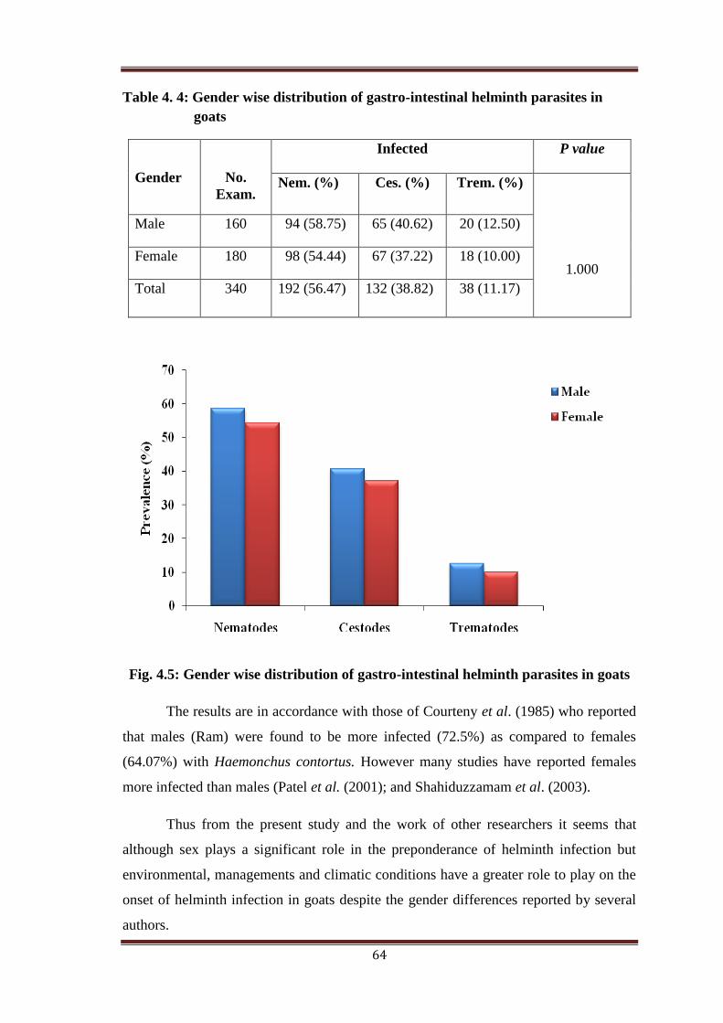
64
Table 4. 4: Gender wise distribution of gastro-intestinal helminth parasites in
goats
Gender
No.
Exam.
Infected P value
Nem. (%) Ces. (%) Trem. (%)
1.000
Male 160 94 (58.75) 65 (40.62) 20 (12.50)
Female 180 98 (54.44) 67 (37.22) 18 (10.00)
Total 340 192 (56.47) 132 (38.82) 38 (11.17)
Fig. 4.5: Gender wise distribution of gastro-intestinal helminth parasites in goats
The results are in accordance with those of Courteny et al. (1985) who reported
that males (Ram) were found to be more infected (72.5%) as compared to females
(64.07%) with Haemonchus contortus. However many studies have reported females
more infected than males (Patel et al. (2001); and Shahiduzzamam et al. (2003).
Thus from the present study and the work of other researchers it seems that
although sex plays a significant role in the preponderance of helminth infection but
environmental, managements and climatic conditions have a greater role to play on the
onset of helminth infection in goats despite the gender differences reported by several
authors.
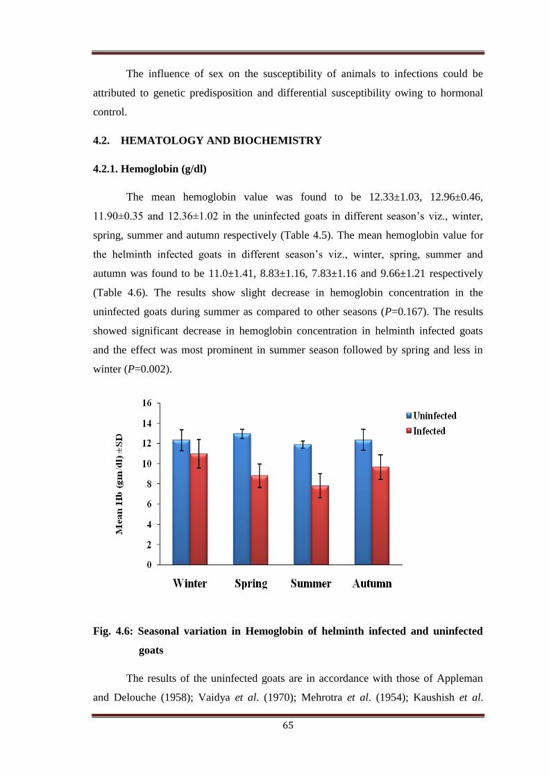
65
The influence of sex on the susceptibility of animals to infections could be
attributed to genetic predisposition and differential susceptibility owing to hormonal
control.
4.2. HEMATOLOGY AND BIOCHEMISTRY
4.2.1. Hemoglobin (g/dl)
The mean hemoglobin value was found to be 12.33±1.03, 12.96±0.46,
11.90±0.35 and 12.36±1.02 in the uninfected goats in different season’s viz., winter,
spring, summer and autumn respectively (Table 4.5). The mean hemoglobin value for
the helminth infected goats in different season’s viz., winter, spring, summer and
autumn was found to be 11.0±1.41, 8.83±1.16, 7.83±1.16 and 9.66±1.21 respectively
(Table 4.6). The results show slight decrease in hemoglobin concentration in the
uninfected goats during summer as compared to other seasons (P=0.167). The results
showed significant decrease in hemoglobin concentration in helminth infected goats
and the effect was most prominent in summer season followed by spring and less in
winter (P=0.002).
Fig. 4.6: Seasonal variation in Hemoglobin of helminth infected and uninfected
goats
The results of the uninfected goats are in accordance with those of Appleman
and Delouche (1958); Vaidya et al. (1970); Mehrotra et al. (1954); Kaushish et al.

66
(1976). Abdelatif et al. (2009) reported the decrease in haemoglobin concentration in
summer as compared to spring and winter in the uninfected goats. The results of the
helminth infected goats are in accordance with Teleb et al. (2007); El-Aziz et al.
(2002). Bordoloi et al. (2012) reported the decrease in hemoglobin concentration in
sheep experimentally infected with Haemonchus. Sulaiman et al. (2010) also reported
decrease in Hb concentration in goats infected with Babesia ovis. Waweru et al. (1999);
Ahmad et al. (2006) and Matanovic et al. (2007) also showed decrease in Hb
concentration in helminth infected ruminants.
In the uninfected goats the decrease in haemoglobin concentration in the
summer season may be due to increase in ambient temperature (Appleman and
Delouche (1958); Bianca, 1968; Prasetyo et al. 1984), and haemodilution. The decline
is also attributed to depression of thyroid secreation which is associated with decreased
erythopoises. Thyroid hormones increase the proliferation rate of erythroid progenitors
(Popovic et al. 1977; Dainiak et al. 1978) and enhance the production of erythropoietic
growth factors (Dainiak et al.1986; Fandrey et al. 1994). The relatively higher values in
spring may be associated with the nutritional status of the goat and the climatic
conditions (Payne et al. 1990).
The reduction in Hb concentration in the helminth infected goats may be
attributed to the acute loss of blood by sucking activity of various parasites especially
Haemonchus contortus and Ostertagia ostertagi which suck 0.05ml blood/worm/day
(Soulsby, 1986). Coles (1986); Kaneko et al. (1997); Kramer (2000); El-Sayed et al.
(2003); Lotfy et al. (2003); Omran and El-Kholany (2003) reported that the severe
anemia may be due to a chronic liver inflammation, which causes depression of
erythrogenesis.
4.2.2. Packed Cell Volume (PCV) (%)
The mean value for packed cell volume in the uninfected goats was found to be
36.16±1.60, 38.25±1.20, 35.10±1.13 and 36.06±1.59 in different seasons viz., winter,
spring, summer and autumn respectively (Table 4.5). The mean PCV value of the
helminth infected goats in different seasons viz., winter, spring, summer and autumn
was found to be 34.5±2.42, 26.16±2.31, 24.33±0.81 and 31.16±2.40 respectively (Table
4.6). The results show decrease in packed cell volume percentage in uninfected goats
during summer as compared to other seasons (P=0.007). The results show significant

67
decrease in packed cell volume percentage in helminth infected goats and the effect
was most prominent in summer and spring season as compared to winter season
(P=0.001).
Fig. 4.7: Seasonal variation in PCV of helminth infected and uninfected goats
The results of the uninfected goats are in accordance with those of Appleman
and Delouche (1958). Abdelatif et al. (2009) reported the decrease in packed cell
volume percentage in summer as compared to spring and winter in the uninfected goats.
Similar results in the helminth infected goats have been reported by Teleb et al. (2007);
Eguale and Abie (2003); Bordoloi et al. (2012). Sulaiman et al. (2010) reported
decrease in PCV in goats infected with Babesia ovis. Mottelib et al. (1992) also
reported decrease in PCV in nematode infected goats and sheep.
In the uninfected goats the decrease in haemoglobin concentration in the
summer season may be due to increase in ambient temperature (Appleman and
Delouche (1958); Bianca, 1968; Prasetyo et al. 1984), and haemodilution. The decline
is also attributed to depression of thyroid secreation which is associated with decreased
erythopoises. Thyroid hormones increase the proliferation rate of erythroid progenitors
(Popovic et al. 1977; Dainiak et al. 1978) and enhance the production of erythropoietic
growth factors. The relatively higher values in spring may be associated with the
nutritional status of the goat and the climatic conditions (Payne et al. 1970).

68
The reduction in PCV in the helminth infected goats may be attributed to the
acute loss of blood by sucking activity of various parasites especially Haemonchus
contortus and Ostertagia ostertagi which suck 0.05ml blood/worm/day (Soulsby,
1986).
4.2.3: Erythrocyte Sedimentation Rate (ESR) (mm/hr)
The ESR in the uninfected goats was found to be 8.08±0.75, 8.0±0.80, 8.6±0.95
and 8.25±1.05 in different seasons viz., winter, spring, summer and autumn
respectively (Table 4.5). The mean ESR value of the helminth infected goats in
different seasons viz., winter, spring, summer and autumn was found to be 8.85±0.65,
11.11±1.60, 13.48±0.98 and 9.25±0.69 respectively (Table 4.6). The results show
significant increase in ESR in helminth infected goats and the effect was most
prominent in summer season and less in winter (P=0.001).
Fig. 4.8: Seasonal variation in ESR of helminth infected and uninfected goats
The results of uninfected goats showed slight variation in ESR in the different
seasons (P=0.667). The results of the helminth infected goats are in accordance with
Taimur et al. (1993) who reported increase in ESR in cattle experimentally infected
with Fasciola gigantica. Aatish et al. (2007) also reported the increase in ESR by the
mange mite infestation in sheep in Pakistan.
Increase in ESR may be attributed to increased level of gamma globulins
because of the increased helminth infection.

69
4.2.4: RBC Count (106/mm
3)
The mean RBC count was found to be 13.83±0.75, 14.48±0.46, 12.88±0.27 and
13.96±0.63 in different seasons viz., winter, spring, summer and autumn respectively in
the uninfected goats (Table 4.5). The mean RBC values for the helminth infected goats
was found to be 12.83±1.47, 9.5±0.54, 8.16±1.16 and 11.0±0.89 in different seasons
viz., winter, spring, summer and autumn respectively (Table 4.6). The results show
decrease in RBC count in the uninfected goats during summer as compared to other
seasons (P=0.001). The results show significant decrease in RBC count in helminth
infected goats and the effect was most prominent in summer and spring season and less
in winter (P=0.001) (Plate 7).
Fig. 4.9: Seasonal variation in RBC count of helminth infected and uninfected
goats
The results of the uninfected goats are in accordance with those of Appleman
and Delouche (1958); Vaidya et al. (1970); Mehrotra et al. (1954); Kaushish et al.
(1976). Abdelatif et al. (2009) reported the decrease in RBC count in summer as
compared to spring and winter in the uninfected goats. The results of the helminth
infected goats are in accordance with those of Sulaiman et al. (2010); Teleb et al.
(2007); El-Aziz et al. (2002); Ahmed et al. (2006) and Matanović et al. (2007).
Mottelib et al. (1992) reported decrease in RBC count in nematode infected sheep and
goats. Esmaeilnejad et al. (2012) reported decrease in Red Blood Cells in goats and
sheep infected with Babesia ovis.

70
In the uninfected goats the decrease in RBC count in the summer season may be
due to increase in ambient temperature (Appleman and Delouche (1958); Bianca, 1968;
Prasetyo et al. 1984), and haemodilution. The decline is also attributed to depression of
thyroid secreation which is associated with decreased erythopoises. Thyroid hormones
increase the proliferation rate of erythroid progenitors (Popovic et al. 1977; Dainiak et
al. 1978) and enhance the production of erythropoietic growth factors (Dainiak et al.
1986; Fandrey et al. 1994). The relatively higher values in spring may be associated
with the nutritional status of the goat and the climatic conditions (Payne et al. 1970).
The reduction in RBC count in the helminth infected goats may be attributed to
the acute loss of blood by sucking activity of various parasites especially Haemonchus
contortus and Ostertagia ostertagi which suck 0.05ml blood/worm/day (Soulsby,
1986). The reduction is comparatively much higher compared to the environmental
changes. Coles (1986); Kaneko et al. (1997); Kramer (2000); El-Sayed et al. (2003);
Lotfy et al. (2003); Omran and El-Kholany (2003) reported that the severe anemia may
be due to a chronic liver inflammation, which causes depression of erythrogenesis.
4.2.5. WBC Count (103/mm
3)
The mean WBC count was found to be 9.0±0.89, 9.21±0.90, 8.08±0.57 and
9.06±0.92 goats in different seasons viz., winter, spring, summer and autumn
respectively in the uninfected goats (Table 4.5). The mean WBC count value for the
helminth infected goats in different seasons viz., winter, spring, summer and autumn
was found to be 9.16±1.16, 10.83±1.16, 11.83±0.75 and 9.66±0.81 respectively (Table
4.6). The results showed no marked variation but slight decrease in WBC count in
uninfected goats during summer (P=0.11). The results show significant increase in
WBC count in helminth infected goats and the effect was most prominent in summer
season and less in winter (P=0.001).

71
Fig. 4.10: Seasonal variation in WBC count of helminth infected and uninfected
goats
In the uninfected goats similar results have been reported by (Vaidya et al.
1970; Vergula et al. 1985) in goats and (Hassan et al. 1966; Hassan et al. 1987) in
sheep. The results of the helminth infected goats are in accordance with those of Teleb
et al. (2007); Waweru et al. (1999); El-Sayed et al. (2003); Lotfy et al. (2003) and
Omranand El-Kholany (2003). Mottelib et al. (1992) reported increase in WBC count
in nematode infected goats and sheep. Esmaeilnejad et al. (2012) also reported increase
in WBC count in goats and sheep infected with Babesia ovis.
The reduction in the WBC count in the summer season in the uninfected goats
could be associated with physiological responses to hot climate which include decrease
in food take and expansion of plasma volume resulting in haemodilution. Increase in
WBC count in the helminth infected goats may be attributed to the immune response of
body against the helminth parasites as a means of self defense.
4.2.6: Differential Leukocyte Count (DLC) (%)
The mean percentage value for neutrophils in the uninfected goats in different
seasons viz., winter, spring, summer and autumn were found to be 31.16±0.75,
31.99±0.23, 32.40±0.49 and 31.35±1.34 respectively (P=0.05) (Table 4.7). The mean
percentage value for lymphocytes in the uninfected goats in different seasons viz.,
winter, spring, summer and autumn were found to be 50.66±0.81, 51.20±0.90,
51.61±0.97 and 50.76±1.03 respectively (P=0.29) (Table 4.7). The mean percentage

72
value for eosinophils in the uninfected goats in different seasons viz., winter, spring,
summer and autumn were found to be 3.0±0.89, 3.3±0.30, 3.7±0.47 and 3.2±0.49
respectively (P=0.23) (Table 4.7). The mean percentage value for monocytes in the
uninfected goats in different seasons viz., winter, spring, summer and autumn were
found to be 3.0±0.63, 2.86±0.30, 2.83±0.26 and 2.9±0.42 respectively (P=0.90) (Table
4.7). The mean percentage value for basophils in the uninfected goats in different
seasons viz., winter, spring, summer and autumn were found to be 2.0±0.89, 2.11±0.24,
2.83±0.57 and 2.05±0.26 respectively (P=0.05) (Table 4.7). The mean percentage value
for neutrophil in the helminth infected goats in different seasons viz., winter, spring,
summer and autumn were found to be 31.83±0.75, 37.66±1.03, 41.83±0.75 and
33.66±1.03 respectively (P=0.001) (Table 4.8). The mean percentage value for
lymphocyte in the helminth infected goats in different seasons viz., winter, spring,
summer and autumn were found to be 51.5±1.04, 64.16±1.16, 71.83±1.16 and 54±0.89
respectively (P=0.001) (Table 4.8). The mean percentage value for eosinophil in the
helminth infected goats in different seasons viz., winter, spring, summer and autumn
were found to be 3.16±0.75, 5.0±0.63, 6.16±0.75 and 3.83±0.75 respectively (P=0.001)
(Table 4.8). The mean percentage value for monocyte in the helminth infected goats in
different seasons viz., winter, spring, summer and autumn were found to be 2.83±0.40,
2.16±0.25, 1.16±0.75 and 2.5±0.54 respectively (P=0.001) (Table 4.8). The mean
percentage value for basophil in the helminth infected goats in different seasons viz.,
winter, spring, summer and autumn were found to be 2.0±0.63, 3.0±0.63, 3.16±0.75
and 2.66±0.81 respectively (P=0.04) (Table 4.8). The results showed not much
variation in the DLC in the uninfected goats. The results showed a significant increase
in neutrophils and lymphocytes and slight increase in eosinophils and basophils
whereas monocytes decrease slightly in the helminth infected goats. (Plate 7).

73
Fig.4.11: Seasonal variation in DLC count of helminth infected (I) and uninfected
goats (UI)
The results are in accordance with those of Waweru et al. (1999); El-Sayed et
al. (2003); Lotfy et al. (2003) and Omranand El-Kholany (2003). Teleb et al (2007)
reported increase in neutrophils, lymphocytes, eosinophils and basophils and also
reported decrease in monocytes. Moreover, eosinophilia has been reported to be
proportional to the degree of antigenic stimulation or parasitic burden in helminthes
infections (Ackerman et al., 1981). This is normally linked to antigen antibody reaction
which occurred when the sensitivity to the protein of the parasites has developed or
when the secretory products were released within the blood (Jain, 1993); associated
with cellular- mediated immunity (Duffus et al., 1980). Leukocytosis and eosinophilia
detected in the present study were similar to those previously reported by Sykes et al.
(1980); Zhang et al. (2005) and Ahmed et al. (2006).

74
Table 4.5: Haematological parameters of uninfected goats
Parameters Range (Mean ± SD)
Winter Spring Summer Autumn P value
Hb
(g/dl) 8-13 12.33±1.03 12.96±0.46 11.90±0.35 12.36±1.02 0.167
RBC
(106/ul)
8-17 13.83±0.75 14.48±0.46 12.88±0.27 13.96±0.63 0.001
WBC
( 103/ul)
4-12 9.0±0.89 9.21±0.90 8.08±0.57 9.06±0.92 0.111
PCV
(%) 23-39 36.16±1.60 38.25±1.20 35.10±1.13 36.06±1.59 0.007
ESR
(mm/hr) 5-15 8.08±0.75 8.0±0.80 8.60±0.95 8.25±1.05 0.667
Table 4.6: Haematological parameters of helminth infected goats
Parameters Range (Mean ± SD)
Winter Spring Summer Autumn P value
Hb
(g/dl)
8-13 11.0±1.41 8.83±1.16 7.83±1.16 9.66±1.21 0.002
RBC
(106/ul)
8-17 12.83±1.47 9.50±0.54 8.16±1.16 11.00±0.89 0.001
WBC
( 103/ul)
4-12 9.16±1.16 10.83±1.16 11.83±0.75 9.66±0.81 0.001
PCV
(%)
23-39 34.50±2.42 26.16±2.31 24.33±0.81 31.16±2.40 0.001
ESR
(mm/hr)
5-15 8.85±0.65 11.11±1.60 13.48±0.98 9.25±0.69 0.001

75
Table 4.7: Differential Leukocyte Count of uninfected goats
Table 4.8: Differential Leukocyte Count of helminth infected goats
Cells Range Mean±SD
Winter Spring Summer Autumn P value
Neutrophil 20-45 31.16±0.75 31.99±0.23 32.40±0.49 31.35±1.34 0.05
Lymphocyte 40-75 50.66±0.81 51.20±0.90 51.61±0.97 50.76±1.03 0.29
Eosinophil 0-7 3.0±0.89 3.30±0.30 3.70±0.47 3.20±0.49 0.23
Monocyte 0-4 3.0±0.63 2.86±0.30 2.83±0.26 2.90±0.42 0.90
Basophil 0-4 2.0±0.89 2.11±0.24 2.83±0.57 2.05±0.26 0.05
Cells Range Mean±SD
Winter Spring Summer Autumn P value
Neutrophil 20-45 31.83±0.75 37.66±1.03 41.83±0.75 33.66±1.03 0.001
Lymphocyte 40-75 51.50±1.04 64.16±1.16 71.83±1.16 54.0±0.89 0.001
Eosinophil 0-7 3.16±0.75 5.0±0.63 6.16±0.75 3.83±0.75 0.001
Monocyte 0-4 2.83±0.40 2.16±0.25 1.16±0.75 2.50±0.54 0.001
Basophil 0-4 2.0±0.63 3.0±0.63 3.16±0.75 2.66±0.81 0.048

76
4.2.7. Total Protein (g/dl)
The mean total protein was found to be 7.06±0.28, 7.02±0.35, 7.04±0.46 and
7.04±0.48 in different seasons viz., winter, spring, summer and autumn respectively in
the uninfected goats (Table 4.9). The mean total protein value for the helminth infected
goats was found to be 7.06±0.29, 7.0±0.20, 6.83±0.42 and 7.03±0.27 in different
seasons viz., winter, spring, summer and autumn respectively (Table 4.10). The results
showed no significant changes in total protein content in the uninfected goats (P=0.99).
The results showed the decrease in total protein content in the helminth infected goats
and was most prevalent in the summer season and less in winter (P=0.57).
Fig. 4.12: Seasonal variation in Total Protein content of helminth infected and
uninfected goats
The results of the helminth infected goats are in accordance with that of Knox et
al. (1993) who reported decrease in the total protein content in the small ruminants
infected with nematode parasites. Rehman and Collins (1990) also reported the
decrease in total protein content in goats. Teleb et al. (2007) reported the decrease in
total protein content in serum of Farafrasheep experimentally infected with Fasciola
gigantica.
The decrease in total protein is due to haematophagous parasites especially
Haemonchus contortus and Ostertagia ostertagi which suck 0.05ml of blood/worm/day
(Soulsby, 1986). The infection of liver and destruction of liver parenchyma also
resulted in alteration in protein values (Ismail et al. (1990); Mohamed (2000) and
Matanovic et al. (2007).
77
4.2.8. Albumin (g/dl)
The mean albumin was found to be 2.35±0.20, 2.40±0.30, 1.93±0.33 and
2.31±0.31 in different seasons viz., winter, spring, summer and autumn respectively in
the uninfected goats (Table 4.9). The mean albumin value of the helminth infected
goats was found to be 2.34±0.20, 2.24±0.18, 1.90±0.20 and 2.30±0.18 in different
seasons viz., winter, spring, summer and autumn respectively (Table 4.10). The results
showed the decrease in albumin content in the uninfected goats in the summer season
as compared to other seasons (P=0.04). The results showed the decrease in albumin
content in the helminth infected goats and was found most prevalent in summer and
less in winter (P=0.003).
Fig. 4.13: Seasonal variation in Albumin content of helminth infected and
uninfected goats
In the uninfected goats similar results have been reported by Hassan et al.
(1966); Abdelatif et al. (2009). Similar results have been reported in helminth infected
goats by Ahmad et al. (1990), who studied the serum protein changes of lambs
experimentally infected with Haemonchus contortus and reported a marked decrease in
albumin content. Teleb et al. (2007) reported the decrease in albumin protein content in
serum of Farafrasheep experimentally infected with Fasciola gigantica. However,
Ahmad et al. (2006) detected no significant change in albumin levels in sheep infected
with Fasciola gigantica.
The decrease in albumin level in the summer in the uninfected goats could be
attributed in part to expansion of plasma volume resulting in haemodilution. The
plasma volume increases by 17% during tropical summer in Merino sheep (Macfarlane
et al. 1966).
The decrease in albumin levels in the helminth infected goats may be attributed
to reduced albumin synthesis caused by liver damage and haematophagous feeding
behavior of various parasites.

78
4.2.9. Globulin (g/dl)
The mean globulin was found to be 4.71±0.34, 4.62±0.42, 5.11±0.16 and
4.73±0.38 in different seasons viz., winter, spring, summer and autumn respectively in
the uninfected goats (Table 4.9). The mean globulin value of the helminth infected
goats in different seasons viz., winter, spring, summer and autumn was found to be
4.72±0.35, 4.76±0.39, 4.92±0.41 and 4.73±0.43 respectively (Table 4.10). The results
showed the increase in globulin content in the uninfected goats in the summer and
spring as compared to other seasons (P=0.09). The results showed the slight increase in
globulin content in the helminth infected goats and was most prevalent in summer and
less in winter (P=0.80).
Fig. 4.14: Seasonal variation in Globulin content of helminth infected and
uninfected goats
The results of uninfected goats are in accordance with those of Al-Busaidi et al.
(2008); Johnson et al. (1995). The results of the helminth infected goats are in
accordance with the Rehman and Collins (1990) who reported the increase in globulin
fraction of plasma proteins in goats. Ahmad et al. (1990) also reported the increase in
α- globulin and β- globulin in the lambs experimentally infected with Haemonchus
contortus and was found highest at the peak of infection. Teleb et al. (2007) also
showed the increase in globulin protein in experimentally infected sheep with Fasciola
gigantica. Amer et al. (2002) and Matanovic et al. (2007) also reported the increase in
globulin levels in helminth infections.
The increase in globulin levels in serum in the uninfected goats may be due to
their potentiated immunity (Al-Busaidi et al. 2008). The increase in globulin levels in
serum in the helminth infected goats may be attributed to the result of immune response

79
to infection (Matanovic et al. 2007) and due to the increase in α & β globulin
production (Duncan et al. 1994).
Table 4.9: Biochemical parameters of uninfected goats
Parameters Range (Mean ± SD)
Winter Spring Summer Autumn P value
Total Protein
(g/dl)
6.0-7.5 7.06±0.28 7.02±0.35 7.04±0.46 7.04±0.48 0.999
Albumin
(g/dl)
1.9-2.4 2.35±0.20 2.40±0.30 1.93±0.33 2.31±0.31 0.041
Globulin
(g/dl)
4.1-5.1 4.71±0.34 4.62±0.42 5.11±0.16 4.73±0.38 0.09
Table 4.10: Biochemical parameters of helminth infected goats
Parameters Range (Mean ± SD)
Winter Spring Summer Autumn P value
Total Protein
(g/dl)
6.0-7.5 7.06±0.29 7.0±0.20 6.83±0.42 7.03±0.27 0.574
Albumin
(g/dl)
1.9-2.4 2.34±0.20 2.24±0.18 1.91±0.20 2.3±0.18 0.003
Globulin
(g/dl)
4.1-5.1 4.72±0.35 4.76±0.39 4.92±0.41 4.73±0.43 0.80
The whole Data was fed into a Microsoft Excel 2010. Primer software was used
for data analysis, chi-square test and t-test were applied. The data was presented as
mean ± standard deviation (SD).

80
Haemonchus egg
Ostertagia egg
Trichuris egg
Nematodirus egg
Fasciola egg
Paramphistomum egg
Dicrocoelium egg
Moneiza egg
Mixed Parasitic Infection
Plate 4: Eggs of various Helminth Parasites

81
Haemonchus sp. Ostertagia sp. Nematodirus sp.
Bunostomum sp. Fasciola sp. Dricocelium sp.
Plate 5: Helminth Parasites

82
Scolex of Moneiza sp. Proglottid of Moneiza sp.
Scolex of Avitellina sp. Gravid segment of Avitellina sp.
Scolex of Stilesia sp. Mature segment of Stilesia sp.
Plate 6: Helminth Parasites

83
RBC’S
Neutrophil Monocyte
Lymphocyte Basophil
Plate 7: RBC’S and WBC’S of goats

84
CHAPTER – 5
Summary, Conclusion and
Recommendations
SUMMARY
The present study deals with the epidemiology and hemato-biochemical
investigations of goats naturally infected with helminth parasites from December 2011
to November 2012 in South Kashmir.
The present study revealed that the infection status by the various species of
helminth parasites is very high. This means that geographical and climatic
conditions of this region are suitable for helminth infection.
The overall percentage of helminth infection was found to be 58.23%.
Out of 340 goats, 192 (56.47%) were found infected with nematodes, 132
goats (38.82%) with Cestode infection, 38 goats (11.17%) with Trematode
infection and 179 (52.64%) were having mixed infection (i.e. more than one
parasite).
The gastrointestinal helminth parasites reported in the infected goats were
Haemonchus sp. (47.94%), Ostertagia sp. (37.94%), Bunostomum sp. (32.94%),
Oesophagostomum sp. (30.0%), Trichuris sp. (35.88%), Nematodirus sp.
(23.82%), Paramphistomum sp. (10.0%), Fasciola sp. (11.17%), Dicrocoelium
sp. (8.23%), Moneizia sp. (21.47%), Stilesia sp. (14.11%), and Avitellina sp.
(18.23%).
This study showed the seasonal pattern of helminth infestation, with high
infection rate in summer (73.46%) followed by spring (68.18%), autumn
(51.16%) and lowest in winter (32.35%).
The study also showed that the young animals are more prone to infection than
the adult ones. Therefore young animals need special attention because of their

85
high susceptibility to infection. They should be included in deworming
programming.
The study also revealed that maximum helminth infection was observed in
males as compared to females. The influence of sex on the susceptibility of
animals to infections could be attributed to genetic predisposition and
differential susceptibility owing to hormonal control.
Hematological studies revealed significant decrease in hemoglobin
concentration, packed cell volume and total erythrocyte count in the helminth
infected goats. Whereas erythrocyte sedimentation rate and total leukocyte
count showed significant increase in the helminth infected goats.
Among biochemical parameters, albumin showed the significant decrease and
globulin showed slight increase in the helminth infected goats where as total
protein showed slight decrease in helminth infected goats.
86
CONCLUSION
It should be concluded that gastro-intestinal parasitism represents a severe
health problem in small ruminant production system, and its consequences can be
extensive ranging from reduced productivity to mortality. Gastro-intestinal
nematodiasis is a major threat and a primary constraint to goat productivity, it
endangers animal welfare worldwide. The main culprit in ruminant nematodiasis is
Haemonchus contortus and Ostertagia ostertagi which causes haemonchosis, anemia
and parasitic gastroenteritis in goats. They cause significant economic losses worldwide
due to their feeding behaviour being haematophagous. It could also be concluded that
changes in the hematological and biochemical parameters in goats infected with
helminth parasites reflects a veterinary problem and causes pathological conditions like
anemia, weight loss, poor wool and milk production. Fascioliasis cause pathological
and necrotic lesions of liver parenchyma and the bile ducts causing hemorrhage, so
modem diagnostic techniques need to be incorporated to estimate the degree of
infection.
The present observation may suggest in planning chemotherapeutic and
prophylactic strategies for the helminth control in the region. With the early diagnosis
of helminthiasis in animals, a treatment schedule could be designed to avoid more
infection and animal losses on the farm level and in turn economical losses. Control of
helminth infection requires knowledge of the epidemiology and ecology of the
parasites. This knowledge is needed not only for planning better strategies of parasitic
control but also provide insight into the natural processes of controlling parasite
population. Present results can be useful to future researchers in this field and can lead
to proper control of parasitic infections in goats of Kashmir Valley.

87
RECOMMENDATIONS
On the basis of the present study following recommendations are given as under.
Regular screening of animals for the presence of helminth infections should be
carried out to understand the disease potential.
Overcrowding at farms should be avoided and good sanitary conditions should
be provided.
Based upon epidemiological principles, Three Check Points to manage and
control helminthiasis in ruminants in our state are suggested and recommended.
(1) Pre grazing: Deworming early spring before the animals are sent to pastures
for grazing. (2) During grazing: Deworming during the middle of grazing
months (somewhere between July- August). (3) Post grazing: Deworming
before the onset of winter.
Animals should be provided with balanced feed containing more than 3%
protein.
Alternatives to chemical control also have viable effect in the treatment of
helminthes, so they must be incorporated alternatively to avoid resistance.
The intiative should be taken at government level to develop and update quality
and quantity of pastures and for animals grazing along with the supplementary
food. Recent studies have indicated that improvement in animal nutrition
especially metabolizable protein can enhance the host response against primary
and secondary infections and to reduce the patho-physiological changes
associated with helminthes.
Further studies are needed on life cycle and larval ecology of these parasites so
as to provide a basis for developing sustainable helminth control strategies for
the temperate climate of the Kashmir Valley. Sustainable and novel control
strategies (manipulation of diet, vaccines, biological control etc.) need to be
employed to control helminthiasis in Jammu and Kashmir.

88
Bibliography
Aatish, F. U., Sindhu, Z., Iqbal, Z., Jabbar, A. and Tasawar, Z. 2007. Prevalence of
Sheep Mange in District Dera Ghazi Khan (Pakistan) and Associated
Hematological/Biochemical Disturbances. Int. J. Agri. Biol., 9: 6.
Abdel-Ali, T. S. 1992. Haematological and biochemical studies on efficiency of
synanthetic against gastro-intestinal nematode parasites in sheep. Assiut Vet.
Med. J., 42(48): 197- 203. (Veterinary Bulletin, 1993 V 63 N-S).
Abdel-abi, I. M., Sherif, N. E., Behnke, J., Zalat, S., Gilbert, Hamada, A., Teama, M.
and Abdel-Rahman, M. 2002. Haematological profile and parasitological survey
of the domestic goats and camels of St. Katherine, Sinai, Egypt. Egyptian
Journal of Biology, 4: 101-109.
Abdelatif, A. M., Ibrahim, M. Y. and Hassan, Y. Y. 2009. Seasonal Variation in
Erythrocytic and Leukocytic Indices and Serum Proteins of Female Nubian
Goats. Middle-East Journal of Scientific Research, 4 (3): 168-174.
Abouzeid, N. Z., Selim. A. M. and El-Hady, K. M. 2010. Prevalence of gastrointestinal
parasites infections in sheep in the Zoo garden and Sinai district and study the
efficacy of anthelmintic drugs in the treatment of these parasites. Journal of
American Science, 6(11): 544-551.
Ackerman, S. J., Gleich, G. J., Weller, P. F. and Ottensen, E. A. 1981. Eosinophilia and
elevated serum levels of eosinophil major basic protein and Charcot-Leyden
crystal protein (lysophospholipase) after treatment of patients with Bancrofts
Filariasis. Journal of Immunology, 127: 1093 -1094.
Addass, P. A., Midau, A. and Babale, D. M. 2010. Haemato-biochemical Findings of
Indigenous Goats in Mubi Adamawa State, Nigeria. Journal of Agriculture and
Social Sciences, 6(1): 14-16.

89
Ahmad, A., Anwarul Hassan., Anwarul Haq and Majeed, M. A. 1990. Serum protein
grams of lambs in experimentally induced Haemonchus contortus infection.
Veterinariski-Arhiv, 60(4): 195-200.
Ahmed, M., and Ansari, J. A. 1987. Prevalence of gastrointestinal nematodes of sheep
and goats in Aligarh (India). Ind. Vet. Med. J., 11:165-170.
Ahmed, M. I., Ambali, A.G. and Baba, S.S. 2006. Hematological and biochemical
responses of Balami sheep to experimental Fasciola gigantica infection. Journal
of Food, Agriculture & Environment, 4(2): 71- 74.
Akhtar, M. Z., Khan, A., Khan, M. Z. and Muhammad, G. 2007. Haemato-Biochemical
Aspects of Parturient Haemoglobinuria in Buffalo. Turk. J. Vet. Anim. Sci.,
31(2): 119-123.
Al-Busaidi, R., Johnson, E. H. and Mahgoub, O. 2008. Seasonal variations of
phagocytic response, immunoglobulin G (IgG) and plasma cortisol levels in
Dhofari goats. Small Ruminant Research, 79: 118-123.
Al-Quaisy, H. H. K., A-Zubaidy, A. J., Altaif, K. L. and Makkawi, T. A. 1987. The
pathogenecity of haemonchosis in sheep and goats in Iraq, Clinical Parasitology
and Haematological findings. Vet. Parasitology, 24: 221-228.
Al-Shaibani, I. R. M., Phulan, M. S., Arijo, A. and Qureshi, T. A. 2008. Epidemiology
of Ovine Gastrointestinal Nematodes in Hyderabad district, Pakistan. Pakistan
Vet. J., 28(3): 125-130.
Amarante, A. F. T., Godoy, W. A. C. and Barbosa, M. A. 1998. Nematode egg count,
PCV and body weight as parameters to identify sheep resistance and
susceptibility to infection by gastro-intestinal nematodes. Aras. Vet., 14(3): 331-
339.
Amer, S. A., Osman, S. A. and Harfoush, M. A. 2002. Epizootical and serological
studies on fascioliasis in cattle. Zag. Vet. J., 30(1): 107-116.
Appleman, R. O. and Delouché, J. C. 1958. Behavioural, physiological and
biochemical responses of goats to temperature 0° to 40°C. J. Animal Sci., 17:
326-335.

90
Astiz, S., Miro, G., Mana, A., Garcia-Romero, C. and Valcarcel, F. 2000. Seasonal
Prevalence of lungworm infections of goats in Costilla- La Mancha. ITEA
Production Animal, 96(2): 95-101.
Bal, M. S., Mahajan, V., Verma, S., Alka., Sharma, S. 2007. Outbreaks of parasitic
gastroenteritis in sheep and goats. Journal of Veterinary Parasitology, 21(1): 93-
95.
Bhat, S. A., Mir, M. R., Qadir, S., Allaie, I., Khan, H. M., Husain, I. and Bilal, S. 2011.
Haematological and Biochemical Parameters of Kashmiri Goats in Different
Climatic Conditions. IJAVMS., 5(5): 481- 487.
Bianca, W., 1968. Thermoregulation. In: Adaptation of Domestic Animals. (Editor
S.E.S. Hafez). Lea and Febiger, Philadelphia.
Boray, J.C. 1985. Flukes of domestic animals. In: SM Gaafar, WE Howard, RE Marsh
(eds) Parasites, pests and predators. Elsevier, Amsterdam, 179 – 218.
Bordoloi, G., Jas, R. and Ghosh, J. D. 2012. Changes in the haemato-biochemical
pattern due to experimentally induced haemonchosis in Sahabadi sheep. J.
Parasit Dis., 36(1): 101-105.
Bottiger, L. E. and Svedberg, C. A. 1967. Normal ESR and age. Br. Med. J., 2: 285.
Brunn, U., Feige, K., Gonsohir, B., Degen, H., Wolffe, K., Gohm, D., Sydler, T. 2000.
Pneumonia caused by Protostrongylus spp. in nine Saanen goats. Tierarztriche
Umschau, 55(6): 338-340, 343-345.
Buttler, R. W. and Yeoman, G. H. 1962. Acute intestinal paramphistomiasis in Zebu
cattle in Tanganyika. Vet. Res., 74: 227-231.
Chakerborty, D. and Lodh, C. 1994. Studies on blood and biochemical profiles in
naturally infected goats. Indian Vet. J., 3: 286-288.
Chaudary, F. R., Khan, M. F. U. and Qayyum, M. (2007). Prevalence of Haemonchus
contortus in naturally infected small ruminants grazing in the Pothar area of
Pakistan. Pakistan Vet. J., 27(2): 73-79.

91
Chavhan, P. B., Khan, L. A., Raut, P. A., Maske, D. K., Rahman, S., Podchalwar, K. S.
and Siddiqui, M. F. M. F. 2008. Prevalence of Nematode parasites of Ruminants
at Nagpur. Veterinary World, 1(5):140.
Coles, E. H. 1986. Veterinary Clinical Pathology. 4th
Ed. W.B. Saunders Company,
Philadelphia, London and Toronto.
Conn, H. J. and Darrow, M. S. 1960. Staining procedure used by the Biological
Staining Commission, 2nd
Baltimore, the Williams and Wilkins Company.
Courteny, C. H., Parker, C. F., McClure, K. E. and Herd, R. P. 1985. Resistance of
exotic and domestic lambs to experimentally infected with Haemonchus
contortus. International Journal of Parasitology, 15: 101-109.
Dacie, J. V. and Levis, S. L. 1975. Practical Haematology. 5th
Edition. Edinbergh
Churchll-Livigstone.
Dainiak, N., Hoffman, R., Maffei, L. A. and Forget, B. G. 1978. Potentiation of human
erythropoiesis in vitro by thyroid hormone. Nature, 272: 260-262.
Dainiak, N., Sutter, D. and Kreczko, S. 1986. The blood picture L-triiodothyronin
augments erythropoietic growth factor release from blood and bone marrow
leukoyctes. Blood, 68: 1289-1297.
Das, M., Prasad, K. D. and Singh, L. B. 2005. Effects of gastrointestinal nematodosis
on the body weight and mortality in Kids. Journal of Veterinary Parasitology,
19(1): 73- 74.
Dhand, N. K., Singh, j., Aradhana and Sandhu, K. S. 2004. Fasciolasis in sheep and
goats: An outbreak and treatment. Journal of Veterinary Parasitology, 18(1): 77-
78.
Dhar, D. N., Sharma, R. L. and Bansal, G. C. 1982. Gastrointestinal nematodes in
sheep in Kashmir. Veterinary Parasitology, 11: 271-272.
Di Gerbo, A. R., Roncari, S., Zanzani, T., Beneetti and Manfriedi, M. J. 2006.
Gastrointestinal parasites in goats farms from Bergamo province. Parasitologia,
(Rome). 48(3): 385-389.

92
Dominguez-Torano I. A., Cuquerella M., Gomez-Munoz M. T., Mendez S., Fernandez-
Perez, F. J. and Alunda J. M. 2000. Vaccination of Manchego lambs against
Haemochus contortus with a somatic fraction (p26/23) of adult parasites.
Parasite Immunol., 22(3):131-138.
Dorny, P., Symoens, C., Jalila, A., Verercrusse, J. and Sami, R. 1995. Strongyle
infections in sheep and goats under the traditional husbandry system in
peninsular Malaysia. Veterinary Parasitology, 56(1/3): 121-136.
Dube, S., Obaimiwe, B. A. and Aisein, M. S. O. 2003. Studies on genus Cotylophoran
Fishoeder, 1901 (Paramphistomidae) recovered from Nigerian cattle. Folia
Veterinaria, 47: 42-47.
Duffus, W. P., Thorne, K. and Oliver, R. 1980. Killing of juvenile Fasciola hepatica by
purified bovine eosinophils proteins. Clin. Exp. Immunol., 40: 336 - 344.
Duncan, J. R., Prasse, K. W. and Mahaffey, E. A. 1994. Veterinary Laboratory
Medicine. Third edition, Iowa State Univ. Press, USA.
Egbe-Nwiyi, T. N., Nwaosu, S. C. and Salami, H. A. 2000. Haematological values of
appararently healthy sheep and goats as influenced by age and sex in arid zone of
Nigeria. Afr. J. Biomed.Res., 3: 109 – 115.
Eguale, T. and Abie, G. 2003. Some clinicopathological observations of a sheep
experimentally infected with Fasciola gigantica. Bulletin of Animal Health and
Production in Africa, 2003. 51(2): 113 -115.
El-Azazy, O. M. E. 1995. Seasonal changes and inhibited development of the
abomassal nematodes in sheep and goats in Saudi Arabia. Veterinary
Parasitology, 58: 91-98.
El-Aziz, M. Z. A., Emara, S. A. and Salem, F. S. 2002. Clinicopathological studies on
fascioliasis among sheep in Giza province. Egyptian Journal of Veterinary
Science, 36: 75-86.
El-Sayed, M., El-Ramady, R. A. and Tawfik, S. A. 2003. Effect of Mirazid on
Fascioliasis and blood chemistry in sheep. Egypt. J. Agric.Res., 81(2): 785-796.

93
England, J. M. and Walford, D. M. 1972. Reassessment of the reliability of
haematocrit. Brit. J. Haematol., 23: 247-253.
England, J. M. and Bain, B. J. 1976. Total and differential leukocytes counts. Brit. J.
Haematol., 33: 1.
Esmaeilnejad, B., Tavassoli, M. and Asri-Rezaei, S. 2012. Investigation of
hematological and biochemical parameters in small ruminants naturally infected
with Babesia ovis. Veterinary Research Forum, 3 (1): 31 – 36.
FAO, (1992). Distribution and impact of helminth diseases of livestock in developing
countries. FAO Animal Production and Health Paper, Vol. 96. FAO, Rome, 221.
Faizal, A. C. M. 1999. Small Ruminants Res., 34: 21-25.
Farooq, Z., Mushtaq, S., Iqbal, Z. and Akhtar, S. 2012. Parasitic Helminths of
Domesticated and Wild Ruminants in Cholistan Desert of Pakistan. International
Journal of Agriculture and Biology, 14(1): 63-68.
Fikry, H. Y., Wafaa, M., Abdel- Razek El-Sadawy, H. A. and Menazi, A. M. 1988. The
relationship between biochemical changes and clinical signs in sheep affected
with liver fluke disease. Z. Vet. J., 1: 113 - 122.
Fandrey, J., Pagel, H., Frede, S., Wolff, M. and Jelkmann, W. 1994. Thyroid hormones
enhance hypoxia induced erythropoietin production in vitro. Experimental
Haematology, 22: 272-277.
Frutschi, T., Kafmann, J., Pfister, K. 1993. Parasitic spectrum and seasonal
epidemiology of gastrointestinal in Gambia. Vet. Parasit., 49(2/4) : 271-288.
Gadahi, J. A., Arshed, M. J., Ali, Q., Javaid, S. B. and S. I. Shah. 2009. Prevalence of
Gastrointestinal Parasites of Sheep and Goat in and around Rawalpindi and
Islamabad, Pakistan. Veterinary World, 2(2): 51-53.
Gadre, A. S., Maske, D. K., Panchbhai, C. G., Gawande, T. R., Kolte, S. W. and
Sirothia, A. R. 2008. Haematological changes in naturally infested dairy animal
at central zone of vidarbha. Veterinary World, 1(2): 47-48.

94
Gatongi, P. M. 1996. Epidemiology and control of haemonchosis of small ruminants in
semi arid Kenya. Kenya Agri. Res. Ins. Inform. Bull., No. 17, April 1996.
Gatongi, P. M., Prichard, R. K., Ranjan, S., Gathuma, J. M., Munyua, W. K.,
Cheruiyot, H. and Scott, M. E. 1998. Hypobiosis of Haemonchus contortus in
natural infections of sheep and goats in a semi-arid area of Kenya. Veterinary
Parasitology, 77: 49–61.
Githigia, S. M., Thamsborg, S. M., Munyen, W. K. and Maingi, N. 2001. Impact of
gastrointestinal helminthes on production in goats in Kenya. Small Ruminant
Research, 42(1): 21-29.
Godara, R., Sharma, R. L. and Sodhi, S. S. 2011. Efficacy of fenbendazole, levamisole
and ivermectin against gastrointestinal nematodes in Jamunapari goats. J.
Parasit. Dis., 35(2): 219–221.
Gwaze, F. R., Chimonyo, M. and Dzama, K. 2010. Nutritionally-related blood
metabolites and faecal egg counts in indigenous Nguni goats of South Africa.
South African Journal of Animal Science, 40 (Issue 5, Supplement 1).
Hammond, J. A. and Sewell, M. M. H. 1990. Diseases caused by helminthes. In:
Sewell, M. M. H., Brocklesby, D. W. (ED), Handbook on Animal Diseases in
the Tropics. Bailliere Tindall, London. 104-159.
Hassan, Y. M. and Hoeller, H. 1966. Observations on the blood composition in cattle
and sheep in Sudan. Sudan Journal of Veterinary Science and Animal
Husbandry, 7: 10-21.
Hassan, G. A., Salem, M. H., El-Nouty, F. D., Okab, A. B. and Latif, M. G. 1987.
Haematoloical changes during summer and winter pregnancies in Barki and
Rahmani sheep (Ovis aries). World Review of Animal Production, XXIII: 89-95.
Hayat, C. S., Khalid, M., Iqbal, Z. and Akhtar, M. 1999. Haematological and
biochemical disturbances associated with Toxocara vitulorum infection in
buffalo calves. Int. J. Agri. Biol., 1: 247-49.

95
Hayat, C. S., Hussain, S. M., Iqbal, Z., Hayat, B. and Akhtar, M. 1996. Effect of
parasitic nematodes on haematology and productivity of sheep. Pak. Vet. J., 16:
81-83.
Herlich, H. 1978. World Anim. Review, 26: 22.
Holmes, P. H. 1993. Interactions between parasites and animal nutrition: the veterinary
consequences. Proc. Nutrit. Soc., 52: 113-120.
Hoste, H. and Chartier, C. 1993. Comparision of the effects on milk production of
concurrent infections with Haemonchus contortus and Trichostrongylus
columbriformis in high and low producing dairy goats. America. J. of Vet. Res.,
54(11): 1886-1893.
Hsiang, Y. K., Lin, J. F., Shu, Y. C. and Wang, J. H. 1990. An investigation on the
endoparasitic species of dairy goat in Taiwan. Journal of Chinese Society of
Veterinary Sciences, 16(3): 211-222.
IEMVT, (1980). Les petits ruminants d’ Afrique Centrale et d’ Afrique d’ Iquest.
Synthese des connaissances actuelles, 295.
Igado, O. O., Ajala, O. O. and Oyeyemi, M. O. 2011. Investigation into the
Hematological and Liver Enzyme Changes at Different Stages of Gestation in
the West African Dwarf Goat (Capra hircus L.). International Journal of Animal
and Veterinary Advances, 3(5): 277-281.
Ijaz, M., Khan, M. S., Avais, M., Ashraf, K., Ali, M. and Saima, M. 2008.
Infection Rate and Chemotherapy of Various Helminths in Goats in and
Around Lahore. Pakistan Vet. J., 28(4): 167-170.
Iqbal, Z., Rasool, G., Hayat, C. S. and Akhtar, M. 1998. Biochemical disturbances
associated with haemonchosis in sheep. Agri. Sci., (Oman). 3: 35-39.
Iqbal, Z., Hayat, C. S., Hayat, B. and Khan, M. N. 1989. Prevalence, organ distribution
and economics of hydatidosis in meat animals at Faisalabad abattoir. Pak. Vet. J.,
9: 70-74.
96
Iqbal, Z., Akhtar, M., Khan, M. N. and Riaz, M. 1993. Prevalence and economic
significance of haemonchosis in sheep and goats slaughtered at Faisalabad
abbatoir. Pak. J. Agri. Sci., 30: 51-53.
International Committee for Standardization in Haematology. 1973. Standard
techniques for the measurements of red cells and plasma volume. A report by the
international committee for the standardization in hematology (I.C.S.H) panel on
diagonostic applications of radioisotopes in hematology. British. J. Haematol.,
25: 801.
Ismail, M. E., El-Sheikh, A., Abdallah, M. A. and Mahmoud, A. A. 1990. Clinical
evaluation of the anthelminitic activity of Fasciola mixed infection in sheep. J.
Drug. Res. Egypt, 19: 173 - 180.
Jain, N.C. 1993. Schalm’s Veterinary Hematology 4th Eds. Lea & Febiger,
Philadelphia. U.S.A.
Jain, S. and Sahni, Y. P. 2010. Biochemical Changes in Goats treated with anthelmintic
indigenous herbs. Veterinary World, 3(7): 315-317.
Jani, R. G. 2008. Prevalence and Haemato-Biochemical Studies of Gastro-intestinal
Parasites of Indian Elephants (Elephas maximus). Veterinary World, 1(10): 296-
298.
Jithendran, K. P. and Bhat, T. K. 2001. Epidemiology and control of parasitism in
nomadic situations in Himachal Pradesh. ENVIS Bulletin: Himalayan ecology
and development, 9(1).
Johnson, E. H., Kass, P. H. and Rosa, J. S. 1995. Effect of energy supplementation and
season on serum immunoglobulin and protein levels in Moxoto goats. Small
Ruminant Res., 15: 121-125.
Kaneko, J. J., Harvey, J. W. and Bruss, M. L. 1997. Clinical Biochemistry of Domestic
Animals, 5th edn. Academic, San Diego, pp 890 - 894.
Kassi, T., Fesus, L., Hendriker, W. M. L., Takats, C., Fok, E., Redel, P., Takaes.,
Nilsson, P. R., Lecure, A. M. W., Vanjasen, J., Bernadine, W. E. and Frankera,
K. 1990. “Is there a relationship between haemoglobin genotype and the innate

97
resistance to experimental Haemonchus contortus infection in merino lambs”. J.
Vet. Parasit., 37(1): 61-77.
Kaushish, S., Bhattia, D. C. and Arora, K.L. 1976. Studies on the adaptability of sheep
to subtopical climate and seasonal changes in rectal temperature,
cardiorespiratory and haematological attributes of Nali sheep. Indian Veterinary
Journal, 33: 760-765.
Khajuria, J. K. and Kapoor, P. R. 2003. Prevalence of parasites in sheep and goats at
Kathua-Jammu. J. Vet. Parasitol., 17: 121–126.
Khan, M. Q., Hayat, C. S., Ilyas, M., Hussain, M. and Iqbal, Z. 1988. Effect of
haemonchosis on body wieght gain and blood values in sheep. Pak. Vet. J., 8:
62-67.
Knox, D. P., Redmon, D. L. and Jones, D. J. 1993. Characterization of proteinase in
extracts of adult Haemonchus contortus, the ovine abomassal nematode.
Veterinary Parasitology, 106(4): 395-404.
Kramer, J. W. 2000. Normal hematology of cattle, sheep and goats. In: Feldman BF,
Zinkl JG, Jain NC (eds) Schalm’s veterinary hematology, 5th edn. Lippincott
Williams & Wilkins, Philadelphia. 1075 – 1084.
Lateef, M., Iqbal, Z., Jabbar, A., Khan, M. N. and Akhtar, M. S. 2005. Epidemiology of
trichostrongylid nematode infections in sheep under traditional husbandry
system in Pakistan. Int. J. Agric. Biol., 7: 596–600.
Leiper, J. W. G. 1957. Animal parasites and their Control Report to Government of
Iraq, FAO series, No. 610. Rome, Italy.
Lepojev, O., Aleksic, N., Kutisic, Z., Dimitrijavic, S., Trajkovski, J. and Panicc, B.
1992. Gastrointestinal strongyles of goats in the Radovis area. Vet. Glasnik,
46(1): 47-50.
Lima, M. M., Faustino, M. A. G., Santana, V. L. A., Santos, N. V. M. and Alves, L. C.
2006. Pernambuco, State, Brazil, through faecal egg count and larval culture.
Evaluation of gastrointestinal helminth infection in goats from the metropolitian
area of Recife. CAB Abstract.

98
Livestock Census Jammu and Kashmir, Livestock, Poultry, Agricultural Machinary and
Implements and Fishery Statistics (2007). Government of India, Ministry of
Agriculture Department of Animal Husbandry and Dairying Krishi Bhavan, New
Delhi.
Lone, B. A., Chishti, M. Z. and Ahmad, F. 2011. Prevalence of Coccidia and
gastrointestinal nematode infection in Goats of Baramullah District of Kashmir
Valley. Global Veterenaria, 7(1):27-30.
Lone, B. A., Chishti, M. Z., Ahmad, F. and Tak, H. 2012. Helminthic infestations in
slaughtered Sheep and Goats of District Ganderbal, Kashmir. DAV International
Journal of Science, 1(1): 2277-5536.
Lotfy, H. S., Mahmoud, S. M. and Abdel-Gawad, M. A. 2003. Some studies on
Fascioliasis in Mid-Egypt. Agric. Res., 81(2): 209 - 227.
Love, S. C. J. and Hutchinson, G. W. 2003. Pathology and diagnosis of internal
parasites in ruminants. Proceedings, 350, Post graduate foundation in Veterinary
Science, University of Sydney: Chapter 16: 309-338.
Macfarlane, W. V., Howard, B. and Morris, R. J. H. 1966. Water metabolism of Merino
sheep shorn during summer. Australian Journal of Agricultural Res., 17: 219-
225.
Magona, J. W. and Musini, G. 2002. Influence of age, grazing system, season and
agroclimatic zone on the prevalence and intensity of gastrointestinal Strongylosis
in Uganda goats. Small Ruminant Research, 44(3): 187-192.
Maiti, S. K., Rao, V. N., Pal, S. and Ali, S. L. 1999. Clinico-haematological and
therapeutic studies in parasitic gastroenteritis in sheep. Indian Vet. J., 76(5): 435-
437.
Makhdoomi, P. M., Nasreen, S. D., Banday, S. and Moulvi, B. A. 1995. Incidence of
different ovine gastrointestinal parasites in Kashmir. Indian Veterinary J., 72:
898-900.

99
Maplestone, P. A. and Bhaduri, N. V. 1940. The Helminth Parasites of Dogs in
Calcutta and their bearings on Human Parasitology. Ind. J. med. Res., 28: 595-
604.
Marshal, P. N., Bentley, S. A. and Levis, S. M. 1975. An evaluation of some
Romanowsky stain prepared from some purified dyes. J. Clin. Path., 28: 680.
Matanović, K., Severin, K., Martinkovic, F., Simpraga, M., Janicki, Z. and Barisicz, J.
2007. Hematological and biochemical changes in organically farmed sheep
naturally infected with Fasciola hepatica. Journal of Parasitology Research,
101(6): 1463 - 1731.
Mattos Junior, D. G. 1991. Occurrence of gastro-intestinal nematodes of Domestic
ruminants in Oriximina, State of Para, Brazil. Arq. Brazil De. Med. Vet. E. Zoo.,
43(2): 189-191.
Mayer, M. C. and Olsen, O. W. 1975. Essentials of Parasitology. 2nd
Edition W.N. C.
Company Publishers. Dubuque. Towa 303.
Mazyad, S. A. and El-Nemr, H. I. 2002. The endoparasites of sheep and goats, and
shepherd in North Sinai Governorate, Egypt. Vet. Parasitol., 107 (1-2): 137-160.
Mbae, C. K., Githiga, S. M., Njoroge, E. M., Magambo, J. K. and Otieno, R. O. 2004.
The prevalence of gastrointestinal nematodes in small ruminants in semi-arid
Turkana District of Kenya. Bulletin of Animal Health and Production in Africa,
52(2): 85-90.
Mehrotra, P. N., Mullick, D. N. and Kehar, N.D. 1954. Seasonal variation in the blood
composition of sheep. Journal of Animal Sci., 13: 1026.
Melikov, Yu. F. and Sadykhov, I. A. 1981. Ecological characteristics of the agents of
hepatic trematode infections in sheep, cattle and buffaloes in Azerbaijan.
Parazitilogichesskie Usledovaniya Azerbaidzhanane. Baku, USSR, Elm: 86-95.
Menkir, M. S. 2 0 0 7 . Ep id em io lo g y, an d an th e l mi n t i c r e s i s t an ce an d i t s
m an agem ent . Doctoral thesis, Swedish University of Agricultural Sciences,
Uppsala, Sweden. MoAC. 2003-04. Statistical information on Nepalese
agriculture.

100
Mir, R. A., Chishti, M. Z., Zarger, M. A., Tak, H. and Dar, F. A. 2008a. Seasonal
prevalence of trematode parasites of sheep (Ovis aries L.) in Kashmir Valley,
India. Nigerian Journal of Parasitology, 29 (2): 80-83.
Mir, R. A., Chishti, M. Z., Zarger, M. A. and Tak, H. 2008b. Effect of Haemonchus contortus
on haematological profile and eye colour in sheep. Journal of Research and
Development, 8:69-73.
Mohamed, D. S. 2000. Effect of helminth parasites of farm animals. Thesis Ph.D. Fac.
of Vet. Medicine- Cairo University.
Molina, E. C., Gonzage, E. A. and Lumbao, L. A. 2005. Prevalence of infection with
Fasciola gigantic and its relationship to carass and liver weights, and fluke and
egg counts in slaughtered cattle and buffaloes in Southern Mindanae,
Philippines. Tropical Animal Health and Production, 37(3): 215-221.
Morimura, O., Yamaguchi, H., Tabiguichi, H., Tanno, T. and Maekawa, M. 1982.
Infection of cattle with juvenile liver flukes. Journal of veterinary Medicine,
730: 267-269.
Mottelib, A. A., Haroon, E. M., Magzoob, M. and El-Basheer, E. 1992. The effect of
gastrointestinal parasites on the blood picture of sheep and goats at Al-Gassim.
Assiut. Vet. Med. J., 28(55): 215-223.
Mughal, F. A., Khan, M. M. and Sheikh, M. A. 1984. Incidence and gross pathology of
fascioliasis in sheep and goats slaughtered at Karachi abattoir, Pakistan. Bulletin
of Zoology, University of Peshawar, 2: 65-68.
Mulcahy, G., O’Connor, F., Clery, D., Hogan, S. F., Dowd, A. J., Andrews, S. J. and
Dalton, J. P. 1999. Immune responses of cattle to experimental anti-Fasciola
hepatica vaccines. Res. Vet. Sci., 67: 27–33.
Mungube, E. O., Bauni, S. M., Tenhagen, B. A., Wamae, L. W., Nginyi, J. M. and
Mugambi, J. M. 2006. The prevalence and economic significance of Fasciola
gigantica and Stilesia sp. in slaughtered animals in the semi-arid coastal Kenya.
Tropical Animal Health and Production, 38(6): 475-483.

101
Muraleedharan, K. 2005. Prevalence of gastrointestinal parasites of livestock in a
central dry zone of Karnataka. Journal of Veterinary Parasitology, 19(1): Print
ISSN: 0971- 6157.
Nabavi, R., Eslami, A., Shokrani, H. R., Bokaie, S., Shayan, P. and Saadati, D. 2011.
Study on the Prevalence, Intensity, Seasonal Dynamics of Abomasal Helminths
in Sheep from Different Climatic Zones of Iran. World Applied Sciences Journal,
12(4): 441-445.
Naem, S and Gorgani, T. 2011. Gastrointestinal parasitic infection of slaughtered sheep
(Zel breed) in Fereidoonkenar city, Iran. Veterinary Research Forum, 2(4): 238-
241.
Nasreen, S., Jeelani, S. G. and Munir, H. 2005. Incidence of gastrointestinal nematodes
in sheep in Kashmir valley. J. of Vet. Parasitology, 19(1): 27-29.
Natter, D. R., Andrew, S. A. and Sajac, A. M. 2003. Responses of hair and wool sheep
to a single fixed dose of infective larvae of Haemonchus contortus. Small
Ruminant Research, 47(3): 221-225.
Nazifi, S., Oryan, A., Bahrami, S. and Razavi, S. M. 2009. Evaluation of
haematological and serum biochemical parameters in Iranian camels (Camelus
dromedarius) infected with haemotrophic Mycoplasma (Eperythrozoon) spp.
Comp. Clin. Pathol., 18:329–332.
Ndao, M., Belot, J., Zinstang, J. and Pfister, K. 1995. Epidemiology of gastro-intestinal
helmenthiasis in small ruminants, from tree cropping system in Senegal. Vet.
Res., 26(2): 132-139.
Ngategize, P. K., Bekele, T. and Tilahun, G. 1993. The financial losses caused by ovine
fascioliosis in the Ethiopian highlands. Tropical animal health and production,
25: 155 - 161.
Nwosu, C. O., Madu, P. P. and Richards, W. S. 2007. Prevalence and seasonal
changes in the population of gastrointestinal nematodes of small ruminants in
the semi- arid zone of north-eastern Nigeria. Vet. Parasitol., 144(1-2):118-124.

102
Odoi, A., Gathuma, J. M., Gachuiri, C. K. and Omore, A. 2007. Risk factors of
gastrointestinal nematode parasite infections in small ruminants kept in
smallholder mixed farms in kenya. BMC Veterinary Research, 3: 1746-6148.
Oduye, O. O. 1976. Haematological values of Nigerian goats and sheep. Tropical
Animal Health Reproduction, 8: 131–136.
Olayemi, F. O., Oboye, O. O., Azeez, I. O., Oyagbemi, A. A. and Soetan, K. O. 2009.
Influence of management systems and sex on haematology of West African
dwarf goat. African Journal of Agricultural Research, 4 (11): 1199-1202.
Omran, H. H. and El-Kholony, K. H. 2003. The influence of Nitroxynil on clinical,
biochemical and hematological parameters in Egyptian buffaloes affected with
liver flukes in Sharkia goveronerate. Egypt. J. Agric.Res., 81(2): 739- 751.
Opara, M. N., Udevi, N. and Okoli, I. C. 2012. Haematological Parameters And Blood
Chemistry Of Apparently Healthy West African Dwarf (Wad) Goats In Owerri,
South Eastern Nigeria. New York Science Journal, 3 (8): 68-72.
Otesile, E. B., Fagbemi, B.O. and Adeyemo, O. 1991. The effect of Trypanosoma
brucei infection on serum biochemical parameters in boars on different planes of
dietary energy. Vet. Parasitol., 40(3-4): 207-216.
Pal, R. A. and Qayyum, M. 1992. Distribution of gastrointestinal helminths of goats in
Swat Valley (NWFP), Pakistan. Pakistan Journal of Zoology, 24(4): 359-360.
Parajuli, L. 2007. A study on intestinal helminth parasites of goat (Capra hircus)
brought to Khasibazar Kalanki (Kathamndu) for Slaughter purpose. M.Sc.
Dissertation submitted to CDZ, T.U.
Parangama, W. D., Horadogoda, N. U., Rajapakse, R. P., Faizal, V. J. and
Dharmavardhane, C. M. 1999. Evaluation of Serum pepsinogen concentration as
a diagnostic aid in Haemonchosis of goats. Trop. Agric. Res., 11: 372-379.
Patel, M. D., Nauriyal, D. S., Hasnani, J. J. and Gupta, R. S. 2001. Prevalence of
gastrointestinal parasitism in goats maintained under semi-intensive and field
management systems. Indian Journal of Veterinary Medicine, 21: 99–101.

103
Pathak, A. K. and Pal, S. 2008. Seasonal Prevalence of Gastrointestinal Parasites in
Goats from Durg District of Chhattisgarh. Veterinary World, 1(5): 136-137.
Pathak, A. K. and Pal, S. 2000. Seasonal Prevalence of Gastrointestinal Parasites in
Goats from Drug District of Chhattisgarh. College of Veterinary Science & Durg
(Chhattisgarh) 491001, India.
Payne, W. J. A. 1990. An Introduction to Animal Husbandry in the Tropics. Longman
Scientific and Technical, England.
Piccione, G., Casella, S., Lutri, L., Vazzana, I., Ferrantelli, V. and Caola, G. 2010.
Reference values for some haematological, haematochemical, and
electrophoretic parameters in the Girgentana goat. Turk. J. Vet. Anim. Sci., 34(2):
197-204.
Popovic, W. J., Brown, J. E. and Adamson, J. W. 1977. The influence of thyroid
hormones on in vitro erythropoiesis. The Journal of Clinical Investigation, 60:
907-913.
Prasetyo, S., Miller, H. M., Scheurman, E. A. and Holmes, J. H. G. 1984. The effect of
heat and humidity on pregnant feral goats. Animal Production, 15: 41-544.
Qamar, M. F., Maqbool, A., Khan, M. S., Ahmad, N. and Muneer, M. A. 2009.
Epidemiology of Haemonchosis in sheep and goats under different
managemental conditions. Veterinary World, 2(11):413-417.
Rafique, S. and Hayat, C. S. 1997. Parasitic infection of sheep and goats in Quetta and
Kalat areas of Buluchistan. Pakistani Veterinary Journal, 17(2): 94-96.
Rahman, W. A. and Collins, G. H. 1990. Changes in live weigh gain, blood content and
worm egg output in goats artificially infected with a sheep derived strain of
Haemonchus contortus. British Vet. J., 146(6): 543-550.
Rajapakse, R. P. V. J., Faizal, A. C. M., Horadagoda, N. U., Dharmawrdan, I. V. P. and
Paranagama, W. D. 2008. An abattoir study on the prevalence of gastrointestinal
nematodes of goats in the dry zone of Sri Lanka. J. Natn. Sci. Foundation, 28(4):
265-275.

104
Rasool, G., Iqbal, Z., Khan, M. N. and Hayat, B. 1995. Haematological disturbances
associated with haemonchosis in sheep. Pak. Vet. J., 15: 159-62.
Raza, M. A., Iqbal, Z., Jabbar, A. and Yaseen, M. 2007. Point prevalence of
gastrointestinal helminthiasis in ruminants in southern Punjab, Pakistan. Journal
of Helminthology, 81: 323–328.
Regasa, F., Sori, T., Dhungume, R. and Kiros, Y. 2003-2004. Epidemiology
of gastro-intestinal parasites of ruminants in western Oromia, Ethiopia. Intern.
J. Appl. Res. Vet. Med., 4(1): 51-57.
Rey, B. 1991. Small ruminant genetic resources and parasite challenge in sub-Sahara,
Africa. In: Proceedings of the Research Planning Workshop Held at ILCA on
Resistance to Endoparasites in Small Ruminants, Addis Ababa, Ethiopia, 1991,
p. 23-32.
Roy, S., Roy, M. and Mishra, S. 2010. Hematological and biochemical profile during
gestation period in Sahiwal cows. Veterinary World, 3(1): 26-28.
Schalm, O. W., Jain, W. C. and Carrol, E. J. 1975. Veterinary haematology, 3rd
Edition
Lea and Febieger, Philadelphia. 1-807.
Shahiduzzaman, M., Alim, M. A., Rahman, M. and Mondal, M. M. H. 2003. Seasonal
influence on the occurrence of Haemonchus contortus in slaughtered black
Bengal goats in Bangladesh. Bangladesh Journal of Veterinary Medicine, 1: 45–
48.
Sharkhuu, T. 2001. Helminths of goats in Mongolia. Veterinary Parasitology, 101(2):
161-169.
Sheikh, G. N., Dar, M. S., Willayat, M. M and Pandit, B. A. 2004. A note on
prevalence of Ovine Fasciolasis in Kashmir Valley. Indian J. Vet. Med., 24(1):
42-43.
Shirale, S. Y., Meshram, M. D. and Khillare, K. P. 2008. Prevalence of Gastrointestinal
Parasites in Cattle of Western Vidarbha Region. Veterinary World, 1(2): 45.

105
Silvestre, A., Chartier, C., Sauve, C. and Cabaret, J. 2000. Relationship between
helminth species diversity, intensity of infection and breeding management in
dairy goats. Vet Parasitol., 9(1- 2): 91-105.
Simmons, A., Leverton, P. and Elbert, G. 1974. Normal laboratory values for
differential white cell counts established by manual and automated cytochemical
methods (Hemalog D). J. Clinic. Path., 27: 55.
Sinclair, K. B. 1967. Physiological aspects of fascioliasis. Second International Liver
Fluke Colloquium, Wageningen.
Singh, K. S. and Srivastava, H. D. 1977. Diagonosis and treatment of helminthic
infections. ICAR: New Delhi.
Singh, R. P., Sahai, B. N. and Jha, G. I. 1984. Histopathology of duodenum and rumen
during experimental infections with Paramphistomum cervi. Vet. Parasitology,
15: 39-46.
Singh, N. K., Singh, H., Jyoti., Haque, M. and Rath, S. S. 2012. Prevalence of parasitic
infections in cattle of Ludhiana district, Punjab. J. Parasit. Dis., 10: 012-119.
Skykes, A. R., Mc Farlene, R. G. and Familton, A. S. 1992. Parasites, Immunity and
antihelminthic resistance. In: Speedy, A. W. (Ed.) Progress in sheep and goat
Research. CAB International, Oxford, U. K., pp. 179-191.
Skykes, A. R. 1994. Parasitism and production in farm animals. Animal Parasitology,
59:155-172.
Soulsby, E. J. L. 1982. Helminth, Arthropods and Protozoa of domesticated animals,
7th
Ed. The English Language Book Society and Bailliere Tindall, London, 763-
773.
Soulsby, E. J. L. 1986. Helminth, Arthropods and Protozoa of domesticated animals
(Text book) Williams and Wilkins Company, Baltimore 7th
Edn., Tindal and
Cassel LTD. 7-8 Henrietta Street London WO2 pp 212-242.
Stear, M. J., Bairden, K., McKeller, Q. A., Scott, I., Strain, I. and Bishop, S. C. 1999.
The relationship between the number and size of Nematodes in the abomasums

106
and the concentration of the pepsinogen in ovine plasma. Res. Vet. Sci., 67(1):
89-92.
Stewart, D. F. 1953. Studies on resistance of sheep to infection with Haemonchus
contortus and Trichostrongylus spp. and on the immunological reactions of
sheep exposed to infection. Aus. J. Agri. Res., 4: 100-117.
Sulaiman, E. G., Arslan, S. H., Al-Obaidi, Q. T. and Daham, E. 2010. Clinical,
haematological and biochemical studies of babesiosis in native goats in Mosul.
Iraqi Journal of Veterinary Sciences, 24(1): 31-35.
Sutar, A. U., Kengar, S. B., Patil, S. S. and Khan, M. R. 2010. Prevalence of
Gastrointestinal Parasites in Goats of Ahmednagar district of Maharashtra.
Veterinary World, 3(10): 456-457.
Sykes, A. R., Coop, R. L. and Rushton, B. 1980. Chronic subclinical fascioliasis in
sheep: effects on food intake, food utilisation and blood constituents. Res. Vet.
Sci., 28: 63 – 70.
Taimur, M. J. F. A., Halder, A. K., Chowdhury, S. M. Z. H. and Islam, S. 1993.
Haematological studies on cattle exposed to Fasciola hepatica infestation. A. J.
A. S., 6(2): 301-303.
Tariq, K. A., Chishti, M. Z., Ahmad, F. and Shawl, A. S. 2008. The epidemiology of
paramphistomosis of sheep (Ovis aries L.) in the north west temperate
Himalayan region of India. Vet. Res. Commun., 32:383–391.
Tariq, K. A., Chishti, M. Z., Ahmad, F. and Shawl, A. S. 2010. Gastro- intestinal
nematode infections in goats relative to season, host, sex and age from the
Kashmir valley, India. Journal of Helminthology, 84:93–97.
Thakur, R. P. and Chanda Thakuri, K. 1992. Helminth parasites of goats in western
Nepal. Veterinary review, 7: 50-52.
Thamaborg, S. H., Boa, M. E., Makundi, A. E. and Kassuki, A. A. 1998. Lungworm
infection (Dictyocaulus viviparous) on dairy cattle farms in tropical highlands of
Tanzania. Tropical Animal Health and Production, 30(2): 93-96.

107
Teleb, F. D., Soliman, E. K. and Abd El-Khalek, T. M. M. 2007. Effect of Fascioliasis
on hematological, serum biochemical and histopathological changes in sheep.
Egyptian Journal of Sheep and Goat Sciences, 2(2):15 – 34.
Umur, S. and Yukuri, B. A. 2005. Seasonal Activity of Gastro-intestinal Nematodes in
Goats in Burdur Region, Turkey. Turk. J. Vet. Anim. Sci., 29: 441-448.
Urquhart, G. M., Armour, J., Duncan, J. L., Dunn, A. M. and Jennings, F. W. 1996.
Veterinary Parasitology, 2nd
edition, pp 196. Blackwell Science Ltd, Blackwell
Publishing Company, Oxford, UK. Pp. 1-138.
Vaidya, M. B., Vaghari, P. M. and Patel, B. M. 1970. Haematological constituents of
blood of goats. Indian Veterinary Journal, 47(8): 642-647.
Valcarceli, F. and Romero, C. G. 1999. Prevalence and seasonal pattern of caprine
Trichostrongyles in a dry area of Central Spain. J. Vet. Med., 46: 673-681.
Van Assendelf, O. W. 1974. Spectrophotometry of haemoglobin derivative. Assen, The
Netherlands Royal Van Gorcum, Ltd.
Vaughan, J. L., Charles, J. A. and Boray, J. C. 1997. Fasciola hepatica infection in
farmed emus (Dromaius novaehollandiae). Aus. Vet. J., 75(11): 811-813.
Vergula, L., Seidel, H. and Garadas, J. 1985. Yearly dynamics of haematological and
biochemical indices in the blood and blood serum of goats. Folia Veterinaria,
29: 3-69.
Vlasoff, A., Leathwick, D. M. and Heath, A. C. G. 2001. The epidemiology of
nematode infections of sheep. New Zealand Veterinary J., 49: 213-221.
Wadhwa, A., Tanwar, R. K., Singla, L. D., Eda, S., Kumar, N and Kumar, Y. 2011.
Prevalence of gastrointestinal helminthes in Cattle and buffaloes in Bikaner,
Rajasthan, India. Veterinary World, 4(9): 417-419.
Waruiru, R. M., Otieno, R. O. and Mutune, M. N. 2005. Gastro-intestinal parasitic
infections of sheep and goats in semi-arid areas of Machakos district, Kenya.
Bulletin of animal health and production in Africa, 53:25-34.

108
Waweru, J. G., Kanyari, P. W. N., Mwangi, D.M., Ngatia, T. A. and Nansen, P. 1999.
Comparative parasitological and hematological changes in two breeds of sheep
infected with Fasciola gigantica. Tropical Animal Health and Production, 31:
363 - 372.
Wharton, D. C and McCarty, R. E. 1972. Experiments and Methods in Biochemistry,
Macmillan Co. New York.
Wintrobes, M. M. 1974. Clinical Haematology. 7th
Edition Philadelphia, Lea and
Febieger. 125.
Yadav, A., Khajuria, J. K. and Khujaria, J. K. 2005. Seasonal prevalence of gastro-
intestinal parasites in sheep and goats of Jammu. Journal of Veterinary
Parasitology, 20(1): 0971-6157.
Zhang, Z. W. Y., Moreau, E., Hope, J. C., Howard, C. J., Huang, W. Y. and Chauvin,
A. 2005. Fasciola hepatica and Fasciola gigantica: comparison of cellular
response to experimental infection in sheep. Exp. Parasitol., 111: 154-59.