hbv diagnosis and treatment - mycmemedia.mycme.com/documents/304/16_lok_final_75920.pdf · hbv...
TRANSCRIPT

HBV Diagnosis and Treatment
Anna S. F. Lok, MD
Alice Lohrman Andrews Professor in Hepatology
Director of Clinical Hepatology
Assistant Dean for Clinical Research
University of Michigan
Ann Arbor, MI, USA

Disclosures
• Grant support
– Bristol-Myers Squibb
– Gilead
– Target Pharma
– NIH, PCORI, Subcontracts from UNC, U Florida
• Intellectual property rights
– UpToDate

Outline
• Diagnosis of HBV
– HBV markers: old and new
• Treatment
– Efficacy and limitations of available therapies
– Indications: when to start
– Which drug
– When to stop

HBsAg Acute/chronic infection
Anti-HBc IgM Recent infection
HBeAg High infectivity
Anti-HBe Low infectivity
Anti-HBs Immunity
Anti-HBc IgG + HBsAg Chronic infection
Anti-HBc IgG + anti-HBs Resolved infection
Serological Markers of HBV Infection
Screening for HBV infection: HBsAg and anti-HBs +/- anti-HBc IgG

Interpretation of HBV Serology
HBsAg Total
anti-HBc
IgM anti-
HBc
Anti-HBs Interpretation
– – – – Not been exposed
+ + – – Chronic infection
+ + + – Acute Infection
– + – + Immunity from past infection
– – – + Immunity after vaccination
–
+ – – Occult / past HBV infection

Concurrent HBsAg and Anti-HBs
• Prevalence
– 5%-60%
– 6.6% in NIH-funded Hepatitis B Research
Network (HBRN)
• Clinical characteristics
– No differences in country of birth, modes of
transmission, AST, ALT, HBeAg, HBV DNA, HBV
genotype; but lower HBsAg level
– Anti-HBs not neutralizing, management as for
other chronic HBV patients who are anti-HBs-

Isolated Anti-HBc+ (HBsAg-, anti-HBs-)
• Most common scenario: past HBV with
spontaneous loss of HBsAg, particularly in
– Persons from endemic areas
– Persons with risk factors for HBV
• Risk behaviors
• HCV or HIV infection
• HBV DNA
– Usually not detected in serum except for those who
are HIV+
– Often detected in liver

Isolated Anti-HBc+ / Occult HBV
• Potential clinical implications
– Antiviral treatment not indicated
– HBV vaccine not necessary
– Underlying liver damage may be present if chronically
infected for decades before HBsAg loss
– Risk of hepatocellular carcinoma may be increased
compared to anti-HBc- persons
– HBV reactivation with reappearance of HBsAg may
occur during potent immunosuppressive therapy

Phases of Chronic HBV Infection

HBsAg Levels during Different Phases of
Chronic HBV Infection
• HBV produces
excess S proteins;
subviral particles
outnumber complete
virions >1000:1
• HBsAg levels lowest
in inactive carriers,
correlate with
cccDNA and immune
control of HBV
Nguyen T, J Hepatol 2010; 52: 508
220 patients
IT = immune tolerance, IC = immune clearance
LR = inactive carrier, ENH = HBeAg- chronic hepatitis

HBsAg Levels Predict Disease Progression in
HBeAg- Patients with Low HBV-DNA Levels
Tseng T, Hepatology 2013;57:441
1068 Taiwanese HBeAg- persons with HBV DNA <2000
IU/mL followed for a mean of 13.0 years

HBV Genotypes
• A-J, difference in geographical distribution
• B/C most common in the US followed by A, D, & E
• Genotype C associated with delayed spontaneous HBeAg
seroconversion
• Genotype C (F) associated with increased risk of HCC
• Genotype A associated with highest rate of interferon-
related HBeAg and HBsAg loss
• No impact on response to nucleos(t)ide analogue therapy
• Testing not clinically indicated except for patients in
whom treatment is indicated and are potential candidates
for interferon therapy

HBV Precore and Core Promoter Variants
• Abolish or decrease HBeAg production, but HBV
replication and HBcAg expression not affected
• Present in most patients with HBeAg- chronic hepatitis
• Geographical distribution related to HBV genotype
• Precore variant most common in genotypes D, B, C,
rarely A
• Core promoter variant less genotype-specific, most
common in genotype C
• Testing not indicated in most clinical settings

AASLD Guidelines for HCC Surveillance
• 2017 guidelines
– Who: adults with cirrhosis but not Child C unless on liver
transplant waiting list
– How: Ultrasound ± alpha-fetoprotein (AFP) q 6 months
• 2005 guidelines for HBsAg+ patients
– Who
• Asian males ≥40, Asian females ≥50, Africans >20
• All patients with cirrhosis
• For noncirrhotics, consider screening if high HBV DNA and
ongoing hepatic necroinflammation
• Family history of HCC
– How (2011 update)
• US q 6 months
–
2005 guidelines for HBsAg+ patients
– Asian males 40, Asian females 50, Africans 20
– All patients with cirrhosis
– Family history of HCC
– For non-cirrhotics, consider screening if high HBV DNA
and ongoing hepatic necroinflammation
Heimbach J, Hepatology 2017 (in press), Bruix J, Hepatology 2005; 42:1208; 2011; 53: 1020

Approved HBV Treatments
• Interferons (IFN)
– Standard IFN alfa - 1992
– Pegylated IFN alfa - 2005
• Nucleos(t)ide analogues
– Lamivudine - 1998
– Adefovir - 2002
– Entecavir - 2005
– Telbivudine - 2006
– Tenofovir disoproxil fumarate - 2008
– Tenofovir alafenamide - 2016

Tenofovir Alafenamide (TAF) vs Tenofovir
Disoproxil Fumarate (TDF) in HBeAg+
and in HBeAg- Patients
HB
V D
NA
<2
9 IU
/mL
, %
64 67
94 93
P = 0.25
P = 0.47
371
581
195
292
268
285
130
140
Chan H, Lancet Gastroenterol Hepatol 2016; 1: 185; Buti M, Lancet Gastrotnerol Hepatol 2016; 1: 196
Response at Week 48

TAF Associated with Less Decrease in Spine
and Hip Bone Mineral Density Than TDF

Decrease in Serum HBV DNA after 1
Year of Treatment
LAM ADV ETV TBV TDF PEG-IFN
Not head-to-head comparison, results from various trials combined
Lo
g1
0 d
ecre
ase i
n H
BV
DN
A
LAM=lamivudine, ADV=adefovir, ETV=entecavir, TBV=telbivudine, TDF=tenofovir, PEG-IFN=peginterferon

11
3 2
5
1.3
10
0
2
4
6
8
10
12
Peg ^ LMV# ADV # ETV # TBV * TDF#HB
sA
g L
os
s
(%)
HBeAg+ Patients
NA NA
^ 3 years off Rx
# 4-5 years on Rx
* 2 years on Rx
8
1
5
0.5 0.30
2
4
6
8
10
12
Peg ^ LMV# ADV # ETV TBV * TDF#
HB
sA
g L
os
s (
%)
HBeAg- Patients
HBsAg Loss after 2-5 Years of Treatment
Peg = peginterferon
LMV = lamivudine
ADV = adefovir
ETV = entecavir
TBV = telbivudine
TDF = tenofovir

Reversal of Fibrosis and Cirrhosis Tenofovir Phase III Trial: Biopsies at Years 0, 1, 5
• 348/641 (54%) had liver biopsy at baseline and Year 5
• 71/96 (74%) with cirrhosis (Ishak Score ≥5) at baseline no longer had
cirrhosis at Year 5
Baselin e Year 1 Year 5
0
10%
20%
30%
40%
50%
60%
70%
80%
90%
100%P
erc
en
tag
e o
f P
ati
en
ts
Ishak Fibrosis Score
654
3210
Pe
rce
nta
ge
of P
atie
nts
Marcellin, P, Lancet 2013; 381: 468

Antiviral Therapy Prevents Disease Progression
% with disease
progression
Time to disease progression (months)
Placebo (n=215) ITT population
Lamivudine (n=436) p=0.001
Lamivudine
Placebo
P=0.001
21%
9%
Liaw YF, NEJM 2004; 351:1521
Bridging fibrosis or cirrhosis, HBeAg+ / HBV DNA >700,000 GEq/ml
Increase CTP score, liver failure or HCC

Antiviral Therapy Decreases Incidence of HCC
651 pts, bridging fibrosis or cirrhosis, HBeAg+ and/or HBV DNA >140,000 IU/mL
Time to diagnosis (months)
Lamivudine
Placebo
P=0.047
After exclusion of cases in yr 1: HR = 0.47; P = 0.052
10%
5%
Liaw YF, NEJM 2004; 351:1521

Risk of HCC Remains after Five Years of ETV
or TDF Therapy in Caucasian CHB Patients
Papatheodoridis G, AASLD 2015, abs 2012
794 adult Caucasian CHB patients
1946 1670 1088 794 498 169 56
Cumulative HCC Incidence
Yearly incidence rate 1.22%
P=0.086
Yearly incidence rate 0.63%
Cu
mu
lative
HC
C in
cid
en
ce,
%
Cumulative HCC Incidence in Relation to Presence of Cirrhosis
No Cir 1346 1156 734 329 127 45
Cir 518 444 304 145 34 6
Cu
mu
lative
HC
C in
cid
en
ce,
%
P=0.046
P=0.818
Cirrhosis
No Cirrhosis
Yearly incidence rate 3.27%
0.45%
Yearly incidence rate 1.07%
0.51%
# pts at risk
HCC risk seems to be decreasing after the first 5 years of ETV/TDF therapy in CHB patients, especially in those with compensated cirrhosis at baseline.
Older age (≥55 yrs) at treatment initiation appears to represent the main risk factor associated with late HCC development

Efficacy of
Currently Available HBV Therapies
• Potent viral suppression
• Reverse hepatic fibrosis / cirrhosis
• Prevent progression to liver failure
BUT
• Low rate of HBsAg loss
• Decrease but not eliminate incidence of HCC

HBV Treatment: for Whom and When?
TREAT
NOW
MONITOR
& DEFER
TREATMENT
UNTIL
INDICATED
TREAT NOW OR
MONITOR?
Risk of Cirrhosis, Liver Failure and HCC
Likelihood of response

Clear-Cut Cases in Which
Treatment Should Be Initiated Now
• Life-threatening liver disease (regardless of HBV DNA and
ALT level)
• Fulminant hepatitis B
• Severe exacerbations of chronic hepatitis B
• Decompensated HBV cirrhosis
• High risk of liver failure/HCC in the near future
• Compensated cirrhosis (any HBV DNA level?)
• HBsAg+ patients who will be starting immunosuppressive
therapy
• HBsAg+ pregnant women with HBV DNA >200,000 IU/mL
• Noncirrhotics at high risk of progressive liver disease

When to Initiate Treatment in Noncirrhotics?

AASLD Guideline Recommendations
Regarding When to Start Treatment
AASLD 2015
HBeAg+
Immune tolerant
Immune active
No treatment except age >40, 3rd trimester pregnancy
Treat, HBV DNA >20,000 IU/mL, ALT elevated, moderate-
severe inflammation / fibrosis
HBeAg-
Inactive
Immune active
No treatment if truly inactive
Treat, HBV DNA >2,000 IU/mL, ALT elevated, moderate-
severe inflammation / fibrosis
Cirrhosis
Compensated
Decompensated
Treat regardless of ALT, especially if HBV DNA >2000
IU/mL
Treat regardless of ALT and HBV DNA
Terrault N, Hepatology 2016; 63: 261

Chen CJ, et al. JAMA. 2006; 295:65
REVEAL Study (n = 3,653), mean age 43
14
10
6
4
2
0
0 1 2 3 4 5 6 7 8 9 10 11 12 13
Cu
mu
lati
ve i
ncid
en
ce o
f H
CC
(%
su
bje
cts
)
Year of follow-up
16
12
8
Baseline HBV DNA level, copies/mL
High Viral Load is Associated with
Increased Incidence of HCC
≥106 (n = 627)
105–<106 (n = 349)
104–<105 (n = 643)
300–<104 (n = 1,161)
<300 (n = 873)
Log rank test of trend
P <0.001
14.9%
12.2%
3.6%
1.4% 1.3%

Outcome of Patients in the Immune-tolerant
Phase is Favorable after 10-Year Follow-up
• 240 patients (130 M: 110 F), mean age 27.6 yr
• Mean FU 10.5 yr (3-20)
• Spontaneous HBeAg seroconversion: 85%
• Reactivation of hepatitis after HBeAg
seroconversion: 2.2%/yr
• Cirrhosis: ~1.5% after 10 yr
• HCC: none
Chu CM, Am J Med 2004; 116: 829

Tenofovir vs Emtricitabine + Tenofovir x 4 Years
Immune Tolerance Phase HBeAg+, HBV DNA ≥8 log10 c/mL, ALT ≤ULN
% o
f p
atie
nts
Chan H, J Hepatol 2013; 58: S45 Response at Week 192
Universal relapse when treatment stopped

Can HBeAg+ Patients in Immune
Tolerance Phase Wait?
• Minimal inflammation / fibrosis
• No to low risk of cirrhosis and HCC during 10-
year follow-up
• Possibility of spontaneous HBeAg
seroconversion and durable remission
• Response to both IFN and nucleos(t)ide
analogue poor

Persistence of HBeAg after Age 40
Associated with Increased Risk of Cirrhosis
0
5
10
15
20
25
30
35
<30 30-39 40-49 >=50
% o
f p
ati
en
ts w
ith
pro
gre
ssio
n t
o c
irrh
os
is
Age at time of HBeAg seroconversion (years)
Chu & Liaw J Viral Hepat 2007; 14: 147

Treatment Interferon Nucleos(t)ide Analogues
Route Parenteral Oral
Duration of
treatment
Finite duration ~12 mos Long duration, yrs to life-long
Antiviral activity Modest, also
immunomodulatory
effects
Potent
ETV/TDF/TAF/TBV >LAM
>ADV
HBsAg loss 1%-3% after 1 yr Rare, 0%-1% after 1 yr
Resistance
mutations
None 0%-25% after 1 yr
LAM>TBV>ADV>ETV/TDF/TAF
Side effects Frequent Rare
Which Treatment?
Interferon or Nucleos(t)ide Analogue?
ADV: adefovir, ETV: entecavir; LAM: lamivudine, TBV: telbivudine; TDF: tenofovir

Combination of Tenofovir and Peg-IFN Increases
Rate of HBsAg Loss Compared with Monotherapy
Marcellin P, Gastroenterol 2016; 150: 134

Combination of Tenofovir and Peg-IFN
Increases HBsAg Loss Only in Genotype A
HBV genotype
Group A B C D A B C D
HBeAg+ HBeAg-
A= TDF+PEG x 48 wk B= TDF+PEG x 16 wk + TDF x 32 wk
C= TDFx120 wk D= PEGx48 wk
HB
sA
g loss, %
Marcellin P, Gastroenterol 2016; 150: 134

Tailoring Treatment to Patient
IFN
• No contraindications
• Willing to try
• Genotype A
• High ALT
Nucleos(t)ide analogues
• Cirrhosis
• Severe flares of chronic hepatitis
• Contraindications to IFN
• Unwilling to try IFN
• Willing to accept long-term
treatment
Entecavir, tenofovir: potent antiviral
activity, high barrier to resistance.
Tenofovir alafenamide: less renal
and bone toxicity vs tenofovir
disoproxil fumarate

When to Stop Interferon Treatment?
Finite duration
– Immunomodulatory effects may persist after cessation of treatment
– Need for parenteral administration, side effects, and high costs
– 48-52 weeks for both HBeAg+ and HBeAg- patients
• Week-12 stop rule for futility, genotype-specific, not validated?
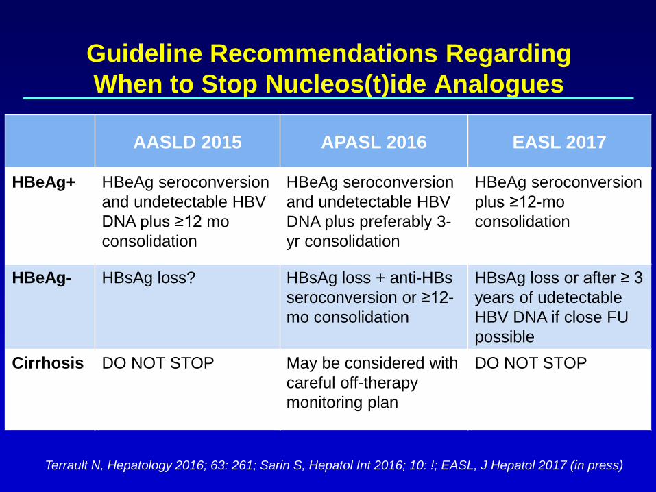
Guideline Recommendations Regarding
When to Stop Nucleos(t)ide Analogues
AASLD 2015 APASL 2016 EASL 2017
HBeAg+ HBeAg seroconversion
and undetectable HBV
DNA plus ≥12 mo
consolidation
HBeAg seroconversion
and undetectable HBV
DNA plus preferably 3-
yr consolidation
HBeAg seroconversion
plus ≥12-mo
consolidation
HBeAg- HBsAg loss? HBsAg loss + anti-HBs
seroconversion or ≥12-
mo consolidation
HBsAg loss or after ≥ 3
years of udetectable
HBV DNA if close FU
possible
Cirrhosis DO NOT STOP May be considered with
careful off-therapy
monitoring plan
DO NOT STOP
Terrault N, Hepatology 2016; 63: 261; Sarin S, Hepatol Int 2016; 10: !; EASL, J Hepatol 2017 (in press)

Risks of Stopping
Nucleos(t)ide Analogues
• Risk of relapse
– HBeAg+ patients who completed ≥12 mos
consolidation therapy after HBeAg seroconversion:
10%-50% viral relapse
– HBeAg- patients who completed >2 yr treatment:
100% viral relapse, ~40% sustained clinical relapse
• Risk of hepatic decompensation
– Limited data, ~3% among cirrhotics
– Depends on vigilance of post-treatment monitoring
