healing of rotator cuff tendons using botulinum toxin a and immobilization … · 2017-08-25 ·...
TRANSCRIPT
RESEARCH ARTICLE Open Access
Healing of rotator cuff tendons usingbotulinum toxin A and immobilization in arat modelMohit N. Gilotra1*, Michael J. Shorofsky1, Jason A. Stein2 and Anand M. Murthi2
Abstract
Background: We evaluated effects of botulinum toxin A (Botox) and cast immobilization on tendon healing in a ratmodel. Injection of Botox into rat supraspinatus was hypothesized to reduce muscle active force and improvedhealing.
Methods: Eighty-four supraspinatus tendons were surgically transected and repaired in 42 Sprague-Dawley rats(transosseous technique). After repair, supraspinatus muscle was injected with saline or Botox (3 or 6 U/kg). Half theshoulders were cast-immobilized for the entire postoperative period; half were allowed free cage activity. Histologywas examined at 2, 4, 8, and 12 weeks. A healing zone cross-sectional area was measured, and biomechanicaltesting of repair strength and tendon viscoelastic properties was conducted at 4 and 12 weeks.
Results: Botox alone and cast immobilization alone exhibited increased ultimate load compared with controls(saline injection, no immobilization) at 4 weeks. No difference in ultimate load occurred between Botox-onlyand cast-only groups. At 12 weeks, the Botox (6 U/kg) plus cast immobilization group was significantly weakest(p < 0.05). A trend was shown toward decreased healing zone cross-sectional areas in casted groups.
Conclusions: Supraspinatus Botox injection after rotator cuff repair might help protect the repair. However, castimmobilization plus Botox administration is harmful to rotator cuff healing in a rat tendon model.
Keywords: Rotator cuff repair, Botulinum toxin A (Botox), Cast immobilization, Tendon healing, Rat model
BackgroundRotator cuff disorders account for 10 % of all shoulderpain in today’s aging population. Direct costs for shoul-der treatment in the United States in the year 2000reached US$7 billion, with no sign of this trend revers-ing [1]. Rates of re-rupture are as high as 90 %, depend-ing on type of imaging, and some rotator cuff repairs donot heal [2]. When the rotator cuff does not heal after arepair, pain might be decreased initially but function andstrength decline [2, 3].Predictive factors that place a repair at risk include
patient age, tear size, time from injury to repair, andpostoperative activity level [4]. Various protocols ofimmobilization have been recommended to allow the
rotator cuff to heal after repair without jeopardizing thelong-term range of motion of the shoulder, but this tenethas been controversial [5]. Most surgeons have preferredinitial immobilization, passive range of motion, andgradual progression to active assisted range of motion[6]. Because of patient compliance issues and biologicalchallenges [7–10], optimizing the postoperative mechan-ical environment remains difficult.Botulinum toxin A (Botox; Allergan, Inc., Irvine, CA)
has been explored as a means of complete paralytic in-ternal immobilization or “bioprotection” [11–13] in thesetting of Achilles tendon and rotator cuff healing. Ourpurpose was to examine rotator cuff healing with intra-muscular Botox injection in comparison with strictimmobilization and with a non-immobilized saline con-trol. We explored this question using two low doses ofbotulinum toxin A: 3 and 6 U/kg. The main objectivewas to learn whether a single Botox injection obviates
* Correspondence: [email protected] of Orthopaedics, University of Maryland School of Medicine,Baltimore, MD 21201, USAFull list of author information is available at the end of the article
© 2016 Gilotra et al. Open Access This article is distributed under the terms of the Creative Commons Attribution 4.0International License (http://creativecommons.org/licenses/by/4.0/), which permits unrestricted use, distribution, andreproduction in any medium, provided you give appropriate credit to the original author(s) and the source, provide a link tothe Creative Commons license, and indicate if changes were made. The Creative Commons Public Domain Dedication waiver(http://creativecommons.org/publicdomain/zero/1.0/) applies to the data made available in this article, unless otherwise stated.
Gilotra et al. BMC Musculoskeletal Disorders (2016) 17:127 DOI 10.1186/s12891-016-0978-y
the need for cast immobilization in a rodent rotator cuffinjury model.We hypothesized that botulinum toxin A injection
into the rat supraspinatus muscle reduces the muscle’sactive contractile force, allowing for improved healing atthe supraspinatus footprint. Although no consensus hasbeen reached regarding what “healing” truly is, weadapted the concept of a healing zone or the area of scarformation as healed to the greater tuberosity footprint.
MethodsAnimal modelAll animal procedures were approved by the Institu-tional Animal Care and Use Committee at the Universityof Maryland. Bilateral supraspinatus tendons of 42male Sprague-Dawley skeletally immature rats (weight,400–450 g) were surgically transected and repairedwith a transosseous technique based on a well-establishedprotocol [14–16]. Bilateral rotator cuff surgery has beenstandardized in multiple animal protocols [17, 18].The animals were anesthetized with 5 % isoflurane in-
duction and 2 % isoflurane maintenance delivered in anoxygen mixture via face masks. After clipping and sterilepreparation were completed, a 1-cm incision was madeover the posterior aspect of the shoulder. The postero-lateral aspect of the deltoid was removed from the acro-mion. The supraspinatus was exposed with adduction ofthe extremity and forearm supination. A 5.0 Prolene su-ture (Ethicon, Somerville, NJ) was passed once throughthe supraspinatus for traction. The tendon was sharplytransected with a No. 15 blade and firmly secured withcompletion of a locking stitch using a modified Mason-Allen technique. The anatomic footprint was burred athigh speed, and a 0.5-mm drill hole was aimed in acranial-caudal direction. The suture was passed throughthe drill hole and tied with the supraspinatus reapproxi-mated to its anatomic footprint on the greater tuberos-ity. This rat injury model is analogous to an acuterotator cuff tear and not a chronic tear.Rat shoulders were randomly allocated by a random-
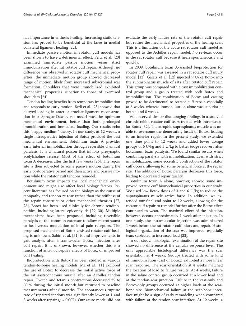
number generator into six experimental groups (Fig. 1),
ensuring one shoulder for a cast and one to be free fromimmobilization while housed in individual cages andallowed free cage activity. A power analysis (Stata 11;StataCorp LP, College Station, TX) that was conductedbased on pilot work assessing ultimate load revealed that60 shoulders were needed in total to provide 80 % powerto detect a 30 % difference in ultimate load betweengroups with a p value of 0.05. One shoulder in eachgroup was reserved for a strictly qualitative histologicalevaluation. Five shoulders in each group underwentbiomechanic evaluation at 4 and 12 weeks, which wasthe main quantitative outcome of the study. Thesupraspinatus muscle was injected intraoperativelywith either saline or botulinum toxin A after repair.The Botox dose administered was either 3 or 6 U/kg,based on previous small-animal bioprotection litera-ture [11, 12]. Dilutions were standardized and volumewas equal for all injections. The control group received sa-line injection into the supraspinatus and no castimmobilization. Half the shoulders remained in caststhroughout the postoperative period. One shoulder ofeach animal was randomly allocated to an immobilizedtreatment group and the contralateral shoulder to a non-immobilized treatment group. All rats were therebysubjected to a cast and the same physiological stress. Aunilateral shoulder was immobilized at 90° of forwardflexion and 20° of abduction, which is a position of neutralweight bearing for the Sprague-Dawley rat. The paw andforearm were not included in the cast, and the animal wasallowed to freely bear weight on both paws.Analgesia (buprenorphine, 0.03 mg/kg, administered
every 12 h) was provided by animal care staff during theimmediate postoperative period. Animals were examineddaily for signs of distress and cast discomfort. Ratsunderwent an obligatory cast change every other week.All cast changes were performed with the animals underanesthesia in a controlled fashion to prevent excessivepassive manipulation of the shoulder.Animals were euthanized at 2, 4, 8, and 12 weeks after
rotator cuff repair. The entire supraspinatus muscle, ten-don, and scar unit were harvested en bloc with the prox-imal humerus. Visual inspection revealed no gross,macroscopic repair failures. Histological analysis wasconducted at all time points. Biomechanical testing wascompleted at the 4- and 12-week time points.
Histological analysisSpecimens from each experimental group and all timepoints underwent histological analysis. Muscle tendonunits were fixed in 4 % paraformaldehyde overnight andthen underwent 2 days of gentle decalcification in 14 %ethylenediaminetetraacetic acid. The specimens werenext embedded in paraffin, sectioned with a microtomeinto 3- to 4-μm sections, and dried. Samples were
Fig. 1 Chart clarifies treatment groups. Shoulders in three of sixtreatment (Tx) groups remained in casts during the entirepostoperative period. Btx = Botox
Gilotra et al. BMC Musculoskeletal Disorders (2016) 17:127 Page 2 of 8

stained with toluidine blue for fibrocartilage detectionand Masson trichrome blue for fibrous tissue detection.Hematoxylin and eosin testing was conducted to assessmuscle cell morphology. Specimens were evaluated by ablinded pathologist specifically for fat formation in themuscle, collagen orientation in the repair, and the orderof fibrocartilage proliferation.
Biomechanical testingSixty shoulders from the 4- and 12-week groups were bio-mechanically tested for repair strength and tendon visco-elastic properties, as previously described [17–19]. Theacromion and deltoid were initially removed, and subper-iosteal dissection of the supraspinatus from its fossa wasperformed. The humerus and tendon were dissected free,and all excess soft-tissue constraints were removed fromthe humeral head. The humerus-tendon unit was storedin phosphate-buffered saline at 4 °C overnight for testingthe next day. The insertion-site scar was measured withdigital calipers, and an approximate area was determinedby using an elliptical assumption: area = π × thickness ×width. A three-dimensional scar volume was calculatedbased on the elliptical area times the scar length.The distal portion of the humerus was potted in poly-
methylmethacrylate. The tendon was then fixed betweentwo pieces of sand paper and was clamped at 90° to thesupraspinatus tendon (Fig. 2). Biomechanical testing wasconducted in a mini-materials testing machine (Electro-Force TestBench system; Bose Corporation, Eden Prairie,MN).The biomechanics loading protocol was based on a
well-established model for testing rat rotator cuff repair[12, 16]. Specimens were initially preloaded to 0.2 N,and then 5 cycles of preconditioning were applied to astrain of 5 %. The strain measurement was based on thedistance from the clamp to the tendon insertion. The
conditioning rate remained a constant 0.1 % per secondfor examination of viscoelastic properties. The specimenwas then stress-relaxed at 5 % strain for 5 min. Tendonswere loaded to failure at a constant rate (0.1 % per sec-ond). Stress was calculated as tensile force applied ÷ initialcalculated cross-sectional area. Stiffness was same force ÷linear displacement, whereas the tendon-scar moduluswas extrapolated from the linear portion of the stress-strain curve. Ultimate load and stress were determinedbased on the point of failure. Anatomic location of failurewas recorded as one of three categories: tendon-scar inter-face, scar-bone interface, or indeterminate (Fig. 3).
Statistical analysisBiomechanical results were subjected to a two-factoranalysis of variance and a Tukey post hoc test to allowfor multiple-group analysis with all groups comparedwith only the non-treated (no Botox or cast) group. Stat-istical significance was set at p < 0.05. Anatomic site offailure was strictly observational, and statistical analysiswas not conducted on histological results because theywere qualitative.
ResultsBefore euthanization, all animals survived the treatmentin good health. No adverse events occurred.
HistologyMuscle atrophy was observed on gross examination; fattyinfiltration was noted on a cellular level. The two groupssubjected to both Botox injection and immobilization ex-hibited wasting in the supraspinatus fossa. The findingswere corroborated with histological findings of extensive
Fig. 2 Harvested sample is shown. Supraspinatus tendon (thick arrow)is clamped at 90° to the humeral head (thin arrow) before load analysis
Fig. 3 Areas of failure. a Failure at the tendon-scar junction. b Failureat the scar-bone interface. Bracket = scar, arrow = point of failure
Gilotra et al. BMC Musculoskeletal Disorders (2016) 17:127 Page 3 of 8

fat infiltration. Cast immobilization alone at 12 weeksechoed similar results: atrophy shown on gross examin-ation and fatty infiltration on histological examination.At 2 and 4 weeks, Botox-only groups had no grosssupraspinatus atrophy but some fatty infiltration wasrevealed by histological examination.At 8 and 12 weeks, however, the Botox-only groups
had no fatty findings. At 2 weeks, all specimens demon-strated a disorganized fibrocartilage scar at the tendon-bone interface. At 8 and 12 weeks, no difference wasobserved in scar organization among all groups. At 4 weeks,shoulders subjected to Botox only, cast immobilizationonly, or Botox plus cast immobilization showed a morelinear formation of collagen compared with the salinecontrol group (Fig. 4).
BiomechanicsThe ultimate load of the repaired supraspinatus tendon inthe 4-week samples showed that the tendons receiving
either dose of Botox alone and those receiving castimmobilization alone were significantly stronger thanthose in the control group (p < 0.05). At 12 weeks, in-creased load to failure was shown for the Botox-only(6 U/kg dose) group compared with the cast-onlygroup and the saline-only control group (Fig. 5). Botox(6 U/kg) in combination with cast immobilization was sig-nificantly the weakest group at both time points (p < 0.05).No statistical difference was observed in the healing
zone cross-sectional scar area or scar volume acrossgroups. The cast-only group at 12 weeks tended to havedecreased scar volume compared with the control group.No difference in stiffness was observed at 4 weeks
(Fig. 6). Shoulders that received 6 U/kg Botox were theleast stiff compared with the 12-week groups and weresignificantly different from control group shoulders atthe 12-week time point (p < 0.05). At 4 weeks, 21 (88 %)of 24 repairs in the cast-only and Botox-only groupscombined failed at the scar-bone interface. All the saline
Fig. 4 Comparison of photomicrographs. a Non-immobilized saline control. b Botox 6 U/kg alone. c Cast alone. Masson’s trichrome blue stain(original magnification, 10×) of scar at 4 weeks shows a disorganized scar for the non-immobilized control and a more organized scar for theBotox-only and cast-only specimens. Rectangles indicate regions of interest
Gilotra et al. BMC Musculoskeletal Disorders (2016) 17:127 Page 4 of 8

control group repairs, however, failed at the tendon-scarjunction. At 12 weeks, gross inspection showed that allspecimens failed at the scar-bone interface.
DiscussionWe assessed whether intraoperative Botox injection op-timizes the early postoperative mechanical environmentin a rat rotator cuff healing model. We paralyzed thesupraspinatus with botulinum toxin A after rotator cuffinjury and repair. We used a lower dose of Botox thanthat used in previous studies [11, 12] to allow some loadat the repair site. Half the shoulders were additionally
treated with immobilization, and the other half wereallowed free cage activity. Cast immobilization in com-bination with 6 U/kg Botox paralysis impaired the repairquality. Botox treatment combined with free cage activ-ity provided repair strength similar to that provided byimmobilization. Some force at the repair site is necessaryfor tendon healing. Early active motion has been shownto be beneficial in preserving hand motion for flexortendon repairs without hampering the healing process[20]. These positive effects of early motion result in adecrease in adhesion formation but might not improvethe quality of the healing tissue [21]. Early motion also
Fig. 5 Bar chart depicts force. Ultimate load at 4 and 12 weeks. At 4 weeks, Botox-only and cast-only groups were significantly stronger than thecontrol group. At 12 weeks, Botox 6 U/kg was the strongest and 6 U/kg with cast immobilization was the weakest. Asterisk = significantvalues (p < 0.05) after Tukey post hoc test. Error bars correspond to standard deviations
Fig. 6 Bar chart depicts stiffness. Shoulders that received 6 U/kg Botox were least stiff at 12 weeks compared with control group shoulders.Asterisk = significant values (p < 0.05) after Tukey post hoc test. Error bars correspond to standard deviations
Gilotra et al. BMC Musculoskeletal Disorders (2016) 17:127 Page 5 of 8
has importance in enthesis healing. Increasing static ten-sion has proved to be beneficial at the knee in medialcollateral ligament healing [22].Immediate passive motion in rotator cuff models has
been shown to have a detrimental effect. Peltz et al. [23]examined immediate passive motion versus strictimmobilization after rat rotator cuff repair. Although nodifference was observed in rotator cuff mechanical prop-erties, the immediate motion group showed decreasedrange of motion, likely from increased subacromial scarformation. Shoulders that were immobilized exhibitedmechanical properties superior to those of exercisedshoulders [24].Tendon healing benefits from temporary immobilization
and responds to early motion. Bedi et al. [25] showed thatdelayed loading in anterior cruciate ligament reconstruc-tion in a Sprague-Dawley rat model was the optimummechanical environment, better than both prolongedimmobilization and immediate loading. Our results echothis “happy medium” theory. In our study, at 12 weeks, asingle intraoperative injection of Botox provided the bestmechanical environment. Botulinum toxin A providesearly internal immobilization through reversible chemicalparalysis. It is a natural poison that inhibits presynapticacetylcholine release. Most of the effect of botulinumtoxin A decreases after the first few weeks [26]. The repairsite is then subjected to some passive motion during theearly postoperative period and then active and passive mo-tion while the rotator cuff tendons remodel.Botulinum toxin impacts the local mechanical envir-
onment and might also affect local biology factors. Re-cent literature has focused on the biology as the cause oftenopathy and tendon re-tear rather than the stiffness ofthe repair construct or other mechanical theories [27,28]. Botox has been used clinically for chronic tendino-pathies, including lateral epicondylitis [29, 30]. Multiplemechanisms have been proposed, including reversibleparalysis of the common extensor to allow microtraumato heal versus modulation of local pain receptors. Theproposed mechanism of Botox-assisted rotator cuff heal-ing is unknown. Şahin et al. [31] found improvements ingait analysis after intramuscular Botox injection aftercuff repair. It is unknown, however, whether this is afunction of anti-nociceptive effects of Botox or improvedcuff healing.Bioprotection with Botox has been studied in various
tendon-to-bone healing models. Ma et al. [11] exploredthe use of Botox to decrease the initial active force ofthe rat gastrocnemius muscle after an Achilles tendonrepair. Twitch and tetanus contractions decreased 25 to50 % during the initial month but returned to baselinemeasurements after 6 months. The spontaneous rupturerate of repaired tendons was significantly lower at 1 and3 weeks after repair (p = 0.007). Our acute model did not
evaluate the early failure rate of the rotator cuff repairbut rather the mechanical properties of the healing scar.This is a limitation of the acute rat rotator cuff model asopposed to the Achilles repair model. No re-tears occurin the rat rotator cuff because it heals spontaneously andquickly.In 2009, botulinum toxin A-assisted bioprotection for
rotator cuff repair was assessed in a rat rotator cuff injurymodel [12]. Galatz et al. [12] injected 9 U/kg Botox intothe supraspinatus muscle of rats after rotator cuff repair.This group was compared with a cast immobilization con-trol group and a group treated with both Botox andimmobilization. The combination of Botox and castingproved to be detrimental to rotator cuff repair, especiallyat 8 weeks, whereas immobilization alone was superior atboth 4 and 8 weeks.We observed similar discouraging findings in a study of
chronic rabbit rotator cuff tears treated with intramuscu-lar Botox [32]. The atrophic supraspinatus muscle was un-able to overcome the denervating insult of Botox, leadingto an inferior repair. In the present study, we extendedone time point to 12 weeks and added lower dosagegroups of 6 U/kg and 3 U/kg to better judge recovery afterbotulinum toxin paralysis. We found similar results whencombining paralysis with immobilization. Even with strictimmobilization, some eccentric contraction of the rotatorcuff occurs, allowing for some beneficial force at the repairsite. The addition of Botox paralysis decreases this force,leading to decreased repair quality.Botulinum toxin A alone, however, showed some im-
proved rotator cuff biomechanical properties in our study.We used low Botox doses of 3 and 6 U/kg to reduce thesupraspinatus muscle active force. In addition, we ex-tended our final end point to 12 weeks, allowing for therotator cuff repair to remodel further after the Botox effectcontinued to wean. The maximal effect of the injection,however, occurs approximately 1 week after injection. Inone study, the intramuscular injection was administered1 week before the rat rotator cuff injury and repair. Histo-logical organization of the scar was improved, especiallytears subjected to increased load [33].In our study, histological examination of the repair site
showed no difference at the cellular response level. Theonly appreciable histological difference was the scarorientation at 4 weeks. Groups treated with some kindof immobilization (cast or Botox) exhibited a more linearscar response. The scar orientation at 4 weeks matchedthe location of load to failure results. At 4 weeks, failurein the saline control group occurred at a lower load andat the tendon-scar junction. Failure in the cast-only andBotox-only groups occurred at higher loads at the scar-bone site. Biomechanical failure at the scar-bone inter-face might be a sign of early remodeling when comparedwith failure at the tendon-scar interface. At 12 weeks, a
Gilotra et al. BMC Musculoskeletal Disorders (2016) 17:127 Page 6 of 8
majority of the constructs showed a linear scar responseand failure at the scar-bone site.This study used a well-established rat rotator cuff in-
jury model that included both cast immobilizationgroups and a saline-only control group. The biomechan-ical and histological protocols have been well described[34], rendering our results comparable with those ofother rodent models. We studied multiple treatmentgroups and varied the dose of botulinum toxin A. Inaddition, we recorded the site of failure, which has notbeen previously described in the literature. Our studyprovides important information in the setting of anacute rotator cuff tear model.A limitation of this study was the acute repair model
in a quadruped. Rotator cuff repairs with healing diffi-culties typically are chronic in nature. It is possible thatoptimizing the mechanical environment with Botoxmight not overcome the poor biological condition ofchronic rotator cuff degeneration. In addition, rotatorcuff muscles in the setting of a chronic tear exhibit base-line fatty degeneration and atrophy [35, 36]. The additiveparalysis effect of Botox in this setting is unknown andmight cause a more deleterious fatty response, as previ-ously shown [32]. The exact paralytic effect of Botox ona small rat supraspinatus muscle is unknown withoutspecific functional muscle testing. Therefore, we areunable to quantify the paralyzed “load” exacted on therepair site.The literature on the topic of botulinum toxin-
assisted rotator cuff repair is confusing. Multiple fac-tors need to be considered, including timing of injec-tion, acute versus chronic repair, combining chemicalparalysis with immobilization, and the species of theanimal model [12, 31–33]. Botox might have its besteffect when injected before an acute repair with minimalimmobilization. Testing in an animal model that exhibitsa clinically relevant incidence of re-tear would help to re-solve a current controversy in the literature.
ConclusionsBotulinum toxin A injection into the supraspinatus mightobviate the need for postoperative immobilization. Chem-ical paralysis in addition to cast immobilization reducesload at the repair site and is harmful to rotator cuff repair.Clinical implications include potentially decreased postop-erative immobilization time, better long-term functionaloutcomes, improved compliance, and reduced stiffnesswith increased protection of tendon repair sites.
Competing interestsThe authors declare that they have no competing interests.
Author contributionsMNG participated in the study design, animal surgeries, analysis of thefindings, and drafting of the final manuscript. MJS helped perform animalsurgeries, conducted data analysis, and wrote part of the manuscript. JAS
conceived the study and participated in the study design and animalsurgeries. AMM participated in the study design and helped draft the finalmanuscript. All authors read and approved the final manuscript.
AcknowledgmentThe authors thank Senior Editor and Writer Dori Kelly, MA, for professionalmanuscript editing and figure formatting. No outside funding was receivedfor this study.
Author details1Department of Orthopaedics, University of Maryland School of Medicine,Baltimore, MD 21201, USA. 2Department of Orthopaedics and SportsMedicine, MedStar Union Memorial Hospital, Baltimore, MD 21218, USA.
Received: 22 October 2015 Accepted: 8 March 2016
References1. Meislin RJ, Sperling JW, Stitik TP. Persistent shoulder pain: epidemiology,
pathophysiology, and diagnosis. Am J Orthop (Belle Mead NJ). 2005;34suppl 12:5–9.
2. Galatz LM, Ball CM, Teefey SA, Middleton WD, Yamaguchi K. The outcomeand repair integrity of completely arthroscopically repaired large andmassive rotator cuff tears. J Bone Joint Surg Am. 2004;86(2):219–24.
3. Kim HM, Teefey SA, Zelig A, Galatz LM, Keener JD, Yamaguchi K. Shoulderstrength in asymptomatic individuals with intact compared with tornrotator cuffs. J Bone Joint Surg Am. 2009;91(2):289–96.
4. Sarver JJ, Peltz CD, Dourte L, Reddy S, Williams GR, Soslowsky LJ. Afterrotator cuff repair, stiffness—but not the loss in range ofmotion—increased transiently for immobilized shoulders in a rat model. JShoulder Elbow Surg. 2008;17 suppl 1:108S–13S.
5. Parsons BO, Gruson KI, Chen DD, Harrison AK, Gladstone J, Flatow EL. Doesslower rehabilitation after arthroscopic rotator cuff repair lead to long-termstiffness? J Shoulder Elbow Surg. 2010;19(7):1034–9.
6. Trenerry K, Walton JR, Murrell GA. Prevention of shoulder stiffness afterrotator cuff repair. Clin Orthop Relat Res. 2005;430:94–9.
7. Blevins FT, Djurasovic M, Flatow EL, Vogel KG. Biology of the rotator cufftendon. Orthop Clin North Am. 1997;28(1):1–16.
8. Roddey TS, Olson SL, Gartsman GM, Hanten WP, Cook KF. A randomizedcontrolled trial comparing 2 instructional approaches to home exerciseinstruction following arthroscopic full-thickness rotator cuff repair surgery. JOrthop Sports Phys Ther. 2002;32(11):548–59.
9. Rodeo SA. Biologic augmentation of rotator cuff tendon repair. J ShoulderElbow Surg. 2007;16 suppl 5:S191–7.
10. Kovacevic D, Rodeo SA. Biological augmentation of rotator cuff tendonrepair. Clin Orthop Relat Res. 2008;466:622–33.
11. Ma J, Shen J, Smith BP, Ritting A, Smith TL, Koman LA. Bioprotection oftendon repair: adjunctive use of botulinum toxin A in Achilles tendon repairin the rat. J Bone Joint Surg Am. 2007;89(10):2241–9.
12. Galatz LM, Charlton N, Das R, Kim HM, Havlioglu N, Thomopoulos S.Complete removal of load is detrimental to rotator cuff healing. J ShoulderElbow Surg. 2009;18(5):669–75.
13. Thomopoulos S, Kim HM, Rothermich SY, Biederstadt C, Das R, Galatz LM.Decreased muscle loading delays maturation of the tendon enthesis duringpostnatal development. J Orthop Res. 2007;25(9):1154–63.
14. Carpenter JE, Thomopoulos S, Flanagan CL, DeBano CM, Soslowsky LJ.Rotator cuff defect healing: a biomechanical and histologic analysis in ananimal model. J Shoulder Elbow Surg. 1998;7(6):599–605.
15. Soslowsky LJ, Carpenter JE, DeBano CM, Banerji I, Moalli MR. Developmentand use of an animal model for investigations on rotator cuff disease. JShoulder Elbow Surg. 1996;5(5):383–92.
16. Galatz LM, Silva MJ, Rothermich SY, Zaegel MA, Havlioglu N, ThomopoulosS. Nicotine delays tendon-to-bone healing in a rat shoulder model. J BoneJoint Surg Am. 2006;88(9):2027–34.
17. Kim HM, Galatz LM, Das R, Havlioglu N, Rothermich SY, Thomopoulos S. Therole of transforming growth factor beta isoforms in tendon-to-bone healing.Connect Tissue Res. 2011;52(2):87–98.
18. Galatz LM, Rothermich SY, Zaegel M, Silva MJ, Havlioglu N, Thomopoulos S.Delayed repair of tendon to bone injuries leads to decreased biomechanicalproperties and bone loss. J Orthop Res. 2005;23(6):1441–7.
Gilotra et al. BMC Musculoskeletal Disorders (2016) 17:127 Page 7 of 8

19. Perry SM, Gupta RR, Van Kleunen J, Ramsey ML, Soslowsky LJ, Glaser DL. Useof small intestine submucosa in a rat model of acute and chronic rotatorcuff tear. J Shoulder Elbow Surg. 2007;16 suppl 5:S179–83.
20. Trumble TE, Vedder NB, Seiler III JG, Hanel DP, Diao E, Pettrone S. Zone-IIflexor tendon repair: a randomized prospective trial of active place-and-holdtherapy compared with passive motion therapy. J Bone Joint Surg Am.2010;92(6):1381–9.
21. Zhao C, Amadio PC, Momose T, Couvreur P, Zobitz ME, An KN. Effect ofsynergistic wrist motion on adhesion formation after repair of partial flexordigitorum profundus tendon lacerations in a canine model in vivo. J BoneJoint Surg Am. 2002;84(1):78–84.
22. Gomez MA, Woo SL, Amiel D, Harwood F, Kitabayashi L, Matyas JR. Theeffects of increased tension on healing medical collateral ligaments. Am JSports Med. 1991;19(4):347–54.
23. Peltz CD, Dourte LM, Kuntz AF, Sarver JJ, Kim SY, Williams GR, et al. Theeffect of postoperative passive motion on rotator cuff healing in a ratmodel. J Bone Joint Surg Am. 2009;91(10):2421–9.
24. Thomopoulos S, Williams GR, Soslowsky LJ. Tendon to bone healing:differences in biomechanical, structural, and compositional properties dueto a range of activity levels. J Biomech Eng. 2003;125(1):106–13.
25. Bedi A, Kovacevic D, Fox AJ, Imhauser CW, Stasiak M, Packer J, et al. Effect ofearly and delayed mechanical loading on tendon-to-bone healing afteranterior cruciate ligament reconstruction. J Bone Joint Surg Am.2010;92(14):2387–401.
26. Wenzel RG. Pharmacology of botulinum neurotoxin serotype A. Am JHealth Syst Pharm. 2004;61(22 suppl 6):S5–10.
27. Oliva F, Osti L, Padulo J, Maffulli N. Epidemiology of the rotator cuff tears: anew incidence related to thyroid disease. Muscles Ligaments Tendons J.2014;4(3):309–14.
28. Via AG, De Cupis M, Spoliti M, Oliva F. Clinical and biological aspects ofrotator cuff tears. Muscles Ligaments Tendons J. 2013;3(2):70–9.
29. Morré HH, Keizer SB, van Os JJ. Treatment of chronic tennis elbow withbotulinum toxin. Lancet. 1997;349(9067):1746.
30. Kahlenberg CA, Knesek M, Terry MA. New developments in the use ofbiologics and other modalities in the management of lateral epicondylitis.Biomed Res Int. 2015;2015:439309.
31. Şahin E, Kalem M, Zehir S, Songur M, Demirtaş M. Effect of intramuscularbotulinum toxin-A in a rat rotator cuff repair model: an experimental study.Acta Orthop Traumatol Turc. 2015;49(4):447–52.
32. Gilotra M, Nguyen T, Christian M, Davis D, Henn 3rd RF, Hasan SA.Botulinum toxin is detrimental to repair of a chronic rotator cuff tear in arabbit model. J Orthop Res. 2015;33(8):1152–7.
33. Ficklscherer A, Hartl TK, Scharf M, Sievers B, Schröder C, Milz S, et al. Effectsof selective paralysis of the supraspinatus muscle using botulinumneurotoxin a in rotator cuff healing in rats. J Orthop Res. 2013;31(5):716–23.
34. Rooney SI, Loro E, Sarver JJ, Peltz CD, Hast MW, Tseng WJ, et al. Exerciseprotocol induces muscle, tendon, and bone adaptations in the rat shoulder.Muscles Ligaments Tendons J. 2015;4(4):413–9.
35. Luan T, Liu X, Easley JT, Ravishankar B, Puttlitz C, Feeley BT. Muscle atrophyand fatty infiltration after an acute rotator cuff repair in a sheep model.Muscles Ligaments Tendons J. 2015;5(2):106–12.
36. Tae SK, Oh JH, Kim SH, Chung SW, Yang JY, Back YW. Evaluation of fattydegeneration of the supraspinatus muscle using a new measuring tool andits correlation between multidetector computed tomography and magneticresonance imaging. Am J Sports Med. 2011;39(3):599–606.
• We accept pre-submission inquiries
• Our selector tool helps you to find the most relevant journal
• We provide round the clock customer support
• Convenient online submission
• Thorough peer review
• Inclusion in PubMed and all major indexing services
• Maximum visibility for your research
Submit your manuscript atwww.biomedcentral.com/submit
Submit your next manuscript to BioMed Central and we will help you at every step:
Gilotra et al. BMC Musculoskeletal Disorders (2016) 17:127 Page 8 of 8