hearing loss: mechanisms revealed by genetics and cell biology
TRANSCRIPT

ANRV394-GE43-18 ARI 10 October 2009 10:48
Hearing Loss: MechanismsRevealed by Geneticsand Cell BiologyAmiel A. Dror and Karen B. AvrahamDepartment of Human Molecular Genetics and Biochemistry, Sackler School of Medicine,Tel Aviv University, Tel Aviv 69978, Israel; email: [email protected],[email protected]
Annu. Rev. Genet. 2009. 43:411–37
First published online as a Review in Advance onAugust 20, 2009
The Annual Review of Genetics is online atgenet.annualreviews.org
This article’s doi:10.1146/annurev-genet-102108-134135
Copyright c© 2009 by Annual Reviews.All rights reserved
0066-4197/09/1201-0411$20.00
Key Words
inner ear, deafness, cochlea, hair cell, mouse, mutation
AbstractHearing loss (HL), or deafness in its most severe form, affects an esti-mated 28 and 22.5 million Americans and Europeans, respectively. Thenumbers are higher in regions such as India and the Middle East, whereconsanguinity contributes to larger numbers of recessively inheritedhearing impairment (HI). As a result of work-related difficulties, edu-cational and developmental delays, and social stigmas and exclusion, theeconomic impact of HL is very high. At the other end of the spectrum,a rich deaf culture, particularly for individuals whose parents and evengrandparents were deaf, is a social movement that believes that deaf-ness is a difference in human experience rather than a disability. Thisreview attempts to cover the remarkable progress made in the field ofthe genetics of HL over the past 20 years. Mutations in a significantnumber of genes have been discovered over the years that contributeto clinically heterogeneous forms of HL, enabling genetic counselingand prediction of progression of HL. Cell biological assays, proteinlocalization in the inner ear, and detailed analysis of spontaneous andtransgenic mouse models have provided an incredibly rich resource forelucidating mechanisms of hereditary hearing loss (HHL). This knowl-edge is providing answers for the families with HL, who contribute agreat deal to the research being performed worldwide.
411
Ann
u. R
ev. G
enet
. 200
9.43
:411
-437
. Dow
nloa
ded
from
ww
w.a
nnua
lrev
iew
s.or
gby
Mos
cow
Sta
te U
nive
rsity
- S
cien
tific
Lib
rary
of
Lom
onos
ov o
n 08
/29/
13. F
or p
erso
nal u
se o
nly.

ANRV394-GE43-18 ARI 10 October 2009 10:48
Cochlea: a snail-shaped organcomprises the auditoryapparatus of the innerear; highly similarbetween human andmouse
HGP: HumanGenome Project
HL: hearing loss
HHL: hereditaryhearing loss
HI: hearingimpairment
Cochlear duct: acomplex of a coiledduct that reaches fromthe basilar toward theapical portion of thecochlea
Endolymph: the fluidinside the scala media
Perilymph: the fluidinside the scalatympani and scalavestibuli
Hair cells: sensorycells of the auditorysystem that reside inthe sensory epitheliumof the cochlea
INTRODUCTION
The exquisite sensory transduction of thecochlea is dependent on a tremendous amountof synchronized processes and mechanisms thatrequire a large battery of protein-coding genesas well as regulatory elements. In the past twodecades, while the Human Genome Project(HGP; http://genomics.energy.gov) was on-going, extensive research on the genetics ofhearing loss (HL) was performed worldwide,leading to the discovery of many genes essen-tial for hearing. However, the cloning of theresponsible genes was not enough, and broadstudies on animal models were conducted inorder to provide a better understanding of thefunction of each gene and its relation to theauditory network. The striking similarities be-tween the human and mouse inner ears pavedthe way to the understanding of many processesand to the gathering of an enormous amount ofinformation about this organ in a way that couldnot have been achieved by just exploring the hu-man ear. In this review, we provide an overviewof the human ear and hereditary hearing loss(HHL), a historical perspective of the researchin this field, a description of protein familiesassociated with deafness grouped by function,and the contribution of mouse models to ourstudy of the pathology and etiology of heredi-tary hearing impairment (HI) in humans. An-other useful model system for studying HL isthe zebrafish, Danio rerio, which has been re-viewed in recent years (111) and will not befurther covered in this review. Understandingthe mechanisms of the inner ear pathways, bothin the functional and the pathological state, isenabling researchers to develop cell- and gene-based therapy treatments that may eventuallybe implemented in humans with HI.
The Complexity of MammalianInner Ear Structure
A mixture of sound waves is constantly gen-erated in the environment and reaches ourears. This rich collection of sounds is scram-bled and contains a wide spectrum of low andhigh tones in a variety of intensities. The ear
is an outstanding organ that can precisely deci-pher between different frequencies and intensi-ties (50), while simultaneously transferring thegathered information to the brain and enablingus to categorize each sound and learn about itsrelative distance and direction (71).
The ear is divided into three anatomicalcompartments: the outer, the middle, and theinner ear (Figure 1a). The main role of theouter ear and the middle ear is to conductthe sound waves from the environment intothe inner ear. The sound waves are capturedby the outer ear auricle and conducted throughthe ear canal to reach the eardrum membrane.The middle ear is composed of three smallbones, the ossicles, that are assembled togetherand serve as a link between the eardrum and theoval window of the fluid-filled inner ear. Uponsound stimulus, vibration of the eardrum willlead to a pistol-like movement of the middle earbones and to movement of the fluids inside theinner ear, the sensory portion of the ear (97).
The inner ear plays two pivotal roles inlife. It contains the sensory organs that enablehearing and that control our balance and spatialorientation. The concealed anatomical positionof the inner ear inside the human head, deeplyembedded in the temporal bone, provides aprotected shield to this tiny, sensitive sensoryorgan. It also serves as an acoustic chamber thatwill support capturing even a very low intensityof sound. The auditory apparatus of the innerear is composed of the cochlea, a snail-shapedorgan surrounded by the temporal bone(Figure 1a). It contains the cochlear ductthat runs along the spiral shape from baseto apex (Figure 1b). This complex, coiledduct is divided by two thin membranes intothree different compartments filled with fluids.The scala media is an endolymph-filled cavitylocated in between the scala vestibuli andscala tympani, perilymph-filled cavities. Thescala media contains the organ of Corti, whichis the sensory epithelium of the auditorysystem (reviewed in Reference 117). The organof Corti sits on the basilar membrane thatseparates the scala media and scala tympani.Specialized sensory cells called hair cells are
412 Dror · Avraham
Ann
u. R
ev. G
enet
. 200
9.43
:411
-437
. Dow
nloa
ded
from
ww
w.a
nnua
lrev
iew
s.or
gby
Mos
cow
Sta
te U
nive
rsity
- S
cien
tific
Lib
rary
of
Lom
onos
ov o
n 08
/29/
13. F
or p
erso
nal u
se o
nly.

ANRV394-GE43-18 ARI 10 October 2009 10:48
ed
c
ba
Inner hair cell
Top view by SEM
Side view by immunohistochemistry
Outer hair cells
Pillar cellsDeiters’ cells
Organ of Corti (sensory epithelium)
Hen
sen
cel
ls
Scalamedia
Scalavestibuli
Cochlear duct
Scalatympani
Cochlea
Ossicles
Tympanicmembrane
Vestibule
Middleear
Innerear
External ear
Ear canal
Temporalbone
Auditorynerve
Figure 1Schematic illustration of the human ear. (a) The ear consists of the outer, middle, and inner ear. (b) A sectionthrough the cochlear duct illustrates the fluid-filled compartments of the inner ear (modified from Reference43). (c) The organ of Corti resides in the scala media, with sensory hair cells surrounded by supporting cellsthat include Deiters’, Hensen, and pillar cells (modified from Reference 43). (d ) Immunohistochemistry withthe inner ear hair cell marker myosin VI, marking the cytoplasm of inner and outer hair cells, and4′,6-diamidino-2-phenylindole (DAPI), marking the nuclei. (e) Scanning electron microscopy image of thetop view of the sensory epithelium reveals the precise arrangement of one row of inner hair cells and threerows of outer hair cells, separated by the pillar cells. Figures designed and created by A. A. Dror.
www.annualreviews.org • Genetics of Hearing Loss 413
Ann
u. R
ev. G
enet
. 200
9.43
:411
-437
. Dow
nloa
ded
from
ww
w.a
nnua
lrev
iew
s.or
gby
Mos
cow
Sta
te U
nive
rsity
- S
cien
tific
Lib
rary
of
Lom
onos
ov o
n 08
/29/
13. F
or p
erso
nal u
se o
nly.

ANRV394-GE43-18 ARI 10 October 2009 10:48
Hair bundle: groupof stereocilia organizedin a typical staircasestructure residing onthe apical surface ofthe hair cells immersedin endolymph
embedded in this sensory epithelium, arrangedin a highly organized conformation of oneinner row and three outer rows of hair cells(Figure 1c–e, Figure 2a). The apical side ofthe hair cells facing the scala media containsactin-rich projections called stereocilia
(Figure 2b). The stereocilia have a typicalstaircase arrangement connected with lateraland tip links stabilizing the mature hair bundlestructure (Figure 2c; reviewed in References36 and 45). The hair cells are covered withthe tectorial membrane, a collagen-rich
Tectorin fibrils
eCollagen bundles
c
Tip li
nks
(Pcdh15 a
nd Cdh23)
Stereocilia
Outerhair cell
Cuticular plate
Cuticular plate
Cldn14
b
Kcnq4
Stereocilia
Outerhair cell
Su
pp
ortin
g ce
ll
Cx26/Cx30 gap junction
K+
K+
K+
K+
Pre
stin
dA
ctin
fil
am
en
ts
Myosin XVa
Myo
sin
V
IIa
Vlgr1/Usherin
Ankle links
Whirlin
Stereocilium
Myosin VI Myosin VI
Har
mo
nin
b
Har
mo
nin
bM
yosi
n
VII
a
Sa
ns
Tip links
Cdh23/Pcdh15
K+
K+
K+
K+
Kcnq1
Kcne1
Endolymph
Stria vascularismarginal cells
f
K+ recycling
Scala mediaendolymph
Scala vestibuliperilymph
Scala tympaniperilymph
Tectorial membrane
Reissner’s membrane
a
Figure 2Schematic illustration of portions of the inner ear, with expression of some proteins that play a role in inner ear function in humans.Only some proteins are shown. (a) Cross section through one turn of a mouse cochlea. (b) Illustration of a single outer hair cell (blue),surrounded by two Deiters’ supporting cells (brown). (c) Enlargement of the hair cell hair bundle with actin-based stereocilia (blue).(d ) A single stereocilium containing actin filaments. (e) Illustration of the tectorial membrane that is composed of thin tectorin fibrils( green) and heavy collagen bundles (blue). ( f ) Marginal cells of the stria vascularis. (Modified from Reference 42). Figures designed andcreated by A. A. Dror.
414 Dror · Avraham
Ann
u. R
ev. G
enet
. 200
9.43
:411
-437
. Dow
nloa
ded
from
ww
w.a
nnua
lrev
iew
s.or
gby
Mos
cow
Sta
te U
nive
rsity
- S
cien
tific
Lib
rary
of
Lom
onos
ov o
n 08
/29/
13. F
or p
erso
nal u
se o
nly.
ANRV394-GE43-18 ARI 10 October 2009 10:48
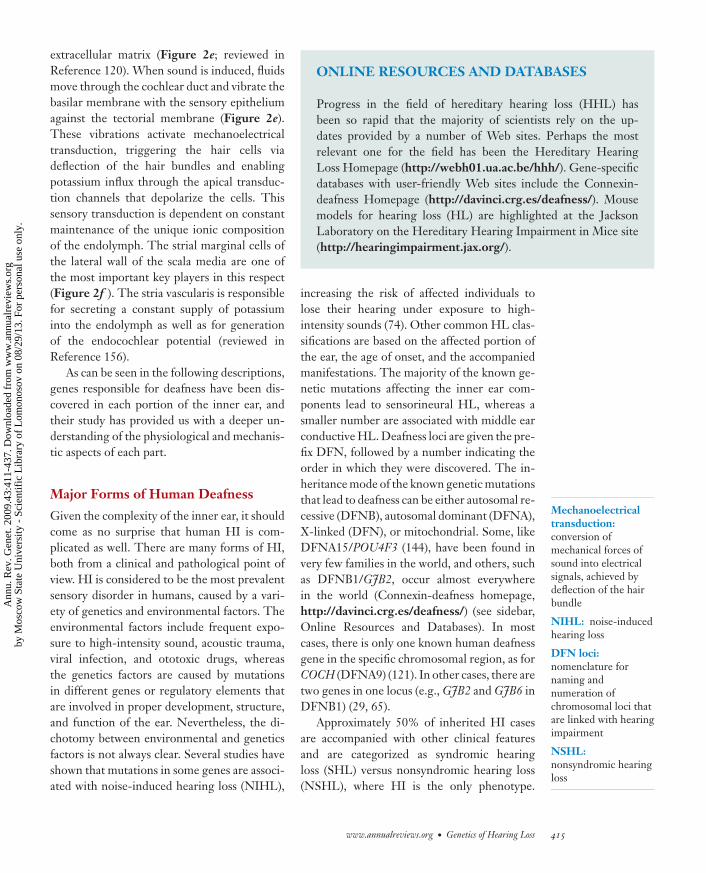
extracellular matrix (Figure 2e; reviewed inReference 120). When sound is induced, fluidsmove through the cochlear duct and vibrate thebasilar membrane with the sensory epitheliumagainst the tectorial membrane (Figure 2e).These vibrations activate mechanoelectricaltransduction, triggering the hair cells viadeflection of the hair bundles and enablingpotassium influx through the apical transduc-tion channels that depolarize the cells. Thissensory transduction is dependent on constantmaintenance of the unique ionic compositionof the endolymph. The strial marginal cells ofthe lateral wall of the scala media are one ofthe most important key players in this respect(Figure 2f ). The stria vascularis is responsiblefor secreting a constant supply of potassiuminto the endolymph as well as for generationof the endocochlear potential (reviewed inReference 156).
As can be seen in the following descriptions,genes responsible for deafness have been dis-covered in each portion of the inner ear, andtheir study has provided us with a deeper un-derstanding of the physiological and mechanis-tic aspects of each part.
Major Forms of Human Deafness
Given the complexity of the inner ear, it shouldcome as no surprise that human HI is com-plicated as well. There are many forms of HI,both from a clinical and pathological point ofview. HI is considered to be the most prevalentsensory disorder in humans, caused by a vari-ety of genetics and environmental factors. Theenvironmental factors include frequent expo-sure to high-intensity sound, acoustic trauma,viral infection, and ototoxic drugs, whereasthe genetics factors are caused by mutationsin different genes or regulatory elements thatare involved in proper development, structure,and function of the ear. Nevertheless, the di-chotomy between environmental and geneticsfactors is not always clear. Several studies haveshown that mutations in some genes are associ-ated with noise-induced hearing loss (NIHL),
Mechanoelectricaltransduction:conversion ofmechanical forces ofsound into electricalsignals, achieved bydeflection of the hairbundle
NIHL: noise-inducedhearing loss
DFN loci:nomenclature fornaming andnumeration ofchromosomal loci thatare linked with hearingimpairment
NSHL:nonsyndromic hearingloss
ONLINE RESOURCES AND DATABASES
Progress in the field of hereditary hearing loss (HHL) hasbeen so rapid that the majority of scientists rely on the up-dates provided by a number of Web sites. Perhaps the mostrelevant one for the field has been the Hereditary HearingLoss Homepage (http://webh01.ua.ac.be/hhh/). Gene-specificdatabases with user-friendly Web sites include the Connexin-deafness Homepage (http://davinci.crg.es/deafness/). Mousemodels for hearing loss (HL) are highlighted at the JacksonLaboratory on the Hereditary Hearing Impairment in Mice site(http://hearingimpairment.jax.org/).
increasing the risk of affected individuals tolose their hearing under exposure to high-intensity sounds (74). Other common HL clas-sifications are based on the affected portion ofthe ear, the age of onset, and the accompaniedmanifestations. The majority of the known ge-netic mutations affecting the inner ear com-ponents lead to sensorineural HL, whereas asmaller number are associated with middle earconductive HL. Deafness loci are given the pre-fix DFN, followed by a number indicating theorder in which they were discovered. The in-heritance mode of the known genetic mutationsthat lead to deafness can be either autosomal re-cessive (DFNB), autosomal dominant (DFNA),X-linked (DFN), or mitochondrial. Some, likeDFNA15/POU4F3 (144), have been found invery few families in the world, and others, suchas DFNB1/GJB2, occur almost everywherein the world (Connexin-deafness homepage,http://davinci.crg.es/deafness/) (see sidebar,Online Resources and Databases). In mostcases, there is only one known human deafnessgene in the specific chromosomal region, as forCOCH (DFNA9) (121). In other cases, there aretwo genes in one locus (e.g., GJB2 and GJB6 inDFNB1) (29, 65).
Approximately 50% of inherited HI casesare accompanied with other clinical featuresand are categorized as syndromic hearingloss (SHL) versus nonsyndromic hearing loss(NSHL), where HI is the only phenotype.
www.annualreviews.org • Genetics of Hearing Loss 415
Ann
u. R
ev. G
enet
. 200
9.43
:411
-437
. Dow
nloa
ded
from
ww
w.a
nnua
lrev
iew
s.or
gby
Mos
cow
Sta
te U
nive
rsity
- S
cien
tific
Lib
rary
of
Lom
onos
ov o
n 08
/29/
13. F
or p
erso
nal u
se o
nly.

ANRV394-GE43-18 ARI 10 October 2009 10:48
SHL: syndromichearing loss
SNL: syndromichearing loss
PS: Pendredsyndrome
ARHL: age-relatedhearing loss
Presbyacusis: age-related hearing loss
Consanguinity:relation that existsbetween persons, withone either descendedfrom the other, ordescended from thesame commonancestor
Interestingly enough, several genes harbor mu-tations leading to both SHL and NSHL,though at times controversies surrounding thepotential lack of obvious syndromic symptomsexist. For example, mutations in myosin VIIa(MYO7A ) occur in both USH1B and thenonsyndromic DFNB2. However, there are pa-tients first diagnosed with the nonsyndromicform that later develop symptoms of USH1B(179). The original DFNB4 family (8), initiallyclaimed to have NSHL, was subsequently foundto have Pendred syndrome (PS). Later re-ports did, however, describe PDS mutations inNSHL (17, 90). In other cases, the demarcationbetween syndromic and nonsyndromic HL ap-pears clear; for example, with cadherin 23 mu-tations, associated with USH1D and DFNB12(12, 13).
Another common classification is based onthe age of onset of the HL and distinguishes be-tween prelingual and postlingual phenotypes,before or after language acquisition, respec-tively. Statistical data shows that one in everyone thousand newborns suffers from profoundHL and another one will become profoundlydeaf before adulthood (99, 132). Furthermore,HI can arise later in age. More than 25% of65 year olds and more than 50% of the pop-ulation older then 80 suffer from different de-grees of age-related hearing loss (ARHL), alsoknown as presbyacusis. Extensive genetic link-age studies of genes for SHL and NSHL infamilies, performed by researchers all over theworld, have yielded more than 46 genes identi-fied to date (Hereditary Hearing Loss Home-page, http://webh01.ua.ac.be/hhh/). In addi-tion to linkage of monogenic forms of HL,efforts are being made by genome-wide asso-ciation studies to mine for genes for NIHLand ARHL. Identification of genes involved inproper hearing is critical to expand our under-standing of how the ear works, to improve di-agnosis, and to develop pioneering therapeuticapproaches in the long run. In addition, it alsoallows for the option of genetic screening dur-ing pregnancy, although this is a controversialissue (18, 104), or at work places with constantexposure to high-intensity sounds.
History of Geneticsof Human Deafness
Reports as early as the sixteenth centurydescribing HHL were not favorable, as theyadvocated preventing the deaf from marryingas they would have deaf children themselves(reviewed in Reference 49). The nature ofrecessive and dominant HL was referredto already in the sixteenth and seventeenthcenturies, with reports about families with con-sanguinity in the former case and otosclerosis inthe latter case. A founder effect was also notedin reference to Martha’s Vineyard, an island offthe coast of Massachusetts in the United States,with the earliest recorded deaf person born in1694 (52). Full social acceptance of the deaf wasexemplified by the fact that all islanders, bothhearing and deaf, learned and communicatedin sign language. Already in 1882, the largeprevalence of HL due to inheritance wasnoted by the statement: “the most frequentcauses of congenital deafness are hereditary. . .”Over the years, many more reports appearedabout HHL. One of the strongest advocatesof preventing marriage among the deaf wasnone other than Alexander Bell, whose motherand wife were both deaf. Although Bell madea tremendous contribution to communica-tion with his invention of the telephone, heproposed elimination of deaf schools, signlanguage, and government jobs for the unfit.Eugenics, which discouraged reproduction bypeople with genetic diseases, continued andhad tragic consequences in Nazi Germany,where 1600 deaf were killed and 17,000 steril-ized. Today, there are many schools of thoughtregarding education and language for the deafand a strong deaf culture in some countries(reviewed in Reference 14). The deaf have a fas-cinating and complex culture. Most interestingis a theater group of deaf (and blind, many ofwhom suffer from Usher syndrome) actors andactresses, Nalaga’at Deaf-Blind Theater Com-pany, who perform in a center that includes arestaurant with blind servers and a cafe withdeaf servers (http://www.nalagaat.org.il/en/).Gallaudet University, located in Washington
416 Dror · Avraham
Ann
u. R
ev. G
enet
. 200
9.43
:411
-437
. Dow
nloa
ded
from
ww
w.a
nnua
lrev
iew
s.or
gby
Mos
cow
Sta
te U
nive
rsity
- S
cien
tific
Lib
rary
of
Lom
onos
ov o
n 08
/29/
13. F
or p
erso
nal u
se o
nly.

ANRV394-GE43-18 ARI 10 October 2009 10:48
DC, is the only institution of higher ed-ucation for the deaf and hard of hearing(http://www.gallaudet.edu/).
Reports of HHL continued to appear, in-cluding one of a genetic form of deafnessdescribed in 1968 as “dominantly inherited lowfrequency hearing loss” (W. Nance, personalcommunication; 148), which turned out to bea dominant mutation at the DFNA6/DFNA14locus when the WFS1 gene was identified (11,173). In 1975, Nance & Sweeney (108) de-scribed the forms of HL, the need for researchto detect carrier status, and the reality that ge-netic counseling was the only course available atthat time. In 1988, the mapping of an X-linkedform of deafness was accomplished in a largeMauritian kindred (153). It was seven years be-fore the responsible gene, POU3F4, was dis-covered (28). In 1992, the first locus for NSHLwas discovered with the mapping of the DFNA1locus in a Costa Rican kindred (88). The generesponsible, diaphanous (DIAPH1), was identi-fied approximately five years later and found toencode a protein that regulates polymerizationof actin (94). Subsequently, approximately 56additional dominant loci have been found, al-though not all of their genes have been iden-tified (Hereditary Hearing Loss Homepage,http://webh01.ua.ac.be/hhh/).
In 1994, the first recessive NSHL locusDFNB1 was mapped (54). Estimates of thenumber of recessive deafness genes were quitelow over the years, ranging from two to sixgenes (41). DFNB1 turned out to be the mostprevalent locus, first discovered in 1994 (54)and followed three years later by the find-ing of the gene connexin 26 (GJB2) (65). To-day, the number of recessive genes stands at26 and is still rising given that another 35loci have been identified, although the cor-responding genes are not yet known for thisgroup (Hereditary Hearing Loss Homepage,http://webh01.ua.ac.be/hhh/).
For SHL, USH2A was the first locus mappedin 1990, which was later identified as theUSH2A gene (37, 70). Subsequently, 32 genesfor nine syndromic forms of HL have beendiscovered, including those for Norrie dis-
ease, Stickler syndrome, Treacher Collins syn-drome, Alport syndrome, Branchio-Oto-Renalsyndrome, and Waardenburg syndrome as wellas those discussed in this chapter for Ushersyndrome, Jervell & Lange-Nielsen syndrome,and PS (Hereditary Hearing Loss Homepage,http://webh01.ua.ac.be/hhh/).
An account of the history of genetics of HLcannot be given without describing the im-mense contribution provided by the mouse. Anaccount of the circling or waltzing mouse isprovided in a review by Ruben (126), wherehe describes the earliest known record fromthe Chinese Han dynasty in 80 BCE. Manyyears later, the Japanese collected mice as pets,most notably for their intriguing coat colors.Dancing/waltzing mice were already describedthen to be poor breeders, and today scientistsworking with homozygote circling mice try torefrain from using the females for mating. TheJapanese had little interest in the circlers, butwhen the mice reached Europe in the nine-teenth century, there was more of an interestin their circling behavior, and they were keptin large cages to observe this phenomenon.Yerkes, in his PhD thesis in 1902 and laterin a book, described the behavioral and audi-tory abnormalities in mice originating from amouse collector in Massachussets (172). Thesemice were later moved to Harvard for furtherstudy. In the early twentieth century, their valuein studying the development and morphologyof the ear became apparent. The deaf and cir-cling shaker1 (sh1) mutant was one of the firststudied, and it is very appropriate that thismutant turned out to be one of the first deafmice whose gene was cloned (48). The shaker1mouse was the crucial factor for helping identifythe USH1B locus (161). Over the years, manymore mouse mutants were instrumental in thediscovery of the human deafness gene (and viceversa). Most significantly, the development andmorphology of inner ears of deaf mouse mu-tants was studied by many before genes ar-rived on the scene, including Malkiat Deol, whoin 1956 described the pirouette, shaker1, andwaltzer mice (32). In 1980, 22 years before itscloning, one of the few noncircling mutants,
www.annualreviews.org • Genetics of Hearing Loss 417
Ann
u. R
ev. G
enet
. 200
9.43
:411
-437
. Dow
nloa
ded
from
ww
w.a
nnua
lrev
iew
s.or
gby
Mos
cow
Sta
te U
nive
rsity
- S
cien
tific
Lib
rary
of
Lom
onos
ov o
n 08
/29/
13. F
or p
erso
nal u
se o
nly.

ANRV394-GE43-18 ARI 10 October 2009 10:48
N-ethyl-N-nitrosourea (ENU):chemical mutagen
deafness (dn), was described in Nature (139). Infact, it was the discovery of both recessive anddominant forms of human deafness with TMC1mutations that led to the finding of the mousegene (79, 152).
The Mouse as a Modelfor Human Deafness
With the completion of the mouse genomedraft sequence, it became clear that the simi-larity between the human and mouse genomesis tremendous. Both have ∼20,000 protein-coding genes, approximately 40% of the mousegenome can be aligned with the human genomeat the nucleotide level, and above 90% of themouse and human genome can be divided intoparallel segments of synteny, conserving geneorder originating from our common ances-tor (23). The divergence of both species oc-curred roughly 90 million years ago, a shortperiod in terms of evolution but long enoughto generate changes at the DNA level, suchas insertions, deletions, duplications, and poly-morphisms. By using comparative genomicsapproaches, it is now possible to identify se-quences that remained conserved during evo-lution. These conserved sequences may serveas clues to illuminate essential regulatory andother key elements. Homo sapiens and Mus mus-culus are both mammals, and therefore, theyshare many common systems in their bodies,as compared with other vertebrates. A good ex-ample for this similarity can be found in theHL field of research. The formidable similari-ties between the human and mouse inner earspaved the way to understand many processesand gather an enormous amount of informationabout this organ in a way that we could not haveachieved by just exploring the human ear. Al-though most of the inner ear studies in humansare performed postmortem, in mice we can ac-cess the ear at any stage of development. Fi-nally, and equally significant, the ability to ma-nipulate the mouse to generate mutants, usinggene-targeted mutagenesis or ENU (15), withinner ear defects has provided unparalleled con-clusions about mechanisms of auditory function
and human HL (For examples, see References24, 30, 38, 43, 124, 151).
FUNCTION OF THE INNER EAR:FROM GENETICS TOCELL BIOLOGY
The discovery of human deafness genes hasprovided a tremendous amount of knowledgeregarding the inner ear. However, in order tounderstand the mechanisms of HL and its asso-ciated pathology, cell biology was required forfunctional assays and gene and protein expres-sion analysis. Together with genetics, cell bio-logical studies supplied vital information aboutcomponents of the inner ear. These includethe cytoskeleton, cell-cell junctions, membranetransport, and regulatory elements, which areexpanded upon in this review. There are othercomponents that are covered, including the ex-tracellular matrix and other structural compo-nents.
The Cytoskeleton
The cytoskeleton is composed of a network ofprotein filaments organized in the cell’s cyto-plasm (5). The ability of a cell to move, to havea particular shape, and to organize its inter-nal components is dependent on this cytoskele-ton. Three types of protein filaments form theframework for the cytoskeleton: intermediatefilaments, microtubules, and actin filaments.Actin filaments are composed of helical poly-mers of actin and are polarized, with tightlyregulated assembly/polymerization and disas-sembly/depolymerization of actin at each end.Motor proteins, including kinesins and dyneinsthat move along microtubules, and myosins thatmove along actin filaments, use the energy ofATP hydrolysis to move along the filaments. Al-though cytoskeletal proteins have similar func-tions in most cell types, the unique structure ofthe hair cells, with their actin-rich stereocilia,requires additional or different functions (re-viewed in Reference 95). Stereocilia bundleshave a characteristic size and shape (Figure 3a),which is determined by the hair cell’s location.
418 Dror · Avraham
Ann
u. R
ev. G
enet
. 200
9.43
:411
-437
. Dow
nloa
ded
from
ww
w.a
nnua
lrev
iew
s.or
gby
Mos
cow
Sta
te U
nive
rsity
- S
cien
tific
Lib
rary
of
Lom
onos
ov o
n 08
/29/
13. F
or p
erso
nal u
se o
nly.

ANRV394-GE43-18 ARI 10 October 2009 10:48
Stereocilia length is tightly regulated, with con-tinuous actin turnover. They contain a rigidparacrystalline array of parallel and polarizedcross-linked actin filaments. The rate of actinpolymerization and depolymerization is depen-dent on a number of factors, including myosinmotors.
A significant number of deafness genesencode proteins associated with the cytoskele-ton in some fashion. These include myosins,outlined below. Mutations in ACTG1, whichencodes a gamma isoform of actin, is associatedwith DFNA20/DFNA26 (147, 178). An innerear-specific form of TRIOBP, coined “ototara,”encodes an actin-binding protein. Associatedwith DFNB28, mutations have been foundin both the Palestinian (135) and Pakistani(119) HI populations. As discussed previously,DFNA1 is associated with diaphanous muta-tions, and the protein diaphanous homolog 1 isspeculated to play a role in actin polymerizationin the inner ear (94). Espins are actin-bundlingproteins that have been shown to be directlyinvolved in stereociliary growth and lengthmaintenance by stabilizing actin filaments(127). Mutations in espin were first found inthe jerker deaf mouse, leading to shorter andthinner stereocilia (Figure 3b) (177), followedby the identification of human mutations (109).Radixin is an example whereby the phenotypein a mouse mutant led to the discovery ofhuman mutations. Radixin-deficient mice werefound to be deaf (72). Radixin is a member ofthe ezrin/radixin/moesin (ERM) protein familyof the band 4.1 superfamily, which cross-linksactin filaments to the plasma membrane.Screening of Pakistani families with markersfrom the region containing radixin, DFNB24,led to the identification of mutations in thisgene (66). Whirler mouse mutants possessshorter than normal stereocilia (Figure 3c)(106), and cloning of the gene indicated that itcodes for a PDZ domain molecule involved inactin dynamics and stereocilia elongation (100).BAC transgenesis confirmed whirlin as thecausative gene for the whirler mouse mutantphenotype. Subsequent sequencing of this genein DFNB31 families, whose locus mapped to
a
2 µm
b
c d
e f
Figure 3A sampling of cochlear outer hair cells from wild-type and mutant mice,demonstrating the effect that mutations in different proteins have on themorphology and function of hair cell stereocilia. High-resolution scanningelectron microscopy reveals abnormal structures in different mutants. (a) Awild-type mouse at postnatal day 21 (p21). (b) The jerker mouse lacking espinat p12 has shorter and thinner stereocilia (127). (c) The whirler mouse lackingwhirlin at p20 has shortened stereocilia (106). (d ) A myosin VIIa-deficientmouse at p28 has longer stereocilia (115). (e) The Tailchaser myosin VI mutantmouse at p21 has branched and fused stereocilia (56). ( f ) A Dicer-PCKO mouseat p38, lacking microRNAs in the hair cells, has rounded hair bundles (43).Scale bars, 2 μm.
www.annualreviews.org • Genetics of Hearing Loss 419
Ann
u. R
ev. G
enet
. 200
9.43
:411
-437
. Dow
nloa
ded
from
ww
w.a
nnua
lrev
iew
s.or
gby
Mos
cow
Sta
te U
nive
rsity
- S
cien
tific
Lib
rary
of
Lom
onos
ov o
n 08
/29/
13. F
or p
erso
nal u
se o
nly.

ANRV394-GE43-18 ARI 10 October 2009 10:48
the homologous mouse chromosomal region,led to the discovery of human mutations (100).
Mutations in the gene SLC26A5, encodingprestin, were found in recessive forms of HL(91). A member of the solute carrier family thatencodes anion transporters, prestin functions asa motor protein specifically in outer hair cells.Most significantly, prestin is the molecular mo-tor that performs electro-mechanical conver-sion, voltage-dependent mechanical changes inthe outer hair cells required for amplification ofsound (176). A mutation in the CCDC50 genein the DFNA44 family encodes Ymer, an effec-tor of epidermal growth factor (EGF)-mediatedcell signaling that may inhibit down-regulationof the EGF receptor (105). Based on its expres-sion in the inner ear, it has been suggested thatit plays a role in the microtubule cytoskeletonand would thus be the first protein implicatedin this portion of the cytoskeleton.
Myosins. Myosins are a family of structuralproteins and among the first proteins foundto be associated with human deafness, via themouse. Eighteen classes are defined by differ-ences in their motor domain, with the similari-ties defining their homology within one family(reviewed in 19). They are actin-activated Mg2+
ATPases that move along actin filaments, us-ing energy from ATP hydrolysis. The head ormotor domain contains both an actin-bindingsite and site of ATPase activity, a neck re-gion with one to six IQ motifs that bind lightchains or calmodulin, and a variable tail do-main that binds cargos. In the inner ear, theyhave specialized functions associated with thestereocilia.
Myosin VIIa was the first protein inthis family to be linked to deafness andis associated with both syndromic Ushersyndrome type 1B (USH1B) and NSHLDFNB2 and DFNA11 (92, 162). One hun-dred mutations in myosin VIIa (MYO7A) havebeen found, with most linked to the syn-dromic form of deafness (Human Gene Muta-tion Database, http://www.hgmd.cf.ac.uk/ac/index.php). Myosin VIIa is expressed in the cy-toplasm and stereocilia of both inner and outer
hair cells of the cochlea (55). As mentioned pre-viously, mutations in the shaker1 (sh1) mousemutant were discovered at the same time as thehuman USH1A mutations (48, 161). Shaker1mice are deaf and exhibit severe vestibular dys-function, manifested as circling. The mecha-nism of this phenotype was revealed by de-tailed analysis of shaker1 mice hair cells, whichshowed that they have raised thresholds fortransduction currents and abnormal transduc-tion channel gating (77). Moreover, the mostrecent data on myosin VIIa mouse mutantsshows compelling evidence that this protein isdirectly involved in the control of actin dy-namics within stereocilia, as it appears to reg-ulate the organization of their height (Figure3d ) (115). Although the hearing phenotype wasreadily apparent in the mice, the eye pheno-type has been more difficult to decipher giventhat they do not demonstrate the retinal de-generation found in humans, although somealleles have reduced electroretinogram ampli-tudes (reviewed in Reference 170). A more thor-ough examination of some of the shaker1 allelesdemonstrated that the photoreceptor pheno-type in the mice is manifested as increased opsinconcentration in the photoreceptor connectingcilium, abnormal retinal pigment epithelium(RPE) melanosome localization, and motilityand defective phagosome localization and di-gestion. From a biochemical point of view, someof the myosin VIIa mutations associated withUSH1B abolish the actin-activated ATPase ac-tivity (159). The complexity of the Usher net-work is further emphasized by the interactionof myosin VIIa with a number of other pro-teins associated with Usher syndrome, includ-ing SANS (USH1G) and harmonin (USH1C),which further interacts with Usherin (USH2A)and VLGR1 (USH2C) (reviewed in Reference76).
Myosin VI (MYO6), the second gene to belinked to deafness, was first found in the deafSnell’s waltzer mice in a positional cloning ap-proach (7). It was only several years later thata human mutation was found. The DFNA22locus was originally represented by one Italianfamily, with a missense mutation in the head
420 Dror · Avraham
Ann
u. R
ev. G
enet
. 200
9.43
:411
-437
. Dow
nloa
ded
from
ww
w.a
nnua
lrev
iew
s.or
gby
Mos
cow
Sta
te U
nive
rsity
- S
cien
tific
Lib
rary
of
Lom
onos
ov o
n 08
/29/
13. F
or p
erso
nal u
se o
nly.

ANRV394-GE43-18 ARI 10 October 2009 10:48
or motor domain of myosin VI (C442Y) (102).In recent years, additional mutations have beenfound in Belgian (59) and Danish (129) fam-ilies. Recessive mutations in myosin VI havebeen found in Pakistani families (3). An essen-tial component of all cells, myosin VI is in-volved in endocytosis, Golgi morphology, re-ceptor clustering, cell migration, and vesicletransport (reviewed in Reference 140). MyosinVI is expressed in the cytoplasm of the haircells, with increased levels in the cuticular plateand to some extent in the stereocilia (55, 56).Myosin VI is in a class of myosins that movetoward the minus end of actin filaments, inthe opposite direction that other characterizedmyosins move (164). In the inner ear, this meansthat myosin VI moves along actin toward thebase of stereocilia. One of the more informativeclues to myosin VI function has been throughan ENU allele of myosin VI, Tailchaser (Tlc)(Figure 3e) (56). A dominant missense muta-tion in the motor domain rendered the proteininactive. Transfection of myosin VI into epithe-lial cells and delivery of vesicles demonstratedthat the Tlc mutant acts as a headless myosin.Furthermore, the ATPase rate for the mutant islower, and it appears that the mutant form canno longer walk along actin, given that the headof the dimer remains stuck to actin. Indeed,myosin VI is concentrated in the tip of the stere-ocilia. The consequence is structural changesin the stereocilia, perhaps by dysregulation ofactin treadmilling, that prevent the mice (andhumans by extrapolation) from hearing. Indeed,a biochemical study of the human dominantmissense mutation demonstrated that the muta-tion affects the ATPase activity and ADP disso-ciation rate of myosin VI, leading to an impairedprocessive movement of myosin VI (130). Iden-tification of the cargos of myosin VI (such asDab2, Sap97, and Gipc) is providing additionalclues regarding myosin VI function (reviewedin Reference 140), but there is still crucial infor-mation missing regarding their role in hair cells.
A mutation in myosin IIIA(MYO3A) hasthus far only been found in an extended Jew-ish family with Iraqi origin (154). Three differ-ent mutations in MYO3A have been found in
DFNB30. This is an unusual form of deafnessgiven that the mutations are recessive in nature,with late onset of HL. Class III myosins have aunique structure relative to the other myosins,as there is a kinase domain at its N-terminal endof the protein, predicted to regulate myosin IIIamotor kinetics (35). In the hair cells, myosinIIIa is expressed at the stereocilia tips, leadingto speculation that it is involved in the mechan-otransduction process (131). Myosin IIIa wasrecently reported to interact with espin, an-other deafness protein, and when coexpressed,they lead to stereocilia elongation and hencemay work together to regulate stereocilia length(128).
Other myosins associated with HL includeMYO1A, encoding a brush border myosin-I in the DFNA48 chromosomal region (33);MYH9, a nonmuscle heavy-chain gene associ-ated with DFNA17 (82); and MYH14, a non-muscle myosin heavy-chain gene responsiblefor DFNA4 (34).
Cell-Cell Junctions
The inner ear fluids’ labyrinth is surrounded bya network of different epithelial cells, includingthe specialized sensory epithelium. Althoughsome of these epithelial cells play a role inmaintaining the homeostasis of the inner earfluids, the entire epithelial network preventsleakage of ions and other essential elementsoutside these fluids. Hence, the occlusion ofcell-cell interfaces is highly important to sup-port and stabilize the inner ear structure. Thecontact between cells is established by a largenumber of cell-cell junctions, including gapjunction, adherens junction, and tight junctionprotein complexes. The gap junction–formingproteins create a direct connection betweenthe cytoplasm of two adjacent cells (reviewed inReference 81). A representative group of con-nexin gap junctions is widely expressed in theinner ear and known to have a role in cell com-munication signaling and homeostasis. Com-pelling evidence for the importance of adherensjunction proteins in inner ear function is nicelydemonstrated by the cadherin transmembrane
www.annualreviews.org • Genetics of Hearing Loss 421
Ann
u. R
ev. G
enet
. 200
9.43
:411
-437
. Dow
nloa
ded
from
ww
w.a
nnua
lrev
iew
s.or
gby
Mos
cow
Sta
te U
nive
rsity
- S
cien
tific
Lib
rary
of
Lom
onos
ov o
n 08
/29/
13. F
or p
erso
nal u
se o
nly.

ANRV394-GE43-18 ARI 10 October 2009 10:48
proteins (reviewed in Reference 136). Cad-herins and protocadherins form cell-to-celljunctions. In the inner ear, they have a uniqueand specialized function as they form the tiplinks, the major site of mechanotransduction atthe apical surface of the hair cell. Furthermore,cell junctions play another role in the auditorysystem, connecting cells to the extracellularmatrix. The tight junctions regulate theparacellular movement of soluble moleculesbetween cells and dictate epithelial transportproperties (reviewed in Reference 142). In theinner ear, tight junctions contribute to themaintenance of the unique ionic composition.
In humans, several cell junction genes wereshown to be involved in HHL. Mutations intwo gap junction genes belong to the connexinfamily and are the major cause for NSHL, high-lighted below. Mutations in the OTOA gene,encoding otoancorin, were shown to be asso-ciated with DFNB22, an autosomal recessiveform of deafness (180). Otoancorin, predictedto be a glycosylphosphatidylinositol-anchoredprotein, was shown to be expressed in thegreater epithelial ridge and in the spiral lim-bus, two attachment zones of the tectorial mem-brane. The collagen-rich tectorial membrane isa highly important extracellular matrix in theauditory system that resides on top of the sen-sory hair cells and participates in their mechani-cal triggering. The restricted expression patternof otoancorin underlies the tectorial membraneattachment area, proposing that otoancorinfunctions as an anchoring protein mediating theattachment of the tectorial membrane. Otherproteins involved in cell-cell junctions, but notdiscussed in detail in this review, include claudin14 (DFNB29) (168) and tricellulin (DFNB49)(118), two tight junction proteins; and cadherin23 (DFNB12) (12, 13) and protocaderin 15(DFNB23) (4), two adherens junction proteinsthat were shown to form the tip links betweenstereocilia of the hair bundle (64).
Gap junction proteins. Connexins (Cx) arepart of a family of gap junction proteinsthat reside in the cell membrane and createchannels that connect the cytoplasm of two
adjacent cells. Gap junctions can transfer bothsmall and large components, enabling traffick-ing of ions, metabolites, and second messengermolecules that are essential for intracellularcommunication (reviewed in Reference 81).The assembly of gap junctions requires thateach of the contacting cells will contribute oneconnexon, also known as a hemichannel. Asingle connexon is composed of six connexintransmembrane subunits, creating half of thechannel. When two connexons of neighbor-ing cells are docked together, an active gapjunction is generated. Annotation of the hu-man genome revealed 21 genes belonging tothe connexin family (reviewed in Reference169), whereas mutations in five of them,Cx26, Cx30, Cx31, Cx32, and Cx43, are in-volved in human deafness (Connexins-deafness,Homepage, http://davinci.crg.es/deafness).The heterogeneity of connexins involved indifferent human-inherited pathologies includ-ing the inner ear highlights their importanceand function in cell-to-cell signaling and home-ostasis of physiological processes (reviewed inReferences 47, 166). In this review, we concen-trate on the most recent findings on Cx26 andCx30, two prominent members of the connexinfamily in the inner ear.
The GJB2 gene, encoding connexin 26(Cx26), and the GJB6 gene, encoding connexin30 (Cx30), reside in close proximity to one an-other in the human genome and were mappedto the same DFNB1 locus (29, 65). Mutationsin both genes are the most abundant geneticalterations that lead to autosomal recessivenonsyndromic congenital HI in humans. Inthe inner ear, it was shown that Cx26 andCx30 hemichannels coassemble to generateheteromeric gap junction channels (2, 40).Conditional disruption of Cx26 and null Cx30cause mouse models to be profoundly deaf, ex-hibiting severe hair cell loss and supporting celldegeneration. Moreover, electrophysiologicaltests showed that these mice lack the essentialendocochlear potential (24, 141), which waslater related to the endothelial barrier ofthe stria vascularis capillaries in the Cx30knockout mice (25). Interestingly, in both
422 Dror · Avraham
Ann
u. R
ev. G
enet
. 200
9.43
:411
-437
. Dow
nloa
ded
from
ww
w.a
nnua
lrev
iew
s.or
gby
Mos
cow
Sta
te U
nive
rsity
- S
cien
tific
Lib
rary
of
Lom
onos
ov o
n 08
/29/
13. F
or p
erso
nal u
se o
nly.

ANRV394-GE43-18 ARI 10 October 2009 10:48
mice, the unaffected gene retains the normalpattern of expression, forming homomericgap junctions, but it cannot compensate forthe loss of the other counterpart. Murineexpression studies showed that Cx26 and Cx30are colocalized and broadly expressed in severalcell populations surrounding the scala media,including the supporting cells of the sensoryepithelium, in the spiral ligament, and in thebasilar portion of the stria vascularis (69, 84).The continuity of Cx26 and Cx30 expression allalong the potassium recycling route, combinedwith the impaired endocochlear potentialobserved in the mouse models, led to theassumption that both connexins might have animportant role in potassium propagation andhomeostasis in the inner ear (175). Neverthe-less, despite this suggestion, no direct evidencehas yet been shown to support this hypothesis.Recent studies strongly suggest that Cx26 andCx30 play a crucial role in cell-to-cell signalingin the auditory system. Electrophysiologicalpatch clamp measurements on cochlear flatpreparations from Cx30 null mice showed thatthe Cx26 homomeric channel retained con-ductance for ions but failed to propagate largermetabolites such as glucose to the cells, whichcan be transferred in the wild-type heteromericchannel. The reduction of glucose levels ele-vated the reactive oxygen species (ROS) in thecells, which eventually led to cell death (21, 22).In vitro studies with recombinant expressionof Cx26 showed that several human mutationsdo not affect ion coupling but strongly inhibitbiochemical coupling (10, 174). Additionallytwo companion articles showed that connexindeficiency dramatically decreases the calciumsignal propagation in organotypic cochlearcultures. The cytosolic changes in the calciumlevels affect the expression of NF-κB, acalcium-sensitive transcription factor that alsocontrols connexin expression. Furthermore,the findings showed a coordinated regulationof Cx26 and Cx30 genes both on the transcrip-tional and functional level, which implicatesintracellular calcium levels and activation ofthe NF-κB signaling pathway (6, 112). Thesefindings highlight new functions of connexins
in intracellular signaling pathways of the innerear cells, providing mechanisms that explainthe observed auditory pathology in humans.
Membrane Transport
The inner ear fluids have two pivotal roles thatare essential for stimulating and activating thesensory hair cells. The first role is to absorband mediate the mechanical forces of the soundwaves from the conductive portions of the ear(the outer and middle ear) to the hair cells viafluctuation of the hair bundle. The second roleis inherent in their unique ionic composition,which is crucial for the mechanoelectrical trans-duction of the hearing cascade.
Two extracellular fluids are encased in thecochlear duct: the endolymph in the scala me-dia and the perilymph in the scala tympani andscala vestibuli (Figure 1). Both fluids are highlydifferent in composition from other physiolog-ical fluids in the human body. The most promi-nent ionic characteristic of the endolymph isits high potassium and low sodium concentra-tions, compared with low potassium and highsodium concentrations in the perilymph. Upondeflection of the hair bundle stereocilia, me-chanically gated ion channels are open, and aninflux of primarily potassium, but also calciumand other cations from the endolymph, depo-larizes the cells (reviewed in Reference 26). Theefflux of potassium reaches the perilymph andis constantly recycled back into the endolymphthrough the spiral ligament and stria vascularispathway in order to support the transduction ofsound (reviewed in Reference 57). The mainte-nance of the inner ear fluids occupies a variety ofcell types expressing a wide range of ion chan-nels and transporters that need to work syner-gistically. Hence, a long list of human geneticmutations leads to HL as a result of dysfunctionin one of the proteins that are responsible forinner ear homeostasis.
Transporters. The maintenance of the innerear fluids’ volume is no less important than theircompositions. Mutations in the solute carriergene SLC26A4 encoding the pendrin protein
www.annualreviews.org • Genetics of Hearing Loss 423
Ann
u. R
ev. G
enet
. 200
9.43
:411
-437
. Dow
nloa
ded
from
ww
w.a
nnua
lrev
iew
s.or
gby
Mos
cow
Sta
te U
nive
rsity
- S
cien
tific
Lib
rary
of
Lom
onos
ov o
n 08
/29/
13. F
or p
erso
nal u
se o
nly.

ANRV394-GE43-18 ARI 10 October 2009 10:48
lead to prelingual deafness diagnosed with hy-drops of the inner ear cavities. In the cochlea,the increased volume can lead to expansion ofthe cochlear duct observed with Mondini dys-plasia when the apical two cochlear turns aremerged together. In the vestibule, the endolym-phatic sac and duct are bulged, together withenlargement of the vestibular aqueducts (EVA)(116). Patient diagnosis, most often obtainedby CT scan and MRI, demonstrates the abnor-mal characteristic of the inner ear and confirmsthe pathology (46, 114). Pendrin mutations inhumans can lead to either autosomal recessivenonsyndromic deafness (DFNB4) or syndromicdeafness known as PS, accompanied by enlarge-ment of the thyroid gland or goiter. PS is con-sidered to be the most common syndromic formof deafness (90). Pendrin is a transmembraneprotein, functions as an anion transporter, andis expressed in several tissues in the human bodyincluding the thyroid, kidney, and inner ear(39, 75, 125). In the murine cochlea, pendrin ismostly expressed in the spiral prominence cellsand actively transports bicarbonate ions into theendolymph. Pendrin knockout mice are pro-foundly deaf, exhibiting bulged endolymphaticspaces of the inner ear with striking similarity tothe pathology observed in humans (38). Never-theless, pendrin null mice do not develop thy-roid clinical manifestations, and therefore therelation between pendrin mutations and syn-dromic thyroid pathology has yet to be de-termined. Electrophysiological measurementsshowed that pendrin disruption in mice leadsto deafness via lack of an endocochler potential(EP), which subsequently was discovered to bedue to the loss of the KCNJ10 potassium chan-nel in the stria vascularis (137, 157). Extensivestudies on the mouse model revealed that loss ofpendrin bicarbonate secretion leads to acidifica-tion of the endolymph and inhibits the activityof pH-sensitive channels TRPV5 and TRPV6,resulting in an increase in calcium ion concen-tration (107, 158). Taken together, these resultspropose that pendrin is responsible for bicar-bonate ion secretion into the endolymph, pro-viding a buffering agent that supports the del-icate equilibrium of the osmotic environment
of the inner ear fluids and prevents dramaticchanges in pH.
Ion channels. KCNQ (Kv7) is a smallfamily of potassium channels, yet is highlyimportant as four out of the five KCNQmembers (KCNQ1 to KCNQ5) are known tobe involved in different human diseases suchas epilepsy, cardiac arrhythmia, and deafness(78, 110, 123, 155). All KCNQ proteins sharesimilar structural properties containing sixtransmembrane domains. When four KCNQsubunits are assembled together, an activechannel is generated. The fourth transmem-brane domain in each subunit is characterizedby positively charged residues that are be-lieved to have a role in voltage sensing andaccordingly affect the channel conformationand accessibility to potassium (reviewed inReference 31). Hence, KCNQ proteins areclassified as voltage-gated channels that dependon the membrane potential and can be activatedupon depolarization of the cell membrane. Thepivotal role of potassium in the inner ear fluidsand its dynamics are emphasized by the fact thattwo members of the KCNQ family, KCNQ1and KCNQ4, are essential for normal hearing.
Mutations in the KCNQ1 gene in hu-mans can lead to either inherited autosomaldominant long-QT (LQT) syndrome or theautosomal recessive Jervell and Lange-Nielsen( JLN) syndrome (110, 155). Both syndromesare characterized with cardiac arrhythmia, butthe latter manifests congenital deafness as well.In the cochlea, KCNQ1 is associated withthe KCNE1 peptide, and both are colocalizedto the apical cell layer of the stria vascularisfacing the endolymph (Figure 2f ). Thisvaluable interaction changes the conductanceproperties of the channel, enabling the secre-tion of potassium into the endolymph (62, 122).Interestingly, it was shown that human muta-tions in the KCNE1 subunit can lead to JLNsyndrome as well (133, 143), emphasizing thatKCNQ1 cannot compensate for the absenceof the KCNE1 subunit in the inner ear. Thus,disruption of either Kcnq1 or Kcne1 in themouse leads to dysfunction of the channel and
424 Dror · Avraham
Ann
u. R
ev. G
enet
. 200
9.43
:411
-437
. Dow
nloa
ded
from
ww
w.a
nnua
lrev
iew
s.or
gby
Mos
cow
Sta
te U
nive
rsity
- S
cien
tific
Lib
rary
of
Lom
onos
ov o
n 08
/29/
13. F
or p
erso
nal u
se o
nly.

ANRV394-GE43-18 ARI 10 October 2009 10:48
deafness with severe morphological abnormal-ities of the inner ear, including complete lossof sensory hair cells and stria vascularis atrophy(20, 85). In both mice, a drastic decrease ofthe endolymph fluid volume was observed, andcollapse of Reisner’s membrane on top of thesensory epithelium resembles the pathologyobserved, in humans (44). The availability ofmouse models elucidated the function of theKCNQ1/KCNE1 channel in generating theelectrochemical gradient of the endolymphand in maintaining its high potassium levels.The substantial roles of KCNQ1/KCNE1channels in endolymph formation and home-ostasis explain how mutations in eithergene lead to severe congenital deafness inhuman.
Another member of the KCNQ family thatwas shown to be implicated in hearing impair-ment is the KCNQ4 gene, which was mappedto the DFNA2 locus (78). Human mutations inKCNQ4 are known to cause slowly progressiveHL inherited in an autosomal dominantmode. Expression studies in mice showedthat KCNQ4 is expressed in the basolateralmembrane of the outer hair cells and moreprominently at the basilar region of the cochlea(Figure 2b) (68). The gradient of KCNQ4expression fits with the affected individuals’ au-diograms, indicating an earlier high-frequencytone loss, which correlates with the basilarregion of the cochlea (27). Whereas KCNQ1activity in the inner ear controls extracellularfluid homeostasis by secreting potassium intothe endolymph, KCNQ4 activity is restrictedto the intracellular electrical signaling pathwayof the outer hair cells. The hair bundles ofthe outer hair cells are constantly immersedin potassium-rich endolymph, and upon de-flection of the stereocilia, influx of potassiumreaches the cytoplasm and depolarizes the cells.At this point, KCNQ4 channels dictate theefflux of potassium outside the cell in order tobring the cell back to the excitatory condition,ready to be activated by another sound. Disrup-tion of the KCNQ4 channel in mice strikinglymimics the human hearing phenotype andreveals that the cause for the progressive HL is
TF: transcriptionfactors
due to outer hair cell degeneration. Moreover,comprehensive electrophysiological measure-ments confirmed the hypothesis that theconstant potassium overloading of the outerhair cells leads to cell death (67). Additionalstudies on mice postulating that the hearingimpairment observed in humans cannot beexplained only by outer hair cell dysfunctionrevealed that KCNQ4 is differentially ex-pressed in the inner hair cells and in the spiralganglion, suggesting an additional effect ofKCNQ4 on the electrical signaling in the innerear (9).
Regulatory Elements
Cellular proliferation and differentiation, aswell as structure and function, are controlled byregulated gene expression. Efficient and preciseregulation of gene expression is implementedat the stage of transcription of DNA sequencesinto RNA by transcription factors (TFs). Thesefactors are products of genes, whose speci-ficity of transcription depends on binding oftrans-acting factors to cis-binding regulatory el-ements (134). Several TFs harbor mutationsleading to HL. Two are from the POU do-main transcription factor family, with a bipar-tite DNA-binding domain that includes a ho-meodomain and POU domain. These includethe DFN3 X-linked POU3F4 gene (28). At leastone mutation in the DFNA15 POU4F3 genemay be associated with the loss of a nuclear lo-calization signal (163). The EYA4 TF is crucialin a tightly controlled network of genes dur-ing inner ear development and is clearly rele-vant for later function of the organ of Corti,as mutations in this gene lead to progressiveautosomal dominant HL DFNA10 (160). Fi-nally, TFCP2L3, a mammalian homolog of theDrosophila gene grainyhead, is associated withmutations in DFNA28 HL (113). Most com-pelling, an association study in two independentpopulations placed this gene as a susceptibilitygene for ARHL (146). Although we do not ex-pand on TFs further in this review, other majortypes of newly discovered regulatory elementswill be discussed in the next section.
www.annualreviews.org • Genetics of Hearing Loss 425
Ann
u. R
ev. G
enet
. 200
9.43
:411
-437
. Dow
nloa
ded
from
ww
w.a
nnua
lrev
iew
s.or
gby
Mos
cow
Sta
te U
nive
rsity
- S
cien
tific
Lib
rary
of
Lom
onos
ov o
n 08
/29/
13. F
or p
erso
nal u
se o
nly.

ANRV394-GE43-18 ARI 10 October 2009 10:48
microRNAs
microRNAs (miRNAS), like TFs, are regula-tory elements that alter gene expression (60).However, their mode of action is differentthan TFs. In mammals, cleavage by a num-ber of RNase III endonucleases, includingDicer, generate miRNAs that eventually actas single-stranded molecules to pair withpartially complementary sequences in the 3′
untranslated region of target mRNAs. Theminimal miRNA sequence that is required fortarget identification is 6–8 nucleotides and iscalled the miRNA seed. They are relativelynew to the scene of HHL. This may comeas no surprise, given that, although the firstmiRNA was discovered in 1993 (87), theirsignficance only became apparent when a largernumber was described in invertebrates andvertebrates (80, 83, 86). The most widely useddatabase, miRBase (http://microrna.sanger.ac.uk/sequences/), lists no less than 9539miRNAs representing 100 species in Release13.0, as of March 2009 (51). Homo sapiensand Mus musculus have 706 and 547 miRNAslisted, respectively. The zebrafish (Danio rerio),another organism useful for inner ear research(reviewed in Reference 111), has 336 miRNAs.
miRNAs in the mammalian inner ear werefirst described in 2006 with the identificationof miRNAs in sensory epithelium of mouseinner ears (165). Microarray analysis of the 344miRNAs that were known at the time revealedthat approximately one third are expressed inthe inner ear. Most interesting, a triad foundearlier to be expressed in the zebrafish innerear hair cells (167) was detected specificallyin mouse cochlear and vestibular hair cells,as well as by in situ hybridization. miR-183,miR-96, and miR-182 are clustered within4 kb and transcribed in the order shown. Thiscluster maps to the critical region of DFNA50,a dominant progressive form of human HL.Most significant, sequencing of miR-96 in twoDFNA50 families revealed mutations in theseed region (103). This is the first report of amiRNA mutation in a Mendelian disorder. Aswith other forms of human deafness, the mouse
has been extremely informative about the effectof miRNA mutations on the inner ear. Fortu-itously, another mutation in the seed region ofthe same miR was found at the same time in theENU-generated diminuendo mouse mutant,which also develops a progressive HL (89).Microarray analysis of organ of Corti tissue, aswell as bioinformatic-based Sylamer analysis,revealed altered expression of hundreds ofgenes in diminuendo. In particular, some pro-teins that are already known to be expressedin the sensory cells or associated with deafnessthemselves were downregulated, including on-comodulin, prestin, Pitpnm1, Gfi1, and Ptprq,although this is by far not a complete list of itstargets. Clearly, the HLs in both the humanand mouse forms of miR mutations are due toaltered expression of many genes. Overall, lossof miRNAs in the inner ear by reducing Dicer,either at an early or late developmental stage,leads to HL in the mouse. A Pax2-Cre;Dicerconditional knockout was generated in whichCre expression, under the control of a Pax2 pro-moter at early stages of inner ear development,in the otic placode, led to floxed deletion ofDicer in the inner ear sensory neurons as well asin the supporting cells and hair cells in all innerear sensory epithelia. As a result, the inner earsdeveloped abnormally, but hearing could notbe examined given that the mice died duringembryogenesis (138). Removal of Dicer later indevelopment of the inner ear, through Pou4f3-Cre expression in hair cells, led to deafness inmonth-old mice, with loss of hair cell stere-ocilia as well as disorganization of hair bundlesand stereocilia fusion (Figure 3f ), suggestingthat miRNAs are crucial for hair cell survival(43).
Other Inner Ear Components
Given the complexity of the inner ear, exhib-ited in Figures 1 and 2, it should come as nosurprise that there are many other genes associ-ated with HL (Hereditary Hearing Loss Home-page, http://webh01.ua.ac.be/hhh/), whoseproteins play a role in other functional entitiesof the ear.
426 Dror · Avraham
Ann
u. R
ev. G
enet
. 200
9.43
:411
-437
. Dow
nloa
ded
from
ww
w.a
nnua
lrev
iew
s.or
gby
Mos
cow
Sta
te U
nive
rsity
- S
cien
tific
Lib
rary
of
Lom
onos
ov o
n 08
/29/
13. F
or p
erso
nal u
se o
nly.

ANRV394-GE43-18 ARI 10 October 2009 10:48
Defects in the extracellular matrix of theinner ear are associated with HHL. Formsof both autosomal dominant and recessiveDFNA8/DFNA12 and DFNB21 have beenfound with mutations in the TECTA gene,which encodes α-tectorin, a component ofthe tectorial membrane (149). Mutations inCOL11A2 affect the triple-helix domain of thecollagen, type XI, alpha 2 protein, found inDFNA13 families (101). Mice deficient in thisprotein have moderate-to-severe HL associatedwith tectorial membrane abnormalities.
Other proteins include stereocilin, a hairbundle protein. Mutations in the gene STRClead to DFNB16 HL (150). A study of stere-ocilin null mice revealed the nature of the ori-gin of cochlear waveform distortions, which areessential for speech intelligibility (151). TheDFNA5 locus contains a gene, DFNA5, withmutations leading to HL; the gene was giventhe same name as the locus since for manyyears its protein identity or function was notknown. Now, it appears to encode an apoptosis-inducing protein (145; K. Op de Beeck, G. VanCamp, S. Thys, N. Cools, I. Callebaut, unpub-lished results).
Whereas the majority of the known deafnessgenes are implicated with cochlear pathologies,DFNB59 is the first reported gene that leads todeafness via neuronal dysfunction along the au-ditory cascade. Mutations in the DFNB59 geneencoding the pejvakin protein are associatedwith autosomal recessive auditory neuropathywith bilateral prelingual HI (30). Interestingly,multiple sequence alignment analysis showsthat pejvakin is a paralog of DFNA5. Expres-sion studies in the mouse inner ear show thatpejvakin is expressed all along the afferentauditory pathway. Pejvakin disruption in miceleads to deafness with impaired neural trans-mission, mimicking the observed pathology inhumans.
Heterozygote mutations in the Wolframsyndrome 1 gene, WFS1, are associated withprogressive hearing loss DFNA6/DFNA14(11, 173). Homozygote mutations in the WFS1gene lead to Wolfram syndrome, an autosomal
recessive disorder with diabetes mellitus, opticatrophy, and sometimes, but not always, deaf-ness. The involvement of COCH, originallyfound in a human embryonic inner ear cDNAlibrary, in human deafness was discoveredin a unique fashion (121). Coch expressionwas detected in temporal bones derived fromDFNA9 patients, and subsequent examinationof the DNA from these patients led to thediscovery of several mutations.
TMC1 mutations were found in bothrecessive (DFNB7/DFNB11) and dominant(DFNA36) forms of human HL (79). The dis-covery of the human genes led to the identifica-tion of the Tmc1 mutations in the mouse, wherethe pathological consequences of Tmc1 muta-tions could be studied (152). CRYM, an NADP-regulated thyroid-binding protein, may be in-volved in potassium-ion recycling (1). Giventhat the potential connection of CRYM in theinner ear was initially detected through cDNAmicroarray analysis and only then screened formutations in HL, this gene is not found in fami-lies assigned a DFN number. Otoferlin (OTOF)mutations underlie DFNB9 prelingual deafness(171). Once again, the mouse was instrumentalin revealing a significant function in the innerear. A study of Otof-deficient mice revealed thatthe protein this gene encodes is essential forsynaptic vesicle exocytosis at the auditory rib-bon synapse (124).
The hurry-scurry (hscy) mouse model is agood example of how mouse genetics can con-tribute to the identification of new human deaf-ness genes (93). A positional cloning approachrevealed that a missense mutation in the Tmhsgene (tetraspan membrane protein of hair cellstereocilia) leads to deafness in hurry-scurrymice. The human TMHS gene, also knownas LHFPL5 (lipoma HMGIC fusion partner-like 5), was mapped to the DFNB66/67 lo-cus on chromosome 6 in the region homol-ogous to mouse chromosome 17. Subsequentsequencing of the gene revealed mutations inTMHS in several families with recessively in-herited NSHL (For example, see Reference63).
www.annualreviews.org • Genetics of Hearing Loss 427
Ann
u. R
ev. G
enet
. 200
9.43
:411
-437
. Dow
nloa
ded
from
ww
w.a
nnua
lrev
iew
s.or
gby
Mos
cow
Sta
te U
nive
rsity
- S
cien
tific
Lib
rary
of
Lom
onos
ov o
n 08
/29/
13. F
or p
erso
nal u
se o
nly.

ANRV394-GE43-18 ARI 10 October 2009 10:48
THE FUTURE OF RESEARCH INTHE GENETICS OF HL
Research in the genetics of HL has providedtremendous progress in our ability to diagnosemutations in HHL. Although most centers onlyscreen for mutations in GJB2 and GJB3, indica-tions are often present for recommendation ofscreening particular genes. The question oftenarises: Which of the 46 known genes should bescreened for? (58) For example, the presence ofEVA is an indication for screening for SLC26A4mutations (116). Identifying mutations in spe-cific ethnic groups, or categories of types ofHL, such as association with vestibular dys-function (121), can define which genes shouldbe checked. Indeed, there are centers aroundthe world that have a battery of tests available.Now more than ever, otolaryngologists and pe-diatricians should be aware of genetic testingalgorithms for avoiding costly medical examsand replace them with relatively easy genetictests in order to discover the etiology of HLin an individual (16). Knowing the mutationcan allow the physician to make predictionsregarding progression of HL or other abnor-malities. However, more efficient methods ofgenetic testing are required in order to screenfor all known genetic mutations simultaneously.The newly developed OtoChip, which is basedon the Affymetrix CustomSeq GeneChips, se-quences ∼70,000 bases across 19 genes forHL, Usher syndrome, and PS (H. Rehm, per-sonal communication). The OtoChip Test waslaunched into clinical use in June 2009 (http://pcpgm.partners.org/LMM). New sequenc-ing technologies will undoubtably be appliedto enhance diagnostic capabilities as well (96).
Beyond the monogenic forms of HL, manyof which have been elucidated and describedabove, there are many other forms of hear-ing disorders with a genetic etiology. Com-plex forms of HL, including ARHL and NIHL,are due to both genetic and environmental fac-tors. The first association studies for NIHL arecurrently underway, yielding promising resultswith the integration of high-throughput geno-typing methods (reviewed in Reference 73).These include genes that protect against oxida-tive stress, and ion channels such as KCNQ1and KCNE1. DNA variants that may influencesusceptibility to ARHL are being discoveredas well, including a gene already implicated inNSHL, TFCP2L3 (113, 146).
One of the greatest challenges in inner earresearch is to discover strategies to save our dy-ing hair cells. Although we are born with ap-proximately 15,000 hair cells, once they die,there is no chance to rescue them. Severalstrategies are being employed in an attempt tosave or create new hair cells in the inner ear.Work so far has been performed in animal mod-els, including guinea pigs and mice. Inner eargene therapy has been conducted by retroviralinsertion of the Atoh1 gene into the cochlea ofdeafened guinea pigs (61) and in utero Atoh1gene transfer to produce functional supernu-merary hair cells in the mouse cochlea (53). Cellreplacement therapy is being designed based oninner ear stem cells that can differentiate to haircells and neurons (98). Further studies in thegenetics of HL will undoubtedly contribute toenhanced diagnostics and development of ther-apeutics, serving the HI community that sup-ports and contributes to research in this area.
SUMMARY POINTS
1. The inner ear is an exquisite sensory organ that requires a myriad of genes and regulatoryelements for its proper function. The significant worldwide efforts to clone these geneshas yielded more then 130 mapped loci and 46 cloned genes, with still more to be found.
2. Knowing the identity of deafness genes is a first step in a long-distance run for under-standing complex biological processes of the auditory and vestibular systems. Studies arecurrently being conducted to delineate the function of the genes and the proteins theyencode as part of complex regulatory networks.
428 Dror · Avraham
Ann
u. R
ev. G
enet
. 200
9.43
:411
-437
. Dow
nloa
ded
from
ww
w.a
nnua
lrev
iew
s.or
gby
Mos
cow
Sta
te U
nive
rsity
- S
cien
tific
Lib
rary
of
Lom
onos
ov o
n 08
/29/
13. F
or p
erso
nal u
se o
nly.

ANRV394-GE43-18 ARI 10 October 2009 10:48
3. As demonstrated in this review, the genes known to be involved in human deafness encodeproteins that are categorized under a wide spectrum of biological functions.
4. The striking similarity between the human and mouse inner ear places the mouse modelat the frontier of research studies, enabling scientists to decode and analyze humanpathologies. The availability of state-of-the-art technologies facilitates the generation ofmouse models for each genetic alteration underlying human HL.
5. The genetics of the HL field of research has made it feasible to identify mutations bygenetic screening, and in the future, screening can be used to identify individuals at high-risk for NIHL so that they can alter lifestyles to avoid frequent exposure to high-intensitysounds.
6. In the long run, the current comprehensive genetic and cell biological studies will estab-lish a concrete basis for pioneering diagnostic and therapeutic approaches for HL, whichwill offer treatment for HI individuals as well as for preventive treatment for high-riskpopulations in order to avoid HL before it begins.
DISCLOSURE STATEMENT
The authors are not aware of any affiliations, memberships, funding, or financial holdings thatmight be perceived as affecting the objectivity of this review.
ACKNOWLEDGMENTS
Research on hereditary hearing loss in the Karen B. Avraham laboratory is supported by the IsraelScience Foundation (ISF), the Israel Ministry of Health (MOH), the U.S. National Institutesof Health-NIDCD, and the European Commission FP6 Integrated Projects EuroHear and Eu-modic. This review is dedicated to the many collaborators, graduate students and postdoctoralfellows who have worked with and in the Avraham group over the years and made our field suchan exciting one to work in.
LITERATURE CITED
1. Abe S, Katagiri T, Saito-Hisaminato A, Usami S, Inoue Y, et al. 2003. Identification of CRYM as acandidate responsible for nonsyndromic deafness, through cDNA microarray analysis of human cochlearand vestibular tissues. Am. J. Hum. Genet. 72:73–82
2. Ahmad S, Chen S, Sun J, Lin X. 2003. Connexins 26 and 30 are coassembled to form gap junctions inthe cochlea of mice. Biochem. Biophys. Res. Commun. 307:362–68
3. Ahmed ZM, Morell RJ, Riazuddin S, Gropman A, Shaukat S, et al. 2003. Mutations of MYO6 areassociated with recessive deafness, DFNB37. Am. J. Hum. Genet. 72:1315–22
4. Ahmed ZM, Riazuddin S, Ahmad J, Bernstein SL, Guo Y, et al. 2003. PCDH15 is expressed in theneurosensory epithelium of the eye and ear and mutant alleles are responsible for both USH1F andDFNB23. Hum. Mol. Genet. 12:3215–23
5. Alberts B. 1998. Cytoskeleton. In Essential Cell Biology: An Introduction to the Molecular Biology of the Cell,pp. 513–44. London: Taylor & Francis
6. Anselmi F, Hernandez VH, Crispino G, Seydel A, Ortolano S, et al. 2008. ATP release through connexinhemichannels and gap junction transfer of second messengers propagate Ca2+ signals across the innerear. Proc. Natl. Acad. Sci. USA 105:18770–75
www.annualreviews.org • Genetics of Hearing Loss 429
Ann
u. R
ev. G
enet
. 200
9.43
:411
-437
. Dow
nloa
ded
from
ww
w.a
nnua
lrev
iew
s.or
gby
Mos
cow
Sta
te U
nive
rsity
- S
cien
tific
Lib
rary
of
Lom
onos
ov o
n 08
/29/
13. F
or p
erso
nal u
se o
nly.

ANRV394-GE43-18 ARI 10 October 2009 10:48
7. Avraham KB, Hasson T, Steel KP, Kingsley DM, Russell LB, et al. 1995. The mouse Snell’s waltzerdeafness gene encodes an unconventional myosin required for structural integrity of inner ear hair cells.Nat. Genet. 11:369–75
8. Baldwin CT, Weiss S, Farrer LA, De Stefano AL, Adair R, et al. 1995. Linkage of congenital, recessivedeafness (DFNB4) to chromosome 7q31 and evidence for genetic heterogeneity in the Middle EasternDruze population. Hum. Mol. Genet. 4:1637–42
9. Beisel KW, Rocha-Sanchez SM, Morris KA, Nie L, Feng F, et al. 2005. Differential expression ofKCNQ4 in inner hair cells and sensory neurons is the basis of progressive high-frequency hearing loss.J. Neurosci. 25:9285–93
10. Beltramello M, Piazza V, Bukauskas FF, Pozzan T, Mammano F. 2005. Impaired permeability toIns(1,4,5)P3 in a mutant connexin underlies recessive hereditary deafness. Nat. Cell Biol. 7:63–69
11. Bespalova IN, Van Camp G, Bom SJ, Brown DJ, Cryns K, et al. 2001. Mutations in the Wolframsyndrome 1 gene (WFS1) are a common cause of low frequency sensorineural hearing loss. Hum. Mol.Genet. 10:2501–8
12. Bolz H, von Brederlow B, Ramirez A, Bryda EC, Kutsche K, et al. 2001. Mutation of CDH23, encodinga new member of the cadherin gene family, causes Usher syndrome type 1D. Nat. Genet. 27:108–12
13. Bork JM, Peters LM, Riazuddin S, Bernstein SL, Ahmed ZM, et al. 2001. Usher syndrome 1D and non-syndromic autosomal recessive deafness DFNB12 are caused by allelic mutations of the novel cadherin-like gene CDH23. Am. J. Hum. Genet. 68:26–37
14. Branson J, Miller D. 2002. Damned for Their Difference. Washington DC: Gallaudet Univ. Press15. Brown SDM, Wurst W, Kuhn R, Hancock J. 2009. The functional annotation of mammalian genomes:
the challenge of phenotyping. Annu. Rev. Genet. 43:305–3316. Brownstein Z, Avraham KB. 2009. Deafness genes in Israel: implications for diagnostics in the clinic.
Pediatr. Res. 66:128–3417. Brownstein ZN, Dror AA, Gilony D, Migirov L, Hirschberg K, Avraham KB. 2008. A novel SLC26A4
(PDS) deafness mutation retained in the endoplasmic reticulum. Arch. Otolaryngol. Head Neck Surg.134:403–7
18. Brunger JW, Murray GS, O’Riordan M, Matthews AL, Smith RJ, Robin NH. 2000. Parental attitudestoward genetic testing for pediatric deafness. Am. J. Hum. Genet. 67:1621–25
19. Buss F, Spudich G, Kendrick-Jones J. 2004. Myosin VI: cellular functions and motor properties. Annu.Rev. Cell Dev. Biol. 20:649–76
20. Casimiro MC, Knollmann BC, Ebert SN, Vary JC Jr, Greene AE, et al. 2001. Targeted disruption ofthe Kcnq1 gene produces a mouse model of Jervell and Lange-Nielsen Syndrome. Proc. Natl. Acad. Sci.USA 98:2526–31
21. Chang Q, Tang W, Ahmad S, Stong B, Leu G, Lin X. 2009. Functional studies reveal new mechanismsfor deafness caused by connexin mutations. Otol. Neurotol. 30:237–40
22. Chang Q, Tang W, Ahmad S, Zhou B, Lin X. 2008. Gap junction mediated intercellular metabolitetransfer in the cochlea is compromised in connexin30 null mice. PLoS ONE 3:e4088
23. Church DM, Goodstadt L, Hillier LW, Zody MC, Goldstein S, et al. 2009. Lineage-specific biologyrevealed by a finished genome assembly of the mouse. PLoS Biol. 7:e1000112
24. Cohen-Salmon M, Ott T, Michel V, Hardelin JP, Perfettini I, et al. 2002. Targeted ablation of connexin26in the inner ear epithelial gap junction network causes hearing impairment and cell death. Curr. Biol.12:1106–11
25. Cohen-Salmon M, Regnault B, Cayet N, Caille D, Demuth K, et al. 2007. Connexin30 deficiency causesinstrastrial fluid-blood barrier disruption within the cochlear stria vascularis. Proc. Natl. Acad. Sci. USA104:6229–34
26. Corey DP. 2006. What is the hair cell transduction channel? J. Physiol. 576:23–2827. Coucke PJ, Van Hauwe P, Kelley PM, Kunst H, Schatteman I, et al. 1999. Mutations in the KCNQ4 gene
are responsible for autosomal dominant deafness in four DFNA2 families. Hum. Mol. Genet. 8:1321–2828. de Kok YJ, Van Der Maarel SM, Bitner-Glindzicz M, Huber I, Monaco AP, et al. 1995. Association
between X-linked mixed deafness and mutations in the POU domain gene POU3F4. Science 267:685–8829. del Castillo I, Villamar M, Moreno-Pelayo MA, del Castillo FJ, Alvarez A, et al. 2002. A deletion involving
the connexin 30 gene in nonsyndromic hearing impairment. N. Engl. J. Med. 346:243–49
430 Dror · Avraham
Ann
u. R
ev. G
enet
. 200
9.43
:411
-437
. Dow
nloa
ded
from
ww
w.a
nnua
lrev
iew
s.or
gby
Mos
cow
Sta
te U
nive
rsity
- S
cien
tific
Lib
rary
of
Lom
onos
ov o
n 08
/29/
13. F
or p
erso
nal u
se o
nly.

ANRV394-GE43-18 ARI 10 October 2009 10:48
30. Delmaghani S, del Castillo FJ, Michel V, Leibovici M, Aghaie A, et al. 2006. Mutations in the geneencoding pejvakin, a newly identified protein of the afferent auditory pathway, cause DFNB59 auditoryneuropathy. Nat. Genet. 38:770–78
31. Delmas P, Brown DA. 2005. Pathways modulating neural KCNQ/M (Kv7) potassium channels.Nat. Rev. Neurosci. 6:850–62
32. Deol MS. 1956. The anatomy and development of the mutants pirouette, shaker-1 and waltzer in themouse. Proc. R. Soc. Lond. B Biol. Sci. 145:206–13
33. Donaudy F, Ferrara A, Esposito L, Hertzano R, Ben-David O, et al. 2003. Multiple mutations of MYO1A,a cochlear-expressed gene, in sensorineural hearing loss. Am. J. Hum. Genet. 72:1571–77
34. Donaudy F, Snoeckx R, Pfister M, Zenner HP, Blin N, et al. 2004. Nonmuscle myosin heavy-chaingene MYH14 is expressed in cochlea and mutated in patients affected by autosomal dominant hearingimpairment (DFNA4). Am. J. Hum. Genet. 74:770–76
35. Dose AC, Ananthanarayanan S, Moore JE, Corsa AC, Burnside B, Yengo CM. 2008. The kinase domainalters the kinetic properties of the myosin IIIA motor. Biochemistry 47:2485–96
36. El-Amraoui A, Petit C. 2005. Usher I syndrome: unraveling the mechanisms that underlie the cohesionof the growing hair bundle in inner ear sensory cells. J. Cell Sci. 118:4593–603
37. Eudy JD, Weston MD, Yao S, Hoover DM, Rehm HL, et al. 1998. Mutation of a gene encoding a proteinwith extracellular matrix motifs in Usher syndrome type IIa. Science 280:1753–57
38. Everett LA, Belyantseva IA, Noben-Trauth K, Cantos R, Chen A, et al. 2001. Targeted disruption ofmouse Pds provides insight about the inner-ear defects encountered in Pendred syndrome. Hum. Mol.Genet. 10:153–61
39. Everett LA, Morsli H, Wu DK, Green ED. 1999. Expression pattern of the mouse ortholog of thePendred’s syndrome gene (Pds) suggests a key role for pendrin in the inner ear. Proc. Natl. Acad. Sci. USA96:9727–32
40. Forge A, Marziano NK, Casalotti SO, Becker DL, Jagger D. 2003. The inner ear contains heteromericchannels composed of Cx26 and Cx30 and deafness-related mutations in Cx26 have a dominant negativeeffect on Cx30. Cell Commun. Adhes. 10:341–46
41. Fraser GR. 1964. Profound childhood deafness. J. Med. Genet. 38:118–5142. Friedman LM, Dror AA, Avraham KB. 2007. Mouse models to study inner ear development and hered-
itary hearing loss. Int. J. Dev. Biol. 51:609–3143. Friedman LM, Dror AA, Mor E, Tenne T, Toren G, et al. 2009. MicroRNAs are essential for development
and function of inner ear hair cells in vertebrates. Proc. Natl. Acad. Sci. USA 106:7915–2044. Friedmann I, Fraser GR, Froggatt P. 1968. Pathology of the ear in the cardio-auditory syndrome of
Jervell and Lange-Nielsen. Report of a third case with an appendix on possible linkage with the Rhblood group locus. J. Laryngol. Otol. 82:883–96
45. Frolenkov GI, Belyantseva IA, Friedman TB, Griffith AJ. 2004. Genetic insights into the morphogenesisof inner ear hair cells. Nat. Rev. Genet. 5:489–98
46. Fugazzola L, Mannavola D, Cerutti N, Maghnie M, Pagella F, et al. 2000. Molecular analysis of thePendred’s syndrome gene and magnetic resonance imaging studies of the inner ear are essential for thediagnosis of true Pendred’s syndrome. J. Clin. Endocrinol. Metab. 85:2469–75
47. Gerido DA, White TW. 2004. Connexin disorders of the ear, skin, and lens. Biochim. Biophys. Acta1662:159–70
48. Gibson F, Walsh J, Mburu P, Varela A, Brown KA, et al. 1995. A type VII myosin encoded by the mousedeafness gene shaker-1. Nature 374:62–64
49. Gorlin RJ. 2004. Genetic hearing loss—a brief history. In Hereditary Hearing Loss and Its Syndromes, ed.HV Toriello, W Reardon, RJ Gorlin, pp. 1–2. Baltimore: JHU Press
50. Greenwood DD. 1990. A cochlear frequency-position function for several species–29 years later.J. Acoust. Soc. Am. 87:2592–605
51. Griffiths-Jones S, Saini HK, van Dongen S, Enright AJ. 2008. miRBase: tools for microRNA genomics.Nucleic Acids Res. 36:D154–58
52. Groce N. 1985. Everyone Here Spoke Sign Language: Hereditary Deafness on Martha’s Vineyard. Cambridge,MA: Harvard Univ. Press
www.annualreviews.org • Genetics of Hearing Loss 431
Ann
u. R
ev. G
enet
. 200
9.43
:411
-437
. Dow
nloa
ded
from
ww
w.a
nnua
lrev
iew
s.or
gby
Mos
cow
Sta
te U
nive
rsity
- S
cien
tific
Lib
rary
of
Lom
onos
ov o
n 08
/29/
13. F
or p
erso
nal u
se o
nly.

ANRV394-GE43-18 ARI 10 October 2009 10:48
53. Gubbels SP, Woessner DW, Mitchell JC, Ricci AJ, Brigande JV. 2008. Functional auditory hair cellsproduced in the mammalian cochlea by in utero gene transfer. Nature 455:537–41
54. Guilford P, Ben Arab S, Blanchard S, Levilliers J, Weissenbach J, et al. 1994. A nonsyndrome form ofneurosensory, recessive deafness maps to the pericentromeric region of chromosome 13q. Nat. Genet.6:24–28
55. Hasson T, Gillespie PG, Garcia JA, MacDonald RB, Zhao Y, et al. 1997. Unconventional myosins ininner-ear sensory epithelia. J. Cell Biol. 137:1287–307
56. Hertzano R, Shalit E, Rzadzinska AK, Dror AA, Song L, et al. 2008. A Myo6 mutation destroys coordi-nation between the myosin heads, revealing new functions of myosin VI in the stereocilia of mammalianinner ear hair cells. PLoS Genet. 4:e1000207
57. Hibino H, Kurachi Y. 2006. Molecular and physiological bases of the K+ circulation in the mammalianinner ear. Physiology (Bethesda) 21:336–45
58. Hilgert N, Smith RJ, Van Camp G. 2009. Forty-six genes causing nonsyndromic hearing impairment:which ones should be analyzed in DNA diagnostics? Mutat. Res. 681:189–96
59. Hilgert N, Topsakal V, van Dinther J, Offeciers E, Van de Heyning P, Van Camp G. 2008. A splice-site mutation and overexpression of MYO6 cause a similar phenotype in two families with autosomaldominant hearing loss. Eur. J. Hum. Genet. 16:593–602
60. Hobert O. 2008. Gene regulation by transcription factors and microRNAs. Science 319:1785–8661. Izumikawa M, Minoda R, Kawamoto K, Abrashkin KA, Swiderski DL, et al. 2005. Auditory hair cell
replacement and hearing improvement by Atoh1 gene therapy in deaf mammals. Nat. Med. 11:271–7662. Jespersen T, Grunnet M, Olesen SP. 2005. The KCNQ1 potassium channel: from gene to physiological
function. Physiology (Bethesda) 20:408–1663. Kalay E, Li Y, Uzumcu A, Uyguner O, Collin RW, et al. 2006. Mutations in the lipoma HMGIC
fusion partner-like 5 (LHFPL5) gene cause autosomal recessive nonsyndromic hearing loss. Hum. Mutat.27:633–39
64. Kazmierczak P, Sakaguchi H, Tokita J, Wilson-Kubalek EM, Milligan RA, et al. 2007. Cadherin 23 andprotocadherin 15 interact to form tip-link filaments in sensory hair cells. Nature 449:87–91
65. Kelsell DP, Dunlop J, Stevens HP, Lench NJ, Liang JN, et al. 1997. Connexin 26 mutations in hereditarynonsyndromic sensorineural deafness. Nature 387:80–83
66. Khan SY, Ahmed ZM, Shabbir MI, Kitajiri S, Kalsoom S, et al. 2007. Mutations of the RDX gene causenonsyndromic hearing loss at the DFNB24 locus. Hum. Mutat. 28:417–23
67. Kharkovets T, Dedek K, Maier H, Schweizer M, Khimich D, et al. 2006. Mice with altered KCNQ4K+ channels implicate sensory outer hair cells in human progressive deafness. EMBO J. 25:642–52
68. Kharkovets T, Hardelin JP, Safieddine S, Schweizer M, El-Amraoui A, et al. 2000. KCNQ4, a K+
channel mutated in a form of dominant deafness, is expressed in the inner ear and the central auditorypathway. Proc. Natl. Acad. Sci. USA 97:4333–38
69. Kikuchi T, Kimura RS, Paul DL, Adams JC. 1995. Gap junctions in the rat cochlea: immunohistochem-ical and ultrastructural analysis. Anat. Embryol. (Berl.) 191:101–18
70. Kimberling WJ, Weston MD, Moller C, Davenport SL, Shugart YY, et al. 1990. Localization of Ushersyndrome type II to chromosome 1q. Genomics 7:245–49
71. King AJ, Schnupp JW, Doubell TP. 2001. The shape of ears to come: dynamic coding of auditory space.Trends Cogn. Sci. 5:261–70
72. Kitajiri S, Fukumoto K, Hata M, Sasaki H, Katsuno T, et al. 2004. Radixin deficiency causes deafnessassociated with progressive degeneration of cochlear stereocilia. J. Cell Biol. 166:559–70
73. Konings A, Van Laer L, Van Camp G. 2009. Genetic studies on noise-induced hearing loss: a review.Ear. Hear. 30:151–59
74. Konings A, Van Laer L, Wiktorek-Smagur A, Rajkowska E, Pawelczyk M, et al. 2009. Candidate geneassociation study for noise-induced hearing loss in two independent noise-exposed populations. Ann.Hum. Genet. 73:215–24
75. Kopp P, Pesce L, Solis SJ. 2008. Pendred syndrome and iodide transport in the thyroid. Trends Endocrinol.Metab. 19:260–68
76. Kremer H, van Wijk E, Marker T, Wolfrum U, Roepman R. 2006. Usher syndrome: molecular links ofpathogenesis, proteins and pathways. Hum. Mol. Genet. 15:R262–70
432 Dror · Avraham
Ann
u. R
ev. G
enet
. 200
9.43
:411
-437
. Dow
nloa
ded
from
ww
w.a
nnua
lrev
iew
s.or
gby
Mos
cow
Sta
te U
nive
rsity
- S
cien
tific
Lib
rary
of
Lom
onos
ov o
n 08
/29/
13. F
or p
erso
nal u
se o
nly.

ANRV394-GE43-18 ARI 10 October 2009 10:48
77. Kros CJ, Marcotti W, van Netten SM, Self TJ, Libby RT, et al. 2002. Reduced climbing and increasedslipping adaptation in cochlear hair cells of mice with Myo7a mutations. Nat. Neurosci. 5:41–47
78. Kubisch C, Schroeder BC, Friedrich T, Lutjohann B, El-Amraoui A, et al. 1999. KCNQ4, a novelpotassium channel expressed in sensory outer hair cells, is mutated in dominant deafness. Cell 96:437–46
79. Kurima K, Peters LM, Yang Y, Riazuddin S, Ahmed ZM, et al. 2002. Dominant and recessive deafnesscaused by mutations of a novel gene, TMC1, required for cochlear hair-cell function. Nat. Genet. 30:277–84
80. Lagos-Quintana M, Rauhut R, Lendeckel W, Tuschl T. 2001. Identification of novel genes coding forsmall expressed RNAs. Science 294:853–58
81. Laird DW. 2006. Life cycle of connexins in health and disease. Biochem. J. 394:527–4382. Lalwani AK, Goldstein JA, Kelley MJ, Luxford W, Castelein CM, Mhatre AN. 2000. Human nonsyn-
dromic hereditary deafness DFNA17 is due to a mutation in nonmuscle myosin MYH9. Am. J. Hum.Genet. 67:1121–28
83. Lau NC, Lim LP, Weinstein EG, Bartel DP. 2001. An abundant class of tiny RNAs with probableregulatory roles in Caenorhabditis elegans. Science 294:858–62
84. Lautermann J, ten Cate WJ, Altenhoff P, Grummer R, Traub O, et al. 1998. Expression of the gap-junction connexins 26 and 30 in the rat cochlea. Cell Tissue Res. 294:415–20
85. Lee MP, Ravenel JD, Hu RJ, Lustig LR, Tomaselli G, et al. 2000. Targeted disruption of the Kvlqt1 genecauses deafness and gastric hyperplasia in mice. J. Clin. Investig. 106:1447–55
86. Lee RC, Ambros V. 2001. An extensive class of small RNAs in Caenorhabditis elegans. Science 294:862–6487. Lee RC, Feinbaum RL, Ambros V. 1993. The C. elegans heterochronic gene lin-4 encodes small RNAs
with antisense complementarity to lin-14. Cell 75:843–5488. Leon PE, Raventos H, Lynch E, Morrow J, King MC. 1992. The gene for an inherited form of deafness
maps to chromosome 5q31. Proc. Natl. Acad. Sci. USA 89:5181–8489. Lewis M, Quint E, Glazier AM, Fuchs H, Hrabe De Angelis M, et al. 2009. An ENU-induced mutation
of miR-96 associated with progressive hearing loss in mice. Nat. Genet. 41:614–1890. Li XC, Everett LA, Lalwani AK, Desmukh D, Friedman TB, et al. 1998. A mutation in PDS causes
nonsyndromic recessive deafness. Nat. Genet. 18:215–1791. Liu XZ, Ouyang XM, Xia XJ, Zheng J, Pandya A, et al. 2003. Prestin, a cochlear motor protein, is
defective in nonsyndromic hearing loss. Hum. Mol. Genet. 12:1155–6292. Liu XZ, Walsh J, Tamagawa Y, Kitamura K, Nishizawa M, et al. 1997. Autosomal dominant nonsyn-
dromic deafness caused by a mutation in the myosin VIIA gene. Nat. Genet. 17:268–6993. Longo-Guess CM, Gagnon LH, Cook SA, Wu J, Zheng QY, Johnson KR. 2005. A missense mutation
in the previously undescribed gene Tmhs underlies deafness in hurry-scurry (hscy) mice. Proc. Natl. Acad.Sci. USA 102:7894–99
94. Lynch ED, Lee MK, Morrow JE, Welcsh PL, Leon PE, King MC. 1997. Nonsyndromic deafnessDFNA1 associated with mutation of a human homolog of the Drosophila gene diaphanous. Science278:1315–18
95. Manor U, Kachar B. 2008. Dynamic length regulation of sensory stereocilia. Semin. Cell Dev. Biol.19:502–10
96. Mardis ER. 2008. The impact of next-generation sequencing technology on genetics. Trends Genet.24:133–41
97. Martin HJ. 2003. The auditory system. In Neuroanatomy pp. 191–206. New York: McGraw-HillProfessional
98. Martinez-Monedero R, Yi E, Oshima K, Glowatzki E, Edge AS. 2008. Differentiation of inner ear stemcells to functional sensory neurons. Dev. Neurobiol. 68:669–84
99. Mason JA, Herrmann KR. 1998. Universal infant hearing screening by automated auditory brainstemresponse measurement. Pediatrics 101:221–28
100. Mburu P, Mustapha M, Varela A, Weil D, El-Amraoui A, et al. 2003. Defects in whirlin, a PDZ do-main molecule involved in stereocilia elongation, cause deafness in the whirler mouse and families withDFNB31. Nat. Genet. 34:421–28
101. McGuirt WT, Prasad SD, Griffith AJ, Kunst HP, Green GE, et al. 1999. Mutations in COL11A2 causenonsyndromic hearing loss (DFNA13). Nat. Genet. 23:413–19
www.annualreviews.org • Genetics of Hearing Loss 433
Ann
u. R
ev. G
enet
. 200
9.43
:411
-437
. Dow
nloa
ded
from
ww
w.a
nnua
lrev
iew
s.or
gby
Mos
cow
Sta
te U
nive
rsity
- S
cien
tific
Lib
rary
of
Lom
onos
ov o
n 08
/29/
13. F
or p
erso
nal u
se o
nly.

ANRV394-GE43-18 ARI 10 October 2009 10:48
102. Melchionda S, Ahituv N, Bisceglia L, Sobe T, Glaser F, et al. 2001. MYO6, the human homologue of thegene responsible for deafness in Snell’s waltzer mice, is mutated in autosomal dominant nonsyndromichearing loss. Am. J. Hum. Genet. 69:635–40
103. Mencıa A, Modamio-Høybjør S, Redshaw N, Morın M, Mayo-Merino F, et al. 2009. Mutations in theseed region of human miR-96 are responsible for nonsyndromic progressive hearing loss. Nat. Genet.41:609–13
104. Middleton A, Hewison J, Mueller RF. 1998. Attitudes of deaf adults toward genetic testing for hereditarydeafness. Am. J. Hum. Genet. 63:1175–80
105. Modamio-Hoybjor S, Mencia A, Goodyear R, del Castillo I, Richardson G, et al. 2007. A mutationin CCDC50, a gene encoding an effector of epidermal growth factor-mediated cell signaling, causesprogressive hearing loss. Am. J. Hum. Genet. 80:1076–89
106. Mogensen MM, Rzadzinska A, Steel KP. 2007. The deaf mouse mutant whirler suggests a role for whirlinin actin filament dynamics and stereocilia development. Cell Motil. Cytoskeleton 64:496–508
107. Nakaya K, Harbidge DG, Wangemann P, Schultz BD, Green ED, et al. 2007. Lack of pendrin HCO3-transport elevates vestibular endolymphatic [Ca2+] by inhibition of acid-sensitive TRPV5 and TRPV6channels. Am. J. Physiol. Renal. Physiol. 292:F1314–21
108. Nance WE, Sweeney A. 1975. Symposium on sensorineural hearing loss in children: early detection andintervention. Genetic factors in deafness of early life. Otolaryngol. Clin. North Am. 8:19–48
109. Naz S, Griffith AJ, Riazuddin S, Hampton LL, Battey JF, Jr, et al. 2004. Mutations of ESPN causeautosomal recessive deafness and vestibular dysfunction. J. Med. Genet. 41:591–95
110. Neyroud N, Tesson F, Denjoy I, Leibovici M, Donger C, et al. 1997. A novel mutation in the potas-sium channel gene KVLQT1 causes the Jervell and Lange-Nielsen cardioauditory syndrome. Nat. Genet.15:186–89
111. Nicolson T. 2005. The genetics of hearing and balance in zebrafish. Annu. Rev. Genet. 39:9–22112. Ortolano S, Di Pasquale G, Crispino G, Anselmi F, Mammano F, Chiorini JA. 2008. Coordinated
control of connexin 26 and connexin 30 at the regulatory and functional level in the inner ear. Proc. Natl.Acad. Sci. USA 105:18776–81
113. Peters LM, Anderson DW, Griffith AJ, Grundfast KM, San Agustin TB, et al. 2002. Mutation of atranscription factor, TFCP2L3, causes progressive autosomal dominant hearing loss, DFNA28. Hum.Mol. Genet. 11:2877–85
114. Phelps PD, Coffey RA, Trembath RC, Luxon LM, Grossman AB, et al. 1998. Radiological malformationsof the ear in Pendred syndrome. Clin. Radiol. 53:268–73
115. Prosser HM, Rzadzinska AK, Steel KP, Bradley A. 2008. Mosaic complementation demonstrates aregulatory role for myosin VIIa in actin dynamics of stereocilia. Mol. Cell Biol. 28:1702–12
116. Pryor SP, Madeo AC, Reynolds JC, Sarlis NJ, Arnos KS, et al. 2005. SLC26A4/PDS genotype-phenotypecorrelation in hearing loss with enlargement of the vestibular aqueduct (EVA): evidence that Pendredsyndrome and nonsyndromic EVA are distinct clinical and genetic entities. J. Med. Genet. 42:159–65
117. Raphael Y, Altschuler RA. 2003. Structure and innervation of the cochlea. Brain Res. Bull. 60:397–422118. Riazuddin S, Ahmed ZM, Fanning AS, Lagziel A, Kitajiri S, et al. 2006. Tricellulin is a tight-junction
protein necessary for hearing. Am. J. Hum. Genet. 79:1040–51119. Riazuddin S, Khan SN, Ahmed ZM, Ghosh M, Caution K, et al. 2006. Mutations in TRIOBP, which
encodes a putative cytoskeletal-organizing protein, are associated with nonsyndromic recessive deafness.Am. J. Hum. Genet. 78:137–43
120. Richardson GP, Lukashkin AN, Russell IJ. 2008. The tectorial membrane: one slice of a complex cochlearsandwich. Curr. Opin. Otolaryngol. Head Neck Surg. 16:458–64
121. Robertson NG, Lu L, Heller S, Merchant SN, Eavey RD, et al. 1998. Mutations in a novel cochlear genecause DFNA9, a human nonsyndromic deafness with vestibular dysfunction. Nat. Genet. 20:299–303
122. Rocheleau JM, Kobertz WR. 2008. KCNE peptides differently affect voltage sensor equilibrium andequilibration rates in KCNQ1 K+ channels. J. Gen. Physiol. 131:59–68
123. Rogawski MA. 2000. KCNQ2/KCNQ3 K+ channels and the molecular pathogenesis of epilepsy:implications for therapy. Trends Neurosci. 23:393–98
124. Roux I, Safieddine S, Nouvian R, Grati M, Simmler MC, et al. 2006. Otoferlin, defective in a humandeafness form, is essential for exocytosis at the auditory ribbon synapse. Cell 127:277–89
434 Dror · Avraham
Ann
u. R
ev. G
enet
. 200
9.43
:411
-437
. Dow
nloa
ded
from
ww
w.a
nnua
lrev
iew
s.or
gby
Mos
cow
Sta
te U
nive
rsity
- S
cien
tific
Lib
rary
of
Lom
onos
ov o
n 08
/29/
13. F
or p
erso
nal u
se o
nly.

ANRV394-GE43-18 ARI 10 October 2009 10:48
125. Royaux IE, Wall SM, Karniski LP, Everett LA, Suzuki K, et al. 2001. Pendrin, encoded by the Pendredsyndrome gene, resides in the apical region of renal intercalated cells and mediates bicarbonate secretion.Proc. Natl. Acad. Sci. USA 98:4221–26
126. Ruben RJ. 2005. The mouse: from pet to paradigm. The mouse and the ear: A brief history. Otol. Jpn.15:259–64
127. Rzadzinska A, Schneider M, Noben-Trauth K, Bartles JR, Kachar B. 2005. Balanced levels of Espin arecritical for stereociliary growth and length maintenance. Cell Motil. Cytoskeleton 62:157–65
128. Salles FT, Merritt RC Jr, Manor U, Dougherty GW, Sousa AD, et al. 2009. Myosin IIIa boosts elonga-tion of stereocilia by transporting espin 1 to the plus ends of actin filaments. Nat. Cell Biol. 11(4):443–50
129. Sanggaard KM, Kjaer KW, Eiberg H, Nurnberg G, Nurnberg P, et al. 2008. A novel nonsense mutationin MYO6 is associated with progressive nonsyndromic hearing loss in a Danish DFNA22 family. Am. J.Med. Genet. A 146A:1017–25
130. Sato O, White HD, Inoue A, Belknap B, Ikebe R, Ikebe M. 2004. Human deafness mutation of myosinVI (C442Y) accelerates the ADP dissociation rate. J. Biol. Chem. 279:28844–54
131. Schneider ME, Dose AC, Salles FT, Chang W, Erickson FL, et al. 2006. A new compartment at stereociliatips defined by spatial and temporal patterns of myosin IIIa expression. J. Neurosci. 26:10243–52
132. Schrijver I. 2004. Hereditary nonsyndromic sensorineural hearing loss: transforming silence to sound.J. Mol. Diagn. 6:275–84
133. Schulze-Bahr E, Wang Q, Wedekind H, Haverkamp W, Chen Q, et al. 1997. KCNE1 mutations causeJervell and Lange-Nielsen syndrome. Nat. Genet. 17:267–68
134. Semenza G. 1998. Transcription Factors and Human Disease. New York: Oxford Univ. Press135. Shahin H, Walsh T, Sobe T, Abu Sa’ed J, Abu Rayan A, et al. 2006. Mutations in a novel isoform
of TRIOBP that encodes a filamentous-actin binding protein are responsible for DFNB28 recessivenonsyndromic hearing loss. Am. J. Hum. Genet. 78:144–52
136. Shin JB, Gillespie PG. 2009. Unraveling cadherin 23’s role in development and mechanotransduction.Proc. Natl. Acad. Sci. USA 106:4959–60
137. Singh R, Wangemann P. 2008. Free radical stress-mediated loss of Kcnj10 protein expression in striavascularis contributes to deafness in Pendred syndrome mouse model. Am. J. Physiol. Renal. Physiol.294:F139–48
138. Soukup GA, Fritzsch B, Pierce ML, Weston MD, Jahan I, et al. 2009. Residual microRNA expressiondictates the extent of inner ear development in conditional Dicer knockout mice. Dev. Biol. 328:328–41
139. Steel KP, Bock GR. 1980. The nature of inherited deafness in deafness mice. Nature 288:159–61140. Sweeney HL, Houdusse A. 2007. What can myosin VI do in cells? Curr. Opin. Cell Biol. 19:57–66141. Teubner B, Michel V, Pesch J, Lautermann J, Cohen-Salmon M, et al. 2003. Connexin30 (Gjb6)-
deficiency causes severe hearing impairment and lack of endocochlear potential. Hum. Mol. Genet. 12:13–21
142. Tsukita S, Furuse M, Itoh M. 2001. Multifunctional strands in tight junctions. Nat. Rev. Mol. Cell Biol.2:285–93
143. Tyson J, Tranebjaerg L, Bellman S, Wren C, Taylor JF, et al. 1997. IsK and KvLQT1: mutation in eitherof the two subunits of the slow component of the delayed rectifier potassium channel can cause Jervelland Lange-Nielsen syndrome. Hum. Mol. Genet. 6:2179–85
144. Vahava O, Morell R, Lynch ED, Weiss S, Kagan ME, et al. 1998. Mutation in transcription factorPOU4F3 associated with inherited progressive hearing loss in humans. Science 279:1950–54
145. Van Laer L, Huizing EH, Verstreken M, van Zuijlen D, Wauters JG, et al. 1998. Nonsyndromic hearingimpairment is associated with a mutation in DFNA5. Nat. Genet. 20:194–97
146. Van Laer L, Van Eyken E, Fransen E, Huyghe JR, Topsakal V, et al. 2008. The grainyhead like 2gene (GRHL2), alias TFCP2L3, is associated with age-related hearing impairment. Hum. Mol. Genet.17:159–69
147. van Wijk E, Krieger E, Kemperman MH, De Leenheer EM, Huygen PL, et al. 2003. A mutation inthe gamma actin 1 (ACTG1) gene causes autosomal dominant hearing loss (DFNA20/26). J. Med. Genet.40:879–84
148. Vanderbilt Study Group. 1968. Dominantly inherited low-frequency hearing loss. Arch. Otolaryngol.88:242–50
www.annualreviews.org • Genetics of Hearing Loss 435
Ann
u. R
ev. G
enet
. 200
9.43
:411
-437
. Dow
nloa
ded
from
ww
w.a
nnua
lrev
iew
s.or
gby
Mos
cow
Sta
te U
nive
rsity
- S
cien
tific
Lib
rary
of
Lom
onos
ov o
n 08
/29/
13. F
or p
erso
nal u
se o
nly.

ANRV394-GE43-18 ARI 10 October 2009 10:48
149. Verhoeven K, Van Laer L, Kirschhofer K, Legan PK, Hughes DC, et al. 1998. Mutations in the humanalpha-tectorin gene cause autosomal dominant nonsyndromic hearing impairment. Nat. Genet. 19:60–62
150. Verpy E, Masmoudi S, Zwaenepoel I, Leibovici M, Hutchin TP, et al. 2001. Mutations in a new geneencoding a protein of the hair bundle cause nonsyndromic deafness at the DFNB16 locus. Nat. Genet.29:345–49
151. Verpy E, Weil D, Leibovici M, Goodyear RJ, Hamard G, et al. 2008. Stereocilin-deficient mice revealthe origin of cochlear waveform distortions. Nature 456:255–58
152. Vreugde S, Erven A, Kros CJ, Marcotti W, Fuchs H, et al. 2002. Beethoven, a mouse model for dominant,progressive hearing loss DFNA36. Nat. Genet. 30:257–58
153. Wallis C, Ballo R, Wallis G, Beighton P, Goldblatt J. 1988. X-linked mixed deafness with stapes fixationin a Mauritian kindred: linkage to Xq probe pDP34. Genomics 3:299–301
154. Walsh T, Walsh V, Vreugde S, Hertzano R, Shahin H, et al. 2002. From flies’ eyes to our ears: mutationsin a human class III myosin cause progressive nonsyndromic hearing loss DFNB30. Proc. Natl. Acad. Sci.USA 99:7518–23
155. Wang Q, Curran ME, Splawski I, Burn TC, Millholland JM, et al. 1996. Positional cloning of a novelpotassium channel gene: KVLQT1 mutations cause cardiac arrhythmias. Nat. Genet. 12:17–23
156. Wangemann P. 2006. Supporting sensory transduction: cochlear fluid homeostasis and the endocochlearpotential. J. Physiol. 576:11–21
157. Wangemann P, Itza EM, Albrecht B, Wu T, Jabba SV, et al. 2004. Loss of KCNJ10 protein expressionabolishes endocochlear potential and causes deafness in Pendred syndrome mouse model. BMC Med.2:30
158. Wangemann P, Nakaya K, Wu T, Maganti RJ, Itza EM, et al. 2007. Loss of cochlear HCO3- secre-tion causes deafness via endolymphatic acidification and inhibition of Ca2+ reabsorption in a Pendredsyndrome mouse model. Am. J. Physiol. Renal Physiol. 292:F1345–53
159. Watanabe S, Umeki N, Ikebe R, Ikebe M. 2008. Impacts of Usher syndrome type IB mutations on humanmyosin VIIa motor function. Biochemistry 47:9505–13
160. Wayne S, Robertson NG, DeClau F, Chen N, Verhoeven K, et al. 2001. Mutations in the transcriptionalactivator EYA4 cause late-onset deafness at the DFNA10 locus. Hum. Mol. Genet. 10:195–200
161. Weil D, Blanchard S, Kaplan J, Guilford P, Gibson F, et al. 1995. Defective myosin VIIA gene responsiblefor Usher syndrome type 1B. Nature 374:60–61
162. Weil D, Kussel P, Blanchard S, Levy G, Levi-Acobas F, et al. 1997. The autosomal recessive isolateddeafness, DFNB2, and the Usher 1B syndrome are allelic defects of the myosin-VIIA gene. Nat. Genet.16:191–93
163. Weiss S, Gottfried I, Mayrose I, Khare SL, Xiang M, et al. 2003. The DFNA15 deafness mutation affectsPOU4F3 protein stability, localization, and transcriptional activity. Mol. Cell Biol. 23:7957–64
164. Wells AL, Lin AW, Chen LQ, Safer D, Cain SM, et al. 1999. Myosin VI is an actin-based motor thatmoves backwards. Nature 401:505–8
165. Weston MD, Pierce ML, Rocha-Sanchez S, Beisel KW, Soukup GA. 2006. MicroRNA gene expressionin the mouse inner ear. Brain Res. 1111:95–104
166. White TW, Paul DL. 1999. Genetic diseases and gene knockouts reveal diverse connexin functions.Annu. Rev. Physiol. 61:283–310
167. Wienholds E, Kloosterman WP, Miska E, Alvarez-Saavedra E, Berezikov E, et al. 2005. MicroRNAexpression in zebrafish embryonic development. Science 309:310–11
168. Wilcox ER, Burton QL, Naz S, Riazuddin S, Smith TN, et al. 2001. Mutations in the gene encodingtight junction claudin-14 cause autosomal recessive deafness DFNB29. Cell 104:165–72
169. Willecke K, Eiberger J, Degen J, Eckardt D, Romualdi A, et al. 2002. Structural and functional diversityof connexin genes in the mouse and human genome. Biol. Chem. 383:725–37
170. Williams DS. 2008. Usher syndrome: animal models, retinal function of Usher proteins, and prospectsfor gene therapy. Vision Res. 48:433–41
171. Yasunaga S, Grati M, Cohen-Salmon M, El-Amraoui A, Mustapha M, et al. 1999. A mutation in OTOF,encoding otoferlin, a FER-1-like protein, causes DFNB9, a nonsyndromic form of deafness. Nat. Genet.21:363–69
436 Dror · Avraham
Ann
u. R
ev. G
enet
. 200
9.43
:411
-437
. Dow
nloa
ded
from
ww
w.a
nnua
lrev
iew
s.or
gby
Mos
cow
Sta
te U
nive
rsity
- S
cien
tific
Lib
rary
of
Lom
onos
ov o
n 08
/29/
13. F
or p
erso
nal u
se o
nly.

ANRV394-GE43-18 ARI 10 October 2009 10:48
172. Yerkes RM. 1907. The Dancing Mouse: A Study in Animal Behavior. New York: Macmillan Company173. Young TL, Ives E, Lynch E, Person R, Snook S, et al. 2001. Non-syndromic progressive hearing loss
DFNA38 is caused by heterozygous missense mutation in the Wolfram syndrome gene WFS1. Hum.Mol. Genet. 10:2509–14
174. Zhang Y, Tang W, Ahmad S, Sipp JA, Chen P, Lin X. 2005. Gap junction-mediated intercellular bio-chemical coupling in cochlear supporting cells is required for normal cochlear functions. Proc. Natl. Acad.Sci. USA 102:15201–6
175. Zhao HB, Kikuchi T, Ngezahayo A, White TW. 2006. Gap junctions and cochlear homeostasis.J. Membr. Biol. 209:177–86
176. Zheng J, Shen W, He DZ, Long KB, Madison LD, Dallos P. 2000. Prestin is the motor protein ofcochlear outer hair cells. Nature 405:149–55
177. Zheng L, Sekerkova G, Vranich K, Tilney LG, Mugnaini E, Bartles JR. 2000. The deaf jerker mousehas a mutation in the gene encoding the espin actin-bundling proteins of hair cell stereocilia and lacksespins. Cell 102:377–85
178. Zhu M, Yang T, Wei S, DeWan AT, Morell RJ, et al. 2003. Mutations in the gamma-actin gene (ACTG1)are associated with dominant progressive deafness (DFNA20/26). Am. J. Hum. Genet. 73:1082–91
179. Zina ZB, Masmoudi S, Ayadi H, Chaker F, Ghorbel AM, et al. 2001. From DFNB2 to Usher syndrome:variable expressivity of the same disease. Am. J. Med. Genet. 101:181–83
180. Zwaenepoel I, Mustapha M, Leibovici M, Verpy E, Goodyear R, et al. 2002. Otoancorin, an innerear protein restricted to the interface between the apical surface of sensory epithelia and their overlyingacellular gels, is defective in autosomal recessive deafness DFNB22. Proc. Natl. Acad. Sci. USA 99:6240–45
RELATED RESOURCES
1. Brown SD, Hardisty-Hughes RE, Mburu P. 2008. Quiet as a mouse: dissecting the molecularand genetic basis of hearing. Nat. Rev. Genet. 9:277–90
2. Konings A, Van Laer L, Van Camp G. 2009. Genetic studies on noise-induced hearing loss: areview. Ear Hear. 30:151–9
3. Leibovici M, Safieddine S, Petit C. 2008. Mouse models for human hereditary deafness. Curr.Top Dev. Biol. 84:385–429
4. Raphael Y, Kim YH, Osumi Y, Izumikawa M. 2007. Non-sensory cells in the deafened organof Corti: approaches for repair. Int. J. Dev. Biol. 51:649–54
5. Soukup GA. 2009. Little but loud: Small RNAs have a resounding affect on ear development.Brain Res. 1277:104–14
6. Toriello HV, Reardon W, Gorlin RJ. 2004. Hereditary Hearing Loss and Its Syndromes. Baltimore:JHU Press
www.annualreviews.org • Genetics of Hearing Loss 437
Ann
u. R
ev. G
enet
. 200
9.43
:411
-437
. Dow
nloa
ded
from
ww
w.a
nnua
lrev
iew
s.or
gby
Mos
cow
Sta
te U
nive
rsity
- S
cien
tific
Lib
rary
of
Lom
onos
ov o
n 08
/29/
13. F
or p
erso
nal u
se o
nly.

AR394-FM ARI 14 October 2009 19:16
Annual Review ofGenetics
Volume 43, 2009 Contents
Genetic and Epigenetic Mechanisms Underlying Cell-SurfaceVariability in Protozoa and FungiKevin J. Verstrepen and Gerald R. Fink � � � � � � � � � � � � � � � � � � � � � � � � � � � � � � � � � � � � � � � � � � � � � � � � � � � � � � 1
Regressive Evolution in Astyanax CavefishWilliam R. Jeffery � � � � � � � � � � � � � � � � � � � � � � � � � � � � � � � � � � � � � � � � � � � � � � � � � � � � � � � � � � � � � � � � � � � � � � � � � � � � �25
Mimivirus and its VirophageJean-Michel Claverie and Chantal Abergel � � � � � � � � � � � � � � � � � � � � � � � � � � � � � � � � � � � � � � � � � � � � � � � � � �49
Regulation Mechanisms and Signaling Pathways of AutophagyCongcong He and Daniel J. Klionsky � � � � � � � � � � � � � � � � � � � � � � � � � � � � � � � � � � � � � � � � � � � � � � � � � � � � � � � � �67
The Role of Mitochondria in ApoptosisChunxin Wang and Richard J. Youle � � � � � � � � � � � � � � � � � � � � � � � � � � � � � � � � � � � � � � � � � � � � � � � � � � � � � � � � �95
Biomineralization in Humans: Making the Hard Choices in LifeKenneth M. Weiss, Kazuhiko Kawasaki, and Anne V. Buchanan � � � � � � � � � � � � � � � � � � � � � � � � 119
Active DNA Demethylation Mediated by DNA GlycosylasesJian-Kang Zhu � � � � � � � � � � � � � � � � � � � � � � � � � � � � � � � � � � � � � � � � � � � � � � � � � � � � � � � � � � � � � � � � � � � � � � � � � � � � � � 143
Gene Amplification and Adaptive Evolution in BacteriaDan I. Andersson and Diarmaid Hughes � � � � � � � � � � � � � � � � � � � � � � � � � � � � � � � � � � � � � � � � � � � � � � � � � � 167
Bacterial Quorum-Sensing Network ArchitecturesWai-Leung Ng and Bonnie L. Bassler � � � � � � � � � � � � � � � � � � � � � � � � � � � � � � � � � � � � � � � � � � � � � � � � � � � � � � 197
How the Fanconi Anemia Pathway Guards the GenomeGeorge-Lucian Moldovan and Alan D. D’Andrea � � � � � � � � � � � � � � � � � � � � � � � � � � � � � � � � � � � � � � � � 223
Nucleomorph GenomesChrista Moore and John M. Archibald � � � � � � � � � � � � � � � � � � � � � � � � � � � � � � � � � � � � � � � � � � � � � � � � � � � � � 251
Mechanism of Auxin-Regulated Gene Expression in PlantsElisabeth J. Chapman and Mark Estelle � � � � � � � � � � � � � � � � � � � � � � � � � � � � � � � � � � � � � � � � � � � � � � � � � � � 265
Maize Centromeres: Structure, Function, EpigeneticsJames A. Birchler and Fangpu Han � � � � � � � � � � � � � � � � � � � � � � � � � � � � � � � � � � � � � � � � � � � � � � � � � � � � � � � � 287
vi
Ann
u. R
ev. G
enet
. 200
9.43
:411
-437
. Dow
nloa
ded
from
ww
w.a
nnua
lrev
iew
s.or
gby
Mos
cow
Sta
te U
nive
rsity
- S
cien
tific
Lib
rary
of
Lom
onos
ov o
n 08
/29/
13. F
or p
erso
nal u
se o
nly.

AR394-FM ARI 14 October 2009 19:16
The Functional Annotation of Mammalian Genomes: The Challengeof PhenotypingSteve D.M. Brown, Wolfgang Wurst, Ralf Kuhn, and John Hancock � � � � � � � � � � � � � � � � � � � 305
Thioredoxins and Glutaredoxins: Unifying Elements in Redox BiologyYves Meyer, Bob B. Buchanan, Florence Vignols, and Jean-Philippe Reichheld � � � � � � � � � � 335
Roles for BMP4 and CAM1 in Shaping the Jaw: Evo-Devo and BeyondKevin J. Parsons and R. Craig Albertson � � � � � � � � � � � � � � � � � � � � � � � � � � � � � � � � � � � � � � � � � � � � � � � � � � 369
Regulation of Tissue Growth through Nutrient SensingVille Hietakangas and Stephen M. Cohen � � � � � � � � � � � � � � � � � � � � � � � � � � � � � � � � � � � � � � � � � � � � � � � � � 389
Hearing Loss: Mechanisms Revealed by Genetics and Cell BiologyAmiel A. Dror and Karen B. Avraham � � � � � � � � � � � � � � � � � � � � � � � � � � � � � � � � � � � � � � � � � � � � � � � � � � � � 411
The Kinetochore and the Centromere: A Working Long DistanceRelationshipMarcin R. Przewloka and David M. Glover � � � � � � � � � � � � � � � � � � � � � � � � � � � � � � � � � � � � � � � � � � � � � � 439
Multiple Roles for Heterochromatin Protein 1 Genes in DrosophilaDanielle Vermaak and Harmit S. Malik � � � � � � � � � � � � � � � � � � � � � � � � � � � � � � � � � � � � � � � � � � � � � � � � � � 467
Genetic Control of Programmed Cell Death During AnimalDevelopmentBarbara Conradt � � � � � � � � � � � � � � � � � � � � � � � � � � � � � � � � � � � � � � � � � � � � � � � � � � � � � � � � � � � � � � � � � � � � � � � � � � � � 493
Cohesin: Its Roles and MechanismsKim Nasmyth and Christian H. Haering � � � � � � � � � � � � � � � � � � � � � � � � � � � � � � � � � � � � � � � � � � � � � � � � � � 525
Histones: Annotating ChromatinEric I. Campos and Danny Reinberg � � � � � � � � � � � � � � � � � � � � � � � � � � � � � � � � � � � � � � � � � � � � � � � � � � � � � � � 559
Systematic Mapping of Genetic Interaction NetworksScott J. Dixon, Michael Costanzo, Charles Boone, Brenda Andrews,
and Anastasia Baryshnikova � � � � � � � � � � � � � � � � � � � � � � � � � � � � � � � � � � � � � � � � � � � � � � � � � � � � � � � � � � � � � � 601
Errata
An online log of corrections to Annual Review of Genetics articles may be found at http://genet.annualreviews.org/errata.shtml
Contents vii
Ann
u. R
ev. G
enet
. 200
9.43
:411
-437
. Dow
nloa
ded
from
ww
w.a
nnua
lrev
iew
s.or
gby
Mos
cow
Sta
te U
nive
rsity
- S
cien
tific
Lib
rary
of
Lom
onos
ov o
n 08
/29/
13. F
or p
erso
nal u
se o
nly.