heat and cold in medicine - university of...
TRANSCRIPT
Heat and Cold in Medicine

Physical Basis of Heat and Temperature
• Matter is composed of molecules that are in motion, In gas or liquid the molecules move about hitting one another or the walls of the container; even in a solid the molecules have some motion about the sites that they occupy within the crystal structure. The fact that the molecules move means that they have kinetic energy is related to temperature. In order to increase the temperature of a gas it is necessary to increase the average kinetic energy of its molecules. This can be done by putting the gas in contact with a flame. The energy transferred from the flame to the gas causing the temperature rise is called heat . The reverse, heat can be removed from substance to lower the temperature are referred to as the cryogenic region.
Thermometry and Temperature Scales
• Temperature is difficult to measure directly, so usually measure it indirectly by measuring one of many physical properties that change with temperature.
• A- In the thermometer, a temperature increase causes the mercury to expand.

Thermistor
• Initially the four resistors are equal, that is, the bridge is balanced by symmetry, the voltages at each end of the meter are equal and no current flows through the meter. A temperature change causes the thermistor resistance to change. The voltage at each end become unequal, causing current to flow and the meter deflection can be calibrated for temperature.

Figure1.The resistance of a thermistor T can be measured with a simple bridge circuit to determine the temperature.
Thermistors are used quit often in medicine because of their sensitivity and responds rapidly to temperature change.
Thermistors are occasionally placed in nose to monitor the breathing rate of patients by showing the temperature change between inspired cool air and expired warm air. This instrument is called penumograph.

Thermocouple
• A thermocouple consists of two different metals. If the two junctions at different temperatures, a voltage is produced that depends on the temperature difference. Usually one of junction is kept at a reference temperature such as in ice-water bath. Thermocouple can be made small enough to measure the temperature of individual cell.

Figure 2. Schematic diagram of a thermocouple.

3-Thermography-mapping the body's Temperature
• Measurements of body surface temperature indicate that the surface temperature varies from point to point depending upon external physical factors and internal metabolic and circulatory processes near the skin blood flow near the skin is dominant factor, since the variations in these condition, many researchers have attempted to accurately measure the surface temperature of the body and relate it to pathologic conditions.

The simple method of obtaining a surface temperature map (thermogram). It was found that most breast cancer could be characterized by an elevated skin temperature in the region the cancer, The surface temperature above tumor was typically about 1co higher than that above nearby normal tissue.

One very appealing method of obtaining a thermo gram is
to measure the radiation emitted from the body. In the
infrared (IR) region.
The basic equation describing the radiation emitted by a
body was given by Max Plank
For our purposes the Stefan-Boltizman law for total
radiative power per surface area W is more useful, it is
W = e σT4
W; is total radiative power per surface area
T; is absolute temperature
E; is the emissivity =1 for radiation from the body
σ; is the Stefan Boltizman constant= 5.7x10⁻¹²W/cm2.K4

Example
A. What is the power radiated per square
centimeter from skin at a temperature of 306oK
W = eσT4 = (5.7 x 10⁻¹²)(306)4 = 0.05W/cm2
B. What is the power radiated from a nude
body 1.75m2 in the area
W = (0.05)(1.75 x 104cm2) = 875W

A basic thermographic unit used to measure the radiation emitted from a part of the body. (IR) radiation from a small area of patient is passed by mirror through a chopper to a detector. The chopper changes the continuous radiation to an alternating signal. That it can be more easily amplified. The IR transparent filter removes visible light, and detector converts IR to electrical signal that is proportional to the temperature of the body. The position and magnitude of radiation from the patient displayed on the cathode tube (CRO).

Figure 3. Diagram of a typical thermographic unit.

Thermography has been most commonly used as an aid in detecting breast cancer. It is customary to compare the patterns from two breast. The cancer becomes warmer than the whole. The lower part of the figure is the temperature profile that exists at the horizontal line in the upper of the scan. The thermogram indicates an elevation of temperature in the right breast that could be due to cancer.

Thermogram of a women's chest. The lighter areas are warmer than the darker ones. It indicate an elevated temperature of the right breast.

Thermography has had considerable success in reducing leg amputations in diabetics. The blood supply in a diabetic's leg is usually adequate, but if the tissue breaks down and ulcer formed, the need for the blood in the leg may double. The circulatory problems of the diabetic then become evident; the ulcer does not heal and often becomes infected. With thermography, the presence of a hot spot on the foot can be determined before an ulcer forms. The physician can then use preventive measures such as having the patient wear a special shoe to try to eliminate the hot spot and a void formation of an ulcer.

Heat Therapy
• Two therapeutic effects take place in a heated area;
• 1- primary There is an increase in metabolism resulting in a relaxation of the capillary system (vasodilation).
• 2- There is an increase in blood flow as blood moves into cool the heated area.
• The relaxation and increased blood flow are beneficial to damage tissue.
The physical methods of producing in the body are.

-Conductive method
•Heat will transfer by conduction from the warmer object to the cooler one. The total heat transferred will depend on the area of contact, the temperature difference, the time of contact, and the thermal conductivity of the material conductive heating used in treating conditions such as arthritis, sprain and strain, contusions, sinusitis and back pain.
Radiant (IR) heat
•Is used for heating of the body. Wavelengths used between 800 and 40,000nm. The wave penetrate the skin about 3mm and increase the surface temperature. Excessive exposure causes reddening (erythematic) and some times swelling (edema). Radiative heating is generally used for the same conditions as conductive heating , but is considered to be more effective because the heat penetrates deeper.

Alternating electric current (short-wave).
• Heat from diathermy penetrates deeper into the body than radiant
• And conductive heat. It is useful for internal heating and has been used in the treatment of inflammation of skeleton, bursitis and neuralgia.
• Two different methods are used for transferring the electromagnetic energy into the body in short wave diathermy.

A- In one, the part of the body to be treated is placed between two metal plate-like electrodes energized by the high –frequency voltage. The body tissue between the plates acts like an electrolytic solution. The charged particles are attracted to one plate and the other depending upon the sign of the alternating voltage on the plates: this results in resistive heating.

Figure 4. Location of capacitor plates for short wav diathermy.
Figure 4. Location of capacitor plates for short wav
diathermy. Figure 4. Location of capacitor plates for short wav
diathermy.
Figure 4. Location of capacitor plates for short wav diathermy.

B- The second method of transferring short wave energy into the body is magnetic induction. A coil is placed around the body region to be treated. The alternating current in the coil results in an alternating magnetic field in the tissue, producing joule heating in the body region being treated. It has been used in relieving muscle spasms, pain from protruded intervertebral discs, degenerative joint disease, and bursitis.

Figure 5. Location of induction coil around knee for short-wave.

Microwave diathermy
• Is usually easier to apply than short wave diathermy. The microwaves are produced in a special tube called a magnetron and are then emitted from the applicator (antenna). The antenna is usually designed so that it can be placed several inches from the region to be treated. Microwaves from antenna penetrate deep into the tissue, causing a temperature rise and deep heating. Microwave is used in the treatment of fractures, sprains and strains, bursitis, injuries to tendons, and arthritis.
• The frequency used in microwave diathermy is 2450 MHz, causing more uniform heating around body region.

Ultrasound waves
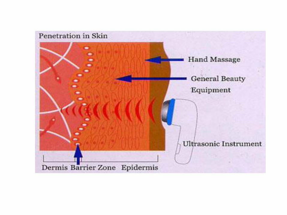
• Are also used for deep heating of body tissue. These waves are completely different from electromagnetic waves. They produce mechanical motion like audible sound waves except the frequency is much higher (usually near 1MHz). In ultrasonic diathermy, power levels of several watts per square centimeter are usually used and the sound source is directly in contact with the body. As the ultrasonic wave move through the body the practices in the tissues move back and forth. The movement is similar to micro massage and results in heating of the tissues. Ultrasonic heating has been found useful in relieving the tightness and scarring that often occur in joint disease. It greatly aids joints that have limited motion. It useful for depositing heat in the bones because the absorb ultrasound energy more effectively than does soft tissue.

Use Cold in Medicine
• Cryogenices is the science and technology of producing and using very low temperatures. The study of low temperature effects in biology and medicine is called coryibiology.
• Cryogenic methods used in medicine. Low temperature have been used for long term preservation of blood, sperm, bone marrow, and tissues. Studies of their relationship to the hibernation of animals are under way and long term preservation of man being considered.
•

Cryosurgery
• Cryogenic methods are also to destroy cell. This application is called cryosurgery. Cryosurgery has several advantages.
• 1-There is little bleeding in the destroyed area.
• 2-The volume of tissue destroyed can be controlled by the temperature of the cryosurgical probe.
• 3-There is little pain sensation because low temperatures tend to desensitize the nerves.

One of uses of cryosurgery was in the treatment of Parkinson's disease. Parkinson's causes uncontrolled tremors in the arms and legs. It is possible to stop the tremors by surgically destroying the part of thalamus in the brain that controls the the transmission of nerve impulses to other parts of the nervous system. One common use of cryosurgery is in the treatment of tumors and warts.


Also use to repair of a detached retina and cataract surgery (the removal of a darkened lens). Perhaps the result of an accident, the retina becomes detached from the wall of eye ball. This produces a blurred spot in the vision because the light rays are not focused at the correct spot. If the a cold tip applied to outside of the eye ball in the vicinity of the detachment, a reaction occurs that acts weld the retina to wall of the eyeball. The technique does not appear to damage the eye. In cryosurgical extraction of the lens, a cold probe is touched to the front surface of the lens. The probe sticks to the lens, making the lens easy to remove.