helminth parasites of the southern sea otter enhydra ... · the southern sea otter enhydra lutris...
TRANSCRIPT
DISEASES OF AQUATIC ORGANISMSDis Aquat Org
Vol. 53: 77–88, 2003 Published January 22
INTRODUCTION
The southern sea otter Enhydra lutris nereis waslisted as ‘threatened’ in 1977 under the EndangeredSpecies Act of 1973. Previous reports of the gastroin-testinal parasites of southern sea otters have described4 species of acanthocephalans, Profilicollis (= Polymor-
phus, see Nickol et al. 1999) altmani, P. kenti, P. major,and Corynosoma enhydri (see Hennessy & Morejohn1977), and 1 species of digenean, Microphallus pirum(see Margolis et al. 1997).
In a recent study investigating mortality in the south-ern sea otter, Thomas & Cole (1996) found that peri-tonitis caused by extra-intestinal migration of larval
© Inter-Research 2003 · www.int-res.com*Corresponding author. Email: [email protected]
Helminth parasites of the southern sea otter Enhydralutris nereis in central California: abundance,
distribution and pathology
Karl A. Mayer1, Murray D. Dailey2,*, Melissa A. Miller3
1Moss Landing Marine Laboratories, 8272 Moss Landing Rd., Moss Landing, California 95039, USA2Marine Mammal Center, Marin Headlands, 1065 Ft. Cronkhite, Sausalito, California 94965, USA
3Marine Wildlife Veterinary Care and Research Center, 1451 Schaffer Rd., Santa Cruz, California 95060, USA
ABSTRACT: From October 1997 to May 2001, the gastrointestinal tracts from 162 beach-cast south-ern sea otters Enhydra lutris nereis were examined for helminth parasites and associated lesions.Carcasses were collected opportunistically in central California between Pt. San Pedro and Pt.Arguello. The primary goals of this study were to examine spatial and temporal variability in mortal-ity due to parasite infection, identify factors associated with increased risk of infection, and illustratethe process of intestinal perforation by Profilicollis spp. Two genera and 4 species of acanthocepha-lans (Profilicollis altmani, P. kenti, P. major, Corynosoma enhydri) were found in 46.3% (Profilicollisspp.) and 94.4% (C. enhydri) of the carcasses examined. Three species of Digenea (Microphalluspirum, M. nicolli, Plenosoma minimum) were found in 47% of carcasses, at times in massive numbers(>3000 per cm2). This is the first report of the latter 2 species from the sea otter. Mortality resultingfrom infection by Profilicollis spp. occurred in 13.0% (n = 21) of sampled carcasses, either directly,due to perforation of the intestinal wall and peritonitis (9.9%, n = 16), or indirectly, due to inhibitionof host nutrient uptake or depletion of host energy reserves to fight chronic infections (3.1%, n = 5).The most massive infections (<8760 parasites), and all cases of intestinal perforation occurred in car-casses infected by P. altmani and/or P. kenti. Mortality due to infection by Profilicollis spp. occurredmore frequently among juvenile and old-adult females (χ2 = 17.479, df = 9, p = 0.045) from sand andmixed habitats in Monterey and Santa Cruz in the north of the sea otter range (χ2 = 9.84, df = 4, p =0.045). Spatial differences in sea otter mortality coincided with the relative distributions of Profilicol-lis altmani, P. kenti, and P. major, and may reflect differences in sea otter diet, or differences in inten-sity of infection in intermediate hosts. Mortality rate due to infection by Profilicollis spp. decreasedbetween 1998 and 2001, though differences were not significant (χ2 = 3.983, df = 3, p = 0.40), and mayvary on multi-year cycles due to environmental factors such as density of definitive hosts (e.g. the surfscoter Melanitta perspicillata), or El Niño. Corynosoma enhydri did not cause significant damage tothe intestine of the host, even when present in great numbers.
KEY WORDS: Sea otter · Enhydra lutris nereis · Mortality · Parasites · Acanthocephala · Intestinalperforation · Pathology · Peritonitis · Digenea
Resale or republication not permitted without written consent of the publisher

Dis Aquat Org 53: 77–88, 2003
acanthocephalans (Profilicollis spp.) was the cause ofdeath in 14% of sampled carcasses (n = 195), andprevalence of this disease appeared to increase in fre-quency between 1992 and 1995. By comparison, Hen-nessy & Morejohn (1977) found only 1 case of extra-intestinal migration by P. kenti in a sample of 80 seaotter carcasses collected in the late-1960s/early-1970s.The relative distribution of these parasites within thesouthern sea otter population has not been described,nor has the pathology and host reaction associatedwith penetration and perforation of the intestinal wall.
Acanthocephalans have complex life cycles involv-ing a free-living egg stage, an arthropod intermediatehost, and a vertebrate definitive host (Hyman 1951).Transmission of profilicollid acanthocephalans to thesea otter occurs through ingestion of the sand crabEmerita analoga and the spiny mole crab Blepharipodaoccidentalis (see Hennessy & Morejohn 1977). Therock crab Cancer irroratus was identified as an inter-mediate host for Profilicollis major on the east coast ofthe United States (Schmidt & MacLean 1978), but C.irroratus has not been verified as an intermediate hostin California. Definitive hosts for Profilicollis spp.include gulls (Larus spp.), scoters (Melanitta spp.) andthe sea duck (Clangula clangula; see Hennessy &Morejohn 1977). Sexually mature adult stages of Pro-filicollis spp. have not been found in the sea otter(Thomas & Cole 1996). An intermediate host has notbeen identified for Corynosoma enhydri, and the seaotter is the only known definitive host for this parasite(Hennessy & Morejohn 1977). Unlike Profilicollis spp.,C. enhydri has not been found to deleteriously affectthe sea otter, even when present in great numbers(Thomas & Cole 1996).
The goals of the present study were to (1) examinespatial and temporal variability in mean parasite den-sity (Rohde 1982) and mortality due to infection byProfilicollis spp. on a sample of southern sea ottercarcasses, and (2) identify factors associated withincreased risk of infection. In addition we describedspatial and temporal variability in infection by Coryno-soma enhydri, illustrated the process of intestinal per-foration by Profilicollis spp., and identified 2 species ofDigenea not previously noted in the southern sea otter.
MATERIALS AND METHODS
Sea otter carcass collection and necropsy. Intactgastro-intestinal tracts were removed from 162 seaotter carcasses opportunistically recovered in centralCalifornia (Pt. San Pedro to Pt.Arguello) betweenOctober 1997 and May 2001 (Fig. 1). This sample rep-resents 25% (n = 651) of southern sea otter carcassesrecovered during that period (B. Hatfield, US Geologi-
cal Survey, Biological Resources Division, pers.comm.). The condition of carcasses examined in thisstudy ranged from freshly dead to advanced decompo-sition, and was determined by field biologists at thetime of carcass recovery (J. Ames, California Depart-ment of Fish and Game, pers. comm.). Age category ofeach animal (pup, juvenile, sub-adult, adult, old-adult)was estimated based on total body length, dentition,and tooth wear. Date of carcass recovery was groupedinto seasons as follows: autumn (September throughNovember), winter (December through February),spring (March through May), and summer (Junethrough August).
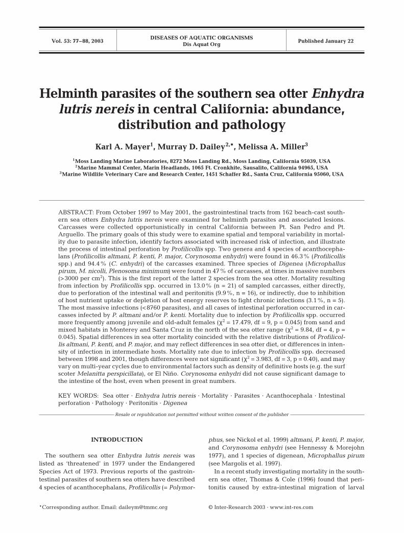
For spatial comparison, carcass recovery sites weregrouped from north to south into 11 contiguous areasbased on predominant habitat-type (sand, rock, ormixed sand and rock; Table 1). Habitat-type was usedas the basis for spatial analysis because this factor in-fluences distributions of crustacean intermediatehosts: Areas of rock habitat contained relatively largeamounts of kelp and predominantly rocky coastline;areas of sand habitat contained large stretches of sandbeach, no rocky coast, and little or no kelp; and areas ofmixed habitat contained some rocky coast, occasionalheadlands and coves with sand beaches, and little or nokelp (Laidre et al. 2001). Recovery sites betweenPt. Piños and Pt. Joe, and Pescadero Pt. and Pt. Lobos(Fig. 1) were classified as mixed instead of rock, despitehaving large amounts of kelp, because areas of sandybeach occur amidst rocky coastline in both areas.
Most sea otter carcasses were stored at the MarineWildlife Veterinary Care and Research Center (MWV-CRC) in Santa Cruz, California prior to examination.Necropsies were performed by a pathologist or fieldbiologist, who determined a cause of death wheneverpossible. The entire intestinal tract was removed dur-ing necropsy and frozen for later examination.
Parasite sampling and identification. The intestinaltract was thawed, placed on a table, separated intometer-long segments, opened, washed, and examinedgrossly for the presence of parasites. Acanthocepha-lans were relaxed in fresh water prior to fixation.Acanthocephalans and digeneans were fixed in alco-hol-formalin-acetic acid (AFA) solution and stored in70% ethanol. Staining and mounting of specimens foridentification followed Dailey (1996). Species of Profili-collis were distinguished using morphologic and mor-phometric characteristics of the proboscis, including,number of proboscis hooks per row, number of rows,and diameter of proboscis (Amin 1992). Additionalacanthocephalan and digenean parasites collectedfrom southern sea otter carcasses by the NationalWildlife Health Center in Madison, Wisconsin, wereutilized for taxonomic determination, but were notincluded in analyses.
78
Mayer et al.: Helminth parasites of the southern sea otter
Histopathological examination. Sections of intestineand omentum containing acanthocephalans fromselected freshly dead otters were immersion-fixed in10% neutral buffered formalin. These tissues weredehydrated in ethanol, embedded in paraffin, sectionedat 5 µm and stained with hematoxylin and eosin(H&E). Step sections and/or 5 µm serial sectionswere completed from some paraffin blocks ofintestine containing transmural acanthocephalanparasite profiles to document anatomic details ofthe parasites as well as host-tissue reaction.
Statistical analyses. Two variables were con-sidered in statistical comparisons: (1) meanabundance (the total number of parasitesdivided by the total number of hosts, stratifiedby age/sex examined, including those with noparasites, Rohde 1982), and (2) rate of mortalitydue to infection by Profilicollis spp. and Coryno-soma enhydri were considered independently incalculating abundance of infection. Mortalitydue to infection by Profilicollis spp. occurred inthe following cases: (1) carcasses in whichintestinal perforation was observed and no othermore obvious cause of death was assigned(acanthocephalan peritonitis), and (2) cases inwhich no intestinal perforation was observed,but abundance of Profilicollis spp. was suffi-ciently great as to affect nutrient uptake by thehost, or cause depletion of host energy reservesto fight infection (intestinal acanthocephaladio-sis). Intensity of infection was defined as thenumber of parasites per infected host. Preva-lence was defined as the percent of host individ-uals infected (Rohde 1982).
A Kruskal-Wallis test (Zar 1996) was used to com-pare mean density of infection by Profilicollis spp. andCorynosoma enhydri among 4 factors: (1) sea otterage and sex category (pup, juvenile, sub-adult, adult,old-adult, male, female), (2) carcass recovery area
79
Table 1. Enhydra lutris nereis. Carcass recovery areas were grouped based on habitat-type. Rocky habitat contained moderate tolarge amounts of kelp and moderate to large amounts of rocky substrate; sandy habitat contained no rocky coast, no kelp, and nosub-tidal rocky substrate; and mixed habitat contained some rocky coast, occasional headlands and coves, little or no kelp, and
minimal sub-tidal rocky substrate (Laidre et al. 2001)
Recovery area Habitat Carcass Distance along 10 mtype sample depth contour (km)
North coast Pt. San Pedro – Pt. Ano Nuevo Mixed 4 70
Santa Cruz Pt. Ano Nuevo – Soquel Pt. Mixed 8 43Soquel Pt. – Moss Landing Sand 25 27.5
Monterey Moss Landing – Monterey Municipal Wharf Sand 23 27.5Pt. Piños – Pt. Joe, Pescadero Pt. – Pt. Lobos Mixed 19 10.5Monterey Municipal Wharf – Pt. Piños, Rock 23 26Pt. Joe - Pescadero Pt., Pt. Lobos – Yankee Pt.
Central Range Yankee Pt. – Cayucos Pier Rock 14 178
Morro Bay Cayucos Pier – Hazard Canyon Sand 25 18.5Hazard Canyon – Shell Beach Rock 5 50.5
Pismo Beach Shell Beach – Pt. Sal Sand 16 26.5
South coast Purisima Pt. – Pt. Arguello Sand 2 20.5
Fig. 1. Enhydra lutris nereis. Recovery locations of 162 sampled seaotter carcasses, opportunistically collected between October 1997 andMay 2001 by a salvage network coordinated by the US Geologic Sur-vey Biological Resources Division in San Simeon, CA. Carcasses with>350 Profilicollis spp. were considered heavily infected. Sectionsof coastline were grouped into broader recovery areas (indicatedby brackets) based on predominant habitat type (see Table 1)
Dis Aquat Org 53: 77–88, 2003
(Table 1), (3) season of carcass recovery (autumn, win-ter, spring, summer), and (4) year of carcass recovery(1998, 1999, 2000, 2001). In 1997, carcasses were sam-pled only in November and December (n = 6), andwere therefore not included in among-year analyses.A non-parametric Kruskal-Wallis test was employedafter log transformation of data failed to correct devia-tions from assumptions of normality and homosce-dasticity. A non-parametric multiple comparison test(Q-test, Zar 1996) was used to determine which levelsof a factor differed if the null hypothesis was rejected.
Rate of mortality due to infection by Profilicollis spp.was compared among age and sex category, carcassrecovery area, season and year of carcass recoveryusing a chi-square test (Zar 1996). Expected frequen-cies were calculated by multiplying the total percentmortality for all sampled carcasses (13.0%) by the sam-ple size for each factor level. The null hypothesis in allchi-square tests was that ‘observed frequency of mor-tality was equal among levels of a factor.’
RESULTS
Acanthocephalans
Two genera and 4 species of acanthocephalans (Cory-nosoma enhydri, Profilicollis altmani, P. kenti, and P.major) were found in our sample of sea otter carcassesfrom central California. Intensity of infection rangedfrom 1 to 6063 for C. enhydri, and 1 to 8760 for Profili-collis spp.
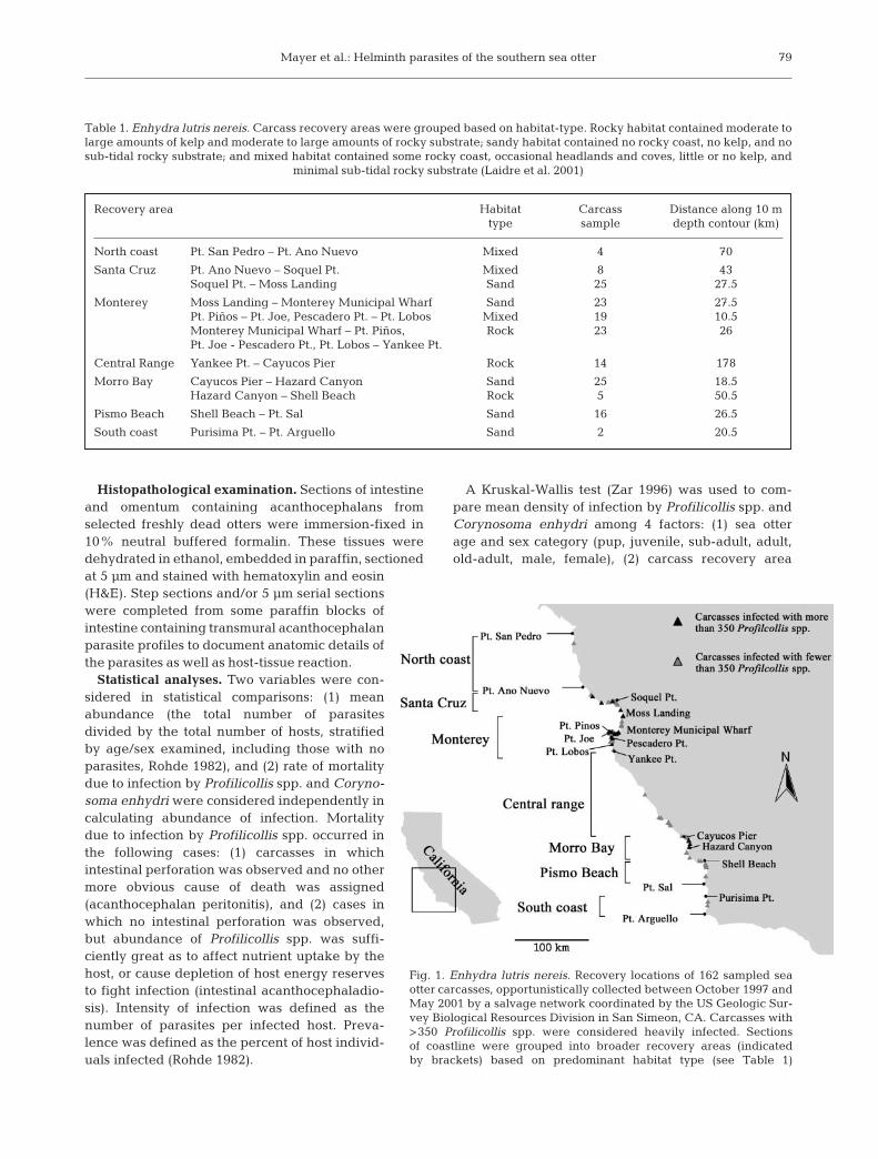
Profilicollis spp. were found in 75 of 162 (46.3%) car-casses sampled. Within a sub-sample of 49 infectedcarcasses, 56% of identified profilicollids were P. alt-mani, 34% were P. kenti, and 10% were P. major. Theoverall mortality rate due to infection by Profilicollisspp. was 13.0% (n = 21). Acanthocephalan peritonitiswas either the primary or secondary cause of death in16 cases, while intestinal acanthocephaladiosis con-tributed to mortality in 5 cases. Most carcasses witheither acanthocephalan peritonitis or intestinal acan-thocephaladiosis were also emaciated. Acanthocepha-lan peritonitis occurred in 14 of 16 (87.5%) carcasses inwhich intensity of infection by P. altmani and P. kentiwas greater than 350, but did not occur in carcassesinfected only by P. major (Fig. 2). Intestinal perforationwas noted in only 1 carcass with fewer than 350 para-sites.
Among sampled carcasses, mean density of infec-tion was significantly greater among juvenile andold-adult females than adult females (Qjuvenile vs adult =3.53, p = 0.02; Qold-adult vs adult = 3.42, p = 0.03,Fig. 3A). Differences in mean density among otherage/sex categories were not significant. Rate of mor-tality due to infection by Profilicollis spp. was sig-nificantly greater than expected among sub-adultmales (36.4%, n = 11) and old-adult females (37.5%,n = 8), and significantly less than expected amongadult females (2.5%, n = 40; χ2 = 17.479, df = 9, p =0.03).
Spatial patterns in mean density of Profilicollis spp.and percent mortality were similar. Within our sample,mean density of Profilicollis spp. was significantly
80
0
1
2
3
1 10 100 1000 10000Intensity of intestinal infection
Rel
ativ
e de
nsity
of
abdo
min
al P
rofil
icol
lis s
pp.
P. major (n=20)
P. altmani and P. kenti (n=14)
Fig. 2. Enhydra lutris nereis. Data from a sub-sample of carcasses infected by Profilicollis altmani and/or P. kenti (n = 14), or onlyby P. major (n = 20). P. altmani and P. kenti were considered together because they appear to share intermediate hosts (Emeritaanaloga and Blepharipoda occidentalis), whereas P. major is transmitted to the sea otter via another, unidentified species of crab.Carcasses simultaneously infected by P. altmani, P. kenti and P. major were not considered (n = 15). Relative density (0 = none, 1 =light, 2 = moderate, 3 = heavy) of abdominal parasites was used for a qualitative estimate of the severity of intestinal perforation.A threshold intensity of infection may exist around 350 parasites: intestinal perforation occurred in 9 of 10 carcasses with >350parasites, and in 1 of 24 carcasses with <350 parasites. Intestinal perforation did not occur in carcasses infected only by P. major

Mayer et al.: Helminth parasites of the southern sea otter
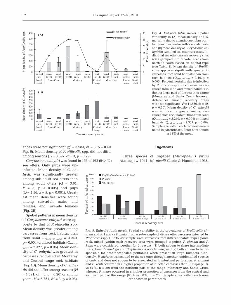
greater in sea otter carcasses from sand habitat thanfrom rock habitat (Qsand vs rock = 3.42, p = 0.002, Fig. 4A).Mean density in mixed habitat was greater than inrock and less than in sand, but differences were notsignificant (Qmixed vs rock = 2.19, p = 0.09). Acantho-cephalan peritonitis was found in only 1 of 40 (2.5%)sampled carcasses from rock habitats.
A particularly high concentration of heavily infectedcarcasses (>350 parasites) was found in a 4.5 kmstretch of coastline just north of Monterey MunicipalWharf in south Monterey Bay. In this area, 6 of 13(46.1%) sampled carcasses had profilicollid infectionsof greater than 800 parasites. The cause of death in 5 of6 cases was acanthocephalan peritonitis, and in 1 case,intestinal acanthocephaladiosis. All 6 were juvenile orsub-adult sea otters recovered in 1998 or 1999.
Mortality due to acanthocephalan peritonitis or in-testinal acanthocephaladiasis was also relatively highamong sampled beach cast otters from mixed habitatbetween Pt. Piños and Pt. Lobos, just south of Mon-terey (23%, n = 21), and from sand habitat betweenSoquel Pt. and Moss Landing in northern MontereyBay (20%, n = 25). Mortality due to Profilicollis spp.occurred less frequently among sampled beach castotters from sand habitat in Morro Bay (Cayucos Pierto Hazard Canyon, 12%, n = 25) despite a relativelyhigh mean density (Fig. 4A). In this area, 3 of 25 car-
casses with acanthocephalan peritonitis had particu-larly massive infections (2200 to 8760 parasites): 2 in-dividuals were old-adult females, 1 was a juvenilefemale.
Mortality due to infection by Profilicollis spp. was notfound in sampled beach cast otters from Pismo Beachand South coast sand habitats (Fig. 4A). Within theseareas, Profilicollis spp. were found in 58% (n = 17) ofsampled carcasses, and intensity of infection rangedfrom 1 to 483; however, 99% of profilicollids wereidentified as the less-pathologic species, P. major. Con-versely, P. altmani and P. kenti occurred in a higherproportion of infected carcasses than P. major (83 vs51%, n = 29) in a sub-sample of infected carcasses fromMonterey and Santa Cruz in the northern part of thesea otter range (Fig. 5). The higher prevalence of P. alt-mani and P. kenti in the north coincided with higherrates of mortality observed in these recovery areas(Fig. 4A).
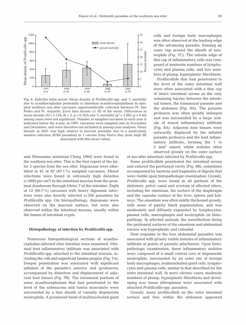
Among sampled carcasses, inter-annual differences inmean density of Profilicollis spp. were not significant(H = 5.116, df = 3, p = 0.16). Mean density in 2001(x = 519, SE = 485, n = 18) was high due to a particularlymassive infection (8760 parasites) in 1 carcass fromMorro Bay (reflected by high SE associated with themean value, Fig. 6). Rates of mortality were greater in1998 and 1999 than in 2000 and 2001; however, differ-
81
0
200
400
600
800
1000
1200
1400
1600
1800
2000
pup juvenile sub-adult adult old-adult(n=16) (n=24) (n=21) (n=75) (n=19)
Mea
n de
nsity
of
C. e
nhyd
ri males
females
males
females
0
500
1000
1500
2000
2500
3000
pup juvenile sub-adult adult old-adult(n=16) (n=24) (n=21) (n=75) (n=19)
Mea
n de
nsity
of
Pro
filic
ollis
spp
.
(A)
(B)
*
*
*
*
*
*
Fig. 3. Enhydra lutris nereis. Mean density of(A) Profilicollis spp. and (B) Corynosoma en-hydri by sea otter age and sex category. As-terisks (*) indicate significant differencesamong age/sex categories. Prevalence ofProfilicollis spp. infection within each agecategory was as follows: pups (19%), juve-niles (54%), sub-adults (38%), adults (48%),and old-adults (53%). Within each agecategory, prevalence did not differ betweenmales and females, except among sub-adults, where 64% of males and 10% of fe-males were infected. Prevalence of infectionby C. enhydri was 100% for all age cate-gories except pups (50%). Sample sizes ineach age category are indicated in parenthe-ses. Error bars denote ±1 SE of the mean
Dis Aquat Org 53: 77–88, 2003
ences were not significant (χ2 = 3.983, df = 3, p = 0.40;Fig. 6). Mean density of Profilicollis spp. did not differamong seasons (H = 3.697, df = 3, p = 0.29).
Corynosoma enhydri was found in 153 of 162 (94.4%)sea otters. Only pups were un-infected. Mean density of C. en-hydri was significantly greateramong sub-adult sea otters thanamong adult otters (Q = 3.61,k = 5, p < 0.005) and pups(Q = 4.56, k = 5, p < 0.001). Great-est mean densities were foundamong sub-adult males andfemales, and juvenile females(Fig. 3B).
Spatial patterns in mean densityof Corynosoma enhydri were op-posite to that of Profilicollis spp.Mean density was greater amongcarcasses from rock habitat thanfrom sand (Qrock vs sand = 3.240,p = 0.004) or mixed habitats (Qrock vs
mixed = 2.327, p = 0.06). Mean den-sity of C. enhydri was greatest incarcasses recovered in Montereyand Central range rock habitats(Fig. 4B). Mean density of C. enhy-dri did not differ among seasons (H= 4.591, df = 3, p = 0.20) or amongyears (H = 6.751, df = 3, p = 0.08).
Digeneans
Three species of Digenea (Microphallus pirumAfanassjew 1941, M. nicolli Cable & Hunninen 1938,
82
0
200
400
600
800
1000
1200
1400
mixed mixed sand sand mixed rock rock sand rock sand sand(n=4) (n=8) (n=25) (n=24) (n=21) (n=20) (n=14) (n=25) (n=6) (n=15) (n=2)Northcoast
Santa Cruz Monterey CentralRange
Morro Bay PismoBeach
Southcoast
Mea
n de
nsit
y of
Pro
fili
coll
is s
pp.
0
5
10
15
20
25
30
Per
cent
mor
tali
ty
Mean density.
Percent mortality
0200400600800
10001200140016001800
mixed mixed sand sand mixed rock rock sand rock sand sand(n=4) (n=8) (n=25) (n=24) (n=21) (n=20) (n=14) (n=25) (n=5) (n=15) (n=2)Northcoast
Santa Cruz Monterey CentralRange
Morro Bay PismoBeach
Southcoast
Carcass recovery areas
Mea
n de
nsit
y of
C. e
nhyd
ri
(A)
(B)
Fig. 4. Enhydra lutris nereis. Spatialvariability in (A) mean density and %mortality due to acanthocephalan peri-tonitis or intestinal acanthocephaladiosisand (B) mean density of Corynosoma en-hydri in sampled sea otter carcasses. In-dividual sea otter carcass recovery siteswere grouped into broader areas fromnorth to south based on habitat-type(see Table 1). Mean density of Profili-collis spp. was significantly greater incarcasses from sand habitats than fromrock habitats (Qsand vs rock = 3.10, p =0.005). Percent mortality due to infectionby Profilicollis spp. was greatest in car-casses from sand and mixed habitats inthe northern part of the sea otter range(Monterey and Santa Cruz); howeverdifferences among recovery areaswere not significant (χ2 = 11.856, df = 10,p = 0.30). Mean density of C. enhydriwas significantly greater among car-casses from rock habitat than from sand(Qrock vs sand = 3.240, p = 0.004) or mixedhabitats (Qrock vs mixed = 2.327, p = 0.06).Sample size within each recovery area isnoted in parentheses. Error bars denote
±1 SE of the mean
0
10
20
30
40
50
60
70
80
90
100
(n = 11 ) (n= 1 8) (n= 3 ) (n = 9 ) (n= 6 ) (n = 2 )San ta Cru z M on terey C en tral R an ge M o rro B ay P is m o B each S ou th coast
Carcass recovery area
Perc
ent o
f in
fect
ed c
arca
sses Profilicollis altmani and P. kenti
P. major
All 3 species
Fig. 5. Enhydra lutris nereis. Spatial variability in the prevalence of Profilicollis alt-mani and P. kenti vs P. major from a sub-sample of 49 sea otter carcasses infected byProfilicollis spp. Due to low sample sizes, carcasses from different habitat types (sand,rock, mixed) within each recovery area were grouped together. P. altmani and P.kenti were considered together for 2 reasons: (1) both appear to share intermediatehosts, Emerita analoga and Blepharipoda occidentalis, and (2) both appear to be re-sponsible for acanthocephalan peritonitis when present in large numbers. Con-versely, P. major is transmitted to the sea otter through another, unidentified speciesof crab, and does not appear to be associated with intestinal perforation. P. altmaniand P. kenti occurred in a higher proportion of infected carcasses than P. major (79%vs 51%, n = 39) from the northern part of the range (Monterey and Santa Cruz),whereas P. major occurred in a higher proportion of carcasses from the central andsouthern part of the range (85% vs 30%, n = 20). Sample sizes within each area
are shown in parentheses
(A)
(B)
Mayer et al.: Helminth parasites of the southern sea otter
and Plenosoma minimum Ching 1960) were found inthe southern sea otter. This is the first report of the lat-ter 2 species from the sea otter. Digeneans were iden-tified in 41 of 87 (47.1%) sampled carcasses. Mixedinfections were found in extremely high densities(>3000 per cm2) in the intestinal mucosa from the prox-imal duodenum through Meter 7 of the intestine. Eightof 12 (66.7%) carcasses with heavy digenean infec-tions were also heavily infected (>350 parasites) byProfilicollis spp. On histopathology, digeneans wereobserved on the mucosal surface, but were alsoobserved within the intestinal mucosa, usually withinthe lumen of intestinal crypts.
Histopathology of infection by Profilicollis spp.
Numerous histopathological sections of acantho-cephalan-infected otter intestine were examined. Min-imal host inflammatory infiltrate was associated withProfillicollis spp. attached to the intestinal mucosa, in-cluding the villi and superficial lamina propria (Fig. 7A).Deeper penetration was associated with significantinflation of the parasite’s anterior end (proboscis),accompanied by distortion and displacement of adja-cent host tissues (Fig. 7B). The intramural portions ofsome acanthocephalans that had penetrated to thelevel of the submucosa and tunica muscularis weresurrounded by a thin sheath of variably degenerateneutrophils. A prominent band of multinucleated giant
cells and foreign body macrophageswas often observed at the leading edgeof the advancing parasite, forming anouter cap around the sheath of neu-trophils (Fig. 7C). The outside edge ofthis cap of inflammatory cells was com-posed of moderate numbers of lympho-cytes and plasma cells, and low num-bers of plump, hyperplastic fibroblasts.
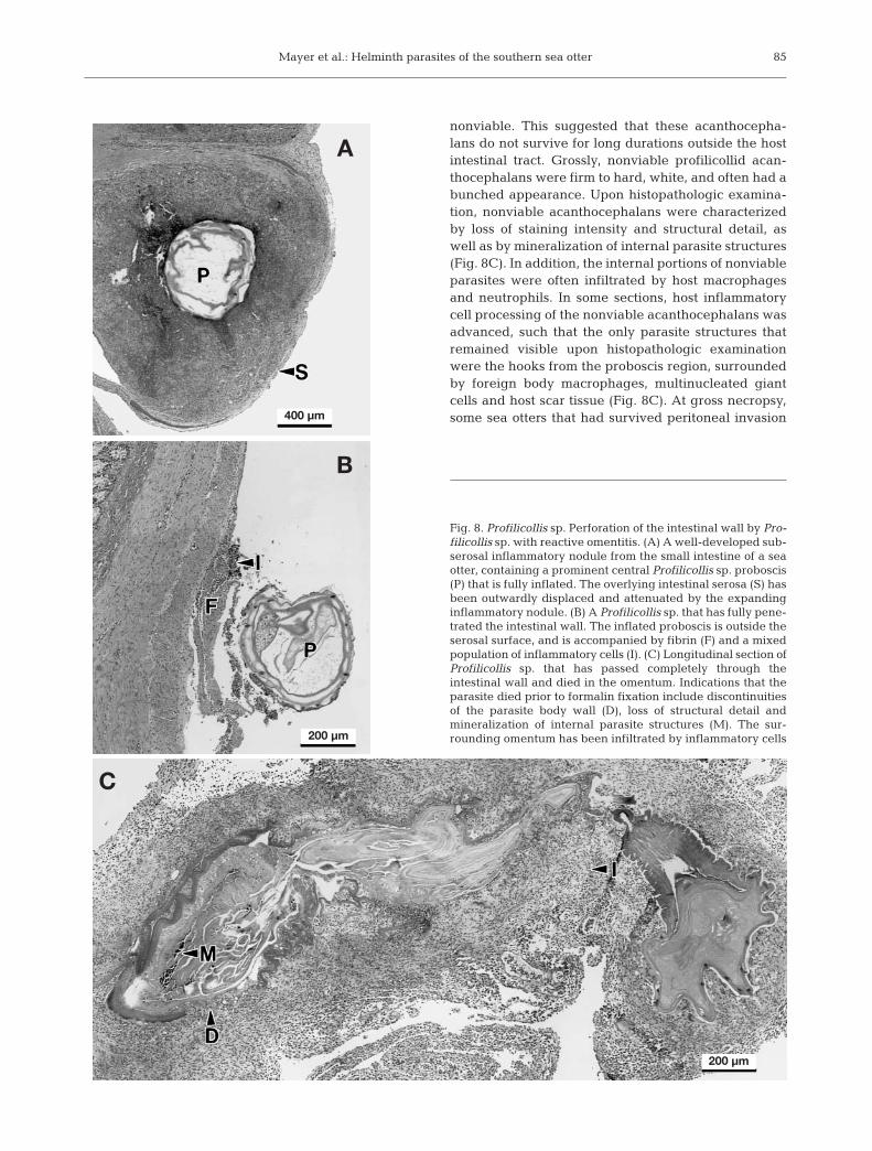
Profilicollids that had penetrated tothe level of the outer intestinal wallwere often associated with a thin capof intact intestinal serosa as the onlyremaining barrier between the intesti-nal lumen, the transmural parasite andthe abdomen (Fig. 8A). The parasiteproboscis was often greatly enlargedand was surrounded by a large nod-ule of mixed inflammatory infiltrate(Fig. 8A). Adjacent host tissues wereoutwardly displaced by the inflatedparasite proboscis and the host inflam-matory infiltrate, forming the 1 to2 mm3 raised, white nodules oftenobserved grossly on the outer surface
of sea otter intestines infected by Profilicollis spp. Some profilicollids penetrated the intestinal serosa
and entered the peritoneal cavity (Fig. 8B), sometimesaccompanied by bacteria and fragments of digesta thatwere visible upon histopathologic examination. Grossly,Profilicollis spp. were found in all portions of theabdomen, pelvic canal and scrotum of affected otters,including the omentum, the surface of the diaphragmand the capsular surface of the liver, spleen and kid-neys. The omentum was often mildly thickened grossly,with areas of patchy black pigmentation, and wasmoderately and diffusely expanded by lymphocytes,plasma cells, macrophages and neutrophils on histo-pathlogy. In affected animals, the mesothelium liningthe peritoneal surfaces of the omentum and abdominalviscera was hyperplastic and cuboidal.
Host response to the free abdominal parasites wasassociated with grossly visible nodules of inflammatoryinfiltrate at points of parasite attachment. Upon histo-pathologic examination, these inflammatory noduleswere composed of a small central core of degenerateneutrophils, surrounded by an outer rim of foreignbody macrophages, multinucleated giant cells, lympho-cytes and plasma cells, similar to that described for theouter intestinal wall. In more chronic cases, moderatenumbers of plump, hyperplastic fibroblasts and devel-oping scar tissue (fibroplasia) were associated withattached Profilicollis spp. parasites.
Grossly, many profilicollids on the outer intestinalsurface and free within the abdomen appeared
83
0
200
400
600
800
1000
1200
1997n=6
1998n=43
1999n=66
2000n=30
2001n=18
Mea
n de
nsity
of
Pro
filic
ollis
spp
.
0
5
10
15
20
25
30
35
Perc
ent m
orta
lity
mean density
percent mortality
Fig. 6. Enhydra lutris nereis. Mean density of Profilicollis spp. and % mortalitydue to acanthocephalan peritonitis or intestinal acanthocephaladiasis in sam-pled southern sea otter carcasses opportunistically collected between Pt. SanPedro and Pt. Arguello. Error bars denote ±1 SE of the mean. Differences inmean density (H = 5.116, df = 3, p = 0.163) and % mortality (χ2 = 3.983, p = 0.40)among years were not significant. Number of sampled carcasses in each year isindicated below the x-axis. In 1997, carcasses were sampled only in Novemberand December, and were therefore not included in among-year analyses. Meandensity in 2001 was high relative to percent mortality due to a particularlymassive infection (8760 parasites) in 1 carcass from Morro Bay (note high SE
associated with this mean value)

Dis Aquat Org 53: 77–88, 200384
Fig. 7. Profilicollis sp. Multiple cross-sections of sea otter small intestine that extend from the intestinal lumen (left of figure) to theabdominal cavity (right of figure). (A) Profilicollis sp. has penetrated the intestinal mucosa to the level of the muscularis mucosa(MM). The intestinal villi (V) have been effaced or displaced laterally in the area of parasite penetration. Note the inflated pro-boscis (P) with prominent surface hooks, and the lack of a significant host inflammatory response. (B) Profilicollis sp. has pene-trated to the external sheath of the tunica muscularis (TM). The proboscis is fully inflated. Some outward displacement of the ex-ternal sheath of the tunica muscularis and serosa (S) is apparent. Mildly increasing numbers of inflammatory cells (I) are presentin the surrounding tissue. (C) Intestinal perforation by Profilicollis sp. is imminent. Large numbers of degenerate neutrophils arepresent in a raised thin-walled inflammatory nodule (N) located just below the serosal surface of the intestine. Surrounding theneutrophils is a prominent zone of reactive macrophages and multinucleated cells, interspersed with lymphocytes and plasma
cells. The proboscis (P) has collapsed, perhaps artifactually
200 µm
200 µm
400 µm
C
B
A
Mayer et al.: Helminth parasites of the southern sea otter
nonviable. This suggested that these acanthocepha-lans do not survive for long durations outside the hostintestinal tract. Grossly, nonviable profilicollid acan-thocephalans were firm to hard, white, and often had abunched appearance. Upon histopathologic examina-tion, nonviable acanthocephalans were characterizedby loss of staining intensity and structural detail, aswell as by mineralization of internal parasite structures(Fig. 8C). In addition, the internal portions of nonviableparasites were often infiltrated by host macrophagesand neutrophils. In some sections, host inflammatorycell processing of the nonviable acanthocephalans wasadvanced, such that the only parasite structures thatremained visible upon histopathologic examinationwere the hooks from the proboscis region, surroundedby foreign body macrophages, multinucleated giantcells and host scar tissue (Fig. 8C). At gross necropsy,some sea otters that had survived peritoneal invasion
85
Fig. 8. Profilicollis sp. Perforation of the intestinal wall by Pro-filicollis sp. with reactive omentitis. (A) A well-developed sub-serosal inflammatory nodule from the small intestine of a seaotter, containing a prominent central Profilicollis sp. proboscis(P) that is fully inflated. The overlying intestinal serosa (S) hasbeen outwardly displaced and attenuated by the expandinginflammatory nodule. (B) A Profilicollis sp. that has fully pene-trated the intestinal wall. The inflated proboscis is outside theserosal surface, and is accompanied by fibrin (F) and a mixedpopulation of inflammatory cells (I). (C) Longitudinal section ofProfilicollis sp. that has passed completely through theintestinal wall and died in the omentum. Indications that theparasite died prior to formalin fixation include discontinuitiesof the parasite body wall (D), loss of structural detail andmineralization of internal parasite structures (M). The sur-rounding omentum has been infiltrated by inflammatory cells
400 µm
200 µm
200 µm
A
B
C

Dis Aquat Org 53: 77–88, 2003
by acanthocephalans had multiple areas of omentaladhesion and small mineralized serosal nodules, pre-sumably points of scar (connective) tissue formationcaused by degenerating acanthocephalan parasites.
In contrast, in all sections examined, points of intesti-nal attachment by Corynosoma enhydri were limitedto the intestinal villi and superficial lamina propria andwere not associated with significant host inflammatoryresponse. In addition, significant inflation of theanterior portion of C. enhydri was not observed uponhistopathologic examination. None of the intra- orextra-intestinal profilicollids examined appeared to bepatent (egg-producing) while intra-intestinal C. enhydri were patent.
DISCUSSION
Data collected opportunistically from beach-cast car-casses represent a biased sample of mortality in thesouthern sea otter population. Potential sources of biasinclude: (1) Non-random disappearance of carcasses—mortality occurring farther offshore, such as fromshark attacks or entanglement in fishing gear—maybe under-represented in beach-cast samples becausethese carcasses would be less likely to be deposited onshore; (2) Recovery bias—most carcasses are recov-ered from easily accessible areas adjacent to popula-tion centers, including Santa Cruz, Monterey, MorroBay, and Pismo Beach. Conversely, very few carcassesare recovered from rocky habitat in the center of thesea otter range (between Yankee Pt. and CayucosPier), and causes of mortality affecting sea otters in thisarea are unknown; (3) Post-mortem drift/decomposi-tion—carcasses may wash ashore shortly after death,or drift some distance prior to deposition dependingon numerous environmental factors such as location,cause of death, wind and current direction, tidal state,waves, and physical characteristics of the coastline.Post-mortem drift potentially confounds spatial differ-ences in mean parasite density and mortality rate.
In this study, we did not attempt to quantify the po-tential sources of bias listed above. As a result, conclu-sions related to spatial and temporal variability in den-sity of Profilicollis spp. and Corynosoma enhydri andrate of mortality due to infection by Profilicollis spp.were limited to the sample population of carcasses, andnot extrapolated more broadly to the southern sea otterpopulation. Nevertheless, despite the limited scope ofconclusions, certain factors associated with greater riskof infection and mortality emerged.
Among sampled sea otter carcasses, Profilicollis spp.were most abundant in juvenile and old-adult females.Thomas & Cole (1996) reported most cases of acantho-cephalan peritonitis in younger otters (pups and juve-
niles). The occurrence of greater Profilicollis spp.infections among juvenile and old-adult females in oursample could be explained by several hypotheses,including food limitation in the southern sea otter pop-ulation.
In a food-limited population, nutritional stress woulddisproportionately affect the poorest feeders (Ralls &Siniff 1990). The poorest feeders may be at a competi-tive disadvantage relative to other age/sex groups, andtherefore more susceptible to disease. In the southernsea otter, juvenile females spend a greater proportionof time feeding than juvenile males and adults (Ralls& Siniff 1990) and have lower survival rates (Siniff &Ralls 1991). No studies have documented nutritionalstress on old-adult females in the southern sea otter;however, in a resource-limited population of northernsea otters (Amchitka Island), individual body conditionwas poorer among old-adult females (>12 yr) thanyounger adults (4 to 12 yr; Monson et al. 2000).
Alternatively, heavy parasite burdens in young andold females in our sample could be explained byincreased parasite exposure through diet. Emeritaanaloga and Blepharipoda occidentalis are seasonallyabundant along sandy beaches and would be rela-tively easy for sea otters to capture. These crustaceans,therefore, may be more heavily targeted by recentlyweaned juvenile sea otters just learning to feed inde-pendently, or by old females struggling to meet energyrequirements with prey that is more difficult to cap-ture. A combination of increased exposure to parasitesand poor body condition may have contributed to highmortality due to Profilicollis spp. among juvenile andold-adult female sea otters in this study.
The adverse effects of infection by Profilicollis spp.also appeared to be concentrated geographically insand and mixed-bottom habitats in Monterey andSanta Cruz in the north, and to a lesser extent, MorroBay in the south. One hypothesis for observed spatialdifferences in mortality is that prevalence of interme-diate hosts in sea otter diets varied among recoveryareas. The relative distributions of P. altmani, P. kenti,and P. major appeared to support this hypothesis. Pro-filicollis altmani and P. kenti, transmitted to the seaotter through ingestion of sand crabs, were moreprevalent than P. major among sampled carcasses fromMonterey and Santa Cruz. Conversely, P. altmani andP. kenti rarely occurred in sampled carcasses from thesouth end of the range, where mortality due to acan-thocephalan peritonitis was not found. In this area,otters may be less likely to forage on Emerita analogaand Blepharipoda occidentalis, and more likely toforage on Cancer spp. or other species of crabs orbivalves. Unfortunately, conclusions about spatial vari-ability in feeding patterns of sea otters based on datafrom stranded carcasses were potentially confounded
86

Mayer et al.: Helminth parasites of the southern sea otter
by the possibility of post-mortem drift. Alternatively,differential risk of infection based on area and habitattype could be a function of the density of parasites inintermediate hosts. Ongoing research is focused onwhether density of Profilicollis spp. in intermediatehosts or prevalence of intermediate hosts in sea otterdiets is correlated with observed spatial patterns ofprofilicollid density in sea otter carcasses.
Among sampled carcasses, mortality due to Profili-collis spp. decreased between 1998 and 2001. Thistrend is the opposite of the general increase in mortal-ity identified by Thomas & Cole (1996) in the early1990s. These results suggest that sea otter mortalitydue to Profilicollis spp. may vary on multi-year cycles.The causes of this variability have not been investi-gated. One possibility is that greater prevalence ofheavy profilicollid infections among southern seaotters (such as in 1998 and 1999) may coincide withgreater densities of definitive hosts (particularly surfscoters Melanitta perspicillata) in Monterey Bay. Year-to-year changes in the density of surf scoters could ulti-mately have an impact on sea otter mortality throughtransmission of a greater number of P. altmani and P.kenti eggs to Emerita analoga and Blepharipoda occi-dentalis. The factors that determine the relative densi-ties of surf scoters along the central California coastare unknown. Alternatively, exposure to parasites mayvary temporally due to large-scale environmental per-turbations (such as El Niño) that adversely affect theabundance and overall health of marine organisms atseveral trophic levels.
Of the 3 species of Digenea found in the southern seaotter during this study, only one, Microphallus pirum,has been previously reported (Margolis et al. 1997).Two species, Microphallus nicolli (= Spelotremanicolli) and Plenosoma minimum, were found in mixedinfections with M. pirum at a maximum of 3750 percm2. Heavy digenean infections tended to occur in car-casses that were also heavily infected by Profilicollisspp. When present in great numbers, digeneans mayact to exacerbate the effects of profilicollids. Micro-phallus pirum was considered pathogenic, throughmechanical damage, when found at 3000 per cm2 inAlaskan otters (Rausch 1953). Metacercaria of M.nicolli, which were infective to mice, have been previ-ously reported in Emerita analoga collected from SantaBarbara, California (Ching 1965).
Based on results from gross necropsy and histo-pathological examination, sea otter mortality associ-ated with heavy acanthocephalan burdens is multi-factorial. First, high intra-intestinal acanthocephalanburdens are associated with significant melena, ordigested blood. The exact mechanism of intestinalhemorrhage is unknown, but may be related to endo-parasitism, and/or result from concurrent gastrointes-
tinal erosions or ulceration. Chronic gastrointestinalbleeding leads to chronic intestinal protein and ironloss, and in severe cases, osmotic diarrhea. This extra-nutritional burden, along with potential interferencewith or competition for nutrient uptake by heavy bur-dens of acanthocephalans and digeneans, could besignificant, especially for very young, very old, or sickotters.
As Profilicollis spp. reach high numbers and begin topenetrate the intestinal wall, an extra-nutritional bur-den could be further compounded by developmentof chronic inflammatory disease, requiring significanthost energy-reserves to surround, kill, and phago-cytize thousands of invading worms. The process ofintestinal wall perforation by profilicollid acantho-cephalan parasites appears to be due to parasite pro-boscis inflation and parasite migration through hosttissues. In addition, degenerate neutrophils and in-testinal bacteria distributed along the sides of theadvancing parasite may contribute to the process ofmural perforation through liquefactive necrosis ofadjacent host tissues. These otters must also cope withdigesta and opportunistic pathogens that gain accessto the peritoneum through perforations in the intestinalwall. In some cases the immediate cause of deathappears to be malnutrition due to depletion of energyreserves, while in others it is secondary septic peri-tonitis or systemic bacterial infection.
Although overall prevalence of Profilicollis spp. washigh (46.3%) in our sample of southern sea otters,acanthocephalan peritonitis and intestinal acantho-cephaladiosis seemed to occur more frequently amongjuvenile and old-adult females, and in sea otters fromsand and mixed habitats in the northern part of therange. Risk of infection by P. altmani and P. kenti mayvary among years due to environmental factors suchas changes in density of definitive hosts, or El Niño;however, further research is necessary to determinewhether inter-annual differences in mortality are sig-nificant. Future research should endeavor to clarify thesignificance of variability in risk of infection to thesouthern sea otter population through comparative for-aging studies, determination of density of Profilicollisspp. in intermediate hosts, and quantification of biasesassociated with use of carcass data.
Acknowledgements. We wish to thank Jack Ames, Mike Har-ris, Dave Jessup and the staff and volunteers of the MarineWildlife Veterinary Care and Research Center in Santa Cruz,CA, for conducting sea otter necropsies, collecting samples,and offering the use of facilities and resources. Also ourthanks to the California Department of Fish and Game, SantaCruz, CA, Friends of the Sea Otter, Monterey, CA, and TheMarine Mammal Center, Sausalito, CA, for financial support,and to Jim Harvey, Brian Hatfield, Nancy Thomas, RebeccaCole, Viviana Wong, and Christine Kreuder for technicalcontributions.
87

Dis Aquat Org 53: 77–88, 2003
LITERATURE CITED
Amin OM (1992) Review of the genus Polymorphus Luhe,1911 (Acanthocephala: Polymorphidae), with the syn-onymization of Hexaglandula Petrochenko, 1950, andSubcorynosoma Hoklova, 1967, and a key to the species.Qatar Univ Sci J 12:115–123
Ching HL (1965) Systematic notes on some North Americanmicrophallid trematodes. Proc Helminthol Soc Wash 32:140–148
Dailey MD (1996) Essentials of Parasitology, 6th edn. WCBrown, Dubuque, IA
Hennessy SL, Morejohn VJ (1977) Acanthocephalan parasitesof the sea otter, Enhydra lutris, off coastal California. CalifFish Game 63:268–272
Hyman LH (1951) The pseudocoelomate Bilateria—PhylumAcanthocephala. Chapter XII,. In: The invertebrates:Acanthocephala, Aschelminthes, and Entoprocta, Vol. III.McGraw-Hill, New York, p 1–52
Laidre KL, Jameson RJ, DeMaster DP (2001) An estimation ofcarrying capacity for sea otters along the California coast.Mar Mamm Sci 17(2):294–309
Margolis L, Groff JM, Johnson SC, McDonald TE, Kent ML,Blaylock RB (1997) Helminth parasites of sea otters (Enhy-dra lutris) from Prince William Sound, Alaska: compari-sons with other populations of sea otters and comments on
the origin of their parasites. J Helminthol Soc Wash 64:161–168
Monson DH, Estes JA, Bodkin JL, Siniff DB (2000) Life historyplasticity and population regulation in sea otters. Oikos90:457–468
Nickol BB, Crompton DWT, Searle DW (1999) Reintroductionof Profilicollis Meyer, 1931 as a genus in Acanthocephala:significance of the intermediate host. J Parasitol 85:716–718
Ralls K, Siniff DB (1990) Time budgets and activity patterns inCalifornia sea otters. J Wildlife Man 54(2):251–259
Rausch R (1953) Studies on the helminth fauna of Alaska, XIII.Disease in the sea otter, with special reference to helminthparasites. Ecology 34:584–604
Rohde K (1982) Ecology of marine parasites. University ofQueensland Press, St. Lucia
Schmidt GD, MacLean SA (1978) Polymorphus (Profilicollis)major Lundstrom 1942 juveniles in rock crabs, Cancerirroratus, from Maine. J Parasitol 64:953–954
Siniff DB, Ralls K (1991) Reproduction, survival and tag loss inCalifornia sea otters. Mar Mamm Sci 7(3):211–229
Thomas NJ, Cole RA (1996) The risk of disease and threatsto the wild population. Endangered Species Update 13:23–27
Zar JH (1996) Biostatistical Analysis, 3rd edn. Prentice Hall,Upper Saddle River, NJ
88
Editorial responsibility: Otto Kinne (Managing Editor),Oldendorf, Germany
Submitted: August 22, 2001; Accepted: June 12, 2002Proofs received from author(s): January 14, 2002