hemopoietic stemcells: stochastic differentiation and ... · pdf fileoncetheinfection...
TRANSCRIPT

Environmental Health PerspectivesVol. 80, pp. 199-207, 1989
Hemopoietic Stem Cells: StochasticDifferentiation and Humoral Control ofProliferation
by Makio Ogawa*
The central feature of hemopoiesis is life-long, stable cell renewal. This process is supported by hemopoieticstem cells which, in the steady state, appear to be dormant in cell cycling. The entry into cell cycle of thedormant stem cells may be promoted by such factors as interleukin-1, interleukin-6 (IL-6), and granulocytecolony-stimulating factor (G-CSF). Once the stem cells leave Go and begin proliferation, the subsequent processis characterized by continued proliferation and differentiation. While several models of stem cell differenti-ation have been proposed, micromanipulation studies of individual progenitors suggest that the commitmentof multipotential progenitors to single lineages is a random (stochastic) process. The proliferation of earlyhemopoietic progenitors requires the presence of interleukin-3 (IL-3), and the intermediate process appearsto be supported by granulocyte/macrophage colony-stimulating factor (GM-CSF). Once the progenitors arecommitted to individual lineages, the subsequent maturation process appears to be supported by late-acting,lineage-specific factors such as erythropoietin and G-CSF. Synthesis of a hemopoietic factor may take placein different cell types and is regulated by multiple factors The physiological regulator of erythropoiesis iserythropoietin, which, by a feedback mechanism, provides fine control of erythrocyte production. Feedbackmechanisms for leukocyte production have not been identified. It is possible that there is no feedback regu-lator of leukopoiesis. In this model, leukocyte production in the steady state is maintained at a geneticallydetermined level. When an infection occurs, the bacterial lipopolysaccharides may augment the productionof interleukin la and (3, tumor necrosis factor, macrophage colony-stimulating factor, etc. These factors cir-culate and stimulate the production in the marrow of G-CSF, GM-CSF, IL-3, IL-6, etc., with resultant leu-kocytosis. Once the infection subsides, the production of the factors and leukopoiesis return to the steady-state levels.
IntroductionThe central feature of the hemopoietic system is its con-
tinuous process of cell turnover. It is estimated that ap-proximately 200 billion erythrocytes (1) and 60 billion neu-trophilic leukocytes (2) are produced and destroyed dailyin a man weighing 70 kg. This life-long cell renewal pro-cess is supported by stem cells that possess the ability toself-renew and to produce progenitors that are commit-ted to differentiation in single lineages. The mechanismsregulating stem cell functions have intrigued not onlyhematologists but also biologists interested in cellulardifferentiation and have been the object of active inves-tigations and model building. During the last three de-cades, studies using functional assays for hemopoietic pro-genitors, i.e., the in vivo spleen colony assay for murinestem cells (3) and clonal cell culture assays for progenitorsat various stages of development (4), have resulted in sig-
*Veterans Administration Medical Center and Department of Medi-cine, Medical University of South Carolina, Charleston, SC 29403.
nificant elucidation of the mechanisms of hemopoiesis. Inaddition, these assays allowed identification of severaltypes of hemopoietic factors. Recent clinical trials indicatethat some of the recombinant proteins promise to be ef-fective in the treatment with patients with hemopoieticdisorders. In this review, I will summarize first the cur-rent understanding of the stem cell kinetics and differen-tiation and second the mechanisms of the hemopoietic fac-tors regulating the dynamics of cell turnover.
ProgenitorsGeneral ConsiderationAvailable evidence suggests that at a given time,
hemopoietic cells occupying the entire hemopoietic tissuemay be supplied by a small number of stem cell clones.Mulligan and his associates (5,6) reconstituted the hemo-poietic system of mice by transferring a retrovirally la-beled clone of stem cells, thereby demonstrating that thedescendants of a single stem cell can generate enough

M. OGAWA
cells to occupy the entire lympho-hemopoietic system.Follow-up studies of such animals indicated that hemopoi-esis may be supported by sequential activation of differentstem cell clones (6). This clonal succession of hemopoieticstem cells in vivo had also been evinced by Mintz et al.(7) who injected suspensions of liver cells from 13-daymouse fetuses into the placental circulation of WIW or
Wf/Wf mice and obtained mice whose hemopoiesis was
supported by more than one lone of stem cells. Serial ob-servations of the erythrocyte types up to 60 weeks aftertransplantation revealed a complementary rise and fall inthe proportion of the cells of different genotypes. Theseobservations clearly demonstrated major proliferativepotentials of a single stem cell and the exponential in-crease in the incidences of the progenitors as they pro-
gress through the developmental process.
Cell-Cycle Dormancy of Stem CellsIt is generally held that in the steady state, most
hemopoietic stem cells are not engaged in active prolifer-ation. Experimental evidence for this concept has beenprovided by investigators in several laboratories. Beckeret al. (8) demonstrated that short-term exposure to3H-thymidine with high specific activity does not killcolony-forming units in spleen (CFU-S). Similarly, multi-lineage colony formation in culture was not affected by thebrief exposure to 3H-thymidine with high specific activ-ity (9). Hodgson and Bradley (10) reported that high-dose5-fluorouracil (5-FU), which preferentially kills cells in thecell cycle, does not affect cells that form late-appearingspleen colonies termed pre-CFU-S In our laboratory, se-
quential observations of the growth of individual mul-tipotential progenitors in culture also suggested that mostprimitive multipotential progenitors are dormant in cellproliferation (11). The concept of a true resting state wasoriginally proposed by Lajtha (12) and was based on
studies of liver cell regeneration. He envisioned that thisstate is metabolically distinct from the other phases of thecell cycle, and coined the term Go state. Lajtha proposedthat hemopoietic stem cells are in Go since cell-cycle dor-mancy confers the stem cells time to repair DNA dam-age and thus allows maintenance of the genetic integrityof the stem cell populations (13). The mechanisms that ini-tiate the cell division of the stem cells are not known. La-
tha proposed that this is a random process (13). As willbe discussed later in this review, there is evidence thatinterleukin-1, interleukin-6, and granulocyte colony-stimulating factors appear to shorten the Go period andmay induce the stem cells to begin active proliferation. Itis possible that these factors, like the other hemopoieticfactors, act as survival and/or growth factors on the mostprimitive hemopoietic stem cells and that the survival ofstem cells and the duration of Go state may depend on theambient levels of these factors.
Self-Renewal of Stem CellsWhen a stem cell divides, its progeny can self-renew or
differentiate. Following the development of the spleen
colony assay, Till and his associates (14) transplantedsingle-cell suspensions of individual spleen colonies intosecondary recipient mice and observed a very heter-ogeneous distribution of the incidences of CFU-S in in-dividual spleen colonies. The frequency distribution couldnot be fitted by a Poisson distribution but could be ap-proximated by a y distribution (14). Simultaneously, theycaried out a computer simulation of a birth and deathmodel in which they envisioned that self-renewal of CFU-S is a birth process and that loss of colony-forming abil-ity associated with differentiation is a death process. Thissimulation of the model based on generation of randomnumbers also resulted in a y distribution of CFU-S inci-dences. Based on these data, they proposed that self-renewal or differentiation is a stochastic (random) process.In our laboratory, we analyzed distributions of colony-
forming cells for blast cell colonies and those of multiline-age colonies by replating individual blast cell colonies. Weassumed that the blast cell colony formation and mul-tilineage colony formation represent the culture equiva-lent of the birth and death processes, respectively (15).The frequency distributions of the secondary blast cellcolonies and multilineage colonies could be approximatedby variates of y distributions. Therefore, the studies invivo and in culture mutually corroborated and were con-sistent with the concept that self-renewal and commit-ment to differentiation of an individual stem cell is astochastic process.We also carried out serial observations (mapping) of the
development multi-potential blast cell colonies (11) fromspleen cells of 5-FU-treated mice (10). Blast cell coloniesemerged after variable lag times and once identified,grew with relatively fixed cell doubling times and devel-oped into multilineage colonies. These observations wereconsistent with the notion that in the steady-state the dor-mant stem cells enter cell-cycle randomly and that oncecommitted to differentiation, their progeny proliferate ata relatively constant cell doubling time.
Differentiation of Stem CellsSeveral models for stem cell differentiation have been
proposed (16). Two deterministic models propose that ex-ternal factors instruct individual stem cells regardingtheir directions of differentiation. The hemopoietic induc-tive microenvironment (HIM) model of Trentin and his as-sociates (17,18) features small anatomical niches that arespecific for individual lineages and determine the commit-ment of stem cells. The stem cell competition model envi-sions control of stem cell differentiation by lineage-specifichumoral factors. For example, VanZant and Goldwasser(19,20) proposed that exposure of a stem cell in a certaincell cycle phase to erythropoietin or colony-stimulatingfactors determines the commitment to the erythroid orgranulocytic lineage. In addition, a model of predeter-mined, sequential loss of lineage potentials has been pro-posed (21). As discussed previously in our review (16),specific cziticisms may be raised against the data support-ing these models. Most importantly, these models arebased on studies of populations of colony-forming cells
200

HEMOPOIETIC STEM CELLS
rather than on individual progenitors. Identification ofmurine blast cell colonies with high replating efficiencies(22) provided us with a unique opportunity to micro-manipulate single progenitors and study differentiationpotentials of single stem cells.When we carried out cytological analysis of multiline-
age colonies that were derived from single progenitorcells and were cultured under identical conditions, we ob-served varying lineage combinations expressed in in-dividual colonies (23). Subsequently, studies of paired pro-genitors revealed heterologous pairs of single andmultilineage colonies expressing diverse lineage com-binations (24). These results were consistent with the con-cept that the lineage selection by the progenitors at thetime of cell division is a random process. Sequentialmanipulation of paired progenitors and analysis of thedifferentials of colonies derived from these progenitors in-dicated that the apparent random commitment takesplace sequentially during stem cell differentiation (25).Analysis of single and paired human progenitors also re-vealed similar results and were consistent with the notionthat the principle of human stem cell differentiation is arandom restriction in the types and number of lineagepotentials (26,27).In the analysis of human multilineage colonies of single
cell origin, we observed that individual lineages were rep-resented by a variable number of cells. For example, amixed colony consisted of 1340 erythrocytes and 4 eosi-nophilic leukocytes (26). This observation suggested thatthe number of times that a progenitor divides after com-mitment to a single lineage is variable. This observationis also in agreement with earlier observations in replat-ing analysis of single lineage colonies. Investigators hadobserved variable replating efficiencies and heterogene-ous size distributions of secondary colonies. For example,Wu (28) reported that the cumulative frequency distribu-tion of the numbers of secondary T-lymphocyte coloniesper primary T-lymphocyte colony could be approximatedby a y distribution. Analysis of the size of secondary mastcell colonies and computer simulation of a modification ofthe birth and death model of Till et al. (14) also suggestedthat mast cell proliferation in culture is a stochastic pro-cess (29). Ibgether these data are consistent with the no-tion that both differentiation and proliferation ofhemopoietic progenitors are separate stochastic pro-cesses.
Humoral FactorsGeneral ConsiderationsWhile differentiation may be a stochastic process,
proliferation and survival of progenitors appear to be un-der the control of humoral factors. Recently, a number ofhemopoietic factors have been identified, purified, andmolecularly cloned. The major hemopoietic factors includeerythropoietin (Ep) (30-32), a physiological regulator oferythropoiesis; granulocyte/macrophage colony-stimulating factor (GM-CSF) (33-36); granulocyte (G)CSF(37-39); interleukin-3 (IL-3) (40-43); and macro-
phage/monocyte (M)-CSF (44-46). Except for Ep, thephysiological roles of these factors have not yet beenclearly established. However, recent clinical trials of someof the factors indicate that they may possess significanttherapeutic potential in the treatment of some of thehemopoietic disorders. Readers are referred to recentreviews (47-49) for detailed information on the biosynthe-sis, biochemistry and molecular biology of these proteins.G-CSF was initially identified as a differentiation-
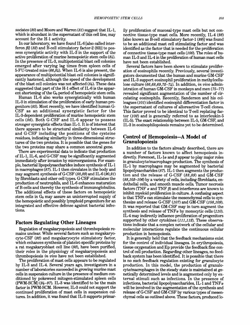
inducing factor for leukemic cell lines (37). Some investi-gators have proposed that hemopoietic factors inducedifferentiation of progenitors in normal hemopoiesis. Asreviewed earlier, our micromanipulation studies ofdifferentiation of single and paired progenitors suggestedthat stem cell differentiation is not a directed process. Theprimary function of the humoral factors is to support thesurvival and/or proliferation of hemopoietic progenitors.In this model, the apparent induction of differentiation isa mere reflection of survival/proliferation of a populationof progenitors that are supported by a humoral factor andthe death of populations of progenitors that are not sup-ported by the factor. Based on these concepts, I have sum-marized the recent evidence on the targets of the in-dividual hemopoietic progenitors. The factors arearbitrarily divided into late-acting, lineage-specific factors,early-acting, lineage-nonspecific factors and those affect-ing dormant stem cells. The proliferative kinetics of thestem cells and the targets of the major hemopoietic fac-tors are schematically presented in Figure 1.
Late-Acting, Lineage-Specific FactorsEp is a physiological regulator of erythropoiesis and is
secreted by the kidney. The recombinant protein has beengiven to patients with chronic renal failure and intracta-ble anemia with remarkable therapeutic success (50).While some controversy still exists, the cells that are themost sensitive to Ep appear to be late erythroid progen-itors that include erythroid colony-forming units (CFU-E) (51), erythroid cluster-forming units (52), and pronor-moblasts. Since these cells are present in large numbers,are close to terminal maturation and are actively engaged
c0
(a
C01)01)
0
Generation Time
R RS
IL-6G-CSF IL-33 DSIL-1
GM-CSF-
EpG-CF P
RS-renewing stem cellDS-differentiating stem cell
FIGURE 1. The model depicts the cell cycle dormancy of the stem cellsand active proliferation of their progenies once they begin differen-tiation. It also illustrates the principle targets of the majorhemopoietic factors.
201

M. OGAWA
in cell proliferation, a slight fluctuation in the ambient Eplevels would result in rapid changes in the production andrelease of reticulocytes. Experimental data suggest thatmultipotential stem cells (53) and early erythroid progen-itors (54-56) are not under the control of Ep.G-CSF appears to be a late-acting, neutrophil-specific
factor. It has been demonstrated that in serum-containingcultures G-CSF supports the formation of neutrophil/mac-rophage colonies and pure neutrophil colonies (57),whereas in serum-free culture, it supports formation ofonly neutrophil colonies (58). In our laboratory, we studiedthe effects of G-CSF alone and in combination with otherfactors on colony fonnation by purified human progenitorsin serum-free culture (59). G-CSF revealed significant syn-ergism with earlier-acting factors such as IL-3 and GM-CSF in support of the formation of neutrophil colonies.When G-CSF was injected into animals, prompt and dose-dependent neutrophilic granulocytosis was observed(60-62). In addition, the differential revealed predomi-nance of mature neutrophils (61). Together, these obser-vations support the concept that G-CSF is a late-acting,neutrophil-specific factor. Interestingly, G-CSF appearsalso to play a role in stimulation of active proliferation ofdormant stem cells. This will be elaborated in more de-tail later.M-CSF has long been considered as a late-acting macro-
phage/monocyte-specific factor. Recently, CSF-HU thatis abundant in human urine (63) was proved to be humanM-CSF (46). A clinical trial of this material produced mod-est elevation in the granulocyte and macrophage countsin patients (63). It was also reported that during preg-nancy, there is concomitant increase in the serum levelsof M-CSF and blood monocyte counts (64). In serum-freeculture of purified human progenitors, M-CSF alone or incombination with GM-CSF, G-CSF, IL-3, and Ep was anineffective factor for macrophage colony formation (59).It is possible that M-CSF needs to interact with other fac-tors in serum in macrophage colony fornation.
Early-Acting, Lineage-Nonspecific FactorsIL-3 appears to be an important factor for proliferation
of early hemopoietic progenitors. Purified murine IL-3supports the growth of various types of colonies, includ-ing multilineage colonies (65-67) and blast cell colonies(66,68). Gibbon IL-3, which is 93% homologous with hu-man IL-3, supported the formation of human multiline-age and blast cell colonies (69). We also observed thatdelayed addition of murine IL-3 to cultures 7 days aftercell plating decreases the number of multipotential blastcell colonies to one-half the number in cultures with IL-3added on day 0. It did not, however, alter the prolifera-tive and differentiative characteristics of late-emerging,multipotential blast cell colonies (66).Based on these observations, we proposed that IL-3
does not trigger stem cells into active proliferation but isrequired for the continued proliferation of early multipo-tential progenitors. Subsequently, we observed that thedevelopment of multipotential blast cell colonies requiresless IL-3 than the process of multilineage colony forma-
tion from blast cell colonies (68). These observations sug-gested that during stem cell development, the early multi-potential progenitors are sensitive to IL-3, whereas asthey gradually differentiate, the sensitivity to IL-3 slowlydeclines.More recently, we examined the effects of human IL-3
on colony formation by purified hemopoietic progenitorsin serum-free culture. IL-3 alone was not effective in sup-port of colony formation except for a few small eosinophilcolonies (59). Serial observation of the serum-free culturedishes containing human IL-3 revealed that blast cellclusters appear after varying lag time, reach approxi-mately the 50 cell stage, and degenerate in the absenceof late-acting, lineage-specific factors. IL-3 revealed sig-nificant synergism with Ep and G-CSF in support oferythroid bursts and neutrophil colonies, respectively.These observations indicated that IL-3 supports progen-itors that are in the early stages of hemopoietic develop-ment, but it does not support the terminal maturationprocess (59).The primary function of GM-CSF may also be to sup-
port the intermediate stages of hemopoietic development.It may not support the terminal neutrophil/macrophagematuration process effectively. Although it was originallyidentified as the factor that supported neutrophil/macro-phage colonies in culture, Metcalf et al. (70) in 1980 ob-served that the few cell divisions of multipotential progen-itors are supported by murine GM-CSF. Using serialtransfer of multipotential blast cell colonies, we demon-strated that a subpopulation of the multipotential progen-itors that respond to IL-3 also respond to GM-CSF (71).Investigators in several laboratories (72-74) have reportedthat human GM-CSF supports multilineage colony forma-tion in serum-containing culture and possesses significantburst-promoting activity (BPA). In the studies of humanenriched progenitors in serum-free culture, we observedthat GM-CSF alone did not support colony formation ex-cept for a few small eosinophil colonies (59). However, incombination with G-CSF and Ep, GM-CSF effectivelysupported a large number of neutrophil colonies anderythroid bursts. Recently, administration of the recom-binant GM-CSF to primates (75,76) and patients with ac-quired immunodeficiency syndrome (77) raised the levelsof circulating neutrophils, eosinophils, and monocytes.These observations indicate that the primary target ofGM-CSF is a population of multipotential progenitors thatare intermediate between those responding to IL-3 andthose sensitive to late-acting, lineage-specific factors.
Factors That Affect Proliferation ofDormant Hemopoietic Stem CellsVery recently, several factors have been identified that
appear to induce proliferation of hemopoietic stem cellsin Go. Stanley and his co-workers (78) reported thathemopoietin-1 (H-1), which was purified from human blad-der carcinoma cell line, 5637 (79), acts synergistically withIL-3 in support of proliferation of murine hemopoieticstem cells. The recent reports by Mochizuki and her as-
202
HEMOPOIETIC STEM CELLS
sociates (80) and Moore and Warren (81) suggest that IL-1,which is abundant in the supernatant of this cell line, mayaccount for the H-1 activity.In our laboratory, we have found IL-6 [also called inter-
feron P2 (82) and B-cell stimulatory factor-2 (83)] to pos-sess synergistic activity with IL-3 in the support of theactive proliferation of murine hemopoietic stem cells (84).In the presence of IL-3, multipotential blast cell coloniesemerged after varying lag times from spleen cells of5-FU-treated mice (66,84). When IL-6 is also present, theappearance of multipotential blast cell colonies is signifi-cantly hastened, although the speed of the developmentof the blast cell colonies was not affected (84). These datasuggested that part of the H-1 effect of IL-6 is the appar-ent shortening of the Go period of hemopoietic stem cells.Human IL-6 also works synergistically with human
IL-3 in stimulation of the proliferation of early human pro-genitors (85). Most recently, we have identified human G-CSF as an additional synergistic factor for theIL-3-dependent proliferation of murine hemopoietic stemcells (86). Both G-CSF and IL-6 appear to possessstronger synergistic effects than IL-1. It is of interest thatthere appears to be structural similarity between IL-6and G-CSF including the positions of the cysteineresidues, indicating similarity in three-dimensional struc-tures of the two proteins. It is possible that the genes forthe two proteins may share a common ancestral gene.There are experimental data to indicate that production
of IL-1, IL-6, and G-CSF may be significantly augmentedimmediately after invasion by microorganisms. For exam-ple, bacterial lipopolysaccharides induce synthesis of IL-1in macrophages (87). IL-1 then circulates in the body andmay augment synthesis of G-CSF (88,89) and IL-6 (90,91)by fibroblasts and other cell types. G-CSF stimulates theproduction of neutrophils, and IL-6 enhances maturationof B-cells and thereby the synthesis of immunoglobulins.The additional effects of these factors on hemopoieticstem cells in Go may assure an uninterrupted supply ofthe hemopoietic and possibly lymphoid progenitors for anintegrated and effective defense against bacterial infec-tions.
Factors Regulating Other LineagesRegulation of megakaryopoiesis and thrombopoiesis re-
mains unclear. While several factors such as megakaryo-cyte-CSF (92) and megakaryocyte stimulatory factor,which enhances synthesis of platelet-specific proteins bya rat megakaryoblast cell line (93), have been purified,their roles in the physiology of megakaryopoiesis andthrombopoiesis in vivo have not been established.The proliferation of mast cells appears to be regulated
by IL-3 and IL-4. Several years ago, investigators in anumber of laboratories succeeded in growing murine mastcells in suspension culture in the presence of medium con-ditioned by pokeweed mitogen-stimulated spleen cells(PWM-SCM) (94-97). IL-3 was identified to be the mainfactor in PWM-SCM. However, IL-3 could not support thecontinued proliferation of mast cells in suspension cul-tures. In addition, it was found that IL-3 supports primar-
ily proliferation of mucosal-type mast cells but not con-nective tissue-type mast cells. More recently, IL-4 (98)[also known as B-cell stimulatory factor-1 (99)] was foundto be an additional mast cell stimulating factor and wasidentified as the factor that is needed for the proliferationof connective tissue-type mast cells (100). The roles of hu-man IL-3 and IL4 in the proliferation ofhuman mast cellshave not been established.Several factors have been shown to stimulate prolifer-
ation of eosinophils recently. Previously, several investi-gators documented that the human and murine GM-CSFand IL-3 support eosinophil proliferation in methylcellu-lose culture (66,68,69,72-74). In addition, in vivo admin-istration ofhuman GM-CSF in monkeys and men (75-77)revealed significant augmentation of the number of cir-culating eosinophils. Recently, Sanderson and his col-leagues (101) identified eosinophil differentiation factor inthe supernatant of cultures of alloreactive T-cell clones.This factor proved to be identical to T-cell replating fac-tor (102) and is generally referred to as interleukin-5(IL-5). The exact relationship between IL-5, GM-CSF, andIL-3 in eosinophilopoiesis remains yet to be determined.
Control of Hemopoiesis-A Model ofGranulopoiesisIn addition to the factors already described, there are
a number of factors known to affect hemopoiesis in-directly. Foremost, IL-ia and p appear to play major rolesin granulocyte/macrophage production. The synthesis ofIL-1 by macrophages may be augmented by bacteriallipopolysaccharides (87). IL-1 then augments the produc-tion and the release of G-CSF (88,89) and GM-CSF(88,103-106) by a variety of cells, including fibroblasts, en-dothelial cells, and smooth muscle cells. Tumor necrosisfactors (TNF a and TNF P) and interferons are known toinhibit myeloid proliferation in culture. A confusing aspectis that TNFa can stimulate the mesenchymal cells to syn-thesize and release G-CSF (107) and GM-CSF (108,109).It was reported that GM-CSF may in turn augment thesynthesis and release of TNFa by monocytic cells (110).IL-4 may indirectly influence proliferation of progenitorssupported by other cytokines (111,112). These observa-tions indicate that a complex network of the cellular andmolecular interactions regulate the continuous cellularproduction in hemopoiesis.
It is generally held that the feedback mechanisms existfor the control of individual lineages. In erythropoiesis,tissue oxygenation and Ep provide the feedback fine con-trol of cell production. Regarding other lineages, no feed-back system has been identified. It is possible that thereis no such feedback regulation existing for granulocyteproduction. In this model, the production of granulo-cyte/macrophages in the steady state is maintained at ge-netically determined levels and is augmented only by ex-ternal stimuli such as infections. In the presence ofinfections, bacterial lipopolysaccharides, IL-1 and TNFawill be involved in the augmentation of the synthesis andrelease ofG-CSF and GM-CSF by various types of mesen-chymal cells as outlined above. These factors, produced lo-
203

204 M. OGAWA
cally in the bone marrow, in turn augment the productionof neutrophils, macrophages, and eosinophils Once the ex-ternal stimuli are removed, the production of the leuko-cytes reverts back to the steady-state level. Since thelifespans of leukocytes are brief, only coarse regulationof the cellular proliferation may be necessary. The widenormal ranges of granulocyte counts may attest to thishypothesis
This work was supported by NIH grant AM32294 and the VeteransAdministration. M. Ogawa is a Veterans Administration Medical Inves-tigator. The author thanks Dr. Pamela N. Pharr and Linda S. Vann fortheir assistance in the preparation of this review.
REFERENCES
1. Erslev, A. J. Production of erythrocytes. In: Hematology (W. J. Wil-liams, E. Beutler, A. J. Erslev, M. A. Lichtman, Eds.), McGraw-Hill, New York, 1983, pp. 365-376.
2. Dancey, J. T., Deubelbeiss, K. A., Harker, L. A., and Finch, C. A.Neutrophil kinetics in man. J. Clin. Invest. 58: 705-715 (1976).
3. Till, J. E., and McCulloch, E. A. A direct measurement of the radi-ation sensitivity of normal mouse bone marrow cells. Radiat. Res.14: 213-222 (1961).
4. Golde, D. W. Methods in Hematology. Hematopoiesis. ChurchillLivingstone, New York, 1984.
5. Williams, D. A., Lemischka, I. R., Nathan, D. G., and Mulligan,R. C. Introduction of new genetic material into pluripotent hae-matopoietic stem cells of the mouse. Nature 310: 476-480.
6. Lemischka, I. R., Raulet, D. H., and Mulligan, R. C. Developmen-tal potential and dynamic behavior or hematopoietic stem cells. Cell45: 917-927 (1986).
7. Mintz, B., Anthony, K., and Litwin, S. Monoclonal derivation ofmouse myeloid and lymphoid lineages from totipotenthematopoietic stem cells experimentally engrafted from fetal hosts.Proc. Natl. Acad. Sci. (U.S.) 81: 7835-7839 (1984).
8. Becker, A. J., McCulloch, E. A., Siminovitch, L., and Till, J. E. Theeffect of differing demands for blood cell production on DNA syn-thesis by hemopoietic colony forming cells of mice. Blood 26:296-308 (1965).
9. Hara, H., and Ogawa, M. Murine hemopoietic colonies in culturecontaining normoblasts, macrophages, and megakaryocytes. Am.J. Hematol. 4: 23-34 (1978).
10. Hodgson, G. S., and Bradley, T. R. Properties of haematopoieticstem cells surviving 5-fluorouracil treatment: Evidence for a pre-CFU-S cell? Nature 281: 381-382 (1979).
11. Suda, T., Suda, J., and Ogawa, M. Proliferative kinetics and differen-tiation of murine blast cell colonies in culture: Evidence for varia-ble Go periods and constant doubling rates of early pluripotenthemopoietic progenitors J. Cell. Physiol. 117: 308-318 (1983).
12. Lajtha, L. G. On the concept of the cell cycle. J. Cell. Comp. Phys-iol. 62(suppl. 1): 143-144 (1963).
13. Lajtha, L. G. Stem cell concepts. Differentiation 14: 23-34 (1979).14. TMl, J. E., McCulloch, E. A., and Siminovitch, L. A stochastic model
of stem cell proliferation, based on the growth of spleen colonyforming cells. Proc. Natl. Acad. Sci. (U.S.) 51: 29-36 (1964).
15. Nakahata, T., Gross, A. J., and Ogawa, M. A stochastic model ofself-renewal and commitment to differentiation of the primitivehemopoietic stem cells in culture. J. Cell. Physiol. 113: 455-458(1982).
16. Ogawa, M., Porter, P. N., and Nakahata, T. Renewal and commit-ment to differentiation of hemopoietic stem cells: An interpretivereview. Blood 61: 823-829 (1983).
17. Curry, J. L., and Trentin, J. J. Hemopoietic spleen colony studies:I. Growth and differentiation. Dev. Biol. 15: 395-413 (1967).
18. Trentin, J. J. Influence of hematopoietic organ stroma(hematopoietic inductive microenvironments) on stem cell differen-tiation. In: Regulation of Hematopoiesis (A. S. Gordon, Ed.),Appleton-Century-Crofts, New York, 1970, pp. 161-186.
19. VanZant, G., and Goldwasser, E. Simultaneous effects oferythropoietin and colony-stimulating factor on bone marrow cells.Science 198: 733-735 (1977).
20. Goldwasser, E. Erythropoietin and differentiation of red blood cells.Fed. Proc. 34: 2285-2292 (1975).
21. Johnson, G. R. Is erythropoiesis an obligatory step in the commit-ment of multipotential hematopoietic stem cells? In: Experimen-tal Hematology Thday (S. J. Baum, G. D. Ledney, and A. Kahn,Eds.), Springer-Verlag, New York, 1981, pp. 13-20.
22. Nakahata, T., and Ogawa, M. Identification in culture of a class ofhemopoietic colony-forming units with extensive capability to self-renew and generate multipotential colonies. Proc. Natl. Acad. Sci.(U.S.) 79: 3843-3847 (1982).
23. Suda, T., Suda, J., and Ogawa, M. Single-cell origin of mousehemopoietic colonies expressing multiple lineages in variable com-binations. Proc. Natl. Acad. Sci. (U.S.) 80: 6689-6693 (1983).
24. Suda, T., Suda, J., and Ogawa, M. Disparate differentiation inmouse hemopoietic colonies derived from paired progenitors. Proc.Natl. Acad. Sci. (U.S.) 81: 2520-2524 (1984).
25. Suda, J., Suda, T., and Ogawa, M. Analysis of differentiation ofmouse hemopoietic stem cells in culture by sequential replating ofpaired progenitors. Blood 64: 393-399 (1984).
26. Leary, A. G., Ogawa, M., Strauss, L. C., and Civin, C. I. Single cellorigin of multilineage colonies in culture: Evidence that differen-tiation of multipotent progenitors and restriction of proliferativepotential of monopotent progenitors are stochastic processes. J.Clin. Invest. 74: 2193-2197 (1984).
27. Leary, A. G., Strauss, L. C., Civin, C. I., and Ogawa, M. Disparatedifferentiation in hemopoietic colonies derived from human pairedprogenitor Blood 66: 327-332 (1985).
28. Wu, A. M. Regulation of self-renewal on human T lymphocytecolony-forming units (TL-CFUs). J. Cell. Physiol. 117: 101-108(1983).
29. Pharr, P. N., Nedelman, J., Downs, H. P., Ogawa, M., and Gross,A. J. A stochastic model for mast cell proliferation in culture- J. Cell.Physiol. 125: 379-386 (1985).
30. Miyake, T., Kung, CK.-H., and Goldwasser, E. Purification of hu-man erythropoietin. J. Biol. Chem. 252: 5558-5564 (1977).
31. Lin, F-K., Suggs, S., Lin, C;H., Browne, J. K., Smalling, R., Egrie,J. C., Chen, K. K., Fox, G. M., Martin, F., Stabinsky, Z., Badrawi,S. M., Lai, P.-H., and Goldwasser, E. Cloning and expression of thehuman erythropoietin gene. Proc. Natl. Acad. Sci. (U.S.) 82:7580-7584 (1985).
32. Jacobs, K., Schoemaker, C., Rudersdorf, R., Neill, S. D., Kaufman,R. J., Mufson, A., Seehra, J., Jones, S. S., Hewick, R., Fritch, E. F.,Kawakita, M., Shimizu, T., and Miyake, T. Isolation and characteri-zation of genomic and cDNA clones of human erythropoietin. Na-ture 313: 806-810 (1985).
33. Burgess, A. W., Camakaris, J., and Metcalf, D. Purification andproperties of colony-stimulating factor from mouse lung-conditionedmedium. J. Biol. Chem. 252: 1998-2003 (1977).
34. Gough, N. M., Gough, J., Metcalf, D., Kelso, A., Grail, D., Nicola,N. A., Burgess, A. W., and Dunn, A. R. Molecular cloning of cDNAencoding a murnne haematopoietic growth regulator, granulocyte-macrophage colony stimulating factor. Nature 309: 763-767 (1984).
35. Gasson, J. C., Weisbart, R. H., Kaufman, S. E., Clark, S. C., He-wick, R. M., Wong, G. G., and Golde, D. W. Purified humangranulocyte-macrophage colony-stimulating factor: Direct actionon neutrophils. Science 226: 1339-1342 (1984).
36. Wong, G. G., Witek, J. S., Temple, P. A., Wilkens, K. M., Leary,A. C., Luxenburg, D. P., Jones, S. S., Brown, E. L., Kay, R. M., Orr,E. C., Shoemaker, C., Golde, D. W., Kaufman, R. J., and Hewick,R. M. Human GM-CSF: Molecular cloning of cDNA and purifica-tion of the natural and recombinant proteins. Science 228: 810-815(1985).
37. Nicola, N. A., Metcalf, D., Matsumoto, M., and Johnson, G. R.Purifi'cation of a factor inducing differentiation in murine my-elomonocytic leukemia cells. Identification as a granulocyte colony-stimulating factor. J. Biol. Chem. 258: 9017-9023 (1983).
38. Welte, K., Platzer, E., Lu, L., Gabrilove, J. L., Levi, E., Martels-mann, R., and Moore, M. A. Purification and biochemical charac-terization of human pluripotent hematopoietic colony-stimulatingfactor. Proc. Natl. Acad. Sci. (U.S.) 82: 1526-1530 (1985).

HEMOPOIETIC STEM CELLS 205
39. Nagata, S., Thuchiya, M., Asano, S., Kaziro, Y., Yamazaki, T.,Yamamoto, O., Hirata, Y, Kubota, N., Oheda, M., Nomme, H., andOno, M. Molecular cloning and expression of cDNA for humangranulocyte colony-stimulating factor. Nature 319: 415-417 (1986).
40. Ihle, J. N., Keller, J., Henderson, L., Klein, F., and Palaszynski, E.Procedures for the purification of interleukin 3 to homogeneity. J.Immunol. 129: 2431-2436 (1982).
41. Yokota, T., Lee, F., Rennick, D., Hall, C., Arai, N., Mosmann, T.,Nabel, G., Cantor, H., and Arai, K. Isolation and characterizationof a mouse cDNA clone that expresses mast-cell growth-factor ac-tivity in monkey cells. Proc. Natl. Acad. Sci. (U.S.) 81: 1070-1074(1984).
42. Fung, M. C., Hapel, A. J., Ymer, S., Cohen, D. R., Johnson, R. M.,Campbell, H. D., and Young, I. G. Molecular cloning of cDNA formurine interleukin-3. Nature 307: 233-237 (1984).
43. Yang Y.-C., Ciarletta, A. R, Temple, P. A., Chung, M. P., Kovacic, S.,Witek-Giannotti, J. S., Leary, A. C., Kriz, R., Donahue, R. E., Wong,G. G., and Clark, S. C. Human IL-3 (multi CSF): Identification byexpression cloning of a novel hematopoietic growth factor relatedto murine IL-3. Cell 47: 3-10 (1986).
44. Stanley, E. R., and Heard, P. M. Factors regulating macrophageproduction and growth. Purification and some properties of thecolony stimulating factor from medium conditioned by mouse Lcells. J. Biol. Chem. 252: 4305-4312 (1977).
45. Kawasaki, E. S., Ledner, M. B., Wang, A. M., Arsdell, J. V., War-ren, M. K., Cayne, M. Y., Schiveickart, V. L., Lee, M.-T., Wilson,K. J., Boosman, A., Stanley, E. R., Ralph, P., and Mark, D. F.Molecular cloning of a complementary DNA encoding humanmacrophage-specific colony-stimulating factor (CSF-1). Science 230:291-296 (1985).
46. Wong, G. G., Temple, P. A., Leary, A. C., Witek-Giannotti, J. S.,Yang, Y.-C., Ciarletta, A. B., Chung, M., Murtha, P., Kriz, R., Kauf-man, R. J., Ferenz, C. R., Sibley, B. S., Turner, K. J., Hewick, R. M.,Clark, S. C., Yanai, N., Yokota, H., Yamada, M., Saito, M.,Motoyoshi, K., and Takaku, F. Human CSF-1: Molecular cloningand expression of 4-kb cDNA encoding the human urinary protein.Science 235: 1504-1508 (1987).
47. Metcalf, D. The granulocyte-macrophage colony-stimulating factor.Science 229: 16-22 (1985).
48. Metcalf, D. The molecular biology and functions of the granulocyte-macrophage colony-stimulating factors Blood 67: 257-267 (1986).
49. Clark, S. C., and Kamen, R. The human hematopoietic colony-stimulating factors. Science 236: 1229-1237 (1987).
50. Eschbach, J. W., Egrie, J. C., Downing, M. R., Browne, J. K., andAdamson, J. W. Correction of the anemia of end-stage renal diseasewith recombinant human erythropoietin. N. Engl. J. Med. 316:73-78(1987).
51. Gregory, C. J. Erythropoietin sensitivity as a differentiation markerin the hemopoietic system: Studies of three erythropoietic colonyresponses in culture. J. Cell. Physiol. 89: 289-302 (1976).
52. Ouellette, P. L., and Monette, F. C. Erythroid progenitors formingclusters in vitro demonstrate high erythropoietin sensitivity. J.Cell. Physiol. 105: 181-184 (1980).
53. Bruce, W. R., and McCulloch, E. A. The effect of erythropoieticstimulation on the hemopoietic colony-forming cells of mice. Blood23: 216-232 (1964).
54. Hara, H., and Ogawa, M. Erythropoietic precursors in mice undererythropoietic stimulation and suppression. Exp. Hematol. 5:141-148(1977).
55. Iscove, N. N. The role of erythropoietin in the regulation of popu-lation size and cell cycling of early and late erythroid precursorsin mouse bone marrow. Cell lisue Kinet. 10: 323-334 (1977).
56. Kimura, H., Finch, C. A., and Adamson, J. W. Hematopoiesis inthe rat: Quantitation of hematopoietic progenitors and the responseto iron deficiency anemia. J. Cell. Physiol. 126: 298-306 (1986).
57. Metcalf, D., and Nicola, N. A. Proliferative effects of purifiedgranulocyte colony-stimulating factor (G-CSF) on normal mousehemopoietic cells. J. Cell. Physiol. 116: 198-206 (1983).
58. Eliason, J. F. Granulocyte-macrophage colony formation in serum-free culture: Effects of purified colony-stimulating factors andmodulation by hydrocortisone. J. Cell. Physiol. 128: 231-238 (1986).
59. Sonoda, Y, Yang, Y.-C., Wong, G. G., Clark, S. C., and Ogawa, M.Analysis in serum-free culture of the targets of recombinant hu-
man hemopoietic growth factors: Interleukin-3 and granulo-cyte/macrophage colony-stimulating factors are specific for earlydevelopmental stages. Proc. Natl. Acad. Sci. (U.S.) 85: 4360-4364(1988).
60. Shimamura, M., Kobayashi, Y., Yuo, A., Urabe, A., Okabe, T.,Komatsu, Y, Itoh, S., and Takaku, F. Effect ofhuman recombinantgranulocyte colony-stimulating factor on hematopoietic injury inmice induced by 5-fluorouracil. Blood 69: 353-355 (1987).
61. Welte, K., Bonilla, A. M., Gillio, A. P., Boone, T. C., Potter, G. K.,Gabrilove, J. L., Moore, M. A. S., O'Reilly, R. J., and Souza, L. M.Recombinant human granulocyte colony-stimulating factor: Effectson hematopoiesis in normal and cyclophosphamide-treated pri-mates. J. Exp. Med. 165: 941-948 (1987).
62. Cohen, A. M., Zsebo, K. M., Inoue, H., Hines, D., Boone, T. C.,Chazin, V. R., Tsai, L., Ritch, T., and Souza, L. M. In vivo stimu-lation of granulopoiesis by recombinant granulocyte colony-stimulating factor. Proc. Natl. Acad. Sci. (U.S.) 84: 2484-2488 (1987).
63. Motoyoshi, K., Takaku, F., and Miura, Y. High serum colony-stimulating activity of leukocytopenic patients after intravenousinfusions of human urinary colony-stimulating factor. Blood 62:685-688 (1983).
64. Bartocci, A., Pollard, J. W, and Stanley, E. R. Regulation of colony-stimulating factor 1 during pregnancy. J. Exp. Med. 164: 956-961(1986).
65. Ihle, J. N., Keller, J., Oroszlan, S., Henderson, L. E., Copeland,T. D., Fitch, F., Prystowsky, M. R, Goldwasser, E., Schrader, J. W,Palaszynski, E., Dy, M., and Lebel, B. Biologic properties ofhomogeneous interleukin 3. I. Demonstration ofWEHI-3 growthfactor activity, mast cell growth factor activity, P cell-stimulatingfactor activity, colony-stimulating factor activity and histamine-producing cell-stimulating factor activity. J. Immunol. 131: 282-287(1983).
66. Suda, T., Suda, J., Ogawa, M., and Ihle, J. N. Permissive role of in-terleukin 3 (IL-3) in proliferation and differentiation of multipoten-tial hemopoietic progenitors in culture. J. Cell. Physiol. 124: 182-190(1985).
67. Rennick, D. M., Lee, F. D., Yokota, T., Arai, K., Cantor, H., andNabel, G. J. A cloned MCGF cDNA encodes a multilineagehematopoietic growth factor: Multiple activities of interleukin 3.J. Immunol. 134: 910-914 (1985).
68. Koike, K., Ihle, J. N., and Ogawa, M. Declining sensitivity to in-terleukin 3 ofmurine multipotential hemopoietic progenitors duringtheir development: Application to a culture system that favors blastcell colony formation. J. Clin. Invest. 77: 894-899 (1986).
69. Leary, A. G., Yang, Y.-C., Clark, S. C., Gasson, J. C., Golde, D. W.,and Ogawa, M. Recombinant gibbon interleukin-3 (IL-3) supportsformation of human multilineage colonies and blast cell colonies inculture: Comparison with recombinant human granulocyte-macrophage colony-stimulating factor (GM-CSF). Blood 70:1343-1348 (1987).
70. Metcalf, D., Johnson, G. R., and Burgess, A. W. Direct stimulationby purified GM-CSF of the proliferation of multipotential anderythroid precursors. Blood 55: 138-147 (1980).
71. Koike, K., Ogawa, M., Ihle, J. N., Miyake, T., Shimizu, T.,Miyajima, A., Yokota, T., and Arai, K. Recombinant murinegranulocyte-macrophage (GM) colony-stimulating factor supportsformation ofGM and multipotential blast cell colonies in culture:Comparison with the effects of interleukin-3. J. Cell. Physiol. 131:458-464(1987).
72. Sieff, C. A., Emerson, S. G., Donahue, R. E., Nathan, D. G., Wang,E. A., Wong, G. G., and Clark, S. C. Human recombinantgranulocyte-macrophage colony-stimulating factor: A multilineagehemopoietin. Science 230: 1171-1173 (1985).
73. Gabrilove, J. L., Welte, K., Harris, P., Platzer, E., Lu, L., Levi, E.,Mertelsmann, R., and Moore, M. A. S. Pluripoietin a: A second hu-man hematopoietic colony-stimulating factor produced by the hu-man bladder carcinoma cell line 5637. Proc. Natl. Acad. Sci. (U.S.)83: 2478-2482 (1986).
74. Kaushansky, K., O'Hara, P. J., Berkner, K., Segel, G. M., Hagen,F. S., and Adamson, J. W Genomic cloning, characterization, andmultilineage growth-promoting activity of human granulocyte-macrophage colony-stimulating factor. Proc. Natl. Acad. Sci. (U.S.)83: 3101-3105 (1986).

206 M. OGAWA
75. Donahue, R. E., Wang, E. A., Stone, D. K., Kamen, R., Wong, G. G.,Sehgal, P. K., Nathan, D. G., and Clark, S. C. Stimulation of hae-matopoiesis in primates by continuous infusion of recombinant hu-man GM-CSF. Nature 321: 872-875 (1986).
76. Nienhuis, A. W, Donahue, R. E., Karlsson, S., Clark, S. C., Agri-cola, B., Antinoff, N., Pierce, J. E., Turner, P., Anderson, W F., andNathan, D. G. Recombinant human granulocyte-macrophagecolony-stimulating factor (GM-CSF) shortens the period of neu-tropenia after autologous bone marrow transplantation in a primatemodel. J. Clin. Invest. 80: 573-577 (1987).
77. Groopman, J. E., Mitsuyasu, R. T., DeLeo, M. J., Oette, D. H., andGolde, D. W. Effect of recombinant human granulocyte-macrophagecolony-stimulating factor on myelopoiesis in the acquired im-munodeficiency syndrome. N. Engl. J. Med. 317: 593-598 (1987).
78. Stanley, E. R., Bartocci, A., Patinkin, D., Rosendaal, M., and Brad-ley, T. R. Regulation of very primitive, multipotent, hemopoieticcells by hemopoietin-1. Cell 45: 667-674 (1986).
79. Jubinsky, P. T., and Stanley, E. R. Purification of hemopoietin 1:A multilineage hemopoietic growth factor. Proc. Natl. Acad. Sci.(U.S.) 82: 2764-2768 (1985). __
80. Mochizuki, D. Y., Eisenman, J. A., Conlon, P. J., Larsen, A. D., andTushinski, R. J. Interleukin 1 regulates hematopoietic activity, arole previously ascribed to hemopoietin 1. Proc. Natl. Acad. Sci.(U.S.) 84: 5267-5271 (1987).
81. Moore. M. A. S., and Warren, D. J. Synergy of interleukin-1 andgranulocyte colony-stimulating factor: In vivo stimulation of stem-cell recovery and hematopoietic regeneration following5-fluorouracil treatment of mice. Proc. Natl. Acad. Sci. (U.S.) 84:7134-7138(1987).
82. Zilberstein, A., Ruggieri, R., Korn, J. H., and Revel, M. Structureand expression of cDNA and genes for human interferon-beta-2,a distinct species inducible by growth stimulatory cytokines. EMBOJ. 5: 2529-2537 (1986).
83. Hirano, T., Yasukawa, K., Harada, H., Taga, T., Watanabe, Y., Mat-suda, T., Kashiwamura, S., Nakajima, K., Koyoma, K., Iwamatsu, A.,Isunasawa, S., Sakiyama, F., Matsui, H., Takahara, Y., Taniguchi,T., and Kishimoto, T. Complementary DNA for a novel human in-terleukin (BSF-2) that induces B lymphocytes to produce immu-noglobulin. Nature 324: 73-77 (1986).
84. Ikebuchi, K., Wong, G. G., Clark, S. C., Ihle, J. N., Hirai, Y., andOgawa, M. Interleukin-6 enhancement of interleukin-3-dependentproliferation of multipotential hemopoietic progenitors Proc. Natl.Acad. Sci. (U.S.) 84: 9035-9039 (1987).
85. Leary, A. G., Ikebuchi, K., Wong, G. G., Yang, Y.-C., Clark, S. C.,and Ogawa, M. Interleukin-6 is a synergistic factor for humanhemopoietic stem cells. Blood 70(suppl. 1): 176a 1987.
86. Ikebuchi, K., Clark, S, C., Ihle, J. N., Souza, L. M., and Ogawa, M.Granulocyte colony-stimulating factor enhances inter-leukin-3-dependent proliferation of multipotential hemopoietic pro-genitors. Proc. Natl. Acad. Sci. (U.S.) 85: 3445-3449 (1988).
87. Dinarello, C. A. Interleukin-1. Rev. Infect. Dis. 6: 51-95 (1984).88. Rennick, D., Yang, G., Gemmell, L., and Lee, F. Control of hemopoi-
esis by a bone marrow stromal cell clone: Lipopolysaccharide- andinterleukin-1-inducible production of colony-stimulating factors.Blood 69: 682-691 (1987).
89. Broudy, V. C., Kaushansky, K., Harlan, J. M., and Adamson, J.WInterleukin 1 stimulates human endothelial cells to producegranulocyte-macrophage colony-stimulating factor and granulocytecolony-stimulating factor. J. Immunol. 139: 464-468 (1987).
90. Yang, Y.-C., Thai, S., Wong, G. G., and Clark, S. C. Interleukin-1 reg-ulation of hematopoietic growth factor production by humanstromal fibroblasts. J. Cell. Physiol. 134: 292-296 (1988).
91. Yasukawa, K., Hirano, T., Watanabe, Y, Muratani, KY, Matsuda, T.,Nakai, S., and Kishimoto, T. Structure and gene expression of hu-man B cell stimulatory factor-2 (BSF-2/IL-6) gene. EMBO J. 5:2529-2537(1987).
92. Hoffman, R., Yang, H. H., Bruno, E., and Straneva, J. E. Purifi-cation and partial characterization of a megakaryocyte colony-stimulating factor from human plasma. J. Clin. Invest. 75:1174-1182 (1985).
93. Tayrien, G., and Rosenberg, R. D. Purification and properties ofa megakaryocyte stimulatory factor present both in the serum-freeconditioned medium of human embryonic kidney cells and in throm-bocytopenic plasma. J. Biol. Chem. 262: 3262-3268 (1987).
94. Schrader, J. W, Lewis, S. J., Clark-Lewis, I., and Culvenor, J. G.The persisting (P) cell: Histamine content, regulation by a T cell-derived factor, origin from a bone marrow precursor, and relation-ship to mast cells. Proc. Natl. Acad. Sci. (U.S.) 78: 323-327 (1981).
95. Nagao, K., Yokoro, K., and Aaronson, S. A. Continuous lines ofbasophil/mast cells derived from normal mouse bone marrow.Science 212: 333-335 (1981).
96. Thrtian, G., Yung, Y P., Guy-Grand, D., and Moore, M. A. S. Long-term in vitro culture of murine mast cells. 1. Description of agrowth factor-dependent culture technique. J. Immunol. 127:788-794 (1981).
97. Razin, E., Cordon-Cordo, C., and Good, R. A. Selective growth ofa population of human basophil cells in vitro. Proc. Natl. Acad. Sci.(U.S.) 78: 5793-5796 (1981).
98. Lee, F., Yokota, T., Otsuka, T., Meyerson, P., VEiaret, D., Coffman, R.,Mosmann, T., Rennick, D., Roehm, N., Smith, C., Zlotnik, A., andArai, K. Isolation and characterization of a mouse interleukincDNA clone that expresses B-cell stimulatory factor 1 activitiesand T-cell- and mast-cell-stimulating activities. Proc. Natl. Acad.Sci. (U.S.) 83: 2061-2065 (1986).
99. Howard, M., Farrar, J., Hilfiker, M., Johnson, B., Takatsu, K.,Hamoaka, T., and Paul,W E. Identification of a T cell-derived Bcell growth factor distinct from interleukin-2. J. Exp. Med. 155:914-923 (1982).
100. Hamaguchi, Y., Kanakura, Y, Fujita, J., Thkeda, S., Nakano, T.,Tarui, S., Honjo, T., and Kitamura, Y Interleukin 4 as an essentialfactor for in vitro clonal growth of murine connective tissue-typemast cells. J. Exp. Med. 165: 268-273 (1987).
101. Sanderson, C. J., O'Garra, A., Warren, D. J., and Klaus, G. G. B.Eosinophil differentiation factor also has B-cell growth factor ac-tivity: Proposed name interleukin 4. Proc. Natl. Acad. Sci. (U.S.)83: 437-440 (1986).
102. Kinashi, T., Harada, N., Severinson, E., Tanabe, T., Sideras, P.,Konishi, M., Azuma, C., Tominaga, A., Bergstedt-Lindqvist, S.,Thkahashi, M., Matsuda, F., Yaoita, Y., Takatsu, K., and Honjo, T.Cloning of complementary DNA encoding T-cell replacing factorand identity with B-cell growth factor II. Nature 324: 70-73 (1986).
103. Zucali, J. R., Dinarello, C. A., Oblon, D. J., Gross, M. A., Ander-son, L., and Weiner, R. S. Interleukin 1 stimulates fibroblasts toproduce granulocyte-macrophage colony-stimulating activity andprostaglandin E2. J. Clin. Invest. 77: 1857-1863 (1986).
104. Bagby, G. C. Jr., Dinarello, C. A., Wallace, P., Wagner, C.,Hefeneider, S., and McCall, E. Interleukin 1 stimulates granulo-cyte macrophage colony-stimulating activity release by vascularendothelial cells. J. Clin. Invest. 78: 1316-1323 (1986).
105. Fibbe,W E., vanDanume, J., Billiau, A., Voogt, P. J., Duinkerken, N.,Kluck, P. M. C., and Falkenburg, J. H. F. Interleukin-1 (22-K fac-tor) induces release of granulocyte-macrophage colony-stimulatingactivity from human mononuclear phagocytes. Blood 68: 1316-1321(1986).
106. Sieff, C. A., Tsai, S., and Faller, D. V. Interleukin 1 induces culturedhuman endothelial cell production of granulocyte-macrophagecolony-stimulating factor. J. Clin. Invest. 79: 48-51 (1987).
107. Koeffler, H. P., Gasson, J., Ranyard, J., Souza, L., Shepard, M., andMunker, R. Recombinant human TNFa stimulates production ofgranulocyte colony-stimulating factor. Blood 70: 55-59 (1987).
108. Munker, R., Gasson, J., Ogawa, M., and Koeffler, H. P. Recombinanthuman TNF induces production of granulocyte-monocyte colony-stimulating factor. Nature 323: 79-82 (1986).
109. Broudy, V. C., Kaushansky, K., Segal, G. M., Harlan, J. M., andAdamson, J. W Tumor necrosis factor type a stimulates human en-dothelial cells to produce granulocyte/macrophage colony-stimulating factor. Proc. Natl. Acad. Sci. (U.S.) 83: 7467-7471 (1986).
110. Cannistra, S. A., Rambaldi, A., Spriggs, D. R., Herrmann, F.,Kufe, D., and Griffin, J. D. Human granulocyte-macrophage colony-stimulating factor induces expression of the tumor necrosis factor

HEMOPOIETIC STEM CELLS 207
gene by the U937 cell line and by normal human monocytes. J. Clin.Invest. 79: 1720-1728 (1987).
111. Peschel, C., Paul,W E., Ohara, J., and Green, I. Effects of B cellstimulatory factor-limterleukin 4 on hematopoietic progenitor cells.Blood 70: 254-263 (1987).
112. Rennick, D., Yang, G., Muller-Sieburg, C., Smith, C., Arai, N.,Takabe, Y, and Gemmell, L. Interleukin 4 (B-cell stimulatory fac-tor 1) can enhance or antagonize the factor-dependent growth ofhemopoietic progenitor cells. Proc. Natl. Acad. Sci. (U.S.) 84:6889-6893 (1987).