high performance boronic acid-containing hydrogel for ... · phenylboronic acid, cross-linker n,...
TRANSCRIPT

RSC Advances
PAPER
Ope
n A
cces
s A
rtic
le. P
ublis
hed
on 2
4 A
ugus
t 201
7. D
ownl
oade
d on
24/
08/2
017
09:4
6:01
. T
his
artic
le is
lice
nsed
und
er a
Cre
ativ
e C
omm
ons
Attr
ibut
ion
3.0
Unp
orte
d L
icen
ce.
View Article OnlineView Journal | View Issue
High performanc
aDivision of Nanophotonics, CAS Center f
Center for Nanoscience and Technology, Be
nanoctr.cn; Tel: +86-010-82545720bThe Armed Police General Hospital, BeijingcHarvard Medical School, Wellman Center f
Hospital, 65 Landsdowne Street, CambridgedHarvard-MIT Division of Health Sciences an
Technology, Cambridge, Massachusetts 021eUniversity of Birmingham, Birmingham B15
† Electronic supplementary informa10.1039/c7ra06965k
Cite this: RSC Adv., 2017, 7, 41384
Received 22nd June 2017Accepted 17th August 2017
DOI: 10.1039/c7ra06965k
rsc.li/rsc-advances
41384 | RSC Adv., 2017, 7, 41384–4139
e boronic acid-containinghydrogel for biocompatible continuous glucosemonitoring†
Qian Dou,a Debo Hu,a Hongkai Gao,b Yongmei Zhang,b Ali K. Yetisen, cd
Haider Butt, e Jing Wang,a Guangjun Nie*a and Qing Dai *a
Rapid and robust hydrogels are essential in realizing continuous glucosemonitoring in diabetes monitoring.
However, existing hydrogels are limited in satisfying all of the sensory requirements such as detection range,
response time, recoverability and biocompatibility. Here, we have developed a surface-initiated
polymerization method to chemically immobilize a nano-boronic acid-hydrogel membrane onto
a quartz crystal, then used a quartz crystal microbalance (QCM) to achieve real-time monitoring of
glucose. The experimental results show that this hydrogel possesses enhanced binding properties to
glucose under physiological conditions (pH 7.0–7.5) and blood glucose concentration (BGC) (1.1–
33.3 mM). Moreover, our hydrogel displayed a rapid response time (�100 s) to glucose, high
biocompatibility in vivo through an animal model. The hydrogel has a great potential as a sensitive
glucose probe for implantable continuous glucose sensors.
Introduction
Continuous glucose monitoring systems (CGMS) are the mostadvanced method for the self-management of diabetes.1–4
Quartz crystal microbalance (QCM) is a mass-sensitive sensorcharacterized by its high sensitivity, fast response and goodoperability.5,6 It can achieve continuous monitoring of glucoseconcentration in a solution by recording the frequency shi ofa quartz crystal before and aer contact with the solution.7–9 Thekey challenge for a QCM-based detection platform is to developa highly-sensitive glucose probe for monitoring in patients'subcutaneous tissues, as the glucose concentrations in inter-stitial uid can be correlated with blood glucose under steady-state conditions.10,11 Boronic acid derivatives offer stability,durability, and low cost for application in CGMS develop-ment.12–16 In particular, boronic acid-containing hydrogels havehigh sensitivity to glucose and offer biocompatibility, and havebecome a promising material for dynamic glucose monitoringin vivo.17–21 For example, Sugnaux et al.22 synthesized glucose-
or Excellence in Nanoscience, National
ijing 100190, P. R. China. E-mail: daiq@
, 100039, China
or Photomedicine, Massachusetts General
, Massachusetts 02139, USA
d Technology, Massachusetts Institute of
39, USA
2TT, UK
tion (ESI) available. See DOI:
0
sensitive polymer brushes with controllable thickness viasurface reversible addition–fragmentation chain-transfer(RAFT) polymerization of 3-methacrylamido phenylboronicacid. The detection range was from 0 to 100 mM; however, thissensor operated outside the physiological conditions (pH 9.0) inphosphate-buffered saline (PBS) solutions. To enable analysisunder physiological pH, Zhang et al.23 have reduced the pKa
value of the hydrogels by adding acrylamide, and constructeda polymerized crystalline colloidal array. This optical sensordetected glucose ranging from 0 to 50 mM at pH 7.4, but hadlow sensitivity (10 mM), which was not suitable for accuratedetection of glucose required by CGMS. Ye et al.24 achieveda high sensitivity (0.5 mM) for glucose by optimizing theconcentration of the acrylamide monomer, 3-methacrylamidophenylboronic acid, cross-linker N,N-methylenebisacrylamidoand solvent dimethyl sulfoxide during the hydrogel preparation,but the detection range was limited to 0.10 to 2.50 mM. Inaddition, the response time of currently reported boronic acid-containing hydrogels are generally longer than 5 min,22–25 whichis not within the realm of practical real-timemonitoring of BGC.Therefore, to realize the potential of boronic acid-containinghydrogel sensors in CGMS, new hydrogel formulations suit-able for fast and sensitive detection of glucose under physio-logical conditions are urgently needed.
In this study, we fabricated an implantable sensor that meetsthe physiological requirements for glucose detection, andevaluated its performance through QCM detection. The3-acrylamidophenylboronic acid (3-APBA) is commerciallyavailable and widely used for the detection of glucose. But3-APBA (pKa > 8) have a low sensitivity at the pH of human
This journal is © The Royal Society of Chemistry 2017
Paper RSC Advances
Ope
n A
cces
s A
rtic
le. P
ublis
hed
on 2
4 A
ugus
t 201
7. D
ownl
oade
d on
24/
08/2
017
09:4
6:01
. T
his
artic
le is
lice
nsed
und
er a
Cre
ativ
e C
omm
ons
Attr
ibut
ion
3.0
Unp
orte
d L
icen
ce.
View Article Online
interstitial uid (7.0–7.5),26,27 we developed a hydrogel systembased on 3-APBA as the glucose-sensing component, and withacrylamide as the monomer and N,N0-methylenebisacrylamideas the crosslinking agent for hydrogel synthesis. The numerousamine groups introduced into the hydrogel compositemembrane hence help to reduce its pKa value, and enhance thecomplexation ability and glucose responsivity of the hybridmembrane at physiological pH.28 Molar ratios of monomers toprepare the hydrogel composition were optimized to operate atphysiological glucose concentrations in human interstitial uid(1.1–33.3 mM). Furthermore, since the real-time sensingcapacity of the existing hydrogel sensors was primarily limitedby the low association rate of hydrogel lm with glucose, weemployed a surface-initiated polymerization method to chemi-cally immobilize the nanogel onto a quartz crystal, with thethickness of the nanoscale hydrogel lm controlled to improveits response time. By tuning the dimensions of the hydrogelnetwork via varying hydrogel formulations and reaction time,the contact between free glucose molecules and hydrogelnetwork can be maximized. This new boronic acid-containinghydrogel sensor detects glucose at pH 7.0–7.5 within a detec-tion range of 1.1–33.3 mM (typical physiological BGC) anda rapid response time (�100 s) as compared to existing sensorsbased on boronic acid in the literature. Subcutaneous implan-tation experiments in rats indicated that the hydrogel hasbiocompatibility and durability, and is potentially suitable forin vivo glucose monitoring.
Results and discussionSynthesis and characterization
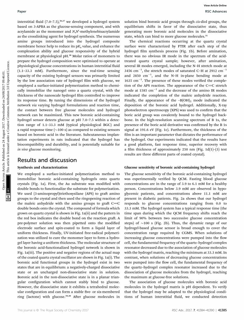
We employed a surface-initiated polymerization method toimmobilize boronic acid-containing hydrogels onto quartzcrystals (Fig. 1a). First, the Au substrate was modied withdouble bonds to functionalize the substrate for polymerization.We used (3-aminopropyl)triethoxysilane (APS) to gra aminogroups to the crystal and then used the ringopening reaction ofthe maleic anhydride with the amino groups to gra C]Cdouble bonds onto the crystal. The compound of formula whichgrown on quartz crystal is shown in Fig. 1a(ii) and the pattern inthe red box indicates the double bond on the reaction gra. Apre-polymer solution was then pipetted onto the modiedelectrode surface and spin-coated to form a liquid layer ofuniform thickness. Finally, UV-initiated free-radical polymeri-zation was utilized to cure the monomer layer to form a hydro-gel layer having a uniform thickness. Themolecular structure ofthe boronic acid-functionalized hydrogel network is shown inFig. 1a(iii). The positive and negative regions of the actual mapof the coated quartz crystal oscillator are shown in Fig. 1a(i). Theboronic acid functional groups in the hydrogel exist in twostates that are in equilibrium: a negatively-charged dissociativestate or an uncharged non-dissociative state in solution.Boronic acid in the non-dissociative state is in a planar trian-gular conguration which cannot stably bind to glucose.However, the dissociative state it exhibits a tetrahedral molec-ular conguration and can form a stable ve- or six-memberedring (lactone) with glucose.29,30 Aer glucose molecules in
This journal is © The Royal Society of Chemistry 2017
solution bind boronic acid groups through cis-diol groups, theequilibrium shis in favor of the dissociative state, thusgenerating more boronic acid molecules in the dissociativestate, which can bind to more glucose molecules.31
The chemical reactions occurring at the quartz crystalsurface were characterized by FTIR aer each step of thehydrogel lm synthesis process (Fig. 1b). Before amination,there was no obvious IR mode in the spectrum of the acid-treated quartz crystal sample; however, aer amination,several IR modes emerged, including the N–H stretch mode at3456 cm�1, the stretch modes of saturated C–H at 2912 cm�1
and 2850 cm�1, and the N–H in-plane bending mode at1635 cm�1. The presence of these modes veried the comple-tion of the APS reaction. The appearance of the C]C stretchmode at 1585 cm�1 and the decrease of the amino IR modesindicated the completion of the maleic anhydride reaction.Finally, the appearance of the –B(OH)2 mode indicated thedeposition of the boronic acid hydrogel. Additionally, X-rayphotoelectron spectroscopy (XPS) was used to conrm that theboric acid group was covalently bound to the hydrogel back-bone. In the high-resolution scanning spectrum of B 1s, thepresence of the boric acid derivative was conrmed by the B 1ssignal at 191.6 eV (Fig. 1c). Furthermore, the thickness of thelm is an important parameter that dictates the performance ofthe hydrogel. Our experiments indicated that the results havea good platform, fast response time, superior recovery witha lm thickness of approximately 250 nm (Fig. 1d(1)–(3) testresults are three different parts of coated crystal).
Glucose sensitivity of boronic acid-containing hydrogel
The glucose sensitivity of the boronic acid-containing hydrogelwas experimentally veried by QCM. Fasting blood glucoseconcentrations are in the range of 3.9 to 6.1 mM for a healthyperson. Concentrations below 3.9 mM are observed in hypo-glycemic patients, and concentrations above 11.1 mM arepresent in diabetic patients. Fig. 2a shows that our hydrogelresponds to glucose concentrations ranging from 0.0 to33.3 mM. The hydrogel system has a typical response time (thetime span during which the QCM frequency shis reach thelimit of 90% between two successive glucose concentrationsteps) of �100 s (Fig. 2b). Thus, the dynamic range of ourhydrogel-based glucose sensor is broad enough to cover theconcentration range required by CGMS. When solutions ofincreasing glucose concentrations were pumped into the owcell, the fundamental frequency of the quartz–hydrogel complexresonator decreased due to the association of glucose moleculeswith the hydrogel matrix, reaching the minimum at 33.3 mM. Incontrast, when solutions of decreasing glucose concentrationswere pumped into the ow cell, the fundamental frequency ofthe quartz–hydrogel complex resonator increased due to thedissociation of glucose molecules from the hydrogel, reachingthe maximum at glucose-free solutions.
The association of glucose molecules with boronic acidmolecules in the hydrogel matrix is pH dependent. To verifythat the hydrogel may be adapted to the physiological condi-tions of human interstitial uid, we conducted detection
RSC Adv., 2017, 7, 41384–41390 | 41385

Fig. 1 The synthesis and characterization. (a) Synthesis of the hydrogel-coated quartz crystal. (b) FTIR spectra at different stages of reaction. (c) B1s XPS spectra of the hydrogel film. (d) Hydrogel thickness characterized by AFM.
RSC Advances Paper
Ope
n A
cces
s A
rtic
le. P
ublis
hed
on 2
4 A
ugus
t 201
7. D
ownl
oade
d on
24/
08/2
017
09:4
6:01
. T
his
artic
le is
lice
nsed
und
er a
Cre
ativ
e C
omm
ons
Attr
ibut
ion
3.0
Unp
orte
d L
icen
ce.
View Article Online
experiments with glucose solutions (0.0–33.3 mM) of pH valuesranging from 7.0 to 7.5 (Fig. 2d). The hydrogel was sensitive toglucose and exhibited linearity at different pH values.Maximum glucose sensitivity was observed at pH 7.5, becausehigher pH facilitates the association of glucose molecules withboronic acid molecules in the hydrogel network. Fig. 2c showsthat the frequency shi is linearly proportional to the glucoseconcentration over the range of 0.0–33.3 mM. Deviations fromthis linear relationship are seen at glucose concentrations above9.4 mM. The larger error at higher glucose concentrations maybe due to the conversion between the 1 : 1 binding mode andthe 2 : 1 cross-linking mode between the boronic acid andglucose molecules.23,32,33 To obtain optimal linear ttingbetween frequency shi and glucose concentration, we per-formed the tting in the ranges of 0.0–9.4 mM and9.4–33.3 mM. The linear coefficients were 0.9971 (pH 7.0),0.9934 (pH 7.3), and 0.9983 (pH 7.5) in the range of 0 to 9.4 mM;and 0.9870 (pH 7.0), 0.9637 (pH 7.3), and 0.9921 (pH 7.5) in therange of 9.4 to 33.3 mM. The reversibility of the hydrogel was
41386 | RSC Adv., 2017, 7, 41384–41390
tested by alternately pumping solutions of low (glucose-freesolutions) and high (3.9 mM) glucose concentrations into theow cell. Aer eleven association–dissociation cycles, thehydrogel maintained its glucose sensitivity (Fig. 2e), thusshowing the sensor has a good recoverability to glucose.
While glucose is predominant, human blood also containsother sugars such as fructose, galactose, sucrose and so on.34–36
However, the effect of these sugars, even at such relatively lowconcentration (<0.1 mM (ref. 37–39)), may be to act as inter-ferents to any glucose measurement using phenylboronic acids.To assess the inuence of possible competitive binding of0.1 mM of other saccharides on the glucose, an experiment wascarried out in which the sensor chip was exposed to 3.9 mMglucose with and without 0.1 mM of other saccharides. Fig. 2fshows that the presence of fructose, maltose and lactose lead toa slight increase in response frequency, compared to the pres-ence of glucose alone. But QCM-based sensor is also able todetect physiologically relevant glucose concentrations also inthe presence of other competing sugars within the error range.
This journal is © The Royal Society of Chemistry 2017

Fig. 2 Glucose sensing. (a) Frequency shift with varying glucose concentrations. (b) Response time. (c) Relationship between frequency shift andglucose concentration. (d) Glucose responses at different pH. (e) Reversibility. (f) Interference.
Paper RSC Advances
Ope
n A
cces
s A
rtic
le. P
ublis
hed
on 2
4 A
ugus
t 201
7. D
ownl
oade
d on
24/
08/2
017
09:4
6:01
. T
his
artic
le is
lice
nsed
und
er a
Cre
ativ
e C
omm
ons
Attr
ibut
ion
3.0
Unp
orte
d L
icen
ce.
View Article Online
Biocompatibility of the glucose sensors
To assess the biocompatibility of the hydrogel sensor, we usedmicroscopy to image L929 cells exposed to the coated chips. Nosignicant difference was observed between cells in the exper-imental group and the control group (consisting of a reagentcontrol and a negative control) (Fig. 3a). The experimentalgroup cells exhibited normal cellular morphology, intactcellular membranes, and no exfoliation or dissolution; thus,there was no evidence of cytotoxicity. We also used the methyl-thiazolyl-tetrazolium (MTT) assay to evaluate the cytotoxicity ofthe hydrogel-coated chips, according to the national standardGB/T 16886-2001.40 MTT results showed that the relative incre-ment rate (RGR) were greater than 90% for incubation periods
This journal is © The Royal Society of Chemistry 2017
of 24 h and 48 h (Fig. 3b). According to the GB/T 16175-1996,41
this is categorized as a grade 1 reaction. These results conrmedthat the hydrogel-coated chips were not toxic to cells.
Aer subcutaneous implantation of the chips into SD rats,tissues that were in direct contact with the coated chips wereimaged, and no difference was observed between the experi-mental group and the control group. Tissues were imaged at7 days post-implantation (Fig. 3c). Aer surgery, all the SD rats'diet, activities and defecation were normal. We did not nd thesurgical incision of rats with pus and local tissue necrosis(Fig. S3†). At 7 days post-implantation, tissue morphology wasnormal, and there was no evidence of inammatory cell inl-tration into the tissue. No brous tissue twined around the
RSC Adv., 2017, 7, 41384–41390 | 41387
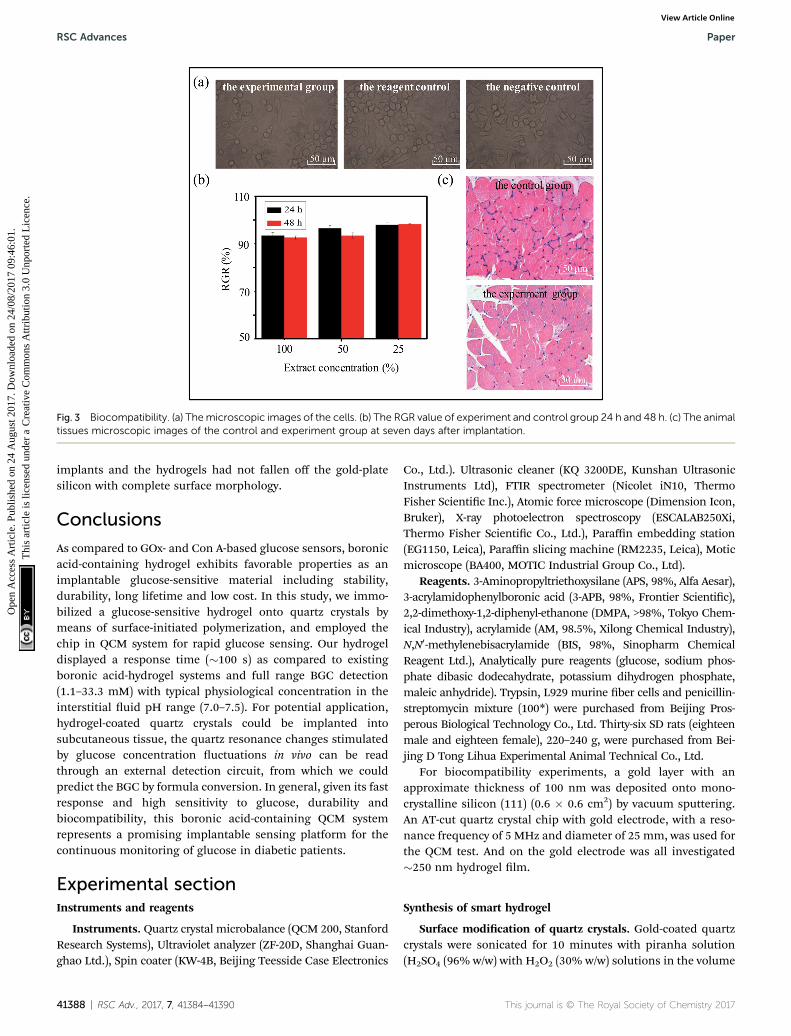
Fig. 3 Biocompatibility. (a) Themicroscopic images of the cells. (b) The RGR value of experiment and control group 24 h and 48 h. (c) The animaltissues microscopic images of the control and experiment group at seven days after implantation.
RSC Advances Paper
Ope
n A
cces
s A
rtic
le. P
ublis
hed
on 2
4 A
ugus
t 201
7. D
ownl
oade
d on
24/
08/2
017
09:4
6:01
. T
his
artic
le is
lice
nsed
und
er a
Cre
ativ
e C
omm
ons
Attr
ibut
ion
3.0
Unp
orte
d L
icen
ce.
View Article Online
implants and the hydrogels had not fallen off the gold-platesilicon with complete surface morphology.
Conclusions
As compared to GOx- and Con A-based glucose sensors, boronicacid-containing hydrogel exhibits favorable properties as animplantable glucose-sensitive material including stability,durability, long lifetime and low cost. In this study, we immo-bilized a glucose-sensitive hydrogel onto quartz crystals bymeans of surface-initiated polymerization, and employed thechip in QCM system for rapid glucose sensing. Our hydrogeldisplayed a response time (�100 s) as compared to existingboronic acid-hydrogel systems and full range BGC detection(1.1–33.3 mM) with typical physiological concentration in theinterstitial uid pH range (7.0–7.5). For potential application,hydrogel-coated quartz crystals could be implanted intosubcutaneous tissue, the quartz resonance changes stimulatedby glucose concentration uctuations in vivo can be readthrough an external detection circuit, from which we couldpredict the BGC by formula conversion. In general, given its fastresponse and high sensitivity to glucose, durability andbiocompatibility, this boronic acid-containing QCM systemrepresents a promising implantable sensing platform for thecontinuous monitoring of glucose in diabetic patients.
Experimental sectionInstruments and reagents
Instruments. Quartz crystal microbalance (QCM 200, StanfordResearch Systems), Ultraviolet analyzer (ZF-20D, Shanghai Guan-ghao Ltd.), Spin coater (KW-4B, Beijing Teesside Case Electronics
41388 | RSC Adv., 2017, 7, 41384–41390
Co., Ltd.). Ultrasonic cleaner (KQ 3200DE, Kunshan UltrasonicInstruments Ltd), FTIR spectrometer (Nicolet iN10, ThermoFisher Scientic Inc.), Atomic force microscope (Dimension Icon,Bruker), X-ray photoelectron spectroscopy (ESCALAB250Xi,Thermo Fisher Scientic Co., Ltd.), Paraffin embedding station(EG1150, Leica), Paraffin slicing machine (RM2235, Leica), Moticmicroscope (BA400, MOTIC Industrial Group Co., Ltd).
Reagents. 3-Aminopropyltriethoxysilane (APS, 98%, Alfa Aesar),3-acrylamidophenylboronic acid (3-APB, 98%, Frontier Scientic),2,2-dimethoxy-1,2-diphenyl-ethanone (DMPA, >98%, Tokyo Chem-ical Industry), acrylamide (AM, 98.5%, Xilong Chemical Industry),N,N0-methylenebisacrylamide (BIS, 98%, Sinopharm ChemicalReagent Ltd.), Analytically pure reagents (glucose, sodium phos-phate dibasic dodecahydrate, potassium dihydrogen phosphate,maleic anhydride). Trypsin, L929 murine ber cells and penicillin-streptomycin mixture (100*) were purchased from Beijing Pros-perous Biological Technology Co., Ltd. Thirty-six SD rats (eighteenmale and eighteen female), 220–240 g, were purchased from Bei-jing D Tong Lihua Experimental Animal Technical Co., Ltd.
For biocompatibility experiments, a gold layer with anapproximate thickness of 100 nm was deposited onto mono-crystalline silicon (111) (0.6 � 0.6 cm2) by vacuum sputtering.An AT-cut quartz crystal chip with gold electrode, with a reso-nance frequency of 5 MHz and diameter of 25 mm, was used forthe QCM test. And on the gold electrode was all investigated�250 nm hydrogel lm.
Synthesis of smart hydrogel
Surface modication of quartz crystals. Gold-coated quartzcrystals were sonicated for 10 minutes with piranha solution(H2SO4 (96% w/w) with H2O2 (30% w/w) solutions in the volume
This journal is © The Royal Society of Chemistry 2017

Paper RSC Advances
Ope
n A
cces
s A
rtic
le. P
ublis
hed
on 2
4 A
ugus
t 201
7. D
ownl
oade
d on
24/
08/2
017
09:4
6:01
. T
his
artic
le is
lice
nsed
und
er a
Cre
ativ
e C
omm
ons
Attr
ibut
ion
3.0
Unp
orte
d L
icen
ce.
View Article Online
ratio 7 : 3.). Processed quartz crystals were washed with redis-tilled water, then were placed in acetone, ethanol and redistilledwater, respectively, each solution was sonicated for 10 minutes,in an ultrasonic cleaner. Aer the quartz crystals were driedwith nitrogen, they were immersed in a mixed solution of3-aminopropyltriethoxysilane and methylbenzene (v/v 1 : 10)under nitrogen. Twelve hours later, the quartz crystals wererinsed with ethanol and subsequently dried with nitrogen. Thedried quartz crystals were then immersed in a mixed solution ofmaleic anhydride and dimethylformamide (2%) for twenty-fourhours before being rinsed with ethanol and dried withnitrogen.42,43
Deposition of smart hydrogel lms. First, we prepareda 5 mol L�1 pre-polymer solution consisting of 18% 3-APB, 2%BIS, 78% AM, and 2% DMPA (by mass) in dimethyl sulfoxide asthe solvent. Second, 30 mL of pre-polymer solution was depos-ited onto the upper electrode of the quartz crystal and spin-coated for one minute (3500 rpm). Third, the coated quartzcrystals were placed under ultraviolet irradiation (l ¼ 365 nm)under nitrogen for 30 min for polymerization. Finally, the lm-coated quartz crystals were repeatedly rinsed with ethanol andredistilled water.
Instruction manual. We remained the waste piranha solu-tion alone placed in the glass waste bottle, sealed with plasticlm and opened a few small hole. Then placed bottle in a fumehood.
Verication of glucose sensitivity
A quartz crystal coated with a glucose-sensitive hydrogel lmwas dried with nitrogen and installed into the ow cell of theQCM 200 system. PBS (0.1 mol L�1) was pumped into the owcell continuously, and the frequency of the crystal was moni-tored in real time using the QCM data acquisition soware.Aer the frequency was stabilized (frequency shi # �2 Hz in10 min), we evaluated the glucose detection capacity of thematerial. Solutions (3 mL) of increasing and then decreasingglucose concentrations (from 0.0 to 33.3 mmol L�1 with PBS,and then in reverse) were pumped into the ow cell every10 min, and the frequency shi DF was recorded for eachglucose concentration. Several cycles of this experiment wereconducted under different pH levels from 7.0 to 7.5 to mimicthe human interstitial uid environment. Further experimentswere conducted to verify the reversibility of the lm by repeat-edly pumping glucose solutions of 0.0 and 3.9 mmol L�1
concentrations into the ow cell.
Biological experiment
All animal experiments were performed complying with theNational Institutes of health (NIH) guidelines for the care and useof laboratory animals of National Center for Nanoscience andTechnology Animal Study Committee's requirements and accord-ing to the protocol approved by the Institutional Animal Care.
Cell toxicity
(1) Quantitative evaluation. The culture medium used in theexperiment was the dulbecco's minimum essential medium
This journal is © The Royal Society of Chemistry 2017
(DMEM) culture medium containing 10% fetal bovine serum(FBS) and 1% penicillin–streptomycin. The detailed operationalprocedures for the MTT assay are as follows: (1) Extraction: eachchip was soaked in 4 mL of culture medium and extracted at37 �C for 24 h. The same conditions were treated with negativecontrast (high density polyethylene) and reagent contrast (underthe condition of without test materials, according to the extrac-tion conditions and test steps to get the extraction medium). (2)Cell inoculation: a single cell suspension was prepared in culturemedium, and 5000 cells were seeded in 96 well plates witha volume of 200 mL per well. (3) Extraction solution treatment:culturemedium was removed aer cells fully adhered to the platewall, and 200 mL of leaching solution was added to each well. Thedifferent concentration leaching solution (25%, 50% or 100%)was obtained by diluting the culture solution. Cells were cultured24 h and 48 h aer the addition of the extraction solution. For allexperiments, three replicate chips were used for each data point,and six parallel wells were used for each dilution. (4) Colorreaction: MTT was dissolved in PBS (pH 7.4), and 20 mL(5 mg mL�1) was added to each well. (5) Termination reaction:cells were incubated in MTT for 4 h, subsequently, the superna-tant was removed and 150 mL of dimethyl sulfoxide was added toeach well to dissolve the crystals, and the plate was placed on theshaker with low-speed oscillation. (6) Colorimetric detection: theabsorbance was detected by the enzyme-linked immune sorbentassay (ELISA) at a wavelength of 492 nm wavelength, and therelative increment rate (RGR ¼ (Aexperiment/Areagent) � 100%) werecalculated. For quantitative evaluation of cytotoxicity, thefollowingmetrics were used: level 0,$100% or higher; level 1, 75–99%; level 2, 50–74%; level 3, 25–49%; level 4, 1–24%, level 5, 0.
(2) Qualitative evaluation. Cells were seeded in 6-well platesand were cultured under 5% CO2 at 37 �C in the incubator. Aerthe cells had adhered to the plate, they were incubated in theextracted solution for 24 h and were then evaluated by micros-copy for cell morphology, vacuolization, loss, cell dissolved, andmembrane integrity, and were scoring accordingly. The scoringmethod was as follows: 0, no cell toxicity; 1, mild cytotoxicity; 2,moderate cytotoxicity; 3, severe cytotoxicity.
Implant experiment
Test samples and control samples (high-density Teon) wereimplanted subcutaneously in the backs of rats. A total of 12 ratswere used (six male, six female), and each group receivingimplants for 7 days. Aer implantation, rats were observed andevaluated daily. Aer 7 implantation period, twelve rats weresacriced to visualize the implant and the surrounding tissues.Paraffin sectioning, HE staining, and light microscopy wereperformed to evaluate the degree of tissue reaction. Addition-ally, the surface morphology of the chip was evaluated for anychanges post-implantation. Chips that exhibited any changes inmorphology, lm position, decomposition, or extensive pres-ence of biomass were considered to fail the implantation test.
Conflicts of interest
There are no conicts to declare.
RSC Adv., 2017, 7, 41384–41390 | 41389
RSC Advances Paper
Ope
n A
cces
s A
rtic
le. P
ublis
hed
on 2
4 A
ugus
t 201
7. D
ownl
oade
d on
24/
08/2
017
09:4
6:01
. T
his
artic
le is
lice
nsed
und
er a
Cre
ativ
e C
omm
ons
Attr
ibut
ion
3.0
Unp
orte
d L
icen
ce.
View Article Online
Acknowledgements
This work was supported by the National Basic ResearchProgram of China (Grant No. 2015CB932400, 2016YFA0201600),the National Natural Science Foundation of China (Grant No.51372045, 11504063), the Bureau of International Cooperation,Chinese Academy of Science (121D11KYSB20130013), and thekey program of the bureau of Frontier Sciences and EductionChinese Academy of Sciences (QYZDB-SSW-SLH021).
Notes and references
1 B. Kovatchev, L. Heinemann, S. Anderson and W. Clarke,Diabetes Care, 2008, 31, 1160–1164.
2 T. S. Bailey, H. C. Zisser and S. K. Garg, Diabetes Technol.Ther., 2007, 9, 203–210.
3 I. M. Wentholt, J. B. Hoekstra, M. A. Vollebregt, J. HansDevries and A. A. Hart, Diabetes Care, 2005, 28, 2871–2876.
4 G. McGarraugh, Diabetes Technol. Ther., 2009, 11, S17–S24.5 M. V. Voinova, M. Jonson and B. Kasemo, Biosens.Bioelectron., 2002, 17, 835–841.
6 M. Rodahl, F. Hook and B. Kasemo, Anal. Chem., 1996, 68,2219–2227.
7 M. Rodahl and B. Kasemo, Sens. Actuators, B, 1996, 37, 111–116.
8 X.-L. Su and Y. Li, Biosens. Bioelectron., 2005, 21, 840–848.9 R. Hao, D. Wang, X. Zhang, G. Zuo, H. Wei, R. Yang,Z. Zhang, Z. Cheng, Y. Guo, Z. Cui and Y. Zhou, Biosens.Bioelectron., 2009, 24, 1330–1335.
10 F. R. Kaufman, J. Austin, A. Neinstein, L. Jeng, M. Halvorson,D. J. Devoe and P. Pitukcheewanont, J. Pediatr., 2002, 141,625–630.
11 D. C. Klonoff, Diabetes Care, 2005, 28, 1231–1239.12 H.-C. Wang, H. Zhou, B. Chen, P. M. Mendes, J. S. Fossey,
T. D. James and Yi-T. Long, Analyst, 2013, 138, 7146–7151.13 K. Ngamdee, T. Noipa, S. Martwiset and T. Tuntulani, Sens.
Actuators, B, 2011, 160, 129–138.14 C. Shimpuku, R. Ozawa, A. Sasaki, F. Sato, T. Hashimoto,
A. Yamauchi, I. Suzuk and T. Hayashita, Chem. Commun.,2009, 13, 1709–1711.
15 A. P. Davis and R. S. Wareham, Angew. Chem., 1999, 38,2978–2996.
16 S. L. Wiskur, H. Aithaddou and J. J. Lavigne, Acc. Chem. Res.,2001, 34, 963–972.
17 Y. Guan and Y. Zhang, Chem. Soc. Rev., 2013, 42, 8106–8121.18 J. N. Cambre and B. S. Sumerlin, Polymer, 2011, 52, 4631–
4643.19 K. Lacina, P. Skladal and T. D. James, Chem. Cent. J., 2014, 8,
60–77.20 C. C. Deng, W. L. A. Brooks, K. A. Abboud and B. S. Sumerlin,
ACS Macro Lett., 2015, 4, 220–224.
41390 | RSC Adv., 2017, 7, 41384–41390
21 X. Jin, X. Zhang, Z. Wu, D. Teng, X. Zhang, Y. Wang, Z. Wangand C. Li, Biomacromolecules, 2009, 10, 1337–1345.
22 C. Sugnaux and H.-A. Klok,Macromol. Rapid Commun., 2014,35, 1402–1407.
23 C. Zhang, G. G. Cano and P. V. Braun, Adv. Mater., 2014, 26,5678–5683.
24 G. Ye and X. Wang, Biosens. Bioelectron., 2010, 26, 772–777.25 J. T. Suri, D. B. Cordes, F. E. Cappuccio, R. A. Wessling and
B. Singaram, Angew. Chem., Int. Ed., 2003, 42, 5857–5859.26 D. Shno, A. Kubo, Y. Murata, Y. Koyama, K. Kataoka,
A. Kikuchi, Y. Sakurai and T. Okano, J. Biomater. Sci.,Polym. Ed., 1996, 7, 697–705.
27 C. Ancla, V. Lapeyre, I. Gosse, B. Catargi and V. Ravaine,Langmuir, 2011, 27, 12693–12701.
28 I. Hisamitsu, K. Kataoka, T. Okano and Y. Sakurai, Pharm.Res., 1997, 14, 289–293.
29 K. Kataoka, H. Miyazaki, M. Bunya, T. Okano and Y. Sakurai,J. Am. Chem. Soc., 1998, 120, 12694–12695.
30 R. Gabai, N. Sallacan, V. Chegel, T. Bourenko, E. Katz andI. Willner, J. Phys. Chem. B, 2001, 105, 8196–8202.
31 A. M. Horgan, A. J. Marshall, S. J. Kew, K. E. S. Dean,C. D. Creasey and S. Kabilan, Biosens. Bioelectron., 2006,21, 1838–1845.
32 V. L. Alexeev, A. C. Sharma, A. V. Goponenko, S. Das,I. K. Lednev, C. S. wilcox, D. N. Finegold and S. A. Asher,Anal. Chem., 2003, 75, 2316–2323.
33 Y. J. Huang, W. J. Ouyang, X. Wu, Z. Li, J. S. Fossey,T. D. James and Y.-B. Jiang, J. Am. Chem. Soc., 2013, 135,1700–1703.
34 S. Tierney, S. Volden and B. T. Stokke, Biosens. Bioelectron.,2009, 24, 2034–2039.
35 G. Ye and X. Wang, Biosens. Bioelectron., 2010, 26, 772–777.36 M.-C. Lee, S. Kabilan, A. Hussain, X. Yang, J. Blyth and
C. R. Lowe, Anal. Chem., 2004, 76, 5748–5755.37 A. M. Horgan, A. J. Marshall, S. J. Kew, K. E. S. Dean,
C. D. Creasey and S. Kabilan, Biosens. Bioelectron., 2006,21, 1838–1845.
38 T. D. James, K. R. A. Samankumara Sandanayake andS. Shinkai, Angew. Chem., Int. Ed. Engl., 1994, 33, 2207–2209.
39 H. Eggert, J. Frederiksen, C. Morin and J. C. Norrild, J. Org.Chem., 1999, 64, 3846–3852.
40 State Administration of Quality Supervision, Inspection andQuarantine of the People's Republic of China, GB/T16886,Standards Press of China, Beijing, 2001.
41 National Technical Supervision Bureau, GB/T 16175,Standards Press of China, Beijing, 1996.
42 M. Lazerges, H. Perrot, N. Rabehagasoa and C. Compere,Biosensors, 2012, 2, 245–254.
43 P. Vejayakumaran, I. A. Rahman, C. S. Sipaut, J. Ismail andC. K. Chee, J. Colloid Interface Sci., 2008, 328, 81–91.
This journal is © The Royal Society of Chemistry 2017