high resolution x-ray ct as enabling technology for ... · pdf filehigh resolution x-ray ct as...
TRANSCRIPT
High resolution X-ray CT as enabling
technology for material research
Veerle Cnudde Dept. of Geology & Soil Science - UGCT, Ghent University, Belgium
With credits to the entire UGCT team (www.ugct.ugent.be)
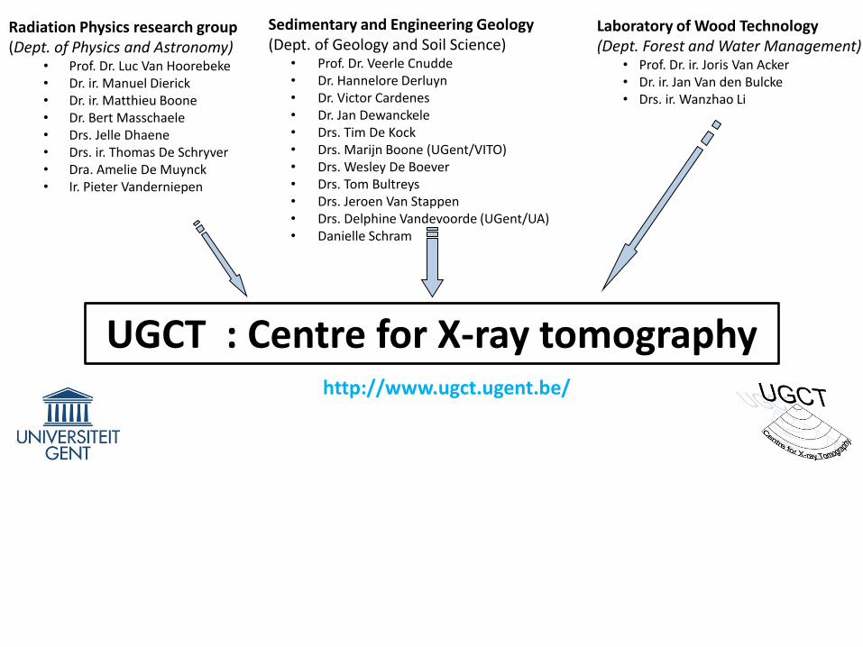
Sedimentary and Engineering Geology (Dept. of Geology and Soil Science)
• Prof. Dr. Veerle Cnudde • Dr. Hannelore Derluyn • Dr. Victor Cardenes • Dr. Jan Dewanckele • Drs. Tim De Kock • Drs. Marijn Boone (UGent/VITO) • Drs. Wesley De Boever • Drs. Tom Bultreys • Drs. Jeroen Van Stappen • Drs. Delphine Vandevoorde (UGent/UA) • Danielle Schram
UGCT : Centre for X-ray tomography http://www.ugct.ugent.be/
Radiation Physics research group (Dept. of Physics and Astronomy)
• Prof. Dr. Luc Van Hoorebeke • Dr. ir. Manuel Dierick • Dr. ir. Matthieu Boone • Dr. Bert Masschaele • Drs. Jelle Dhaene • Drs. ir. Thomas De Schryver • Dra. Amelie De Muynck • Ir. Pieter Vanderniepen
Laboratory of Wood Technology (Dept. Forest and Water Management)
• Prof. Dr. ir. Joris Van Acker • Dr. ir. Jan Van den Bulcke • Drs. ir. Wanzhao Li

UGCT : Centre for X-ray tomography
XRE Inside Matters
http://www.ugct.ugent.be/
www.insidematters.eu http://www.xre.be/

UGCT : Centre for X-ray tomography
• Perform research on and with high resolution X-ray CT
• Control and optimize complete workflow
– Hardware: custom designed and built CT scanners
– Hardware: peripheral equipment (climate chambers, pressure stage ...)
– Software: scanner operation
– Software: tomographic reconstruction (Octopus)
– Software: 3D analysis (Morpho+/Octopus Analysis)
• Applied material research for the characterization of wood, stone, concrete, plastics, foams, food, metal, biological material, ….)
www.octopusimaging.eu

What is X-ray micro-tomography?

(1991, John O’Brien, the New Yorker Magazine)

X-ray source
Sample
X-ray detector
Tomography: the principle
dExEµEIIi
ii
)(exp0

X-ray source
Sample
X-ray detector
Tomography: the principle
sMM
dR
11
SOD
SDDM
R: Resolution
d: resolution detector
s: spot size X-ray source
M: magnification
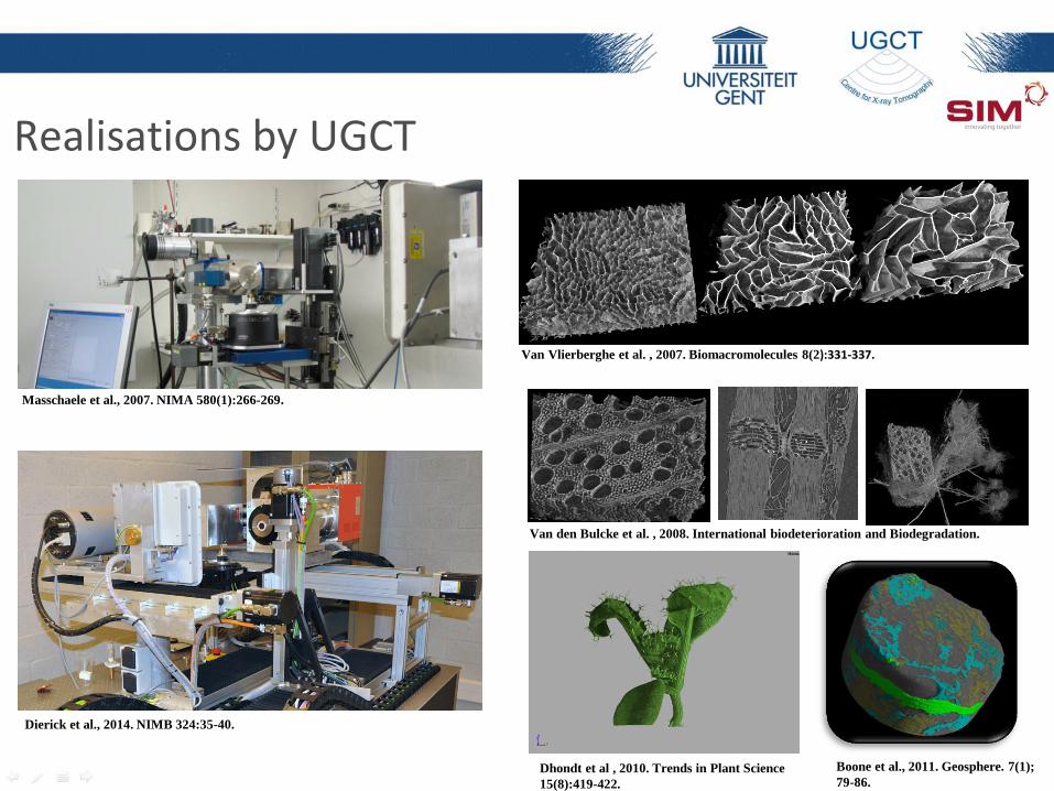
Realisations by UGCT
Van Vlierberghe et al. , 2007. Biomacromolecules 8(2):331-337.
Dhondt et al , 2010. Trends in Plant Science
15(8):419-422.
Masschaele et al., 2007. NIMA 580(1):266-269.
Dierick et al., 2014. NIMB 324:35-40.
Van den Bulcke et al. , 2008. International biodeterioration and Biodegradation.
Boone et al., 2011. Geosphere. 7(1);
79-86.
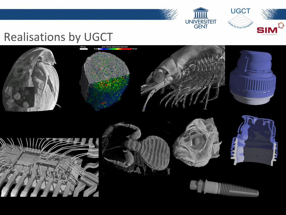
Realisations by UGCT
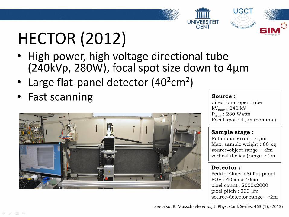
HECTOR (2012) • High power, high voltage directional tube
(240kVp, 280W), focal spot size down to 4µm • Large flat-panel detector (40²cm²) • Fast scanning
See also: B. Masschaele et al., J. Phys. Conf. Series. 463 (1), (2013)
Source : directional open tube
kVmax : 240 kV
Pmax : 280 Watts
Focal spot : 4 µm (nominal)
Detector : Perkin Elmer aSi flat panel
FOV : 40cm x 40cm
pixel count : 2000x2000
pixel pitch : 200 µm
source-detector range : ~2m
Sample stage : Rotational error : ~1µm
Max. sample weight : 80 kg
source-object range : ~2m
vertical (helical)range :~1m

HECTOR (2012)

Subsample A (4 µm resolution) discrimination of mineral grains and pore space
Subsample B (2.8 µm resolution) discrimination between different minerals (quartz, feldspar, clays) Microporosity visible
Sample size Resolution

EMCT (2012) • Gantry based system
• Environmental control (Temperature, pressure, ...)
• Continuous scanning
• Ultra-fast scanning (<30 sec)
• Maximum resolution 5µm
Dierick et al., Nucl. Inst. & Meth. 324 (0), (2014)
Source :
directional closed tube
kVmax : 130 kV
Pmax : 39 Watts
Focal spot : 5 µm (nominal)
Detector :
CMOS flat panel
pixel count : 1316x1312
pixel pitch : 100 µm
source-detector range : 15-40cm
Sample stage :
Rotational error : <3µm
Max. sample weight : 50 kg
source-object range : ~2m
vertical (helical)range : ~1m

X-ray CT add-on modules
Pressure Cell (120 bar) Freezing Cell (-20°C) Climatic chamber Pressure/tensile stage

Acquiring, or developing extra add-on modules, such as an in-situ tensile/compression cell.
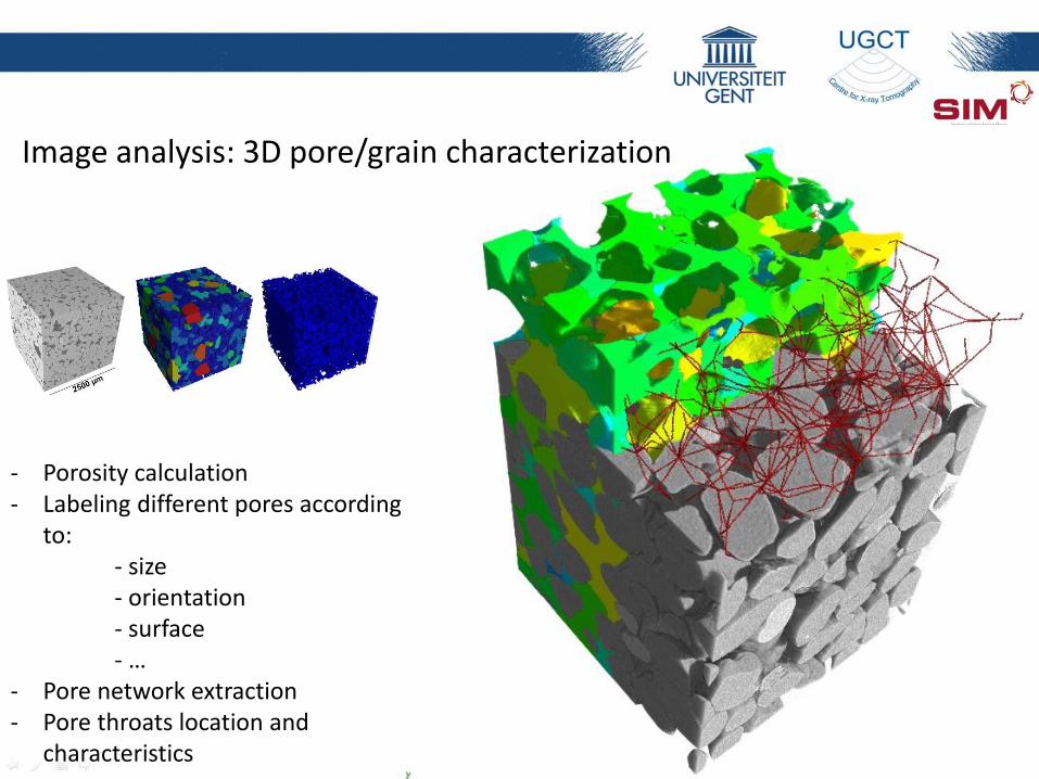
- Porosity calculation - Labeling different pores according
to: - size - orientation - surface - … - Pore network extraction - Pore throats location and
characteristics
Image analysis: 3D pore/grain characterization

Pore Network Modelling for
multi-phase flow
Mineral grains
Pore space
Prediction of macroscopical behaviour based on microscopical study (PNM)

Water displaced by non wetting phase
APPLICATIONS OF HIGH RESOLUTION X-RAY CT AS CHARACTERIZATION AND MONITORING TECHNIQUE

(a)
(c)
(b)
(d)
Mšené sandstone + halite crystals (red) (a-b) after 1st cycle of wetting and drying at 20%RH and (c-d) after 2 additional cycles.
Salt precipitation migration into pores close to surface controlled by RH cycling
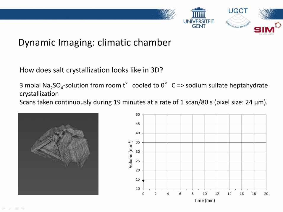
How does salt crystallization looks like in 3D? 3 molal Na2SO4-solution from room t°cooled to 0°C => sodium sulfate heptahydrate crystallization Scans taken continuously during 19 minutes at a rate of 1 scan/80 s (pixel size: 24 µm).
Dynamic Imaging: climatic chamber
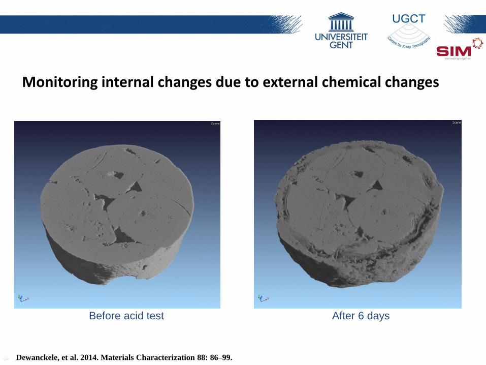
Monitoring internal changes due to external chemical changes
Before acid test After 6 days
Dewanckele, et al. 2014. Materials Characterization 88: 86–99.
.

Dynamic Imaging: freezing cell

Semi-saturated conditions
B
CO2
18 scans in total Total period: 14 hours Each scan: 2’20” Resolution: 18 µm Total porosity: 42.5 % brine: 35.4% CO2 : 7.1%
Dynamic Imaging: wollastonite (CaSiO3) carbonatation

Semi-saturated conditions
CO2
CaCO3
Dynamic Imaging: wollastonite carbonatation
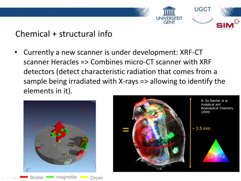
Chemical + structural info
• Currently a new scanner is under development: XRF-CT scanner Heracles => Combines micro-CT scanner with XRF detectors (detect characteristic radiation that comes from a sample being irradiated with X-rays => allowing to identify the elements in it).
Biotite magnetite Zircon
SR-µ-XRF
micro-CT
+ +
2D-µ-XRF CT-µ-XRF
~ 3
.5 m
m

Conclusions:
- HRXCT is an ideal 3D characterization technique
- a wide range of new and dynamic experiments are
now possible using lab-based HRXCT
- spatial and temperal resolution are still increasing
- besides on the hardware level, also on the software
level progress is being made

Thank you for your attention!
Prof. Dr. Veerle Cnudde
Department of Geology and Soil Science - UGCT, Ghent University, Krijgslaan 281/S8 9000 Ghent
Belgium
0032-9-2644580
Special thanks to: Entire UGCT team
SEPOCOM
SECEMIN
ISHECO
VITO