histo-anatomical, cytogenetic, and …phdthesis.uaic.ro/phdthesis/tudose (afemei), marinela,...
TRANSCRIPT

1
UNIVERSITATEA ,,ALEXANDRU IOAN CUZA” – IASI
FACULTATEA DE BIOLOGIE
HISTO-ANATOMICAL, CYTOGENETIC, AND
BIOCHEMICAL RESEARCHES ON THE INULA L.
SPECIES FROM THE ROMANIAN FLORA
PHD. THESIS-ABSTRACT
SCIENTIFIC COORDINATOR:
PROF. DR. CONSTANTIN TOMA
Corresponding Member of the Romanian Academy

2
INTRODUCTION 1
CHAPTER I.
HISTORY OF THE RESEARCHES REFERRING TO HIETO-ANATOMICAL,
CYTOGENETIC AND BIOCHEMICAL STUDIES OF SOME SPECIES BELONGING TO
THE ASTERACEAE FAMILY AND INULA GENUS
3
I.1. Some data referring to the history of the histo-anatomical researches 3
I.2. The history of the cytogenetic researches 7
I.3. The history of the biochemical researches 11
CHAPTER II
MATERIALS AND RESEARCH METHODS
16
II.1. Material methods used in the histi-anatimical researches
II.2. Cytogenetic determinations – the study of the mitotic chromosomes in species if the
Inula genus
II.2.1. Elaboration of the cariotype for some of the Inula L. species
16
18
21
II.3. Biochemical determinations
II.3.1. The cytological method to high light inuline
II.3.2. Thr qualitative phytochemical analysis to determine the active principles of
nula helenium
II.3.2.1. TLC analysis for flavons
II.3.2.2. TLC analysis for polyphenolcharboxylic acids
II.3.2.3. TLC analysis for triterpens
II.3.2.4. TLC analysis for volatile compounds
II.3.3. The quantitative phytochemical analysis
II.3.3.1. The spectrophotometric determination of flavonoids
II.3.3.2. The spectrophotometric determination of the polyphenolcharboxylic
acids
II.3.3.3. The determination of the frutosan content expressed in fruitose
II.3.3.4. The determination of the total aminoacid content expressed in glutamic
acid
23
23
24
26
26
26
26
27
27
29
31
32
CHAPTER III
THE INULA L. GENUS TAXONOMY AND THE BOTANICAL CHARACTERIZATION OF
THE STUDIED SPECIES
III.1. General data about the Asteraceae family and about the Inula L. genus
III.2. Morphological characterization of the Inula L. species of the Romannian
flora
III.2.1. The morphology of the mature Inula L. plants
III.2.1.1.Inula helenium L.
III.2.1.2.Inula germanica L.
III.2.1.3.Inula salicina L.
III.2.1.4.Inula hirta L.
III.2.1.5.Inula ensifolia L.
III.2.1.6.Inula britannica L.
III.2.1.7.Inula oculus-christi L.
III.2.1.8.Inula conyza DC.
III.2.1.9.Inula bifrons (Gou.) L.
III.2.1.10.Inula spiraeifolia L.
III.2.2. Some data regarding the plant morphology of some Inula species
III.3. Morphological data referring to the hybrids of the Inula L. genus
III.4. Data about corology, ecology, variability and the importance of the Inula
species
III.4.1. Data about the species corology
III.4.1.1. The extent of the Inula species in Europe
III.4.2. The ecology of the Inula species
33
33
35
35
50
52
53
53
56
57
57
60

3
III.4.3. The variability of the Inula genus species
III.4.4. Importanța
III.4.4.1. Compoziția chimic
III.4.4.2. Utilizări terapeutice
III.4.4.3. Mod de administrare
III.4.4.4. Credințe populare asociate cu planta
III.4.5. The cytological highlighting of inuline in Inula helenium L.
61
65
65
66
66
CHAPTER IV
THE STRUCTURE OF THE VEGETATIVE ORGANS OF THE INULA L. SPECIES
IV.1 The structure of the vegetative organs of Inula L. plantlets
IV.1.1.Inula helenium L.
IV.1.2.Inula hirta L.
IV.1.3.Inula ensifolia L.
IV.1.4.Inula britannica L.
IV.2. The structure of the vegetative organs of the mature plants of Inula L.
IV.2.1.Inula oculus-christi L.
IV. 2.2.Inula salicina ssp. salicina L.
IV. 2.3.Inula salicina ssp. aspera L.
IV.2.4.Inula germanica L.
IV.2.5.Inula helenium L.
IV.2.6.Inula ensifolia L.
IV.2.7.Inula hirta L.
IV.2.8.Inula britannica L.
IV.2.9.Inula bifrons (Gou.) L.
IV.2 10.Inula conyza DC.
IV.3. Discussions
67
67
78
170
CHAPTER V
THE MICROMORPHOLOGICAL ASPECTS IN INULA SPECIES
V.I. Micromorphological Aspects of the Inula L. leaf
V.1.1.Inula oculus-christi L.
V.1.2.Inula ensifolia L.
V.1.3.Inula hirta L.
V.1.4.Inula salicina ssp. salicina L.
V.1.5.Inula salicina ssp. aspera L.
V.1.6.Inula germanica L.
V.1.7.Inula britannica L.
V.1.8.Inula helenium L.
V.1.9.Inula conyza DC.
V.2. Data regarding the fruit morphology of the Inula L species
176
189
CHAPTER VI
THE CHARACTERIZATION OF THE MITOTIV CHROMOSOMES OF INULA SPECIES IN
STUDY
VI.1. Morphological characteristics of the chromosomes of Inula spiraeifolia L.
VI.2. Morphological characteristics of the Inula ensifolia L. chromosomes
VI.3. Morphological characteristics Inula hirta L. chromosomes
VI.4. Morphological characteristics of the Inula helenium L. chromosomes
Conclusions
194
194
198
202
209
212
CHAPTER VII
THE QUALITATIVE AND QUANTITATIVE DETERMINATION OF THE INULA
HELENIUM (ELECAMPANE) ACTIVE PRINCIPLES
VII.1. The qualitative analysis
VII.2. The quantitative phytochemical analysis
213
213
214
CONCLUSIONS 220
BIBLIOGRAPHY 223

4
INTRODUCTION
The Inula L. genus is part of the Asteraceae family and comprises about 120 species,
extended in the temperate area of Europe and Asia (Nyarady, 1964). In the Romanian flora
there are 10 species, out of which, one has no longer been confirmed by scientific literature,
older (Nyarady, 1964) or newer (Oprea, 2005). In the same time, six hybrids are cited
(Nyarady, 1964) or 5 hybrids in the N-E of the country (Chifu et al., 2006).
Of all the Inula species growing in our country, Inula helenium L. Is considered a
valuable medicinal plant (Perrot and Paris, 1971). In recent works of pharmacognosy and
phytochemistry other speciees of Inul are mentioned: Inula viscosa (Nikolakaki and
Christodoulakis 2004), Inula germanica, Inula ensifolia, Inula britannica, Inula crithmoides,
Inula conyza (Perrot and Paris, 1971).
The anatomical structure of the Inula species has very little been studied. In the
treateses of dycotyledonous plant anatomy (Metcalfe and Chalk, 1972) or of the angiosperms
in general (Napp-Zinn, 1973, 1974), only the Inula genus is mentioned. In some atlases of
medicinal plant microscopy (Terpilo, 1961; Toma and Rugina, 1998) only Inula helenium L.
is given importance.
In the present thesis, a special attention is given to the histo-anatomical study of the
vegetative organs of the Inula species, otherwise little studied, except Inula helenium which
interested some authors.
The aim of thePhd thesisa was the histo-anatomical, cytogenetic and biochemical of
some plant species belonging to the Inula genus from the North-East of Romania.
The objectives of the research:
A comparative anatomy study on the Inula L. species from the Romanian flora,
highlighting the traces with taxonomic value, and also some of anatomo-ecological order,
insisting on the structure and locations of the secretory canals, of the tector hairs and of the
secretory ones by the help of classical and modern microscopy (SEM);
The characterization of the mitotic chromosomes of some Inula species;
The qualitative and quantitative phytochemical analysis of the active principles
of Inula helenium L by chromatogrphic methods (CSS) and their dosing by means of
spectrometry.

5
The Inula L. genus is part of Spermatophyta division, the Magnoliophytina
(Angiospermae) subdivision, the Magnoliopsida (Dicotyledonatae) class, subclass Asteridae,
the Asterales ( Compositales) order, the family: Asteraceae (Compositae), subfamily
Asteroideae (Tubuliflorae), the Inuleae tribe (Ciocirlan, 2000).
The Inula genus comprises plant species with ligulated flowers and tube formed, of the
same colour; this genus is a kin of Senecio, Arnica, Tagetes, Tussilago etc. (Stefan and Oprea,
2007).
All the 10 species of Inula in the present study are from the wild flora, as follows:
Inula helenium L., Inula oculus-christi L., Inula salicina with ssp. salicina and ssp. aspera
(Poir.) Hayek, Inula ensifolia L., Inula hirta L., Inula bifrons (Gou.) L., Inula conyza DC.,
Inula spiraeifolia L. and Inula germanica L.
Inula helenium L. (orig.) Inula britannica L. (orig.)
fig.1 fig.2

6
Inula oculus-christi L. (orig.) Inula salicina (http: // luirig. altervista.org /
naturaitaliana)
Inula ensifolia L. (orig.)
Inula hirta L. (http: // luirig. altervista.org /
naturaitaliana)
Inula bifrons (Gou.) L. (http: // luirig.
altervista.org / naturaitaliana) Inula conyza DC. (http: // luirig. altervista.org /
naturaitaliana)
fig.3
fig.4
fig.5 fig.6
fig.7 fig.8

7
Inula germanica L. (orig.) Inula spiraeifolia L. (http: // luirig.
altervista.org / naturaitaliana)
The general part has 3 chapters: The history of the researches regarding the structure
and chemical composition of the Asteraceae species, of those belonging to the Inula genus,
especially; The taxonomy of the Inula genus and the morphological characterization of the
species; Materials and methods of research. The investigated material was collected from
different locations in Moldavia and from the Danube Delta, including the natural reservations.
The methods are the currently used ones in the researches of photonic and electronic
microscopy, in the cytogenetic and biochemical researches (chromatography,
spectrophotometry).
The special part has 4 chapters: The structure of the plantlets and mature plants
vegetative organs (in the antesis stage); Micromorphological aspects of the leaf and fruit
surface; The characterization of the mitotic chromosomes; The qualitative and quantitative
determination of the active principles of Inula helenium (a species with medicinal value).
fig.9 fig.10

8
The structure of the plantlets and mature plants vegetative organs
• The structure of the vegetative organs of Inula L. plantlets (drawings I, II)
After the analysis of the vegetative organs structure of the plantlets belonging to the 4
species studied we highlighted the following aspects:
The root presents rhizoderm with big cells, with all the cell walls thin; the external
one is visibly bilging in case of Inula ensifolia. Here and there one may notice adsorbant
hairs. The thickness of the cortical parenchyma varies: 7-8 strata of the meatic type for Inula
helenium, 5 strata for Inula ensifolia and 4 strata for Inula hirta and Inula britannica. In the
case of all the analyzed species, the endoderm presents Caspary thickenings in the radial cell
walls. The stele is of several types: diarh at the Inula ensifolia plantlets, triarh, for the Inula
helenium and Inula hirta, tetrarh for the Inula britannica plantlets. At the level of the central
cylinder (of the tetrarh type) of Inula britannica there is a transfer to the secondary structure
so that along with the 4 primary wooden fascicles there also appeared secondary wooden
vessels.
The hypocotyl presents an epidermis with tangent or isodiametric prolongued cells
(Inula ensifolia), with all the walls thin and with the exterior wall thicker than the others (I.
helenium, I. hirta, I. britannica). Here and there, at the level of the epidermis there are already
very numerous uniserial multicell tector hairs at Inula hirta. The number of the cortical strata
differs: 10-12 for Inula helenium, 5-6 strata for Inula ensifolia and Inula hirta, 4-5 for Inula
britannica. At the internal face of the crust one my notice secretory canals (Inula helenium
and Inula hirta). The structure is primary in case of all species; with Inula britannica, the
structure is intermediary between that of the root and the bladed. Those are visible as well as
4 free fascicles, and the woodeen vessels have irregular distribution, the ones of the organ axis
being much smaller; among the wooden vessels with the walls moderately thickened and
lignified there are cells of cellulose parenchyma.
The leaf. In the cros section, the foliar limb presents an outstanding median nervure
on the inferior face, with a libero-wooden conducting fascicle, having mechanic fibres at both
poles. The epidermis presents two types of hairs: tector, bicellular (I. helenium) and
pluricellular, either thick and short (I. helenium, I. hirta), or long and thin (I. hirta, I. ensifolia
and I. britannica); secretory hairs with a relatively long pedicel, formed of two lines of cells
(biseriated) with bicellular gland (I. helenium, I. hirta). For all the plants we analyzed, the
mesophill is thin, homogenous, of the lacunous type, so the limb has an isofacial bifacial
structure.

9
Drawing I. Plantlets of Inula. Cross sections of the foliar limb- detail of the median nervure: I.
helenium (fig. 1), I. hirta (fig. 2); I. britannica (fig. 3); pluricellular tector hairs: I. helenium (fig. 4, 5),
I. hirta (fig. 6), I. ensifolia (fig. 7), I. britannica (fig. 8) (orig.)
fig.1 fig.2
fig.3 fig.5 fig.4
fig.6 fig.7 fig.8

10
Drawing II. Inula plantlets. Cross sections through the foliar limb, secretory hairs: scheme (fig. 9),
detail: I. helenium (fig. 10), I. hirta (fig. 11-13) (orig.)
fig.10
fig.11 fig.12 fig.13
fig.9

11
• The structure of the vegetative organs of mature Inula L. plants
The results regarding the histo-anatomical structure of the vegetative organs of the
Inula specie from the Romanian flora assert, on the one hand, the data contained by scientific
literature (especially referring to I. helenium), and, on the other hand, they complete the same
data as there very few titles referring to the anatomy of these taxons.
Terpilo (1961) analyzes the root structure of Inula helenium L., highlighting the
following characteristics:
the presence of an exterior epidermis;
the presence of the secretory canals in the primary, secondary crust and at the
level of the liber and the secondary wood;
in parenchym cells of the root and rhizome there is inuline in the form of
sphere shapes crystal.
Metcalfe and Chalk (1972) refer to some peculiarities of the Inula genus:
the presence of anomocytic stomata;
the frequency of uniserial pluricellular hairs, with the terminal cell very long,
and the a very thick wall;
the presence of secretory cavities in the underground parts.
Toma and Rugina (1998) investigated the vegetative organs (root, stem and leaf) of
Inula helenium, stating the following:
the root presents a typical secondary structure, as a result of the activity of the
two lateral meristems: cambium and felogene; at itd lecel:
- the liber presents riddled tubes, annex cells and a great quantity of amiliferous
parenchym;
- the wood is formed of vessels of different diameter, isolated or grouped;
- in vecinity of the vessels there are cells with sphere shaped crystal of inuline;
- the secretory canals are present in al the anatomical areas of the root.
at the stem level:
- the epidermis presents long secretory and tector hairs, a prezintă peri tectori şi
secretori lungi, uniserial; the secretory ones having hte pland pluricellular on
levels;
- the crust is defferentiated between a collencyme ring and an internal
parenchymatic area;

12
- the central cylinder is formed of numerous libero-wooden conducting fascicles
of the colateral-open type, surrounded by a thick gurdle of sclerenchym at the
margins of the liber.
at the level of the leaf:
- the limb is hypostomatic;
- the tector hairs are also very numerous on the inferior face of the limb and very
rare on the superior one;
- in the cross section, the median vervure is very prominent on the inferior face
of the limb and less on the superior one;
- the limb has bifacial ecvifacial structure.
Fahn (1988) said that:
the secretory hairs of many composites are pluricllular and biserial; these are
either pedicelled, or sesilled;
the cells on their top secrete terpens, lipides, flavonoidic aglicons and wax
(lipofilic material);
in case of Inula viscosa, the secretory hairs produce terpens, lipids,
polysaccharides, proteins;
the substances secreted are accumulated under the cuticule, and after the
coticule is torn they are eliminated to the exterior.
Nikolakaki and Christodoulakis (2004) made a study on the secretory structure in the
Inula viscosa leaf, underlining the following peculiariries:
the limb is amfistomatic; the stomata being of the anomocytic type;
the presence of long tector hairs and numerous secretory hairs;
the secretory hairs are short, pluricellular, formed of pedicell and head with
two cells.
After analyzing the structure of the vegetative organs of the studied species, we
noticed the following aspects:
1. The root (drawing III). At the level of the rhizoderm one may notice
adsorbant hairs of variable length, with very thin walls.
The cortical parenchyma presents a different number of cell strata: 6-7 (Inula
conyza), 10 (Inula oculus-christi), 10-12 (Inula britannica and Inula germanica) and 16-18
(Inula ensifolia). In case of I. germanica, here and there, in the thickness of the internal
cortical parenchyma one may notice secretory canals, localized near the liberien fascicles. In

13
the internal stratum of the cortical parenchyma there are cells with helenine (I. hirta, I.
helenium, I. conyza) or cells with tanin (I. britannica).
Helenine is a mixture of sesquiterpenic lactons, used in lung diseases, and externally
as lotions against ulcer and arthritis (Grigorescu et al., 2001). In the I. britannica and I.
Helenium species, in the cortical parenchyma, between the cells there are aerial canals gaps.
With the majority of the studied species, the cortical parenchyma ends with an endoderm of a
special type Caspary thickenings in the radial walls.
The presence of the aerial lacunary canals in the roots of I. britannica and I. helenium
represents an adaptation of the plant to the environmental conditions, were the phreatic water
is close to the soil surface, proving the mezohygrophile or optionally halophile character
(Inula britannica) mentioned by Ciocarlan (2000).
The stel is of several typesi: triarh (I. britannica, I. conyza), tetrarh (I. oculus-christi,
I. helenium), pentarh (I. hirta), hexarh (I. salicina ssp. salicina, I. ensifolia) and septarh (Inula
salicina ssp. aspera, I. germanica).
The speies studied had a primary structure of the root; only I. hirta, I. britannica and I.
conyza show a secondary structure at the level of the central cylinder, based on the cambium.
2. The rhizome (drawing IV). The epidermic celles that are isodiametric or
slightly tangently prolongued, with the outer wall thicker than the others.
The crust is relatively thick,cellulose-parenchymatic, of the meatic type, formed of a
variable number of cell strata: 7-8 (I. ensifolia), 8-10 (I. oculus-christi), 12-14 (I. germanica),
12-16 (I. salicina ssp. salicina), 16-18 (I. salicina ssp. aspera).
In I. oculus-christi, I. salicina ssp. salicina, I. salicina ssp. aspera, I. ensifolia, I. hirta,
I. germanica and I. britannica, in the interior strata of the crust there are bir secretory canals,
tangently prolongued (I. germanica, I. britannica), with the colector canal surrounded by a
stratum of epitelial cells. These canals are more often localizedbetween the girdles of
priliberien sclerenchematic fibres. With I. Helenium, in the thick cortical parenchyma, in the
thick ring of secondare phloem abd the very thick xylem ring there dominate the secretory
canals, as Terpilo (1961) noticed.
The conducting tisseus from the central cylinder of the rhizome have secondary
structure in the majority of the species. The phellogen differentiates in I. ensifolia and I.
helenium, generating suber to the exterior and phellodem to the interior.
3. The stem (drawing V). The epidermis has isodiametri ca nd isomorphous
cells with the external and internal walls thicker than the others; the external wall is covered
by a thin curicule. In some species (I. oculus-christi, I. germanica, I. hirta, I. bifrons, I.

14
conyza) one may notice, at the level of the epidermis, the presence of pluricellular tector hairs
and of biserial secretory hairs.
In Inula hirta, Inula salicina ssp. salicina and Inula ensifolia we see some conducting
fascisles in the crust, completely surrounded by sclerenchematic fibres.
The central cylinder is rather thick, with agreat number of libero-wooden conducting
fascicles, separated by medular rays of different width, having cells with the walls moderately
thickened and lignified at the wood level. The conducting fascicles present, at the margins of
the liber, each a very thick girdle of sclerenchematic fibres, with thickened and intensly
lignified walls.
The pith is either cellulose-parenchymatic, or parenchymatic-lignified, the cells
having the walls moderately thickened.
Towards the basis, the general structure remains the same, with the difference that the cross
section contour becomes almost circular, the wood quantity is greater, the lignifications is
more intense and the pith thicker, some of the cells of the last get disorganized, resulting in
some aerial cavities with irregular margins. At the basis of the stem, the number of the hairs is
reduced, the number of the mechanical cortical fibres increases, as well as the thickness of the
periliberien mechanical girdles.
4. The leaf (drawings VI, VII). The petiole present at the basal leaves of I.
oculus-christi, I. helenium and I. conyza in the cross section is realtively cordiform (I. oculus-
christi) or approximately circular (I. helenium, I. conyza), with lateral-abaxial ribs that
separate a superficial adaxial ditch (I. oculus-christi). On the abaxial face of the petiole, the
tector hairs are numerous, thick, pluricellular, with very long terminal cells, and the secretory
hairs are pluricellular, biserial (I. helenium and I. conyza). In I. oculus-christi, the tector and
the secretory hairs are uniformly distributed both on the adaxial face and of the abaxial one. In
the fundamental parenchyma, the conducting tissues are represented by the libero-wooden
fascicles, disposed on an arch.
The limb is amfistomatic (I. salicina ssp. salicina, I. ensifolia, I. hirta, I. britannica, I.
conyza) or hypostomatic (I. salicina ssp.aspera, I. germanica). The limb structure is
heterofacial bifacial (dorsiventral),with unistratified palisadic tissue (I. salicina ssp. salicina,
I. salicina ssp.aspera, I. helenium, I. bifrons, I. britannica) or bistratified (I. germanica). In I.
ensifolia, I. hirta, I. conyza, the limb presents an isofacial bifacial structure (the mesophyl is
homogenous, relatively compact, formed of rounded cells, only with meates between them).

15
As aresult of the histo-anatomical analysis of the vegetative organs, we highlight
several differences an similarities between the two subspecies of I. salicina: ssp. salicina and
ssp. aspera .
Thus, at root level, I. salicina ssp. Salicina presents 6 wooden liber fascicles while in
I. salicina ssp. aspera, there are 7-8 wooden conducting fascicles, that alternate with as many
liber fascicles. In both subspecies, in the rhizome, in the internal stratum of the internal
cortical parenchyma there are secretory canals.
The contour of the superior third cross section of the aerial stem is circular ribbed,
with 8 relatively proeminent ribs and relatively deep valecules in case of the I. salicina ssp.
salicina subspecies and circular, unregularly ribbed, with different size ribs in case of the I.
salicina ssp. aspera subspecies.
At the level of the inferior aerial stem third, I. salicina ssp. aspera has thich, uniserial
tector hairs, and also visibly proeminent stoamata over the protecting tissue level.
In the I. salicina ssp. Salicina subspecies, the limb is amfistomatic, while in I. salicina
ssp. aspera, the stomata are missing from the suoerior epidermis (so the limb is
hypostomatic). In Inula salicina ssp. aspera, near the nervures of the foliar limb there are
long, uniserial tector hairs.

16
Drawing III. Root cross sections – central cylinder detail: I. salicina ssp. salicina (fig. 14), I.
germanica (fig. 15), I. helenium (fig. 16), I. ensifolia (fig. 17), I. britannica (fig. 18), I. conyza (fig.
19) (orig.)
fig.14 fig.15
fig.16 fig.17
fig.19 fig.18

17
Drawing IV. Rhizome cross section I. oculus-christi (fig. 20), I. salicina ssp. salicina (fig. 21), I.
salicina ssp. aspera (fig. 22), I. germanica (fig. 23), I. ensifolia (fig. 24), I. britannica (fig. 25) (orig.)
fig.20 fig.21
fig.22 fig.23
fig.25 fig.24

18
Drawing V. Aerial stem cross section: I. salicina ssp. salicina (fig. 26), I. salicina ssp. aspera (fig.
27), I. helenium (fig. 28), I. hirta (fig. 29), I. britannica (fig. 30), I. bifrons (fig. 31) (orig.)
fig.27
fig.28 fig.29
fig.30 fig.31
fig.26

19
Drawing VI. Leaf cross section: I. salicina ssp. salicina (fig. 32), I. helenium (fig. 33), I. ensifolia
(fig. 34), I. hirta (fig. 35), I. britannica (fig. 36); I. bifrons: tector and secretory hairs (fig. 37) (orig.)
fig.32 fig.33
fig.34 fig.35
fig.36 fig.37

20
Drawing VII. Petiole cross section: general view I. oculus-christi (fig. 38); detail: conducting fascicle,
aerial cavities, tector and secretory hairs. helenium (fig. 39-41); detail: aerial cavities and conducting
fascicles I. conyza (fig. 42, 43) (orig.)
fig.38 fig.39
fig.40 fig.41
fig.42 fig.43

21
Morphological aspects of the Inula L. species
• Morphological aspects of the Inula L. leaf (drawing VIII)
As aresult of the foliar surface of the Inula species, with the help of the electronic
microscope with scavenging, we highlighted the following:
- the external surface epidermic cell walls has a different conformation:
ondulated in the I. ensifolia, I. oculus-christi and I. Germanica species, with cuticular
crests at I. britannica or with obvious scratches at I. salicina ssp. aspera;
- the stomata are present in th esuperior epidermis of I. oculus-christi, I.
ensifolia, I. salicina ssp. salicina, I. britannica, I. hirta, I. conyza. These are missing
from the superior epidermis of the Inula salicina ssp. Aspera species;
- the stomata are surrounded by annex cells with the exterior surface modified,
presenting either lamellar excrescences of epicuticullar wax (la I. ensifolia), or
radially disposed ondulations (I. salicina ssp. salicina) or proeminent crests that limit
each stomatic apparatus (I. britannica);
- in most of the cases, the opening of the stomata is surrounded by a waxless
cuticullar collaret (I. germanica) or by moderate ondulations of the cuticule, disposed
in parallel to the stomata axis (I. britannica);
- at the level of the superior epidermis one may notice two types of hairs:
uniserial plirucellular tector hairs, (the majority with 1, 2, 3 and 4 short basal cells and
and one or two long and very long terminal cells) and secretory hairs with clavated
gland;
- the external surface of the cells from the inferior epidermis presents
ondulations (I. oculus-christi), parallel cuticullar scratches (I. ensifolia, I. salicina ssp.
aspera) or frequent cuticullar crests (I. britannica);
- in case of all the analyzed speceis, the inferior epidermis there are stomata. The
annex cell walls of the stomata present ont eir surface wax lamellas either aranged,
isolated, non-stratified (I. ensifolia), or orderly disposed, with parallel lamellas (I.
salicina ssp. aspera). In I. hirta and I. Germanica species, the opening of the stomata
is surrounded a waxed collaret;
- at the level of the inferior epidermis we observe two types of hairs:
pluricellular tector hairs, long, numerous, mostly disposed on the nervures (I. oculus-
christi, I. hirta, I. salicina ssp. aspera, I. helenium, I. conyza, I. britannica) and
secretory hairs, a greater number in the I. Helenium species.

22
Drawing VIII. I. oculus-christi: secretory and tector hairs (fig. 44); I. ensifolia: detail: inferior
epidermic stomatum (fig. 45); I. salicina ssp. salicina: detail: superior epidermic stomatum (fig. 46); I.
britannica: detail: inferior epidermic secretary hair (fig. 47); I. helenium: secretory and tector hairs of
the inferior epidermis (fig. 48); I. conyza: tector and secretory hair of the inferior epidermis fig. 49)
(orig.)
fig.44 fig.45
fig.46
fig.48 fig.49
fig.47

23
Date regarding the morphology of the fruits of the Inula species (fig. 50-53)
Boyko (2011) studies by means of the electronic microscope the surface of the
different Asteraceae species achenes. On the basis of the results, there have been identified
diverse morphological types of hairs. The authore do not characterize the species of the Inula
genus.
The surface of the pericarp in the speccies of Inula studied by us is ribbed. The
pericarp presents narrow polygonal cells, with an extremely smooth exterior wall (I. helenium,
I. hirta) or alongated (I. germanica, I. conyza), sometimes with sharp tips (I. hirta), many
times covered by a thicker cuticullar complex, slightly goffered (I. ensifolia) or with an
amorphous aspect (I. germanica, I. conyza). On the surface of the pericarp there are relatively
short tector hairs, with a sharp tip (I. ensifolia), with short, slightly maimed hairs (I. bifrons)
or robust alongated ones (I. conyza).
Inula ensifolia- achene: detail: pericarp with tector hairs (fig. 50); Inula hirta- achene, detail: pericarp
cells (fig. 51); Inula conyza: detail: tector hairs onthe surface of the pericarp (fig. 52); Inula bifrons,
detail: tector hairs on the pericarp surface (fig. 53) (orig.)
fig.50 fig.51
fig.52 fig.53

24
• The characterization of the mitotic chromosomes of the studied
Inula L. species
• The morphological characteristics of the Inula spiraefolia L. chromosomes
The analysis of the well exposed chromosomes, with less than two superpositions,
allowed us to establish thediploid number to be equal with 16, so that 2n=16, in concordance
with th data of scientific literature and the cariotype achieving (fig.54 and 55).
Taking into account the ratio among the branches (BL/BS), the centromeric index, the
relative length and the branch differences (BL-BS), we could establish the fact that the Inula
spiraefolia L. presents a single type of chromosomes: median (m), all the eight pairs of
homologues (I – VIII) the relation between the branches from 1.09 and 1.49.
fig.54. Metaphase – Inula spiraeifolia L. (2n=16)
fig.55. Cariotype Inula spiraeifolia L. (2n=16)

25
• The morphological characterisitics of the Inula ensifolia L. chromosomes
In the works we consulted we found that with the Inula ensifolia L. species, the
number of the chromosomes in the somatic cells is 2n = 16, all the samples studied being
diploid. In the speciality literature, we found a single work (Kamari et al., 2008) describing
the morphology and typolgy of this species chromocomes, yet in case of one triploid variant
2n = 24 =3x = 16m + 4mSAT + 2smSAT + 2tSAT our study has the character of novelty.
Taking into account the relation among he branches (BL/BS), the centromeric index,
the relative length and the difference of the branches (BL-BS), we coulg establish the fact that
the Inula ensifolia L. species presents two types of chromosmes median (m): the pairs I – VII
(having th erelation between the branches between 1.03 and 1.53) and submedians (sm): pair
VIII, having the ratio between the branches of 1,.7 (fig.57).
fig.56. Metaphase – Inula ensifolia L. (2n=16)
fig.57.Cariotype - Inula ensifolia L. (2n=16)

26
• The morphological characteristics of the Inula hirta L. species
chromosomes
In case of the Inula hirta L. species, we tried to characterize the chromosome from the
morphometrical and typological point of view due to the stages stated in chapter II;
unfortunately, this objective could not totally be finalized: becuse of the different degree of
spiral winding of some homologues we obtained only two variants of cariotype.
Taking into account the relation among the branches (BL/BS), the centromeric index,
the relative length and the difference of the branches (BL-BS), we could establish the fact that
that the Inula hirta L. species presints two types of chromosomes: median (m), the pairs of
homologues I, II, III, IV, VII și VIII (having th ratio between tyhe branches from 1.15 and
1.50) and the submedians, the homologues pairs V and VI (having the ratio between the
branches from 2.13 and 2.54) (variant 1; fig. 59).
fig.58. Metaphase 1 – Inula hirta L. (2n=16)
fig.59. Cariotype 1 - Inula hirta L. (2n=16)
In case of metaphase 2, taking into account the ratio between the branches (BL/BS), the
centromeric index, the relative length and the branches difference (BL-BS), we could establish
on this metaphase too that the Inula hirta L. presents two types of median chromosomes:
median (m), the homologues pairs I, II, IV, V, VI and VII (having the ratio between the
branches from 1.05 to 1.64) and submedians, the homologues pairs III and VIII (having the
ratio between the branches from 1.82 and 2.27) (fig 61).
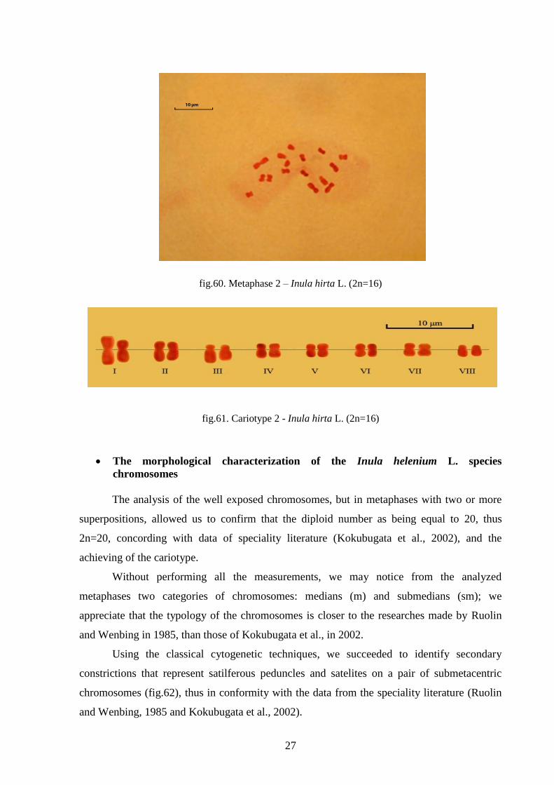
27
fig.60. Metaphase 2 – Inula hirta L. (2n=16)
fig.61. Cariotype 2 - Inula hirta L. (2n=16)
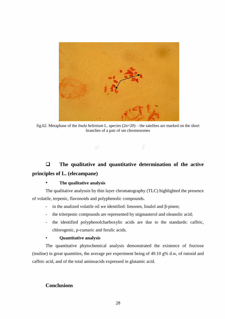
The morphological characterization of the Inula helenium L. species
chromosomes
The analysis of the well exposed chromosomes, but in metaphases with two or more
superpositions, allowed us to confirm that the diploid number as being equal to 20, thus
2n=20, concording with data of speciality literature (Kokubugata et al., 2002), and the
achieving of the cariotype.
Without performing all the measurements, we may notice from the analyzed
metaphases two categories of chromosomes: medians (m) and submedians (sm); we
appreciate that the typology of the chromosomes is closer to the researches made by Ruolin
and Wenbing in 1985, than those of Kokubugata et al., in 2002.
Using the classical cytogenetic techniques, we succeeded to identify secondary
constrictions that represent satilferous peduncles and satelites on a pair of submetacentric
chromosomes (fig.62), thus in conformity with the data from the speciality literature (Ruolin
and Wenbing, 1985 and Kokubugata et al., 2002).
28
fig.62. Metaphase of the Inula helenium L. species (2n=20) – the satelites are marked on the short
branches of a pair of sm chromosomes
The qualitative and quantitative determination of the active
principles of L. (elecampane)
• The qualitative analysis
The qualitative analyssis by thin layer chromatography (TLC) highlighted the presence
of volatile, terpenic, flavonoids and polyphenolic compounds.
- in the analized volatile oil we identified: limonen, linalol and β-pinen;
- the triterpenic compounds are represented by stigmasterol and oleanolic acid;
- the identified polyphenolcharboxylic acids are due to the standards: caffeic,
chlorogenic, p-cumaric and ferulic acids.
• Quantitative analysis
The quantitative phytochemical analysis demonstrated the existence of fructose
(inuline) in great quantities, the average per experiment being of 49.10 g% d.w, of rutosid and
caffeic acid, and of the total aminoacids expressed in glutamic acid.
Conclusions

29
The results regarding the histo-anatomic of the vegetative organs of the Inula
species from the Romanian flora confirmed, on the one hand the already known data (special
reference to I. helenium), and on the other hand completed the data of the speciality literature,
known being the fact that there are few articles referring to the antomy of these species.
From the anatomical point of view:
- in case of the analyzed plantlets and mature plants, at the level of the leaf we
identified two types: massive and thick tector hairs, that differ in size, some being bicellular,
others pluricellular, with great basal cells, sometimes almost izodiametric, and secretory
hairs with a long pedicell, formed of two lines of cells (biserial) with bicellular gland, in a
great number in I. helenium.
- at the root level of the mature plants, in the internal stratum of the cortical
parenchyma, in the majority of the analyzed species, there are cells with helenine, excepting
I. germanica, which presents in the thickness of the interior cortical parenchyma secretory
canals. The secretory canals are present especially in the rhizome; the secretory canal
disposition differs, inmost of the cases are localized in the internal strarta of the crust, among
the girdles of sclerenchimatic periliberien fibers.
From the cytogenetic point of view:
- we achieved the cariotype and the idiogram of 3 Inula: Inula spiraeifolia, Inula
ensifolia and Inula hirta species, all with 2n=16. The 3 species differ by the morphological
types of the chromosomes ; in I. spiraeifolia, all the chromosomes are of the median type (m);
in I. ensifolia, the cariotype is represented by the 7 pairs of the median type (m) and a pair of
submedian chromosomes (sm); and I. hirta, as wel as in the precedent species we identified 2
types of chromosomes: median and submedian (variant 1: the pairs V and VI; variant 2: the
pairs III and VIII). With I. helenium, we could establish only the number of diploid
chromosomes, 2n=20, and from the photos obtained we could distinguish chromosomes with
satelites.
After the phytochemical studiy achieved on samples of Inula helenium, the
Miclauseni-Iasi we noticed: the presence of the volatile compounds (limonen, linalol and β-
pinen), terpenic compounds (stigmasterol and oleanolic acid), flavonoidic compounds
(cvercetol and rutosid, also being present some of their aglicons) and polyphenolic
compoungs (caffeic, chlorogenic, p-cumaric and ferulic acids) in vegetal extracts of Inula
helenium and it was demonstrated the existence of fruictosans (inuline) in great quantities, the
average per experiment beingd of 49.10 g% d.w, of rutosid and caffeic acid, and also of he
total aminoacids expressed in glutamic acid.

30
Selective bibliography:
1. Andrei M., 1997, Morfologia generală a plantelor, Editura Enciclopedică, București: 163
2. Andrei M., Predan Genţiana M.I., 2003, Practicum de morfologia şi anatomia plantelor,
Editura Ştiinţelor Agricole, Bucureşti: 113
3. Ardelean A., Mohan Gh., 2008, Flora medicinală a României, Editura All, Bucureşti: 331-
332
4. Băra I.I., Cîmpeanu Mirela, 2003, Genetica, Editura Corson: 118–119
5. Boyko E.V., 2011, Trichomes of achenes of Asteraceae. I. Covering hairs, Turczaninowia,
14 (2): 130-144
6. Bruneton J., 1995, Pharmacognosy, phytochemistry, medicinal plants, Ed. Lavoisier,
Paris: 503–504
7. Ciocârlan V., 2000, Flora ilustrată a României (Pteridophyta et Spermatophyta), Editura
Ceres, București, (Ediția a II-a revizuită și adăugită): 782-784
8. Ciulei I., Grigorescu E., Stănescu Ursula, 1993, Plante medicinale, fitochimie şi
fitoterapie-tratat de farmacognozie, vol. I, Editura Medicală, Bucureşti: 67-68
9. Esau Katherine, 1954, Primary vascular differentiation in plants, Biol. Rev., 29: 46-86
10. Fahn A., 1988, Secretory tissues in vascular plants, New. Phytol., 108: 229-257
11. Grigorescu E., Lazăr M.I., Stănescu Ursula, Ciulei I., 2001, Index fitoterapeutic, Editura
Cantes, Iași: 275-277
12. Kamari G., Blanché C., Siljak–Yakovlev S., 2008, Mediterranean chromosome number
reports – 18, Flora Medit.,18: 563-610
13. Kokubugata G., Kondo K., Tatarenko Irina V. , Kulikov P.V., Knyasev M.S., Ryabinina
Z.N., 2002, Cytological studies of 13 Asteraceae species from Russia, Chromosome
Science, 6: 67-72
14. Metcalfe C.R., Chalk L., 1972, Anatomy of the Dicotyledons, Clarendon Press, Oxford,
2: 782-785
15. Nyárády E.I., 1964, Fam Compositae. In: Flora R.P. Române, Editura Academiei
Române, București: 9: 264-291
16. Ștefan N., Oprea A., 2007, Botanică sistematică, Editura Univ. ,,Al.I.Cuza”, Iași: 398,
422-423
17. Terpilo N.I., 1961, Anatomiceskij atlas lekarstvenyh rastenij, Gosud. Mediținskoe
izdatel'stvo U.S.S.R., Kiev: 333-335
18. Toma C., Rugină Rodica, 1998, Anatomia plantelor medicinale. Atlas., Editura Acad.
Rom., București: 110-113