histological processing techniques for the … · histological processing techniques for the ......
TRANSCRIPT

Rev. Brasil. Biol., 61(2): 341-345
HISTOLOGICAL TECHNIQUES FOR STUDY OF DUGESIIDAE DEVELOPMENT 341
HISTOLOGICAL PROCESSING TECHNIQUES FOR THESTUDY OF DUGESIIDAE DEVELOPMENT
(PLATYHELMINTHES, TRICLADIDA, PALUDICOLA)
VARA, D. C. da, LEAL-ZANCHET, A. M. and LIZARDO-DAUDT, H. M.Instituto de Pesquisas de Planárias e Laboratório de Embriologia, Centro de Ciências da Saúde, Universidade do
Vale do Rio dos Sinos, Av. Unisinos, 950, São Leopoldo, RS, Brazil
Correspondence to: Ana Maria Leal-Zanchet, Centro de Ciências da Saúde, UNISINOS, Av. Unisinos, 950,CEP 93022-000, São Leopoldo, RS, Brazil, e-mail: [email protected]
Received January 12, 2000 – Accepted April 13, 2000 – Distributed May 31, 2001
(With 8 figures)
ABSTRACT
The objective of the present study was to adapt techniques for the histological processing of Dugesiidaecocoons for the study of embryo development. The cocoons were fixed with formalin, SUSA, Bouinor paraformaldehyde/glutaraldehyde and subsequently embedded in Paraplast or glycol methacry-late (Historesin). Paraplast embedding yielded reasonable results only after the cocoon was perfo-rated or fixed for a prolonged period of time using softening techniques with acid solutions. Whenthe SUSA or Bouin fixative and Historesin embedding techniques were used the results were goodfor light microscopical analysis. Fixation with paraformaldehyde/glutaraldehyde and glycol meth-acrylate embedding resulted in better tissue preservation, and did not require prolonged fixation orsoftening techniques. Thus, we suggest this technique for light microscopical analysis of embryo de-velopment in Dugesiidae.
Key words: embryo development, techniques, glycol methacrilate, Tricladida.
RESUMO
Técnicas de processamento histológico para estudos do desenvolvimento embrionário deDugesiidae (Platyhelminthes, Tricladida, Paludicola)
Este trabalho teve por objetivo adequar técnicas ao processamento histológico de casulos de Dugesiidaepara a realização de estudos do desenvolvimento embrionário. Os casulos foram fixados em formol10%, SUSA, Bouin ou paraformaldeído/glutaraldeído, e, subseqüentemente, incluídos em paraplastoou glicol-metacrilato (Historesin). A inclusão em paraplasto forneceu resultados razoáveis apenasapós fixação por tempo prolongado e utilização de técnicas de amolecimento por soluções ácidas ouperfuração do casulo. Utilizando-se fixação em SUSA ou Bouin e inclusão em Historesin, foram obtidosbons resultados para análise ao microscópio óptico. Porém, a fixação com paraformaldeído/gluta-raldeído e a inclusão em Historesin resultaram, sob todos os aspectos, em uma melhor preservaçãodos tecidos, além de dispensar o uso de técnicas demoradas de fixação e amolecimento. Assim, sugere-se a utilização dessa técnica para estudos ao microscópio óptico do desenvolvimento embrionáriode Dugesiidae.
Palavras-chave: desenvolvimento embrionário, técnicas, glicol-metacrilato, Tricladida.
INTRODUCTION
Few data are available about the embryodevelopment of freshwater triclads (Le Moigne,1963). However, morphological and morphogenetic
characters concerned with oocyte and yolk cellstructure, cleavage patterns and modes of gas-trulation have been used to elucidate phylogeneticrelationships among the Turbellaria (Thomas,1986).

Rev. Brasil. Biol., 61(2): 341-345
342 VARA, D. C. da, LEAL-ZANCHET, A. M. and LIZARDO-DAUDT, H. M.
The embryos of triclads have a rigid capsule,the cocoon, which consists of sclerotized and kera-tinized proteins (Minganti, 1957). This constitutionhampers the penetration of fixatives and infiltratingsolutions. Thus, we verified that the histologicalprocessing of the cocoons should be adapted forlight microscopy in order to study the embryodevelopment of Dugesiidae.
MATERIAL AND METHODS
We used cocoons of Girardia tigrina (Girard,1850) specimens maintained in the laboratory ata constant temperature of 22°C. Two embeddingtechniques were tested: Paraplast and glycol metha-crylate (Historesin). All processed material wastested in terms of fixation time, ranging from 2h to five days (maximum).
The Paraplast-embedded material was fixedin 10% formalin, SUSA or Bouin (Michalany,1980). The cocoons fixed with these solutions weretreated with acids commonly used for histology,such as nitric acid, trichloroacetic acid and formicacids (Paulete-Vanrell, 1967). After softening, thecocoons were immersed in sodium sulfate for 24hours in order to neutralize the action of the acidson the embryonic tissues.
The subsequent steps for histological pro-cessing were as follows: dehydration in ethanolabsolute and clearing with toluene (Michalany,1980), or dehydration in increasing concentrationsof ethanol and clearing with isopropanol P.A. (Hau-ser, 1952). Cocoons fixed either in SUSA or inBouin were also processed for Paraplast embeddingby the method of Hauser (1952) without previousacid treatment. In this case these samples wereperforated with a needle to facilitate the infiltrationof the embedding medium. The Paraplast-embed-ded material was cut into 7 µm thick sections andstained with hematoxylin/eosin (HE).
For glycol methacrylate embedding we usedcocoons fixed in paraformaldehyde/glutaraldehyde,SUSA or Bouin. Fixation in paraformaldehyde/glutaraldehyde was carried out by a variation ofthe Karnovsky’s fixative solution using 4% aqueousglutaraldehyde and 4% paraformaldehyde in 0.05M phosphate buffer (pH 7.2) 1:1 solution (Plattnert,1975). The material for this procedure was fixedfor four hours and then washed in Sörensen’s phos-
phate buffer (Ruthmann, 1966). The cocoons fixedin SUSA or Bouin were washed in 70% ethanol.After washing the material was dehydrated withincreasing concentrations of ethanol (15-30 min.in each bath), infiltrated with a solution of 50 mlHistoresin mixed with 0.5 g activator in a mag-netic stirrer, and embedded in a mixture of infil-tration solution and hardening agent. The materialembedded in Historesin was cut into 2 µm thicksections and stained with toluidine blue (Spurlocket al., 1966) or methylene blue/basic fuchsin(Bennett et al., 1976).
RESULTS
The histological sections were examinedto determine whether the tissues had been proper-ly preserved and to choose the technique that yiel-ded better results. The results are summarized inTable 1.
The most adequate preservation of Paraplast-embedded tissues was obtained with the followingmethods: (1) fixation in Bouin for five days followedby immersion in 5% trichloroacetic acid for an equalperiod of time; and (2) fixation in Bouin or SUSAfor five days and later perforation of the cocoonto facilitate Paraplast infiltration.
The results obtained with the two differentmethods of dehydration and clearing for Paraplastembedding were similar.
Fixation in SUSA or Bouin followed by theHistoresin embedding procedure yielded good re-sults for light microscopical analysis. However,fixation in paraformaldehyde/glutaraldehyde andHistoresin embedding resulted in better tissuepreservation.
DISCUSSION
In order to analyze the embryonic develop-ment of Polycelis nigra (Platyhelminthes: Tricla-dida), Le Moigne (1963) used fixation in Hellyor alcoholic Bouin and paraffin embedding. Tofacilitate the infiltration of the fixative, the inves-tigator either perforated the cocoons or removedthe embryos from the capsule in more advancedstages. However, he obtained poor tissue con-servation with the presence of many artifacts dueto the rigidity of the cocoon wall.

Rev. Brasil. Biol., 61(2): 341-345
HISTOLOGICAL TECHNIQUES FOR STUDY OF DUGESIIDAE DEVELOPMENT 343
Fixative SofteningTechnique Clearing Embedding
MediumResults
10% Formalin 5% nitric acid toluene Paraplast –
10% Formalin 5% trichloroaceticacid
toluene Paraplast –
Bouin 5% formic acid toluene Paraplast –
Bouin 5% trichloroaceticacid
toluene Paraplast + –
SUSA* ** isopropanol Paraplast + –
Bouin* ** isopropanol Paraplast + –
SUSA ** infiltration solution glycol methacrylate +
Bouin ** infiltration solution glycol methacrylate +
Glutaraldehyde/paraformaldehyde
** infiltration solution glycol methacrylate + +
* = With perforation.** = Technique not used.– = Poor tissue preservation, presence of many artifacts.+ – = Reasonable tissue preservation, presence of artifacts.+ = Good tissue preservation.
Fig. 1 — Cocoons of Girardia tigrina. Fig. 2 — Histological section (7 µm) of material fixed in Bouin’s fluid with per-foration of the cocoon wall and embedded in Paraplast. The arrow shows artifacts. Scale bar: 50 µm. Fig. 3 — Histologi-cal section (7 µm) of material fixed in formaldehyde, treated with formic acid, and embedded in Paraplast. The arrow showsartifacts. Scale bar: 50 µm. Fig. 4 — Histological section (7 µm) of material fixed in formaldehyde, treated with trichlo-roacetic acid, and embedded in Paraplast. The arrow shows artifacts. Scale bar: 50 µm.
TABLE 1
Techniques tested for the histological processing of the cocoon of Girardia tigrina specimens.
Rev. Brasil. Biol., 61(2): 341-345
344 VARA, D. C. da, LEAL-ZANCHET, A. M. and LIZARDO-DAUDT, H. M.
In order to analyze the development of Ma-crostomum appendiculatum (Platyhelminthes: Ma-crostomida), Seilern-Aspang (1957) used fixationof the cocoons in Gelei, dehydration in ethanolP.A., clearing in isopropanol P.A. heated to 60ºCand embedding in paraffin. He concluded that thistechnique permitted the utilization of only 60%of the processed material due to the presence ofmany artifacts.
In the present study, using techniques similarto the above-cited studies, we observed that onlyfixation in Bouin for a prolonged period of timeor perforation of the cocoon permits reasonabletissue preservation.
The fixative solutions tested in the presentstudy were selected based on the results reportedby Silva et al. (1997) for the histological processingof adult specimens of G. tigrina.
However, the greatest difficulty, that wasobserved in our study, was the softening of thecocoon to permit infiltration of the embeddingmedium and microtome sectioning. Epoxy resins
have been used as embryo tissue embedding mediafor transmission electron microscopical studies (LeMoigne, 1966; Gremigni & Domenici, 1975).Although these resins can also be used for lightmicroscopical analysis, glycol methacrylate shouldbe preferentially used for this purpose since it hasseveral advantages, such as lower cost, rapid infil-tration and less wear of glass knives.
In the present study, an appropriate infil-tration of tricladid embryonic tissues was obtainedwith glycol methacrylate after fixation in para-formaldehyde/glutaraldehyde. It avoids prolongedperiods of fixation and immersion in acid, pre-venting the occurrence of artifacts.
Thus, we suggest this technique for lightmicroscopical analysis of embryo development inDugesiidae.
Acknowledgments — The authors are grateful to the laboratorytechnicians Welcy Hilier Santos for animal maintenance andcocoon collection, Jaqueline Cavalheiro Rodrigues for helpingwith the preparation of the solutions, and Teresinha Henselde Oliveira for helping with the photography work.
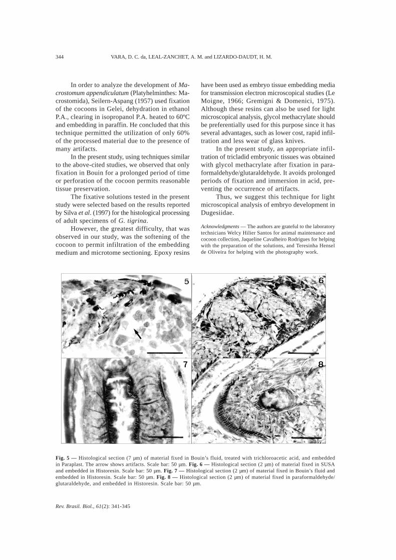
Fig. 5 — Histological section (7 µm) of material fixed in Bouin’s fluid, treated with trichloroacetic acid, and embeddedin Paraplast. The arrow shows artifacts. Scale bar: 50 µm. Fig. 6 — Histological section (2 µm) of material fixed in SUSAand embedded in Historesin. Scale bar: 50 µm. Fig. 7 — Histological section (2 µm) of material fixed in Bouin’s fluid andembedded in Historesin. Scale bar: 50 µm. Fig. 8 — Histological section (2 µm) of material fixed in paraformaldehyde/glutaraldehyde, and embedded in Historesin. Scale bar: 50 µm.

Rev. Brasil. Biol., 61(2): 341-345
HISTOLOGICAL TECHNIQUES FOR STUDY OF DUGESIIDAE DEVELOPMENT 345
REFERENCES
BENNETT, H. S., WYRICK, A. D., LEE, S. W. & MCNEIL,J. H., 1976, Science and art in preparing tissues embed-ded in plastic for light microscopy, with special referenceto glycol methacrylate, glass knives and simple stains.Stain Technol., 51(2): 71-97.
GREMIGNI, V. & DOMENICI, L., 1975, Genesis, compo-sition, and fate of cortical granules in the eggs ofPolycelis nigra (Turbellaria, Tricladida). J. Ultrastr. Res.,50: 277-283.
HAUSER, J., 1952, Ausschaltung des Xylols in derhistologischen Technik. Mikroskopie, 7(5-6): 208-211.
LE MOIGNE, A., 1963, Étude du développementembrionnaire de Polycelis nigra. Bull. Soc. Zool. France,88(4): 403-422.
LE MOIGNE, A., 1966, Étude au microscope électroniquede cellules d’embryons de Polycelis (Turbellarié, Tricla-dide), au début de leur développement. C. R. Acad. Sc.Paris, 263: 550-553.
MICHALANY, J., 1980, Técnica histológica em anatomiapatológica. Editora Pedagógica e Universitária Ltda., SãoPaulo, 277p.
MINGANTI, A., 1957, Sulla constituzione chimica degli in-volucri ovulari negli animali. Boll. Zool., 25: 55-88.
PAULETE-VANRELL, J., 1967, Guia de Técnica Microscó-pica. Faculdade de Filosofia, Ciências e Letras de SãoLeopoldo, São Leopoldo, não paginado.
PLATTNERT, N., 1975, Die chemische Fixierung biologis-cher Objekte für die Eletronenmikroskopie, pp. 3-43. In:G. Schimmel & W. Vogell (eds.), Methodensammlung derElektronenmikroskopie. Wissenschaftliche Verlagsgesells-chaft mbH, Stuttgart.
RUTHMANN, A., 1966, Methoden der Zellforschung.Franckh’sche Verlagshandlung, Stuttgart, 301p.
SEILERN-ASPANG, F., 1957, Die Entwicklung vonMacrostomum appendiculatum (Fabricius). Zool. Jb., Abt.Anat. u. Ontog., 76: 311-330.
SILVA, N. M. S., LEAL-ZANCHET, A. M. & HAUSER, J.,1997, Analysis of the efficiency of different solutions forthe fixation of Girardia tigrina (Turbellaria, Tricladida,Paludicola). Braz. J. Morphol. Sci., 14(2): 271-274.
SPURLOCK, B. O., SKINNER, M. S. & KATTINE, A. A.,1966, Simple rapid method for staining epoxi-embeddedspecimens for light microscopy with the polychromaticstaining Paragon-1031. Am. J. Clin. Pathol., 46: 252-258.
THOMAS, M. B., 1986, Embryology of the Turbellaria andits phylogenetic significance. Hidrobiologia, 132: 105-115.