histological tools
TRANSCRIPT
HISTOLOGICAL
TOOLSPresented to: Presented by:
Dr. Ashutosh Nirola Tejinder Pal Singh
Dr. Madhu Gupta
Dr. Vikram Bali
Dr. Priyanka Sharma
Dr. Kanika Singla
Dr. Priyanka Singla
CONTENTS
INTRODUCTION
MICROSCOPY
BIOPSY
TISSUE CULTURE
STAINING
HISTOCHEMISTRY AND CYTOCHEMISTRY
CELL FRACTIONATION
IMMUNOCYTOCHEMISTRY
HYBRIDIZATION TECHNIQUE
SUMMARY
INTRODUCTION
Histology is the study of the microscopic
anatomy of cells and tissues of plants and animals.
- performed by examining a thin slice (section)
of tissue under a light microscope or electron
microscope. enhanced through the use of
histological stains. Histology is an essential tool
of biology and medicine.
- Histopathology, the microscopic study of diseased
tissue, is an important tool in anatomical
pathology.
- Pathologists, are the personnel who perform
histopathological examination and provide
diagnostic information based on their observations
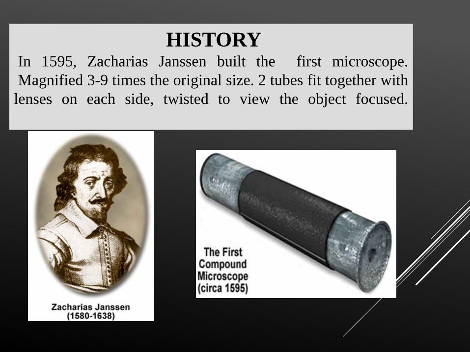
HISTORYIn 1595, Zacharias Janssen built the first microscope.
Magnified 3-9 times the original size. 2 tubes fit together with
lenses on each side, twisted to view the object focused.
ROBERT HOOK -17 TH
CENTURY- At the age of 27- designed a compoundmicroscope-magnified the object by 30 times.
- Using these scopes he made the greatestcontribution in a book he published entitled‘Micrographia’ - detailed view of all theobjects viewed under the microscope.
.
ANTONY VAN
LEEUWENHOEK.
ANTONY VAN LEEUWENHOEK –
FATHER OF MICROSCOPY
- After reading Hooke’s Micrographia- he learned togrind lenses and created over 500 microscopes( 4 inchlong).
- He preferred simple microscopes unlike Janssen andHooke (compound microscopes - 2 lenses).
- Magnified objects by 200 times.
- Until he discovered bacteria .
- In 1683- described the substance
removed from his own teeth, a little
white matter.
- Under microscope, saw many ‘very
little living animalcules’.
- Later termed as bacteria- the
founder being AV Leeuwenhoek.
SIGNIFICANCE
- Detailed structure of chromosomes bearing genes and themechanism underlying genetic expressions in cells are studied underhistology revealing relationship of histology with genetics.
- Several different types of cells are involved in immunologicalreactions.
- These cells are involved in combating disease causing organismsand describe the cellular basis for the body’s reaction to sensitizingagents like pollens etc.
“Histology plays a great role in immunology”
PREPARATION OF TISSUES FOR
MICROSCOPIC EXAMINATION
- The most common procedure used in the study of tissuesis the preparation of histological sections .
- Under light microscope, tissues are examined by
transillumination, Since tissues are too thick for
transillumination they should be sectioned .
Chemical Fixation with Formaldehyde or Others :
1. are used to preserve tissue from degradation
2. to maintain the structure of the cell and of sub-cellular components such as cell
organelles.
3. Main action of these aldehyde fixatives is to cross-link amino groups in proteins
through the formation of CH2 (methylene) linkage.
Fixing
FIXATIVES
Light microscopy - 10% neutral buffered
formalin (4% formaldehyde in phosphate
buffered saline).
Electron microscopy - glutaraldehyde,
usually as a 2.5% solution in phosphate
buffered saline.
FROZEN SECTION
FIXATION
- Frozen section is a rapid way to fix and mount
histology sections.
- It is done using a refrigeration device called a
cryostat.
- The frozen tissue - sliced using a microtome.
- Frozen slices are mounted on a glass slide ,stained the
same way as other methods.
- To fix tissue for certain stain such as antibody linked
immunofluorescence staining.
- Used to determine if a tumour is malignant when it is
found incidentally during surgery on a patient.
PROCESSING -
DEHYDRATION, CLEARING,
AND INFILTRATION
Aim of Tissue Processing
- Remove water from tissues
- Replace it with a medium that solidifies to allow thinsections to be cut.
1. 5 μm (micrometres; 1000 micrometres = 1 mm) thickfor light microscopy
2. 80-100 nm (nanometre; 1,000,000 nanometres =1 mm) thick for electron microscopy
- Light microscopy - paraffin wax is most
frequently used.
- Immiscible with water, the main constituent of
biological tissue.
- Water must first be removed in the process of
dehydration.
- Samples transferred -baths of progressively
more concentrated ethanol to remove the water.
- Hydrophobic clearing agent (such as xylene) to
remove the alcohol.
- A molten paraffin wax, the infiltration agent,
which replaces the xylene.
- Paraffin wax does not provide a sufficiently hard matrix for cutting very thin
sections for electron microscopy.
- Instead, resins are used.
- Epoxy resins are the most commonly employed embedding media.
- OTHER TYPES OF EMBEDDING MEDIA
Carbowax-It is a water soluble wax. Therefore tissues are directly
transferred to water soluble wax after fixation and washing.
Methacrylate: It is easily miscible with alcohol and gives a clear and hard
block when polymerized.
Agar embedding: It is mainly used in double embedding. Used in FNAC.
Gelatin: Its melting point is less than the melting point of agar. Gelatin may
be used when frozen sections are required on friable and necrotic tissues.
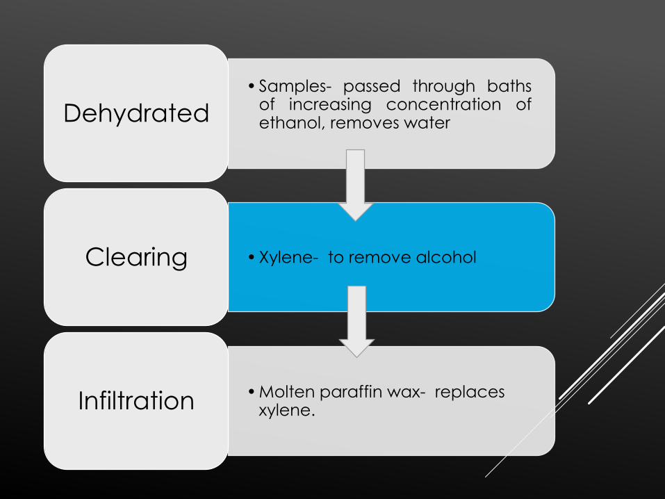
• Samples- passed through bathsof increasing concentration ofethanol, removes waterDehydrated
• Xylene- to remove alcoholClearing
• Molten paraffin wax- replaces xylene.Infiltration
EMBEDDING
- Tissues are ready for external embedding
- Tissues are placed in the moulds with liquid embedding material.(agar, gelatin,
wax.),hardened.
- Formalin-fixed, paraffin-embedded (FFPE) tissues may be stored indefinitely at
room temperature, and nucleic acids (both DNA and RNA) may be recovered from
them decades after fixation, making FFPE tissues an important resource for
historical studies in medicine.
SECTIONING
- Vertical sectioning - perpendicular to the surface
of the tissue- usual method.
- Horizontal sectioning - evaluation of the hair
follicles and pilosebaceous units. Tangential to
horizontal sectioning is done in Mohs surgery.
STAINING
- Staining - contrast to the tissue & highlights particular features of interest.
- Hematoxylin and eosin (H&E stain) is the most commonly used light microscopically stain in histology and histopathology.
- Hematoxylin, a basic dye, stains nuclei blue due to an affinity to nucleic acids in the cell nucleus;
- Eosin, an acidic dye, stains the cytoplasm pink.
- Extracellular structure ,collagen and other fibers.
- In connective tissue stain pink.
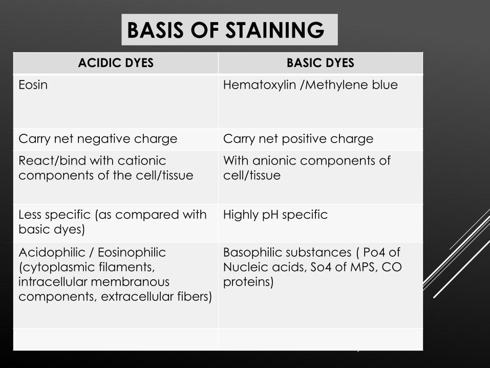
ACIDIC DYES BASIC DYES
Eosin Hematoxylin /Methylene blue
Carry net negative charge Carry net positive charge
React/bind with cationic
components of the cell/tissue
With anionic components of
cell/tissue
Less specific (as compared with
basic dyes)
Highly pH specific
Acidophilic / Eosinophilic
(cytoplasmic filaments,
intracellular membranous
components, extracellular fibers)
Basophilic substances ( Po4 of
Nucleic acids, So4 of MPS, CO
proteins)
BASIS OF STAINING
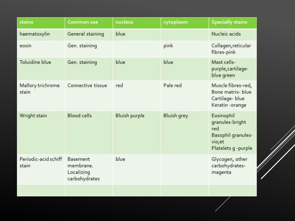
- ELASTIC STAINS -chemically react with elastic fibers in aorta, elastic
cartilage ,and in connective tissue –black/blue.
- RETICULAR STAINS –stain type 3 collagen –blue/black.
- METAL STAINS – stain reticular fibres, golgi apparatus & neurofibrils.
FEULGEN STAIN FOR NUCLEAR PROTEIN
1. Acid hydrolyses or cleaves proteins from deoxyribose of DNA leads
to opening of sugar group & formation of aldehyde.
2. Schiff binds and gives magenta color to aldehyde.
3. Can be useful to quantify amount of DNA ( by using spectrophotometry
of Feulgen stained tissue).
TRICHOME STAINS –these are dyes with multiple
constituents mixed together each dye staining different
structure .MALLORY”S TRICHOME most common.
- NUCLEI –BLACK
- CYTOPLASM,KERATIN,MUSCLE FIBRE –RED/PINK
-CONNECTIVE TISSUE-BLUE
-NISSL’S STAIN –BASIC DYE ,it stains basophillic structures
nuclei ,ribosomes.
STAINS USING CHEMICAL
REACTION
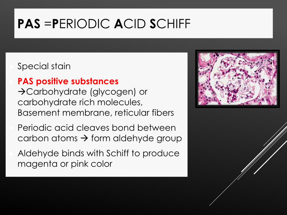
Special stain
PAS positive substances Carbohydrate (glycogen) or
carbohydrate rich molecules,
Basement membrane, reticular fibers
Periodic acid cleaves bond between carbon atoms form aldehyde group
Aldehyde binds with Schiff to produce
magenta or pink color
PAS =PERIODIC ACID SCHIFF
MICROSCOPE
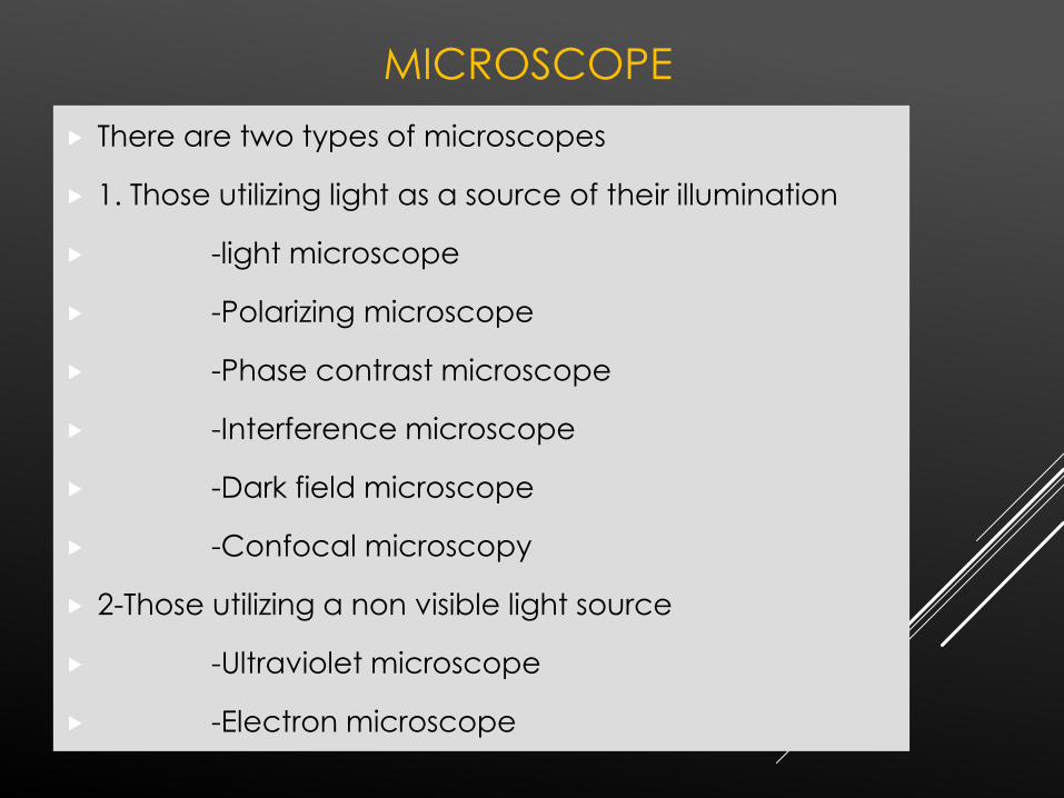
There are two types of microscopes
1. Those utilizing light as a source of their illumination
-light microscope
-Polarizing microscope
-Phase contrast microscope
-Interference microscope
-Dark field microscope
-Confocal microscopy
2-Those utilizing a non visible light source
-Ultraviolet microscope
-Electron microscope

A microscope is an instrument designed to make fine details visible.
The microscope must accomplish three tasks: produce a
magnified image of the specimen (magnification), separate
the details in the image (resolution), and render the details
visible to the eye, camera, or other imaging device
(contrast).
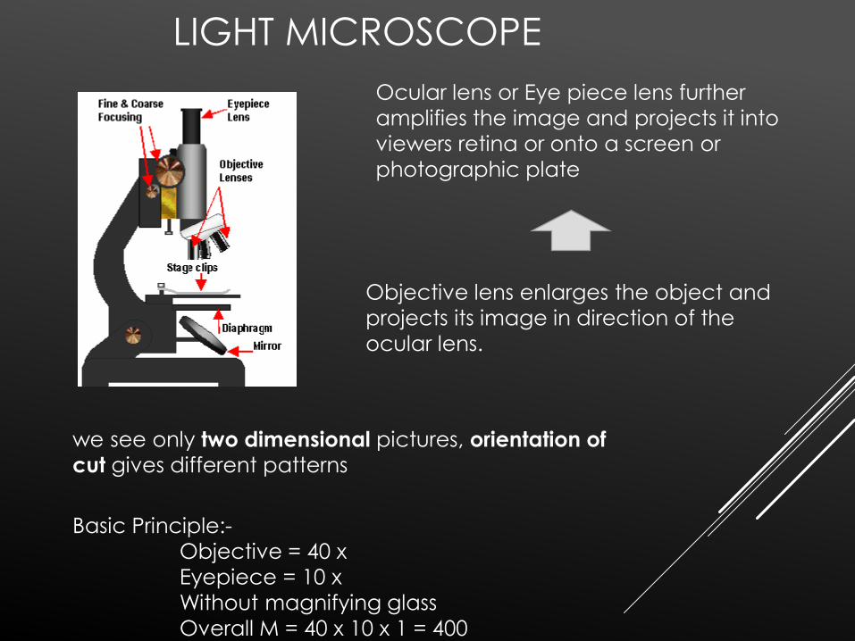
LIGHT MICROSCOPE
we see only two dimensional pictures, orientation of
cut gives different patterns
Ocular lens or Eye piece lens further
amplifies the image and projects it into
viewers retina or onto a screen or
photographic plate
Objective lens enlarges the object and
projects its image in direction of the
ocular lens.
Basic Principle:-
Objective = 40 x
Eyepiece = 10 x
Without magnifying glass
Overall M = 40 x 10 x 1 = 400
PHASE CONTRAST MICROSCOPY
Examine unstained preparations, for fresh specimens, living cells.
Based on the fact that light passing through media with different refractive
index slows down and changes direction.
This forms phase differences between two adjoining regions.
These phase differences by means of special optical system are transformed
into differences of light intensity so that the image becomes visible.
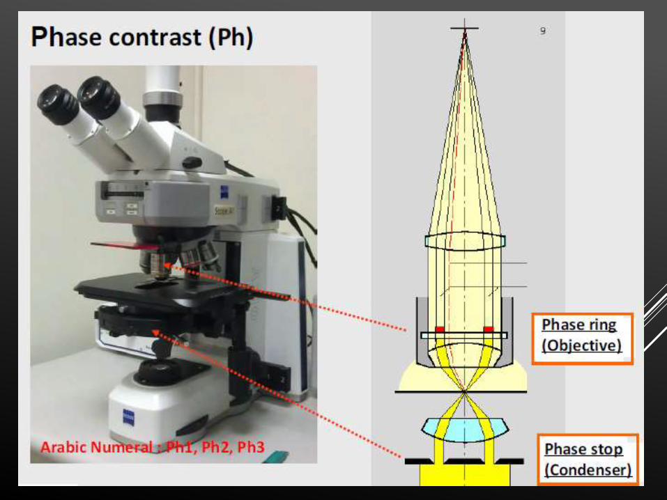
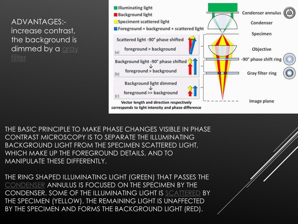
THE BASIC PRINCIPLE TO MAKE PHASE CHANGES VISIBLE IN PHASE
CONTRAST MICROSCOPY IS TO SEPARATE THE ILLUMINATING
BACKGROUND LIGHT FROM THE SPECIMEN SCATTERED LIGHT,
WHICH MAKE UP THE FOREGROUND DETAILS, AND TO
MANIPULATE THESE DIFFERENTLY.
THE RING SHAPED ILLUMINATING LIGHT (GREEN) THAT PASSES THE
CONDENSER ANNULUS IS FOCUSED ON THE SPECIMEN BY THE
CONDENSER. SOME OF THE ILLUMINATING LIGHT IS SCATTERED BY
THE SPECIMEN (YELLOW). THE REMAINING LIGHT IS UNAFFECTED
BY THE SPECIMEN AND FORMS THE BACKGROUND LIGHT (RED).
ADVANTAGES:-
increase contrast,
the background is
dimmed by a gray
filter
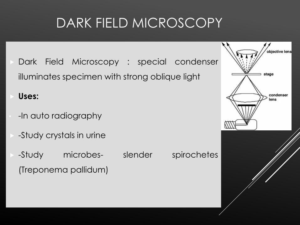
DARK FIELD MICROSCOPY
Dark Field Microscopy : special condenser
illuminates specimen with strong oblique light
Uses:
• -In auto radiography
-Study crystals in urine
-Study microbes- slender spirochetes
(Treponema pallidum)
DISADVANTAGES
THE MAIN LIMITATION OF DARK FIELD MICROSCOPY IS THE
LOW LIGHT LEVELS SEEN IN THE FINAL IMAGE. THIS MEANS
THE SAMPLE MUST BE VERY STRONGLY ILLUMINATED,
WHICH CAN CAUSE DAMAGE TO THE SAMPLE.
ADVANTAGES
Dark field microscopy is a very simple yet
effective technique and well suited for uses
involving live and unstained biological samples,
such as a smear from a tissue culture or individual, water-borne, single-celled organisms.
Considering the simplicity of the setup, the
quality of images obtained from this technique is
impressive.
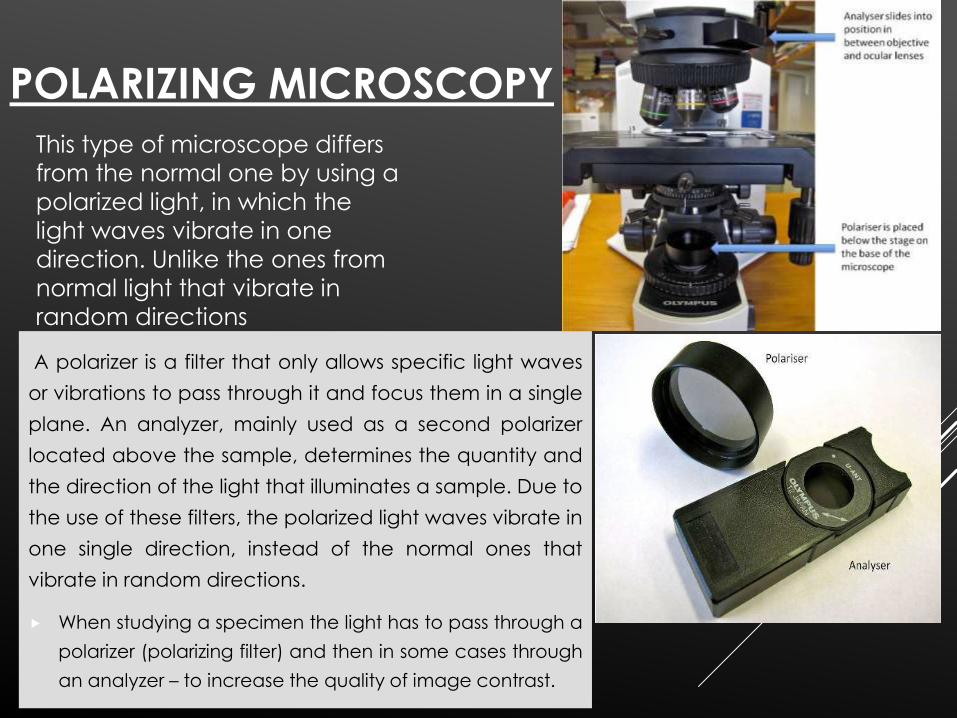
POLARIZING MICROSCOPY
A polarizer is a filter that only allows specific light waves
or vibrations to pass through it and focus them in a single
plane. An analyzer, mainly used as a second polarizer
located above the sample, determines the quantity and
the direction of the light that illuminates a sample. Due to
the use of these filters, the polarized light waves vibrate in
one single direction, instead of the normal ones that
vibrate in random directions.
When studying a specimen the light has to pass through a
polarizer (polarizing filter) and then in some cases through
an analyzer – to increase the quality of image contrast.
This type of microscope differs
from the normal one by using a
polarized light, in which the
light waves vibrate in one
direction. Unlike the ones from
normal light that vibrate in
random directions
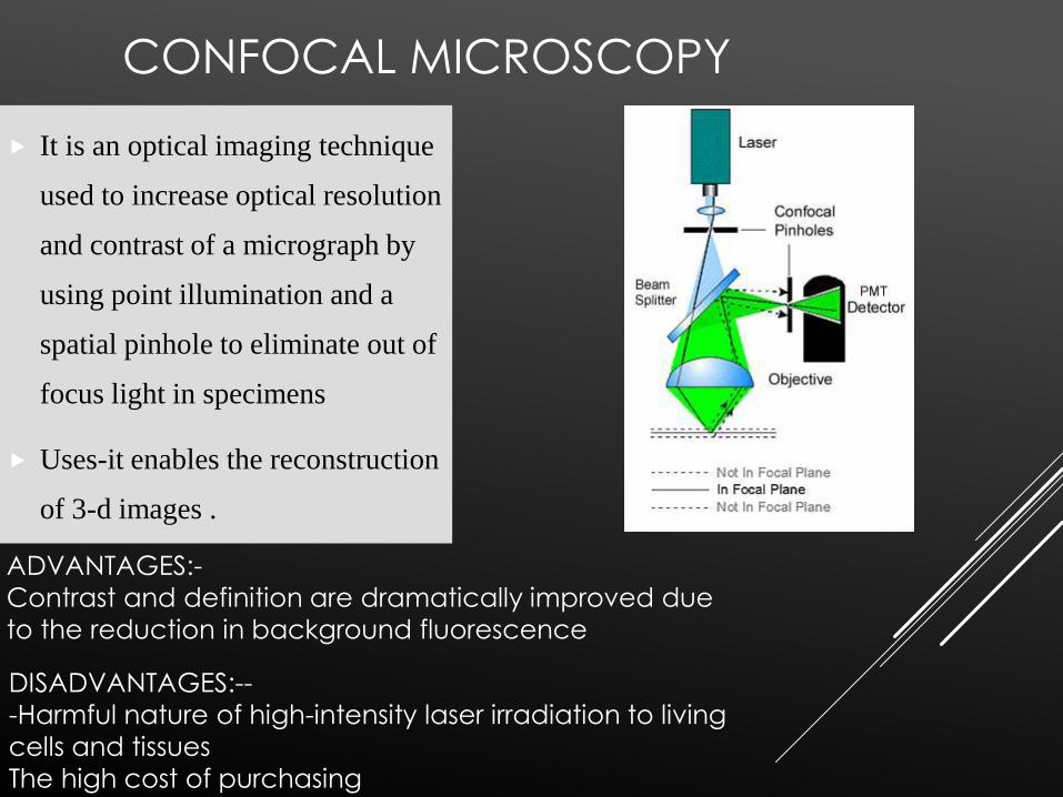
CONFOCAL MICROSCOPY
It is an optical imaging technique
used to increase optical resolution
and contrast of a micrograph by
using point illumination and a
spatial pinhole to eliminate out of
focus light in specimens
Uses-it enables the reconstruction
of 3-d images .
ADVANTAGES:-
Contrast and definition are dramatically improved due
to the reduction in background fluorescence
DISADVANTAGES:--
-Harmful nature of high-intensity laser irradiation to living
cells and tissues
The high cost of purchasing
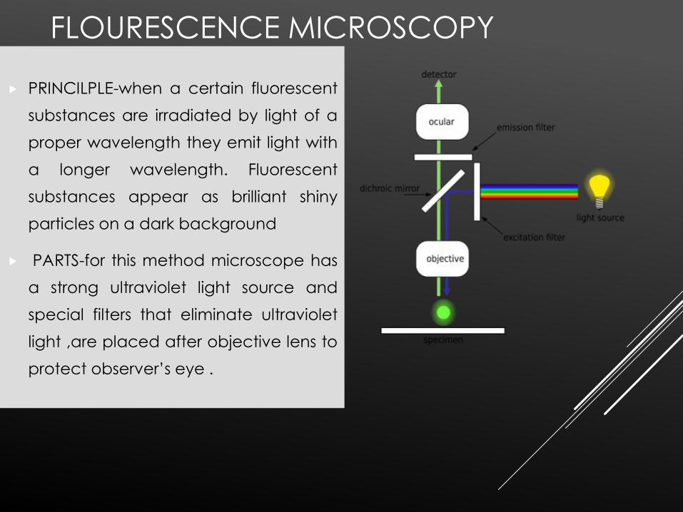
FLOURESCENCE MICROSCOPY
PRINCILPLE-when a certain fluorescent
substances are irradiated by light of a
proper wavelength they emit light with
a longer wavelength. Fluorescent
substances appear as brilliant shiny
particles on a dark background
PARTS-for this method microscope has
a strong ultraviolet light source and
special filters that eliminate ultraviolet
light ,are placed after objective lens to
protect observer’s eye .
Dyes used -acridine orange,auramine (tuberclebacilli),quinacrine(Q banding of chromosomes)
Acridine-DNA complex emits a yellow green light and
RNA Acridine orange emits a reddish orange .
It is thus possible to identify and localize nucleic acidsin cells
ELECTRON MICROSCOPY
Electron Microscope (EM): Electrons are suchsmall particles that, like photons in light, theyact as waves. A beam of electrons passesthrough the specimen, then through a seriesof lenses that magnify the image.Types:Transmission (TEM), scanning (SEM)
Mechanism: similar to LM except that beam ofelectrons replace light source
Recording: photoelectric plate or video detector
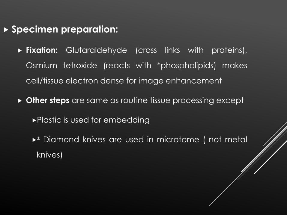
Specimen preparation:
Fixation: Glutaraldehyde (cross links with proteins),
Osmium tetroxide (reacts with *phospholipids) makes
cell/tissue electron dense for image enhancement
Other steps are same as routine tissue processing except
Plastic is used for embedding
± Diamond knives are used in microtome ( not metal
knives)
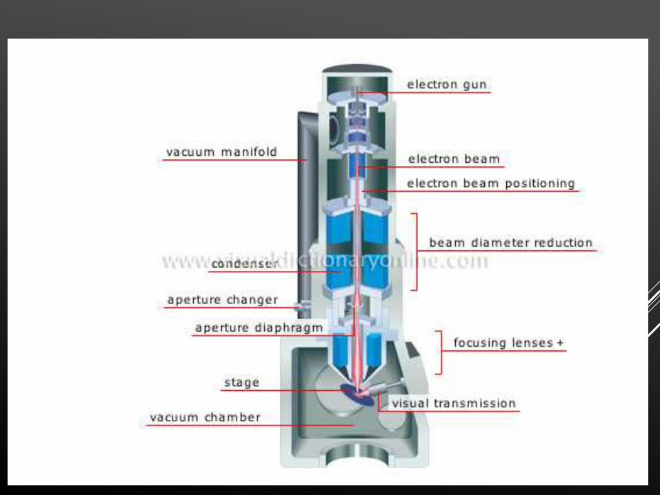
WORKING – electrons are produced by high
temperature heating of a metallic filament(cathode) in a vacuum.The emitted electrons are then submitted
to a potential difference of appx 60-100 kv .
The anode is a metallic plate with a small hole in its
centre.electrons are accelerated from the cathode
to anode .some of these particles pass through the
central opening in the anode forming a constant
stream of electrons.The beam is deflected by
electromagnetic lenses in a way roughly analogous to
optical microscope .The image obtained is further enlarged by one or two projecting lenses and is finally
seen on a flourescent screen on a flourescent screen
or is projected onto photographic plates .
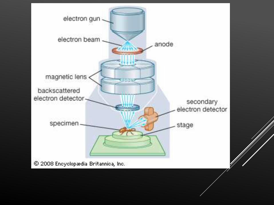
SCANNING ELECTRON MICROSCOPE
It permits pseudo 3-dimentionsal view of the surfaces
of cells tissues and organs
Scanning (SEM)
It differs from TEM that electron beam passes across the surface of spectrum (not thru specimen as in TEM)
Resembles Television
Can see 3D pictures
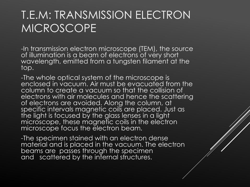
T.E.M: TRANSMISSION ELECTRON
MICROSCOPE
-In transmission electron microscope (TEM), the source of illumination is a beam of electrons of very short wavelength, emitted from a tungsten filament at the top.
-The whole optical system of the microscope is enclosed in vacuum. Air must be evacuated from the column to create a vacuum so that the collision of electrons with air molecules and hence the scattering of electrons are avoided. Along the column, at specific intervals magnetic coils are placed. Just as the light is focused by the glass lenses in a light microscope, these magnetic coils in the electron microscope focus the electron beam.
-The specimen stained with an electron dense material and is placed in the vacuum. The electron beams are passes through the specimen and scattered by the internal structures.
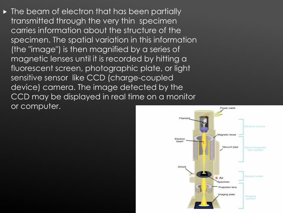
The beam of electron that has been partially
transmitted through the very thin specimen
carries information about the structure of the
specimen. The spatial variation in this information
(the "image") is then magnified by a series of
magnetic lenses until it is recorded by hitting a
fluorescent screen, photographic plate, or light
sensitive sensor like CCD (charge-coupled device) camera. The image detected by the
CCD may be displayed in real time on a monitor
or computer.
Atomic Force M: most powerful tool tostudy surface topography
Non – optical M: works like finger tip
Has highest resolution power – 50 pm
No need to put specimen in vaccum
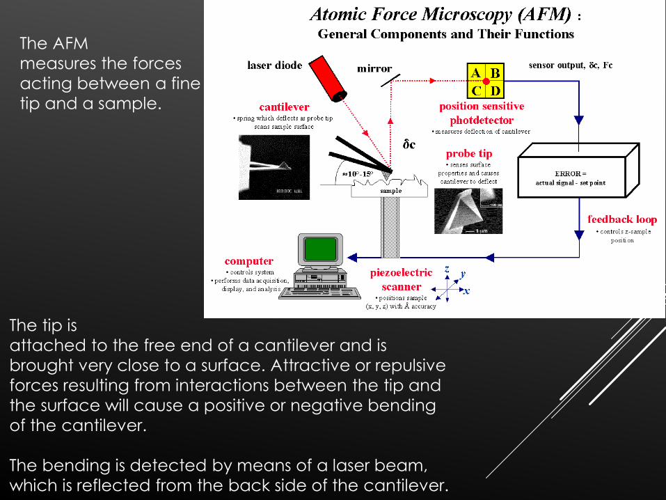
The tip is
attached to the free end of a cantilever and is
brought very close to a surface. Attractive or repulsive
forces resulting from interactions between the tip and
the surface will cause a positive or negative bending
of the cantilever.
The bending is detected by means of a laser beam,
which is reflected from the back side of the cantilever.
The AFM
measures the forces
acting between a fine
tip and a sample.
EXAMINING LIVING TISSUES
(CULTURE MEDIA)
Prolonged study of living tissues can be achievedby culturing them in solutions that containnecessary nutrients to keep them alive.
Cells are treated prior with trypsin-isolated-cultivated insuspension - spread out on a glasssurface to which they adhere spontaneously as asingle layer of cells.
Culture medium should be changed frequentlysince nutrients become depleted and toxicproducts of metabolism accumulate. Rigorousaseptic techniques are necessary during processof cell cultivation in order to avoid contaminationof culture media
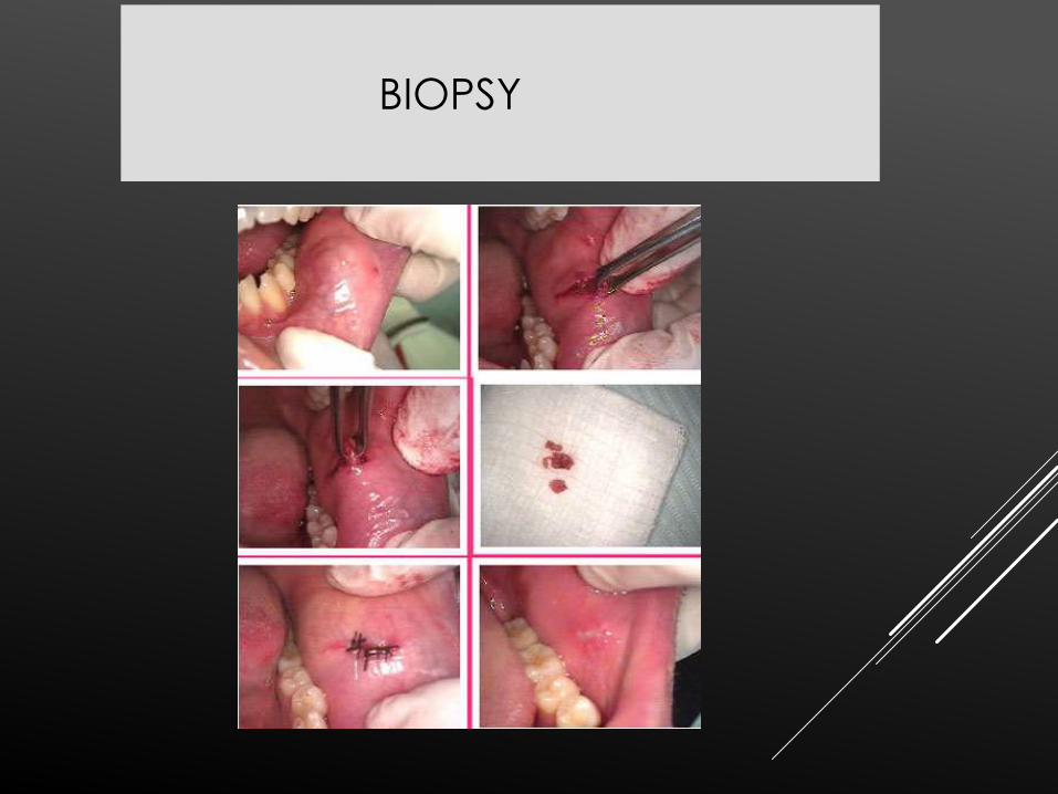
BIOPSY
BIOPSY – it is a controlled and deliberate
removal of tissues from a living organism for
the purpose of microscopic examination.
PURPOSE :
1. Make a speedy, definitive, accurate diagnosis.
2. To aid in determining the prognosis of the lesion.
3. Aids in deciding the best treatment possible.
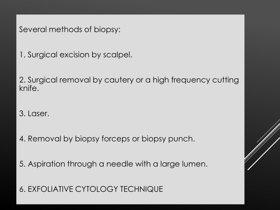
Several methods of biopsy:
1. Surgical excision by scalpel.
2. Surgical removal by cautery or a high frequency cutting knife.
3. Laser.
4. Removal by biopsy forceps or biopsy punch.
5. Aspiration through a needle with a large lumen.
6. EXFOLIATIVE CYTOLOGY TECHNIQUE
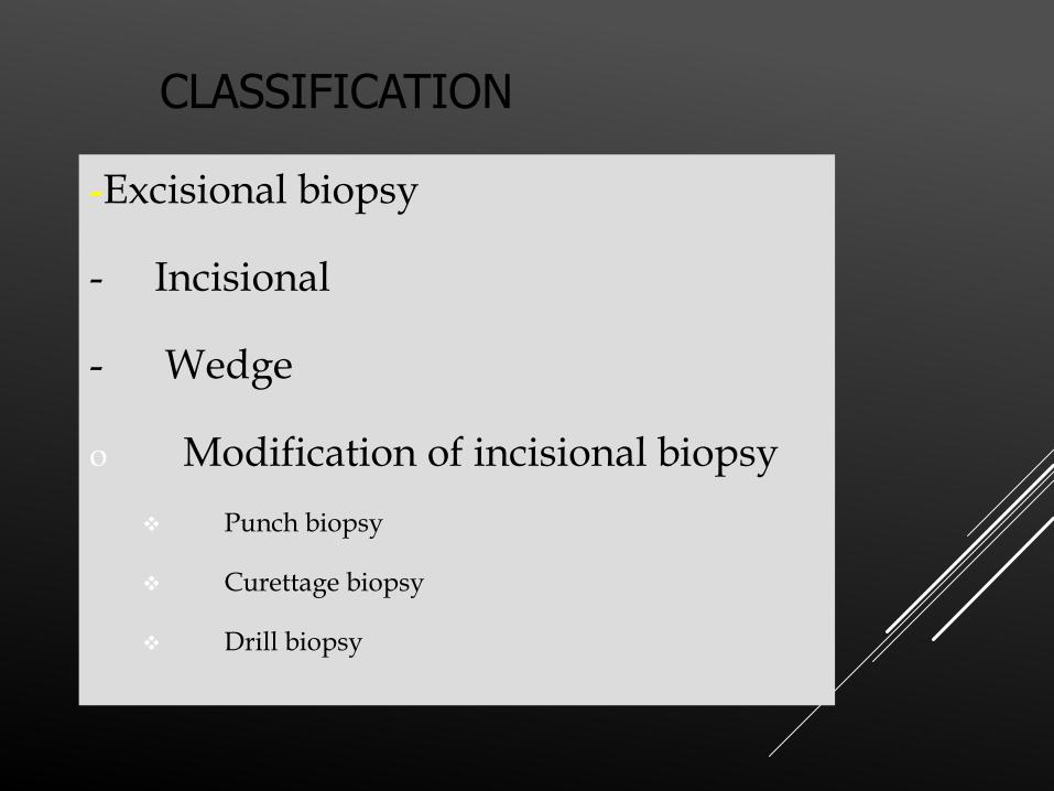
CLASSIFICATION
-Excisional biopsy
- Incisional
- Wedge
o Modification of incisional biopsy
Punch biopsy
Curettage biopsy
Drill biopsy
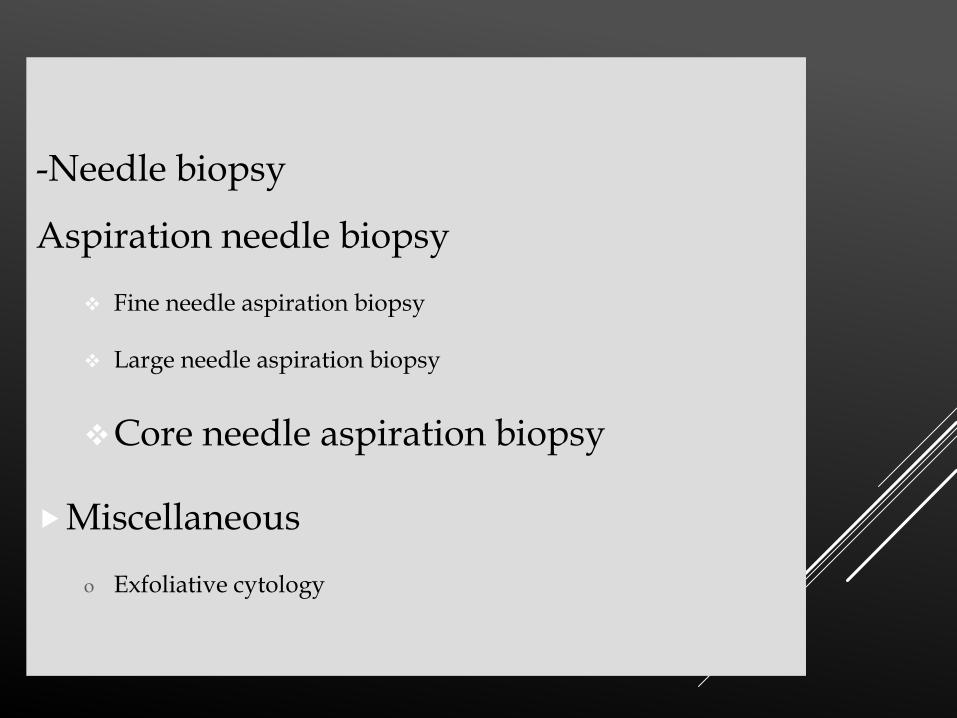
-Needle biopsy
Aspiration needle biopsy
Fine needle aspiration biopsy
Large needle aspiration biopsy
Core needle aspiration biopsy
Miscellaneous
o Exfoliative cytology
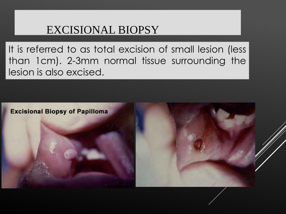
EXCISIONAL BIOPSY
It is referred to as total excision of small lesion (less
than 1cm). 2-3mm normal tissue surrounding the
lesion is also excised.
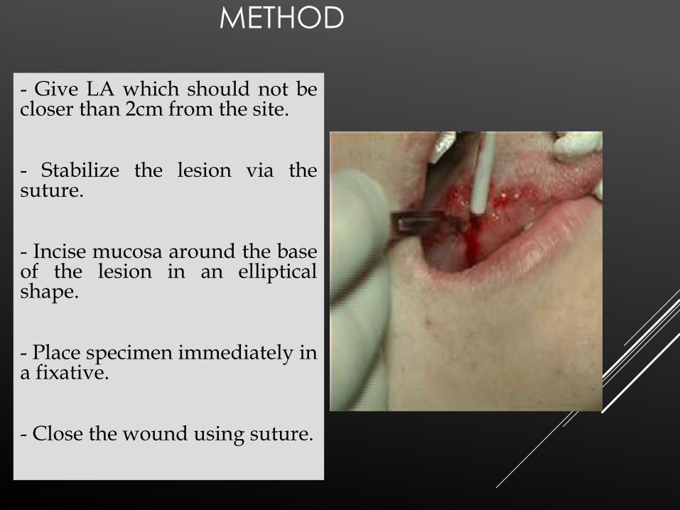
METHOD
- Give LA which should not becloser than 2cm from the site.
- Stabilize the lesion via thesuture.
- Incise mucosa around the baseof the lesion in an ellipticalshape.
- Place specimen immediately ina fixative.
- Close the wound using suture.
INCISIONAL BIOPSY:
-Some lesions are too large to excise initially without having established diagnosis or are of such a nature that excision would be inadvisable in such instances a small section is removed for examination called incisional or diagnostic biopsy.
-It samples only a particular or representative part of the
lesion
-Lesion is larger than 1cm in diameter
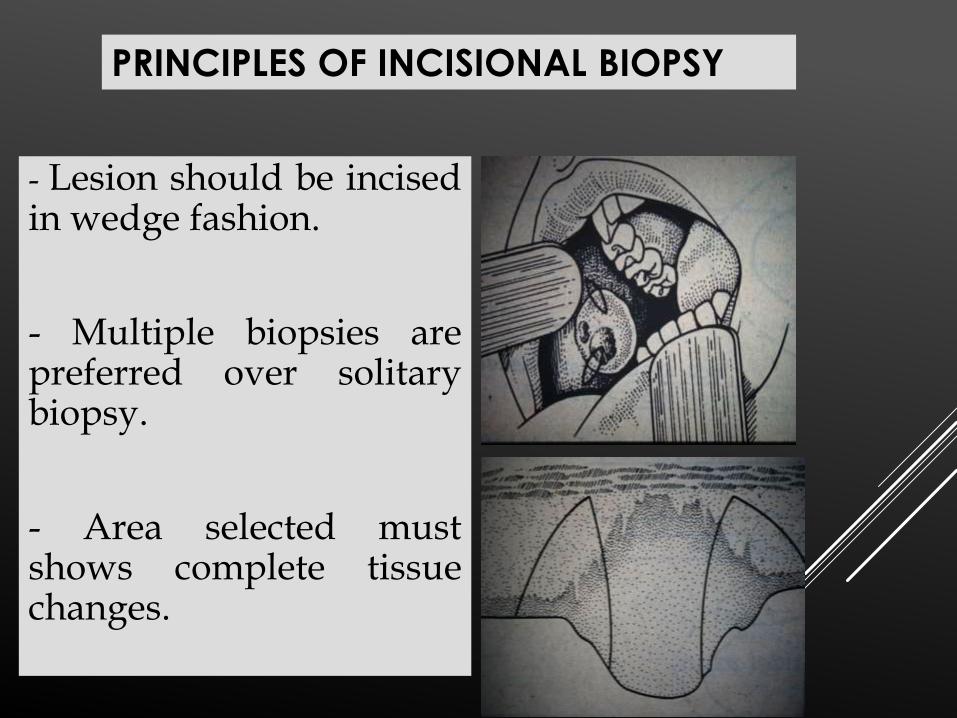
- Lesion should be incisedin wedge fashion.
- Multiple biopsies arepreferred over solitarybiopsy.
- Area selected mustshows complete tissuechanges.
PRINCIPLES OF INCISIONAL BIOPSY
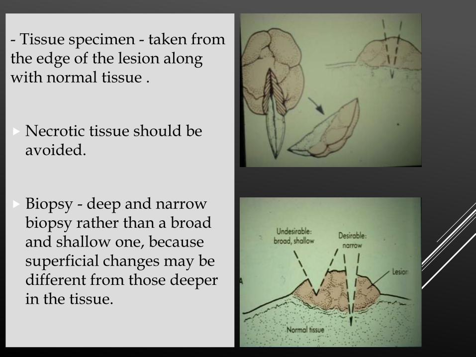
- Tissue specimen - taken from the edge of the lesion along with normal tissue .
Necrotic tissue should be avoided.
Biopsy - deep and narrow biopsy rather than a broad and shallow one, because superficial changes may be different from those deeper in the tissue.
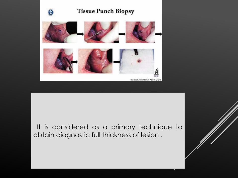
PUNCH BIOPSY
It is considered as a primary technique to
obtain diagnostic full thickness of lesion .
CURETTAGE BIOPSY-
A curette is a surgical instrument with a curved spoon
like tip and a long handle designed for scraping out
body cavities for tissue diagnosis or therapeutic
purposes. The samples produced are usually soft
tissue but may include bone fragments as well.
- The curettage is usually diagnostic for pathological
purposes, but can also be therapeutic in treating the
condition.
TECHNIQUE
Most common method - Fountain Pen Technique
The curette is held between the thumb, index
and middle finger
The skin is stretched with finger of other hand
Other common method - Potato Peeler
Technique
Handle of curette is held in the distal inter-digital
fold of the index finger supported by other fingersof curetting hand
Thumb provides a stable base
CELL ASPIRATE
-Aspiration biopsy is the use of a needle and
syringe to penetrate a lesion for aspiration of its
content
It is relatively painless
Use of local analgesia optional
Capable of producing immediate results
TECHNIQUE
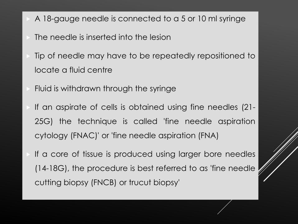
A 18-gauge needle is connected to a 5 or 10 ml syringe
The needle is inserted into the lesion
Tip of needle may have to be repeatedly repositioned to
locate a fluid centre
Fluid is withdrawn through the syringe
If an aspirate of cells is obtained using fine needles (21-
25G) the technique is called 'fine needle aspiration
cytology (FNAC)' or 'fine needle aspiration (FNA)
If a core of tissue is produced using larger bore needles
(14-18G), the procedure is best referred to as 'fine needle
cutting biopsy (FNCB) or trucut biopsy'
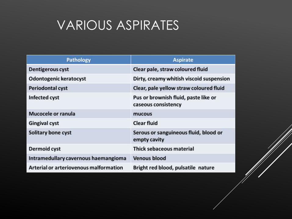
VARIOUS ASPIRATES
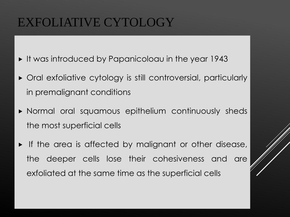
EXFOLIATIVE CYTOLOGY
It was introduced by Papanicoloau in the year 1943
Oral exfoliative cytology is still controversial, particularly
in premalignant conditions
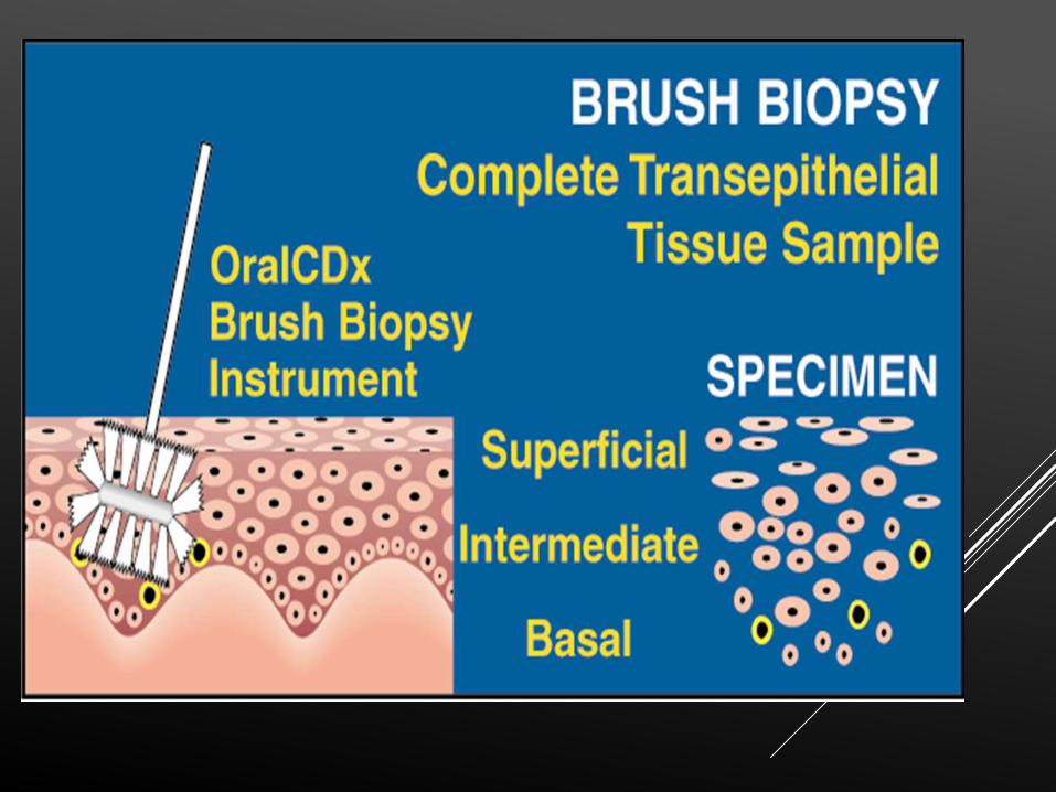
Normal oral squamous epithelium continuously sheds
the most superficial cells
If the area is affected by malignant or other disease,
the deeper cells lose their cohesiveness and are
exfoliated at the same time as the superficial cells

Exfoliative cytology has
not been a very
diagnostic or useful
screening method for oral
cancer because
hyperkeratosis and keratin
itself interfere with cell
obtainment and a greater
proportion of diagnostic
cells are below the surface
(most at the basement
membrane level)
CYTOLOGICAL SMEARS FALL UNDER
FOLLOWING CATEGORIES
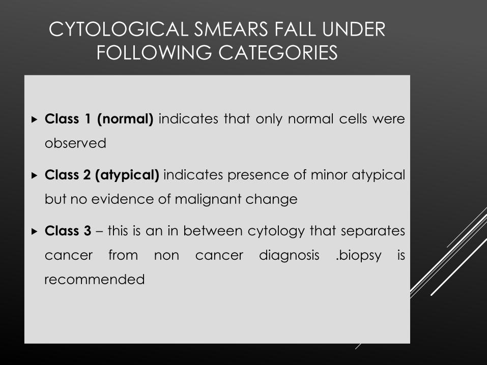
Class 1 (normal) indicates that only normal cells were
observed
Class 2 (atypical) indicates presence of minor atypical
but no evidence of malignant change
Class 3 – this is an in between cytology that separates
cancer from non cancer diagnosis .biopsy is
recommended
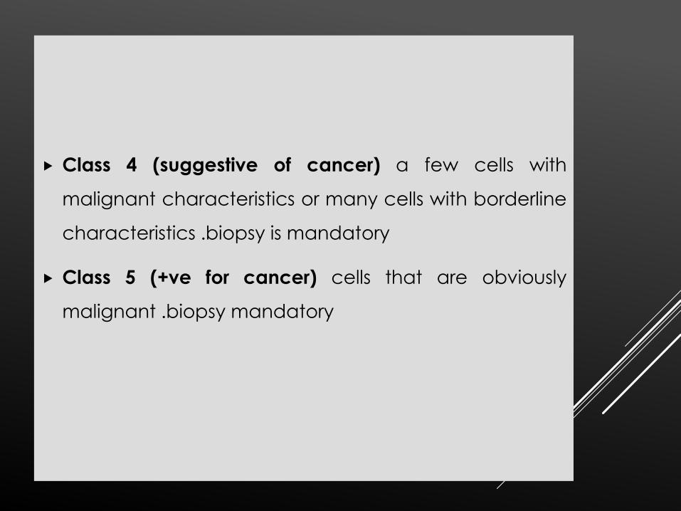
Class 4 (suggestive of cancer) a few cells with
malignant characteristics or many cells with borderline
characteristics .biopsy is mandatory
Class 5 (+ve for cancer) cells that are obviously
malignant .biopsy mandatory
HISTOCHEMISTRY AND CYTOCHEMISTRY
• Are used to indicate methods for localization different
substances in tissue sections
• They are based on specific chemical reactions or on
high affinity interactions between macromolecules
• Both methods usually produce insoluble coloured or
electron dense compounds that enable the
localization of specific substances by means of light or
electron microscopy
NUCLEIC ACIDS-DNA can be identified and quantified in
cell nuclei using feulgen reaction which produces a red
color in presence of dna
PROTEINS –specific enzymes such as RNAase
,DNAase,collagenase and elastase that digest cell and
tissue components are used extensively
POLYSACCRIDES and OLIGOSACCRIDES –glycogen
which can be demonstrated by PAS .
GLYCOSAMINOGLYCANS react strongly with ALCIAN
BLUE DYE
Neutral glycoproteins react with PAS but are not
digested by glycogenolytic enzymes .
REACTIONS FREQUENTLY USED IN
LABORATORY DIAGNOSIS
PAS-AMYLASE REACTION and alcian blue reaction –
glycogen and glycosaminoglycans
ENZYMES –acid phosphatase –THE GOMORI METHOD –
consist of incubating formalin fixed tissue sections in a
solution containing sodium glycerophosphate and
lead nitrate buffered to ph.5. The enzymes hydrolyses
the glycerophosphate liberating phosphate ions that
react with lead nitrate to produce an insoluble
,electron dispersing , colourless ppt of lead phosphate
at the site of enzymatic activity
In a second step the preparation is immersed in a solution
of ammonium sulfide that reacts
With lead phosphate to produce a blak ppt of lead sulphite
.this method permits the localization of this enzyme’s activity
and is frequently used to demonstrate lysosomes
,cytoplasmic organelles that contain acid phosphates .
DEHYDROGENASES-these enzyme remove hydrogen from
one substrate and trasfer hydrogen from one substance
and transfer it to an other .the enzyme transports
hydrogen from the substrate to the tetrazole and
reduces it to an intensely coloured insoluble compound
FORMAZAN which ppt at the site of enzyme active
example is succinate dehydrogenase .
Peroxidase –a section of adequately fixed tissue are
incubated in a solution containing hydrogen peroxide
and 3,3’diaminoazobenzidine ,it is oxidised in presence
of peroxidase resulting in black ppt.
IMMUNOCYTOCHEMISTRY
It is based on coupling of immunoglobulin's to substances
that render them visible in microscope without causing a
loss of antibody’s biological activity
METHODS OF LABELLING ANTIBODIES
1.COUPLING WITH A FLOURESCENT ANTIBODIES
2.COUPLING WITH AN ENZYME-Most common enzyme used
is peroxidase
3.COUPLING WITH A COLOURED ELECTRON SCATTERING
COMPOUND e.g gold particle
There are three methods –
Direct, Indirect AND Indirect Method With Signal
Amplification
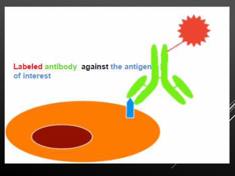
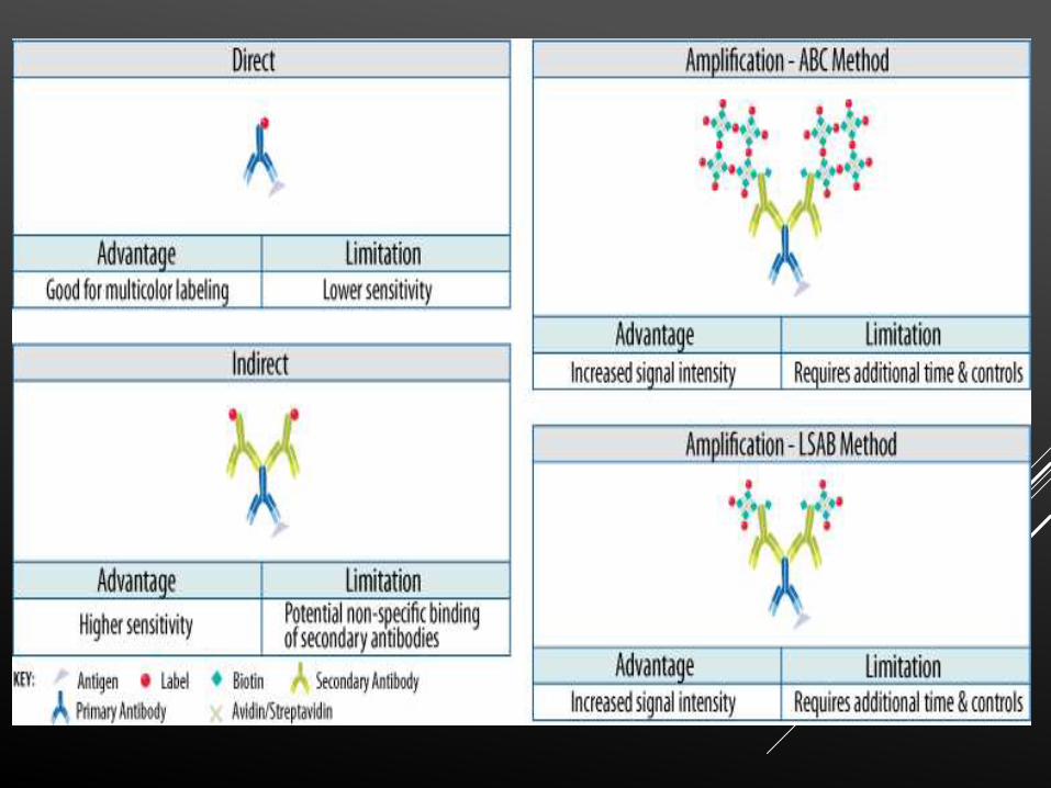
Direct method –The detection of the complex is based
upon the molecule conjugated to the antibody.
Advantages of direct detection include the ease of use
for multicolor staining and the elimination of concerns
regarding non-specific binding of the secondary antibody.
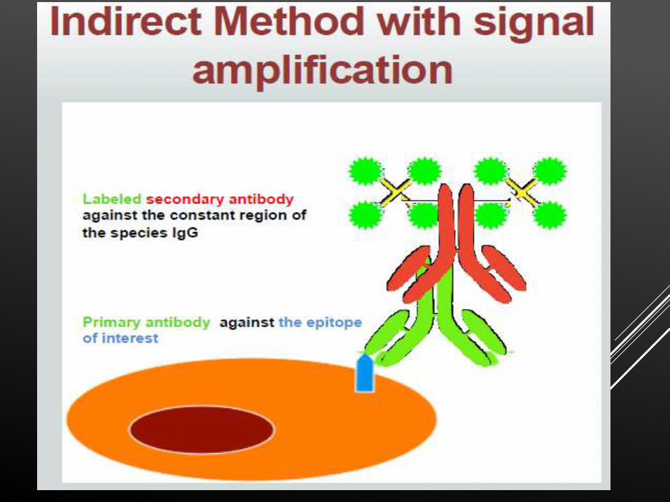
INDIRECT METHOD indirect detection methods generally have
a higher level of sensitivity and generate a more intense
signal. The signal is amplified using the indirect method
because of the potential for at least two labeled secondary
antibodies to bind to each primary antibody..

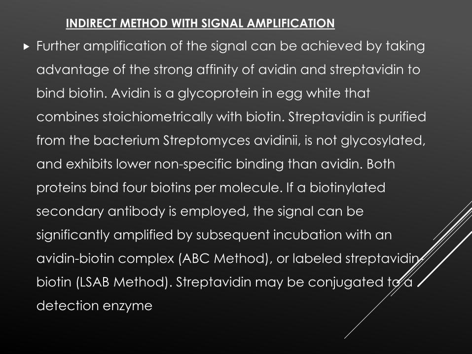
Further amplification of the signal can be achieved by taking
advantage of the strong affinity of avidin and streptavidin to
bind biotin. Avidin is a glycoprotein in egg white that
combines stoichiometrically with biotin. Streptavidin is purified
from the bacterium Streptomyces avidinii, is not glycosylated,
and exhibits lower non-specific binding than avidin. Both
proteins bind four biotins per molecule. If a biotinylated
secondary antibody is employed, the signal can be
significantly amplified by subsequent incubation with an
avidin-biotin complex (ABC Method), or labeled streptavidin-
biotin (LSAB Method). Streptavidin may be conjugated to a
detection enzyme
INDIRECT METHOD WITH SIGNAL AMPLIFICATION
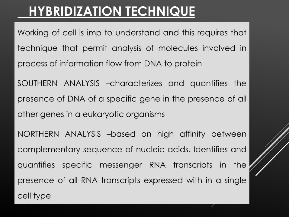
HYBRIDIZATION TECHNIQUE
Working of cell is imp to understand and this requires that
technique that permit analysis of molecules involved in
process of information flow from DNA to protein
SOUTHERN ANALYSIS –characterizes and quantifies the
presence of DNA of a specific gene in the presence of all
other genes in a eukaryotic organisms
NORTHERN ANALYSIS –based on high affinity between
complementary sequence of nucleic acids. Identifies and
quantifies specific messenger RNA transcripts in the
presence of all RNA transcripts expressed with in a single
cell type
Western Analysis- detects a single protein
species from among all other proteins expressed
in a single cell or tissue. It is based on high
affinity and specificity between antibodies and
antigens
NUCLEIC ACID PROBES
A probe is a nucleic acid molecule (single-stranded DNA
or RNA) with a strong affinity with a specific target (DNA
or RNA sequence).
DNA PROBES- use segments of single stranded nucleic acid
labelled with an enzyme or radioisotope that is able to
hybridize to the complementry nucleic acid sequence and
thus detects the presence of particular microorganisms
HYBRIDISATION refers to pairing of complementry DNA
strands to produce a double stranded nucleic acid

-More specific oligonucleotide
probes complementary to variable
regions of 16s rRNA
Bacterial genes have been
identified .these bacterial 16s rRNA
genes contain both regions shared
by different bacteria and short
streches of variable regions shared
by specific organisms of same
species or genes .
LECTIN HISTOCHEMISTRY
Lectins are proteins derived mainly from plant seeds thatbind to cell surface carbohydrates with high affinity andspecificity
Different lectins bind to specific sequences of sugarresidues.
They bind to cell surface glycoproteins, proteoglycansand glycolipids and are widely used to characterizemembrane molecules containing specific sequences ofsugar residues.
Lectins are usually labeled with peroxidase to makepossible their identification.
SUMMARY
The small size of cells and matrix components
make histology dependent on use of tools
advances in chemical, biology, physiology,
immunology and pathology and interactions
among these contribute to better knowledge of
tissue biology
Familiarity with the tools and methods of any
branch of science is essential for a proper
understanding of the subject. It was a review of
some common tools used in histology
REFERENCES
Basic Histology (9th Edition) Lange
Shafer’s Oral Pathology (5th Edition)
Orban’s Oral Histology (13th edition)
Dye, Stains & special probes in histology (Wolf D, HulmannN.D)
Cell Biology Lab Manual ( Dr. William H. Heidcamp)
Immunihistochemistry (Eldem Sadikoglou)
Molecular Hybridization Technique Of Nucleic Acid: ISSN1843-6099
Oral Histology (Antonio Nancy 6th Edition)
Microscope : Basic & Beyond (Mortimer Abramowitz Vol.1)
Textbook of Human Histology (Dr. Veena Bharioke)
A Book Of Microbiology –Ananthnarayan

THANK
YOU