history, virulence genes, identification and antimicrobial ... abdulrahman... ·...
TRANSCRIPT

Int.J.Curr.Microbiol.App.Sci (2018) 7(1): 2136-2154
2136
Review Article https://doi.org/10.20546/ijcmas.2018.701.258
History, Virulence Genes, Identification and Antimicrobial Resistance of
Enterococcus faecalis Isolated from Diabetic Foot Patients
Ahmad Abdulrahman AL Bloushy1, 2*
and Ayman Elbehiry3,4
1Al Bukayriyah General Hospital, Qassim, Kingdom of Saudi Arabia
2Department of Botany and Microbiology, College of Science, King Saud University,
P.O. Box 2455, Riyadh 11451, Saudi Arabia 3Department of Bacteriology, Mycology and Immunology, Faculty of Veterinary Medicine,
Sadat City University, Sadat, Egypt 4Department of Public Health, College of Public Health and Health Informatics, Qassim
University, Buraidah, Saudi Arabia
*Corresponding author
A B S T R A C T
Enterococci
Enterococcus is a large genus of lactic acid
bacteria that have the ability to grow under
various aggressive conditions. Enterococci are
Gram-positive cocci that often occur in pairs
(diplococci) or short chains, and are difficult
to distinguish from streptococci on physical
characteristics alone (Gilmore et al., 2002).
Enterococcus species are facultative anaerobic
organisms that can survive at 60°C for short
times and can grow in high salt
concentrations. Recently, the genus
Enterococcus is composed of thirty-eight
species, Enterococcus faecalis (E. faecalis)
and Enterococcus faecium (E. faecium) are
considered the most common commensal two
species normally inhabitant in the intestine of
both humans and animals.(Gilmore et al.,
2002; John and Carvalho, 2011). Up to 1984,
International Journal of Current Microbiology and Applied Sciences ISSN: 2319-7706 Volume 7 Number 01 (2018) Journal homepage: http://www.ijcmas.com
Diabetic foot infections (DFIs) is a chronic form of diabetes mellitus (DM) associated
with a high economic and social problem worldwide. Approximately 60% of these
amputations is preceded by the presence of infected ulcers. Recently, Enterococci,
mainly Entrococcus feacalis (E. faecalis), is considered one of the most frequent
microorganisms isolated from hospital associated infections in in many parts of the
world. The aim of this manuscript is to provide a present concept review on the history
of infections, virulence genes, identification and antimicrobial resistance of E. faecalis
isolated from patients suffering from diabetic foot infections which are considered the
most thoughtful and common problems encountered in patients with diabetes mellitus.
A literature review on E. faecalis with emphasis on history of diseases, virulence
genes, anti-microbial resistance and identification methods has been carried out in
details.
K e y w o r d s
Virulence genes,
Enterococcus
faecalis
Accepted:
16 December 2017
Available Online:
10 January 2018
Article Info
Int.J.Curr.Microbiol.App.Sci (2018) 7(1): 2136-2154
2137
E. faecalis was recognized as Streptococcus
faecalis. Formerly researchers considered this
bacterium as part of the genus Streptococcus.
As stated by the Centers for Disease Control
and Prevention (CDC), E. faecalis is
responsible for about 80% of human
infections.
History of Enterococcus feacalis diseases
Enterococci are opportunistic microorganisms
that become pathogenic due to defect in the
immune system. Currently, (Olawale et al.,
2011) illustrated that Enterococcus species
have the ability to cause hospitalized
infections, especially of the urinary tract,
surgical sites, and blood stream. Moses et al.,
(2012) indicated that Enterococcus are
considered the third isolated organism
between hospitalized infections in the USA
and the most common isolated
microorganisms in blood stream. Enterococci
are also considered one of main causes of
infectious endocarditis after staphylococci and
streptococci. Approximately 90% enterococcal
endocarditis are caused by E. faecalis, with
fewer than 5% affected by E. faecium. The
morbidity and mortality of enterococcal
endocarditis are high. The proportion of
patients demanding cardiac operation 42% and
the 1-year mortality ratio 29% have endured
almost unaffected for the previous 30 years,
and modern data demonstration that they may
even be increasing (Miro et al., 2013).
Primarily in the last two decades; these strains
have occurred in hospitalized infections in the
United States. In a previous study,
Vancomycin Resistant Enterococci (VRE)
form approximately 43% of all the enterococci
isolates, a sum that is in height when one
reflects the fact that vancomycin is not
presented for clinical usage in Nigeria. The
VRE isolates include two E. faecalis and the
one E. faecium were isolated and resistant to
eight tested antibiotics (Olawale et al.,
2011).The high prevalence of Enterococci has
developed as a public health threat.
Anvarinejad et al., (2015) indicated that the
Enterococci are the most common bacteria
present in immune-compromised patients,
such as diabetics, and in their foot ulcers, but
their part in corruptions at these sites is not
clearly detected.
Diabetic foot infections
Diabetic foot (DF) is a chronic form of
diabetes mellitus (DM) associated with a high
economic and social problem. The danger of
emerging a foot ulcer in a patient with
diabetes ranges from 15% to 25% (Doria et
al., 2016). Diabetic Foot Ulcer (DFU)
represents one of the major problems of
Diabetes Mellitus (DM) with a yearly
incidence of 10% among diabetic patients. It is
calculated that 15% of diabetic patients are
suffering from ulcers through their lifetime,
and 10-30% of these cases ultimately develop
to an amputation. The rate of infections is a
significant causative issue for this case, as
according to this review of literature; about
60% of these amputations is preceded by the
presence of infected ulcers. The mortality after
5 years in patients suffering of a lower limb
amputation is ranged from50% to60% (Perich
et al., 2010; Berlanga et al., 2005). Most
Diabetic foot infections (DFIs) require a
polybacterial etiology, presence of
enterococcal strains as part of the multifaceted
diabetic foot microbiota. Former studies
pointed to that the Enterococcus genus is one
of the greatest gram positive pathogenic
microorganisms in DFI samples (Semedo-
Lemsaddek et al., 2016).
The diabetic patients are approximately 366
million worldwide and it is expected to exceed
half billion by 2030 (Quilici et al., 2016). Foot
ulcers are one of the major health concerns
and hospitalization among patients with
diabetes, worldwide. In the USA, the yearly

Int.J.Curr.Microbiol.App.Sci (2018) 7(1): 2136-2154
2138
price of foot ulcers is estimated at US$11
billion (Brechow et al., 2013). In Brazil, the
population 30 years old and over with type 2
diabetes is calculated at 6.5 million. Rezende
et al., (2010) clarified that about 323 000 cases
of foot ulcers were recorded yearly,
97 thousand of which need hospitalization.
Addition to the prices of handling infection,
diabetic patients is suffered from the danger of
limb amputation, with rates of about
40timescomplex than in persons of normal
cases. Several studies have explained the
incidence of diabetic foot to be on the order of
(3- 4%), accounting for unevenly 11 million
patients with this complaint in 2014 (Santos et
al., 2013).
The controlling of both obesity and diabetes
and their associated problems is considered as
a substantial economic burden. McInnes,
(2012) indicated that the yearly cost of
diabetic foot disease to healthcare agencies in
the United Kingdom (UK) exceeds £732
million equating to £1 in every £150 of the
NHS financial plan. Throughout the high
incidence of these circumstances, these costs
are likely to intensify. Control of diabetic
patients plays an important role in reducing
the danger of emerging micro-vascular
problems (UK Prospective Diabetes Study
Group, 1998). A reduction in weight has been
exposed to develop glycaemic controller, and
weight loss is now considered as one of the
strength steps of management in type 2
diabetes (Gooday et al., 2014).
Aerobic gram-positive cocci are the main
bacteria that colonize and extremely infect
skin (Mathangi and Prabhakaran, 2013;
Hartemann- Heurtier et al., 2004). A total of
86 diabetic patients were investigated and
Enterococcus spp. were frequently isolated
from 34 (39.5%) patients consisting of 20
males (59%) and 14 females (41%), and
showed a high degree of resistance to
vancomycin. Knowing of the causative
microbes in Diabetic Foot Infections (DFIs)
and their antibiotic susceptibility profiles is an
important step for an appropriate treatment
and eradication of infection (Anvarinejad et
al., 2015). In Portugal all Enterococcus
species isolated from diabetic foot infections
were considered as resistant to different
antibacterial agents, gelatinase and cytolysin
creators, and the majority also established the
aptitude to harvest biofilms these
consequences show the importance of
enterococci in diabetic foot infection advance
and persistence, particularly concerning their
biofilm forming aptitude and resistance to
clinically pertinent antibiotics (Semedo-
Lemsaddek et al., 2016). Currently,
enterococci have developed one of the greatest
public nosocomial infections, with patients
having a great mortality rate of about 61 %
Fisher and Phillips (2009).
Additionally, wound and soft tissue infections
due to Enterococcus spp. are progressively
increase. Risk factors for colonization and
infection include earlier use of antibiotics.
Nevertheless, data concerning the soft tissue
and wound infections due to Enterococcus
spp. and its resistance form among traumatic
patients are uncommon (Rajkumari et al.,
2014). Enterococcus genusis considered as
one of the common gram positive pathogenic
bacteria in DFIs samples, causative to the
tenacity or cruelty of the disease and principal
to higher morbidity and mortality degrees
(Lipsky et al., 2013). (Alzahrani et al., 2013)
reported that more researches on DFDs are
needed in maximum of the Arabs’ republics
predominantly those in the Gulf Cooperation
Council (GCC) area which informed very high
incidence degrees and are predictable to hold
these degrees for the next years.
Cellulitis
The historical cellulitis is usually defined as a
non-necrotizing infection of the skin and
Int.J.Curr.Microbiol.App.Sci (2018) 7(1): 2136-2154
2139
subcutaneous tissues, frequently from acute
infection. Cellulitis commonly monitors a
breach in the skin, although a portal of entry
may not be visible; the breach may include
microscopic skin changes or invasive abilities
of certain microorganisms. Skin and soft
tissue infections without a clear trauma, scar,
drainage, or abscess is mainly caused by
streptococci, Staphylococcus aureus, and
community acquired Methicillin Resistant
Staphylococcus aureus (MRSA), is most
common pathogen of this infection (Stevens et
al., 2014).
Deep skin and soft tissue infections
Skin and soft tissue infections (SSTIs) are a
significant reason of high morbidity and
mortality rates among nosocomial infections
and are considered as a main therapeutic
research for clinicians. SSTIs are similarly an
important cause for life threatening bacteremia
and metastatic abscesses. Gram positive
bacteria, such as S. aureus and S.pyogenes,
represent the primary isolated bacteria from
SSTIs, while gram negative bacteria are
originating in chronic wound infections
Cardona and Wilson (2015). Among culture
established SSTIs in the USA, the frequent
bacterial reason is S. aureus, although P.
aeruginosa, Enterococcus spp., E. coli, and
BHS have also been recognized as significant
sources of certain types of SSTIs (Ray et al.,
2013).
Osteomyelitis
Osteomyelitis is a provocative procedure
relating cortical and cancellous bone. In the
maxillofacial area, the jawbone is the major
affected bone (Zemann et al., 2011). Recently,
Enterococcus cecorum has been recognized as
an emergent avian pathogen, related by
spondylitis, femoral head necrosis, and
osteomyelitis in broiler and broiler breeder
flocks in Scotland (Stalker et al., 2010).
Enterococcus vertebral osteomyelitis is a rare
infection that can occur by hematogenous
spread from an infection of the urinary tract
(Kow et al., 2014).
Enterococcus virulence genes
Enterococcus species have numerous
virulence factors such as enterococcal surface
protein (Esp) and aggregation substance (Agg)
which increase the process of colonization in
epithelial lining of the host cells (Soheili et al.,
2014). Seventy-nine of Malaysian enterococci
isolates were examined for the presence of
different virulence genes. Soheili et al., (2014)
found that pilB, fms8, efaAfm, and sgrA genes
of Enterococci are predominant in all medical
isolates. In addition, incidence of gene coding
for Esp has been commonly identified in
medical isolates than commensal isolates
(Giridhara Upadhyaya et al., 2010).
The aggregation substance (Agg) is a
pheromone inducible surface protein of
Enterococcus faecalis essential for cell to cell
connection throughout conjugation and for
adhesion to eukaryotic cells. Medeiros et al.,
(2014) demonstrated that this protein
facilitates aggregation of donor and recipient
bacteria and supports the conjugative plasmid
transmission throughout microbial
conjugation. In Brazil the asa1, gelE and esp
virulence genes were recognized in 38%, 60%
& 76% of all isolates, correspondingly
(Comerlato et al., 2013) and the first two
genes were predominant in E. faecalis than in
E. faecium. Moreover, various factors such as
gelatinase, hemolysin, aggregation substance
(AS), enterococcal surface protein (Esp),
MSCRAMM Ace (microbial surface
component recognizing adhesive matrix
molecule adhesion of collagen from
Enterococci), serine protease, cell wall
polysaccharide, capsule and superoxide have
been associated with the virulence of E.
faecalis in animal models (Giridhara
Int.J.Curr.Microbiol.App.Sci (2018) 7(1): 2136-2154
2140
Upadhyaya et al., 2010). Gelatinase is a
protease enzyme formed by E. faecalis. It is
accomplished of hydrolyzing collagen, casein,
hemoglobin and other peptides. Giridhara
Upadhyaya et al., (2010) clarified that
hemolysin is a cytolytic protein associated
with of red blood cells of human, horse and
rabbit origin. This protein is associated with
the increasing severity of infections.
Alternatively, in Portugal (Semedo-
Lemsaddek et al., 2016), stated that the
pathogenesis of foot ulceration is complicated,
the death is excessive and ulcers are
frequently occurred and lead to severe and
chronic infections of the foot. Most DFIs have
a poly-microbial etiology, enterococcal strains
actuality portion of the multifaceted diabetic
foot microbiota. The higher rate of E. faecalis
among the diabetic foot ulcer enterococci
relates to the predictable, as this species is
reflected the highest pathogenic of this genus,
being usually related with medical samples.
El-Tahawy (2000), described that the
frequency of Enterococci in diabetic foot
infections in Saudi Arabian patients, has been
increasing, and indicated that the high
incidence might be due to former using of
different. Whereas; in Turkey (Turhan et al.,
2013), established that the second most
frequent Gram positive bacterium was
Enterococcus spp. With about 30% isolated
from diabetic foot ulcers.
Fischer and Phillips, (2009) found that he
extracellular surface protein (Esp), encoded by
the esp gene, is a cell wall-associated protein
which contributes as adhesin for the pathogen
to host tissue establishment and persistence in
urinary tract infections. This protein is
associated with increased virulence (Shankar
et al., 1999). Furthermore, (Koch et al., 2004
and Lebreton et al., 2009) indicated that E.
faecalis species contain a special type of
protein called cell surface protein which is
considered as adhesion of collagen (Ace), and
play an important role in the association of
microorganisms to host cell matrix proteins,
for example collagen I, IV and laminin.
Several studies on the pathogenesis of
enterococci demonstrated that Ace protein
may have a positive effect in. In India the
different genes in responsible for virulence of
Enterococcus species were identified by
multiplex PCR and at smallest one of the five
was noticed in all the scientific vancomycin
resistant enterococci (VRE) and vancomycin
sensitive enterococci (VSE). esp, gel E, and
hyl genes were establish to be meaningfully
advanced in medical VRE (Biswas et al.,
2016). Of the fecal isolates, occurrence of esp,
gel E, and asa1 was importantly greater in
VRE as compared to VSE. In addition, in
Turky the vanA MDR1 gene was noticed in all
VRE isolates (Saba Copur et al., 2016).
Furthermore, from the virulence genes, hylesp
was discovered in E. faecium, an
Enterococcus with the uppermost resistance to
vancomycin, and gelE was discovered in E.
faecalis, an Enterococcus with the maximum
sensitivity to vancomycin. Three or more
virulence genes were recognized only in VSE
strains. We reflect that it is an important
outcome that VSE had more virulence genes
than VRE. Esp was only noticed in VRE E.
faecium strains. In India, E. faecalis, 16
isolates (12.9%) and 4 isolates (3.2%) showed
resistance to vancomycin and teicoplanin by
disc diffusion correspondingly (Rengaraj et
al., 2016). Van A was discovered in 2, van B
in 7 and one had both van A and van B. In
China, Sun et al., (2012) stated that E. faecalis
showed vancomycin and teicoplanin MIC
results at ≥256 μg/mL and harbored vanA, but
for 1 vanB carrying strain (MIC, 32 and 1
μg/mL, correspondingly). In Pakistan,
Yameen et al., (2013) informed that VRE
presented resistance to teicoplanin and
vancomycin together and none was resistant to
linezolid and inupristin/dalfopristin.
Frequently, MICs of vancomycin for adenoid
and perirectal VRE were 512 mg/L and 64 to
512 mg/L separately. VRE were detected in

Int.J.Curr.Microbiol.App.Sci (2018) 7(1): 2136-2154
2141
patients with long-lasting hospitalization, from
city areas and those having pneumonia. In
Malaysia, Raja, (2007) indicated that 9% of
Enterococcus spp. from 287 microorganisms
isolated from diabetic foot infections. In India,
Vinodkumar et al., (2011) found that a whole
of 65.6% of Enterococcus spp. were isolated
from diabetic foot infections with high level
aminoglycoside resistance (HLAR). Multidrug
resistance and associated resistance of HLAR
strains to other antibiotics were moderately
high. The nosocomial infection of
Enterococcus species has increased in recent
years, in addition to growing resistance to the
glycopeptide vancomycin antimicrobial drugs.
Understanding of ecology, epidemiology and
virulence of Enterococcus spp. is significant
for preventive numerous infections, and in
addition stop the development of antimicrobial
resistance Fisher and Phillips, (2009). The
degree virulence and pathogenicity have been
described by genotypic methods. Klibi et al.,
(2007) indicated that numerous genes isolated
from resistant enterococci (agg, cpd, cylLLS,
esp, gelE, ace, fsrB) were encoded to
virulence factors for example the production
of gelatinase and hemolysin, adherence to
caco-2 and hep-2 cells, and ability for biofilm
formation. Different isolates of E. faecalis and
E. faecium exhibited varied strategies of
virulence factors.). In India Bhatt et al., (2014)
established that VanA gene was observed in
all the fourteen isolates by Multiplex PCR.
One of the PCR amplicons was directed for
sequencing and the sequence established was
submitted in the GenBank (GenBank
accession no. KF181100).
Mechanism of resistance to antimicrobials
Resistance to various antibiotics is considered
as a standard biological phenomenon. The
plan of using each antibiotic as a therapeutic
agent has been followed through the
recognition in the lab of microbial strains that
are resistant, i.e. capable to increase in the
presence of drug cares greater than the
attentions in humans getting therapeutic
amounts Davies and Davies, (2010). Such
resistance might be a characteristic associated
with the entire species or occurs in the
vulnerable strains by alteration or gene
transfer. Resistance genes encode numerous
devices which let bacteria to resist the
inhibitory properties of exact antimicrobial
agents Malachowa and DeLeo, (2010). Gram
negative bacteria use four mechanisms of
resistance to survive to the antibiotic
treatment.
Efflux of antibiotics from bacteria
Enterococcus faecalis is supposed to have a
high level of resistance to various types of
antibiotics. Drug efflux pump proteins in
microorganisms drop into five distinct
proteins and they are typically encoded by
chromosomal genes./The Colony blotting
presented that the Enterococcus faecalis
isolates protected multidrug efflux pump
genes (Chouchania et al., 2012). Multidrug
efflux pump concept, sanitization, and
sequencing showed the spreading of mefA and
msrA/msrB efflux pumps.
Outer membrane permeability
Most of antibacterial agents enter the
microbial cell to reach their target site
wherever they can affect with the usual role of
the microbial organism. Porin channels are the
passages through which these agents would
usually annoyed the microbial outer
membrane. Certain microorganisms protect
themselves by elimination these antimicrobial
complexes from incoming past their cell walls.
Diversities of Gram negative microorganisms
decrease the uptake of various antimicrobial
groups, for instance aminoglycosides and β-
lactams groups, throughout the adaptation of
the plasma membrane porin channel.
denEngelsen et al., (2009) demonstrated that

Int.J.Curr.Microbiol.App.Sci (2018) 7(1): 2136-2154
2142
the prevention of these antimicrobials from
achievement their actions, are based on the
ribosomes and the penicillin binding proteins
(PBPs).
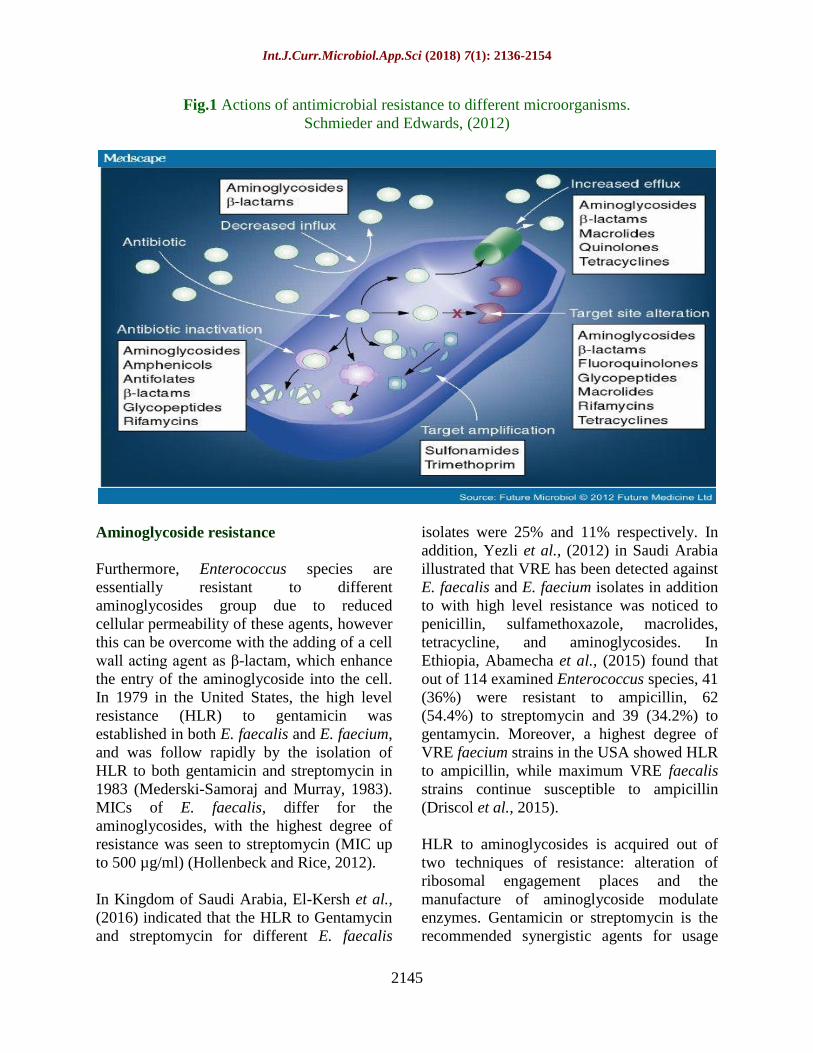
Modification of the antimicrobial target
Several microbial species can resistant
antibiotics through covering their target sites
(Figure 1). Consequently, despite the presence
of an entire and active antimicrobial complex,
no subsequent binding or inhibition will take
site Schmieder and Edwards (2012).
Modification of the antimicrobial enzymatic
activity
Ampicillin and penicillin are considered the
most common β-lactam antibiotics against
enterococci by inhibiting the synthesis of
peptidoglycan, which is one of the basic
structures of the bacterial cell wall. Penicillin-
binding proteins (PBPs) are the corner stones
for synthesis of the cell wall of microbial cells
and they can be classified into two main
groups: class A, which are functional enzymes
that composed, d-trans peptidase and trans
glycosylase, and class B, which have one the
transpeptidase range and depend on the trans
glycosylase action of other enzymes (Miller et
al., 2014).
Natural resistance
Natural resistance is the innate aptitude of a
microbial species to resist action of specific
antibiotics and concluded its characteristic
structural or useful characteristics, which
permit tolerance of a specific drug or
antimicrobial class, such as normal resistance
of E coli to penicillin (Martinez, 2002).
Acquired resistance
Acquired resistance is occurring once specific
bacteria obtain the aptitude to resist the action
of a specific antibacterial agent to which it
was earlier susceptible. van Hoek et al.,
(2011) stated that exert their action through
the mutation of genes implicated in usual
physiological procedures and cellular
constructions, from the acquirement of
external resistance genes or from a mixture of
these two mechanisms, such as when E. coli
resistant to ampicillin.
Vertical gene transfer
Assassination susceptible microorganisms
while permitting strains with resistance to that
specific antibiotic to live and grow. Characters
for such resistance are then vertically
delivered on to daughter cells thru cell
division, then making a resistant populace
which can then feast and be additional sources
of resistance genes for other strains
(Lawrence, 2005).
Horizontal gene transfer
The antibiotic resistance genes are transfer on
plasmids, transposons or integrons that can
performance as vectors that transmission these
genes to other memberships of the same
microbial species, as well as to
microorganisms in additional genus or species.
Horizontal gene transmission may arise via
three main techniques: transformation,
transduction or conjugation (Vogan and
Higgs, 2011). Antibiotic resistance has been
acquired, and has dispersed through
enterococci, via horizontal transfer of mobile
genetic elements. This transfer has been
facilitated mostly via conjugative plasmids of
the pheromone-responsive and wide host
range incompatibility group 18 types and
lately they played a significant part in
mediating transmission of vancomycin
resistance from enterococci to methicillin-
resistant strains of Staphylococcus aureus
(Palmer et al., 2010).
Int.J.Curr.Microbiol.App.Sci (2018) 7(1): 2136-2154
2143
Vancomycin resistant enterococci (VRE)
Vancomycin resistant enterococci (VRE) are a
sort of microorganisms termed enterococci
that have established resistance to numerous
antimicrobial agents, particularly vancomycin.
CDC, (2011) indicated that Enterococcus
species live in our guts and on our skin,
commonly without adverse effects
nevertheless if they develop resistant to
antibiotics, they can lead to grave infections,
particularly in societies who are ill or weak.
These infections can arise anywhere in the
body. Certain common sites contain the guts,
the urinary tract, and wounds. VRE exert its
action when it attacks the bloodstream.
Furthermore, it can be presented into a wound.
Infection is additional possible in publics with
chronic diseases like diabetes or patients who
have lately received antibiotics. It is also
further joint in patients with indwelling
devices like intravenous lines or urinary
catheters and those with compromised
immune systems. When medical isolates of
these enterococcal species with acquired
vancomycin resistance initiated to show in the
late 1980s, it encouraged significant changes
in testing of enterococci in the medical
microbiology laboratory, infection control of
enterococci, and management of enterococcal
infections (Eliopoulos and Gold, 2001).
Driscol, Crank, (2015) in USA established that
E. faecalis is considered the most common
cause of joint infections, nevertheless E.
faecium is an extra resistant to antibiotics with
a half of nosocomial isolates in the US
producing resistance to ampicillin and
vancomycin. Rendering to the National
HealthCare Safety Network (NHSN), from
2009 to 2010, (35.5%) of enterococcal
hospital related infections were resistant to
vancomycin, ranking as the 2nd
greatest public
reason of nosocomial infections in the US
(Sievert et al., 2013). In contrast, Canada has a
lesser frequency of VRE; rendering to
CANWARD, (6%) of enterococci in Canada
were resistant to vancomycin from 2007 to
2011 (Zhanel et al., 2013; Lochan et al.,
2016). In South African reported that VRE
was found in 8 of 55 patients screened.
Infected and colonized patients were isolated
in the unit throughout their admission and
strict interaction precaution infection control
applies were established. The vanA gene was
recognized in all of the isolates (Tripathi et al.,
2016).
In India found that E. faecalis (72, 61%) and
E. faecium (46, 39%). All 118 vancomycin
resistant isolates were vanA genotype
(minimum inhibitory concentration [MIC] to
vancomycin ≥64 μg/ml and MIC to
teicoplanin ≥32 μg/ml) and none of the
isolates was vanB genotype. Multivariate
logistic reversion analysis recognized
ventilator provision and hospital stay for ≥48
h as sovereign risk factors related with VR E.
faecalis and VR E. faecium infection or
colonisation. Hospital stay ≥48 h was the only
sovereign risk factor for mortality in patients
infected with vancomycin resistant
enterococci. Amberpet et al., (2016) conveyed
that Mainstream of the isolates were
Enterococcus faecium (77.2 %) followed by
Enterococcus faecalis (23.8%). All the VRE
isolates were positive for vanA gene.
Augmented period of hospital stay, younger
age, consumption of ceftriaxone and
vancomycin were established to be
significantly related with VRE colonization in
MICU. Amongst VRE colonized patients, (6,
4.5%) acquired VRE infection. Karimzadeh et
al., (2016) in Iran reported that all
Enterococcus spp. isolates within the 3 years
were resistant to oxacillin. The rate of
vancomycin resistant enterococci (VRE)
augmented from 40.63% in 2013 to 72.73% in
2015. Enterococcus spp. resistance rates to
aminoglycosides during 3 years were above
85%.Hospitalization, surgical processes, and,
particularly, lengthy or broad spectrum
antibiotic treatment may dispose patients to
Int.J.Curr.Microbiol.App.Sci (2018) 7(1): 2136-2154
2144
colonization and/or infection with antibiotic
resistant bacteria (e.g., MRSA or vancomycin-
resistant enterococci [VRE]) (Hartemann-
Heurtier et al., 2004). Vancomycin (or
glycopeptide) intermediate S. aureus has been
isolated in numerous nations. Of note, the first
2conveyedsuitcases of vancomycin resistant S.
aureus each implicated a diabetic patient with
a foot infection CDC (2002). FURLANETO-
MAIA et al., (2014) in Sao Paulo found that
all isolates of E. faecium and E. faecalis we
observed 100% arrangement. Resistance
incidences were advanced in E. faecium than
E. faecalis the resistance degrees gained were
greater for erythromycin (86.7%), vancomycin
(80.0%), tetracycline (43.35) and gentamicin
(33.3%). The relationship between disk
diffusion and automation revealed a
convention for the plurality of the antibiotics
with category agreement degrees of more than
80%. In India, Bhatt et al., (2014)
demonstrated that 14, (14.6%) out of 96
Enterocoocus spp isolates, were resistant to
vancomycin via vancomycin E test method
(MIC32mg/ml).
Phenotypes, genotypes of glycopeptide
resistance in Enterococci
Microbial cell walls are consisting of
peptidoglycan that is made once cell wall
pentapeptide precursors finish in D-Ala-D-Ala
translocate subsequently the cytoplasm to the
cell external and are combined into emerging
peptidoglycan by trans-glycosylation, creating
cross links via trans-peptidation to strengthen
the cell wall (Mainardi et al., 2008).
The main report of enterococci resistant to
great absorptions of glycopeptide
antimicrobial agents such as vancomycin and
teicoplanin was published in 1988, once
Uttley et al., indicated that the incidence of an
outbreak of vancomycin resistant E. faecium
infecting patients in a hospital renal unit. In
relation to glycopeptide resistance, there are
six phenotypes, three of which are frequently
arising. The VanA phenotype has an inducible
high level of resistance to vancomycin in
addition to teicoplanin (encoded by the VanA
gene). The VanB phenotype (encoded by two
vanB genes) has an excessive resistance to
vancomycin only. The VanC phenotype
(encoded by two vanC genes) reveals a non-
inducible squat level resistance to vancomycin
(Eliopoulos and Gold, 2001).
Van A and Van B are the greatest clinically
important phenotypes and are typically seen
amongst E. faecalis and E. faecium isolates.
Van C is both intrinsic and specific in E.
gallinarum and E. casseliflavus. Since they are
intrinsic relatively than acquired, they signify
a dissimilar impact/significance for hospital
epidemiology; final speciation can have
import for infection control tenacities.
Recently, both ampicillin and vancomycin
exert the most common resistance to E.
faecium isolates than with E. faecalis. Forbes
et al., (2014) indicated that vancomycin is
highest – E. faecium strains which have the
vanA gene. In 2002, the threat of VRE
colonization and infections improved when
the first patient case of VRE transmitting vanA
resistance genes to methicillin-resistant
Staphylococcus aureus (MRSA) to form a
vancomycin-resistant Staphylococcus aureus
(VRSA) isolate was detected (Chang et al.,
2003).
Praharaj et al., (2013) illustrated that 32 out of
367 isolates of Enterococcus species isolated,
were established resistant to vancomycin after
MIC testing. VanA was the the most common
phenotype of vancomycin resistance. An
occurrence of heterogeneity in isolates of VRE
with the vanA gene cluster with respects to
resistance to teicoplanin and the cohabitation
of vanA and vanC1 gene clusters in an isolate
of E. gallinarum which allow a high level
glycopeptide resistance to the isolate.
Int.J.Curr.Microbiol.App.Sci (2018) 7(1): 2136-2154
2145
Fig.1 Actions of antimicrobial resistance to different microorganisms.
Schmieder and Edwards, (2012)
Aminoglycoside resistance
Furthermore, Enterococcus species are
essentially resistant to different
aminoglycosides group due to reduced
cellular permeability of these agents, however
this can be overcome with the adding of a cell
wall acting agent as β-lactam, which enhance
the entry of the aminoglycoside into the cell.
In 1979 in the United States, the high level
resistance (HLR) to gentamicin was
established in both E. faecalis and E. faecium,
and was follow rapidly by the isolation of
HLR to both gentamicin and streptomycin in
1983 (Mederski-Samoraj and Murray, 1983).
MICs of E. faecalis, differ for the
aminoglycosides, with the highest degree of
resistance was seen to streptomycin (MIC up
to 500 µg/ml) (Hollenbeck and Rice, 2012).
In Kingdom of Saudi Arabia, El-Kersh et al.,
(2016) indicated that the HLR to Gentamycin
and streptomycin for different E. faecalis
isolates were 25% and 11% respectively. In
addition, Yezli et al., (2012) in Saudi Arabia
illustrated that VRE has been detected against
E. faecalis and E. faecium isolates in addition
to with high level resistance was noticed to
penicillin, sulfamethoxazole, macrolides,
tetracycline, and aminoglycosides. In
Ethiopia, Abamecha et al., (2015) found that
out of 114 examined Enterococcus species, 41
(36%) were resistant to ampicillin, 62
(54.4%) to streptomycin and 39 (34.2%) to
gentamycin. Moreover, a highest degree of
VRE faecium strains in the USA showed HLR
to ampicillin, while maximum VRE faecalis
strains continue susceptible to ampicillin
(Driscol et al., 2015).
HLR to aminoglycosides is acquired out of
two techniques of resistance: alteration of
ribosomal engagement places and the
manufacture of aminoglycoside modulate
enzymes. Gentamicin or streptomycin is the
recommended synergistic agents for usage

Int.J.Curr.Microbiol.App.Sci (2018) 7(1): 2136-2154
2146
with β-lactams to get bactericidal action.
Eliopoulos (1993) indicated that the presence
of HLR to aminoglycosides terminates the
bactericidal action found with β-lactam and
aminoglycoside synergy in medical pursuit.
An increased frequency of elevation level of
resistance to aminoglycoside antibiotics (MIC
> 8,000 μg/mL) in medical isolates of
enterococci has been described which were
also resistant to synergism with the
penicillins. Mittal et al., (2016) demonstrated
that the emergence of multidrug resistant
enterococci to frequently utilized
antimicrobial agents, e.g., aminoglycosides
and cephalosporin’s, is due to their capability
to achieve and transfer the drug resistance
gene, leading to increase the level
aminoglycoside (HLAR) and glycopeptide
resistance against enterococci. In Germany,
Werner et al., (2012) stated that 64 E.faecalis
and 37 E. faecium isolates did not show a
particular multi resistance phenotype and
resistances to glycopeptides and antibiotics.
Moreover, in India, Vinodkumar et al., (2011)
conveyed that a total of 65.6% of
Enterococcus spp. showed HLAR.
β-lactam resistance
Enterococci apply a squat level intrinsic
resistance to β-lactams due to penicillin-
binding proteins (PBPs) with a squat empathy
for these agents. Related to streptococci, E.
faecalis is 10–100-fold less sensitive to
penicillin, and matched to E. faecalis, E.
faecium is 4–16fold less susceptible.
Consequently, enterococci are tolerant to of
the various β-lactam antibiotics, Nevertheless,
if bactericidal action is required to treat severe
infections such as endocarditis or meningitis,
a synergistic bactericidal mixture of a β-
lactam with an aminoglycoside can be utilized
(Arias et al., 2010). High-level β-lactam
resistance in enterococci is primarily due to
two main techniques: the production of low-
affinity PBP5, or the production of β-
lactamases. Overproduction of PBP5 with
low-affinity compulsory to β-lactams is
distinguishing of E. faecium but unusual
amongst E. faecalis. Actually, in the US most
VRE faecium strains express high-level
resistance (HLR) to ampicillin, whereas
several VRE faecalis strains continue
sensitivity to ampicillin. The production of β-
lactamases is rare in Enterococcus species,
nevertheless they can be a precursor of HLR
by hydrolyzing β-lactams earlier they reach
their target in the cell wall. It is practically
worldwide due to E. faecalis strains and is
constitutive and inoculum dependent (Cattoir
et al., 2013).
E. faecalis chromosome does not comprise
any extra glycosyl transferase-related genes,
these comments designate that glycan chain
polymerization in the triple mutant is did by a
novel type of glycosyl transferase. The last
enzyme was not reserved by moenomycin,
subsequently deletion of the three classes A
PBP genes led to high-level resistance to this
glycosyl transferase inhibitor (Arbeloa et al.,
2004). Enterococci have an intrinsic low
vulnerability or resistance to β-lactams.
Enterococcus faecalis naturally has least
inhibitory concentricity (MICs) for penicillin
of 2–8 mg L, these significant human
microorganisms have been the topic of
intense molecular revisions, together with
Enterococcus hirae, which is more of a
veterinary anxiety (Hujer et al., 2005).
In USA, Smith et al., (2015) illustrated that
Enterococcus faecalis (Efc) and Enterococcus
faecium (Efm) are regularly resistant to
different antibiotics such as vancomycin and
β-lactams (BLs). Fifteen Efc and 20 Efm
strains were assessed for daptomycin
improvement by mixture MICs. Daptomycin
MICs were found by micro dilution in the
absence and presence of ceftaroline,
ertapenem, cefepime, ceftriaxone, cefotaxime,
cefazolin and ampicillin.
Int.J.Curr.Microbiol.App.Sci (2018) 7(1): 2136-2154
2147
Phenotypic, molecular and mass
spectrometry identification of E. faecalis
Phynotypic identification by Vitek2
compact system
VITEK 2 cards which includes tests for
Antimicrobial Susceptibility Testing (AST),
which are FDA approved. The Vitek 2 AST
(BioMe´rieux Vitek 2, France) uses
Cefotaxime and Ceftazidime, only (at 0.5
μg/mL) and in mixture with Clavulanic acid
(4 μg/mL). Inoculation of the cards is
identical to that performed for steady VITEK
2 cards. Analysis of all wells is achieved
mechanically once the growth control well
has got a set threshold (4-15 hours of
incubation). A predetermined reduction in the
growth of the Cefotaxime or Ceftazidime
wells comprising Clavulanic acid, compared
with the level of growth in the well with the
Cephalosporin alone, shows occurrence of
ESBLs. Sensitivity and specificity of the
technique out do 90% (Winstanley and
Courvalin, 2011).
Molecular identification by PCR assay
The aim of the polymerase chain reaction
(PCR) is to identify and describe genes. PCR
is an in vitro technique for amplifying a DNA
sequence via a heat stable polymerase and
two primers, one complementary to the (+)
strand at one end of the sequence to be
amplified and the other complementary to the
(+) strand at the other end. The recently
synthesized DNA strands then assist as
templates for the similar primers and
succeeding rounds of primer annealing; strand
elongation and dissociation harvest a greatly
particular amplification of the sequence. PCR
can be used in ecological observing assays to
discover the presence or nonexistence of a
DNA sequence in a sample, for example, a
gene particular for an infectious viral particle
or bacterium (Olsen, 2012). It is developed by
Kary Mullis in the 1980s. PCR is established
on using the capability of DNA polymerase to
manufacture new strand of DNA
complementary to the existing template
strand. In order to DNA polymerase can add a
nucleotide one onto a preexisting 3'-OH
group; it requests a primer to which it can add
the first nucleotide. This obligation makes it
potential to delineate a particular area of
template sequence that the investigator wants
to amplify. Aboud et al., (2013) indicated that
at the ending of the PCR response, the
particular sequence will be accrued in billions
of copies (amplicons).
By way of the heat, degree in the tube passes
into the Tm range and settles at the Ta
temperature, the greatest probable number of
primer molecules relative to the number of
obtainable targets will have found those
targets and will lay down in stable duplexes
(Carr and Moore, 2012). Agarose gel
electrophoresis is employed for size parting of
the PCR output. The size(s) of PCR products
is determined by comparison with a DNA
ladder (a molecular weight marker), which
comprises DNA fragments of identified size,
run on the gel alongside the PCR products
(Lee et al., 2012). In Germany, Werner et al.,
(2012) described that Molecular typing of the
64 isolates of E. faecalis showed three PFGE
clusters of associated strains represented by 3
MLST types (ST40, ST211, ST268). In
China, Sun et al., (2012) demonstrated that E.
faecalis showed vancomycin and teicoplanin
MIC results at ≥256 μg/mL and harbored
vanA, excluding for 1vanB-carrying strain
(MIC, 32 and 1 μg/mL, correspondingly).In
addition, in USA, Ferguson et al., (2016)
stated that multiplex PCR was used to liken
the delivery of virulence genes amongst E.
faecalis and E. faecium isolated from coasts
in Southern California and Puerto Rico to
isolates from potential foundations counting
humans, animals, birds, and plants. All 5
virulence genes were discovering in E.
Int.J.Curr.Microbiol.App.Sci (2018) 7(1): 2136-2154
2148
faecalis and E. faecium from coastline water,
typically amongst E. faecalis. gelE was the
maximum joint amongst isolates from all
exporters types. In Iran, Honarm et al., (2012)
established that routine analysis and PCR for
all inoculated blood samples with ≥5 cfu/ml
was positive. Meanwhile for PCR and routine
assays was ten hours and five days,
respectively PCR is a further rapid and
sensitive assay for simultaneous discovery
and description for E.faecalis, and
determination of its sensitivity pattern to
vancomycin.
In Germany, Dalpke et al., (2016) stated that
the susceptibilities of the various PCR
formats were 84 to 100% for vanA and 83.7 to
100% for vanB; specificities were 96.8 to
100% for vanA and 81.8 to 97% for vanB. In
China, He et al., (2016) indicated that nine
optrA-carrying plasmids were conjugated into
E. faecalis JH2-2 and the trans conjugants
exhibited the optrA-associated phenotype.
The specific and rapid detection and
quantification of ace, esp and gelE genes
compared to conventional PCR assays, thus
allowing the rapid and direct safety
assessment of Enterococcus genus in food
samples (Abouelnage et al., 2016). In Serbia,
Stojanovic et al., (2014) indicated that E.
faecalis was discovered in 49% (25/51),
When individuality was made between the
intracanal medications, there was an
important variance in the number of PCR
positive samples between S1 and S2, S1 and
S3, but not between S2 and S3 samples.
FURLANETO-MAIA L et al., (2014) in Sao
Paulo found that the PCR-based assay, the
van (A) gene was detected in 100% of
vancomycin resistant enterococci. This
evaluation is simple to conduct and steadfast
in the identification of clinically pertinent
enterococci. The data acquired supported the
requirement for a development of the
automated system to detect some enterococci.
Proteomic identification by Mass Matrix
assisted laser desorption/ionization (MALDI)
The term matrix assisted laser desorption
ionization (MALDI) was invented in 1985 by
Franz Hillenkamp, Michael Karas and their
colleagues (Karas et al., 1985). Based bio-
typing is an emerging method for high
throughput and quick bacterial ID. Due to its
comparatively greater accuracy, inclusive
database of clinically significant bacteria and
low price compared to other bacterial ID
techniques, MALDI has started changing
current applies prevalent in clinical diagnosis.
Nevertheless, applicability of MALDI in the
area of bacterial research is still partial mostly
due to the absence of data on non-clinical
bacteria (Alatoom et al., 2011). Matrix-
Assisted Laser Desorption/Ionization Time-
of-Flight Mass-Spectrometry (MALDI -TOF
MS) is a quick and reliable method for
microbial identification as most results gotby
this are like to that of 16S rRNA gene
sequence analysis but at a quick rate and at a
lesser price. This method is based on
fingerprinting analyses of mainly ribosomal
proteins, which are manufactured under all
growth circumstances and are the most
plentiful cellular proteins (Rahi et al., 2016).
MALDI-TOF MS has been used to
characterize a wide variety of bacteria
including bacteria, fungi, and viruses the
competence of MALDI-TOF to quickly
characterize bacteria favors its potential uses
in multiple areas including medical
diagnostics, biodefense, ecological
monitoring, and food quality control.
MALDI-TOF MS is appropriate for high-
throughput and quick bacterial identification
at low prices and is a different for
conventional laboratory biochemical and
molecular identification systems (Giebel et
al., 2010; ElBehiry et al., 2014; Elbehiry et
al., 2016). In Germany, Werner et al., (2012)
indicated that the conventional and MALDI

Int.J.Curr.Microbiol.App.Sci (2018) 7(1): 2136-2154
2149
TOF MS analyses identified 64 Enterococcus
faecalis and 37 Enterococcus faeciumisolates,
which were confirmed by species-specific
PCRs. In Zagreb, Dobranic et al., (2016)
stated that MALDI-TOF MS identification
presented 100% concordance with API 20
Strep in the identification of Enterococcus
faecalis.
References
Abamecha, A., Wondafrash, B., and Abdissa, A.
2015. Antimicrobial resistance profile of
Enterococcus species isolated from intestinal
tracts of hospitalized patients in Jimma,
Ethiopia. BMC Research. 8:213
Aboud, M., Oh, H.H., McCord, B. 2013. Rapid
direct PCR for forensic genotyping in under
25 min. Electrophoresis.34(11):1539–1547.
Abouelnage, M., Lamas, A., Guarddon, M.,
Osman, M., Miranda, M., and Cepeda, A.
2016. Assessment of food safety using a new
real-time PCR assay for detection and
quantification of virulence factors of
enterococci in food samples. JOAM. DOI:
10.1111/jam.13306.
Alatoom, A. A., Cunningham, S. A., Ihde, S. M.,
Mandrekar, J., and Patel, R. 2011.
Comparison of direct colony method versus
extraction method for identification of gram-
positive cocci by use of Bruker Biotyper
matrix-assisted laser desorption ionization-
time of flight mass spectrometry. J. Clin.
Microbiol. 49: 868–2873.
Alzahrani, O.H., Badahdah, Y.S., Bamakrid,
M.S., Alfayez, A.S., Alsaeedi, M.S.,
Mansouri, A.M., and Alzahrani, A.H. 2013.
The Diabetic Foot Research in Arabs’
Countries. OJEMD. 3(3): 157-165.
Amberpet, R., Sistla, S., Parija, S.M., and Thabah,
M.M. 2016. Screening for Intestinal
Colonization with Vancomycin Resistant
Enterococci and Associated Risk Factors
among Patients Admitted to an Adult
Intensive Care Unit of a Large Teaching
Hospital. JCDR. 10(9): 6–9. Anvarinejad, M., Pouladfar, G., Japoni, A.,
Bolandparvaz, S., Satiary, Z., Abbasi, P., and
Mardaneh, J. 2015. Isolation and Antibiotic
Susceptibility of the Microorganisms Isolated
from Diabetic Foot Infections in Nemazee
Hospital, Southern Iran. Journal of
Pathogens.ID 3287967
Arbeloa, A., Segal, H., Hugonnet, J.E.,
Josseaume, N., Dubost, L., Brouard, J.P.,
Gutmann, L., Mengin-Lecreulx, D., and
Arthur, M. 2004. Role of class A penicillin-
binding proteins inPBP5-mediated beta-
lactam resistance in Enterococcus faecalis. J
Bacteriol.186(5):1221-8.
Arias, C.A., Contreras, G.A., and Murray, B.E.
2016. Management of multidrug-resistant
enterococcal infections. Clin Microbiol
Infect.16(6):555–562.
Berlanga, J., Cibrian, D., Guillén, I., Freyre, F.,
Alba, J.S., and López-Saura, P. 2005.
Methylglyoxal administration induces
diabetes-like microvascular changes and
perturbs the healing process of cutaneous
wounds. ClinSci (Lond), 109(1):83-95.
Bhatt, M., Sahni, B., Praharaj, S., Grover, C.,
Kumar, C., Chaudhari, S., and Khajuria, A.
2014. Detection of glycopeptide resistance
genes in enterococci by multiplex PCR.
MJAFI. 71 (1):43–47.
Biswas P.P., Aninda, and Sen, S.D. 2016.
Molecular Characterization of Virulence
Genes in Vancomycin-Resistant and
Vancomycin-Sensitive Enterococci. J Glob
Infect Dis. 8(1): 16–24
Brechow, A., Slesaczeck, T., and Münch, D.
2013. Improving major amputation rates in
the multicomplex diabetic foot patient: focus
on the severity of peripheral arterial disease.
Therapeutic Advances in Endocrinology and
Metabolism. 4 (3): 83–94.
Cardona, A.F., and Wilson, S.E. 2015. Skin and
Soft-Tissue Infections: A Critical Review and
the Role of Telavancin in Their Treatment.
Clin Infect Dis. 61 (2): 69-78.
Carr, A.C., Moore, S.D. 2012. Lucia, Alejandro,
ed. "Robust quantification of polymerase
chain reactions using global fitting". PLOS
ONE. 7 (5): e37640.
Cattoir, V., and Leclercq, R. 2013. Twenty-five
years of shared life with vancomycin-
resistant enterococci: is it time to divorce? J
Antimicrob Chemother.68(4):731–742.
Centers for Disease Control and Prevention. 2011.
VRE in Healthcare Settings, Retrieved from

Int.J.Curr.Microbiol.App.Sci (2018) 7(1): 2136-2154
2150
https://www.cdc.gov/hai/organisms/vre/vre.ht
ml
Centers for Disease Control Preventio.
Vancomycin-resistant Staphylococcus
aureus—Pennsylvania, 2002. MMWR Morb
Mortal Wkly Rep. 51:902
Chang, S., Sievert, D.M., and Hageman, J.C.
2003. Infection with vancomycin-resistant
Staphylococcus aureus containing the vanA
resistance gene. N Engl J Med.348(14):1342–
1347.
Chouchania, C., El Salabib, A., Marrakchia, R.,
Ferchichid, L., and Walshb, T.R. 2012. First
report of mefA and msrA/msrB multidrug
efflux pumps associated with blaTEM-1 β-
lactamase in Enterococcus faecalis.
International Journal of Infectious Diseases.
16(2):104–109.
Comerlato, C.B., de Resende, M.C., Caierao, J.,
and Azevedo, P.A. 2013. Presence of
virulence factors in Enterococcus faecalis
and Enterococcus faecium susceptible and
resistant to vancomycin. Mem Inst Oswaldo
Cruz. 108(5): 590–595.
Dalpke, A.H., Hofko, M., and Zimmermann, S.
2016. Development of a Real-Time PCR
Protocol Requiring Minimal Handling for
Detection of Vancomycin-Resistant
Enterococci with the Fully Automated BD
Max System. J. Clin. Microbiol. 54 (9):
2321-2329.
Davies, J. and Davies, D. 2010. Origins and
Evolution of Antibiotic Resistance.
Microbiol. Mol. Biol. Rev. 74 (3): 417-433.
Den Engelsen, C., van der Werf, C., Matute, A.J.,
Delgado, E., Schurink, C.A., and Hoepelman,
A.I. 2009. Infectious diseases and the use of
antibiotics in outpatients at the emergency
department of the University Hospital of
León, Nicaragua. Int J Infect. 3(3):349-354.
Dobranic, V., Kazazic, S., Filipovic, I., Mikulec,
N., and Zdolec, N. 2016. Composition of raw
cow’s milk microbiota and identification of
enterococci by MALDI-TOF MS - short
communication. Veterinarski arhiv. 86 (4):
581-590.
Doria, M., Rosado, V., Pacheco, L.R., Hernández,
M., Betriu, A., Valls, J., Franch-Nadal, J.,
and Fernández Mauricio, D. 2016.
Prevalence of Diabetic Foot Disease in
Patients with Diabetes Mellitus under Renal
Replacement Therapy in Lleida, Spain.
Biomed Res Int., ID 7217586.
Driscol, T., and Crank, C. 2015. Vancomycin-
resistant enterococcal infections:
epidemiology, clinical manifestations, and
optimal management. Infect Drug Resist. 8:
217–230.
El Behiry A., Zahran R.N., Marzouk E., Al-Dabib
M. 2014. Phenotypical and mass spectral
assessment methods for identification of
some contagious mastitis pathogens.
American J Microbiol. 5: 1-10.
Elbehiry, A., Al-Dubaib, M., Marzouk, E.,
Osman, S., and Edrees, H. 2016.
Performance of MALDI biotyper compared
with Vitek (™) 2 compact system for fast
identification and discrimination of
Staphylococcus species isolated from bovine
mastitis Microbiologyopen. 5(6): 1061–1070.
Eliopoulos, G.M. 1993. Aminoglycoside resistant
enterococcal endocarditis. Infect Dis Clin
North Am.7(1):117–133.
Eliopoulos, G.M., and Gold, H.S. 2001.
Vancomycin-Resistant Enterococci:
Mechanisms and Clinical Observations. Clin
Infect Dis.33 (2): 210-219.
El-Kersh, T.A., Marie, M.A., Al-Sheikh, Y.A.,
Al-Agamy, M.H., and Al-Bloushy, A.A.
2016. Prevalence and risk factors of early
fecal carriage of Enterococcus faecalis and
Staphylococcus spp and their antimicrobial
resistant patterns among healthy neonates
born in a hospital setting in central Saudi
Arabia. Saudi Med J. 37(3): 280–287.
El-Tahawy, A.T. 2000. Bacteriology of diabetic
foot infections., MD, PhD (UK), Saudi
Medical Journal. 21 (4): 344-347.
Enterococcus faecalis and Porphyromonas
gingivalis in Infected Root Canals and Their
Susceptibility to Endodontic Treatment
Procedures: A Molecular Study. Srp Arh
Celok Lek. 142(9-10):535-541.
Ferguson, D.M., Talavera, G.N., Hernández, L.R.,
Weisberg, S.B., Ambrose, R.F., and Jay, J.A.
2016. Virulence Genes among Enterococcus
faecalis and Enterococcus faecium Isolated
from Coastal Beaches and Human and
Nonhuman Sources in Southern California
and Puerto Rico. Journal of Pathogens. ID
3437214:7.
Int.J.Curr.Microbiol.App.Sci (2018) 7(1): 2136-2154
2151
Fisher, K., and Phillips, C. 2009. The ecology,
epidemiology and virulence of Enterococcus.
Microbiology. 155(6):1749-57.
Forbes, B.A., Sahm, D.F., and Weissfeld, A.S.
2014. Diagnostic Microbiology. 13th ed.
Mosby.
Furlaneto-maia, L., Rocha, K., Siqueira, V., and
Furlaneto, M. 2014. Comparison between
automated system and pcr-based method for
identification and antimicrobial susceptibility
profile of clinical Enterococcus spp. Rev Inst
Med Trop Sao Paulo. 56(2):97-103.
Giebel, R., Worden, C., Rust, S.M., Kleinheinz,
G.T., Robbins, M. and Sandrin, T.R. 2010.
Microbial fingerprinting using matrix-
assisted laser desorption ionization time-of-
flight mass spectrometry (MALDI-TOF MS)
applications and challenges. Adv Appl
Microbiol. 71: 149–184. Gilmore, M.S., Clewell, D.B., Courvalin, P.,
Dunny G.M., Murray B.E., and Louis B. Rice
L.S. 2002. The Enterococci: Pathogenesis,
Molecular Biology, and Antibiotic Resistance
and Infection Control. ISBN: 978-1-55581-
234-8.
Giridhara Upadhyaya, P. M., Umapathy, B. L.,
and Ravikumar, K. L. 2010. Comparative
Study for the Presence of Enterococcal
Virulence Factors Gelatinase, Hemolysin and
Biofilm Among Clinical and Commensal
Isolates of Enterococcus faecalis. J Lab
Physicians. 2(2): 100–104.
Gooday, C., Murchison, R., and Dhatariya, K.
2014. Complex relationships requiring long-
term follow-up: Obesity, bariatric surgery-
induced diabetic remission, and the diabetic
foot. The Diabetic Foot Journal. 17 (1): 20–4.
Hartemann-Heurtier, A. J., Robert, S.,
Jacqueminet R. 2004. Diabetic foot ulcer and
multidrug-resistant organisms: risk factors
and impact. Diabet. Med. 21(7):710-5.
Hartemann-Heurtier, A., Robert, J., and
Jacqueminet, S. 2004. Diabetic foot ulcer and
multidrug-resistant organisms: risk factors
and impact. Diabet Med. 21:710-5.
He, T., Shen, Y., Schwarz, S., Cai, J., Lv, Y., Li,
J., FeBler, A., and Zhang, R. 2016. Genetic
environment of the transferable
oxazolidinone/phenicol resistance gene optrA
in Enterococcus faecalis isolates of human
and animal origin. J. Antimicrob. Chemother.
doi: 10.1093.
Hollenbeck, B.L. and Rice, L.B. 2012. Intrinsic
and acquired resistance mechanisms in
Enterococcus. Virulence.15. 3(5): 421–569.
Honarm, H., Ghavidel, M.F., Nikokar, I.,
Taromsari, M.R. 2012. Evaluation of a PCR
Assay to Detect Enterococcus faecalis in
Blood and Determine Glycopeptides
Resistance Genes: Van A and Van B. Iran J
Med Sci. 37(3): 194–199.
Hujer, A.M., Kania, M., Gerken, T., Anderson,
V.E., Buynak, J.D., Ge, X., Caspers, P., Page,
M.G., Rice, L.B.and Bonomo, R.A. 2005.
Structure-activity relationships of different β-
lactam antibiotics against a soluble form of
Enterococcus faecium PBP5, a type II
bacterial transpeptidase. Antimicrob Agents
Chemother.49: 612–618.
John, V.U., and Carvalho, J. 2011. Enterococcus:
review of its physiology, pathogenesis,
diseases and the challenges it poses for
clinical microbiology. J. Front. Biol., 6: 357.
Karas, M., Bachmann, D., and Hillenkamp, F.
1985. "Influence of the Wavelength in High-
Irradiance Ultraviolet Laser Desorption Mass
Spectrometry of Organic Molecules".
Analytical Chemistry. 57 (14): 2935–9.
Karimzadeh, I., Mirzaee, M. N. Sadeghimanesh
Sagheb, M.M. 2016. Antimicrobial resistance
pattern of Gram-positive bacteria during
three consecutive years at the nephrology
ward of a tertiary referral hospital in Shiraz,
Southwest Iran. JRPP. 5(4): 238-247.
Klibi, N., Ben Slama, K., Sáenz, Y., Masmoudi,
A., Zanetti, S., Sechi, L.A., Boudabous, A.,
and Torresb, C. 2007. Detection of virulence
factors in high-level gentamicin-resistant
Enterococcus faecalis and Enterococcus
faecium isolates from a Tunisian hospital.
Canadian Journal of Microbiology. 53(3):
372-379.
Koch, S., Hufnage, M., Theilacker, C., and
Huebner, J. 2004. Enterococcal infections:
host response, therapeutic, and prophylactic
possibilities. Vaccine, 22:822–30.
Kow, N., and Ferzandi, T.R. 2014. Enterococcus
osteomyelitis secondary to pyelonephritis.
International Urogynecology Journal.
24(4):691–692.

Int.J.Curr.Microbiol.App.Sci (2018) 7(1): 2136-2154
2152
Lawrence, J.G. 2005. Horizontal and Vertical
Gene Transfer: The Life History of
Pathogens. Contrib. Microbiol. 12: 255-271.
Lebreton, F., Riboulet-Bisson, E., Serror, P.,
Sanguinetti, M., Posteraro, B., Torelli, R.,
Hartke, A., Auffray, Y., and Giard, J.C. 2009.
Ace, Which encodes an adhesin in
Enterococcus faecalis, is regulated by Ers
and is involved in virulence. Infect Immun.
77(7):2832-9.
Lee, P.Y., Costumbrado, J., Hsu, C.Y., and Kim,
Y.H. 2012. Agarose Gel Electrophoresis for
the Separation of DNA Fragments. J. Vis.
Exp. (62), e3923.
Lipsky, B.A., Richard, J.L., and Lavigne, J.P.
2013. Diabetic foot ulcer microbiome: one
small step for molecular microbiology One
giant leap for understanding diabetic foot
ulcers? Diabetes. 62(3):679-81.
Lochan, H., Moodle, C., Rip, D., Bamford, C.,
Hendricks, M., Davidson, A., and Eley, B.
2016. Emergence of vancomycin-resistant
Enterococcus at a tertiary paediatric hospital
in South Africa. South African Medical
Journal. 106(6):562-566.
Mainardi, J.L., Villet, R., Bugg, T.D., Mayer, C.,
and Arthur, M. 2008. Evolution of
peptidoglycan biosynthesis under the
selective pressure of antibiotics in Gram-
positive bacteria. FEMS Microbiol
Rev.32(2):386–408.
Malachowa, N. and DeLeo, F.R. 2010. Mobile
genetic elements of Staphylococcus aureus.
Cell Mol Life Sci. 67(18): 3057–3071.
Martinez, J.L. and Baquero, F. 2002. Interactions
among Strategies Associated with Bacterial
Infection: Pathogenicity, Epidemicity, and
Antibiotic Resistance. Clin. Microbiol. Rev.
15(4): 647–679.
Mathangi, T., and Prabhakaran, P. 2013.
Prevalence of Bacteria Isolated from Type 2
Diabetic Foot Ulcers and the Antibiotic
Susceptibility Pattern.
Int.J.Curr.Microbiol.App. 2: 329-337.
McInnes, A.D. 2012. Diabetic foot disease in the
United Kingdom: about time to put feet first,
Journal of Foot and Ankle Research. 26 (5):
1757-1146.
Medeiros, A.W., Pereira, R.I., Oliveira, D.V.,
Martins, P.D., dAzevedo, P.A., Van der
Sand, S., Frazzon, J., and Frazzon, A.P. 2014.
Molecular detection of virulence factors
among food and clinical Enterococcus
faecalis strains in South Brazil. Braz J
Microbiol. 45(1): 327–332.
Mederski-Samoraj, B.D., and Murray, B.E. 1983.
High-level resistance to gentamicin in
clinical isolates of enterococci. J Infect
Dis.147(4):751–757.
Miller, W.R., Munita, J.M., and Arias, C.A. 2014.
Mechanisms of antibiotic resistance in
enterococci. Expert Rev Anti Infect Ther.
12(10): 1221–1236.
Miro, J.M., Pericas, J.M., and Rio, A.D. 2013. A
New Era for Treating Enterococcus Faecalis
Endocarditis: Ampicillin plus Short-Course
Gentamicin or Ampicillin plus Ceftriaxone;
that is the Question! Published online. doi:
10.1161/CIRCULATIONAHA.113.002431.
Mittal, S., Singla, P., Deep, A., Bala, K., Sikka,
R., Garg, M., and Chaudhary, U. 2016.
Vancomycin and High Level
Aminoglycoside Resistance in Enterococcus
spp. in a Tertiary Health Care Centre: A
Therapeutic Concern. Journal of Pathogens.
ID 8262561.
Moses, V., Jerobin, J., Nair, A., Sathyendara, S.,
Balaji, V., George, A., and Peter, J.V. 2012.
Enterococcal Bacteremia is Associated with
Prolonged Stay in the Medical Intensive Care
Unit. J Glob Infect Dis., 4(1): 26–30.
Olawale, K.O., Fadiora, S.O., and Taiwo, S.S.
2011. Prevalence of Hospital-Acquired
Enterococci Infections in Two Primary-Care
Hospitals in Osogbo, Southwestern Nigeria.
Afr J Infect Dis., 5(2): 40–46.
Olsen, J.L. 2012. Polymerase Chain Reaction.
Encyclopedia of Immunotoxicology. 715-
720.
Palmer, K.L., Kos, V.N., and Gilmore, M.S. 2010.
Horizontal gene transfer and the genomics of
enterococcal antibiotic resistance. Curr Opin
Microbiol. 13(5):632-9.
Perich, A.P., González, R.M., Valdés, E., and
Arranz, M.C., 2010. Desarrollo de diabetes
mellitus en pacientes con tolerancia a la
glucosaalterada: Seguimiento de 18 años.
Rev Cubana Endocrinol, 13(2): 0-0.
Praharaj, I., Sujatha, S., Parija, S. 2013.
Phenotypic & genotypic characterization of
vancomycin resistant Enterococcus isolates

Int.J.Curr.Microbiol.App.Sci (2018) 7(1): 2136-2154
2153
from clinical specimens. Indian J Med
Res.138(4): 549–556.
Quilici, M.T., Del Fiol, F., Vieira, A.E., and
Toledo, M.I. 2016. Risk Factors for Foot
Amputation in Patients Hospitalized for
Diabetic Foot Infection. Journal of Diabetes
Research, ID 8931508, 8.
Rahi, P., Prakash, O., and Shouche, Y. 2016.
Matrix-Assisted Laser Desorption/Ionization
Time-of-Flight Mass-Spectrometry (MALDI-
TOF MS) Based Microbial Identifications:
Challenges and Scopes for Microbial
Ecologists. Front Microbiol. 7: 1359.
Raja, N.S. 2007. Microbiology of diabetic foot
infections in a teaching hospital in Malaysia:
a retrospective study of 194 cases. J
Microbiol. Immunol. Infect.40(1):39-44.
Rajkumari, N., Mathur, P., and Misra, M.C. 2014.
Soft tissue and wound infections due to
Enterococcus spp. among hospitalized
trauma patients in a developing country. J
Global Infect Dis. (6):189-93.
Ray, G.T., Suaya, J.A., and Baxter, R. 2013.
Incidence, microbiology, and patient
characteristics of skin and soft-tissue
infections in a U.S. population: a
retrospective population-based study BMC
Infectious Diseases, 13:252.
Rengaraj, R., Mariappan, S., Sekar, U., and
Kamalanadhan, A. 2016. Detection of
Vancomycin
Resistance among Enterococcus faecalis and
Staphylococcus aureus. J Clin Diagn Res.
10(2): 4-6.
Rezende, K.F., Ferraz, M.B., and Malerbi, D.A.
2010. ―Predicted annual costs for in patients
with diabetes and foot ulcers in a developing
country—a simulation of the current situation
in Brazil,‖ Diabetic Medicine. 27(1):109–
112.
Saba Copur, S., Şahin, F., and Goçmen, J.S. 2016.
Determination of virulence and multidrug
resistance genes with polymerase chain
reaction method in vancomycin sensitive and
resistant enterococci isolated from clinical
samples. Turk J Med Sci. 46(3):877-91. Santos, I.C., Sobreira, C.M., Nunes, E.N., and
Morais, M.C. 2013. ―The prevalence and
factors associated with diabetic foot
amputations,‖ Ciencia e Saude Coletiva.18
(10):3007–3014.
Schmieder, R., and Edwards, R. 2012. Insights
into Antibiotic Resistance through
Metagenomic Approaches. Future Microbiol.
7(1):73-89.
Semedo-Lemsaddek, T., Mottola, C., Alves-
Barroco, C., Cavaco-Silva, P., Tavares, L.,
and Oliveira M. 2016. Characterization of
multidrug-resistant diabetic foot ulcer
enterococci. Enferm Infecc Microbiol Clin.,
34(2):114–6.
Shankar, V., Baghdayan, A.S., Huycke, M.M.,
Lindahl, G., and Gilmore, M.S. 1999.
Infection-Derived Enterococcus
faecalisStrains Are Enriched in esp, a Gene
Encoding a Novel Surface Protein. Infect.
Immun. 67(1): 193-200.
Sievert, D.M., Ricks, P., Edwards, J.R.,
Schneider, A., Patel, J., Srinivasan, A.,
Kallen, A., Limbago, B., and Fridkin, S.
2013. National Healthcare Safety Network
(NHSN) Team and Participating NHSN
Facilities. Infect Control Hosp Epidemiol.
34(1):1-14.
Smith, J.R., Barber, K.E., Raut, A., Aboutaleb,
M., Sakoulas, G., and Rybak, M.J. 2015. β-
Lactam combinations with daptomycin
provide synergy against vancomycin resistant
Enterococcus faecalis and Enterococcus
faecium. J Antimicrob Chemother.; 70(6):
1738-43.
Soheili, S., Ghafourian, S., Sekawi, Z., Neela, V.,
Sadeghifard, N., Ramli, R., and Hamat, R.A.
2014. Wide Distribution of Virulence Genes
among Enterococcus faecium and
Enterococcus faecalis Clinical Isolates. The
Scientific World Journal. (2014) ID 623174:6 Stalker, M.J., Brash, M.L., Weisz, A., Ouckama,
R.M., and Slavic, D. 2010. Arthritis and
osteomyelitis associated with Enterococcus
cecorum infection in broiler and broiler
breeder chickens in Ontario, Canada Stalkerc.
J Vet Diagn.22:643–645.
Stevens, D.L., Bisno, A.L., Chambers, H.F.,
Dellinger, E.P., Goldstein, E.J., and Gorbach,
S.L. 2014. Practice guidelines for the
diagnosis and management of skin and soft
tissue infections: 2014 update by the
Infectious Diseases Society of America. Clin
Infect Dis. 59 (2):147-59.
Stojanovic, N., Krunic, J., Popovic, B., Stojicic,
S., and Zivkovic, S. 2014. Prevalence of

Int.J.Curr.Microbiol.App.Sci (2018) 7(1): 2136-2154
2154
Sun, H., Wang, H., Xu, Y., Jones, R.N., Costello,
A.J., Liu, Y., Li, G., Chen, M., and Mendes,
R.E. 2012. Molecular characterization of
vancomycin-resistant Enterococcus spp.
clinical isolates recovered from hospitalized
patients among several medical institutions in
China. Diagn Microbiol Infect Dis.74(4):399-
403.
Tripathi, A., Shukla, S.K., Singh, A., and Prasad,
K.N. 2016. Prevalence, outcome and risk
factor associated with vancomycin-resistant
Enterococcus faecalis and Enterococcus
faecium at a Tertiary Care Hospital in
Northern India. Indian Journal of Medical
Microbiology. 34(1): 38-45.
Turhan, V., Mutluoglu, M., Acar, A., Hatipoglu,
M., Onem, Y., Uzun, G., Ay, H., Oncul, O.,
and Gorenek, L. 2013. Increasing incidence
of Gram-negative organisms in bacterial
agents isolated from diabetic foot ulcers.
JIDC. 10(7): doi:10.3855/jidc.2967.
Van Hoek, A., Mevius, D., Guerra, B., Mullany,
P., Roberts, A.P. and Aarts, H.J.M. 2011.
Acquired Antibiotic Resistance Genes: An
Overview. Front Microbiol. 2(203):
doi:0.3389/fmicb.2011.00203.
Vinodkumar,.CS., Srinivasa, H., Basavarajappa,
K.G., Geethalakshmi, S., and Bandekar, N.
2011. Isolation of bacteriophages to multi-
drug resistant Enterococci obtained from
diabetic foot: a novel antimicrobial agent
waiting in the shelf?. Indian J Pathol
Microbiol.54(1):90-5.
Vinodkumar, C.S., Srinivasa, H., Basavarajappa,
K.G., Geethalakshmi, S., and Bandekar, N.
2011. Isolation of bacteriophages to multi-
drug resistant Enterococci obtained from
diabetic foot: a novel antimicrobial agent
waiting in the shelf? Indian J Pathol
Microbiol. 54(1):90-5.
Vogan, A.A. and Higgs, P.G. 2011. The
advantages and disadvantages of horizontal
gene transfer and the emergence of the first
species. Biology Direct. 6(1): doi:
10.1186/1745-6150-6-1.
Werner, G., Fleige, C., Fessler, A.T., Timke, M.,
Kostrzewa, M., Zischka, M., Peters, T.
Kaspar, H., and Schwarz, S. 2012. Improved
identification including MALDI-TOF mass
spectrometry analysis of group D
streptococci from bovine mastitis and
subsequent molecular characterization of
corresponding Enterococcus faecalis and
Enterococcus faecium isolates. Vet
Microbiol.160(1-2):162-9.
Winstanley, T. and Courvalin, P. 2011. Expert
Systems in Clinical Microbiology. Clin.
Microbiol. Rev. 24 (3): 515-556.
Yameen, M.A., Iram, S., Mannan, A., Khan, S.A.,
and Akhtar, N. 2013. Nasal and perirectal
colonization of vancomycin sensitive and
resistant enterococci in patients of paediatrics
ICU (PICU) of tertiary health care facilities.
BMC Infect Dis. 13:156. Yezli, S., Shibl, A.M., Livermore, D.M., and
Memish, Z.A. 2012. Antimicrobial resistance
among Gram-positive pathogens in Saudi
Arabia. J Chemother. 24(3):125-36.
Zemann, W., Feichtinger, M., Pau, M., and
Kärcher, H. 2011. Primary osteomyelitis of
the mandibular condyle—a rare case. Oral
and Maxillofacial Surgery. 15(2): 109–111.
Zhanel, G.G., Adam, H.J., and Baxter, M.R. 2013.
Canadian Antimicrobial Resistance Alliance.
Antimicrobial susceptibility of 22746
pathogens from Canadian hospitals: results of
the CANWARD 2007–2011 study. J
Antimicrob Chemother. 68(1): 7–22.
How to cite this article:
Ahmad Abdulrahman AL Bloushy and Ayman Elbehiry. 2018. History, Virulence Genes,
Identification and Antimicrobial Resistance of Enterococcus faecalis Isolated from Diabetic
Foot Patients. Int.J.Curr.Microbiol.App.Sci. 7(01): 2136-2154.
doi: https://doi.org/10.20546/ijcmas.2018.701.258