hkmd vol.21 no 6 june 2016
TRANSCRIPT
VOL.21 NO.6 June 2016
OFFICIAL PUBLICATION FOR THE FEDERATION OF MEDICAL SOCIETIES OF HONG KONG ISSN 1812 - 1691
Dentistry
1
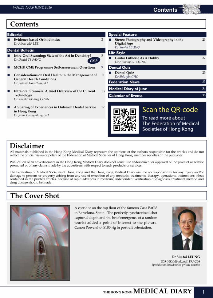
VOL.21 NO.6 JUNE 2016 Contents
Contents
The Cover Shot
Disclaimer All materials published in the Hong Kong Medical Diary represent the opinions of the authors responsible for the articles and do not reflect the official views or policy of the Federation of Medical Societies of Hong Kong, member societies or the publisher.
Publication of an advertisement in the Hong Kong Medical Diary does not constitute endorsement or approval of the product or service promoted or of any claims made by the advertisers with respect to such products or services.
The Federation of Medical Societies of Hong Kong and the Hong Kong Medical Diary assume no responsibility for any injury and/or damage to persons or property arising from any use of execution of any methods, treatments, therapy, operations, instructions, ideas contained in the printed articles. Because of rapid advances in medicine, independent verification of diagnoses, treatment method and drug dosage should be made.
A corridor on the top floor of the famous Casa Batlló in Barcelona, Spain. The perfectly synchronised shot captured depth and the brief emergence of a random tourist added a point of interest to the picture. Canon Powershot S100 rig in portrait orientation.
Editorialn Evidence-based Orthodontics
Dr Albert MP LEE2
Dental Bulletinn Intra-Oral Scanning: State of the Art in Dentistry?
Dr Daniel TS FANG5
n MCHK CME Programme Self-assessment Questions 9
n Considerations on Oral Health in the Management of General Health ConditionsDr Frankie Hon-ching SO
11
n Intra-oral Scanners: A Brief Overview of the Current TechnologyDr Ronald Yik-long CHAN
13
n A Sharing of Experiences in Outreach Dental Service in Hong KongDr Jerry Kwong-shing LIU
17
Special Featuren Stereo Photography and Videography in the
Digital AgeDr Siu-fai LEUNG
21
Life Stylen Guitar Lutherie As A Hobby
Dr Anthony SF CHING27
Dental Quizn Dental Quiz
Dr Shiu-yin CHO25
Federation News 33
Medical Diary of June 34
Calendar of Events 35
CME
To read more aboutThe Federation of MedicalSocieties of Hong Kong
Scan the QR-code
Dr Siu-fai LEUNGBDS (HK) MSc (Lond.) FRACDS
Specialist in Endodontics, private practice
2
VOL.21 NO.6 JUNE 2016
Published by The Federation of Medical Societies of Hong Kong
EDITOR-IN-CHIEFDr MOK Chun-on莫鎮安醫生
EDITORSProf CHAN Chi-fung, Godfrey陳志峰教授 (Paediatrics)Dr CHAN Chi-kuen 陳志權醫生 (Gastroenterology & Hepatology) Dr KING Wing-keung, Walter金永強醫生 (Plastic Surgery)Dr LO See-kit, Raymond勞思傑醫生 (Geriatric Medicine)
EDITORIAL BOARDDr AU Wing-yan, Thomas 區永仁醫生 (Haematology and Haematological Oncology)Dr CHAK Wai-kwong 翟偉光醫生 (Paediatrics)Dr CHAN Chun-kwong, Jane 陳真光醫生 (Respiratory Medicine) Dr CHAN Hau-ngai, Kingsley 陳厚毅醫生 (Dermatology & Venereology) Dr CHAN, Norman 陳諾醫生 (Diabetes, Endocrinology & Metabolism) Dr CHEUNG Fuk-chi, Eric 張復熾醫生 (Psychiatry)Dr CHIANG Chung-seung 蔣忠想醫生 (Cardiology) Prof CHIM Chor-sang, James 詹楚生教授 (Haematology and Haematological Oncology)Dr CHONG Lai-yin 莊禮賢醫生 (Dermatology & Venereology) Dr CHUNG Chi-chiu, Cliff 鍾志超醫生 (General Surgery) Dr FONG To-sang, Dawson 方道生醫生 (Neurosurgery) Dr HSUE Chan-chee, Victor 徐成之醫生 (Clinical Oncology)Dr KWOK Po-yin, Samuel 郭寶賢醫生 (General Surgery) Dr LAM Siu-keung 林兆強醫生 (Obstetrics & Gynaecology)Dr LAM Wai-man, Wendy 林慧文醫生 (Radiology) Dr LEE Kin-man, Philip 李健民醫生 (Oral & Maxillofacial Surgery)Dr LEE Man-piu, Albert 李文彪醫生 (Dentistry) Dr LI Fuk-him, Dominic 李福謙醫生 (Obstetrics & Gynaecology)Prof LI Ka-wah, Michael, BBS李家驊醫生 (General Surgery)Dr LO Chor Man 盧礎文醫生 (Emergency Medicine)Dr LO Kwok-wing, Patrick 盧國榮醫生 (Diabetes, Endocrinology & Metabolism)Dr MA Hon-ming, Ernest 馬漢明醫生 (Rehabilitation)Dr MAN Chi-wai 文志衛醫生 (Urology) Dr NG Wah Shan 伍華山醫生 (Emergency Medicine)Dr PANG Chi-wang, Peter 彭志宏醫生 (Plastic Surgery)Dr TSANG Kin-lun 曾建倫醫生 (Neurology)Dr TSANG Wai-kay 曾偉基醫生 (Nephrology)Dr WONG Bun-lap, Bernard 黃品立醫生 (Cardiology) Dr YAU Tsz-kok 游子覺醫生 (Clinical Oncology)Prof YU Chun-ho, Simon 余俊豪教授 (Radiology) Dr YUEN Shi-yin, Nancy 袁淑賢醫生 (Ophthalmology)
Design and Production
Evidence-based Orthodontics
In providing dental treatment for patients, dental practitioners are faced with the dilemma of making the best clinical decision for patients with less uncertainties. This is usually based on the dentists’ background training, knowledge and experience. However in dealing with uncertainties, the best clinical evidence is based on research in ascending importance including case reports, case series, case control studies, prospective cohort studies, randomised clinical trial studies and systemic review of treatment results.
Uncertainties present in all clinical decisions and are best reduced by research. In contrast, uncertainties will increase by claims based on low evidence. In a recent presentation at the 23rd Convocation of the Royal Australasian College of Dental Surgeons by Professor Kevin O’Brien, Professor of Orthodontics at the University of Manchester, he illustrated examples in orthodontics to discuss areas of dentistry with uncertainties in decision making by clinicians.
One of the best examples is the effect of early orthodontic treatment for Class II malocclusions in young children with large overjets versus delayed treatment in adolescents. Claims for the benefit of early treatment include shorter treatment time, skeletal change, reduction in trauma, no extraction required and to a certain extent to improve breathing in young children.
However clinical research and evidence based on randomised clinical trials illustrate that there are no significant differences in early treatment in young children as compared to delayed treatment in adolescents. The only benefit of early orthodontic treatment for Class II malocclusions in young children with large overjets is to reduce the incidence of incisal trauma. There is a 9% reduction of dental trauma with early treatment as compared to delayed treatment in adolescents. However from the public health point of view, in order to prevent one child from experiencing incisal trauma, ten children need to be treated earlier. Therefore, except for the reason of preventing dental trauma, claims for the benefit of early treatment in dental and skeletal factors are low from the evidence-based data.
Nowadays there are many claims by commercial products for faster orthodontic treatment time and different treatment methods with low evidence-based information on the market. Further researches are required to assist dental practitioners and specialists to make clinical decisions in many aspects of dentistry especially in dealing with treatment outcomes and uncertainties.
BDS, MSc, FRACDS, FCDSHK(Paed Dent)FHKAM(Dental Surgery)
Editor
www.apro.com.hk
Dr Albert MP LEE
Dr Albert MP LEE
Editorial
Dental BulletinVOL.21 NO.6 JUNE 2016
5
Intra-Oral Scanning: State of the Art in Dentistry?Dr Daniel TS FANGBDS(U. London), MDS(HK), AdvDipProsthodont(HK), MRD RCS(Ed), MRACDS(Pros), MGDS RCS(Ed), MFGDP(UK), FRACDS, FICD, FCDSHK (Pros), FHKAM (Dental Surgery)Hon. Clinical Associate Professor, Faculty of Dentistry, University of Hong KongSpecialist in Prosthodontics, Private Practice
Dr Daniel TS FANG
This article has been selected by the Editorial Board of the Hong Kong Medical Diary for participants in the CME programme of the Medical Council of Hong Kong (MCHK) to complete the following self-assessment questions in order to be awarded 1 CME credit under the programme upon returning the completed answer sheet to the Federation Secretariat on or before 30 June 2016.
IntroductionWe live in a digital world. Technological changes have been making an impact in various aspects of our daily lives (van der Zande et al., 2013). These changes have also gained ground in the development of dentistry (Bauer, 2001; Eaton, 2008). Clinic records and file keeping in practice management, photography and radiology in diagnosis, navigation implant surgery and CAD-CAM restorations in treatment provision, digitalisation has gained popularity.
One such digital innovation has made significant inroads to daily dental practice ---- intra-oral scanning. Francois Duret introduced the first intra-oral digital scanner for restorative dentistry (Duret et al., 1971). The past 30 years have seen rapid advancement of the digital intraoral impression technique (Ender et al., 2003; Reich, 2007; Christensen, 2008; Beuer, 2008; Birnbaum, 2008; Christensen, 2009)
There are 4Ps to the intra-oral scanning technology: Potential: What does it do?Process: How does it work?Probabilities: How accurate is it?Problems: What are the challenges?
Potential: What does it do?Analog impression procedures use an elastomeric impression material to generate an imprint of the oral situation. With the imprint, a stone cast is poured. Then an intracoronal (post-core, inlay, onlay) or extracoronal restoration (crown, bridge) is fabricated.
Intra-oral scanning technology uses a 3-dimensional camera to capture the data from the area of the tooth preparation, adjacent and opposing structures, and then convert them to virtual impressions in a digital format (Patzelt et al., 2014; Yuzbasioglu et al., 2014; Zandparsa, 2014; Sannino et al., 2015). The restoration can then be fabricated using computer-aided design software and computer numerical control milling machines (Ng et al., 2014; Pradies et al., 2015). Contrary to the conventional analog methods, a physical stone cast is not needed, but can be produced using 3D rapid prototyping technology (Bosch et al., 2014).
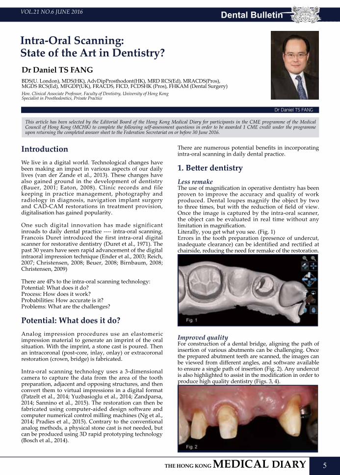
There are numerous potential benefits in incorporating intra-oral scanning in daily dental practice. 1. Better dentistryLess remakeThe use of magnification in operative dentistry has been proven to improve the accuracy and quality of work produced. Dental loupes magnify the object by two to three times, but with the reduction of field of view. Once the image is captured by the intra-oral scanner, the object can be evaluated in real time without any limitation in magnification. Literally, you get what you see. (Fig. 1)Errors in the tooth preparation (presence of undercut, inadequate clearance) can be identified and rectified at chairside, reducing the need for remake of the restoration.
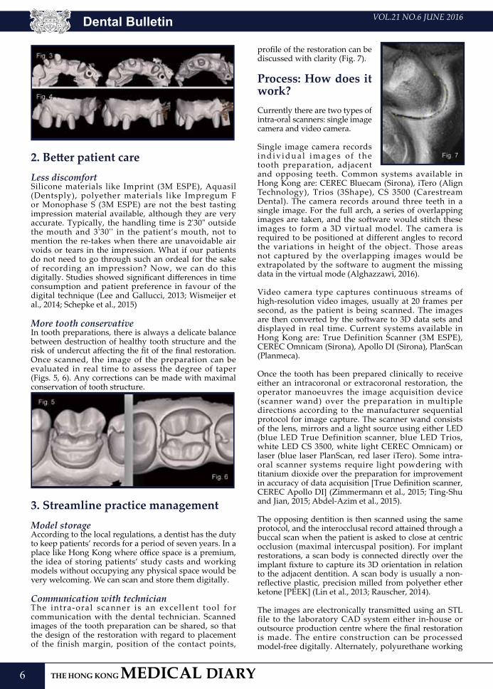
Improved quality For construction of a dental bridge, aligning the path of insertion of various abutments can be challenging. Once the prepared abutment teeth are scanned, the images can be viewed from different angles, and software available to ensure a single path of insertion (Fig. 2). Any undercut is also highlighted to assist in the modification in order to produce high quality dentistry (Figs. 3, 4).
Dental Bulletin VOL.21 NO.6 JUNE 2016
6
2. Better patient careLess discomfortSilicone materials like Imprint (3M ESPE), Aquasil (Dentsply), polyether materials like Impregum F or Monophase S (3M ESPE) are not the best tasting impression material available, although they are very accurate. Typically, the handling time is 2'30" outside the mouth and 3'30'' in the patient’s mouth, not to mention the re-takes when there are unavoidable air voids or tears in the impression. What if our patients do not need to go through such an ordeal for the sake of recording an impression? Now, we can do this digitally. Studies showed significant differences in time consumption and patient preference in favour of the digital technique (Lee and Gallucci, 2013; Wismeijer et al., 2014; Schepke et al., 2015)
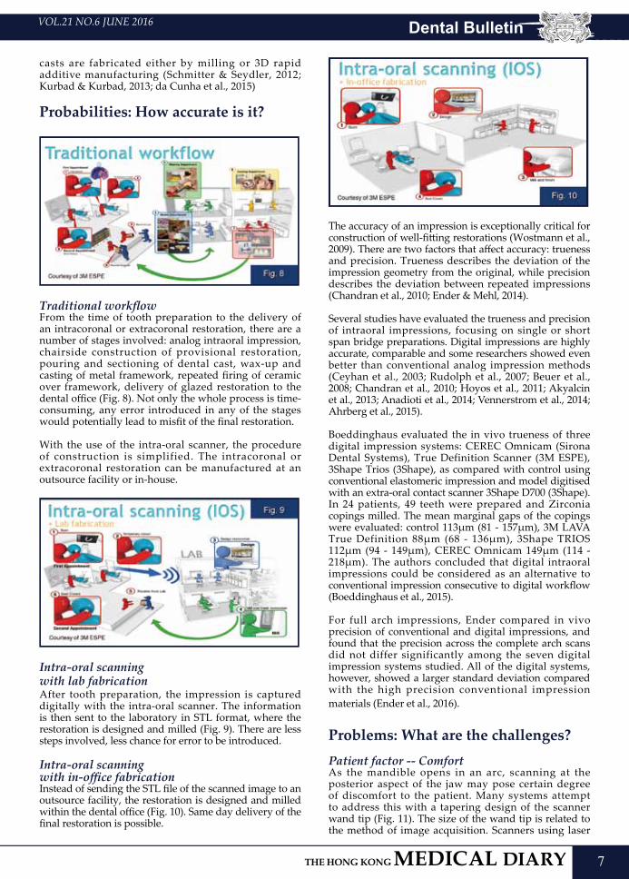
More tooth conservativeIn tooth preparations, there is always a delicate balance between destruction of healthy tooth structure and the risk of undercut affecting the fit of the final restoration. Once scanned, the image of the preparation can be evaluated in real time to assess the degree of taper (Figs. 5, 6). Any corrections can be made with maximal conservation of tooth structure.
3. Streamline practice managementModel storageAccording to the local regulations, a dentist has the duty to keep patients’ records for a period of seven years. In a place like Hong Kong where office space is a premium, the idea of storing patients’ study casts and working models without occupying any physical space would be very welcoming. We can scan and store them digitally.
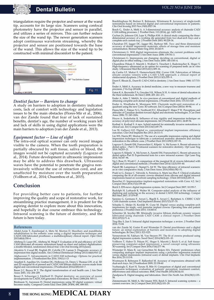
Communication with technicianThe intra-oral scanner is an excel lent tool for communication with the dental technician. Scanned images of the tooth preparation can be shared, so that the design of the restoration with regard to placement of the finish margin, position of the contact points,
profile of the restoration can be discussed with clarity (Fig. 7).
Process: How does it work?Currently there are two types of intra-oral scanners: single image camera and video camera.
Single image camera records ind iv idua l images o f the tooth preparation, adjacent and opposing teeth. Common systems available in Hong Kong are: CEREC Bluecam (Sirona), iTero (Align Technology), Trios (3Shape), CS 3500 (Carestream Dental). The camera records around three teeth in a single image. For the full arch, a series of overlapping images are taken, and the software would stitch these images to form a 3D virtual model. The camera is required to be positioned at different angles to record the variations in height of the object. Those areas not captured by the overlapping images would be extrapolated by the software to augment the missing data in the virtual mode (Alghazzawi, 2016).
Video camera type captures continuous streams of high-resolution video images, usually at 20 frames per second, as the patient is being scanned. The images are then converted by the software to 3D data sets and displayed in real time. Current systems available in Hong Kong are: True Definition Scanner (3M ESPE), CEREC Omnicam (Sirona), Apollo DI (Sirona), PlanScan (Planmeca).
Once the tooth has been prepared clinically to receive either an intracoronal or extracoronal restoration, the operator manoeuvres the image acquisition device (scanner wand) over the preparation in multiple directions according to the manufacturer sequential protocol for image capture. The scanner wand consists of the lens, mirrors and a light source using either LED (blue LED True Definition scanner, blue LED Trios, white LED CS 3500, white light CEREC Omnicam) or laser (blue laser PlanScan, red laser iTero). Some intra-oral scanner systems require light powdering with titanium dioxide over the preparation for improvement in accuracy of data acquisition [True Definition scanner, CEREC Apollo DI] (Zimmermann et al., 2015; Ting-Shu and Jian, 2015; Abdel-Azim et al., 2015).
The opposing dentition is then scanned using the same protocol, and the interocclusal record attained through a buccal scan when the patient is asked to close at centric occlusion (maximal intercuspal position). For implant restorations, a scan body is connected directly over the implant fixture to capture its 3D orientation in relation to the adjacent dentition. A scan body is usually a non-reflective plastic, precision milled from polyether ether ketone [PEEK] (Lin et al., 2013; Rauscher, 2014).
The images are electronically transmitted using an STL file to the laboratory CAD system either in-house or outsource production centre where the final restoration is made. The entire construction can be processed model-free digitally. Alternately, polyurethane working
Dental BulletinVOL.21 NO.6 JUNE 2016
7
casts are fabricated either by milling or 3D rapid additive manufacturing (Schmitter & Seydler, 2012; Kurbad & Kurbad, 2013; da Cunha et al., 2015)
Probabilities: How accurate is it?
Traditional workflowFrom the time of tooth preparation to the delivery of an intracoronal or extracoronal restoration, there are a number of stages involved: analog intraoral impression, chairside construction of provisional restoration, pouring and sectioning of dental cast, wax-up and casting of metal framework, repeated firing of ceramic over framework, delivery of glazed restoration to the dental office (Fig. 8). Not only the whole process is time-consuming, any error introduced in any of the stages would potentially lead to misfit of the final restoration.
With the use of the intra-oral scanner, the procedure of construction is simplified. The intracoronal or extracoronal restoration can be manufactured at an outsource facility or in-house.
Intra-oral scanning with lab fabricationAfter tooth preparation, the impression is captured digitally with the intra-oral scanner. The information is then sent to the laboratory in STL format, where the restoration is designed and milled (Fig. 9). There are less steps involved, less chance for error to be introduced.
Intra-oral scanning with in-office fabricationInstead of sending the STL file of the scanned image to an outsource facility, the restoration is designed and milled within the dental office (Fig. 10). Same day delivery of the final restoration is possible.
The accuracy of an impression is exceptionally critical for construction of well-fitting restorations (Wostmann et al., 2009). There are two factors that affect accuracy: trueness and precision. Trueness describes the deviation of the impression geometry from the original, while precision describes the deviation between repeated impressions (Chandran et al., 2010; Ender & Mehl, 2014).
Several studies have evaluated the trueness and precision of intraoral impressions, focusing on single or short span bridge preparations. Digital impressions are highly accurate, comparable and some researchers showed even better than conventional analog impression methods (Ceyhan et al., 2003; Rudolph et al., 2007; Beuer et al., 2008; Chandran et al., 2010; Hoyos et al., 2011; Akyalcin et al., 2013; Anadioti et al., 2014; Vennerstrom et al., 2014; Ahrberg et al., 2015).
Boeddinghaus evaluated the in vivo trueness of three digital impression systems: CEREC Omnicam (Sirona Dental Systems), True Definition Scanner (3M ESPE), 3Shape Trios (3Shape), as compared with control using conventional elastomeric impression and model digitised with an extra-oral contact scanner 3Shape D700 (3Shape). In 24 patients, 49 teeth were prepared and Zirconia copings milled. The mean marginal gaps of the copings were evaluated: control 113μm (81 - 157μm), 3M LAVA True Definition 88μm (68 - 136μm), 3Shape TRIOS 112μm (94 - 149μm), CEREC Omnicam 149μm (114 - 218μm). The authors concluded that digital intraoral impressions could be considered as an alternative to conventional impression consecutive to digital workflow (Boeddinghaus et al., 2015).
For full arch impressions, Ender compared in vivo precision of conventional and digital impressions, and found that the precision across the complete arch scans did not differ significantly among the seven digital impression systems studied. All of the digital systems, however, showed a larger standard deviation compared with the high precision conventional impression materials (Ender et al., 2016).
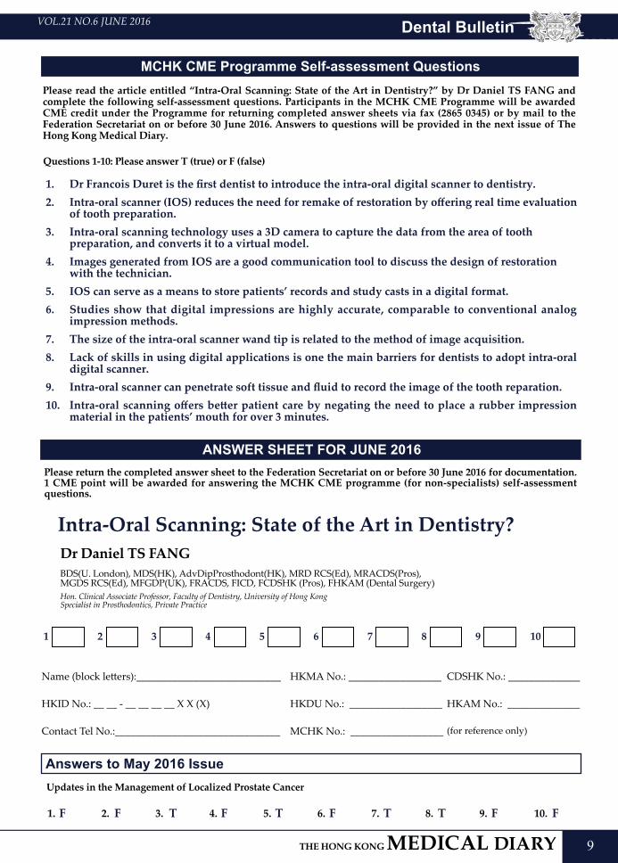
Problems: What are the challenges?Patient factor -- ComfortAs the mandible opens in an arc, scanning at the posterior aspect of the jaw may pose certain degree of discomfort to the patient. Many systems attempt to address this with a tapering design of the scanner wand tip (Fig. 11). The size of the wand tip is related to the method of image acquisition. Scanners using laser
Dental Bulletin VOL.21 NO.6 JUNE 2016
8
triangulation require the projector and sensor at the wand tip, accounts for its large size. Scanners using confocal photometry have the projector and sensor in parallel, and utilizes a series of mirrors. This can further reduce the size of the wand tip. The newer generation scanners adopt continuous waveform streaming, whereby the projector and sensor are positioned towards the base of the wand. This allows the size of the wand tip to be constructed with minimal discomfort to the patient.
Dentist factor -- Barriers to changeA study on barriers to adoption in dentistry indicated costs, lack of comfort with technology and legislation issues to be the main obstacles (Flores-Mir et al., 2006). van der Zande found that fear of lack of sustained benefits, dentist’s age, the number of working years left and lack of skills in using digital applications were the main barriers to adoption (van der Zande et al., 2013).
Equipment factor -- Line of sightThe intra-oral optical scanner can only record images visible to the camera. When the tooth preparation is partially obscured by soft tissue, saliva or blood, the images would not be captured accurately (Logozzo et al., 2014). Future development in ultrasonic impressions may be able to address this drawback. Ultrasonic waves have the potential to penetrate the gingiva non-invasively without the use of retraction cord, and are unaffected by moisture over the tooth preparation (Vollborn et al., 2014; Chuembou et al., 2015).
ConclusionFor providing better care to patients, for further improving the quality and scope of restorative work, for streamlining practice management, it is prudent for the aspiring dentist to explore more about this innovation, and hopefully in due course embrace this technology. Intraoral scanning is the future of dentistry, and the future is here today.
ReferencesAbdel-Azim T, Zandinejad A, Metz M, Morton D. Maxillary and mandibular rehabilitation in the esthetic zone using a digital impression technique and CAD/CAM-fabricated prostheses: a multidisciplinary clinical report. Oper Dent 2015;40(4):350–6Ahrberg D, Lauer HC, Ahrberg M, Weigl P. Evaluation of fit and efficiency of CAD/CAM fabricated all-ceramic restorations based on direct and indirect digitalization: a double-blinded, randomized clinical trial. Clin Oral Investig 2015;(June). Akyalcin S, Cozad BE, English JD, Colville CD, Laman S. Diagnostic accuracy of impression-free digital models. Am J Orthod Dentofacial Orthop 2013;144(6):916–22.Alghazzawi T. Advancements in CAD/CAM technology: Options for practical implementation. J Prosthodont Res 2016; 319(1):1-13 Anadioti E, Aquilino SA, Gratton DG, Holloway JA, Denry I, Thomas GW, et al. 3D and 2D marginal fit of pressed and CAD/CAM lithium disilicate crowns made from digital and conventional impressions. J Prosthodont 2014;23(8):610–7. Bauer J C, Brown W T. The digital transformation of oral health care. J Am Dent Assoc 2001; 132: 204–209.Beuer F, Schweiger J, Edelhoff D. Digital dentistry: an overview of recent developments for CAD/CAM generated restorations. Br Dent J 2008;204: 505-11.Birnbaum N, Aaronson H. Dental impressions using 3D digital scanners: virtual becomes reality. Compend Contin Educ Dent 2008; 29:494, 496, 498-505.
Boeddinghaus M, Breloer E, Rehmann, Wöstmann B. Accuracy of single-tooth restorations based on intraoral digital and conventional impressions in patients. Clin Oral Investig 2015 Nov;19(8):2027-34.Bosch G, Ender A, Mehl A. A 3-dimensional accuracy analysis of chairside CAD/CAM milling processes. J Prosthet Dent, 112 (2014), pp. 1425–1431Ceyhan JA, Johnson GH, Lepe X, Phillips KM. A clinical study comparing the three-dimensional accuracy of a working die generated from two dual-arch trays and a complete-arch custom tray. J Prosthet Dent 2003; 90:228-34.Chandran DT, Jagger DC, Jagger RG, Barbour ME. Two- and three dimensional accuracy of dental impression materials: effects of storage time and moisture contamination. Biomed Mater Eng 2010; 20:243-9.Christensen G. Will digital impressions eliminate the current problems with conventional impressions? J Am Dent Assoc 2008; 139:761-3.Christensen G. Impressions are changing: deciding on conventional, digital or digital plus in-office milling. J Am Dent Assoc 2009; 140:1301-4.Chuembou Pekam F, Marotti J, Wolfart S, Tinschert J, Radermacher K, Heger S. High-frequency ultrasound as an option for scanning of prepared teeth: an in vitro study. Ultrasound Med Biol 2015;41(1):309–16. da Cunha LF, Mukai E, Hamerschmitt RM, Correr GM. Fabrication of lithium silicate ceramic veneers with a CAD/ CAM approach: a clinical report of cleidocranial dysplasia. J Prosthet Dent 2015;113(5):355–9. Duret F, Blouin JL, Duret B. CAD-CAM in dentistry. J Am Dent Assoc 1988; 117:715-20.Ender A, Mehl A. Accuracy in dental medicine, a new way to measure trueness and precision. J Vis Exp 2014;86.Eaton K A, Reynolds P A, Greyden S K, Wilson N H. A vision of dental education in the third millennium. Br Dent J 2008; 205: 261–271.Ender A, Attin T, MehlA. In vivo precision of conventional and digital methods of obtaining complete-arch dental impressions. J Prosthet Dent 2016; 115:313-320Ender A, Wiedhahn K, Mormann WH. Chairside multi-unit restoration of a quadrant using the new Cerec 3D software. Int J Comput Dent 2003;6: 89-94.Flores-Mir C, Palmer N G, Northcott H C, Khurshed F, Major P W. Perceptions and attitudes of Canadian dentists toward digital and electronic technologies. J Can Dent Assoc 2006; 72: 243–243e.Hoyos A, Soderholm K. Influence of tray rigidity and impression technique on accuracy of polyvinyl siloxane impressions. Int J Prosthodont 2011; 24:49-54.Kurbad A, Kurbad S. A new, hybrid material for minimally invasive restorations in clinical use. Int J Comput Dent 2013;16(1):69–79. Lee SJ, Gallucci GO. Digital vs. conventional implant impressions: efficiency outcomes. Clin Oral Implants Res 2013; 24:111-5.Lin WS, Harris BT, Morton D. The use of a scannable impression coping and digital impression technique to fabricate a customized anatomic abutment and zirconia restoration in the esthetic zone. J Prosthet Dent 2013;109(3):187–91.Logozzo S, Zanetti EM, Franceschini G, Kilpela¨ A, Ma¨kynen A. Recent advances in dental optics – Part I: 3D intraoral scanners for restorative dentistry. Opt Laser Eng 2014; 54:203–21. Logozzo S, Kilpela¨ A, Ma¨kynen A, Zanetti EM, Franceschini G. Recent advances in dental optics–Part II: Experimental tests for a new intraoral scanner. Opt Laser Eng 2014; 54:187–96.Ng J, Ruse D, Wyatt C. A comparison of the marginal fit of crowns fabricated with digital and conventional methods. J Prosthet Dent 2014;112(3):555–60. Patzelt SB, Lamprinos C, Stampf S, Att W. The time efficiency of intraoral scanners: an in vitro comparative study. J Am Dent Assoc 2014;145(6):542–51.Pradı´es G, Zarauz C, Valverde A, Ferreiroa A, Martı´nez-Rus F. Clinical evaluation comparing the fit of all-ceramic crowns obtained from silicone and digital intraoral impressions based on wavefront sampling technology. J Dent 2015;43(2):201–8. Rauscher O. Impressionless implant-supported restorations with Cerec 4.2. Int J Comput Dent 2014;17(2):159–68. Reich S. IDS-news: digital impression systems. Int J Comput Dent 2007; 10:195-7.Rudolph H, Luthardt R, Walter M. Computer-aided analysis of the influence of digitizing and surfacing on the accuracy in dental CAD/CAM technology. Comput Biol Med 2007; 37:579-87.Sannino G, Germano F, Arcuri L, Bigelli E, Arcuri C, Barlattani A. CEREC CAD/CAM chairside system. Oral Implantol (Rome) 2015;7(3):57–70.Schepke U, Meijer H, Kerdijk W, Cune M. Digital versus analog complete-arch impressions for single –unit premolar implant crowns: Operating time and patient preference. J Prosthet Dent 2015; 114:403-6Schmitter M, Seydler BB. Minimally invasive lithium disilicate ceramic veneers fabricated using chairside CAD/ CAM: a clinical report. J Prosthet Dent 2012;107(2):71–4. Ting-Shu S, Jian S. Intraoral digital impression technique: a review. J Prosthet Dent 2016; 72-84van der Zande M, Gorter R and Wismeijer D. Dental practitioners and a digital future: an initial exploration of barriers and incentives to adopting digital technologies. Br Den J 2013; E21-E25.Vennerstrom M, Fakhary M, Von Steyern PV. The fit of crowns produced using digital impression systems. Swed Dent J 2014;38(3):101–10. Vollborn T, Habor D, Pekam FC, Heger S, Marotti J, Reich S, et al. Soft tissue-preserving computer-aided impression: a novel concept using ultrasonic 3D-scanning. Int J Comput Dent 2014;17(4):277–96.Wismeijer D, Mans R, van Genuchten M, Reijers HA. Patients’ preferences when comparing analogue implant impressions using a polyether impression material versus digital impressions (intraoral scan) of dental implants. Clin Oral Implants Res 2014; 25:1113-8.Wostmann B, Rehmann P, Balkenhol M. Accuracy of impressions obtained with dual-arch trays. Int J Prosthodont 2009; 22:158-60Yuzbasioglu E, Kurt H, Turunc R, Bilir H. Comparison of digital and conventional impression techniques: evaluation of patients’ perception, treatment comfort, effectiveness and clinical outcomes. BMC Oral Health 2014;30(14):10.Zandparsa R. Digital imaging and fabrication. Dent Clin North Am 2014;58(1):135–58. Zimmermann M, Mehl A, Mo¨rmann WH, Reich S. Intraoral scanning systems – a current overview. Int J Comput Dent 2015;18(2):101–29.
Dental BulletinVOL.21 NO.6 JUNE 2016
9
ANSWER SHEET FOR JUNE 2016
Answers to May 2016 Issue
Please return the completed answer sheet to the Federation Secretariat on or before 30 June 2016 for documentation. 1 CME point will be awarded for answering the MCHK CME programme (for non-specialists) self-assessment questions.
Updates in the Management of Localized Prostate Cancer
1 4 82 5 93 76 10
1. F F T T T F FT F F4. 8.2. 5. 9.3. 7.6. 10.
Name (block letters):____________________________ HKMA No.: __________________ CDSHK No.: _______________
HKID No.: __ __ - __ __ __ __ X X (X) HKDU No.: __________________ HKAM No.: ________________
Contact Tel No.:________________________________ MCHK No.: __________________ (for reference only)
MCHK CME Programme Self-assessment QuestionsPlease read the article entitled “Intra-Oral Scanning: State of the Art in Dentistry?” by Dr Daniel TS FANG and complete the following self-assessment questions. Participants in the MCHK CME Programme will be awarded CME credit under the Programme for returning completed answer sheets via fax (2865 0345) or by mail to the Federation Secretariat on or before 30 June 2016. Answers to questions will be provided in the next issue of The Hong Kong Medical Diary.
Questions 1-10: Please answer T (true) or F (false)
1. Dr Francois Duret is the first dentist to introduce the intra-oral digital scanner to dentistry. 2. Intra-oral scanner (IOS) reduces the need for remake of restoration by offering real time evaluation
of tooth preparation.3. Intra-oral scanning technology uses a 3D camera to capture the data from the area of tooth
preparation, and converts it to a virtual model.4. Images generated from IOS are a good communication tool to discuss the design of restoration
with the technician.5. IOS can serve as a means to store patients’ records and study casts in a digital format.6. Studies show that digital impressions are highly accurate, comparable to conventional analog
impression methods.7. The size of the intra-oral scanner wand tip is related to the method of image acquisition.8. Lack of skills in using digital applications is one the main barriers for dentists to adopt intra-oral
digital scanner.9. Intra-oral scanner can penetrate soft tissue and fluid to record the image of the tooth reparation.10. Intra-oral scanning offers better patient care by negating the need to place a rubber impression
material in the patients’ mouth for over 3 minutes.
Intra-Oral Scanning: State of the Art in Dentistry?Dr Daniel TS FANGBDS(U. London), MDS(HK), AdvDipProsthodont(HK), MRD RCS(Ed), MRACDS(Pros), MGDS RCS(Ed), MFGDP(UK), FRACDS, FICD, FCDSHK (Pros), FHKAM (Dental Surgery)Hon. Clinical Associate Professor, Faculty of Dentistry, University of Hong KongSpecialist in Prosthodontics, Private Practice
2952 8079 | 6898 8919
Please note that prices are listed per person, non-air cruise-only and based on double occupancy. Fares INCLUDE Taxes, Fees & Port Expenses. Terms & Conditions applied. Princess Cruises reserves the right to add, edit, modify, delete any contents without giving any prior notice.
Find Details ofBritish Isles:
All Fares INCLUDE Taxes, Fee & Port Expenses.
SERIES
BRITISH ISLES
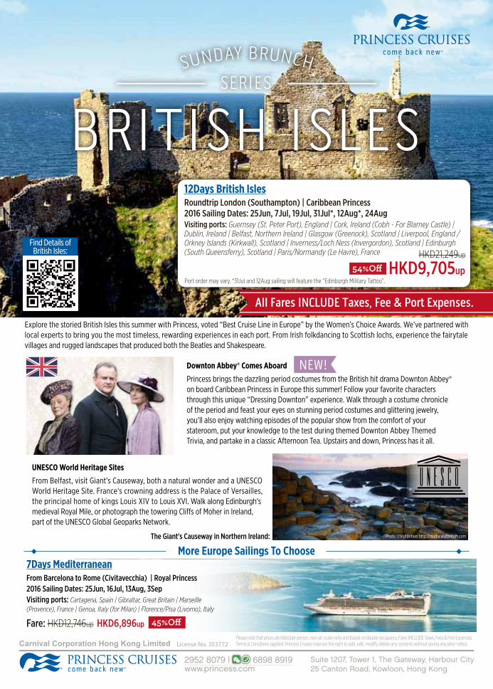
Explore the storied British Isles this summer with Princess, voted “Best Cruise Line in Europe” by the Women’s Choice Awards. We’ve partnered with local experts to bring you the most timeless, rewarding experiences in each port. From Irish folkdancing to Scottish lochs, experience the fairytale villages and rugged landscapes that produced both the Beatles and Shakespeare.
UNESCO World Heritage Sites
From Belfast, visit Giant's Causeway, both a natural wonder and a UNESCO World Heritage Site. France's crowning address is the Palace of Versailles, the principal home of kings Louis XIV to Louis XVI. Walk along Edinburgh's medieval Royal Mile, or photograph the towering Cli�s of Moher in Ireland, part of the UNESCO Global Geoparks Network.
Photo: ©VisitBritain: http://media.visitbritain.comThe Giant's Causeway in Northern Ireland:
7Days MediterraneanFrom Barcelona to Rome (Civitavecchia) | Royal Princess2016 Sailing Dates: 25Jun, 16Jul, 13Aug, 3SepVisiting ports: Cartagena, Spain | Gibraltar, Great Britain | Marseille (Provence), France | Genoa, Italy (for Milan) | Florence/Pisa (Livorno), Italy
More Europe Sailings To Choose
12Days British IslesRoundtrip London (Southampton) | Caribbean Princess2016 Sailing Dates: 25Jun, 7Jul, 19Jul, 31Jul*, 12Aug*, 24Aug Visiting ports: Guernsey (St. Peter Port), England | Cork, Ireland (Cobh - For Blarney Castle) | Dublin, Ireland | Belfast, Northern Ireland | Glasgow (Greenock), Scotland | Liverpool, England / Orkney Islands (Kirkwall), Scotland | Inverness/Loch Ness (Invergordon), Scotland | Edinburgh (South Queensferry), Scotland | Paris/Normandy (Le Havre), France
Port order may vary. *31Jul and 12Aug sailing will feature the “Edinburgh Military Tattoo”.
54%O�HKD21,249up
HKD9,705up
45%O�Fare: HKD12,746up HKD6,896up
Downton Abbey® Comes Aboard
Princess brings the dazzling period costumes from the British hit drama Downton Abbey® on board Caribbean Princess in Europe this summer! Follow your favorite characters through this unique “Dressing Downton” experience. Walk through a costume chronicle of the period and feast your eyes on stunning period costumes and glittering jewelry, you’ll also enjoy watching episodes of the popular show from the comfort of your stateroom, put your knowledge to the test during themed Downton Abbey Themed Trivia, and partake in a classic Afternoon Tea. Upstairs and down, Princess has it all.
NEW!
Dental BulletinVOL.21 NO.6 JUNE 2016
11
Considerations on Oral Health in the Management of General Health ConditionsDr Frankie Hon-ching SO
Dr Frankie Hon-ching SO
BDS(HK), MDS(HK), FHKAM(Dental Surgery), FCDSHK(Community Dentistry)Specialist in Community Dentistry
Many health conditions and their medical treatments may negatively impact on the oral and dental health of the concerned patients. If preventive dental care can be included as part of the management of these patients at a very early stage, deterioration of oral health can be avoided. Poor oral health, dental caries and periodontal diseases may cause discomfort, pain and additional distress to patients already suffering from other diseases. Good oral health will in turn ensure the general health and well-being of the patients which will enhance the effects of medical care provided by medical practitioners.
It is impractical to develop an exhaustive list of medical conditions and medical treatments that may negatively impact on oral and dental health. The following principles are recommended to medical practitioners in both general and specialised practices for identification of cases that preventive dental care should be initiated as early as possible.
Some diseases or treatments may increase the risks in developing new dental diseases among patients, if they may• reduce salivary production;• affect dietary habit;• affect hand movement; or• affect general mobility.
Some diseases or treatments may put the patients at high risks or even contraindicated to receive invasive dental treatments such as dental extractions. The avoidance of invasive dental treatments is an important part of management for patients with diseases or treatments that may• affect haemostasis; or• affect healing capacity, immune functions and body
defence capacity.
Diseases / medications reducing salivary productionReduced salivary production and the resulting dry mouth (sometimes referred to as xerostomia) may put the affected patients at very high risks of developing dental caries, sometimes progressing at a very rapid rate.
Diseases of the salivary glands such as Sojgren’s syndrome in itself may cause dry mouth. The irradiation of salivary glands associated with the radiotherapy for
naso-pharyngeal carcinomas is also a major reason of dry mouth in Hong Kong.
A number of medications, including those prescribed for common chronic diseases such as hypertension and diabetes may also produce the side-effect of dry mouth1,2.
Diseases / medications affecting dietary habitThe frequency and quantity of sugar intake is directly related to the risk of developing dental caries. Sometimes, patients may be advised to take more frequent meals with less quantity each time (少食多餐) because of their specific medical conditions. Patients suffering from diseases of the spleen or had splenectomies may frequently experience hypoglycaemia and require the intake of sugar at a high frequency. These may all increase the risks of developing new dental caries
Diseases / medications affecting hand movementOne of the main strategies in preventing dental diseases is plaque control by daily tooth brushing and interdental cleaning by flossing or interdental brushing. These oral hygiene measures require a fair degree of dexterity in hand movements. Conditions such as stroke, rheumatoid arthritis, Parkinson’s disease and medications that may cause hand tremors will affect the patients’ ability in performing daily oral hygiene. Cognitive impairment arising from dementia or interlectual disabilities can also affect the patients’ ability in self-care.
Diseases / medication affecting general mobilityConditions that render patients home-bound or bed-bound may not only affect their ability in performing daily oral hygiene, but also hinder them from going to the dental clinics to receive professional dental care.
Diseases / medications affecting haemostasisNowadays, anticoagulants are increasingly prescribed due to the common occurrence of coronary heart diseases and transient ischaemic attacks. Difficulties in
Dental Bulletin VOL.21 NO.6 JUNE 2016
12
stopping bleeding definitely have implications if surgical procedures such as dental extractions are necessary.
Diseases / medications affecting healing capacity, immune functions and body defence capacityPatients who had bone marrow or solid organ transplantation, suffering from cancer or had received / receiving chemotherapy are at special risks if surgical procedures such as dental extractions are necessary. The irradiation of the jaws associated with radiotherapy for naso-pharyngeal carcinoma and bisphosphonate type of medications for treating osteoporosis also affect the ability of bone healing. Osteonecrosis is a possible complication in these patients if a dental extraction has to be performed.
The above description is short but the possible list of diseases and medical treatments that may affect oral and dental health can be very extensive. Hopefully the above principles may assist medical practitioners in identifying patients with high oral health risks and to refer these patients to dentists for early preventive dental care.
The two most common dental diseases in Hong Kong are dental caries and periodontal diseases. These are all preventable by adopting an appropriate life-style3. Prevention of dental diseases is more than just ‘more
tooth brushing and less sweets and candies’ (刷多啲牙, 食少啲糖). The prevention of dental diseases and tooth loss must start with prevention and early treatment of dental diseases. Dentists can be partners in prevention by providing individualised advices on daily tooth cleaning, dietary and other oral health-related habits at the regular checkup visits. This can be accomplished only if people in Hong Kong visit dentist regularly for checkup even though they believe that their oral health status is good3.
It is more common for people in Hong Kong to make a medical consultation than a dental one. Medical practitioners are in a good position to inform their patients about the need to prevent dental diseases and the need to avoid dental extractions, especially for patients with diseases or medical treatments as described above. Good oral health is not just limited to teeth and the mouth, but is also contributing to the overall well-being and positively impact on the diseases or health conditions under the care of medical practitioners.
References1. Thomson, W.M., et al., A longitudinal study of medication exposure and
xerostomia among older people. Gerodontology, 2006. 23: p. 205-213.2. Sreebny, L.M. and S.S. Schwartz, A reference guide to drugs and dry mouth
– 2nd edition. Gerodontology, 1997. 14(1): p. 33-47.3. Department of Health, Report on Oral Health Survey 2011. 2013, Department
of Health: Hong Kong.
Dental BulletinVOL.21 NO.6 JUNE 2016
13
Intra-oral scanners: A brief overview of the technology and considerations
Dr Ronald Yik-Long CHAN
IntroductionBeing able to accurately capture the anatomical details in the oral cavity has always been one of the most critical procedures for dental professionals. The concept of impression-taking was first recorded to be around the early 18th century when Philipp Pfaff described a technique of using sealing wax softened in hot water to capture an impression.1 However, the details of the anatomy captured and distortion was a problem to many dentists.
The next big improvement in the concept of impression taking was the invention of elastic impression materials, which were developed by S.L. Pearson at the University of Liverpool in 1955. 2 Later, elastic impression materials included polyethers, polysulfides and also polyvinylsiloxanes. The problems of distortions were reduced but the setting time of the material was prolonged and patients frequently complained of an unpleasant smell or taste.
We have now reached the next major milestone in impression taking by utilising optical technology to digitally capture the anatomical tissues of the oral cavity. Many different brands in the market exist but what are their differences in terms of the technology behind them? Does this technology add value to a dental practice and has the accuracy reached a satisfactory level yet? This article will briefly address these questions.
The TechnologyThe digital workflow currently consists of three main steps. These include data acquisition, data manipulation and computer aided design/manufacturing. All intra-oral scanners (IOS) on the market today attempt to at least accurately perform the first step of this digital workflow. To digitally capture anatomical details, different IOS systems utilise different methods for recording the details in the oral cavity. These methods can be broadly classified into laser beam based systems or light beam based systems. Both methods incident a beam on the surface of the tissue and a camera-like device (charge-coupled device (CCD) or position sensitive detector) is used to record the location of the point at which the beam strikes the object. Most software of these IOS systems does the algorithms based on the known position and angulations of the camera and sensors of the scanner.3
Laser Beam based IOS systemsThis method of image capturing relies on making still
images at different positions which are later rendered into a 3-dimensional object.4 Since lasers are employed and does not scatter as irregularly as light, it does not require a reflecting agent (Ex. Powder). Two main categories of utilising laser beam technologies are the parallel confocal imaging technique and laser triangulation imaging technique.
Parallel confocal imaging technique (Ex. iTero)This technique emits 2 parallel laser beams at a specific focal length, which are bounced off the tissue and back through a laser sensor (Fig. 1). Before entering the laser sensor, a beam splitter is used to lead the reflected beam through a focal filter so that only the image that lies in the focal point of the lens reaches the filter.5 Since the focal distance is already set, the software is able to calculate the distance of the scanned object to the lens by moving the lens up and down in the oral cavity.
Laser triangulation imaging technique (Ex. E4D - PlanScan)This technique utilises a red laser beam with micro mirrors oscillating at around 20000 cycles per second to capture a series of still images from multiple angles.4 The triangulation is done similar to the light based triangulation method described below.
Light beam based IOS systemsThis technology of image capturing uses visible light beams instead of lasers. Since light reflects irregularly on different surface characteristics, a titanium dioxide reflecting agent is sometimes required to create a uniform light dispersion surface. Three main methods utilise this light beam based technology to capture images.
Dr Ronald Yik-Long CHAN
BSc, BDS, MBA
Dental Bulletin VOL.21 NO.6 JUNE 2016
14
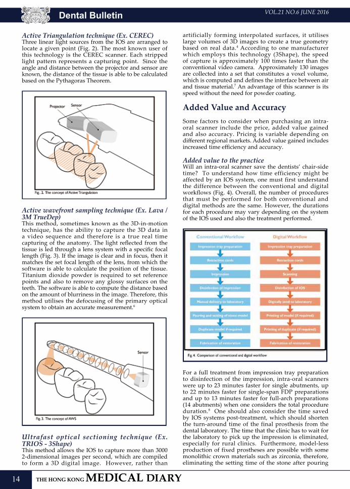
Active Triangulation technique (Ex. CEREC)Three linear light sources from the IOS are arranged to locate a given point (Fig. 2). The most known user of this technology is the CEREC scanner. Each stripped light pattern represents a capturing point. Since the angle and distance between the projector and sensor are known, the distance of the tissue is able to be calculated based on the Pythagoras Theorem.
Active wavefront sampling technique (Ex. Lava / 3M TrueDep) This method, sometimes known as the 3D-in-motion technique, has the ability to capture the 3D data in a video sequence and therefore is a true real time capturing of the anatomy. The light reflected from the tissue is led through a lens system with a specific focal length (Fig. 3). If the image is clear and in focus, then it matches the set focal length of the lens, from which the software is able to calculate the position of the tissue. Titanium dioxide powder is required to set reference points and also to remove any glossy surfaces on the teeth. The software is able to compute the distance based on the amount of blurriness in the image. Therefore, this method utilises the defocusing of the primary optical system to obtain an accurate measurement.6
Ultrafast optical sectioning technique (Ex. TRIOS - 3Shape)This method allows the IOS to capture more than 3000 2-dimensional images per second, which are compiled to form a 3D digital image. However, rather than
artificially forming interpolated surfaces, it utilises large volumes of 3D images to create a true geometry based on real data.4 According to one manufacturer which employs this technology (3Shape), the speed of capture is approximately 100 times faster than the conventional video camera. Approximately 130 images are collected into a set that constitutes a voxel volume, which is computed and defines the interface between air and tissue material.7 An advantage of this scanner is its speed without the need for powder coating.
Added Value and AccuracySome factors to consider when purchasing an intra-oral scanner include the price, added value gained and also accuracy. Pricing is variable depending on different regional markets. Added value gained includes increased time efficiency and accuracy.
Added value to the practiceWill an intra-oral scanner save the dentists’ chair-side time? To understand how time efficiency might be affected by an IOS system, one must first understand the difference between the conventional and digital workflows (Fig. 4). Overall, the number of procedures that must be performed for both conventional and digital methods are the same. However, the durations for each procedure may vary depending on the system of the IOS used and also the treatment performed.
For a full treatment from impression tray preparation to disinfection of the impression, intra-oral scanners were up to 23 minutes faster for single abutments, up to 22 minutes faster for single-span FDP preparations and up to 13 minutes faster for full-arch preparations (14 abutments) when one considers the total procedure duration.8 One should also consider the time saved by IOS systems post-treatment, which should shorten the turn-around time of the final prosthesis from the dental laboratory. The time that the clinic has to wait for the laboratory to pick up the impression is eliminated, especially for rural clinics. Furthermore, model-less production of fixed prostheses are possible with some monolithic crown materials such as zirconia, therefore, eliminating the setting time of the stone after pouring
Dental BulletinVOL.21 NO.6 JUNE 2016
15
the impression. The disadvantages of mould instability, transport, plaster solidification and delamination will largely be overcome with an IOS system.9
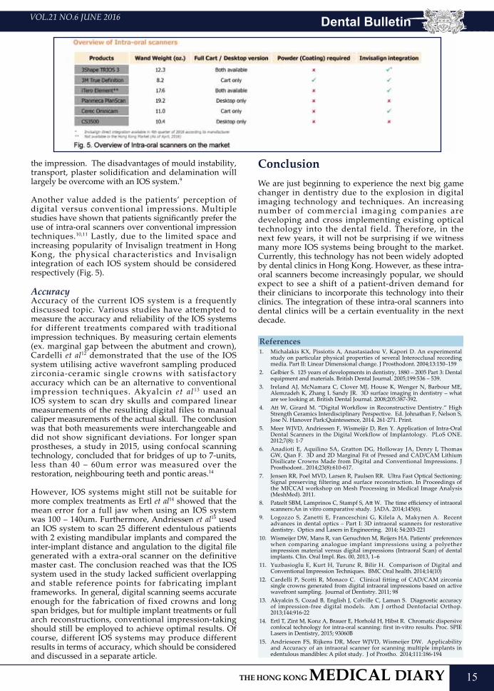
Another value added is the patients’ perception of digital versus conventional impressions. Multiple studies have shown that patients significantly prefer the use of intra-oral scanners over conventional impression techniques.10,11 Lastly, due to the limited space and increasing popularity of Invisalign treatment in Hong Kong, the physical characteristics and Invisalign integration of each IOS system should be considered respectively (Fig. 5).
Accuracy Accuracy of the current IOS system is a frequently discussed topic. Various studies have attempted to measure the accuracy and reliability of the IOS systems for different treatments compared with traditional impression techniques. By measuring certain elements (ex. marginal gap between the abutment and crown), Cardelli et al12 demonstrated that the use of the IOS system utilising active wavefront sampling produced zirconia-ceramic single crowns with satisfactory accuracy which can be an alternative to conventional impression techniques. Akyalcin et al13 used an IOS system to scan dry skulls and compared linear measurements of the resulting digital files to manual caliper measurements of the actual skull. The conclusion was that both measurements were interchangeable and did not show significant deviations. For longer span prostheses, a study in 2015, using confocal scanning technology, concluded that for bridges of up to 7-units, less than 40 – 60um error was measured over the restoration, neighbouring teeth and pontic areas.14
However, IOS systems might still not be suitable for more complex treatments as Ertl et al14 showed that the mean error for a full jaw when using an IOS system was 100 – 140um. Furthermore, Andriessen et al15 used an IOS system to scan 25 different edentulous patients with 2 existing mandibular implants and compared the inter-implant distance and angulation to the digital file generated with a extra-oral scanner on the definitive master cast. The conclusion reached was that the IOS system used in the study lacked sufficient overlapping and stable reference points for fabricating implant frameworks. In general, digital scanning seems accurate enough for the fabrication of fixed crowns and long span bridges, but for multiple implant treatments or full arch reconstructions, conventional impression-taking should still be employed to achieve optimal results. Of course, different IOS systems may produce different results in terms of accuracy, which should be considered and discussed in a separate article.
ConclusionWe are just beginning to experience the next big game changer in dentistry due to the explosion in digital imaging technology and techniques. An increasing number of commercial imaging companies are developing and cross implementing existing optical technology into the dental field. Therefore, in the next few years, it will not be surprising if we witness many more IOS systems being brought to the market. Currently, this technology has not been widely adopted by dental clinics in Hong Kong. However, as these intra-oral scanners become increasingly popular, we should expect to see a shift of a patient-driven demand for their clinicians to incorporate this technology into their clinics. The integration of these intra-oral scanners into dental clinics will be a certain eventuality in the next decade.
References1. Michalakis KX, Pissiotis A, Anastasiadou V, Kapori D. An experimental
study on particular physical properties of several Interocclusal recording media. Part II: Linear Dimensional change. J Prosthodont. 2004;13:150–159
2. Gelbier S. 125 years of developments in dentistry, 1880 – 2005 Part 3: Dental equipment and materials. British Dental Journal. 2005;199:536 – 539.
3. Ireland AJ, McNamara C, Clover MJ, House K, Wenger N, Barbour ME, Alemzadeh K, Zhang L Sandy JR. 3D surface imaging in dentistry – what are we looking at. British Dental Journal. 2008;205:387-392.
4. Att W, Girard M. “Digital Workflow in Reconstructive Dentistry.” High Strength Ceramics Interdisciplinary Perspective. Ed. Johnathan F, Nelson S, Jose N. Hanover Park:Quintessence, 2014. 261-271. Print.
5. Meer WJVD, Andriessen F, Wismeijir D, Ren Y. Application of Intra-Oral Dental Scanners in the Digital Workflow of Implantology. PLoS ONE. 2012;7(8): 1-7
6. Anadioti E, Aquilino SA, Gratton DG, Holloway JA, Denry I, Thomas GW, Qian F. 3D and 2D Marginal Fit of Pressed and CAD/CAM Lithium Disilicate Crowns Made from Digital and Conventional Impressions. J Prosthodont.. 2014;23(8):610-617.
7. Jensen RR, Poel MVD, Larsen R, Paulsen RR. Ultra Fast Optical Sectioning: Signal preserving filtering and surface reconstruction. In Proceedings of the MICCAI workshop on Mesh Processing in Medical Image Analysis (MeshMed). 2011.
8. Patzelt SBM, Lamprinos C, Stampf S, Att W. The time efficiency of intraoral scanners:An in vitro comparative study. JADA. 2014;145(6).
9. Logozzo S, Zanetti E, Franceschini G, Kilela A, Makynen A. Recent advances in dental optics – Part I: 3D intraoral scanners for restorative dentistry. Optics and Lasers in Engineering. 2014; 54:203-221
10. Wismeijer DW, Mans R, van Genuchten M, Reijers HA. Patients’ preferences when comparing analogue implant impressions using a polyether impression material versus digital impressions (Intraoral Scan) of dental implants. Clin. Oral Impl. Res. 00, 2013, 1–6
11. Yuzbasioglu E, Kurt H, Turunc R, Bilir H. Comparison of Digital and Conventional Impression Techniques. BMC Oral health. 2014;14(10)
12. Cardelli P, Scotti R, Monaco C. Clinical fitting of CAD/CAM zirconia single crowns generated from digital intraoral impressions based on active wavefront sampling. Journal of Dentistry. 2011; 98
13. Akyalcin S, Cozad B, English J, Colville C, Laman S. Diagnostic accuracy of impression-free digital models. Am J orthod Dentofacial Orthop. 2013;144:916-22
14. Ertl T, Zint M, Konz A, Brauer E, Horhold H, Hibst R. Chromatic dispersive confocal technology for intra-oral scanning: first in-vitro results. Proc. SPIE Lasers in Dentistry, 2015; 93060B
15. Andrieseen FS, Rijkens DR, Meer WJVD, Wismeijer DW. Applicability and Accuracy of an intraoral scanner for scanning multiple implants in edentulous mandibles: A pilot study. J of Prostho. 2014;111:186-194
Dental BulletinVOL.21 NO.6 JUNE 2016
17
A Sharing of Experiences in Outreach Dental Service in Hong Kong
Dr Jerry Kwong-shing LIU
I am a private dental practitioner, also working as a part time lecturer in the Dental Public Health, Faculty of Dentistry of The University of Hong Kong. After gaining years of experience in providing outreach services for the elderly, I started an NGO in 2005 to provide voluntary outreach dental services for the elders in residential homes.
What is outreach dental service?The scope of outreach dental service in different areas of the world is different. It can be understood as dental treatments restricted to primary prevention and dental health education in some countries. It can also be developed into full dental services including radiography, dental fillings, extractions and dentures making in the other parts of the world. The way to provide services can also be very different. Some outreach teams provide dental treatments in a fully renovated vehicle as a mobile dental clinic. Other teams may carry all their portable equipment to the site for set up, and they provide dental treatments in the activity venue, e.g. elderly homes.
Advantages of outreach dental serviceModern dentistry relies on sophisticated equipment to provide dedicated and precise dental treatments to patients, so as to obtain the optimal results. This approach of service delivery will lead to two undesired consequences. First, dental service will only be delivered in a well-equipped venue such as a dental clinic or hospital. It creates impact in accessing dental services for some groups of the population with travelling difficulties or difficulties in adapting to new environments. Second, the depreciation of the expensive equipment, no matter you have used it or not in a dental practice, will increase the cost of services. Therefore, the better equipped the clinic, the higher the cost of service (often calculated as cost of chairtime) will result. Hence, financial cost becomes another impact of service utilisation for low-income populations.
Outreach dental service is another approach for providing dental services. The principle is to provide dental services in a field environment. By using appropriate equipment and focusing on prevention, service providers work together with existing social services organisations such as NGOs, to provide dental treatments to the patients within the society. The advantages of this service approach are:
1) It improves the accessibility of dental services. Dental services are provided in the venues that people are routinely gathered, e.g. the elderly homes or activity halls.
2) It improves the acceptability of dental treatments, particularly for the groups of special needs that may have difficulties in adapting to new environments, e.g. elders of extreme age, ADHD and autism. Since dental treatments can be carried out in their familiar place in the companion of their caretakers, the psychological barriers to receive dental treatments can be greatly reduced.
3) It reduces the cost of service. Instead of cutting edge technology and advanced equipment, outreach dental service relies on appropriate technology for providing se lected dental treatments that have been proven to be effective. Therefore, the cost of service can be greatly reduced. Furthermore, by minimising the need of special transportation to carry patients to the clinic, the tangible and intangible cost of transportation and escort manpower can be reduced.
Difficulties of providing outreach dental serviceThere are several issues that may be worth discussing, based on my experience in providing outreach dental services. The most important one is the goal of the service. Besides the principle of Primary Health Approach that was proposed by WHO, I also set the goal of my outreach team as improving the quality of life of the patients through effective treatments in the field environment. In fact, treatments with evidence in ideal clinical settings may not necessarily give the same result in the field settings. For example, many outreach services emphasise on primary care and therefore they put a lot of resources in providing scaling (dental cleaning) to their patients on site. However, it is well known that scaling can hardly help periodontal diseases in any sense without meticulous oral hygiene instruction. In other words, periodontal diseases can only be prevented by good toothbrushing and interdental cleaning technique, together with mechanical cleaning of plaque and calculus1. However, it will never be easy and sometimes impossible to teach our elders to hold a conventional toothbrush with their shaking hands and ask them to brush along their gum line thoroughly with their stiff fingers and wrists. As a result, the resources spent on scaling may not be able to generate the desired outcome of disease prevention.
Dr Jerry Kwong-shing LIU
BSc BDS MDS MFGDP(UK)
Dental Bulletin VOL.21 NO.6 JUNE 2016
18
Furthermore, treating periodontal diseases through scaling requires routine reviews and services2. With limited resources, outreach service can hardly revisit the patient frequently. As a result, the therapeutic effect of scaling is reduced tremendously and the resource cannot effectively treat periodontal diseases as well. There are some evidence showing that toothbrushing with electric toothbrushes by caretakers could improve the gingival health of the elders in residential homes3. The primary care of the periodontal conditions of the elders may be more effectively delivered by training the caretakers on the correct way of assisted toothbrushing, increasing the manpower of elderly homes for assisted toothbrushing, instead of purchasing dental manpower to do dental scaling and oral hygiene instruction alone.
The resources of outreach dental service are limited, not only in terms of the financial budget, but also manpower and facilities. Which treatment could be provided in the field environment under an acceptable risk control is therefore controversial, especially when the services are always designed for special needs groups. Dental manpower with adequate training should be able to provide many basic treatments to solve patients’ problems with minimal risks. For example, caries are common in elders4, and dental fillings with Glass Ionomer Cement under atraumatic restorative technique5,6,7 or applying Silver Diamine Fluoride on lesions are both proven to be safe and effective treatments to caries in the field environment8,9,10,11. From my experience, providing partial dentures or complete dentures in the elderly homes can also result in very high success rates with minimal risks as well. On the other hand, not all dental treatments are suitable to be carried out in outreach service, especially those treatments with a high demand on clinical facilities and equipment, like extraction of stable teeth, oral surgery and root canal treatment. The clinical risk and success rate should be considered seriously before they are carried out in the outreach approach.
ConclusionOutreach dental service for special needs groups and elders of extreme age is a valuable service approach. It improves the accessibility and acceptability of dental services in the community. With careful planning of a treatment approach and manpower training, the treatment results of many common dental problems are promising12. This valuable service approach is still waiting for the dental service providers in Hong Kong to explore and develop.
References1. Tonetti MS, Eickhoiz P, Loos BG, Papapanou P, van der Velden U,
Armitage G, Bouchard P, Deinzer R, Dietrich T, Hughes F, Kocher T, Lang NP, Lopez R, Needleman I, Newton T, Nibali L, Pretzl B, Ramseier C, Sanz-Sanchez I, Schiagenhauf U, Suvan JE. Principles in prevention of periodontal disease: Consensus report of group1 of the 11th European Workshop on Periodontology on effective prevention of periodontal and peri-implant diseases. J Clin Periodontol. 2015 Apr;42 Suppl 16: S5-11
2. Worthington HV, Clarkson JE, Bryan G, Beirne PV. Routine scaling and polish for periodontal health in adults. Cochrane Oral Health Group, pub 2013 Nov 7.
3. Kambhu PP, Levy SM. An evaluation of the effectiveness of four mechanical plaque-removal devices when used by a trained care-provider. Spec Care Dent. 1993 Jan-Feb;13(1):9-14.
4. Gregory D, Hyde S. Root caries in older adults. J Calif Dent Assoc. 2015 Aug; 43(8): 439-45.
5. Dorri M, Sheiham A, Marinho VC. Atraumatic restorative treatment versus conventional restorative treatment for the management of dental caries. Cochrane Oral Health Group, pub 2009 Oct 7.
6. Lo EC, Luo Y, Tan HP, Dyson JE, Corbet EF. ART and conventional root restorations in elders after 12 months. J Dnt Res. 2006;85:929-32.
7. da Mata C, Allen PF, Cronin M, O’Mahony D, McKenna G, Woods N. Cost-effectiveness of ART restorations in elderly adults: a randomized clinical trial. Comm Dent Oral Epidemiol. 2014 Feb; 42(1): 79-87.
8. Ferreira de Oliveria MA, Celeste RR, Rodrigues CC. Topical fluoride for treating dental caries. Cochrane Oral Health Group, pub 2002 Jan 21.
9. Mei ML, Lo EC, Chu CH. Clinical use of silver diamine fluoride in dental treatment. Compend Contin Educ Dent. 2016 Feb; 37(2): 93-98
10. Mei ML, Zhao IS, Ito L, Lo EC, Chu CH. Prevention of secondary caries by silver diamine fluoride. Int Dent J. 2015 Dec; doi:10.1111/idj.12207.
11. Yee R, Holmgren C, Mulder J, Lama D, Walker D, van Palenstein Helderman W. Efficacy of silver diamine fluoride for arresting caries treatment. J Dent Res. 2009 Jul;88(7):644-7.
12. Quock RL, Patel SA, Falcao FA, Barros JA. Is a drill-less dental filling possible? Med Hypotheses. 2011 Sep;77(3):315-7.
Vacancies
Rental / For Sale
Commencement
of Practice
Please contact the Federation Secretariat at 2527 8898 for placement of classified advertisement.
Classified AdvertisementClassified Advertisement
從此展現你的燦爛笑容!
Stock Code : 3600.HK
現代牙科 信心可靠的完美牙齒現代牙科榮獲美國 FDA 和 ISO 國際認證,嚴格選用主要來自歐洲進口的原材料,並配以嶄新科技設備和數碼化生產技術,每一個步驟都務求精益求精,為你的牙齒提供最專業最可信賴的品質保證。
這是一個品質揉合科技的笑容!
"Recommended by the American Academy of Sleep Medicine for the treatment of obstructive sleep apnea1"
Moses EliteSerene Silensor EMA
1. For adult patients who are intolerant of CPAP therapy or prefer alternate therapy. Reference : Ramar K, Dort L, Katz S, et al. Clinical Practice Guideline for the Treatment of Obstructive Sleep Apnea and Snoring with Oral Appliance Therapy. Journal of Clinical Sleep Medicine 2015;11(7).
Intra-oral ScannersOffering over 50% discount for selected intra-oral scanners!
Please contact our sales team at +852 3766 0783 or +852 3766 0760 for a demonstration now!
With a vast experience with adjustable mandibular advancement devices, we deliver the supreme quality you need.
21
VOL.21 NO.6 JUNE 2016
Stereo Photography and Videography in the Digital AgeDr Siu-fai LEUNGBDS (HK) MSc (Lond.) FRACDSSpecialist in Endodontics, private practice.
Dr Siu-fai LEUNG
The beauty of stereo or 3D photography lies not only in the objects it captures but more in the spaces between the objects. Revealing spatial relationship on a 2D medium always carries that wow factor and can make an otherwise mundane image interesting. Two eyes and a brain are all we need to interpret the stereo effect and we are doing it every day. Our brains extrapolate the combined images so even low-resolution photographs become very life-like. This article aims to provide a nutshell of the progress of 3D imaging in both leisure and clinical applications in the digital age.
Stereo photography was first invented in 1841 by two Britons, Charles Wheatstone and Fox Talbot, two years after photography was formally introduced. The interest in 3D photography comes and goes, matched every time by breakthroughs in 2D photography. It went into obscurity in 1900 due to the launch of the highly portable Kodak Brownie camera. 3D mania struck again in the 1950s with the introduction of the Sawyer’s Viewmaster from Portland, Oregon, USA. Stereo cards and viewers became very popular. The interest waned again in the 1960 with the invention of the Polaroid camera, which was more portable, did not require a viewer, and gave instant results.
1952 saw the publication of “A stereoscopic atlas of human anatomy” by Dr David L. Bassett of the Stanford Medical School1, which was viewed using the Sawyer’s Viewmaster. The material took more than 10 years to produce and the quality of both the specimens and images were amazing. The archives have been totally updated and digitised2. There are relevant sections on the head and neck, (and tooth anatomy is also available elsewhere3). This is still a very good learning tool for both students and practitioners alike, and a big feast in visual art.
With the advent of digital photography, Fujifilm released the FinePix 3D camera and viewer series in 2009, bringing compact 3D cameras into the modern age. The system allowed naked eye 3D viewing without any accessory. There were even custom-built aftermarket accessories available4. Albeit expensive, these adjuncts expanded the capability, if not the image quality (IQ), of what is otherwise a low-resolution camera. Panasonic offered a similar camera (Lumix DMC 3D1) with superior IQ but could not reveal 3D on board. As these cameras are now discontinued, keen amateur 3D photographers with computer programming know-hows are hacking the Canon Powershot compact cameras (reversibly) to pair them up for synchronised stereo photography and videography5. Digital Single Lens Reflex cameras (DSLRs) can also be wired in a
similar manner but are less desirable due to the weight and shallow depth of field. Also synchronised zooming is more cumbersome. They are limited to professional applications. An alternative for DSLR is to attach a Loreo 3D lens cap in front of the lens. It eliminates the synchronisation problem but the image is limited to portrait format.
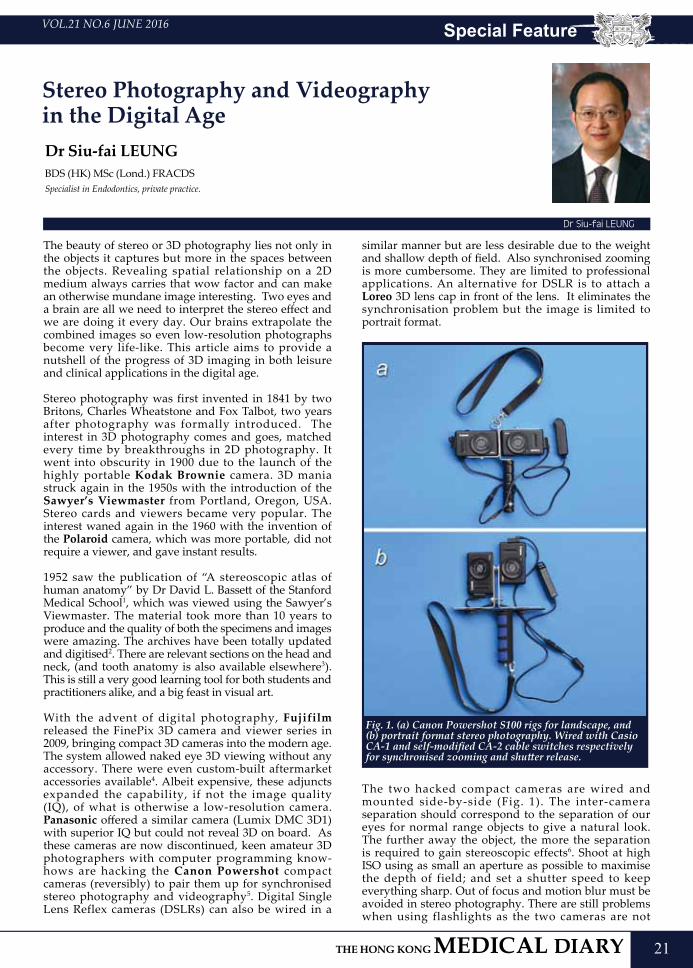
Fig. 1. (a) Canon Powershot S100 rigs for landscape, and (b) portrait format stereo photography. Wired with Casio CA-1 and self-modified CA-2 cable switches respectively for synchronised zooming and shutter release.
The two hacked compact cameras are wired and mounted side-by-side (Fig. 1). The inter-camera separation should correspond to the separation of our eyes for normal range objects to give a natural look. The further away the object, the more the separation is required to gain stereoscopic effects6. Shoot at high ISO using as small an aperture as possible to maximise the depth of field; and set a shutter speed to keep everything sharp. Out of focus and motion blur must be avoided in stereo photography. There are still problems when using flashlights as the two cameras are not
Special Feature
22
VOL.21 NO.6 JUNE 2016
perfectly synchronised. When flash photography is required I usually fall back to either the Fujifilm or the Lumix. The images are then processed by a custom 3D editing software. I picked up a kit from 中南圖書公司a few years back but I found StereoPhotoMaker and StereoMovieMaker5 the preferred choices. It is a very versatile package and is free. The viewing part is what prevents stereo photography from becoming popular. As not many people can casually register stereoscopic effects by viewing left and right images unassisted, special viewers or effects are required, which prevent the wide spread of stereo photography.
Besides the now discontinued Fujifilm viewers, the three common ways of viewing stereo still images include cross-eye viewing; the L/R images are transposed so the eyes are viewing the images diagonally. Sounding odd initially, this is in fact the easier viewing setup for most people. In parallel-eye viewing the images are aligned with the corresponding eyes. This is more difficult to acclimatise and a magnifying viewer is usually required. The simple Google Cardboard is designed for parallel-eye viewing on smartphones. Anaglyph involves adding blue and red tones to the images. A pair of blue and red spectacles (available from黃金電腦商場) is required to visualise the 3D effect. This is arguably the easiest way to view 3D and most popular for printed media. However the altered colours limit this method best suited for less colourful images.
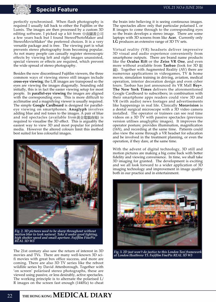
Fig. 2. 3D pictures need to be sharp throughout without motion blur to look natural. Take it under good lighting, high shutter speed and small aperture. Fujifilm FinePix REAL 3D W3
The 21st century also saw the return of interest in 3D movies and TVs. There are many well-known 3D sci-fi movies with great box office success, and more are coming. There are also 3D TV series like the reboot wildlife series by David Attenborough. Together with ‘on screen’ polarised stereo photographs, these are viewed using passive, or less desirably, active spectacles. The working principle is to alternate the polarised L/R images on the screen fast enough (144Hz) to cheat
the brain into believing it is seeing continuous images. The spectacles allow only that particular polarised L or R images to come through to the corresponding eyes so the brain develops a stereo image. There are some laptops with 3D screens from like Acer. Currently only LG produces an extensive range of 3D TV sets.
Virtual reality (VR) headsets deliver impressive 3D visual and audio experience conveniently from smartphone outputs. There are brand name products like the Oculus Rift or the Zeiss VR One, and even more without available from Taobao (look for 3D 眼鏡). Together with Augmented Reality (AV) there are numerous applications in videogames, TV & home movie, simulation training in driving, aviation, medical operation, interior decoration design and even cyber tours. Taobao has just announced its VR Mall Buy+. The New York Times delivers the aforementioned Google Cardboard to subscribers; in combination with their smartphone apps readers could view 3D and VR (with audio) news footages and advertisements like happenings in real life. Clinically Moravision is a dental surgical microscope with a 3D video camera installed. The operator or trainees can see real time videos on a 3D TV with passive spectacles (previous version utilises anaglyphic images). It improves the operator posture; provides illumination, magnification (16X), and recording at the same time. Patients could also view the scene through a VR headset for education and be involved in the treatment planning, or even the operation, if they dare, at the same time.
With the advent of digital technology, 3D still and motion pictures are making a big come back with better fidelity and viewing convenience. In time, we shall take 3D imaging for granted. The development is exciting and we all look forward to a wider application of 3D imaging technology and improvement in image quality both in our practice and in entertainment.
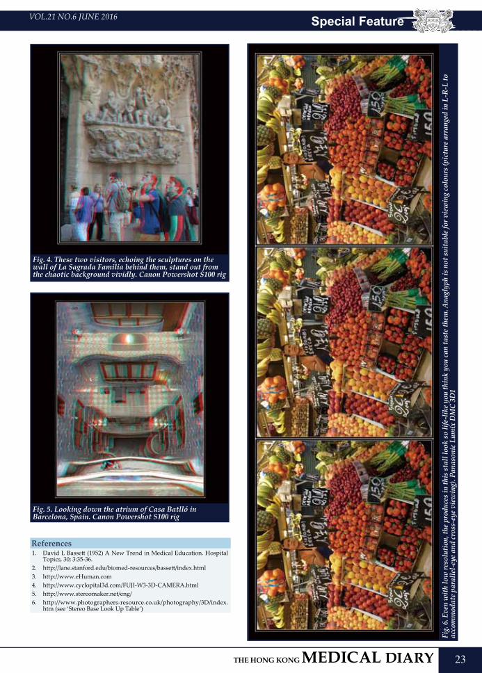
Fig. 3. 2D just won’t do justice to this London Taxi framework at London Heathrow T3. Fujifilm FinePix REAL 3D W3
Special Feature
23
VOL.21 NO.6 JUNE 2016 Special Feature
Fig.
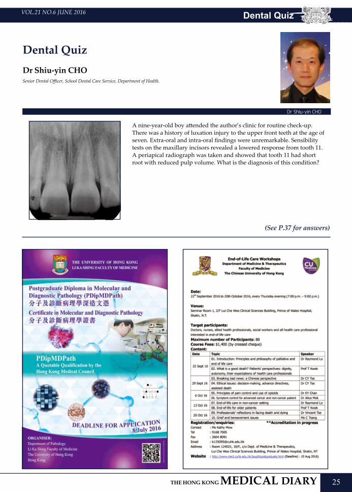
6. E
ven
wit
h lo
w re
solu
tion
, the
pro
duce
s in
this
stal
l loo
k so
life
-lik
e you
thin
k yo
u ca
n ta
ste t
hem
. Ana
glyp
h is
not
suit
able
for v
iew
ing
colo
urs (
pict
ure a
rran
ged
in L
-R-L
to
acco
mm
odat
e par
alle
l-ey
e and
cros
s-ey
e vie
win
g). P
anas
onic
Lum
ix D
MC
3D1
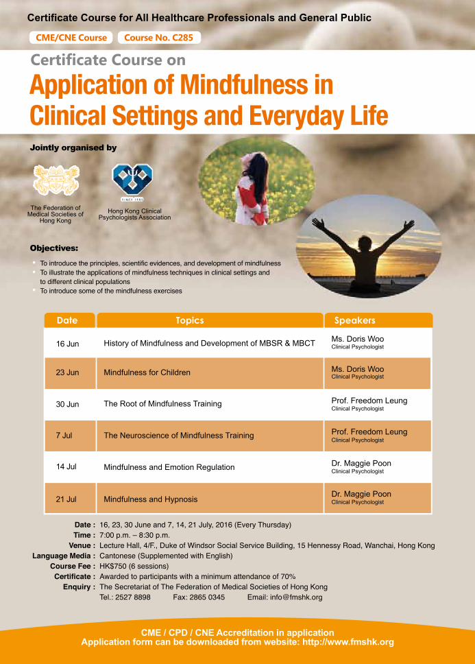
Fig. 4. These two visitors, echoing the sculptures on the wall of La Sagrada Familia behind them, stand out from the chaotic background vividly. Canon Powershot S100 rig
Fig. 5. Looking down the atrium of Casa Batlló in Barcelona, Spain. Canon Powershot S100 rig
References1. David L Bassett (1952) A New Trend in Medical Education. Hospital
Topics, 30; 3:35-36.2. http://lane.stanford.edu/biomed-resources/bassett/index.html3. http://www.eHuman.com4. http://www.cyclopital3d.com/FUJI-W3-3D-CAMERA.html5. http://www.stereomaker.net/eng/6. http://www.photographers-resource.co.uk/photography/3D/index.
htm (see ‘Stereo Base Look Up Table’)
25
VOL.21 NO.6 JUNE 2016 Dental Quiz
Dental QuizDr Shiu-yin CHO
A nine-year-old boy attended the author’s clinic for routine check-up. There was a history of luxation injury to the upper front teeth at the age of seven. Extra-oral and intra-oral findings were unremarkable. Sensibility tests on the maxillary incisors revealed a lowered response from tooth 11. A periapical radiograph was taken and showed that tooth 11 had short root with reduced pulp volume. What is the diagnosis of this condition?
Senior Dental Officer, School Dental Care Service, Department of Health.
(See P.37 for answers)
Dr Shiu-yin CHO
27
VOL.21 NO.6 JUNE 2016 Life Style
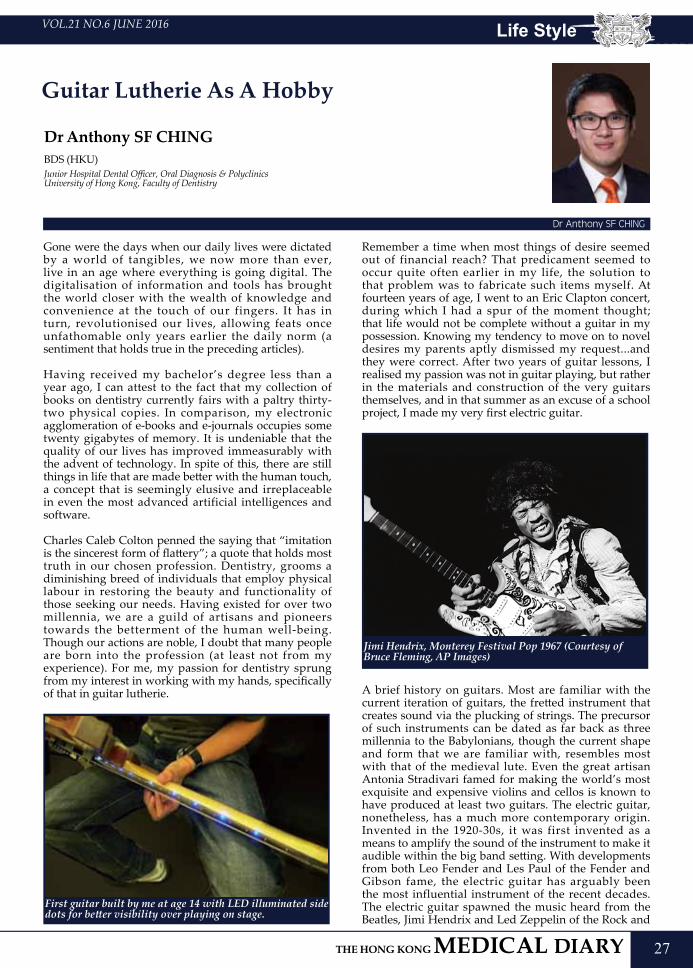
Guitar Lutherie As A Hobby
Dr Anthony SF CHINGBDS (HKU)Junior Hospital Dental Officer, Oral Diagnosis & PolyclinicsUniversity of Hong Kong, Faculty of Dentistry
Dr Anthony SF CHING
Gone were the days when our daily lives were dictated by a world of tangibles, we now more than ever, live in an age where everything is going digital. The digitalisation of information and tools has brought the world closer with the wealth of knowledge and convenience at the touch of our fingers. It has in turn, revolutionised our lives, allowing feats once unfathomable only years earlier the daily norm (a sentiment that holds true in the preceding articles).
Having received my bachelor’s degree less than a year ago, I can attest to the fact that my collection of books on dentistry currently fairs with a paltry thirty-two physical copies. In comparison, my electronic agglomeration of e-books and e-journals occupies some twenty gigabytes of memory. It is undeniable that the quality of our lives has improved immeasurably with the advent of technology. In spite of this, there are still things in life that are made better with the human touch, a concept that is seemingly elusive and irreplaceable in even the most advanced artificial intelligences and software.
Charles Caleb Colton penned the saying that “imitation is the sincerest form of flattery”; a quote that holds most truth in our chosen profession. Dentistry, grooms a diminishing breed of individuals that employ physical labour in restoring the beauty and functionality of those seeking our needs. Having existed for over two millennia, we are a guild of artisans and pioneers towards the betterment of the human well-being. Though our actions are noble, I doubt that many people are born into the profession (at least not from my experience). For me, my passion for dentistry sprung from my interest in working with my hands, specifically of that in guitar lutherie.
First guitar built by me at age 14 with LED illuminated side dots for better visibility over playing on stage.
Remember a time when most things of desire seemed out of financial reach? That predicament seemed to occur quite often earlier in my life, the solution to that problem was to fabricate such items myself. At fourteen years of age, I went to an Eric Clapton concert, during which I had a spur of the moment thought; that life would not be complete without a guitar in my possession. Knowing my tendency to move on to novel desires my parents aptly dismissed my request...and they were correct. After two years of guitar lessons, I realised my passion was not in guitar playing, but rather in the materials and construction of the very guitars themselves, and in that summer as an excuse of a school project, I made my very first electric guitar.
Jimi Hendrix, Monterey Festival Pop 1967 (Courtesy of Bruce Fleming, AP Images)
A brief history on guitars. Most are familiar with the current iteration of guitars, the fretted instrument that creates sound via the plucking of strings. The precursor of such instruments can be dated as far back as three millennia to the Babylonians, though the current shape and form that we are familiar with, resembles most with that of the medieval lute. Even the great artisan Antonia Stradivari famed for making the world’s most exquisite and expensive violins and cellos is known to have produced at least two guitars. The electric guitar, nonetheless, has a much more contemporary origin. Invented in the 1920-30s, it was first invented as a means to amplify the sound of the instrument to make it audible within the big band setting. With developments from both Leo Fender and Les Paul of the Fender and Gibson fame, the electric guitar has arguably been the most influential instrument of the recent decades. The electric guitar spawned the music heard from the Beatles, Jimi Hendrix and Led Zeppelin of the Rock and
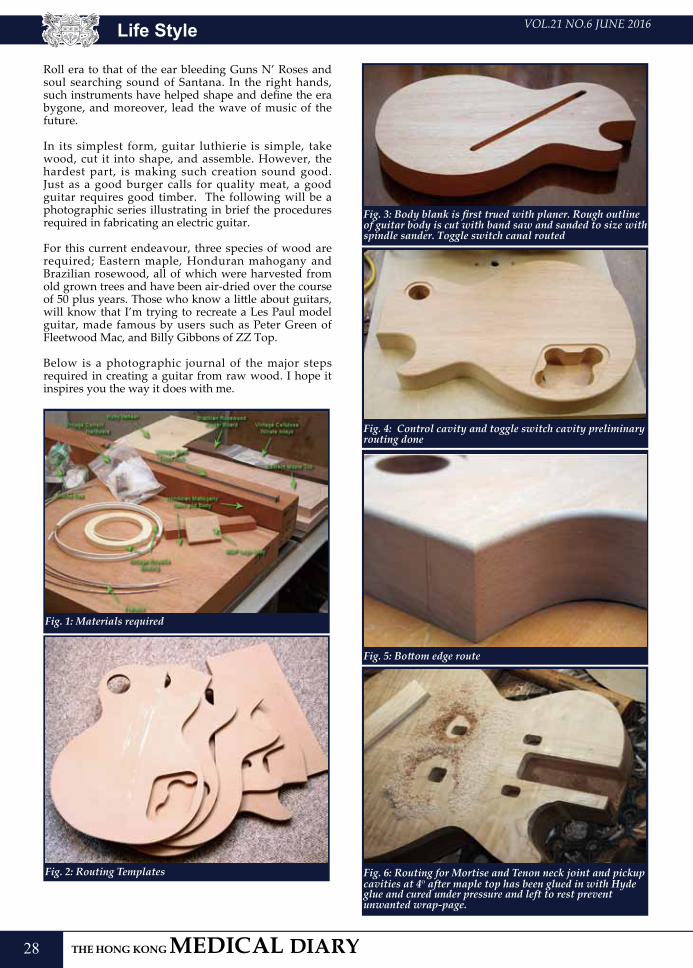
28
VOL.21 NO.6 JUNE 2016
Roll era to that of the ear bleeding Guns N’ Roses and soul searching sound of Santana. In the right hands, such instruments have helped shape and define the era bygone, and moreover, lead the wave of music of the future.
In its simplest form, guitar luthierie is simple, take wood, cut it into shape, and assemble. However, the hardest part, is making such creation sound good. Just as a good burger calls for quality meat, a good guitar requires good timber. The following will be a photographic series illustrating in brief the procedures required in fabricating an electric guitar.
For this current endeavour, three species of wood are required; Eastern maple, Honduran mahogany and Brazilian rosewood, all of which were harvested from old grown trees and have been air-dried over the course of 50 plus years. Those who know a little about guitars, will know that I’m trying to recreate a Les Paul model guitar, made famous by users such as Peter Green of Fleetwood Mac, and Billy Gibbons of ZZ Top.
Below is a photographic journal of the major steps required in creating a guitar from raw wood. I hope it inspires you the way it does with me.
Fig. 1: Materials required
Fig. 2: Routing Templates
Fig. 3: Body blank is first trued with planer. Rough outline of guitar body is cut with band saw and sanded to size with spindle sander. Toggle switch canal routed
Fig. 4: Control cavity and toggle switch cavity preliminary routing done
Fig. 5: Bottom edge route
Fig. 6: Routing for Mortise and Tenon neck joint and pickup cavities at 4o after maple top has been glued in with Hyde glue and cured under pressure and left to rest prevent unwanted wrap-page.
Life Style
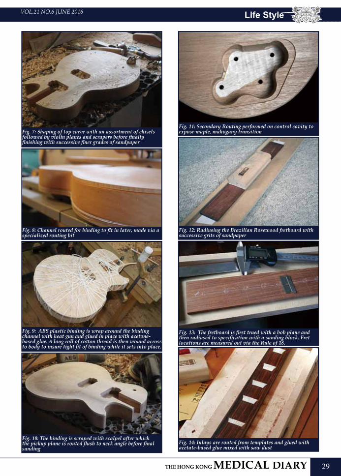
29
VOL.21 NO.6 JUNE 2016 Life Style
Fig. 7: Shaping of top curve with an assortment of chisels followed by violin planes and scrapers before finally finishing with successive finer grades of sandpaper
Fig. 8: Channel routed for binding to fit in later, made via a specialized routing bit
Fig. 9: ABS plastic binding is wrap around the binding channel with heat gun and glued in place with acetone-based glue. A long roll of cotton thread is then wound across to body to insure tight fit of binding while it sets into place.
Fig. 10: The binding is scraped with scalpel after which the pickup plane is routed flush to neck angle before final sanding
Fig. 11: Secondary Routing performed on control cavity to expose maple, mahogany transition
Fig. 12: Radiusing the Brazilian Rosewood fretboard with successive grits of sandpaper
Fig. 13: The fretboard is first trued with a bob plane and then radiused to specification with a sanding block. Fret locations are measured out via the Rule of 18.
Fig. 14: Inlays are routed from templates and glued with acetate-based glue mixed with saw dust
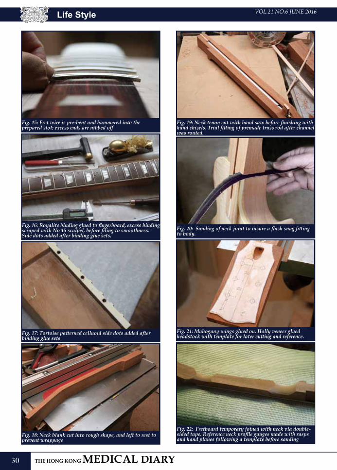
30
VOL.21 NO.6 JUNE 2016
Fig. 15: Fret wire is pre-bent and hammered into the prepared slot; excess ends are nibbed off
Fig. 16: Royalite binding glued to fingerboard, excess binding scraped with No 15 scalpel, before filing to smoothness. Side dots added after binding glue sets.
Fig. 17: Tortoise patterned celluoid side dots added after binding glue sets
Fig. 18: Neck blank cut into rough shape, and left to rest to prevent wrappage
Fig. 19: Neck tenon cut with band saw before finishing with hand chisels. Trial fitting of premade truss rod after channel was routed.
Fig. 20: Sanding of neck joint to insure a flush snug fitting to body.
Fig. 21: Mahogany wings glued on. Holly veneer glued headstock with template for later cutting and reference.
Fig. 22: Fretboard temporary joined with neck via double-sided tape. Reference neck profile gauges made with rasps and hand planes following a template before sanding
Life Style
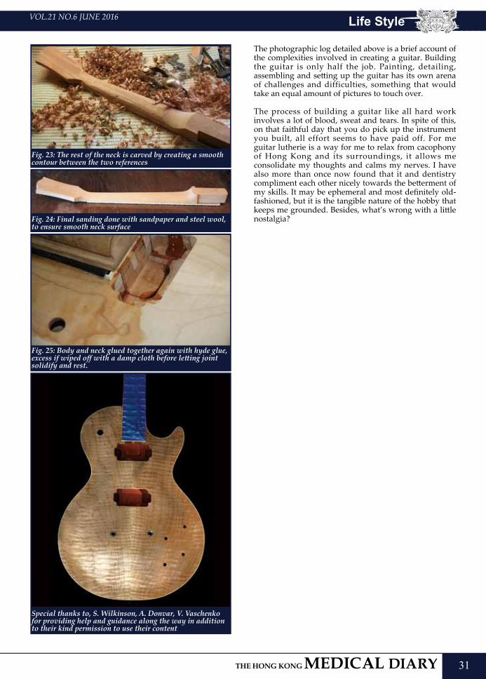
31
VOL.21 NO.6 JUNE 2016
Fig. 23: The rest of the neck is carved by creating a smooth contour between the two references
Fig. 24: Final sanding done with sandpaper and steel wool, to ensure smooth neck surface
Fig. 25: Body and neck glued together again with hyde glue, excess if wiped off with a damp cloth before letting joint solidify and rest.
Special thanks to, S. Wilkinson, A. Donvar, V. Vaschenko for providing help and guidance along the way in addition to their kind permission to use their content
The photographic log detailed above is a brief account of the complexities involved in creating a guitar. Building the guitar is only half the job. Painting, detailing, assembling and setting up the guitar has its own arena of challenges and difficulties, something that would take an equal amount of pictures to touch over.
The process of building a guitar like all hard work involves a lot of blood, sweat and tears. In spite of this, on that faithful day that you do pick up the instrument you built, all effort seems to have paid off. For me guitar lutherie is a way for me to relax from cacophony of Hong Kong and its surroundings, it allows me consolidate my thoughts and calms my nerves. I have also more than once now found that it and dentistry compliment each other nicely towards the betterment of my skills. It may be ephemeral and most definitely old-fashioned, but it is the tangible nature of the hobby that keeps me grounded. Besides, what’s wrong with a little nostalgia?
Life Style
Venue or MeetingFacilities
Member Society(Hourly Rate HK$)
PeakHour
Non-PeakHour
All day Sats, Suns &
Public Holidays
Rental Fees of Meeting Room and Facilities at The Federation of Medical Societies of Hong Kong
Non-Member Society(Hourly Rate HK$)
Multifunction Room I (Max 15 persons)Council Chamber (Max 20 persons)Lecture Hall (Max 100 persons)
150.00
240.00
300.00
105.00
168.00
210.00
225.00
360.00
450.00
250.00
400.00
500.00
175.00
280.00
350.00
375.00
600.00
750.00
PeakHour
Non-PeakHour
All day Sats, Suns &
Public Holidays
(Effective from October 2009)
Non-Peak Hour: 9:30am - 5:30pmPeak Hour: 5:30pm - 10:30pm
LCD Projector Microphone System
500.00 per session50.00 per hour, minimum 2 hours
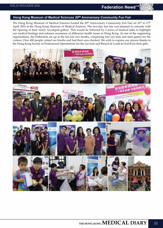
33
VOL.21 NO.6 JUNE 2016
The Hong Kong Museum of Medical Sciences hosted the 20th Anniversary Community Fun Fair on 16th to 17th April 2016 at the Hong Kong Museum of Medical Sciences. The two-day fun fair was planned to coincide with the opening of their newly revamped gallery. This would be followed by a series of medical talks to highlight our medical heritage and enhance awareness of difference health issues in Hong Kong. As one of the supporting organisations, the Federation set up at the fun fair two booths, comprising free eye tests and mini games for the visitors. Over 400 people visited our booths and had their eyes checked. We wish to express our sincere thanks to the Hong Kong Society of Professional Optometrists for the eye tests and Bausch & Lomb & Oral-B for their gifts.
Federation News
Hong Kong Museum of Medical Sciences 20th Anniversary Community Fun Fair
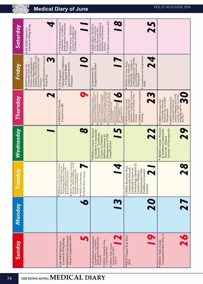
34
VOL.21 NO.6 JUNE 2016Medical Diary of JuneSu
nday
Mon
day
Tues
day
Wed
nesd
ayT
hurs
day
Frid
aySa
turd
ay
1617
18
9103
11
1912
2021
262713
14158
65
22
7
21
4
2323
293030
28
2425
HK
MA
CF
Cha
rity
C
once
rt fo
r SJS
HK
MA
Kow
loon
City
C
omm
unity
Net
wor
k -
Sem
inar
on
Man
agem
ent o
f C
omm
on B
reas
tfeed
ing
Prob
lem
s: W
hat P
rim
ary
Car
e D
octo
rs N
eed
to K
now
and
Pr
actic
e?
17th
Reg
iona
l O
steo
poro
sis
Con
fere
nce
- A C
onjo
int B
attle
ag
ains
t Ost
eopo
rotic
Fr
actu
res
Acu
pres
sure
for
Sym
ptom
atic
Rel
ief
17th
Reg
iona
l Ost
eopo
rosi
s C
onfe
renc
e - A
Con
join
t Ba
ttle
agai
nst O
steo
poro
tic
Frac
ture
s
HK
MA
KEC
N, H
KC
FP &
U
CH
– C
ME
Cou
rse
for
Hea
lth P
erso
nnel
201
6 (S
essi
on 2
): U
nder
stan
ding
D
emen
tia a
nd D
emen
tia
Serv
ices
HK
MA
Kow
loon
Eas
t C
omm
unity
Net
wor
k -
Ref
resh
er T
rain
ing
Cou
rse
on
Dem
entia
for P
rim
ary
Car
e D
octo
rs
HK
MA
Kow
loon
Eas
t C
omm
unity
Net
wor
k -
Prac
tical
Tip
s fo
r GER
D
Man
agem
ent
HK
MA
Hon
g K
ong
East
C
omm
unity
Net
wor
k -
Ann
ual M
eetin
g cu
m C
ME
Lect
ure
on "H
ow to
Avo
id
bein
g Br
ough
t to
the
PIC
?"
HK
MA
Yau
Tsi
m M
ong
Com
mun
ity N
etw
ork
- M
acul
opat
hy -
Upd
ate
on
Ant
ihyp
ergl
ycem
ic T
hera
py
beyo
nd D
iagn
osis
and
M
anag
emen
tA
cupr
essu
re fo
r Sym
ptom
atic
R
elie
f
FMSH
K E
xecu
tive
Com
mitt
ee
Mee
ting
HK
MA
Car
eer S
emin
ar 2
016
CM
E Le
ctur
e - R
efre
sher
C
ours
e fo
r Hea
lth C
are
Prov
ider
s 20
15/2
016
17th
Reg
iona
l Ost
eopo
rosi
s C
onfe
renc
e - A
Con
join
t Ba
ttle
agai
nst O
steo
poro
tic
Frac
ture
s
HK
MA
Ten
pin
Bow
ling
Tour
nam
ent 2
016
HK
MA
Tab
le T
enni
s To
urna
men
t 201
6 (D
ay 1
)
RSC
P D
rago
n Bo
at R
ace
2016
HK
MA
Kow
loon
Wes
t C
omm
unity
Net
wor
k -
Cer
tifica
te C
ours
e on
D
erm
atol
ogy
(Ses
sion
2):
Com
mon
Pitf
alls
in
Man
agem
ent o
f Ski
n Pr
oble
ms
in G
ener
al
Prac
tice
HK
MA
Cen
tral
, Wes
tern
&
Sou
ther
n C
omm
unity
N
etw
ork
- Pat
ient
Jo
urne
y of
Ost
eopo
rosi
s
HK
MA
Tab
le T
enni
s To
urna
men
t 201
6 (D
ay 2
)
HK
MA
Kow
loon
Wes
t Com
mun
ity
Net
wor
k - C
ertifi
cate
Cou
rse
on
Der
mat
olog
y (S
essi
on 1
): D
iagn
osis
an
d M
anag
emen
t of A
cne
and
Ros
acea
in C
linic
al P
ract
ice
HK
MA
Hon
g K
ong
East
Com
mun
ity
Net
wor
k - T
he M
anag
emen
t of H
eart
Fai
lure
- C
urre
nt a
nd N
ew M
edic
atio
n O
verv
iew
HK
MA
Kow
loon
Eas
t Com
mun
ity N
etw
ork
- La
test
CO
PD M
anag
emen
tH
KM
A N
ew T
errit
orie
s Wes
t Com
mun
ity
Net
wor
k - C
ertifi
cate
Cou
rse
on D
iabe
tes
Mel
litus
(Ses
sion
3) -
Str
ateg
ies t
o M
otiv
ate
the
DM
Pat
ient
s to
Ach
ieve
the
Trea
tmen
t Tar
gets
HK
MA
Sha
tin D
octo
rs N
etw
ork
- Bac
k Pa
in
or A
nkyl
osin
g Sp
ondy
litis
: Ref
erra
l Str
ateg
y fo
r Ear
ly R
ecog
nitio
n of
Axi
al
Spon
dylo
arth
ritis
HK
MA
Str
uctu
red
CM
E Pr
ogra
mm
e w
ith H
ong
Kon
g Sa
nato
rium
& H
ospi
tal Y
ear
2016
– A
n U
pdat
e on
N
asop
hary
ngea
l Can
cer
Scre
enin
g
HK
MA
Cen
tral
, Wes
tern
&
Sou
ther
n C
omm
unity
N
etw
ork
- Cer
tifica
te
Cou
rse
on D
iabe
tes
Mel
litus
(Ses
sion
4):
Man
agem
ent o
f DM
C
ompl
icat
ions
HK
MA
Yau
Tsi
m M
ong
Com
mun
ity
Net
wor
k - S
GLT
2 In
hibi
tors
- an
All
Rou
nded
Ant
ihyp
ergl
ycem
ic
Ther
apy
beyo
nd H
bA1c
HK
MA
Cou
ncil
Mee
ting
FMSH
K O
ffice
rs’ M
eetin
g
6th
IDK
D In
tens
ive
Cou
rse
in
Hon
g K
ong
6th
IDK
D In
tens
ive
Cou
rse
in H
ong
Kon
g
6th
IDK
D In
tens
ive
Cou
rse
in H
ong
Kon
gH
KM
APS
2nd
Sea
sona
l Ph
oto
Com
petit
ion
2016
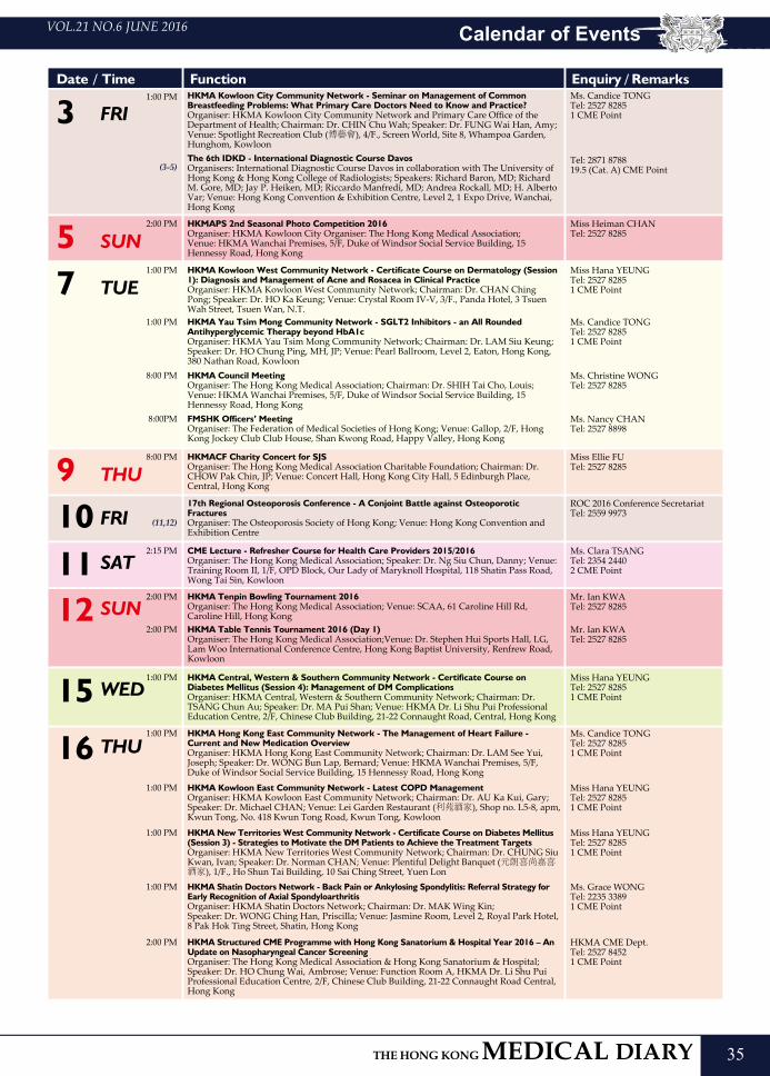
35
VOL.21 NO.6 JUNE 2016 Calendar of Events
Ms. Candice TONGTel: 2527 82851 CME Point
Date / Time Function Enquiry / Remarks
FRI3HKMA Kowloon City Community Network - Seminar on Management of Common Breastfeeding Problems: What Primary Care Doctors Need to Know and Practice?Organiser: HKMA Kowloon City Community Network and Primary Care Office of the Department of Health; Chairman: Dr. CHIN Chu Wah; Speaker: Dr. FUNG Wai Han, Amy;Venue: Spotlight Recreation Club (博藝會), 4/F., Screen World, Site 8, Whampoa Garden, Hunghom, Kowloon
HKMAPS 2nd Seasonal Photo Competition 2016Organiser: HKMA Kowloon City Organiser: The Hong Kong Medical Association;Venue: HKMA Wanchai Premises, 5/F, Duke of Windsor Social Service Building, 15 Hennessy Road, Hong Kong
1:00 PM
Miss Heiman CHANTel: 2527 8285SUN5
2:00 PM
HKMACF Charity Concert for SJSOrganiser: The Hong Kong Medical Association Charitable Foundation; Chairman: Dr. CHOW Pak Chin, JP; Venue: Concert Hall, Hong Kong City Hall, 5 Edinburgh Place, Central, Hong Kong
Miss Ellie FUTel: 2527 8285THU9
8:00 PM
CME Lecture - Refresher Course for Health Care Providers 2015/2016Organiser: The Hong Kong Medical Association; Speaker: Dr. Ng Siu Chun, Danny; Venue: Training Room II, 1/F, OPD Block, Our Lady of Maryknoll Hospital, 118 Shatin Pass Road, Wong Tai Sin, Kowloon
Ms. Clara TSANGTel: 2354 24402 CME PointSAT11
2:15 PM
HKMA Central, Western & Southern Community Network - Certificate Course on Diabetes Mellitus (Session 4): Management of DM ComplicationsOrganiser: HKMA Central, Western & Southern Community Network; Chairman: Dr. TSANG Chun Au; Speaker: Dr. MA Pui Shan; Venue: HKMA Dr. Li Shu Pui Professional Education Centre, 2/F, Chinese Club Building, 21-22 Connaught Road, Central, Hong Kong
Miss Hana YEUNGTel: 2527 82851 CME PointWED15
1:00 PM
HKMA Hong Kong East Community Network - The Management of Heart Failure - Current and New Medication OverviewOrganiser: HKMA Hong Kong East Community Network; Chairman: Dr. LAM See Yui, Joseph; Speaker: Dr. WONG Bun Lap, Bernard; Venue: HKMA Wanchai Premises, 5/F, Duke of Windsor Social Service Building, 15 Hennessy Road, Hong Kong
Ms. Candice TONGTel: 2527 82851 CME PointTHU16
1:00 PM
HKMA Kowloon East Community Network - Latest COPD ManagementOrganiser: HKMA Kowloon East Community Network; Chairman: Dr. AU Ka Kui, Gary; Speaker: Dr. Michael CHAN; Venue: Lei Garden Restaurant (利苑酒家), Shop no. L5-8, apm, Kwun Tong, No. 418 Kwun Tong Road, Kwun Tong, Kowloon
Miss Hana YEUNGTel: 2527 82851 CME Point
1:00 PM
HKMA New Territories West Community Network - Certificate Course on Diabetes Mellitus (Session 3) - Strategies to Motivate the DM Patients to Achieve the Treatment TargetsOrganiser: HKMA New Territories West Community Network; Chairman: Dr. CHUNG Siu Kwan, Ivan; Speaker: Dr. Norman CHAN; Venue: Plentiful Delight Banquet (元朗喜尚嘉喜酒家), 1/F., Ho Shun Tai Building, 10 Sai Ching Street, Yuen Lon
Miss Hana YEUNGTel: 2527 82851 CME Point
1:00 PM
HKMA Shatin Doctors Network - Back Pain or Ankylosing Spondylitis: Referral Strategy for Early Recognition of Axial SpondyloarthritisOrganiser: HKMA Shatin Doctors Network; Chairman: Dr. MAK Wing Kin;Speaker: Dr. WONG Ching Han, Priscilla; Venue: Jasmine Room, Level 2, Royal Park Hotel, 8 Pak Hok Ting Street, Shatin, Hong Kong
Ms. Grace WONGTel: 2235 33891 CME Point
1:00 PM
HKMA Structured CME Programme with Hong Kong Sanatorium & Hospital Year 2016 – An Update on Nasopharyngeal Cancer ScreeningOrganiser: The Hong Kong Medical Association & Hong Kong Sanatorium & Hospital; Speaker: Dr. HO Chung Wai, Ambrose; Venue: Function Room A, HKMA Dr. Li Shu Pui Professional Education Centre, 2/F, Chinese Club Building, 21-22 Connaught Road Central, Hong Kong
HKMA CME Dept.Tel: 2527 84521 CME Point
2:00 PM
HKMA Tenpin Bowling Tournament 2016Organiser: The Hong Kong Medical Association; Venue: SCAA, 61 Caroline Hill Rd, Caroline Hill, Hong Kong
Mr. Ian KWATel: 2527 8285SUN12
2:00 PM
HKMA Table Tennis Tournament 2016 (Day 1)Organiser: The Hong Kong Medical Association;Venue: Dr. Stephen Hui Sports Hall, LG, Lam Woo International Conference Centre, Hong Kong Baptist University, Renfrew Road, Kowloon
Mr. Ian KWATel: 2527 8285
2:00 PM
17th Regional Osteoporosis Conference - A Conjoint Battle against Osteoporotic FracturesOrganiser: The Osteoporosis Society of Hong Kong; Venue: Hong Kong Convention and Exhibition Centre
ROC 2016 Conference SecretariatTel: 2559 9973FRI10
HKMA Kowloon West Community Network - Certificate Course on Dermatology (Session 1): Diagnosis and Management of Acne and Rosacea in Clinical PracticeOrganiser: HKMA Kowloon West Community Network; Chairman: Dr. CHAN Ching Pong; Speaker: Dr. HO Ka Keung; Venue: Crystal Room IV-V, 3/F., Panda Hotel, 3 Tsuen Wah Street, Tsuen Wan, N.T.
Miss Hana YEUNGTel: 2527 82851 CME PointTUE7
1:00 PM
HKMA Yau Tsim Mong Community Network - SGLT2 Inhibitors - an All Rounded Antihyperglycemic Therapy beyond HbA1cOrganiser: HKMA Yau Tsim Mong Community Network; Chairman: Dr. LAM Siu Keung; Speaker: Dr. HO Chung Ping, MH, JP; Venue: Pearl Ballroom, Level 2, Eaton, Hong Kong, 380 Nathan Road, Kowloon
Ms. Candice TONGTel: 2527 82851 CME Point
1:00 PM
HKMA Council MeetingOrganiser: The Hong Kong Medical Association; Chairman: Dr. SHIH Tai Cho, Louis; Venue: HKMA Wanchai Premises, 5/F, Duke of Windsor Social Service Building, 15 Hennessy Road, Hong Kong
Ms. Christine WONGTel: 2527 8285
8:00 PM
FMSHK Officers’ MeetingOrganiser: The Federation of Medical Societies of Hong Kong; Venue: Gallop, 2/F, Hong Kong Jockey Club Club House, Shan Kwong Road, Happy Valley, Hong Kong
Ms. Nancy CHANTel: 2527 8898
8:00PM
(3-5)
(11,12)
Tel: 2871 878819.5 (Cat. A) CME Point
The 6th IDKD - International Diagnostic Course DavosOrganisers: International Diagnostic Course Davos in collaboration with The University of Hong Kong & Hong Kong College of Radiologists; Speakers: Richard Baron, MD; Richard M. Gore, MD; Jay P. Heiken, MD; Riccardo Manfredi, MD; Andrea Rockall, MD; H. Alberto Var; Venue: Hong Kong Convention & Exhibition Centre, Level 2, 1 Expo Drive, Wanchai, Hong Kong

36
VOL.21 NO.6 JUNE 2016Calendar of Events
Ms. Nicki PANGTel: 9320 50765 CNE Point
Date / Time Function Enquiry / Remarks
FRI17Acupressure for Symptomatic ReliefOrganisers: Hong Kong College of Chinese Medicinal Nursing & Continue Health Care Education Centre, Hong Kong Baptist Hospital; Chairman: Ms HUI Yin Hing; Speakers: Mr LAU Pak Sing; Ms PANG Wai Sum Nicki; Venue: Institute of Professional Education Continue Health Care Education Centre
HKMA KECN, HKCFP & UCH – CME Course for Health Personnel 2016 (Session 2): Understanding Dementia and Dementia ServicesOrganiser: HKMA Kowloon East Community Network & Hong Kong College of Family Physicians & United Christian Hospital; Chairman: Dr. MA Ping Kwan, Danny; Speaker: Dr. SHA Kwok Yiu, Edmund; Venue: 1. Lecture Theatre, G/F, Block K, United Christian Hospital (UCH), 130 Hip Wo Street, Kwun Tong, Kowloon 2. Conference Room, G/F, Block K, UCH (video conference)
6:30 PM(24)
Miss Denise KWOKTel: 2527 8285SUN19 RSCP Dragon Boat Race 2016
Organiser: The Hong Kong Institute of Architects; Chairman: Dr. YAM Chun YinVenue: Stanley Main Beach
8:00 AM
Ms. Nancy CHANTel: 2527 8898
THU23FMSHK Executive Committee MeetingOrganiser: The Federation of Medical Societies of Hong Kong; Venue: Council Chamber, 4/F, Duke of Windor Social Service Building, 15 Hennessy Road, Wanchai, Hong Kong
8:00 PM
Ms. Nancy CHANTel: 2527 8898
HKMA Kowloon East Community Network - Refresher Training Course on Dementia for Primary Care DoctorsOrganiser: HKMA Kowloon East Community Network and Institute of Alzheimer’s Education (IAE) of Hong Kong Alzheimer’s Disease Association; Chairman: Dr. AU Ka Kui, Gary; Speaker: Dr. DAI Lok Kwan, David, JP; Venue: Gingko House, G/F, Cheerful Court, 55 Choi Ha Road, Ngau Tau Kok, Kowloon
1:00 PM
Mr. Ian KWATel: 2527 8285SUN26
HKMA Table Tennis Tournament 2016 (Day 2)Organiser: The Hong Kong Medical Association; Venue: Dr. Stephen Hui Sports Hall, LG, Lam Woo International Conference Centre, Hong Kong Baptist University, Renfrew Road, Kowloon
2:00 PM
Miss Hana YEUNGTel: 2527 82851.5 CME PointSAT18
1:30 PM
HKMA Kowloon West Community Network - Certificate Course on Dermatology (Session 2): Common Pitfalls in Management of Skin Problems in General PracticeOrganiser: HKMA Kowloon West Community Network; Chairman: Dr. WONG Wai Hong, Bruce; Speaker: Dr. LAM Wai Sun; Venue: Crystal Room IV-V, 3/F., Panda Hotel, 3 Tsuen Wah Street, Tsuen Wan, N.T.
Miss Hana YEUNGTel: 2527 82851 CME PointTUE21
1:00 PM
HKMA Yau Tsim Mong Community Network - Maculopathy - Update on Antihyperglycemic Therapy beyond Diagnosis and ManagementOrganiser: HKMA Yau Tsim Mong Community Network; Chairman: Dr. CHAN Wai Keung, Ricky; Speaker: Dr. NG Sin Yee, Anita; Venue: Jade Ballroom, Level 2, Eaton, Hong Kong, 380 Nathan Road, Kowloon
Ms. Candice TONGTel: 2527 82851 CME PointFRI24
1:00 PM
HKMA Central, Western & Southern Community Network - Patient Journey of OsteoporosisOrganiser: HKMA Central, Western & Southern Community Network; Chairman: Dr. LAM Ming Yuen; Speaker: Dr. TING Zhao Wei, Rose;Venue: HKMA Dr. Li Shu Pui Professional Education Centre, 2/F, Chinese Club Building, 21-22 Connaught Road, Central, Hong Kong
Miss Hana YEUNGTel: 2527 82851 CME PointWED29
1:00 PM
HKMA Kowloon East Community Network - Practical Tips for GERD ManagementOrganiser: HKMA Kowloon East Community Network; Chairman: Dr. MA Ping Kwan, Danny; Speaker: Dr. NG Ho, Paul; Venue: V Cuisine, 6/F., Holiday Inn Express Hong Kong Kowloon East, 3 Tong Tak Street, Tseung Kwan O
Miss Hana YEUNGTel: 2527 82851 CME PointTHU30
1:00 PM
HKMA Hong Kong East Community Network - Annual Meeting cum CME Lecture on "How to Avoid being Brought to the PIC?"Organiser: HKMA Hong Kong East Community Network; Chairman: Dr. CHAN Nim Tak, Douglas; Speaker: Dr. CHOI Kin; Venue: HKMA Wanchai Premises, 5/F, Duke of Windsor Social Service Building, 15 Hennessy Road, Hong Kong
Ms. Candice TONGTel: 2527 82851 CME Point
1:00 PM
HKMA Career Seminar 2016Organiser: The Hong Kong Medical Association; Chairman: Dr. PONG Chiu Fai, Jeffrey;Venue: LKS Faculty of Medicine, HKU
Miss Kayin LEETel: 2527 8285
3:00 PM
Upcoming Meeting1-3/7/20168:00 AM
Ms. Jana LAMTel: 2871 8790Fax: 2554 07397 CME Point
2016 AIRP Course in Hong KongOrganiser: American Institute for Radiologic Pathology (AIRP) and Hong Kong College of Radiologists (HKCR); Speakers: Dr Mark D. MURPHEY; Dr Marilyn J. SIEGAL, MD; Dr Kelly K KOELLER, MD; Venue: Hong Kong Academy of Medicine Jockey Club Building99 Wong Chuk Hang Road, Aberdeen, Hong Kong SAR
23/7/201612:00 PM
Ms. Eva TSANGTel: 2821 3514Fax: 2865 0345
Hong Kong College of Health Service Executives Annual Conference 2016 - People, Technology, and innovationOrganiser: HKCHSE; Venue: Shanghai room, Level 8, Cordis Hong Kong, 555 Shanghai Street, Mongkok
8-9/10/2016 HKAC 2016 SecretariatTel: 2559 9973
The 9th Hong Kong Allergy Convention - Novel Strategies for Prevention and Treatment of Allergic DisordersOrganiser: Hong Kong Institute of Allergy; Venue: Hong Kong Convention and Exhibition Centre
37
VOL.21 NO.6 JUNE 2016
Answers to Dental Quiz
Answer:
The condition is called pulpal healing by regular bone and cementum. The radiograph showed that the lamina dura had extended into the pulp canal walls and was continuous with that on the external root surfaces. Experimental studies have disclosed various distinct pulpo-dentinal responses after severe luxation and displacement dental injury, which included:
1.2.3.4.5.6.7.
Regular tubular dentineIrregular reparative dentine with diminished tubular structuresIrregular reparative dentine with encapsulated cellsIrregular immature boneRegular mature bone and/or cementumInternal resorptionPulp necrosis
The last two categories were indications of failure to heal and active root canal treatment would be needed.
Reference: Andreasen JO, Andreasen FM and Andersson L. Textbook and colour atlas of traumatic injuries to the teeth. 4th Ed. Munksgaard:Copenhagen, 2007.
Dr Shiu-yin CHO Senior Dental Officer, School Dental Care Service, Department of Health.
Dental Quiz The Federation of Medical Societies of Hong Kong 4/F Duke of Windsor Social Service Building, 15 Hennessy Road, Wanchai, HK Tel: 2527 8898 Fax: 2865 0345
PresidentDr CHAK Wai-kwong, Mario 翟偉光醫生
1st Vice-PresidentDr MAN Chi-wai 文志衞醫生
2nd Vice-PresidentDr CHAN Chun-kwong, Jane 陳真光醫生
Hon. TreasurerMr LEE Cheung-mei, Benjamin 李祥美先生
Hon. SecretaryProf CHEUNG Man-yung, Bernard 張文勇教授
Deputy Hon. SecretaryDr NG Chun-kong 吳振江醫生
Immediate Past President Dr LO See-kit, Raymond 勞思傑醫生
Executive Committee Members
Dr CHAN Hau-ngai, Kingsley 陳厚毅醫生Dr CHAN Sai-kwing 陳世烱醫生Dr HUNG Wai-man 熊偉民醫生Ms KU Wai-yin, Ellen 顧慧賢女士Dr MOK Chun-on 莫鎮安醫生Dr NG Yin-kwok 吳賢國醫生Dr NGUYEN Gia-hung, Desmond 阮家興醫生Dr SO Man-kit, Thomas 蘇文傑醫生Dr TSOI Chun-hing, Ludwig 蔡振興醫生Dr WONG Sau-yan 黃守仁醫生Ms YAP Woan-tyng, Tina 葉婉婷女士Dr YU Chau-leung, Edwin 余秋良醫生Dr YUNG Shu-hang, Patrick 容樹恆醫生
Founder MembersBritish Medical Association (Hong Kong Branch)英國醫學會 ( 香港分會 )
PresidentDr LO See-kit, Raymond 勞思傑醫生
Vice-PresidentDr WU, Adrian 鄔揚源醫生
Hon. SecretaryDr HUNG Che-wai, Terry 洪致偉醫生
Hon. TreasurerDr Jason BROCKWELL
Council RepresentativesDr LO See-kit, Raymond 勞思傑醫生Dr CHEUNG Tse-ming 張子明醫生Tel: 2527 8898 Fax: 2865 0345
The Hong Kong Medical Association香港醫學會
PresidentDr SHIH Tai-cho, Louis, JP 史泰祖醫生 , JP
Vice- PresidentsDr CHAN Yee-shing, Alvin 陳以誠醫生Dr CHOW Pak Chin, JP 周伯展醫生 , JP
Hon. SecretaryDr LAM Tzit-yuen, David 林哲玄醫生
Hon. TreasurerDr LEUNG Chi-chiu 梁子超醫生
Council RepresentativesDr CHAN Yee-shing, Alvin 陳以誠醫生Dr CHOW Pak Chin, JP 周伯展醫生 , JP
Chief ExecutiveMs Jovi LAM 林偉珊女士Tel: 2527 8285 (General Office) 2527 8324 / 2536 9388 (Club House in Wanchai / Central)Fax: 2865 0943 (Wanchai), 2536 9398 (Central)Email: [email protected] Website: http://www.hkma.org
The HKFMS Foundation Limited 香港醫學組織聯會基金 Board of DirectorsPresident
Dr CHAK Wai-kwong, Mario 翟偉光醫生
1st Vice-PresidentDr MAN Chi-wai 文志衞醫生
2nd Vice-PresidentDr CHAN Chun-kwong, Jane 陳真光醫生
Hon. TreasurerMr LEE Cheung-mei, Benjamin 李祥美先生
Hon. SecretaryProf CHEUNG Man-yung, Bernard 張文勇教授
DirectorsMr CHAN Yan-chi, Samuel 陳恩賜先生Dr HUNG Wai-man 熊偉民醫生Ms KU Wai-yin, Ellen 顧慧賢女士Dr LO See-kit, Raymond 勞思傑醫生Dr YU Chak-man, Aaron 余則文醫生