holistic perception of the individual face is specific and ... · a université catholique de...
TRANSCRIPT
Ha
Ta
b
c
a
ARRAA
KASFHR
1
bchali&wd
w(1lt(i
PmT
0d
Neuropsychologia 48 (2010) 4057–4092
Contents lists available at ScienceDirect
Neuropsychologia
journa l homepage: www.e lsev ier .com/ locate /neuropsychologia
olistic perception of the individual face is specific and necessary: Evidence fromn extensive case study of acquired prosopagnosia
homas Busignya,∗, Sven Joubertb, Olivier Felicianc, Mathieu Ceccaldi c, Bruno Rossiona
Université Catholique de Louvain, Louvain-la-Neuve, BelgiumDépartement de Psychologie, Université de Montreal & Centre de Recherche Institut Universitaire de Gériatrie de Montréal, CanadaService de Neurologie et de Neuropsychologie, AP-HM Timone, & Laboratoire Epilepsie et Cognition, INSERM U 751, Université de la Méditerranée, Marseille, France
r t i c l e i n f o
rticle history:eceived 17 March 2010eceived in revised form 5 August 2010ccepted 16 September 2010
a b s t r a c t
We present an extensive investigation (24 experiments) of a new case of prosopagnosia following rightunilateral damage, GG, with the aim of addressing two classical issues: (1) Can a visual recognition impair-ment truly be specific to faces? (2) What is the nature of acquired prosopagnosia? We show that GGrecognizes nonface objects perfectly and quickly, even when it requires fine-grained analysis to individ-
vailable online 25 September 2010
eywords:cquired prosopagnosiapecificityace processingolistic perception
ualize these objects. He is also capable of perceiving objects and faces as integrated wholes, as indicatedby normal Navon effect, 3D-figures perception and perception of Mooney and Arcimboldo face stimuli.However, the patient could not perceive individual faces holistically, showing no inversion, composite, orwhole-part advantage effects for faces. We conclude that an occipito-temporal right hemisphere lesionmay lead to a specific impairment of holistic perception of individual items, a function that appearscritical for normal face recognition but not for object recognition.
ight hemisphere
. Introduction
The ability to recognize people from their face is a fundamentalrain function which holds a high social value. It is also an extremelyomplex function, which is nevertheless performed quite well inuman adults. The adult human brain has developed mechanismsllowing, for instance, recognizing a familiar person from its face iness than half a second (Bruce & Young, 1986), or encoding new facesn memory effortlessly during the entire life (e.g., Bahrick, Bahrick,
Wittinger, 1975). Yet, interestingly, the field of face recognitionas originally based upon the study of people who, following brainamage, have lost this expertise in recognizing faces.
Difficulty in face recognition as a major symptom in patientsith cerebral disease was first reported in the nineteenth century
Charcot, 1883; Quaglino & Borelli, 1867; Wigan, 1844; Wilbrandt,887). However, it was Bodamer (1947) who proposed to iso-
ate the disorder on the basis of three cases, and introduced theerm prosopagnosia from the Greek “prosopon” (face) and “a-gnosia”without knowledge). Prosopagnosia is classically defined as thenability to recognize individual faces following brain damage, an
∗ Corresponding author at: Université Catholique de Louvain (UCL), Faculté desychologie et des Sciences de l’Education (PSP), Unité de Cognition et Développe-ent (CODE), Place du Cardinal Mercier, 10, B-1348 Louvain-la-Neuve, Belgium.
el.: +32 010 47 92 60; fax: +32 010 47 37 74.E-mail address: [email protected] (T. Busigny).
028-3932/$ – see front matter © 2010 Elsevier Ltd. All rights reserved.oi:10.1016/j.neuropsychologia.2010.09.017
© 2010 Elsevier Ltd. All rights reserved.
impairment that cannot be attributed to intellectual deficiencies orlow-level visual problems (Benton, 1980; Bodamer, 1947; Hécaen& Angelergues, 1962; Rondot & Tzavaras, 1969). Prosopagnosicpatients also generally still retain their ability to recognize peo-ple by other cues: the voice or other visual traits such as gait, size,clothes, or even facial features (moustache, scar, freckles, . . .) oraccessories (ear-rings, eyeglasses, piercings, . . .).
Over the years, tens of cases of prosopagnosia following braindamage have been reported, although extensive neuropsycholog-ical investigations of prosopagnosic patients remain quite rare(e.g., Anaki, Kaufman, Freedman, & Moscovitch, 2007; Barton,2008a; Delvenne, Seron, Coyette, & Rossion, 2004; Lhermitte, Chain,Escourolle, Ducarne, & Pillon, 1972; Riddoch, Johnston, Bracewell,Boutsen, & Humphreys, 2008; Rossion et al., 2003; Sergent &Signoret, 1992a; Sergent & Villemure, 1989).
Both in traditional (cognitive) neuropsychology and in moderncognitive neuroscience, the lesion method is seen as an invalu-able and unique way of understanding normal brain function (e.g.,Caramazza, 1986; Damasio & Damasio, 1989; Farah, 1990; Farah,2004; Humphreys & Riddoch, 1987; Shallice, 1988), in particularwith respect to face recognition. Such patient studies contribute toshaping our knowledge and conceptions of the processes involved
in normal face recognition and their underlying neural networks.From a functional point of view, there are two main debatesconcerning prosopagnosia, which have direct implications forunderstanding face recognition: (1) Can the impairment truly berestricted to face recognition (i.e. face-specific)? (2) What is the
4 ycholo
ntTs1&
1
cpBDCr1&ic(tdRprap(mau(aitKawudsWogAochAnfisipnpnRcChifaa
recognition [. . .]. Recognition was based on characteristic details, notfaces per se [. . .]. What seems to be lacking in WL is the ability to createan integrated, unitary percept or a gestalt of a human face enablinghim to assign identity to an individual” (pp. 93, 98).
058 T. Busigny et al. / Neurops
ature of the disorder, that is, what is at the heart of our exper-ise in facial recognition, and which is lost in these patients?hese two issues have proved quite difficult to resolve and aretill debated (e.g., Barton, 2009; Damasio, Damasio, & Van Hoesen,982; De Renzi, 1986a; Hécaen, 1981; Riddoch et al., 2008; RondotTzavaras, 1969; Sergent & Signoret, 1992a).
.1. Prosopagnosia as a face-specific disorder
The issue of the specificity of the disorder has been compli-ated by the fact that most reported cases of prosopagnosia alsoresent with difficulties in basic-level object recognition (e.g.,arton, 2008a; Boutsen & Humphreys, 2002; Damasio et al., 1982;elvenne et al., 2004; Gauthier, Behrmann, & Tarr, 1999; Levine &alvanio, 1989; Steeves et al., 2006). In many other cases, objectecognition abilities were not tested sufficiently (e.g., De Renzi,986a; Ettlin et al., 1992; Tohgi et al., 1994; Young, Flude, Hay,Ellis, 1993). A brief but extensive review of the neuropsycholog-
cal literature points to 13 prosopagnosic patients who could beonsidered as presenting with a face-specific recognition disorderTable 1). De Renzi (1986a) presented patient 4 who performed inhe normal range at object and figure recognition, figure–groundiscrimination, visual closure and segmentation. Patient VA (Deenzi, Faglioni, Grossi, & Nichelli, 1991) could name objects andictures (presented under usual and unusual view) in the normalange, and succeeded at tasks of visual closure, coin discrimination,nd recognition of makes of cars and personal belongings. Anotheratient described by De Renzi, Perani, Carlesimo, Silveri, and Fazio1994), OR, was documented to present with an absence of impair-
ent with respect to object naming, Italian coins discrimination,nd recognition of animals, fruits and vegetables (under usual andnusual views). Takahashi, Kawamura, Hirayama, Shiota, and Isono1995), in a study of four patients, related the case of a patient withpparently no object recognition impairment: case 3 succeededn different tasks including overlapping figures, Gestalt comple-ion test, Kanizsa triangle and real object naming. Schweinberger,los, and Sommer (1995) and Henke, Schweinberger, Grigo, Klos,nd Sommer (1998) showed that the performance of patient MTas preserved in numerous tasks: recognizing overlapping fig-res, Gestalt completion task, object naming, animal naming andifferent series of similar objects to name (fruits and vegetables,ymbols of German industrial brands and cars brands). Patient
B (Buxbaum, Glosser, & Coslett, 1996) presented with preservedbject naming (real objects and drawings) and memory for homo-eneous category of objects (glasses, under different views). Patientnna (De Renzi & di Pellegrino, 1998) succeeded in several tasks:bjects naming (color photographs and line drawings), perceptualategorization, visual segmentation and closure, and memory foromogeneous category of objects (glasses, under different views).nother study (Wada & Yamamoto, 2001) also reported a prosopag-osic patient who could perform well the tasks of overlappinggures, picture copying, recognition of letters and symbols, visualpace perception, object naming (real objects, pictures, line draw-ngs; under usual and unusual view), animal face and famouslace recognition. Prosopagnosic patient PS was able to recog-ize objects perfectly and rapidly (Rossion et al., 2003) and coulderform within-category discrimination for nonface items in theormal range of performance and speed (Busigny, Graf, Mayer, &ossion, 2010; Schiltz et al., 2006). Barton and colleagues reportedase 009 (Barton, 2008a, 2009; Barton & Cherkasova, 2005; Barton,herkasova, Press, Intriligator, & O’Connor, 2004), a patient who
ad no low-level visual impairment and was able to recognizencomplete letters, overlapping figures, real objects, vegetables andruits, presented with a classical Navon effect, and showed somebility to process configurations of dots. Bukach, Bud, Gauthier,nd Tarr, 2006 and Bukach, Le Grand, Kaiser, Bub, and Tanaka, 2008
gia 48 (2010) 4057–4092
related another case of selective impairment for faces, LR, who suc-ceeded easily in tasks of low-level visual processing, silhouettesand object naming (under usual and unusual views). Riddoch et al.(2008) presented the case of FB, who had preserved abilities inlow-level visual processing, non-living and living (birds, flowers,vegetables and fruits) objects naming, and in a task of learning asso-ciations between names and novel multipart objects. Finally, Rivest,Moscovitch, and Black (2009) published the case of DC, who per-formed normally in segmented object recognition, object naming,recognition of famous buildings and dog breeds.
These pure cases of acquired prosopagnosia suggest that someprocesses may be necessary to recognize faces efficiently, and thatthese processes may be selectively disrupted by brain damage.While these processes might also be involved in object recognition,they would not be necessary for this function.
1.2. The holistic perception account of prosopagnosia
Regarding the nature of the impairment in prosopagnosia, aninfluential idea is that such patients have difficulties in perceiving aface as a whole, or a Gestalt. This long-standing view (Galli, 1964) isinspired originally from the Gestaltist approach of visual perception(e.g., Koffka, 1935/1963; Kohler, 1929; 1930/1971; Wertheimer,1925/1967). According to the Gestaltist view and its more modernrevival (e.g., Kubovy & Poremantz, 1981; Navon, 1977; for a reviewsee Kimchi, 1992), a whole item is qualitatively different from thesum of the components, the whole exceeding the sum of its parts.Hence, what takes place in each single part already depends uponwhat the whole is: objects are not only made of featural elements,but also defined by the interactions between these constituents,a property that is called configuration or (w)holistic property (e.g.,Navon, 2003). For instance, a face is a typical visual stimulus madeof parts (eyes, nose, mouth, . . .) that are organized in a whole con-figuration (a symmetrical structure with two eyes on top, above acentral nose and mouth).
The idea that acquired prosopagnosic patients lose their abilityto perceive faces holistically is supported at four levels.
First, many patients have been described as presenting witha configural/holistic1 processing impairment, that is, an inabil-ity to integrate simultaneously different features into a coherentglobal representation (RB, Davidoff, Matthews, & Newcombe, 1986;HJA, Boutsen & Humphreys, 2002; Riddoch & Humphreys, 1987;LH, Levine & Calvanio, 1989; BM, Sergent & Villemure, 1989; WL,Spillmann, Laskowski, Lange, Kasper, & Schmidt, 2000; AR, Saumier,Arguin, & Lassonde, 2001; RC, Wilkinson et al., 2009; PS, Ramon,Busigny, & Rossion, 2010). For example, Levine and Calvanio (1989)described the patient LH as being unable to “get an immediateoverview of a face [. . .] as a whole at a single glance” (p. 159). Theyconceptualized this loss of visual “configural [i.e. holistic] process-ing” as a deficit in visual perception reflected by the inability toderive an “overview of sufficient features to allow structuring or crys-tallization of a coherent concept”. In the same vein, Spillmann et al.(2000) described the prosopagnosic patient WL as following: “hewas unable to form a holistic percept of a given face that would haverevealed its bearer’s identity. Rather, he used conspicuous features for
1 In this paper, in keeping with earlier studies in the field of face recognition andrecent reviews of this issue, the terms holistic and configural are used interchange-ably, as synonyms, to refer to the same process (see McKone & Yovel, 2009; Rossion,2008a; Rossion, 2009).
ycholo
s&Mpupm
haaashEpleSYanqMf
ciiopwap2tlXtaalie2RtB2aSi
1p
(vtseNcp
T. Busigny et al. / Neurops
Second, very few cases of acquired prosopagnosia with pre-erved holistic face processing have been reported (PV, Sergent
Poncet; PC, Sergent & Signoret, 1992a; LR, Bukach et al., 2006).oreover, holistic processing was not tested extensively and with
articularly sensitive tests in these patients, so that it remainsnclear to what extent their holistic processing of faces was trulyreserved (see Ramon et al., 2010). This issue will be addressedore extensively in Section 4.Third, there is to date no solid and more accurate alternative
ypothesis to account for the functional impairment characterizingcquired prosopagnosia. For instance, the few alternative propos-ls in terms of low-level processing are no longer valid. Indeed,n account of prosopagnosia – or visual agnosia – in terms of sen-ory or low-level visual impairments (Bay, 1953; Ettlinger, 1956),as been dismissed for some time (De Haan, Heywood, Young,delstyn, & Newcombe, 1995; Rondot & Tzavaras, 1969), and manyrosopagnosic patients do not suffer from low-level visual prob-
ems (e.g., Bukach et al., 2006; Buxbaum et al., 1996; Delvennet al., 2004; Dixon, Bub, & Arguin, 1998; Eimer & McCarthy, 1999;chweinberger et al., 1995; Sergent & Poncet, 1990; Wada &amamoto, 2001). Even when low-level vision is impaired, suchs color vision (i.e. achromatopsia, as in many cases of prosopag-osia, see Bouvier & Engel, 2006), or visual defects in the left upperuadrant (Bouvier & Engel, 2006; Hécaen & Angelergues, 1962;eadows, 1974) these associated defects cannot account for the
ace recognition impairment (Rondot & Tzavaras, 1969).Fourth and finally, alternative views of prosopagnosia which
onsider this syndrome as a high-level visual defect can be eas-ly integrated into a holistic processing impairment account. Fornstance, it has been suggested that the processing of the regionf the eyes in faces is particularly problematic for prosopagnosicatients (Gloning, Gloning, Hoff, & Tschabitscher, 1966), a proposalhich has received recent empirical support by studies showingreduced diagnosticity of the region of the eyes of faces for the
atients PS (Caldara et al., 2005; Rossion, Kaiser, Bub, & Tanaka,009) and LR (Bukach et al., 2006, 2008). However, the reason whyhese patients do not rely on the eyes region, and fixate this regioness often than normal observers during face recognition (Orban deivry, Ramon, Lefèvre, & Rossion, 2008), may be directly related
o their inability to process individual faces holistically. Indeed, forpatient who cannot encode the individual features of the face
s a single representation, it may be better to focus on an iso-ated feature (e.g., the mouth) which may contain in itself morenformation than each of the elements of the eye region consid-red in isolation (see Caldara et al., 2005; Orban de Xivry et al.,008; Rossion et al., 2009; Van Belle, de Graef, Verfaillie, Busigny, &ossion, 2010). In the same vein, an impairment in perceiving rela-ive distances between features in prosopagnosia (Barton, 2009;arton & Cherkasova, 2005; Barton, Press, Keenan, & O’Connor,002) may be a consequence of the difficulty to perceive the face aswhole (Ramon & Rossion, 2010; Rossion, 2008a; Rossion, 2009;
ekunova & Barton, 2008). These issues will be developed furthern the present paper.
.3. Holistic perception and pure prosopagnosia: a paradox and aroposal
Given these considerations, the holistic perception account ofacquired) prosopagnosia can be considered to be the dominantiew. However, there is at least one important issue that needso be resolved: since object recognition is based – at least to
ome extent – on the ability to perceive an object or a gen-ral visual pattern holistically (e.g., Kimchi, 1992; Kimchi, 2000;avon, 1977), how could prosopagnosia be specific to faces if whatharacterizes prosopagnosic patients is an impairment in holisticerception? This paradox is reinforced by the fact that most casegia 48 (2010) 4057–4092 4059
studies who support the holistic account of prosopagnosia havereported patients who suffer from important object recognitionimpairments, and who were actually tested with non-face objects(e.g., HJA, Riddoch & Humphreys, 1987; LH, Levine & Calvanio,1989; WL, Spillmann et al., 2000; AR, Saumier et al., 2001; CR,Behrmann & Williams, 2007; Gauthier et al., 1999; SM, Behrmann& Kimchi, 2003; Behrmann & Williams, 2007; Gauthier et al., 1999;RN, Behrmann & Kimchi, 2003; NS, Delvenne et al., 2004).
To resolve this issue, one may consider that nonface objects areperceived in a part-based manner (Biederman, 1987; Marr, 1982;Treisman, 1986), while faces only would be perceived holistically(Biederman & Kalocsai, 1997; Moscovitch, Winocur, & Behrmann,1997; see also McKone, Martini, & Nakayama, 2003). However,there are numerous instances in which a nonface object patternis perceived holistically (e.g., Kimchi, 1992; Navon, 1977).
Another way to resolve this issue is by proposing that faces areprocessed more holistically than other objects (Farah, 1990, 2004).That is, while object perception would depend on both part-basedand holistic processes, faces would depend exclusively on holis-tic processes. Depending on severity of the impairment, patientswould be either prosopagnosic only, or prosopagnosic and objectagnosic (Farah, 1990, 2004). However, it is unclear based on thisaccount how “the severity” of the impairment can be assessed, andthus how the recognition impairment may be completely restrictedto faces in certain cases. In fact, this view was inspired by investiga-tions carried out on the patient LH, a case of prosopagnosia who alsopresented with a severe impairment in object recognition (Farah,Wilson, Drain, & Tanaka, 1995; Levine & Calvanio, 1989; Levine,Calvanio, & Wolf, 1980).
Finally, since most case studies supporting the holistic accountof prosopagnosia have reported patients who are impaired at non-face object recognition (Behrmann & Kimchi, 2003; Behrmann &Williams, 2007; Delvenne et al., 2004; Gauthier et al., 1999; Levine& Calvanio, 1989; Saumier et al., 2001; Spillmann et al., 2000), itmay be that the few cases of pure prosopagnosia reported in theliterature (Table 1) do not suffer from an impairment at holistic pro-cessing. However, while holistic object perception has been testedand found normal in some of these patients (see Table 1), their abil-ity to process faces holistically remains largely unclear. Moreover,there is evidence collected from separate studies performed in atleast one of these cases of prosopagnosia (PS) that visual recogni-tion difficulties can be truly restricted to faces (Rossion et al., 2003;Schiltz et al., 2006), and yet concern holistic perception of faces(Busigny & Rossion, 2010; Ramon et al., 2010; Van Belle, de Graef,Verfaillie, Busigny, et al., 2010).
Here we suggest that none of the above proposals is satisfying,and that the key issue is not that face recognition would be “more”holistic than object recognition, or that objects would not be pro-cessed holistically. Rather, we suggest that both basic-level face andobject categorization (“it is a face”; “it is a banana”) rely on holis-tic processes. These processes are impaired in patients sufferingfrom integrative visual agnosia, and these patients also suffer fromprosopagnosia. When an individual item of a visual category hasto be identified and/or differentiated from other individuals fromthe same category, normal observers would rather rely largely onpart-based processes. In contrast, individualizing faces would stillrequire the ability to perceive (the individual item) holistically. Thatis, contrary to nonface objects, fine-grained discrimination of indi-vidual faces would also depend critically on the ability to perceivefaces holistically (Biederman & Kalocsai, 1997).
If this hypothesis is true, a patient with pure prosopagnosia fol-
lowing brain-damage, should have (1) preserved basic-level objectrecognition, even when holistic processing is required; (2) pre-served face detection, even when holistic processing is required;(3) preserved individual level object recognition, that is, when fine-grained discrimination is required. However, the patient should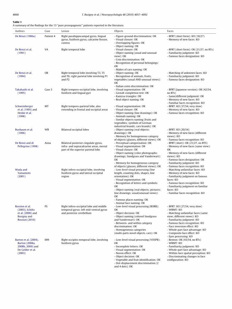
4060 T. Busigny et al. / Neuropsychologia 48 (2010) 4057–4092
Table 1A summary of the findings for the 13 “pure prosopagnosic” patients reported in the literature.
Authors Case Lesion Objects Faces
De Renzi (1986a) Patient 4 Right parahippocampal gyrus, lingualgyrus, fusiform gyrus, calcarine fissure,cuneus
- Figure-ground discrimination: OK- Visual closure: OK- Overlapping figures: OK- Object naming: OK
- BFRT (short form): KO (18/27)- Memory of new faces: KO
De Renzi et al.(1991)
VA Right temporal lobe - Visual closure: OK- Object naming (usual and unusualview): OK- Coin discrimination: OK- Recognition of personal belongings:OK- Makes of cars naming: OK
- BFRT (short form): OK (21/27, no RTs)- Familiarity judgment: KO- Famous faces designation: KO
De Renzi et al.(1994)
OR Right temporal lobe involving T3, T5and T6; right parietal lobe involving P1and P2
- Object naming: OK- Recognition of animals, fruits,vegetables (usual AND unusual views):OK- Italian coins discrimination: OK
- Matching of unknown faces: KO- Familiarity judgment: KO- Famous faces designation: KO
Takahashi et al.(1995)
Case 3 Right temporo-occipital lobe, involvingfusiform and lingual gyri
- Visual segmentation: OK- Gestalt completion test: OK- Kanizsa triangles: OK- Real object naming: OK
- BFRT (Japanese version): OK (42/54,no RTs)- Same/different judgment: OK- Memory of new faces: KO- Familiar faces recognition: KO
Schweinbergeret al. (1995) andHenke et al.(1998)
MT Right temporo-parietal lobe, alsoextending in frontal and occipital areas
- Visual segmentation: OK- Visual closure: OK- Object naming (line drawings): OK- Animals naming: OK- Similar objects naming (fruits andvegetables; symbols of Germanindustrial brands; cars brands): OK
- BFRT: KO (37/54, very slow)- Memory of new faces: KO- Famous faces recognition: KO
Buxbaum et al.(1996)
WB Bilateral occipital lobes - Object naming (real objects;drawings): OK- Memory for homogeneous categoryof objects (glasses, different views): OK
- BFRT: KO (20/54)- Memory of new faces (differentviews): KO- Famous faces recognition: KO
De Renzi and diPellegrino (1998)
Anna Bilateral posterior cingulate gyrus,infra- and supracalcarine areas, mesialpart of the superior parietal lobe
- Perceptual categorization: OK- Visual segmentation: OK- Visual closure: OK- Object naming (color photographs;drawings; Snodgrass and Vanderwart):OK- Memory for homogeneous categoryof objects (glasses, different views): OK
- BFRT (short): OK (21/27, no RTS)- Memory of new faces (same view):OK- Memory of new faces (differentviews): KO- Famous faces designation: OK- Familiarity judgment: KO- Famous faces recognition: KO
Wada andYamamoto(2001)
Right infero-occipital lobe, involvingfusiform gyrus and lateral occipitalregion
- Low-level visual processing (linelength, counting dots, shapes, lineorientation): OK- Visual segmentation: OK- Recognition of letters and symbols:OK- Object naming (real objects; pictures;line drawings; usual/unusual views):OK- Famous places naming: OK- Animal face naming: OK
- Matching unfamiliar faces: KO- Memory of new faces: KO- Familiarity judgment on famousfaces: KO- Famous faces recognition: KO- Familiarity judgment on familiarfaces: KO- Familiar faces recognition: KO
Rossion et al.(2003), Schiltzet al. (2006) andBusigny andRossion (2010)
PS Right infero-occipital lobe and middletemporal gyrus; left mid-ventral gyrusand posterior cerebellum
- Low-level visual processing (BORB):OK- Object decision: OK- Object naming (colored Snodgrassand Vanderwart): OK- Between- and within categorydiscrimination: OK- Homogeneous categories(multi-parts novel objects, cars): OK
- BFRT: KO (27/54, very slow)- WRMT: KO- Matching unfamiliar faces (sameview; different views): KO- Familiarity judgment: KO- Famous faces recognition: KO- Face inversion effect: KO- Whole-part face advantage: KO- Composite face effect: KO- Eyes processing: KO
Barton et al. (2004),Barton (2008a,2008b, 2009) andDe Gelder et al.(2003)
009 Right occipito-temporal lobe, involvingfusiform gyrus
- Low-level visual processing (VOSPB):OK- Incomplete letters: OK- Visual segmentation: OK- Navon effect: OK- Object decision: OK- Vegetable and fruit identification: OK- Dot-displacement discrimination (2and 4 dots): OK
- Benton: OK (43/54, no RTs)- WRMT: KO- Familiarity judgment: KO- Whole-part face advantage: KO- Within-face spatial perception: KO- Discriminating changes in faceconfiguration: KO
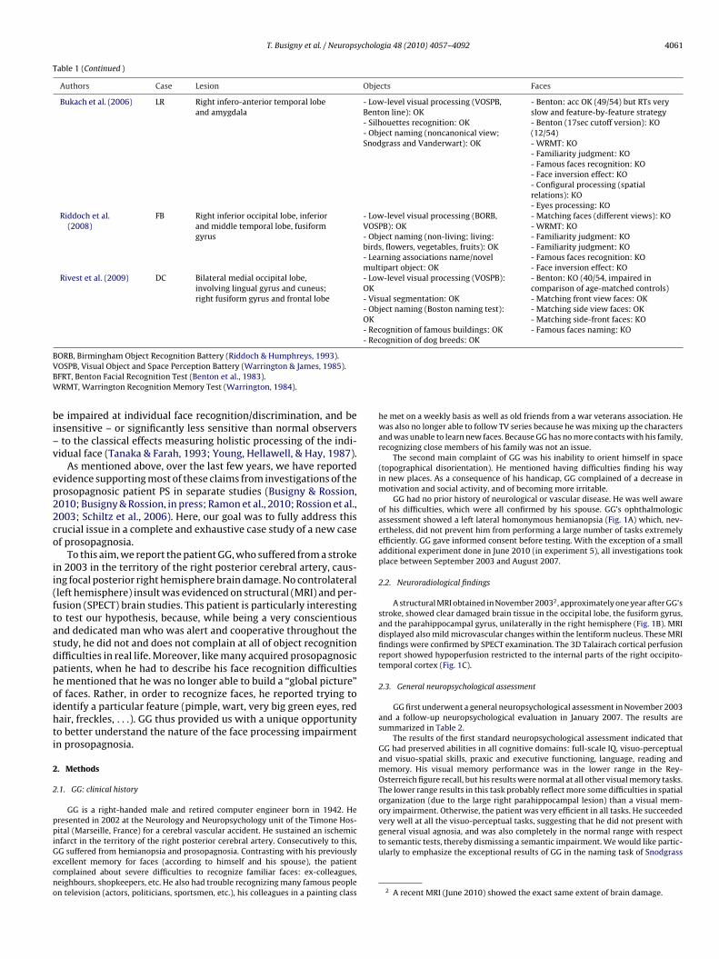
T. Busigny et al. / Neuropsychologia 48 (2010) 4057–4092 4061
Table 1 (Continued )
Authors Case Lesion Objects Faces
Bukach et al. (2006) LR Right infero-anterior temporal lobeand amygdala
- Low-level visual processing (VOSPB,Benton line): OK- Silhouettes recognition: OK- Object naming (noncanonical view;Snodgrass and Vanderwart): OK
- Benton: acc OK (49/54) but RTs veryslow and feature-by-feature strategy- Benton (17sec cutoff version): KO(12/54)- WRMT: KO- Familiarity judgment: KO- Famous faces recognition: KO- Face inversion effect: KO- Configural processing (spatialrelations): KO- Eyes processing: KO
Riddoch et al.(2008)
FB Right inferior occipital lobe, inferiorand middle temporal lobe, fusiformgyrus
- Low-level visual processing (BORB,VOSPB): OK- Object naming (non-living; living:birds, flowers, vegetables, fruits): OK- Learning associations name/novelmultipart object: OK
- Matching faces (different views): KO- WRMT: KO- Familiarity judgment: KO- Familiarity judgment: KO- Famous faces recognition: KO- Face inversion effect: KO
Rivest et al. (2009) DC Bilateral medial occipital lobe,involving lingual gyrus and cuneus;right fusiform gyrus and frontal lobe
- Low-level visual processing (VOSPB):OK- Visual segmentation: OK- Object naming (Boston naming test):OK- Recognition of famous buildings: OK- Recognition of dog breeds: OK
- Benton: KO (40/54, impaired incomparison of age-matched controls)- Matching front view faces: OK- Matching side view faces: OK- Matching side-front faces: KO- Famous faces naming: KO
BVBW
bi–v
ep22co
ii(ftasdphoihti
2
2
ppiGecno
ory impairment. Otherwise, the patient was very efficient in all tasks. He succeededvery well at all the visuo-perceptual tasks, suggesting that he did not present withgeneral visual agnosia, and was also completely in the normal range with respect
ORB, Birmingham Object Recognition Battery (Riddoch & Humphreys, 1993).OSPB, Visual Object and Space Perception Battery (Warrington & James, 1985).FRT, Benton Facial Recognition Test (Benton et al., 1983).RMT, Warrington Recognition Memory Test (Warrington, 1984).
e impaired at individual face recognition/discrimination, and bensensitive – or significantly less sensitive than normal observers
to the classical effects measuring holistic processing of the indi-idual face (Tanaka & Farah, 1993; Young, Hellawell, & Hay, 1987).
As mentioned above, over the last few years, we have reportedvidence supporting most of these claims from investigations of therosopagnosic patient PS in separate studies (Busigny & Rossion,010; Busigny & Rossion, in press; Ramon et al., 2010; Rossion et al.,003; Schiltz et al., 2006). Here, our goal was to fully address thisrucial issue in a complete and exhaustive case study of a new casef prosopagnosia.
To this aim, we report the patient GG, who suffered from a stroken 2003 in the territory of the right posterior cerebral artery, caus-ng focal posterior right hemisphere brain damage. No controlateralleft hemisphere) insult was evidenced on structural (MRI) and per-usion (SPECT) brain studies. This patient is particularly interestingo test our hypothesis, because, while being a very conscientiousnd dedicated man who was alert and cooperative throughout thetudy, he did not and does not complain at all of object recognitionifficulties in real life. Moreover, like many acquired prosopagnosicatients, when he had to describe his face recognition difficultiese mentioned that he was no longer able to build a “global picture”f faces. Rather, in order to recognize faces, he reported trying todentify a particular feature (pimple, wart, very big green eyes, redair, freckles, . . .). GG thus provided us with a unique opportunityo better understand the nature of the face processing impairmentn prosopagnosia.
. Methods
.1. GG: clinical history
GG is a right-handed male and retired computer engineer born in 1942. Heresented in 2002 at the Neurology and Neuropsychology unit of the Timone Hos-ital (Marseille, France) for a cerebral vascular accident. He sustained an ischemic
nfarct in the territory of the right posterior cerebral artery. Consecutively to this,G suffered from hemianopsia and prosopagnosia. Contrasting with his previouslyxcellent memory for faces (according to himself and his spouse), the patientomplained about severe difficulties to recognize familiar faces: ex-colleagues,eighbours, shopkeepers, etc. He also had trouble recognizing many famous peoplen television (actors, politicians, sportsmen, etc.), his colleagues in a painting class
he met on a weekly basis as well as old friends from a war veterans association. Hewas also no longer able to follow TV series because he was mixing up the charactersand was unable to learn new faces. Because GG has no more contacts with his family,recognizing close members of his family was not an issue.
The second main complaint of GG was his inability to orient himself in space(topographical disorientation). He mentioned having difficulties finding his wayin new places. As a consequence of his handicap, GG complained of a decrease inmotivation and social activity, and of becoming more irritable.
GG had no prior history of neurological or vascular disease. He was well awareof his difficulties, which were all confirmed by his spouse. GG’s ophthalmologicassessment showed a left lateral homonymous hemianopsia (Fig. 1A) which, nev-ertheless, did not prevent him from performing a large number of tasks extremelyefficiently. GG gave informed consent before testing. With the exception of a smalladditional experiment done in June 2010 (in experiment 5), all investigations tookplace between September 2003 and August 2007.
2.2. Neuroradiological findings
A structural MRI obtained in November 20032, approximately one year after GG’sstroke, showed clear damaged brain tissue in the occipital lobe, the fusiform gyrus,and the parahippocampal gyrus, unilaterally in the right hemisphere (Fig. 1B). MRIdisplayed also mild microvascular changes within the lentiform nucleus. These MRIfindings were confirmed by SPECT examination. The 3D Talairach cortical perfusionreport showed hypoperfusion restricted to the internal parts of the right occipito-temporal cortex (Fig. 1C).
2.3. General neuropsychological assessment
GG first underwent a general neuropsychological assessment in November 2003and a follow-up neuropsychological evaluation in January 2007. The results aresummarized in Table 2.
The results of the first standard neuropsychological assessment indicated thatGG had preserved abilities in all cognitive domains: full-scale IQ, visuo-perceptualand visuo-spatial skills, praxic and executive functioning, language, reading andmemory. His visual memory performance was in the lower range in the Rey-Osterreich figure recall, but his results were normal at all other visual memory tasks.The lower range results in this task probably reflect more some difficulties in spatialorganization (due to the large right parahippocampal lesion) than a visual mem-
to semantic tests, thereby dismissing a semantic impairment. We would like partic-ularly to emphasize the exceptional results of GG in the naming task of Snodgrass
2 A recent MRI (June 2010) showed the exact same extent of brain damage.

4062 T. Busigny et al. / Neuropsychologia 48 (2010) 4057–4092
F emianl mispht
afhn
pii1tiaopp
ig. 1. (A) GG’s ophthalmologic assessment showing a left lateral homonymous hobe, the fusiform gyrus, and the parahippocampal gyrus, unilaterally in the right hehe internal parts of the right occipito-temporal cortex.
nd Vanderwart (1980). GG correctly named 259 items on 260 (including animals,ruits, vegetables and man-made objects) in a total time of 8min55. In other words,e named each item at an average rate of 2 s, almost perfectly. Thus, GG clearly doesot show any impairment in basic-level object recognition.
In contrast, standard neuropsychological tests showed a clear deficit for facerocessing, in line with the patient’s self-reported complaints. The results showed
mpairments in perceiving and recognizing faces. First, GG obtained impaired scoresn the Benton Facial Recognition Test (Benton, Sivan, Hamsher, Varney, & Spreen,983), more specifically showing difficulties in the second part of the test (in whichhe subject has to match faces presented under different viewpoints and light-
ngs). Second, GG failed the faces subtest of the WMS-III (Weschler, 2001), revealingnterograde difficulties in encoding new faces. Finally, GG was impaired in a taskf naming celebrities, underlying a defect in accessing the identity of well-knowneople via their face. This is a typical pattern of functional impairments in acquiredrosopagnosia, as assessed by conventional neuropsychological tests.opsia. (B) GG’s structural MRI showing clear damaged brain tissue in the occipitalere. (C) 3D Talairach cortical perfusion report showing hypoperfusion restricted to
The control neuropsychological assessment conducted three years later largelyreplicated these observations and showed an efficient global cognitive function-ing (Table 2). Three new tasks of object perception (object decision, categoricaljudgment on different exemplars and categorical judgment on different view-points) showed preserved structural properties and abilities in perceiving objectsacross different viewpoints (even in non-canonical views). Concerning face recog-nition, a slight recovery was noticeable, but the patient still was impaired inthe BFRT (Benton et al., 1983) and the WRMT (Warrington, 1984), and GG stillreported the same difficulties with the recognition of familiar people in real lifecircumstances.
In summary, these results seem to point to a “pure” form of acquiredprosopagnosia in patient GG. This provides us with a unique opportunity toexplore in detail the nature of this patient’s face recognition deficit, whichmay allow us in turn to shed light on theories of normal face perception andrecognition.
T. Busigny et al. / Neuropsychologia 48 (2010) 4057–4092 4063
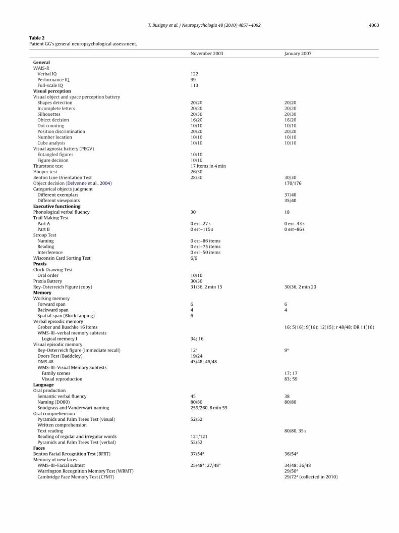
Table 2Patient GG’s general neuropsychological assessment.
November 2003 January 2007
GeneralWAIS-R
Verbal IQ 122Performance IQ 99Full-scale IQ 113
Visual perceptionVisual object and space perception battery
Shapes detection 20/20 20/20Incomplete letters 20/20 20/20Silhouettes 20/30 20/30Object decision 16/20 16/20Dot counting 10/10 10/10Position discrimination 20/20 20/20Number location 10/10 10/10Cube analysis 10/10 10/10
Visual agnosia battery (PEGV)Entangled figures 10/10Figure decision 10/10
Thurstone test 17 items in 4 minHooper test 26/30Benton Line Orientation Test 28/30 30/30Object decision (Delvenne et al., 2004) 170/176Categorical objects judgment
Different exemplars 37/40Different viewpoints 35/40
Executive functioningPhonological verbal fluency 30 18Trail Making Test
Part A 0 err–27 s 0 err–43 sPart B 0 err–115 s 0 err–86 s
Stroop TestNaming 0 err–86 itemsReading 0 err–75 itemsInterference 0 err–50 items
Wisconsin Card Sorting Test 6/6PraxisClock Drawing Test
Oral order 10/10Praxia Battery 30/30Rey-Osterreich Figure (copy) 31/36, 2 min 15 30/36, 2 min 20MemoryWorking memory
Forward span 6 6Backward span 4 4Spatial span (Block tapping) 6
Verbal episodic memoryGrober and Buschke 16 items 16; 5(16); 9(16); 12(15); r 48/48; DR 11(16)WMS-III–verbal memory subtests
Logical memory I 34; 16Visual episodic memory
Rey-Osterreich figure (immediate recall) 12a 9a
Doors Test (Baddeley) 19/24DMS 48 43/48; 46/48WMS-III–Visual Memory Subtests
Family scenes 17; 17Visual reproduction 83; 59
LanguageOral production
Semantic verbal fluency 45 38Naming (DO80) 80/80 80/80Snodgrass and Vanderwart naming 259/260, 8 min 55
Oral comprehensionPyramids and Palm Trees Test (visual) 52/52Written comprehensionText reading 80/80, 35 sReading of regular and irregular words 121/121Pyramids and Palm Trees Test (verbal) 52/52
FacesBenton Facial Recognition Test (BFRT) 37/54a 36/54a
Memory of new facesWMS-III–Facial subtest 25/48a; 27/48a 34/48; 36/48Warrington Recognition Memory Test (WRMT) 29/50a
Cambridge Face Memory Test (CFMT) 29/72a (collected in 2010)
4064 T. Busigny et al. / Neuropsychologia 48 (2010) 4057–4092
Table 2 (Continued )
November 2003 January 2007
Identification of famous peopleFamiliarity judgment 37/50a
Identification from photographs 26/40a 35/40
under
3
idiiwiasZtkal
tghaidospnp
rpfrtpsotaaiP(
awifsisAvopA
Identification from names 39/40Age judgment 40/40
a Indicates impaired scores (below 2 standard deviations from controls’ score or
. Computer experiments
In total, GG was administered with a set of 24 behavioural exper-ments conducted in 2007. These experiments were conducteduring two time periods, in January and August 2007. GG realized
n average 5 tests per day. The order of administration was approx-mately the same than the order reported in the paper: GG began
ith tasks of faces and objects matching, then tasks of visual sim-larity and general global processing, then tasks of face detectionnd finally tasks of face holistic processing. In all experiments, thetimuli were presented using E-prime 1.1 (Schneider, Eschman, &uccolotto, 2002). The patient was positioned at about 40 cm fromhe screen. He was asked to provide a binary response using theeyboard of the laptop computer. Percentages of correct responsesnd response times on correct trials were calculated. RTs that wereonger than two SDs of the mean were discarded.
As means of comparison for GG’s results, five to ten healthy con-rol participants were selected for each experiment, controlling forender, socio-economic background and age. Participants had noistory of neurological or vascular disease, head injury or alcoholbuse, and did not have cognitive complaints. All participants gavenformed consent. The number of control participants and their ageiffer slightly across the experiments. When GG performed as wellr better than controls, we considered that five participants wereufficient to demonstrate normal processing. However, when hiserformance was below that of the five controls, we increased theumber of participants to ensure that GG was truly impaired in thatarticular task.
For intra-subject and intra-group statistical analyses, we usedespectively classical independent sample t-tests and paired sam-le t-tests. These analyses were conducted by SPSS 14.0 within theramework of one-tailed hypothesis (0.05 p value). To compare theesults of GG to the control participants, we used the modified t-est of Crawford and Howell (1998) for single-case studies. Thisrocedure decreases the type 1 error as it tests whether a patient’score is significantly below controls by providing a point estimatef the abnormality of the score. Here we used a 0.05 p value withinhe framework of a unilateral hypothesis. Consequently, all scoresssociated with a p value under 0.05 were considered as reflectingn abnormal result. Analyses were conducted with a computer-zed version of the Crawford & Howell’s method: SINGLIMS.EXE:oint estimate and confidence limits on the abnormality of a test scoreCrawford & Garthwaite, 2002).
Concerning the terminology used in this paper for various tasks,nd to avoid semantic confusions as much as possible, definitionsill be given for some words frequently appearing in the exper-
ments and in this paper in general (following Sergent, 1989). Aace detection task involves a decision as to whether or not a giventimulus is a face. A discrimination (or simultaneous matching)nvolves a comparison between two (or more) simultaneously pre-ented items and a decision as to their sameness or difference.
recognition (or delayed matching) involves a judgment of pre-ious occurrence and therefore whether an item – a face or anbject –has been seen earlier. The comparison is thus between aresently generated representation and a stored representation.categorization involves the classification of the object or face
percentile 5).
into a predetermined category, and different grains of resolutionmay be imposed on the process depending on the type of cate-gorization (e.g., basic level vs. subordinate level). Identification isthe categorization of a face as that of a unique individual whoseidentity must be accessed. Obviously, two of these terms can bevalid to describe a given task, for instance a delayed matchingtask with a distractor is both a recognition and a discrimina-tion/matching task. In this particular situation, both terms can beused.
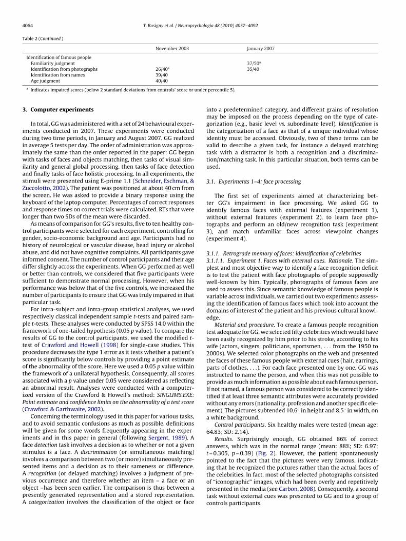
3.1. Experiments 1–4: face processing
The first set of experiments aimed at characterizing bet-ter GG’s impairment in face processing. We asked GG toidentify famous faces with external features (experiment 1),without external features (experiment 2), to learn face pho-tographs and perform an old/new recognition task (experiment3), and match unfamiliar faces across viewpoint changes(experiment 4).
3.1.1. Retrograde memory of faces: identification of celebrities3.1.1.1. Experiment 1. Faces with external cues. Rationale. The sim-plest and most objective way to identify a face recognition deficitis to test the patient with face photographs of people supposedlywell-known by him. Typically, photographs of famous faces areused to assess this. Since semantic knowledge of famous people isvariable across individuals, we carried out two experiments assess-ing the identification of famous faces which took into account thedomains of interest of the patient and his previous cultural knowl-edge.
Material and procedure. To create a famous people recognitiontest adequate for GG, we selected fifty celebrities which would havebeen easily recognized by him prior to his stroke, according to hiswife (actors, singers, politicians, sportsmen, . . . from the 1950 to2000s). We selected color photographs on the web and presentedthe faces of these famous people with external cues (hair, earrings,parts of clothes, . . .). For each face presented one by one, GG wasinstructed to name the person, and when this was not possible toprovide as much information as possible about each famous person.If not named, a famous person was considered to be correctly iden-tified if at least three semantic attributes were accurately providedwithout any errors (nationality, profession and another specific ele-ment). The pictures subtended 10.6◦ in height and 8.5◦ in width, ona white background.
Control participants. Six healthy males were tested (mean age:64.83; SD: 2.14).
Results. Surprisingly enough, GG obtained 86% of correctanswers, which was in the normal range (mean: 88%; SD: 6.97;t = 0.305, p = 0.39) (Fig. 2). However, the patient spontaneouslypointed to the fact that the pictures were very famous, indicat-ing that he recognized the pictures rather than the actual faces of
the celebrities. In fact, most of the selected photographs consistedof “iconographic” images, which had been overly and repetitivelypresented in the media (see Carbon, 2008). Consequently, a secondtask without external cues was presented to GG and to a group ofcontrols participants.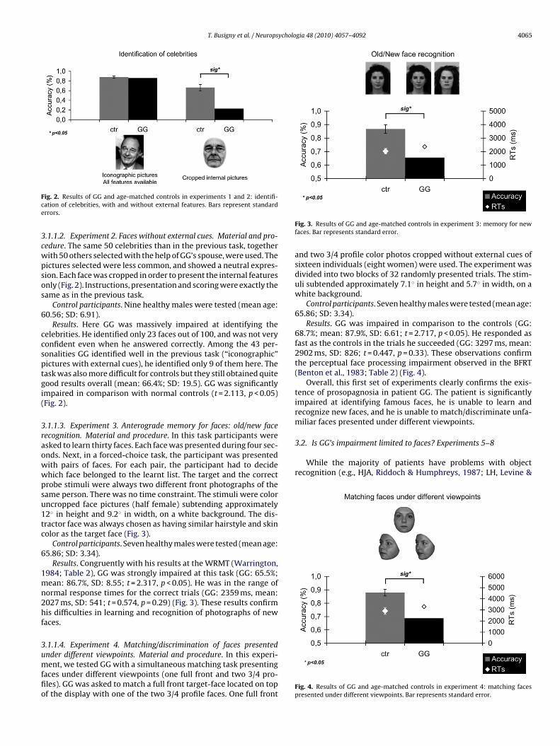
T. Busigny et al. / Neuropsychologia 48 (2010) 4057–4092 4065
Fce
3cwpsos
6
ccsptgi(
3raowwpsu1tc
6
1mn2hf
3umffio
3.2. Is GG’s impairment limited to faces? Experiments 5–8
While the majority of patients have problems with objectrecognition (e.g., HJA, Riddoch & Humphreys, 1987; LH, Levine &
ig. 2. Results of GG and age-matched controls in experiments 1 and 2: identifi-ation of celebrities, with and without external features. Bars represent standardrrors.
.1.1.2. Experiment 2. Faces without external cues. Material and pro-edure. The same 50 celebrities than in the previous task, togetherith 50 others selected with the help of GG’s spouse, were used. Theictures selected were less common, and showed a neutral expres-ion. Each face was cropped in order to present the internal featuresnly (Fig. 2). Instructions, presentation and scoring were exactly theame as in the previous task.
Control participants. Nine healthy males were tested (mean age:0.56; SD: 6.91).
Results. Here GG was massively impaired at identifying theelebrities. He identified only 23 faces out of 100, and was not veryonfident even when he answered correctly. Among the 43 per-onalities GG identified well in the previous task (“iconographic”ictures with external cues), he identified only 9 of them here. Theask was also more difficult for controls but they still obtained quiteood results overall (mean: 66.4%; SD: 19.5). GG was significantlympaired in comparison with normal controls (t = 2.113, p < 0.05)Fig. 2).
.1.1.3. Experiment 3. Anterograde memory for faces: old/new faceecognition. Material and procedure. In this task participants weresked to learn thirty faces. Each face was presented during four sec-nds. Next, in a forced-choice task, the participant was presentedith pairs of faces. For each pair, the participant had to decidehich face belonged to the learnt list. The target and the correctrobe stimuli were always two different front photographs of theame person. There was no time constraint. The stimuli were colorncropped face pictures (half female) subtending approximately2◦ in height and 9.2◦ in width, on a white background. The dis-ractor face was always chosen as having similar hairstyle and skinolor as the target face (Fig. 3).
Control participants. Seven healthy males were tested (mean age:5.86; SD: 3.34).
Results. Congruently with his results at the WRMT (Warrington,984; Table 2), GG was strongly impaired at this task (GG: 65.5%;ean: 86.7%, SD: 8.55; t = 2.317, p < 0.05). He was in the range of
ormal response times for the correct trials (GG: 2359 ms, mean:027 ms, SD: 541; t = 0.574, p = 0.29) (Fig. 3). These results confirmis difficulties in learning and recognition of photographs of new
aces.
.1.1.4. Experiment 4. Matching/discrimination of faces presented
nder different viewpoints. Material and procedure. In this experi-ent, we tested GG with a simultaneous matching task presentingaces under different viewpoints (one full front and two 3/4 pro-les). GG was asked to match a full front target-face located on topf the display with one of the two 3/4 profile faces. One full front
Fig. 3. Results of GG and age-matched controls in experiment 3: memory for newfaces. Bar represents standard error.
and two 3/4 profile color photos cropped without external cues ofsixteen individuals (eight women) were used. The experiment wasdivided into two blocks of 32 randomly presented trials. The stim-uli subtended approximately 7.1◦ in height and 5.7◦ in width, on awhite background.
Control participants. Seven healthy males were tested (mean age:65.86; SD: 3.34).
Results. GG was impaired in comparison to the controls (GG:68.7%; mean: 87.9%, SD: 6.61; t = 2.717, p < 0.05). He responded asfast as the controls in the trials he succeeded (GG: 3297 ms, mean:2902 ms, SD: 826; t = 0.447, p = 0.33). These observations confirmthe perceptual face processing impairment observed in the BFRT(Benton et al., 1983; Table 2) (Fig. 4).
Overall, this first set of experiments clearly confirms the exis-tence of prosopagnosia in patient GG. The patient is significantlyimpaired at identifying famous faces, he is unable to learn andrecognize new faces, and he is unable to match/discriminate unfa-miliar faces presented under different viewpoints.
Fig. 4. Results of GG and age-matched controls in experiment 4: matching facespresented under different viewpoints. Bar represents standard error.
4 ycholo
C&ht1LnparlshatcflsptW
tscc
tB&SBeSS2&L1S(1Yeb&madRe22
mseK&cne2p2
066 T. Busigny et al. / Neurops
alvanio, 1989; AR, Saumier et al., 2001; SM and RN, BehrmannKimchi, 2003; NS, Delvenne et al., 2004), other patients do not
ave such problems (see Table 1 for review). Yet, opponents ofhe specificity hypothesis in prosopagnosia (e.g., Beyn & Knyazeva,962; Bornstein, 1963; Damasio et al., 1982; Gauthier et al., 1999;hermitte et al., 1972) have raised the hypothesis, suggested origi-ally by Faust (1955), that what characterizes prosopagnosia is aarticular problem in recognizing or discriminating objects thatre visually similar, rather than faces in particular. This line ofeasoning originates from the observation that human faces – ateast for a given gender and race – are very similar in shape andurface reflectance (color, texture). Moreover, while most objectsave simply to be categorized at a basic-level (“it’s a chair”) or justfew exemplars have to be identified (“it’s my car”), faces have
o be individualized, a process which goes beyond the basic-levelategorization of a visual stimulus as “a face” (face detection). Inact, face detection does not appear to cause a great deal of prob-ems for many prosopagnosic patients (see next section). There areome variants of this view of prosopagnosia, but they all state thatrosopagnosia is a problem at recognizing/discriminating itemshat are visually similar (Damasio et al., 1982; Gauthier et al., 1999).
hat is the current evidence supporting this view?The specificity of the deficit in prosopagnosia has usually been
ested by asking to categorize items belonging to visually similaruperordinate categories (fruits, flowers, animals, . . .), or to dis-riminate among exemplars of the same category (matching/forcedhoice discrimination tasks).
First, in categorization tasks, patients have been tested with pic-ures of: fruits and vegetables (e.g., Arguin, Bub, & Dudek, 1996;arton, 2008a; De Renzi et al., 1994; Henke et al., 1998; LoperaArdila, 1992; Riddoch & Humphreys, 1987; Riddoch et al., 2008;
chweinberger et al., 1995; Sergent & Villemure, 1989; Stephan,reen, & Caine, 2006); animals (e.g., Damasio et al., 1982; Lhermittet al., 1972; Lopera & Ardila, 1992; Riddoch & Humphreys, 1987;chweinberger et al., 1995; Shuttleworth, Syring, & Allen, 1982;teeves et al., 2006; Tiberghien & Clerc, 1986; Wada & Yamamoto,001); makes of cars (e.g., Davidoff et al., 1986; De Haan, Young,
Newcombe, 1987; De Renzi et al., 1994; Henke et al., 1998;hermitte et al., 1972; Lopera & Ardila, 1992; McNeil & Warrington,991; Schweinberger et al., 1995; Sergent & Signoret, 1992a;tephan et al., 2006; Young, De Haan, & Newcombe, 1990); flowerse.g., Davidoff et al., 1986; De Haan et al., 1987; Lopera & Ardila,992; McNeil & Warrington, 1991; Sergent & Signoret, 1992a;oung et al., 1990); or coins (De Renzi et al., 1991, 1994; Spillmannt al., 2000). Some patients have also been asked to identify famousuildings and places (De Renzi, 1986a; De Renzi et al., 1994; McNeilWarrington, 1991; Wada & Yamamoto, 2001). Even though theajority of prosopagnosic patients performed below normal range
t these tasks, some of them succeeded, as mentioned in the intro-uction (see also Table 1): this was the case for patients VA (Deenzi et al., 1991); WJ (McNeil & Warrington, 1991); OR (De Renzit al., 1994); MT (Schweinberger et al., 1995); WL (Spillmann et al.,000); the patient of Wada & Yamamoto (2001); FB (Riddoch et al.,008); 009 (Barton, 2008a); and DC (Rivest et al., 2009).
Second, matching/discrimination or recognition (delayedatching) tasks with visually similar items were tested in several
tudies with different kinds of stimuli: pairs of glasses (Buxbaumt al., 1996; De Renzi & di Pellegrino, 1998; Farah, Levinson, &lein, 1995); shoes (De Gelder & Rouw, 2000a); houses (De GelderRouw, 2000a); snowflakes (Gauthier et al., 1999); a set of birds,
ars, chairs and boats (Schiltz et al., 2006); and different sets of
ovel objects, Greebles (Gauthier et al., 1999), Scott objects (Rossiont al., 2003), Geons (Behrmann, Peterson, Moscovitch, & Susuki,006), and Fribbles (Behrmann & Williams, 2007). While someatients were impaired in these experiments (Behrmann et al.,006; Behrmann & Williams, 2007; De Gelder & Rouw, 2000a;gia 48 (2010) 4057–4092
Gauthier et al., 1999), others performed in the normal range: thethree patients tested with pairs of glasses scored in the normalrange (Buxbaum et al., 1996; De Renzi & di Pellegrino, 1998; Farah,Levinson, et al., 1995), and PS succeeded in the task with the Scottobjects (Rossion et al., 2003) and could recognize individual itemsof birds, cars, chairs and boats in a delayed presentation modeaccurately and rapidly (Schiltz et al., 2006).
In light of these observations, the interpretation of prosopag-nosia as a problem at categorizing or discriminating items that arevisually similar (Damasio et al., 1982; Gauthier et al., 1999) does notappear to be currently well supported. Moreover, most cases whohave been characterized as having difficulties with visually similarnonface objects already showed massive problems at categorizingobjects that had clear distinctive shapes (e.g., patients 1, 2 and 3;Damasio et al., 1982; RB, Davidoff et al., 1986; PH, De Haan et al.,1987; HJA, Riddoch & Humphreys, 1987; LH, Farah, Wilson, et al.,1995; SM, Gauthier et al., 1999; CR, Gauthier et al., 1999; Marotta,McKeeff, & Behrmann, 2002; DF, Steeves et al., 2006). Finally, theclaim that these patients’ impairments increase relatively morethan normal observers when visual similarity of a distractor itemincreases (Gauthier et al., 1999) is not supported by strong evidence(see Busigny et al., 2010).
Nonetheless, this view remains influential because, in cases whohave been reported to present with an impairment restricted tothe category of faces, the investigation with visually similar non-face objects was not thorough and systematic, and generally didnot take into account patients’ speed (e.g., Barton, 2008a; Buxbaumet al., 1996; De Renzi et al., 1991, 1994; McNeil & Warrington, 1991;Riddoch et al., 2008; Rivest et al., 2009; Schweinberger et al., 1995;Spillmann et al., 2000; Wada & Yamamoto, 2001).
GG does not have any basic-level categorization difficulties,even with items belonging to visually similar categories. In the nextexperiments, we tested him in tasks that require face and objectrecognition at the individual level, taking into account both accu-racy rates and speed. Moreover, contrary to previous studies (e.g.,Gauthier et al., 1999) but in line with our recent investigation ofthe patient PS (Busigny et al., 2010), we manipulated visual sim-ilarity objectively and parametrically both with objects and faces(experiments 7 and 8).
3.2.1. Experiment 5. Face and object discrimination at theindividual level
Material and procedure. The patient and control participantswere presented with individual pictures from different object cat-egories: birds, boats, cars, chairs and faces (see Schiltz et al., 2006).In a delayed two-alternative forced choice decision task, they werefirst presented with a target stimulus belonging to one of thefive categories for one second. After an ISI (white screen) of thesame duration, two probe stimuli (target and distractor) appeared,among which they were asked to indicate which one had beenpreviously presented. To encode the response, participants wereasked to press a key corresponding to the position of the stim-ulus (i.e. right-key if right-stimulus; left-key if left-stimulus); notime constraints were applied but the participants were instructedto respond as accurately and as quickly as possible. The distrac-tor belonged to the same (intra-category discrimination) or toanother category (inter-category discrimination). Photographs offaces were cropped (the external cues were removed) and for allobjects any “external” cue was also removed (e.g., license plates ofthe cars). Twenty-four gray scaled pictures of each category wereused in the two conditions (inter- and intra-category). The exper-
iment was divided into four blocks of 60 randomized trials. Thestimuli subtended approximately the following sizes, respectivelyin height and width: birds (6.4◦ × 9.9◦), boats (8.5◦ × 9.9◦), cars(5◦ × 9.9◦), chairs (9.9◦ × 5.7◦) and faces (9.2◦ × 7.1◦). The pictureswere displayed on a white background.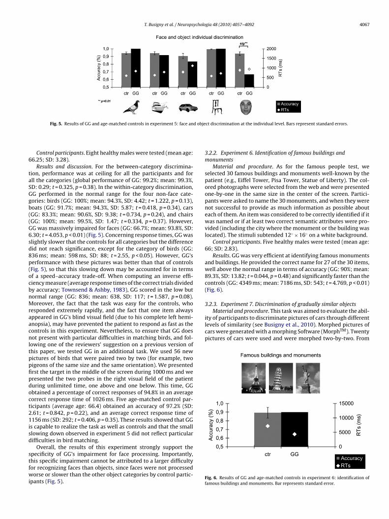
T. Busigny et al. / Neuropsychologia 48 (2010) 4057–4092 4067
d obje
6
taSGgb((G6sd8p(ocbnMraacnltppfipdoct21isd
stfwi
Material and procedure. This task was aimed to evaluate the abil-ity of participants to discriminate pictures of cars through differentlevels of similarity (see Busigny et al., 2010). Morphed pictures ofcars were generated with a morphing Software (MorphTM). Twentypictures of cars were used and were morphed two-by-two. From
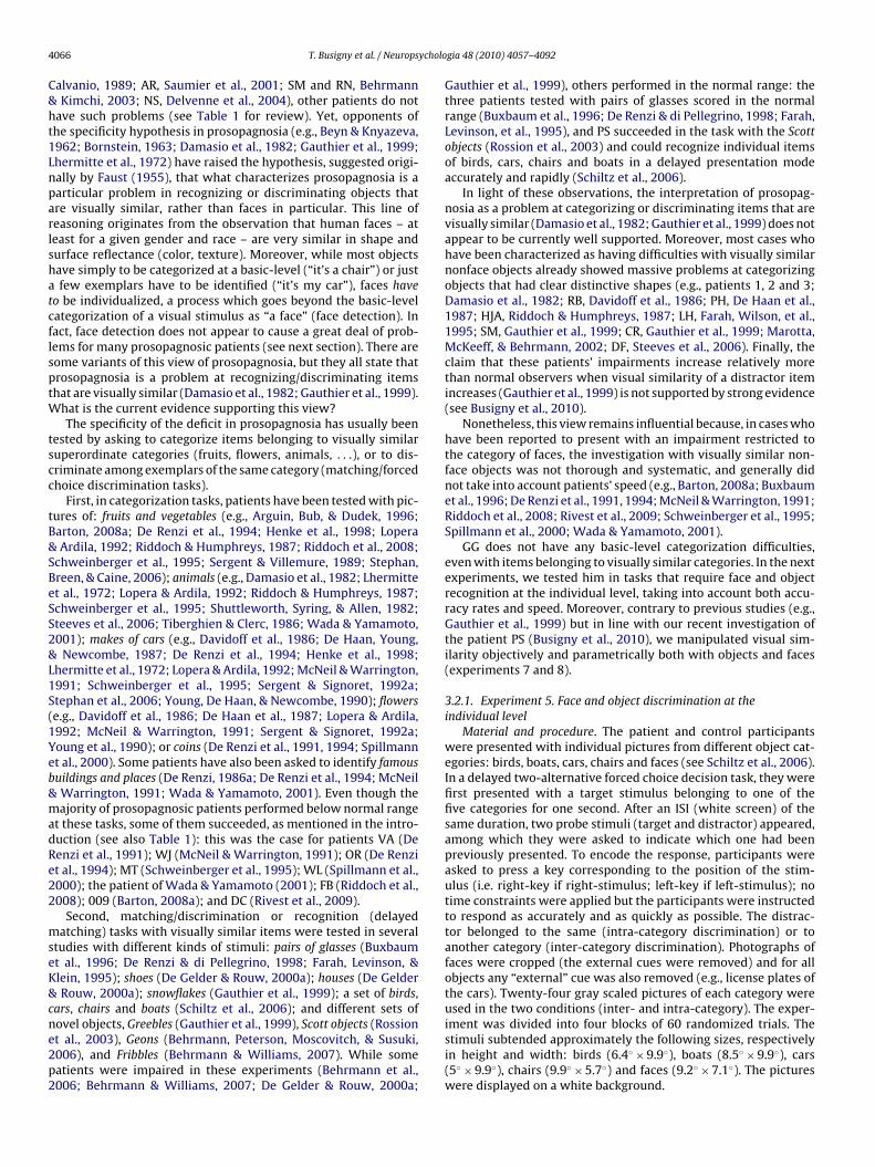
Fig. 5. Results of GG and age-matched controls in experiment 5: face an
Control participants. Eight healthy males were tested (mean age:6.25; SD: 3.28).
Results and discussion. For the between-category discrimina-ion, performance was at ceiling for all the participants and forll the categories (global performance of GG: 99.2%; mean: 99.3%,D: 0.29; t = 0.325, p = 0.38). In the within-category discrimination,G performed in the normal range for the four non-face cate-ories: birds (GG: 100%; mean: 94.3%, SD: 4.42; t = 1.222, p = 0.13),oats (GG: 91.7%; mean: 94.3%, SD: 5.87; t = 0.418, p = 0.34), carsGG: 83.3%; mean: 90.6%, SD: 9.38; t = 0.734, p = 0.24), and chairsGG: 100%; mean: 99.5%, SD: 1.47; t = 0.334, p = 0.37). However,G was massively impaired for faces (GG: 66.7%; mean: 93.8%, SD:.30; t = 4.053, p < 0.01) (Fig. 5). Concerning response times, GG waslightly slower that the controls for all categories but the differenceid not reach significance, except for the category of birds (GG:36 ms; mean: 598 ms, SD: 88; t = 2.55, p < 0.05). However, GG’serformance with these pictures was better than that of controlsFig. 5), so that this slowing down may be accounted for in termsf a speed–accuracy trade-off. When computing an inverse effi-iency measure (average response times of the correct trials dividedy accuracy; Townsend & Ashby, 1983), GG scored in the low butormal range (GG: 836; mean: 638, SD: 117; t = 1.587, p = 0.08).oreover, the fact that the task was easy for the controls, who
esponded extremely rapidly, and the fact that one item alwaysppeared in GG’s blind visual field (due to his complete left hemi-nopsia), may have prevented the patient to respond as fast as theontrols in this experiment. Nevertheless, to ensure that GG doesot present with particular difficulties in matching birds, and fol-
owing one of the reviewers’ suggestion on a previous version ofhis paper, we tested GG in an additional task. We used 56 newictures of birds that were paired two by two (for example, twoigeons of the same size and the same orientation). We presentedrst the target in the middle of the screen during 1000 ms and weresented the two probes in the right visual field of the patienturing unlimited time, one above and one below. This time, GGbtained a percentage of correct responses of 94.8% in an averageorrect response time of 1026 ms. Five age-matched control par-icipants (average age: 66.4) obtained an accuracy of 97.2% (SD:.61; t = 0.842, p = 0.22), and an average correct response time of156 ms (SD: 292; t = 0.406, p = 0.35). These results showed that GG
s capable to realize the task as well as controls and that the smalllowing down observed in experiment 5 did not reflect particularifficulties in bird matching.
Overall, the results of this experiment strongly support the
pecificity of GG’s impairment for face processing. Importantly,his specific impairment cannot be attributed to a larger difficultyor recognizing faces than objects, since faces were not processedorse or slower than the other object categories by control partic-pants (Fig. 5).
ct discrimination at the individual level. Bars represent standard errors.
3.2.2. Experiment 6. Identification of famous buildings andmonuments
Material and procedure. As for the famous people test, weselected 30 famous buildings and monuments well-known by thepatient (e.g., Eiffel Tower, Pisa Tower, Statue of Liberty). The col-ored photographs were selected from the web and were presentedone-by-one in the same size in the center of the screen. Partici-pants were asked to name the 30 monuments, and when they werenot successful to provide as much information as possible abouteach of them. An item was considered to be correctly identified if itwas named or if at least two correct semantic attributes were pro-vided (including the city where the monument or the building waslocated). The stimuli subtended 12◦ × 16◦ on a white background.
Control participants. Five healthy males were tested (mean age:66; SD: 2.83).
Results. GG was very efficient at identifying famous monumentsand buildings. He provided the correct name for 27 of the 30 items,well above the normal range in terms of accuracy (GG: 90%; mean:89.3%, SD: 13.82; t = 0.044, p = 0.48) and significantly faster than thecontrols (GG: 4349 ms; mean: 7186 ms, SD: 543; t = 4.769, p < 0.01)(Fig. 6).
3.2.3. Experiment 7. Discrimination of gradually similar objects
Fig. 6. Results of GG and age-matched controls in experiment 6: identification offamous buildings and monuments. Bar represents standard error.
4068 T. Busigny et al. / Neuropsycholo
Fo(
ew1T(nlwoiawltvsbt
6
avG4ma
ipants, at all levels of visual dissimilarity between the target and
TG
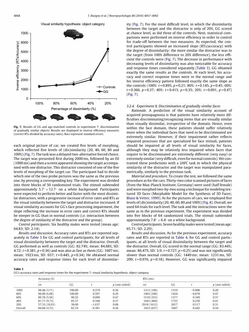
ig. 7. Results of GG and age-matched controls in experiment 7: discriminationf gradually similar objects. Results are displayed in inverse efficiency measurescorrect RTs divided by accuracy rates). Bars represent standard errors.
ach original picture of car, we created five levels of morphing,hich reflected five levels of (dis)similarity (20, 40, 60, 80 and
00%) (Fig. 7). The task was a delayed two-alternative forced choice.he target was presented first during 2000 ms, followed by an ISI1000 ms) and then a screen appeared showing the target accompa-ied with one distractor. This distractor consisted of one of the five
evels of morphing of the target car. The participant had to decidehich one of the two probe pictures was the same as the previous
ne, by pressing a corresponding key. The experiment was dividednto three blocks of 50 randomized trials. The stimuli subtendedpproximately 5.7◦ × 12.7◦ on a white background. Participantsere expected to perform better and faster with the most dissimi-
ar distractors, with a progressive increase of error rates and RTs ashe visual similarity between the target and distractor increased. Ifisual similarity accounts for GG’s face processing impairment, thelope reflecting the increase in error rates and correct RTs shoulde steeper in GG than in normal controls (i.e. interaction betweenhe degree of similarity of the distractor and the group).
Control participants. Six healthy males were tested (mean age:4.83; SD: 2.14).
Results and discussion. Accuracy rates and RTs are reported sep-rately in Table 3 for GG and control participants, for all levels ofisual dissimilarity between the target and the distractor. Overall,
G performed as well as controls (GG: 82.74%; mean: 84.68%, SD:.72; t = 0.381, p = 0.36) and was also as fast as them (GG: 1607 ms;ean: 1923 ms, SD: 657; t = 0.445, p = 0.34). He obtained normalccuracy rates and response times for each level of dissimilar-
able 3G’s accuracy rates and response times for the experiment 7: visual similarity hypothesi
Accuracy (%)
ctr GG t p (one-taile
100% 98.48 (3.71) 100.00 0.379 0.3680% 96.08 (6.08) 100.00 0.597 0.2960% 89.78 (5.56) 90.32 0.090 0.4740% 81.71 (9.73) 85.37 0.348 0.3720% 57.33 (10.93) 38.00 −1.637 0.08Overall 84.68 (4.72) 82.74 −0.381 0.36
gia 48 (2010) 4057–4092
ity (Fig. 7). For the most difficult level, in which the dissimilaritybetween the target and the distractor is only of 20%, GG scoredat chance level, as did three of the controls. Next, statistical com-parisons were performed on inverse efficiency in order to controlfor trade-off between the two measures. As expected, the con-trol participants showed an increased slope (RTs/accuracy) withthe degree of dissimilarity: the more similar the distractor was tothe target (from 100% difference to 20% difference), the less effi-cient the controls were (Fig. 7). The decrease in performance withdecreasing levels of dissimilarity was also noticeable for accuracyand response times considered separately (Table 3). GG obtainedexactly the same results as the controls. At each level, his accu-racy and correct response times were in the normal range andhis inverse efficiency pattern followed exactly the same slope asthe controls (100%: t = 0.895, p = 0.21; 80%: t = 0.145, p = 0.45; 60%:t = 0.366, p = 0.37; 40%: t = 0.415, p = 0.35; 20%: t = 0.091, p = 0.47)(Fig. 7).
3.2.4. Experiment 8. Discrimination of gradually similar facesRationale. A prediction of the visual similarity account of
acquired prosopagnosia is that patients have relatively more dif-ficulties discriminating/recognizing items that are visually similarthan visually dissimilar, irrespective of the domain. Hence, evenwithin the face domain, these patients should suffer relativelymore when the individual faces that need to be discriminated areextremely similar. However, if their impairment rather reflectsimpaired processes that are specialized for face stimuli, patientsshould be impaired at all levels of visual similarity for faces,although they may be relatively less impaired when faces thatrequire to be discriminated are extremely different (very easy) orextremely similar (very difficult, even for normal controls). We con-trasted these predictions with a 2AFC task in which the physicalsimilarity of the distractor and the target was manipulated para-metrically, similarly to the previous task.
Material and procedure. To create the test, we followed the sameprocedure as for the cars. Thirty-two color scanned pictures of faces(from the Max-Planck Institute, Germany) were used (half female)and were morphed two-by-two using a technique for modeling tex-tured 3D faces (Morphable Model for the Synthesis of 3D Faces;Blanz & Vetter, 1999). As for the pictures of cars, we employed fivelevels of (dis)similarity (20, 40, 60, 80 and 100%) (Fig. 8). Overall, weused 64 trials for each level. The task and the instructions were thesame as in the previous experiment. The experiment was dividedinto five blocks of 64 randomized trials. The stimuli subtendedapproximately 7.8◦ × 6.4◦ on a white background.
Control participants. Seven healthy males were tested (mean age:65.71; SD: 2.29).
Results and discussion. As for the previous experiment, accuracyrates and RTs are reported in Table 4, for GG and control partic-
the distractor. Overall, GG scored in the normal range (GG: 83.44%;mean: 86.47%, SD: 3.9; t = 0.727, p = 0.25) and was not significantlyslower than normal controls (GG: 1449 ms; mean: 1231 ms, SD:209; t = 0.976, p = 0.18). However, GG was significantly impaired
s, object category.
RTs (ms)
d) ctr GG t p (one-tailed)
1215 (106) 1319 −0.908 0.201403 (248) 1424 −0.078 0.471510 (353) 1377 0.349 0.371843 (484) 1723 0.230 0.41
2918 (1612) 2017 0.517 0.311923 (657) 1607 0.445 0.34
T. Busigny et al. / Neuropsycholo
FgR
ita8
iapfctc1ptbGp
ni1tda
Navon interference effect (see below). They interpreted the prob-
TG
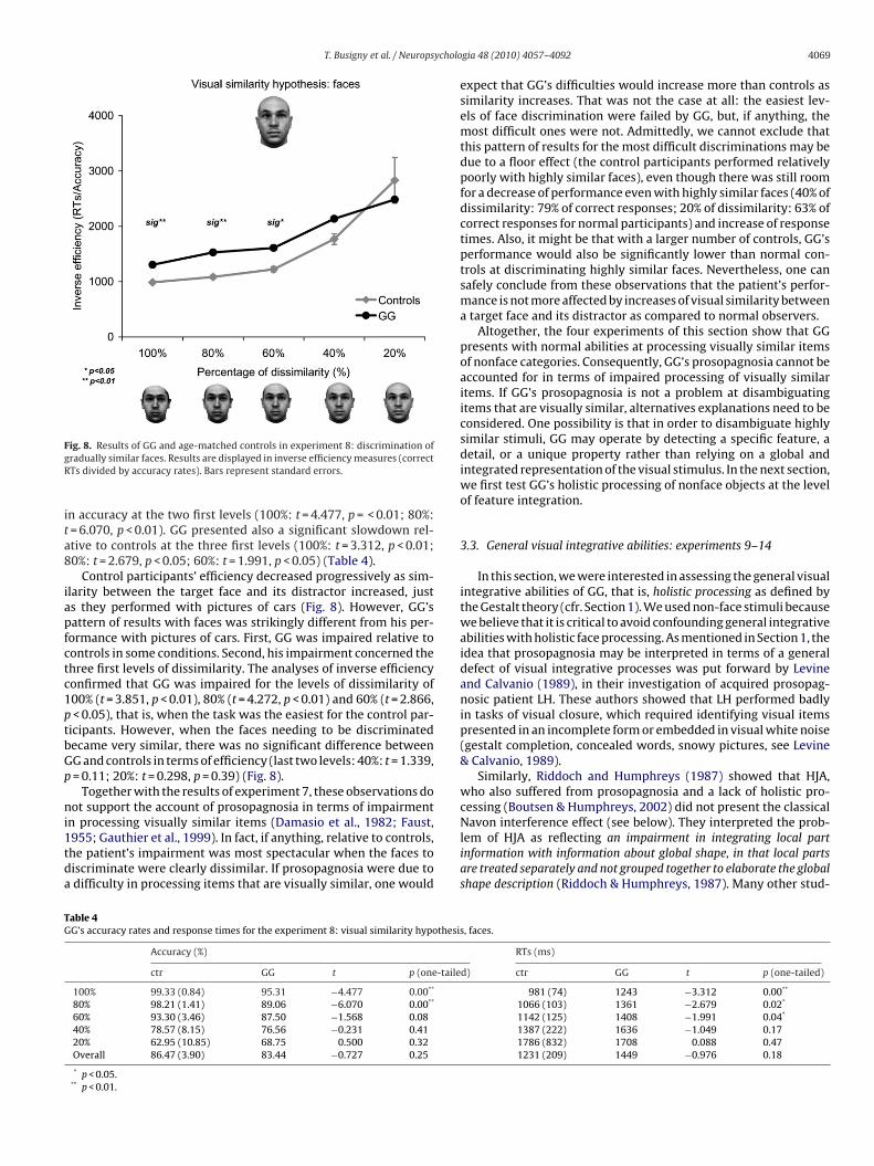
ig. 8. Results of GG and age-matched controls in experiment 8: discrimination ofradually similar faces. Results are displayed in inverse efficiency measures (correctTs divided by accuracy rates). Bars represent standard errors.
n accuracy at the two first levels (100%: t = 4.477, p = < 0.01; 80%:= 6.070, p < 0.01). GG presented also a significant slowdown rel-tive to controls at the three first levels (100%: t = 3.312, p < 0.01;0%: t = 2.679, p < 0.05; 60%: t = 1.991, p < 0.05) (Table 4).
Control participants’ efficiency decreased progressively as sim-larity between the target face and its distractor increased, justs they performed with pictures of cars (Fig. 8). However, GG’sattern of results with faces was strikingly different from his per-ormance with pictures of cars. First, GG was impaired relative toontrols in some conditions. Second, his impairment concerned thehree first levels of dissimilarity. The analyses of inverse efficiencyonfirmed that GG was impaired for the levels of dissimilarity of00% (t = 3.851, p < 0.01), 80% (t = 4.272, p < 0.01) and 60% (t = 2.866,< 0.05), that is, when the task was the easiest for the control par-
icipants. However, when the faces needing to be discriminatedecame very similar, there was no significant difference betweenG and controls in terms of efficiency (last two levels: 40%: t = 1.339,= 0.11; 20%: t = 0.298, p = 0.39) (Fig. 8).
Together with the results of experiment 7, these observations doot support the account of prosopagnosia in terms of impairment
n processing visually similar items (Damasio et al., 1982; Faust,
955; Gauthier et al., 1999). In fact, if anything, relative to controls,he patient’s impairment was most spectacular when the faces toiscriminate were clearly dissimilar. If prosopagnosia were due todifficulty in processing items that are visually similar, one wouldable 4G’s accuracy rates and response times for the experiment 8: visual similarity hypothesis
Accuracy (%)
ctr GG t p (one-taile
100% 99.33 (0.84) 95.31 −4.477 0.00**
80% 98.21 (1.41) 89.06 −6.070 0.00**
60% 93.30 (3.46) 87.50 −1.568 0.0840% 78.57 (8.15) 76.56 −0.231 0.4120% 62.95 (10.85) 68.75 0.500 0.32Overall 86.47 (3.90) 83.44 −0.727 0.25
* p < 0.05.** p < 0.01.
gia 48 (2010) 4057–4092 4069
expect that GG’s difficulties would increase more than controls assimilarity increases. That was not the case at all: the easiest lev-els of face discrimination were failed by GG, but, if anything, themost difficult ones were not. Admittedly, we cannot exclude thatthis pattern of results for the most difficult discriminations may bedue to a floor effect (the control participants performed relativelypoorly with highly similar faces), even though there was still roomfor a decrease of performance even with highly similar faces (40% ofdissimilarity: 79% of correct responses; 20% of dissimilarity: 63% ofcorrect responses for normal participants) and increase of responsetimes. Also, it might be that with a larger number of controls, GG’sperformance would also be significantly lower than normal con-trols at discriminating highly similar faces. Nevertheless, one cansafely conclude from these observations that the patient’s perfor-mance is not more affected by increases of visual similarity betweena target face and its distractor as compared to normal observers.
Altogether, the four experiments of this section show that GGpresents with normal abilities at processing visually similar itemsof nonface categories. Consequently, GG’s prosopagnosia cannot beaccounted for in terms of impaired processing of visually similaritems. If GG’s prosopagnosia is not a problem at disambiguatingitems that are visually similar, alternatives explanations need to beconsidered. One possibility is that in order to disambiguate highlysimilar stimuli, GG may operate by detecting a specific feature, adetail, or a unique property rather than relying on a global andintegrated representation of the visual stimulus. In the next section,we first test GG’s holistic processing of nonface objects at the levelof feature integration.
3.3. General visual integrative abilities: experiments 9–14
In this section, we were interested in assessing the general visualintegrative abilities of GG, that is, holistic processing as defined bythe Gestalt theory (cfr. Section 1). We used non-face stimuli becausewe believe that it is critical to avoid confounding general integrativeabilities with holistic face processing. As mentioned in Section 1, theidea that prosopagnosia may be interpreted in terms of a generaldefect of visual integrative processes was put forward by Levineand Calvanio (1989), in their investigation of acquired prosopag-nosic patient LH. These authors showed that LH performed badlyin tasks of visual closure, which required identifying visual itemspresented in an incomplete form or embedded in visual white noise(gestalt completion, concealed words, snowy pictures, see Levine& Calvanio, 1989).
Similarly, Riddoch and Humphreys (1987) showed that HJA,who also suffered from prosopagnosia and a lack of holistic pro-cessing (Boutsen & Humphreys, 2002) did not present the classical
lem of HJA as reflecting an impairment in integrating local partinformation with information about global shape, in that local partsare treated separately and not grouped together to elaborate the globalshape description (Riddoch & Humphreys, 1987). Many other stud-
, faces.
RTs (ms)
d) ctr GG t p (one-tailed)
981 (74) 1243 −3.312 0.00**
1066 (103) 1361 −2.679 0.02*
1142 (125) 1408 −1.991 0.04*
1387 (222) 1636 −1.049 0.171786 (832) 1708 0.088 0.471231 (209) 1449 −0.976 0.18
4 ycholo
ipfi(hpB1Ht
opRdctvcgNesdp
Gaiwg
3
etts&itp
BfliswoipcbbtTt
6
atm
(
070 T. Busigny et al. / Neurops
es have used tests of visual closure to assess the global configuralrocessing in prosopagnosic patients, using for example the Streetgure-completion test (Street, 1931), the Gollin incomplete picturesGollin, 1960), the Kanisza triangles (Kanizsa, 1955), or the Navonierarchical letters (Navon, 1977). The large majority of acquiredrosopagnosic patients tested with these tasks were impaired (e.g.,ehrmann & Kimchi, 2003; De Renzi, 1986a, 1986b; De Renzi et al.,991; Lê et al., 2002). However, these patients, including LH andJA, suffered from marked deficits for object recognition, that is,
heir impairment reflects general visual integrative agnosia.In contrast, several cases of acquired prosopagnosia with no
bject recognition deficit succeeded in tasks of visual closure: threeatients studied by De Renzi and colleagues [patient no. 4 (Deenzi, 1986a), VA (De Renzi et al., 1991) and Anna (De Renzi &i Pellegrino, 1998)] who presented no impairment in the Street’sompletion test concomitantly with no apparent objects recogni-ion impairment. Another case of prosopagnosia with no generalisual agnosia, MT (Henke et al., 1998), also succeeded the Street’sompletion task. In a recent study, Barton (2009) also showed in aroup study of acquired prosopagnosic patients a general normalavon effect for the group (main effect of the global level and mainffect of the local level). However, the patients were significantlylowed down in comparison with the controls and the individualata of the patients were not provided (see Busigny & Rossion, inress).
Thus, even though some studies tend to show that generalestalt/holistic processing can be preserved in prosopagnosia, theyre rare. Here, we investigated more extensively the general visualntegrative abilities of GG. Our prediction was that the patient
ould succeed the three following experiments and show intacteneral integrative/configural processing of non-face objects.
.3.1. Experiment 9. Navon effectRationale. In his original paper, Navon (1977) tested the hypoth-
sis that perceptual processes proceed from global structuringowards more and more fine-grained analysis, a theory that heermed global addressability, and which is inspired from earliertudies on object perception under the Gestaltist approach (FlavellDraguns, 1957). To test this theory, he created hierarchical letters,
n which global letters are composed with small letters. He showedhat normal observers process the global level better, and that therocessing of the local level is influenced by the global level.
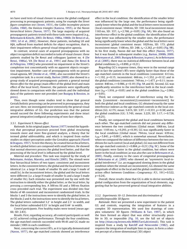
Material and procedure. This task was inspired by the study ofehrmann, Avidan, Marotta, and Kimchi (2005). The stimuli were
our hierarchical letters of two types: consistent and inconsistentetters. In the consistent letters, the global and the local letters weredentical (i.e. a large H made of smaller Hs and a large S made ofmall Ss). In the inconsistent letters, the global and the local lettersere different (i.e. a large H made of smaller Ss and a large S made
f smaller Hs). Each stimulus was presented one by one with thenstruction to identify either the global letter, or the local letter, byressing a corresponding key. A 500 ms ISI and a 500 ms fixationross preceded each trial. The experiment was divided into fourlocks of 48 consistent and inconsistent randomized trials. In thelocks 1 and 3 the instructions were to identify the global letter, inhe blocks 2 and 4, the instructions were to identify the local letters.he global letters subtended 3.2◦ in height and 2.3◦ in width, andhe local letter subtended 0.53◦ in height and 0.44◦ in width.
Control participants. Five healthy males were tested (mean age:6; SD: 2.83).
Results. First, regarding accuracy, all control participants as well
s GG achieved ceiling performance. Through the four conditions,he age-matched controls succeeded with 99.4% and GG made noistake (100% in each condition).Next, concerning the correct RTs, as it is typically demonstrated
Navon, 1977), the age-matched controls showed an interference
gia 48 (2010) 4057–4092
effect in the local condition: the identification of the smaller letterwas influenced by the large one, the performance being signifi-cantly lower when the global and the local letters were inconsistent(local consistent mean: 1044 ms, SD: 330; local inconsistent mean:1165 ms, SD: 337; t4 = 2.766, p < 0.05) (Fig. 9A). We also found aninterference effect in the global condition: the identification of thelarge letter was influenced by the smaller ones, the performancebeing significantly lower when the global and the local letters wereinconsistent (global consistent mean: 1029 ms, SD: 173; globalinconsistent mean: 1108 ms, SD: 248; t4 = 2.262, p < 0.05) (Fig. 9B).In his first study, Navon did not find this effect (Navon, 1977),but it was found in subsequent studies (e.g., Barton, 2008b, 2009;Behrmann et al., 2005; Busigny & Rossion, in press). As in Behrmannet al. (2005), there was no statistical difference between local andglobal conditions (t4 = 0.890, p = 0.21).
Regarding GG’s response times, they were in the normal rangeand in fact the patient’s RTs were slightly faster than those of theage-matched controls in the local conditions (consistent: 613 ms,t = 1.192, p = 0.15; inconsistent: 666 ms, t = 1.352, p = 0.12) and inthe global conditions (consistent: 749 ms, t = 1.477, p = 0.11; incon-sistent: 840 ms, t = 0.986, p = 0.19) (Fig. 9A and B). Second, GG wassignificantly sensitive to the interference both in the local condi-tion (t94 = 1.916, p < 0.05) and in the global condition (t94 = 2.062,p < 0.05).
Third, we computed indexes of interference using the for-mula [(consistent − inconsistent)/(consistent + inconsistent)] forboth the global and local conditions. GG obtained exactly the sameinterference indexes as the age-matched controls in the local con-dition (GG: 4.17%; mean: 5.61%, SD: 3.97; t = 0.331, p = 0.38) and inthe global condition (GG: 5.74%; mean: 3.22%, SD: 3.17; t = 0.726,p = 0.25).
Finally, we compared the global and local conditions betweeneach other. The age-matched controls did not present any differ-ence between the two conditions (Global mean: 1068 ms, Localmean: 1105 ms; t4 = 0.293, p = 0.39). GG was significantly faster inthe local condition (Global mean: 794 ms, Local mean: 639 ms;t190 = 5.841, p < 0.001), which is somewhat unusual. Nevertheless,when we compared the indexes of difference between the two con-ditions for each control (local and global), GG was not different fromthe age-matched controls (t = 0.884, p = 0.21) (Fig. 9C). Some of theparticipants were faster in the global condition, but others werefaster in the local condition (as was also the case in Behrmann et al.,2005). Finally, contrary to the cases of congenital prosopagnosiaof Behrmann et al. (2005) who showed an “asymmetric local-to-global interference” (i.e. an exaggerated slowing down in the globalcondition when the stimuli are inconsistent), GG showed the samemagnitude of effect in the local and the global conditions (no inter-action effect between Condition × Congruency: F(1, 191) = 0.522,p = 0.24).
Overall, these results show that GG is able to derive normally aglobal configuration from the organization of local elements, sug-gesting that he has preserved general visual integrative abilities.
3.3.2. Experiments 10–12. Detection and discrimination ofpossible/impossible 3D figures
Rationale. Here we presented a new experiment to the patientGG and controls, requiring the integration of features in acomplex 3D representation. We used line drawings of com-plex volumetric 3D figures (Williams & Tarr, 1997), in whichthe lines formed an object that was either structurally possi-
ble in 3D, or impossible (Fig. 10, see the full set of objectson http://titan.cog.brown.edu:8080/TarrLab/stimuli). This task isinspired from a study by Ratcliff and Newcombe (1982), andrequires the integration of multiple lines in space, to form a coher-ent percept of a three-dimensional (3D) object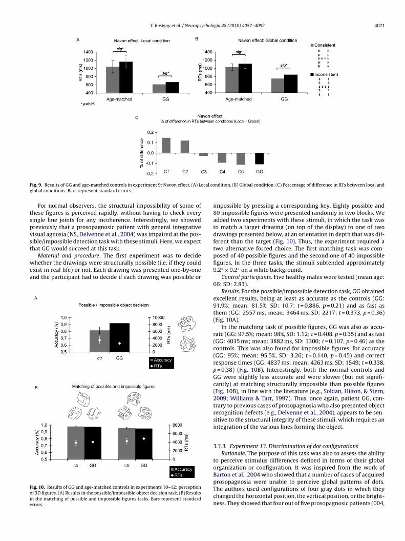
T. Busigny et al. / Neuropsychologia 48 (2010) 4057–4092 4071
F cal cog
tspvst
wea
Foie
ig. 9. Results of GG and age-matched controls in experiment 9: Navon effect. (A) Lolobal conditions. Bars represent standard errors.
For normal observers, the structural impossibility of some ofhese figures is perceived rapidly, without having to check everyingle line joints for any incoherence. Interestingly, we showedreviously that a prosopagnosic patient with general integrativeisual agnosia (NS, Delvenne et al., 2004) was impaired at the pos-ible/impossible detection task with these stimuli. Here, we expecthat GG would succeed at this task.
Material and procedure. The first experiment was to decidehether the drawings were structurally possible (i.e. if they could
xist in real life) or not. Each drawing was presented one-by-onend the participant had to decide if each drawing was possible or
ig. 10. Results of GG and age-matched controls in experiments 10–12: perceptionf 3D figures. (A) Results in the possible/impossible object decision task. (B) Resultsn the matching of possible and impossible figures tasks. Bars represent standardrrors.
ndition. (B) Global condition. (C) Percentage of difference in RTs between local and
impossible by pressing a corresponding key. Eighty possible and80 impossible figures were presented randomly in two blocks. Weadded two experiments with these stimuli, in which the task wasto match a target drawing (on top of the display) to one of twodrawings presented below, at an orientation in depth that was dif-ferent than the target (Fig. 10). Thus, the experiment required atwo-alternative forced choice. The first matching task was com-posed of 40 possible figures and the second one of 40 impossiblefigures. In the three tasks, the stimuli subtended approximately9.2◦ × 9.2◦ on a white background.
Control participants. Five healthy males were tested (mean age:66; SD: 2.83).
Results. For the possible/impossible detection task, GG obtainedexcellent results, being at least as accurate as the controls (GG:91.9%; mean: 81.5%, SD: 10.7; t = 0.886, p = 0.21) and as fast asthem (GG: 2557 ms; mean: 3464 ms, SD: 2217; t = 0.373, p = 0.36)(Fig. 10A).
In the matching task of possible figures, GG was also as accu-rate (GG: 97.5%; mean: 98%, SD: 1.12; t = 0.408, p = 0.35) and as fast(GG: 4035 ms; mean: 3882 ms, SD: 1300; t = 0.107, p = 0.46) as thecontrols. This was also found for impossible figures, for accuracy(GG: 95%; mean: 95.5%, SD: 3.26; t = 0.140, p = 0.45) and correctresponse times (GG: 4837 ms; mean: 4263 ms, SD: 1549; t = 0.338,p = 0.38) (Fig. 10B). Interestingly, both the normal controls andGG were slightly less accurate and were slower (but not signifi-cantly) at matching structurally impossible than possible figures(Fig. 10B), in line with the literature (e.g., Soldan, Hilton, & Stern,2009; Williams & Tarr, 1997). Thus, once again, patient GG, con-trary to previous cases of prosopagnosia who also presented objectrecognition defects (e.g., Delvenne et al., 2004), appears to be sen-sitive to the structural integrity of these stimuli, which requires anintegration of the various lines forming the object.
3.3.3. Experiment 13. Discrimination of dot configurationsRationale. The purpose of this task was also to assess the ability
to perceive stimulus differences defined in terms of their globalorganization or configuration. It was inspired from the work of
Barton et al., 2004 who showed that a number of cases of acquiredprosopagnosia were unable to perceive global patterns of dots.The authors used configurations of four gray dots in which theychanged the horizontal position, the vertical position, or the bright-ness. They showed that four out of five prosopagnosic patients (004,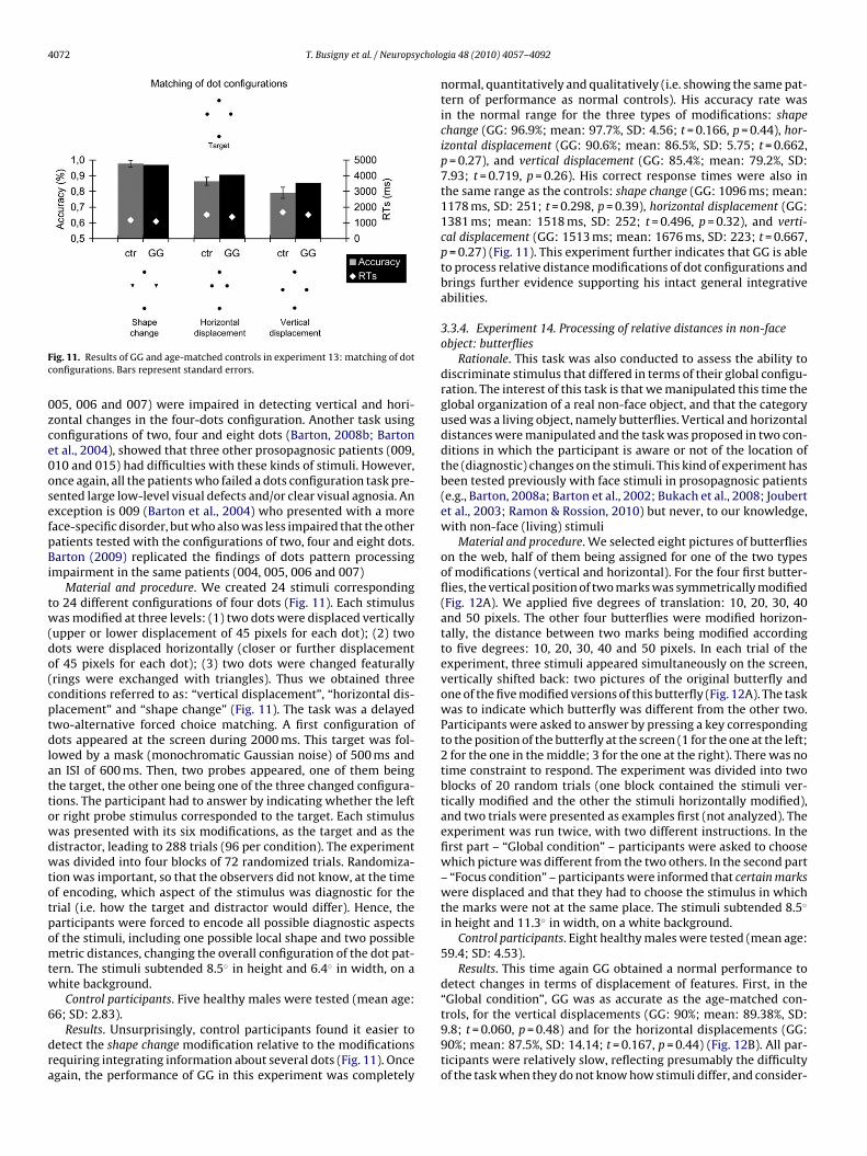
4072 T. Busigny et al. / Neuropsycholo
Fc
0zce0osefpBi
tw(do(cptdlattowdwtotpomtw
6
dra
ig. 11. Results of GG and age-matched controls in experiment 13: matching of dotonfigurations. Bars represent standard errors.
05, 006 and 007) were impaired in detecting vertical and hori-ontal changes in the four-dots configuration. Another task usingonfigurations of two, four and eight dots (Barton, 2008b; Bartont al., 2004), showed that three other prosopagnosic patients (009,10 and 015) had difficulties with these kinds of stimuli. However,nce again, all the patients who failed a dots configuration task pre-ented large low-level visual defects and/or clear visual agnosia. Anxception is 009 (Barton et al., 2004) who presented with a moreace-specific disorder, but who also was less impaired that the otheratients tested with the configurations of two, four and eight dots.arton (2009) replicated the findings of dots pattern processing
mpairment in the same patients (004, 005, 006 and 007)Material and procedure. We created 24 stimuli corresponding
o 24 different configurations of four dots (Fig. 11). Each stimulusas modified at three levels: (1) two dots were displaced vertically
upper or lower displacement of 45 pixels for each dot); (2) twoots were displaced horizontally (closer or further displacementf 45 pixels for each dot); (3) two dots were changed featurallyrings were exchanged with triangles). Thus we obtained threeonditions referred to as: “vertical displacement”, “horizontal dis-lacement” and “shape change” (Fig. 11). The task was a delayedwo-alternative forced choice matching. A first configuration ofots appeared at the screen during 2000 ms. This target was fol-
owed by a mask (monochromatic Gaussian noise) of 500 ms andn ISI of 600 ms. Then, two probes appeared, one of them beinghe target, the other one being one of the three changed configura-ions. The participant had to answer by indicating whether the leftr right probe stimulus corresponded to the target. Each stimulusas presented with its six modifications, as the target and as theistractor, leading to 288 trials (96 per condition). The experimentas divided into four blocks of 72 randomized trials. Randomiza-
ion was important, so that the observers did not know, at the timef encoding, which aspect of the stimulus was diagnostic for therial (i.e. how the target and distractor would differ). Hence, thearticipants were forced to encode all possible diagnostic aspectsf the stimuli, including one possible local shape and two possibleetric distances, changing the overall configuration of the dot pat-
ern. The stimuli subtended 8.5◦ in height and 6.4◦ in width, on ahite background.
Control participants. Five healthy males were tested (mean age:
6; SD: 2.83).Results. Unsurprisingly, control participants found it easier toetect the shape change modification relative to the modificationsequiring integrating information about several dots (Fig. 11). Oncegain, the performance of GG in this experiment was completely
gia 48 (2010) 4057–4092
normal, quantitatively and qualitatively (i.e. showing the same pat-tern of performance as normal controls). His accuracy rate wasin the normal range for the three types of modifications: shapechange (GG: 96.9%; mean: 97.7%, SD: 4.56; t = 0.166, p = 0.44), hor-izontal displacement (GG: 90.6%; mean: 86.5%, SD: 5.75; t = 0.662,p = 0.27), and vertical displacement (GG: 85.4%; mean: 79.2%, SD:7.93; t = 0.719, p = 0.26). His correct response times were also inthe same range as the controls: shape change (GG: 1096 ms; mean:1178 ms, SD: 251; t = 0.298, p = 0.39), horizontal displacement (GG:1381 ms; mean: 1518 ms, SD: 252; t = 0.496, p = 0.32), and verti-cal displacement (GG: 1513 ms; mean: 1676 ms, SD: 223; t = 0.667,p = 0.27) (Fig. 11). This experiment further indicates that GG is ableto process relative distance modifications of dot configurations andbrings further evidence supporting his intact general integrativeabilities.
3.3.4. Experiment 14. Processing of relative distances in non-faceobject: butterflies
Rationale. This task was also conducted to assess the ability todiscriminate stimulus that differed in terms of their global configu-ration. The interest of this task is that we manipulated this time theglobal organization of a real non-face object, and that the categoryused was a living object, namely butterflies. Vertical and horizontaldistances were manipulated and the task was proposed in two con-ditions in which the participant is aware or not of the location ofthe (diagnostic) changes on the stimuli. This kind of experiment hasbeen tested previously with face stimuli in prosopagnosic patients(e.g., Barton, 2008a; Barton et al., 2002; Bukach et al., 2008; Joubertet al., 2003; Ramon & Rossion, 2010) but never, to our knowledge,with non-face (living) stimuli
Material and procedure. We selected eight pictures of butterflieson the web, half of them being assigned for one of the two typesof modifications (vertical and horizontal). For the four first butter-flies, the vertical position of two marks was symmetrically modified(Fig. 12A). We applied five degrees of translation: 10, 20, 30, 40and 50 pixels. The other four butterflies were modified horizon-tally, the distance between two marks being modified accordingto five degrees: 10, 20, 30, 40 and 50 pixels. In each trial of theexperiment, three stimuli appeared simultaneously on the screen,vertically shifted back: two pictures of the original butterfly andone of the five modified versions of this butterfly (Fig. 12A). The taskwas to indicate which butterfly was different from the other two.Participants were asked to answer by pressing a key correspondingto the position of the butterfly at the screen (1 for the one at the left;2 for the one in the middle; 3 for the one at the right). There was notime constraint to respond. The experiment was divided into twoblocks of 20 random trials (one block contained the stimuli ver-tically modified and the other the stimuli horizontally modified),and two trials were presented as examples first (not analyzed). Theexperiment was run twice, with two different instructions. In thefirst part – “Global condition” – participants were asked to choosewhich picture was different from the two others. In the second part– “Focus condition” – participants were informed that certain markswere displaced and that they had to choose the stimulus in whichthe marks were not at the same place. The stimuli subtended 8.5◦
in height and 11.3◦ in width, on a white background.Control participants. Eight healthy males were tested (mean age:
59.4; SD: 4.53).Results. This time again GG obtained a normal performance to
detect changes in terms of displacement of features. First, in the“Global condition”, GG was as accurate as the age-matched con-
trols, for the vertical displacements (GG: 90%; mean: 89.38%, SD:9.8; t = 0.060, p = 0.48) and for the horizontal displacements (GG:90%; mean: 87.5%, SD: 14.14; t = 0.167, p = 0.44) (Fig. 12B). All par-ticipants were relatively slow, reflecting presumably the difficultyof the task when they do not know how stimuli differ, and consider-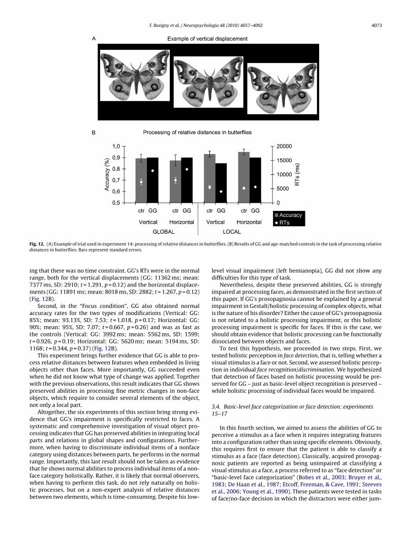
T. Busigny et al. / Neuropsychologia 48 (2010) 4057–4092 4073
F n buttd
ir7m(
a89tt1
cowwpon
dscpmcrtfwtb
ig. 12. (A) Example of trial used in experiment 14: processing of relative distances iistances in butterflies. Bars represent standard errors.
ng that there was no time constraint. GG’s RTs were in the normalange, both for the vertical displacements (GG: 11362 ms; mean:377 ms, SD: 2910; t = 1.291, p = 0.12) and the horizontal displace-ents (GG: 11891 ms; mean: 8018 ms, SD: 2882; t = 1.267, p = 0.12)
Fig. 12B).Second, in the “Focus condition”, GG also obtained normal
ccuracy rates for the two types of modifications (Vertical: GG:5%; mean: 93.13%, SD: 7.53; t = 1.018, p = 0.17; Horizontal: GG:0%; mean: 95%, SD: 7.07; t = 0.667, p = 0.26) and was as fast ashe controls (Vertical: GG: 3992 ms; mean: 5562 ms, SD: 1599;= 0.926, p = 0.19; Horizontal: GG: 5620 ms; mean: 5194 ms, SD:168; t = 0.344, p = 0.37) (Fig. 12B).
This experiment brings further evidence that GG is able to pro-ess relative distances between features when embedded in livingbjects other than faces. More importantly, GG succeeded evenhen he did not know what type of change was applied. Togetherith the previous observations, this result indicates that GG showsreserved abilities in processing fine metric changes in non-facebjects, which require to consider several elements of the object,ot only a local part.
Altogether, the six experiments of this section bring strong evi-ence that GG’s impairment is specifically restricted to faces. Aystematic and comprehensive investigation of visual object pro-essing indicates that GG has preserved abilities in integrating localarts and relations in global shapes and configurations. Further-ore, when having to discriminate individual items of a nonface
ategory using distances between parts, he performs in the normalange. Importantly, this last result should not be taken as evidence
hat he shows normal abilities to process individual items of a non-ace category holistically. Rather, it is likely that normal observers,hen having to perform this task, do not rely naturally on holis-ic processes, but on a non-expert analysis of relative distancesetween two elements, which is time-consuming. Despite his low-
erflies. (B) Results of GG and age-matched controls in the task of processing relative
level visual impairment (left hemianopia), GG did not show anydifficulties for this type of task.
Nevertheless, despite these preserved abilities, GG is stronglyimpaired at processing faces, as demonstrated in the first section ofthis paper. If GG’s prosopagnosia cannot be explained by a generalimpairment in Gestalt/holistic processing of complex objects, whatis the nature of his disorder? Either the cause of GG’s prosopagnosiais not related to a holistic processing impairment, or this holisticprocessing impairment is specific for faces. If this is the case, weshould obtain evidence that holistic processing can be functionallydissociated between objects and faces.
To test this hypothesis, we proceeded in two steps. First, wetested holistic perception in face detection, that is, telling whether avisual stimulus is a face or not. Second, we assessed holistic percep-tion in individual face recognition/discrimination. We hypothesizedthat detection of faces based on holistic processing would be pre-served for GG – just as basic-level object recognition is preserved –while holistic processing of individual faces would be impaired.
3.4. Basic-level face categorization or face detection: experiments15–17
In this fourth section, we aimed to assess the abilities of GG toperceive a stimulus as a face when it requires integrating featuresinto a configuration rather than using specific elements. Obviously,this requires first to ensure that the patient is able to classify astimulus as a face (face detection). Classically, acquired prosopag-nosic patients are reported as being unimpaired at classifying a
visual stimulus as a face, a process referred to as “face detection” or“basic-level face categorization” (Bobes et al., 2003; Bruyer et al.,1983; De Haan et al., 1987; Etcoff, Freeman, & Cave, 1991; Steeveset al., 2006; Young et al., 1990). These patients were tested in tasksof face/no-face decision in which the distractors were either jum-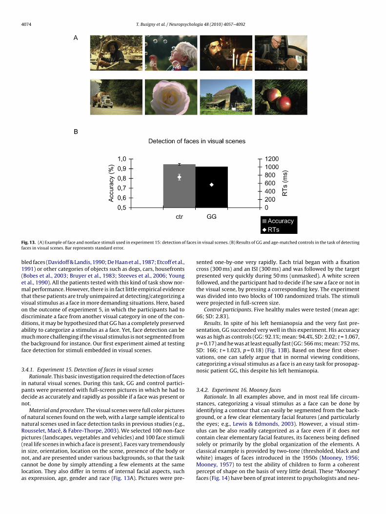
4074 T. Busigny et al. / Neuropsychologia 48 (2010) 4057–4092
F facesf
b1(emtvoddamtf
3
ipdn
onRp(incla
ig. 13. (A) Example of face and nonface stimuli used in experiment 15: detection ofaces in visual scenes. Bar represents standard error.
led faces (Davidoff & Landis, 1990; De Haan et al., 1987; Etcoff et al.,991) or other categories of objects such as dogs, cars, housefrontsBobes et al., 2003; Bruyer et al., 1983; Steeves et al., 2006; Youngt al., 1990). All the patients tested with this kind of task show nor-al performance. However, there is in fact little empirical evidence
hat these patients are truly unimpaired at detecting/categorizing aisual stimulus as a face in more demanding situations. Here, basedn the outcome of experiment 5, in which the participants had toiscriminate a face from another visual category in one of the con-itions, it may be hypothesized that GG has a completely preservedbility to categorize a stimulus as a face. Yet, face detection can beuch more challenging if the visual stimulus is not segmented from
he background for instance. Our first experiment aimed at testingace detection for stimuli embedded in visual scenes.
.4.1. Experiment 15. Detection of faces in visual scenesRationale. This basic investigation required the detection of faces
n natural visual scenes. During this task, GG and control partici-ants were presented with full-screen pictures in which he had toecide as accurately and rapidly as possible if a face was present orot.
Material and procedure. The visual scenes were full color picturesf natural scenes found on the web, with a large sample identical toatural scenes used in face detection tasks in previous studies (e.g.,ousselet, Macé, & Fabre-Thorpe, 2003). We selected 100 non-faceictures (landscapes, vegetables and vehicles) and 100 face stimulireal life scenes in which a face is present). Faces vary tremendously
n size, orientation, location on the scene, presence of the body orot, and are presented under various backgrounds, so that the taskannot be done by simply attending a few elements at the sameocation. They also differ in terms of internal facial aspects, suchs expression, age, gender and race (Fig. 13A). Pictures were pre-in visual scenes. (B) Results of GG and age-matched controls in the task of detecting
sented one-by-one very rapidly. Each trial began with a fixationcross (300 ms) and an ISI (300 ms) and was followed by the targetpresented very quickly during 50 ms (unmasked). A white screenfollowed, and the participant had to decide if he saw a face or not inthe visual scene, by pressing a corresponding key. The experimentwas divided into two blocks of 100 randomized trials. The stimuliwere projected in full-screen size.
Control participants. Five healthy males were tested (mean age:66; SD: 2.83).
Results. In spite of his left hemianopsia and the very fast pre-sentation, GG succeeded very well in this experiment. His accuracywas as high as controls (GG: 92.1%; mean: 94.4%, SD: 2.02; t = 1.067,p = 0.17) and he was at least equally fast (GG: 566 ms; mean: 752 ms,SD: 166; t = 1.023, p = 0.18) (Fig. 13B). Based on these first obser-vations, one can safely argue that in normal viewing conditions,categorizing a visual stimulus as a face is an easy task for prosopag-nosic patient GG, this despite his left hemianopia.
3.4.2. Experiment 16. Mooney facesRationale. In all examples above, and in most real life circum-
stances, categorizing a visual stimulus as a face can be done byidentifying a contour that can easily be segmented from the back-ground, or a few clear elementary facial features (and particularlythe eyes; e.g., Lewis & Edmonds, 2003). However, a visual stim-ulus can be also readily categorized as a face even if it does notcontain clear elementary facial features, its faceness being definedsolely or primarily by the global organization of the elements. A
classical example is provided by two-tone (thresholded, black andwhite) images of faces introduced in the 1950s (Mooney, 1956;Mooney, 1957) to test the ability of children to form a coherentpercept of shape on the basis of very little detail. These “Mooney”faces (Fig. 14) have been of great interest to psychologists and neu-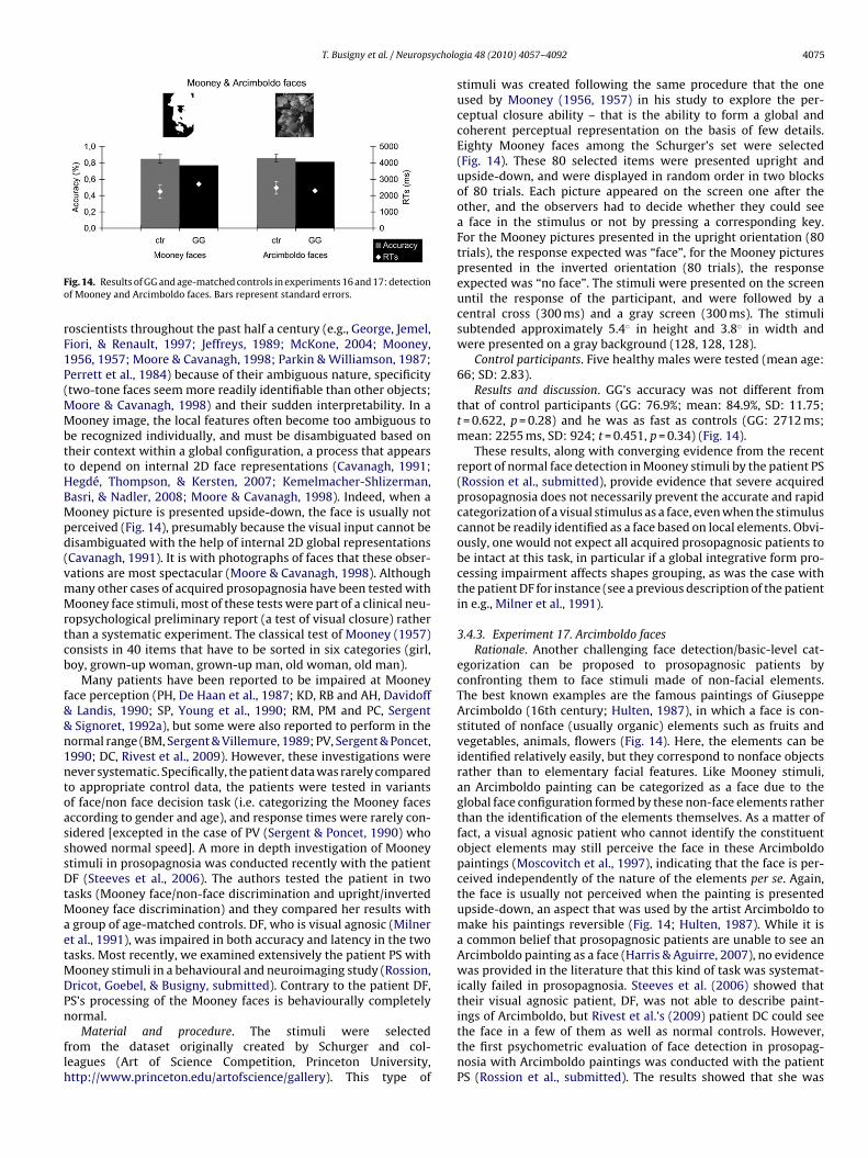
T. Busigny et al. / Neuropsycholo
Fo
rF1P(MMbttHBMpd(vmMrtcb
f&&n1ntoasssDtMaetMDPn
flh
ig. 14. Results of GG and age-matched controls in experiments 16 and 17: detectionf Mooney and Arcimboldo faces. Bars represent standard errors.
oscientists throughout the past half a century (e.g., George, Jemel,iori, & Renault, 1997; Jeffreys, 1989; McKone, 2004; Mooney,956, 1957; Moore & Cavanagh, 1998; Parkin & Williamson, 1987;errett et al., 1984) because of their ambiguous nature, specificitytwo-tone faces seem more readily identifiable than other objects;
oore & Cavanagh, 1998) and their sudden interpretability. In aooney image, the local features often become too ambiguous to
e recognized individually, and must be disambiguated based onheir context within a global configuration, a process that appearso depend on internal 2D face representations (Cavanagh, 1991;egdé, Thompson, & Kersten, 2007; Kemelmacher-Shlizerman,asri, & Nadler, 2008; Moore & Cavanagh, 1998). Indeed, when aooney picture is presented upside-down, the face is usually not
erceived (Fig. 14), presumably because the visual input cannot beisambiguated with the help of internal 2D global representationsCavanagh, 1991). It is with photographs of faces that these obser-ations are most spectacular (Moore & Cavanagh, 1998). Althoughany other cases of acquired prosopagnosia have been tested withooney face stimuli, most of these tests were part of a clinical neu-
opsychological preliminary report (a test of visual closure) ratherhan a systematic experiment. The classical test of Mooney (1957)onsists in 40 items that have to be sorted in six categories (girl,oy, grown-up woman, grown-up man, old woman, old man).
Many patients have been reported to be impaired at Mooneyace perception (PH, De Haan et al., 1987; KD, RB and AH, Davidoff
Landis, 1990; SP, Young et al., 1990; RM, PM and PC, SergentSignoret, 1992a), but some were also reported to perform in the
ormal range (BM, Sergent & Villemure, 1989; PV, Sergent & Poncet,990; DC, Rivest et al., 2009). However, these investigations wereever systematic. Specifically, the patient data was rarely comparedo appropriate control data, the patients were tested in variantsf face/non face decision task (i.e. categorizing the Mooney facesccording to gender and age), and response times were rarely con-idered [excepted in the case of PV (Sergent & Poncet, 1990) whohowed normal speed]. A more in depth investigation of Mooneytimuli in prosopagnosia was conducted recently with the patientF (Steeves et al., 2006). The authors tested the patient in two
asks (Mooney face/non-face discrimination and upright/invertedooney face discrimination) and they compared her results withgroup of age-matched controls. DF, who is visual agnosic (Milnert al., 1991), was impaired in both accuracy and latency in the twoasks. Most recently, we examined extensively the patient PS with
ooney stimuli in a behavioural and neuroimaging study (Rossion,ricot, Goebel, & Busigny, submitted). Contrary to the patient DF,S’s processing of the Mooney faces is behaviourally completely
ormal.Material and procedure. The stimuli were selectedrom the dataset originally created by Schurger and col-eagues (Art of Science Competition, Princeton University,ttp://www.princeton.edu/artofscience/gallery). This type of
gia 48 (2010) 4057–4092 4075
stimuli was created following the same procedure that the oneused by Mooney (1956, 1957) in his study to explore the per-ceptual closure ability – that is the ability to form a global andcoherent perceptual representation on the basis of few details.Eighty Mooney faces among the Schurger’s set were selected(Fig. 14). These 80 selected items were presented upright andupside-down, and were displayed in random order in two blocksof 80 trials. Each picture appeared on the screen one after theother, and the observers had to decide whether they could seea face in the stimulus or not by pressing a corresponding key.For the Mooney pictures presented in the upright orientation (80trials), the response expected was “face”, for the Mooney picturespresented in the inverted orientation (80 trials), the responseexpected was “no face”. The stimuli were presented on the screenuntil the response of the participant, and were followed by acentral cross (300 ms) and a gray screen (300 ms). The stimulisubtended approximately 5.4◦ in height and 3.8◦ in width andwere presented on a gray background (128, 128, 128).
Control participants. Five healthy males were tested (mean age:66; SD: 2.83).
Results and discussion. GG’s accuracy was not different fromthat of control participants (GG: 76.9%; mean: 84.9%, SD: 11.75;t = 0.622, p = 0.28) and he was as fast as controls (GG: 2712 ms;mean: 2255 ms, SD: 924; t = 0.451, p = 0.34) (Fig. 14).
These results, along with converging evidence from the recentreport of normal face detection in Mooney stimuli by the patient PS(Rossion et al., submitted), provide evidence that severe acquiredprosopagnosia does not necessarily prevent the accurate and rapidcategorization of a visual stimulus as a face, even when the stimuluscannot be readily identified as a face based on local elements. Obvi-ously, one would not expect all acquired prosopagnosic patients tobe intact at this task, in particular if a global integrative form pro-cessing impairment affects shapes grouping, as was the case withthe patient DF for instance (see a previous description of the patientin e.g., Milner et al., 1991).
3.4.3. Experiment 17. Arcimboldo facesRationale. Another challenging face detection/basic-level cat-
egorization can be proposed to prosopagnosic patients byconfronting them to face stimuli made of non-facial elements.The best known examples are the famous paintings of GiuseppeArcimboldo (16th century; Hulten, 1987), in which a face is con-stituted of nonface (usually organic) elements such as fruits andvegetables, animals, flowers (Fig. 14). Here, the elements can beidentified relatively easily, but they correspond to nonface objectsrather than to elementary facial features. Like Mooney stimuli,an Arcimboldo painting can be categorized as a face due to theglobal face configuration formed by these non-face elements ratherthan the identification of the elements themselves. As a matter offact, a visual agnosic patient who cannot identify the constituentobject elements may still perceive the face in these Arcimboldopaintings (Moscovitch et al., 1997), indicating that the face is per-ceived independently of the nature of the elements per se. Again,the face is usually not perceived when the painting is presentedupside-down, an aspect that was used by the artist Arcimboldo tomake his paintings reversible (Fig. 14; Hulten, 1987). While it isa common belief that prosopagnosic patients are unable to see anArcimboldo painting as a face (Harris & Aguirre, 2007), no evidencewas provided in the literature that this kind of task was systemat-ically failed in prosopagnosia. Steeves et al. (2006) showed thattheir visual agnosic patient, DF, was not able to describe paint-
ings of Arcimboldo, but Rivest et al.’s (2009) patient DC could seethe face in a few of them as well as normal controls. However,the first psychometric evaluation of face detection in prosopag-nosia with Arcimboldo paintings was conducted with the patientPS (Rossion et al., submitted). The results showed that she was
4 ycholo
at
tHaJtfpwtraatstetfi1
6
ai(2
inmcnt
comoet
nIpm
3
niofuioishsc
076 T. Busigny et al. / Neurops
s accurate and fast as the controls, as she did in the Mooneyask.
Material and procedure. The stimuli used here were inspired byhe paintings of the 16th century artist, Giuseppe Arcimboldo (seeulten, 1987; or for example http://www.artyst.net; 14 stimuli)nd by the creations of the contemporary mosaic portrait artist,ason Mecier (http://www.jasonmecier.com; 26 stimuli). Both ofhem created works of art consisting in faces composed by nonacial elements (vegetables, fruits, animals, candies, stationeries,ebbles, . . .). The pictures were downloaded from the websites andere cropped so that only the area of the face was present. Next,
he pictures were homogenized to have roughly the same size andesolution. In total, 40 color “Arcimboldo” face stimuli were cre-ted (Fig. 14). These 40 created items were presented at uprightnd upside-down orientation, and were displayed randomly inwo blocks of forty trials. The procedure and the instructions wereimilar to the Mooney faces experiment. For the “Arcimboldo” pic-ures presented in the upright orientation (40 trials), the responsexpected was “face”, for the “Arcimboldo” pictures presented inhe inverted orientation (40 trials), the response expected was “noace”. The stimuli subtended approximately 5.3◦ in height and 4.6◦
n width and were presented on a gray background (128, 128,28).
Control participants. Five healthy males were tested (mean age:6; SD: 2.83).
Results and discussion. Again, in this experiment, GG presentedprofile of response that was not different from the control partic-
pants, both for accuracy and response times. He was very accurateGG: 81.3%; mean: 85.8%, SD: 9.98; t = 0.412, p = 0.35) and fast (GG:303 ms; mean: 2481 ms, SD: 865; t = 0.188, p = 0.43) (Fig. 14).
These three experiments indicate clearly that the patient GGs perfectly able to categorize a visual stimulus as a face. He haso complaints in real life about this, and showed normal perfor-ance even in difficult circumstances. In this respect, he shows a
omparable response profile to another case of acquired prosopag-osia, with a different etiology and localization of brain damage,he patient PS (Rossion et al., 2003).
Here, we demonstrated that the problem of the patient is not inlassifying a stimulus as a face, even when it requires an analysisf the whole stimulus configuration rather than specific local ele-ents. These observations are consistent with what we found for
bjects: the patient is able to perceive a global form, and even toxtract local information (details) and take into account changes inerms of distances between these details (cfr. experiment 14).
Nevertheless, we have shown that GG has a massive prosopag-osia, showing great difficulties when having to individualize faces.
n the next section, we proceeded to assessing the nature of hisrosopagnosia by asking him to individualize faces in which weanipulated different kinds of information.
.5. Recognition of individual faces: experiments 18–24
So far, we have demonstrated that GG presents with a recog-ition impairment restricted to individual faces. However, he has
ntact face detection/basic-level categorization, even when a formf global or holistic perception such as for Mooney or Arcimboldo-acelike stimuli is required. In this final set of tasks, we aimed atnderstanding more precisely what causes GG’s difficulties in deal-
ng with individual faces. In a first subsection, we evaluated the rolef specific facial features and relations between them in tasks of
ndividual face recognition (experiments 18 and 19). In the secondubsection we assessed GG’s abilities at processing individual facesolistically using well-known paradigms and effects: face inver-ion (experiments 20–22), whole-part face (experiment 23) andomposite face effects (experiment 24).gia 48 (2010) 4057–4092
3.5.1. Facial features and relations between them in individualface recognition
As briefly indicated in Section 1, there is recent evidence thatacquired prosopagnosia can lead to a lack or reduced sensitivity todiagnostic information located in the eyes area of individual faces.Using a learning paradigm followed by an identification task offaces masked with random apertures (“Bubbles”, Gosselin & Schyns,2001), Caldara et al. (2005) tested the brain-damaged patient PS(Rossion et al., 2003). In contrast to normal viewers, who reliedextensively on localized information on the eyes of the faces, PSneeded much more information to achieve the same level of per-formance, and relied mostly on the mouth and lower contours ofthe faces rather than the eyes. Bukach et al. (2006) showed thatthe brain-damaged prosopagnosic patient LR was able to detectsmall shape changes in the mouth region as well as variations inmetric distances between features of the lower area of the face(e.g., nose–mouth distance), but was strikingly impaired at mak-ing similar judgments on the eyes of faces. Most recently, Bukachet al. (2008) extended these observations on LR and another caseof prosopagnosia (HH) using a Face Dimensions Task in which theparticipant’s sensitivity to parametric manipulations to the shapeand distance of facial features was tested. While the patients per-formed like control participants on all types of changes applied tothe mouth, they were severely impaired for individual face discrim-ination based on the eyes. Similar observations in this experimentwere made with the patient PS (Rossion et al., 2009), who has alsobeen shown to fixate her gaze mainly to the mouth when identify-ing personally familiar faces (Orban de Xivry et al., 2008; Van Belle,de Graef, Verfaillie, Busigny, et al., 2010).
3.5.1.1. Experiment 18. Simultaneous discrimination task with pre-view. Material and procedure. The stimuli were those used inGoffaux and Rossion (2007). In the experiment, there were fourrandomly interleaved stimulus conditions (eyes featural, eyes ver-tical, eyes horizontal, and nose–mouth featural). As usual, facestimuli were free of facial hair, glasses and hairline in order toremove any external cue to face perception. The inner featuresof each face (eyes, nose and mouth in their original spatial rela-tions) were pasted on a generic face shape. Then each stimulusunderwent four kinds of modifications: (1) eyes-featural (contrastof eyes were made darker or lighter, with small modifications ofshape), (2) eyes-vertical (eyes moved upward or downward in theface), (3) eyes-horizontal (smaller or larger inter-ocular distance),and (4) lower part of the face (nose and mouth were exchangedwith those of another face) (see Goffaux & Rossion, 2007). The taskwas a simultaneous matching with a preview of the target. Trialsbegan with the target face presented alone at the top of the screen.After 2000 ms, two probe stimuli located side by side below thetarget appeared on the screen and remained until a response. Oneof the probe stimuli was identical to the target and the distractor– for half of the trials on the left, randomly – was one of the fourmodified faces of this target-face. Participants had unlimited timeto answer by indicating whether the left or right probe stimuluscorresponded to the target. Twenty full-front gray scaled picturesof faces (half males) with a neutral expression were used. Each trialwas repeated twice, leading to 40 trials per condition. The exper-iment was divided into two blocks of 80 randomized trials. Thestimuli subtended approximately 9.2◦ in height and 7.1◦ in width,on a gray background.
Control participants. Five healthy males were tested (mean age:67.7; SD: 2.31).
Results and discussion. Control participants performed the taskvery well for all conditions (Bottom: 92%, SD: 7.58; Eyes featural:91.5%, SD: 8.77; Eyes horizontal: 97%, SD: 2.09; Eyes vertical: 98%,SD: 2.09) (Fig. 15), with no difference between the 4 conditions(F(1,4) = 1.471, p = 0.14). They took on average around 3000 ms to
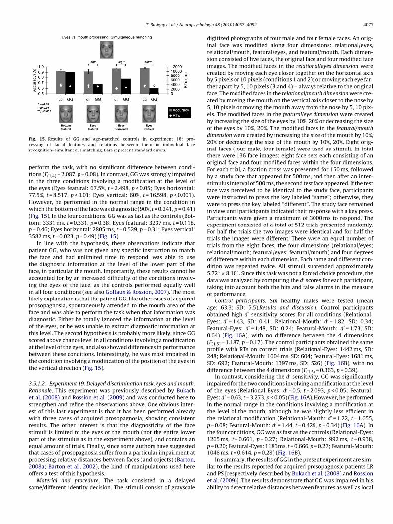
T. Busigny et al. / Neuropsycholo
Fcr
ptit7Hw(tp3
pttfaiilpfdotsabtt
3Resewrspetp2o
s
ig. 15. Results of GG and age-matched controls in experiment 18: pro-essing of facial features and relations between them in individual faceecognition–simultaneous matching. Bars represent standard errors.
erform the task, with no significant difference between condi-ions (F(1,4) = 2.087, p = 0.08). In contrast, GG was strongly impairedn the three conditions involving a modification at the level ofhe eyes (Eyes featural: 67.5%, t = 2.498, p < 0.05; Eyes horizontal:7.5%, t = 8.517, p < 0.01; Eyes vertical: 60%, t = 16.598, p < 0.001).owever, he performed in the normal range in the condition inhich the bottom of the face was diagnostic (90%, t = 0.241, p = 0.41)
Fig. 15). In the four conditions, GG was as fast as the controls (Bot-om: 3331 ms, t = 0.331, p = 0.38; Eyes featural: 3237 ms, t = 0.118,= 0.46; Eyes horizontal: 2805 ms, t = 0.529, p = 0.31; Eyes vertical:582 ms, t = 0.023, p = 0.49) (Fig. 15).
In line with the hypothesis, these observations indicate thatatient GG, who was not given any specific instruction to matchhe face and had unlimited time to respond, was able to usehe diagnostic information at the level of the lower part of theace, in particular the mouth. Importantly, these results cannot beccounted for by an increased difficulty of the conditions involv-ng the eyes of the face, as the controls performed equally welln all four conditions (see also Goffaux & Rossion, 2007). The mostikely explanation is that the patient GG, like other cases of acquiredrosopagnosia, spontaneously attended to the mouth area of theace and was able to perform the task when that information wasiagnostic. Either he totally ignored the information at the levelf the eyes, or he was unable to extract diagnostic information athis level. The second hypothesis is probably more likely, since GGcored above chance level in all conditions involving a modificationt the level of the eyes, and also showed differences in performanceetween these conditions. Interestingly, he was most impaired inhe condition involving a modification of the position of the eyes inhe vertical direction (Fig. 15).
.5.1.2. Experiment 19. Delayed discrimination task, eyes and mouth.ationale. This experiment was previously described by Bukacht al. (2008) and Rossion et al. (2009) and was conducted here totrengthen and refine the observations above. One obvious inter-st of this last experiment is that it has been performed alreadyith three cases of acquired prosopagnosia, showing consistent
esults. The other interest is that the diagnosticity of the facetimuli is limited to the eyes or the mouth (not the entire lowerart of the stimulus as in the experiment above), and contains anqual amount of trials. Finally, since some authors have suggestedhat cases of prosopagnosia suffer from a particular impairment at
rocessing relative distances between faces (and objects) (Barton,008a; Barton et al., 2002), the kind of manipulations used hereffers a test of this hypothesis.Material and procedure. The task consisted in a delayedame/different identity decision. The stimuli consist of grayscale
gia 48 (2010) 4057–4092 4077
digitized photographs of four male and four female faces. An orig-inal face was modified along four dimensions: relational/eyes,relational/mouth, featural/eyes, and featural/mouth. Each dimen-sion consisted of five faces, the original face and four modified faceimages. The modified faces in the relational/eyes dimension werecreated by moving each eye closer together on the horizontal axisby 5 pixels or 10 pixels (conditions 1 and 2); or moving each eye far-ther apart by 5, 10 pixels (3 and 4) – always relative to the originalface. The modified faces in the relational/mouth dimension were cre-ated by moving the mouth on the vertical axis closer to the nose by5, 10 pixels or moving the mouth away from the nose by 5, 10 pix-els. The modified faces in the featural/eye dimension were createdby increasing the size of the eyes by 10%, 20% or decreasing the sizeof the eyes by 10%, 20%. The modified faces in the featural/mouthdimension were created by increasing the size of the mouth by 10%,20% or decreasing the size of the mouth by 10%, 20%. Eight orig-inal faces (four male, four female) were used as stimuli. In totalthere were 136 face images: eight face sets each consisting of anoriginal face and four modified faces within the four dimensions.For each trial, a fixation cross was presented for 150 ms, followedby a study face that appeared for 500 ms, and then after an inter-stimulus interval of 500 ms, the second test face appeared. If the testface was perceived to be identical to the study face, participantswere instructed to press the key labeled “same”; otherwise, theywere to press the key labeled “different”. The study face remainedin view until participants indicated their response with a key press.Participants were given a maximum of 3000 ms to respond. Theexperiment consisted of a total of 512 trials presented randomly.For half the trials the two images were identical and for half thetrials the images were different. There were an equal number oftrials from the eight faces, the four dimensions (relational/eyes;relational/mouth; featural/eyes; featural/mouth) and four degreesof difference within each dimension. Each same and different con-dition was repeated twice. All stimuli subtended approximately5.72◦ × 8.10◦. Since this task was not a forced choice procedure, thedata was analyzed by computing the d′ scores for each participant,taking into account both the hits and false alarms in the measureof performance.
Control participants. Six healthy males were tested (meanage: 63.3; SD: 5.5).Results and discussion. Control participantsobtained high d′ sensitivity scores for all conditions (Relational-Eyes: d′ = 1.43, SD: 0.41; Relational-Mouth: d′ = 1.82, SD: 0.34;Featural-Eyes: d′ = 1.48, SD: 0.24; Featural-Mouth: d′ = 1.73, SD:0.64) (Fig. 16A), with no difference between the 4 dimensions(F(1,5) = 1.187, p = 0.17). The control participants obtained the sameprofile with RTs on correct trials (Relational-Eyes: 1442 ms, SD:248; Relational-Mouth: 1604 ms, SD: 604; Featural-Eyes: 1681 ms,SD: 692; Featural-Mouth: 1397 ms, SD: 526) (Fig. 16B), with nodifference between the 4 dimensions (F(1,5) = 0.363, p = 0.39).
In contrast, considering the d′ sensitivity, GG was significantlyimpaired for the two conditions involving a modification at the levelof the eyes (Relational-Eyes: d′ = 0.5, t = 2.093, p < 0.05; Featural-Eyes: d′ = 0.63, t = 3.273, p < 0.05) (Fig. 16A). However, he performedin the normal range in the conditions involving a modification atthe level of the mouth, although he was slightly less efficient inthe relational modification (Relational-Mouth: d′ = 1.22, t = 1.655,p = 0.08; Featural-Mouth: d′ = 1.44, t = 0.429, p = 0.34) (Fig. 16A). Inthe four conditions, GG was as fast as the controls (Relational-Eyes:1265 ms, t = 0.661, p = 0.27; Relational-Mouth: 992 ms, t = 0.938,p = 0.20; Featural-Eyes: 1183ms, t = 0.666, p = 0.27; Featural-Mouth:1048 ms, t = 0.614, p = 0.28) (Fig. 16B).
In summary, the results of GG in the present experiment are sim-ilar to the results reported for acquired prosopagnosic patients LRand PS [respectively described by Bukach et al. (2008) and Rossionet al. (2009)]. The results demonstrate that GG was impaired in hisability to detect relative distances between features as well as local
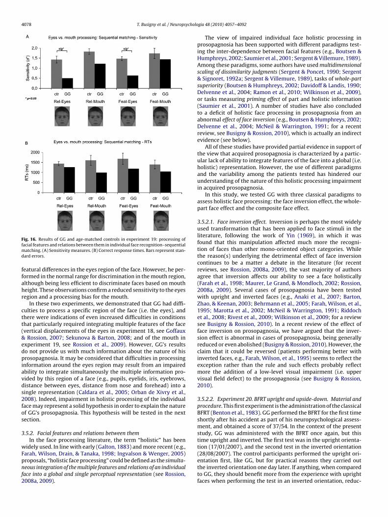
4078 T. Busigny et al. / Neuropsycholo
Ffmd
ffahr
ctt(&edpiavds2fos
3
wFpnf2
ig. 16. Results of GG and age-matched controls in experiment 19: processing ofacial features and relations between them in individual face recognition–sequential
atching. (A) Sensitivity measures. (B) Correct response times. Bars represent stan-ard errors.
eatural differences in the eyes region of the face. However, he per-ormed in the normal range for discrimination in the mouth region,lthough being less efficient to discriminate faces based on moutheight. These observations confirm a reduced sensitivity to the eyesegion and a processing bias for the mouth.
In these two experiments, we demonstrated that GG had diffi-ulties to process a specific region of the face (i.e. the eyes), andhere were indications of even increased difficulties in conditionshat particularly required integrating multiple features of the facevertical displacements of the eyes in experiment 18, see Goffaux
Rossion, 2007; Sekunova & Barton, 2008; and of the mouth inxperiment 19, see Rossion et al., 2009). However, GG’s resultso not provide us with much information about the nature of hisrosopagnosia. It may be considered that difficulties in processing
nformation around the eyes region may result from an impairedbility to integrate simultaneously the multiple information pro-ided by this region of a face (e.g., pupils, eyelids, iris, eyebrows,istance between eyes, distance from nose and forehead) into aingle representation (Caldara et al., 2005; Orban de Xivry et al.,008). Indeed, impairment in holistic processing of the individualace may represent a solid hypothesis in order to explain the naturef GG’s prosopagnosia. This hypothesis will be tested in the nextection.
.5.2. Facial features and relations between themIn the face processing literature, the term “holistic” has been
idely used. In line with early (Galton, 1883) and more recent (e.g.,
arah, Wilson, Drain, & Tanaka, 1998; Ingvalson & Wenger, 2005)roposals, “holistic face processing” could be defined as the simulta-eous integration of the multiple features and relations of an individualace into a global and single perceptual representation (see Rossion,008a, 2009).
gia 48 (2010) 4057–4092
The view of impaired individual face holistic processing inprosopagnosia has been supported with different paradigms test-ing the inter-dependence between facial features (e.g., Boutsen &Humphreys, 2002; Saumier et al., 2001; Sergent & Villemure, 1989).Among these paradigms, some authors have used multidimensionalscaling of dissimilarity judgments (Sergent & Poncet, 1990; Sergent& Signoret, 1992a; Sergent & Villemure, 1989), tasks of whole-partsuperiority (Boutsen & Humphreys, 2002; Davidoff & Landis, 1990;Delvenne et al., 2004; Ramon et al., 2010; Wilkinson et al., 2009),or tasks measuring priming effect of part and holistic information(Saumier et al., 2001). A number of studies have also concludedto a deficit of holistic face processing in prosopagnosia from anabnormal effect of face inversion (e.g., Boutsen & Humphreys, 2002;Delvenne et al., 2004; McNeil & Warrington, 1991; for a recentreview, see Busigny & Rossion, 2010), which is actually an indirectevidence (see below).
All of these studies have provided partial evidence in support ofthe view that acquired prosopagnosia is characterized by a partic-ular lack of ability to integrate features of the face into a global (i.e.holistic) representation. However, the use of different paradigmsand the variability among the patients tested has hindered ourunderstanding of the nature of this holistic processing impairmentin acquired prosopagnosia.
In this study, we tested GG with three classical paradigms toassess holistic face processing: the face inversion effect, the whole-part face effect and the composite face effect.
3.5.2.1. Face inversion effect. Inversion is perhaps the most widelyused transformation that has been applied to face stimuli in theliterature, following the work of Yin (1969), in which it wasfound that this manipulation affected much more the recogni-tion of faces than other mono-oriented object categories. Whilethe reason(s) underlying the detrimental effect of face inversioncontinues to be a matter a debate in the literature (for recentreviews, see Rossion, 2008a, 2009), the vast majority of authorsagree that inversion affects our ability to see a face holistically(Farah et al., 1998; Maurer, Le Grand, & Mondloch, 2002; Rossion,2008a, 2009). Several cases of prosopagnosia have been testedwith upright and inverted faces (e.g., Anaki et al., 2007; Barton,Zhao, & Keenan, 2003; Behrmann et al., 2005; Farah, Wilson, et al.,1995; Marotta et al., 2002; McNeil & Warrington, 1991; Riddochet al., 2008; Rivest et al., 2009; Wilkinson et al., 2009; for a reviewsee Busigny & Rossion, 2010). In a recent review of the effect offace inversion on prosopagnosia, we have argued that the inver-sion effect is abnormal in cases of prosopagnosia, being generallyreduced or even abolished (Busigny & Rossion, 2010). However, theclaim that it could be reversed (patients performing better withinverted faces, e.g., Farah, Wilson, et al., 1995) seems to reflect theexception rather than the rule and such effects probably reflectmore the addition of a low-level visual impairment (i.e. uppervisual field defect) to the prosopagnosia (see Busigny & Rossion,2010).
3.5.2.2. Experiment 20. BFRT upright and upside-down. Material andprocedure. This first experiment is the administration of the classicalBFRT (Benton et al., 1983). GG performed the BFRT for the first timeshortly after his accident as part of his neuropsychological assess-ment, and obtained a score of 37/54. In the context of the presentstudy, GG was administered with the BFRT once again, but thistime upright and inverted. The first test was in the upright orienta-tion (17/01/2007), and the second test in the inverted orientation
(28/08/2007). The control participants performed the upright ori-entation first, like GG, but for practical reasons they carried outthe inverted orientation one day later. If anything, when comparedto GG, they should benefit more from the experience with uprightfaces when performing the test in an inverted orientation, reduc-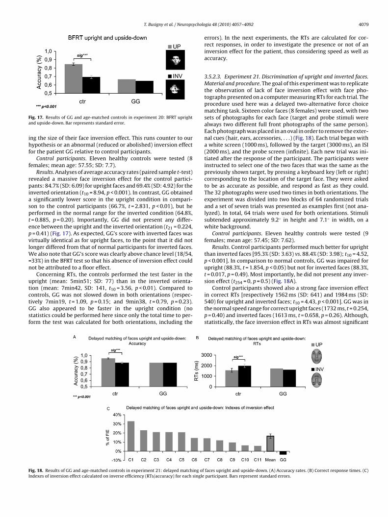
T. Busigny et al. / Neuropsycholo
Fa
ihf
f
rpiasptepvlW=n
utctGsf
FI
ig. 17. Results of GG and age-matched controls in experiment 20: BFRT uprightnd upside-down. Bar represents standard error.
ng the size of their face inversion effect. This runs counter to ourypothesis or an abnormal (reduced or abolished) inversion effect
or the patient GG relative to control participants.Control participants. Eleven healthy controls were tested (8
emales; mean age: 57.55; SD: 7.7).Results. Analyses of average accuracy rates (paired sample t-test)
evealed a massive face inversion effect for the control partici-ants: 84.7% (SD: 6.09) for upright faces and 69.4% (SD: 4.92) for the
nverted orientation (t10 = 8.94, p < 0.001). In contrast, GG obtainedsignificantly lower score in the upright condition in compari-
on to the control participants (66.7%, t = 2.831, p < 0.01), but heerformed in the normal range for the inverted condition (64.8%,= 0.885, p = 0.20). Importantly, GG did not present any differ-nce between the upright and the inverted orientation (t21 = 0.224,= 0.41) (Fig. 17). As expected, GG’s score with inverted faces wasirtually identical as for upright faces, to the point that it did notonger differed from that of normal participants for inverted faces.
e also note that GG’s score was clearly above chance level (18/54,33%) in the BFRT test so that his absence of inversion effect couldot be attributed to a floor effect.
Concerning RTs, the controls performed the test faster in thepright (mean: 5min51; SD: 77) than in the inverted orienta-ion (mean: 7min42, SD: 141, t10 = 3.56, p < 0.01). Compared to
ontrols, GG was not slowed down in both orientations (respec-ively 7min19, t = 1.09, p = 0.15; and 9min38, t = 0.79, p = 0.23).G also appeared to be faster in the upright condition (notatistics could be performed here since only the total time to per-orm the test was calculated for both orientations, including theig. 18. Results of GG and age-matched controls in experiment 21: delayed matching ofndexes of inversion effect calculated on inverse efficiency (RTs/accuracy) for each single
gia 48 (2010) 4057–4092 4079
errors). In the next experiments, the RTs are calculated for cor-rect responses, in order to investigate the presence or not of aninversion effect for the patient, thus considering speed as well asaccuracy.
3.5.2.3. Experiment 21. Discrimination of upright and inverted faces.Material and procedure. The goal of this experiment was to replicatethe observation of lack of face inversion effect with face pho-tographs presented on a computer measuring RTs for each trial. Theprocedure used here was a delayed two-alternative force choicematching task. Sixteen color faces (8 females) were used, with twosets of photographs for each face (target and probe stimuli werealways two different full front photographs of the same person).Each photograph was placed in an oval in order to remove the exter-nal cues (hair, ears, accessories, . . .) (Fig. 18). Each trial began witha white screen (1000 ms), followed by the target (3000 ms), an ISI(2000 ms), and the probe screen (infinite). Each new trial was ini-tiated after the response of the participant. The participants wereinstructed to select one of the two faces that was the same as thepreviously shown target, by pressing a keyboard key (left or right)corresponding to the location of the target face. They were askedto be as accurate as possible, and respond as fast as they could.The 32 photographs were used two times in both orientations. Theexperiment was divided into two blocks of 64 randomized trialsand a set of seven trials was presented as examples first (not ana-lyzed). In total, 64 trials were used for both orientations. Stimulisubtended approximately 9.2◦ in height and 7.1◦ in width, on awhite background.
Control participants. Eleven healthy controls were tested (9females; mean age: 57.45; SD: 7.62).
Results. Control participants performed much better for uprightthan inverted faces [95.3% (SD: 3.63) vs. 88.4% (SD: 3.98); t10 = 4.52,p < 0.001]. In comparison to normal controls, GG was impaired forupright (88.3%, t = 1.854, p < 0.05) but not for inverted faces (88.3%,t = 0.017, p = 0.49). Most importantly, he did not present any inver-sion effect (t254 = 0, p = 0.5) (Fig. 18A).
Control participants showed also a strong face inversion effect
in correct RTs [respectively 1562 ms (SD: 641) and 1984 ms (SD:540) for upright and inverted faces; t10 = 4.43, p < 0.001]. GG was inthe normal speed range for correct upright faces (1732 ms, t = 0.254,p = 0.40) and inverted faces (1613 ms, t = 0.658, p = 0.26). Although,statistically, the face inversion effect in RTs was almost significantfaces upright and upside-down. (A) Accuracy rates. (B) Correct response times. (C)participant. Bars represent standard errors.
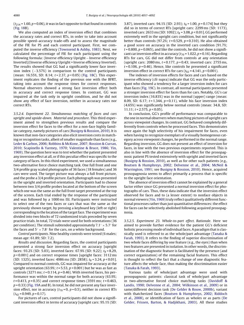
4 ycholo
((
tpopcfITs(itNiisc
3uiiikiL21act3caibwoatpcdppt
m
pfp(Cucftps(
c
080 T. Busigny et al. / Neurops
t214 = 1.60, p = 0.06), it was in fact opposite to that found in controlsFig. 18B).
We also computed an index of inversion effect that combineshe accuracy rates and correct RTs, in order to take into accountossible speed–accuracy trade-offs and to assess the magnitudef the FIE for PS and each control participant. First, we com-uted the inverse efficiency (Townsend & Ashby, 1983). Next, wealculated the percentage of FIE for each participant using theollowing formula: (Inverse efficiency Upright − Inverse efficiencynverted)/(Inverse efficiency Upright + Inverse efficiency Inverted).he results showed that GG had a significantly lower face inver-ion index (−3.57%) in comparison to the control participantsmean: 16.55%, SD: 8.14; t = 2.37, p < 0.05) (Fig. 18C). This exper-ment replicates the finding of the previous one with the BFRT,aking into account the response times for correct responses.ormal observers showed a strong face inversion effect both
n accuracy and correct response times. In contrast, GG wasmpaired at the task only in upright condition, and he did nothow any effect of face inversion, neither in accuracy rates nororrect RTs.
.5.2.4. Experiment 22. Simultaneous matching of faces and carspright and upside-down. Material and procedure. This third exper-
ment aimed to strengthen previous results and compare thenversion effect for faces to the effect for a non-face highly famil-ar category, namely pictures of cars (Busigny & Rossion, 2010). It isnown that non-face categories also elicit inversion costs in match-ng or recognition tasks, albeit of smaller magnitude than faces (e.g.,eder & Carbon, 2006; Robbins & McKone, 2007; Rossion & Curran,010; Scapinello & Yarmey, 1970; Valentine & Bruce, 1986; Yin,969). The question here was to test whether the patient presentedny inversion effect at all, or if this peculiar effect was specific to theategory of faces. In this third experiment, we used a simultaneouswo-alternative force choice matching task. One full front and one/4 profile gray scaled photographs of 36 faces (18 females) and 36ars were used. The target picture was always a full front picture,nd the probe a 3/4 profile picture. Each photograph was presentedn the upright and inverted orientation. Participants had to chooseetween two 3/4 profile probes located at the bottom of the screenhich one was the same as the full front target presented at the top
f the screen. Each trial ended by the response of the participantnd was followed by a 1000 ms ISI. Participants were instructedo select one of the two faces or cars that was the same as thereviously shown target, by pressing a keyboard key (left or right)orresponding to the location of the target face. The experiment wasivided into two blocks of 72 randomized trials preceded by sevenractice trials. In total, 72 trials were used for both orientations (36er condition). The stimuli subtended approximately 7.1◦ × 5.7◦ forhe faces and 5◦ × 7.8◦ for the cars, on a white background.
Control participants. Nine healthy controls were tested (6 males;ean age: 61.89; SD: 7.2).Results and discussion. Regarding faces, the control participants
resented a strong face inversion effect on accuracy [uprightaces: 93.2% (SD: 5.03), inverted faces: 67.9% (SD: 9.22); t8 = 8.29,< 0.001] and on correct response times [upright faces: 3112 ms
SD: 1325), inverted faces: 4986 ms (SD: 2858); t8 = 3.24, p < 0.01].ompared to normal controls, GG was impaired for accuracy at thepright orientation (63.9%; t = 5.53, p < 0.001) but he was as fast asontrols (3271 ms; t = 0.114, p = 0.46). With inverted faces, his per-ormance was within the normal range for both accuracy (63.9%;= 0.413, p = 0.35) and correct response times (3591 ms; t = 0.463,
= 0.33) (Fig. 19A and B). In total, he did not present any face inver-ion effect, nor in accuracy (t70 = 0, p = 0.5), neither in correct RTst42 = 0.949, p = 0.17).
For pictures of cars, control participants did not show a signifi-ant inversion effect in terms of accuracy [upright cars: 95.1% (SD:
gia 48 (2010) 4057–4092
3.87), inverted cars: 94.1% (SD: 2.93); t8 = 1.00, p = 0.174] but theydid so in terms of correct RTs [upright cars: 2299 ms (SD: 1173),inverted cars: 2633 ms (SD: 1092); t8 = 3.08, p < 0.01]. GG performedextremely well in the upright cars condition, but not significantlybetter than controls (97.2%; t = 0.529, p = 0.310). He also obtaineda good score on accuracy in the inverted cars condition (91.7%;t = 0.800, p = 0.065), and like the controls, he did not show a signifi-cant car inversion effect in accuracy (t70 = 1.022, p = 0.16). RegardingRTs for cars, GG did not differ from controls at any orientation(upright cars: 2080 ms, t = 0.177, p = 0.43; inverted cars: 2755 ms,t = 0.106, p = 0.46). Hence, like controls he presented a significantinversion effect in correct RTs for cars (t62 = 4.141, p < 0.001).
The indexes of inversion effects for faces and cars based on theinverse efficiency (cfr supra) indicate that GG was the only partic-ipant who showed a tendency for a larger inversion index for carsthan faces (Fig. 19C). In contrast, all normal participants presenteda stronger inversion effect for faces than for cars. Notably, GG’s carinversion index (16.83%) was in the normal (upper) range (mean:8.09, SD: 6.17; t = 1.344, p = 0.11), while his face inversion index(4.65%) was significantly below normal controls (mean: 34.8, SD:11.11; t = 2.575, p < 0.05).
In conclusion, GG’s profile of performance was comparable tothe one in normal observers when matching pictures of upright carsacross viewpoint changes. In contrast, GG behaved completely dif-ferently from controls when he processed faces. This demonstratesonce again the high selectivity of his impairment for faces, evenwhen having to recognize exemplars of a visually homogenous cat-egory across viewpoint changes, he performed in the normal range.Regarding inversion, GG does not present an effect of inversion forfaces, in line with the two previous experiments reported. This isalso in line with the absence of inversion effect for the prosopag-nosic patient PS tested extensively with upright and inverted faces(Busigny & Rossion, 2010), as well as for other such patients (e.g.,Boutsen & Humphreys, 2002; Delvenne et al., 2004; McNeil &Warrington, 1991; see Busigny & Rossion, 2010). Hence, acquiredprosopagnosia seems to affect primarily a process that is specificto the upright face orientation.
The absence of inversion effect cannot be explained by a generalfactor either since GG presented a normal inversion effect for pho-tographs of cars. Thus, these data indicate that the inversion effectobserved for faces and to (a lesser extent) to nonface objects innormal viewers (Yin, 1969) truly reflect qualitatively different func-tional processes rather than just quantitative differences: the effectfor faces can be selectively abolished following acquired prosopag-nosia.
3.5.2.5. Experiment 23. Whole-to-part effect. Rationale. Here weaimed to provide further evidence for the patient GG’s deficientholistic processing mode of individual faces. A paradigm that is clas-sically used is referred to as the whole/part advantage (Tanaka &Farah, 1993). It refers to the finding of superior discrimination oftwo whole faces differing by one feature (e.g., the eyes) than whentwo features are presented in isolation. In other words, the discrim-ination of the diagnostic feature is facilitated by the presence (andcorrect organization) of the remaining facial features. This effectis thought to reflect the fact that a change of one diagnostic fea-ture affects the whole face, thus making the discrimination easier(Tanaka & Farah, 1993).
Various tasks of whole/part advantage were used withprosopagnosic patients: classical task of whole/part advantagein two-alternative forced choice matching tasks (Davidoff &
Landis, 1990; Delvenne et al., 2004; Wilkinson et al., 2009) or insame/different decision task (De Gelder & Rouw, 2000b), variantwith thatcherized faces (Boutsen & Humphreys, 2002; Riddochet al., 2008), or identification of faces as wholes or as parts (DeGelder, Frissen, Barton, & Hadjikhani, 2003). All these studies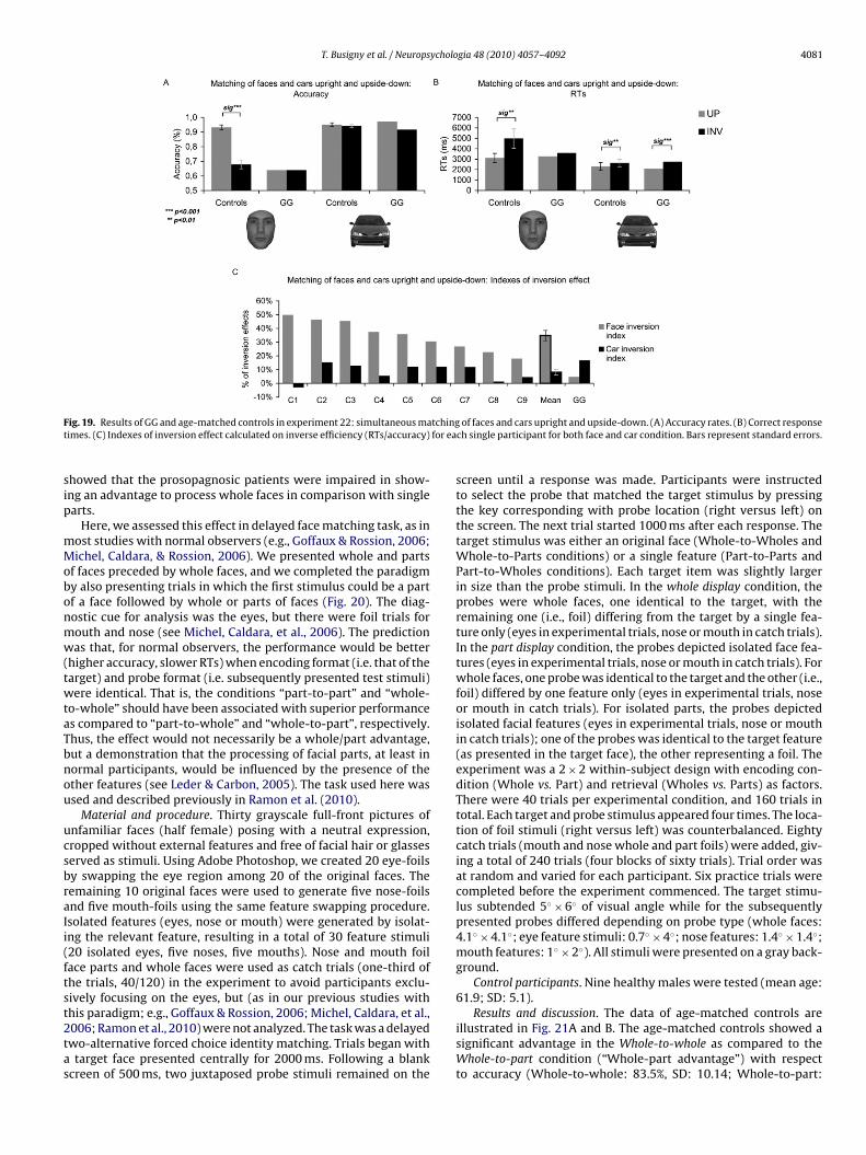
T. Busigny et al. / Neuropsychologia 48 (2010) 4057–4092 4081
F tchingt for ea
sip
mMobonmw(twtaTbnou
ucsbraIi(ftst2tas
ig. 19. Results of GG and age-matched controls in experiment 22: simultaneous maimes. (C) Indexes of inversion effect calculated on inverse efficiency (RTs/accuracy)
howed that the prosopagnosic patients were impaired in show-ng an advantage to process whole faces in comparison with singlearts.
Here, we assessed this effect in delayed face matching task, as inost studies with normal observers (e.g., Goffaux & Rossion, 2006;ichel, Caldara, & Rossion, 2006). We presented whole and parts
f faces preceded by whole faces, and we completed the paradigmy also presenting trials in which the first stimulus could be a partf a face followed by whole or parts of faces (Fig. 20). The diag-ostic cue for analysis was the eyes, but there were foil trials forouth and nose (see Michel, Caldara, et al., 2006). The predictionas that, for normal observers, the performance would be better
higher accuracy, slower RTs) when encoding format (i.e. that of thearget) and probe format (i.e. subsequently presented test stimuli)ere identical. That is, the conditions “part-to-part” and “whole-
o-whole” should have been associated with superior performances compared to “part-to-whole” and “whole-to-part”, respectively.hus, the effect would not necessarily be a whole/part advantage,ut a demonstration that the processing of facial parts, at least inormal participants, would be influenced by the presence of thether features (see Leder & Carbon, 2005). The task used here wassed and described previously in Ramon et al. (2010).
Material and procedure. Thirty grayscale full-front pictures ofnfamiliar faces (half female) posing with a neutral expression,ropped without external features and free of facial hair or glasseserved as stimuli. Using Adobe Photoshop, we created 20 eye-foilsy swapping the eye region among 20 of the original faces. Theemaining 10 original faces were used to generate five nose-foilsnd five mouth-foils using the same feature swapping procedure.solated features (eyes, nose or mouth) were generated by isolat-ng the relevant feature, resulting in a total of 30 feature stimuli20 isolated eyes, five noses, five mouths). Nose and mouth foilace parts and whole faces were used as catch trials (one-third ofhe trials, 40/120) in the experiment to avoid participants exclu-ively focusing on the eyes, but (as in our previous studies with
his paradigm; e.g., Goffaux & Rossion, 2006; Michel, Caldara, et al.,006; Ramon et al., 2010) were not analyzed. The task was a delayedwo-alternative forced choice identity matching. Trials began withtarget face presented centrally for 2000 ms. Following a blankcreen of 500 ms, two juxtaposed probe stimuli remained on the
of faces and cars upright and upside-down. (A) Accuracy rates. (B) Correct responsech single participant for both face and car condition. Bars represent standard errors.
screen until a response was made. Participants were instructedto select the probe that matched the target stimulus by pressingthe key corresponding with probe location (right versus left) onthe screen. The next trial started 1000 ms after each response. Thetarget stimulus was either an original face (Whole-to-Wholes andWhole-to-Parts conditions) or a single feature (Part-to-Parts andPart-to-Wholes conditions). Each target item was slightly largerin size than the probe stimuli. In the whole display condition, theprobes were whole faces, one identical to the target, with theremaining one (i.e., foil) differing from the target by a single fea-ture only (eyes in experimental trials, nose or mouth in catch trials).In the part display condition, the probes depicted isolated face fea-tures (eyes in experimental trials, nose or mouth in catch trials). Forwhole faces, one probe was identical to the target and the other (i.e.,foil) differed by one feature only (eyes in experimental trials, noseor mouth in catch trials). For isolated parts, the probes depictedisolated facial features (eyes in experimental trials, nose or mouthin catch trials); one of the probes was identical to the target feature(as presented in the target face), the other representing a foil. Theexperiment was a 2 × 2 within-subject design with encoding con-dition (Whole vs. Part) and retrieval (Wholes vs. Parts) as factors.There were 40 trials per experimental condition, and 160 trials intotal. Each target and probe stimulus appeared four times. The loca-tion of foil stimuli (right versus left) was counterbalanced. Eightycatch trials (mouth and nose whole and part foils) were added, giv-ing a total of 240 trials (four blocks of sixty trials). Trial order wasat random and varied for each participant. Six practice trials werecompleted before the experiment commenced. The target stimu-lus subtended 5◦ × 6◦ of visual angle while for the subsequentlypresented probes differed depending on probe type (whole faces:4.1◦ × 4.1◦; eye feature stimuli: 0.7◦ × 4◦; nose features: 1.4◦ × 1.4◦;mouth features: 1◦ × 2◦). All stimuli were presented on a gray back-ground.
Control participants. Nine healthy males were tested (mean age:61.9; SD: 5.1).
Results and discussion. The data of age-matched controls areillustrated in Fig. 21A and B. The age-matched controls showed asignificant advantage in the Whole-to-whole as compared to theWhole-to-part condition (“Whole-part advantage”) with respectto accuracy (Whole-to-whole: 83.5%, SD: 10.14; Whole-to-part:

4082 T. Busigny et al. / Neuropsychologia 48 (2010) 4057–4092
n exp
7ttdw5rSpf
twctitPipdt
uapWcieectwe
of context change. These results mean that GG does not present
F
Fig. 20. Example of stimuli used i
8.6%, SD: 11.19; t8 = 2.257, p < 0.05) and correct RTs (Whole-o-whole: 1352 ms, SD: 283; Whole-to-part: 1763 ms, SD: 260;8 = 11.698, p < 0.001). They also showed a significant “Whole-partisadvantage” when comparing the Part-to-part and the Part-to-hole conditions, both in terms of accuracy (Part-to-part: 93.9%, SD:.76; Part-to-whole: 86.4%, SD: 9.27; t8 = 2.917, p < 0.01), and cor-ect RTs (Part-to-part: 1258 ms, SD: 208; Part-to-whole: 1564 ms,D: 292; t8 = 5.48, p < 0.001). These results show that the controlarticipants are better and faster when the encoding and retrievalace context is the same.
GG showed no difference between the Whole-to-part andhe Whole-to-whole conditions, neither in accuracy (Whole-to-hole: 63.8%; Whole-to-part: 68.8%; t158 = 0.665, p = 0.25) nor in
orrect RTs (Whole-to-whole: 1336 ms; Whole-to-part: 1341ms;99 = 0.076, p = 0.47). He even showed a reverse trend of effectn accuracy: he was less efficient for the Whole-to-whole condi-ion as compared to the Whole-to-part (Fig. 21A). Considering theart-to-part and the Part-to-whole conditions, GG showed no effectn accuracy (Part-to-part: 95%; Part-to-whole: 92.5%; t158 = 0.650,= 0.26), but he obtained a significant difference in the rightirection in RTs (Part-to-part: 1232 ms; Part-to-whole: 1398ms;
141 = 3.791, p < 0.001) (Fig. 21B).Indexes were computed using the same formula that was
sed for the inversion effect (cfr supra). Thus we calculatedn index for the condition in which the whole face is firstlyresented (inverse efficiency Whole-to-part–Inverse efficiencyhole-to-whole)/(inverse efficiency Whole-to-part + inverse effi-
iency Whole-to-whole) and for the condition in which the parts firstly presented (inverse efficiency Part-to-whole − inversefficiency Part-to-part)/(inverse efficiency Part-to-whole + inversefficiency Part-to-part). In the whole display condition, each single
ontrol participant obtained a high index of whole-part advan-age (mean = 16.7%, SD: 7.28), but this was not the case for GG,ho even obtained a negative index (−3.6%), significantly differ-nt from the controls (t = 2.641, p < 0.05) (Fig. 22A). Similarly, in the
ig. 21. Results of GG and age-matched controls in experiment 23: whole-to-part effect.
eriment 23: whole-to-part effect.
part display condition, each single participant showed a high indexof part-whole disadvantage (mean = 14.9%, SD: 5.17). Even if GG didnot show a significantly reduced effect in comparison to the con-trols, his effect was the lowest of them all (GG: 7.6%, controls range:[25.4–8.6%], t = 1.332, p = 0.11) (Fig. 22B).
Finally, we computed together the results of the two display con-ditions (whole first and part first) to obtain a general index of contextchange. To obtain this, we put together the condition part-to-partwith the condition whole-to-whole in a new condition called “samecontext”, and the condition part-to-whole with the condition whole-to-part in a new condition called “different context”. Then we usedthe following formula: (same context − different context)/(samecontext + different context). By doing that, we calculated the per-centage loss of performance when the context changes. The indexeswere calculated for inverse efficiency as previously described. Theresults indicate that every single control participant showed a highindex of context change (mean = 15.8%, SD: 3.74), while GG showeda highly reduced effect (3%, t = 3.244, p < 0.01) (Fig. 22C).
In conclusion, the age-matched controls show better per-formance when encoding format and recognition format arecongruent: (1) when they are presented at encoding with a wholeface, they have an advantage recognizing a part embedded in awhole face in contrast to the part presented in isolation (because ofa whole context advantage); (2) when they are presented at encod-ing with a face part, they recognize the part better when presentedin isolation than when it is embedded in a whole face (because ofa whole context interference). In contrast, GG does not show anyof these effects: he shows no whole context advantage, even pre-senting a reverse trend, and he presents reduced whole contextinterference. Most importantly, GG does not show a general effect
the normal effects associated with a processing of the whole faceconfiguration. These observations provide direct evidence that GGdoes not show a classical whole-part effect, and that he processesfaces qualitatively differently than normal observers.
(A) Accuracy rates. (B) Correct response times. Bars represent standard errors.
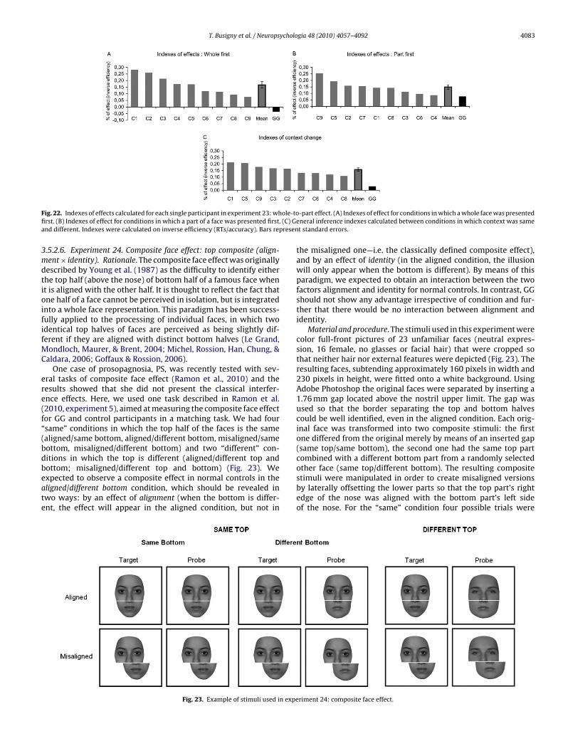
T. Busigny et al. / Neuropsychologia 48 (2010) 4057–4092 4083
F ole-tofi (C) Gea resen
3mdtioififMC
ere(f“(bdbeate
ig. 22. Indexes of effects calculated for each single participant in experiment 23: whrst. (B) Indexes of effect for conditions in which a part of a face was presented first.nd different. Indexes were calculated on inverse efficiency (RTs/accuracy). Bars rep
.5.2.6. Experiment 24. Composite face effect: top composite (align-ent × identity). Rationale. The composite face effect was originally
escribed by Young et al. (1987) as the difficulty to identify eitherhe top half (above the nose) of bottom half of a famous face whent is aligned with the other half. It is thought to reflect the fact thatne half of a face cannot be perceived in isolation, but is integratednto a whole face representation. This paradigm has been success-ully applied to the processing of individual faces, in which twodentical top halves of faces are perceived as being slightly dif-erent if they are aligned with distinct bottom halves (Le Grand,
ondloch, Maurer, & Brent, 2004; Michel, Rossion, Han, Chung, &aldara, 2006; Goffaux & Rossion, 2006).
One case of prosopagnosia, PS, was recently tested with sev-ral tasks of composite face effect (Ramon et al., 2010) and theesults showed that she did not present the classical interfer-nce effects. Here, we used one task described in Ramon et al.2010, experiment 5), aimed at measuring the composite face effector GG and control participants in a matching task. We had foursame” conditions in which the top half of the faces is the samealigned/same bottom, aligned/different bottom, misaligned/sameottom, misaligned/different bottom) and two “different” con-itions in which the top is different (aligned/different top and
ottom; misaligned/different top and bottom) (Fig. 23). Wexpected to observe a composite effect in normal controls in theligned/different bottom condition, which should be revealed inwo ways: by an effect of alignment (when the bottom is differ-nt, the effect will appear in the aligned condition, but not inFig. 23. Example of stimuli used in expe
-part effect. (A) Indexes of effect for conditions in which a whole face was presentedneral inference indexes calculated between conditions in which context was samet standard errors.
the misaligned one—i.e. the classically defined composite effect),and by an effect of identity (in the aligned condition, the illusionwill only appear when the bottom is different). By means of thisparadigm, we expected to obtain an interaction between the twofactors alignment and identity for normal controls. In contrast, GGshould not show any advantage irrespective of condition and fur-ther that there would be no interaction between alignment andidentity.
Material and procedure. The stimuli used in this experiment werecolor full-front pictures of 23 unfamiliar faces (neutral expres-sion, 16 female, no glasses or facial hair) that were cropped sothat neither hair nor external features were depicted (Fig. 23). Theresulting faces, subtending approximately 160 pixels in width and230 pixels in height, were fitted onto a white background. UsingAdobe Photoshop the original faces were separated by inserting a1.76 mm gap located above the nostril upper limit. The gap wasused so that the border separating the top and bottom halvescould be well identified, even in the aligned condition. Each orig-inal face was transformed into two composite stimuli: the firstone differed from the original merely by means of an inserted gap(same top/same bottom), the second one had the same top partcombined with a different bottom part from a randomly selected
other face (same top/different bottom). The resulting compositestimuli were manipulated in order to create misaligned versionsby laterally offsetting the lower parts so that the top part’s rightedge of the nose was aligned with the bottom part’s left sideof the nose. For the “same” condition four possible trials wereriment 24: composite face effect.
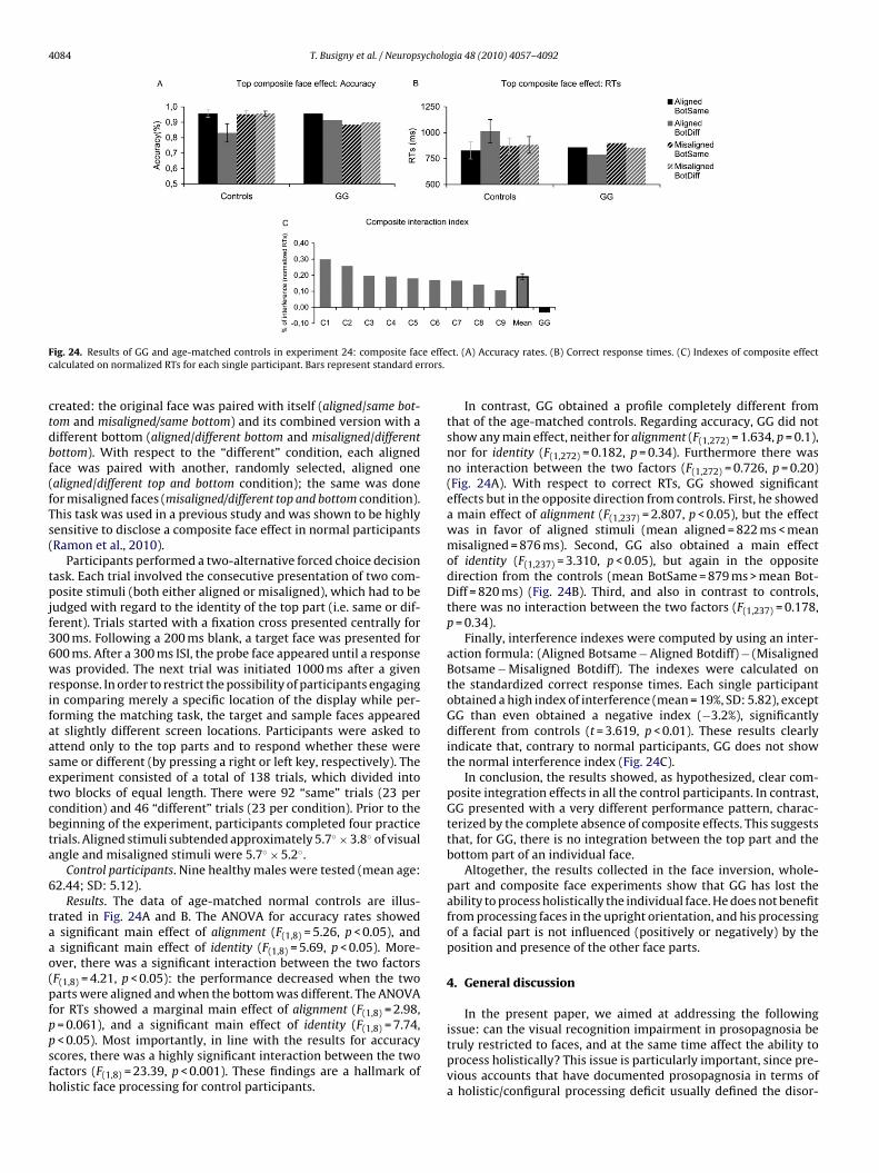
4084 T. Busigny et al. / Neuropsychologia 48 (2010) 4057–4092
F e effec rrors.
ctdbf(fTs(
tpjf36wrifaasetcbta
6
taao(pfppsfh
ig. 24. Results of GG and age-matched controls in experiment 24: composite facalculated on normalized RTs for each single participant. Bars represent standard e
reated: the original face was paired with itself (aligned/same bot-om and misaligned/same bottom) and its combined version with aifferent bottom (aligned/different bottom and misaligned/differentottom). With respect to the “different” condition, each alignedace was paired with another, randomly selected, aligned onealigned/different top and bottom condition); the same was doneor misaligned faces (misaligned/different top and bottom condition).his task was used in a previous study and was shown to be highlyensitive to disclose a composite face effect in normal participantsRamon et al., 2010).
Participants performed a two-alternative forced choice decisionask. Each trial involved the consecutive presentation of two com-osite stimuli (both either aligned or misaligned), which had to be
udged with regard to the identity of the top part (i.e. same or dif-erent). Trials started with a fixation cross presented centrally for00 ms. Following a 200 ms blank, a target face was presented for00 ms. After a 300 ms ISI, the probe face appeared until a responseas provided. The next trial was initiated 1000 ms after a given
esponse. In order to restrict the possibility of participants engagingn comparing merely a specific location of the display while per-orming the matching task, the target and sample faces appearedt slightly different screen locations. Participants were asked tottend only to the top parts and to respond whether these wereame or different (by pressing a right or left key, respectively). Thexperiment consisted of a total of 138 trials, which divided intowo blocks of equal length. There were 92 “same” trials (23 perondition) and 46 “different” trials (23 per condition). Prior to theeginning of the experiment, participants completed four practicerials. Aligned stimuli subtended approximately 5.7◦ × 3.8◦ of visualngle and misaligned stimuli were 5.7◦ × 5.2◦.
Control participants. Nine healthy males were tested (mean age:2.44; SD: 5.12).
Results. The data of age-matched normal controls are illus-rated in Fig. 24A and B. The ANOVA for accuracy rates showed
significant main effect of alignment (F(1,8) = 5.26, p < 0.05), andsignificant main effect of identity (F(1,8) = 5.69, p < 0.05). More-
ver, there was a significant interaction between the two factorsF(1,8) = 4.21, p < 0.05): the performance decreased when the twoarts were aligned and when the bottom was different. The ANOVAor RTs showed a marginal main effect of alignment (F(1,8) = 2.98,
= 0.061), and a significant main effect of identity (F(1,8) = 7.74,< 0.05). Most importantly, in line with the results for accuracycores, there was a highly significant interaction between the twoactors (F(1,8) = 23.39, p < 0.001). These findings are a hallmark ofolistic face processing for control participants.
ct. (A) Accuracy rates. (B) Correct response times. (C) Indexes of composite effect
In contrast, GG obtained a profile completely different fromthat of the age-matched controls. Regarding accuracy, GG did notshow any main effect, neither for alignment (F(1,272) = 1.634, p = 0.1),nor for identity (F(1,272) = 0.182, p = 0.34). Furthermore there wasno interaction between the two factors (F(1,272) = 0.726, p = 0.20)(Fig. 24A). With respect to correct RTs, GG showed significanteffects but in the opposite direction from controls. First, he showeda main effect of alignment (F(1,237) = 2.807, p < 0.05), but the effectwas in favor of aligned stimuli (mean aligned = 822 ms < meanmisaligned = 876 ms). Second, GG also obtained a main effectof identity (F(1,237) = 3.310, p < 0.05), but again in the oppositedirection from the controls (mean BotSame = 879 ms > mean Bot-Diff = 820 ms) (Fig. 24B). Third, and also in contrast to controls,there was no interaction between the two factors (F(1,237) = 0.178,p = 0.34).
Finally, interference indexes were computed by using an inter-action formula: (Aligned Botsame − Aligned Botdiff) − (MisalignedBotsame − Misaligned Botdiff). The indexes were calculated onthe standardized correct response times. Each single participantobtained a high index of interference (mean = 19%, SD: 5.82), exceptGG than even obtained a negative index (−3.2%), significantlydifferent from controls (t = 3.619, p < 0.01). These results clearlyindicate that, contrary to normal participants, GG does not showthe normal interference index (Fig. 24C).
In conclusion, the results showed, as hypothesized, clear com-posite integration effects in all the control participants. In contrast,GG presented with a very different performance pattern, charac-terized by the complete absence of composite effects. This suggeststhat, for GG, there is no integration between the top part and thebottom part of an individual face.
Altogether, the results collected in the face inversion, whole-part and composite face experiments show that GG has lost theability to process holistically the individual face. He does not benefitfrom processing faces in the upright orientation, and his processingof a facial part is not influenced (positively or negatively) by theposition and presence of the other face parts.
4. General discussion
In the present paper, we aimed at addressing the following
issue: can the visual recognition impairment in prosopagnosia betruly restricted to faces, and at the same time affect the ability toprocess holistically? This issue is particularly important, since pre-vious accounts that have documented prosopagnosia in terms ofa holistic/configural processing deficit usually defined the disor-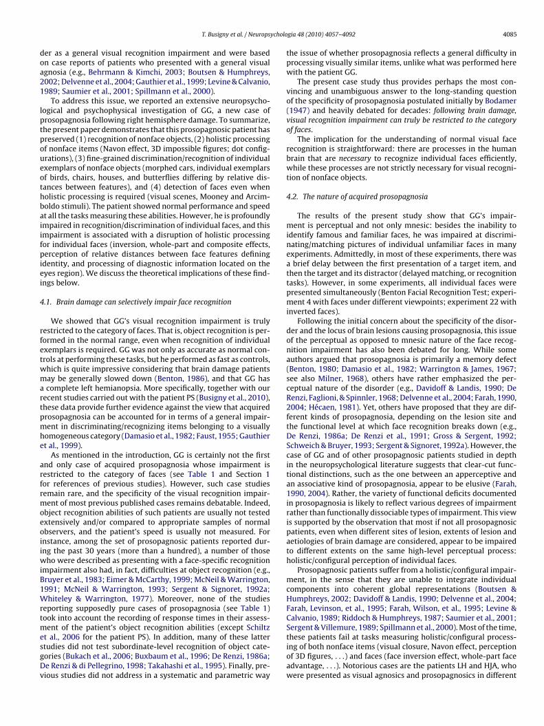
ycholo
doa21
lptpoueothbaiifpiei
4
rfetwmartpmhe
arfrmoeoiiwiB1WrtmesgDv
T. Busigny et al. / Neurops
er as a general visual recognition impairment and were basedn case reports of patients who presented with a general visualgnosia (e.g., Behrmann & Kimchi, 2003; Boutsen & Humphreys,002; Delvenne et al., 2004; Gauthier et al., 1999; Levine & Calvanio,989; Saumier et al., 2001; Spillmann et al., 2000).
To address this issue, we reported an extensive neuropsycho-ogical and psychophysical investigation of GG, a new case ofrosopagnosia following right hemisphere damage. To summarize,he present paper demonstrates that this prosopagnosic patient hasreserved (1) recognition of nonface objects, (2) holistic processingf nonface items (Navon effect, 3D impossible figures; dot config-rations), (3) fine-grained discrimination/recognition of individualxemplars of nonface objects (morphed cars, individual exemplarsf birds, chairs, houses, and butterflies differing by relative dis-ances between features), and (4) detection of faces even whenolistic processing is required (visual scenes, Mooney and Arcim-oldo stimuli). The patient showed normal performance and speedt all the tasks measuring these abilities. However, he is profoundlympaired in recognition/discrimination of individual faces, and thismpairment is associated with a disruption of holistic processingor individual faces (inversion, whole-part and composite effects,erception of relative distances between face features defining
dentity, and processing of diagnostic information located on theyes region). We discuss the theoretical implications of these find-ngs below.
.1. Brain damage can selectively impair face recognition
We showed that GG’s visual recognition impairment is trulyestricted to the category of faces. That is, object recognition is per-ormed in the normal range, even when recognition of individualxemplars is required. GG was not only as accurate as normal con-rols at performing these tasks, but he performed as fast as controls,hich is quite impressive considering that brain damage patientsay be generally slowed down (Benton, 1986), and that GG hascomplete left hemianopsia. More specifically, together with our
ecent studies carried out with the patient PS (Busigny et al., 2010),hese data provide further evidence against the view that acquiredrosopagnosia can be accounted for in terms of a general impair-ent in discriminating/recognizing items belonging to a visually
omogeneous category (Damasio et al., 1982; Faust, 1955; Gauthiert al., 1999).
As mentioned in the introduction, GG is certainly not the firstnd only case of acquired prosopagnosia whose impairment isestricted to the category of faces (see Table 1 and Section 1or references of previous studies). However, such case studiesemain rare, and the specificity of the visual recognition impair-ent of most previous published cases remains debatable. Indeed,
bject recognition abilities of such patients are usually not testedxtensively and/or compared to appropriate samples of normalbservers, and the patient’s speed is usually not measured. Fornstance, among the set of prosopagnosic patients reported dur-ng the past 30 years (more than a hundred), a number of those
ho were described as presenting with a face-specific recognitionmpairment also had, in fact, difficulties at object recognition (e.g.,ruyer et al., 1983; Eimer & McCarthy, 1999; McNeil & Warrington,991; McNeil & Warrington, 1993; Sergent & Signoret, 1992a;hiteley & Warrington, 1977). Moreover, none of the studies
eporting supposedly pure cases of prosopagnosia (see Table 1)ook into account the recording of response times in their assess-
ent of the patient’s object recognition abilities (except Schiltz
t al., 2006 for the patient PS). In addition, many of these lattertudies did not test subordinate-level recognition of object cate-ories (Bukach et al., 2006; Buxbaum et al., 1996; De Renzi, 1986a;e Renzi & di Pellegrino, 1998; Takahashi et al., 1995). Finally, pre-ious studies did not address in a systematic and parametric waygia 48 (2010) 4057–4092 4085
the issue of whether prosopagnosia reflects a general difficulty inprocessing visually similar items, unlike what was performed herewith the patient GG.
The present case study thus provides perhaps the most con-vincing and unambiguous answer to the long-standing questionof the specificity of prosopagnosia postulated initially by Bodamer(1947) and heavily debated for decades: following brain damage,visual recognition impairment can truly be restricted to the categoryof faces.
The implication for the understanding of normal visual facerecognition is straightforward: there are processes in the humanbrain that are necessary to recognize individual faces efficiently,while these processes are not strictly necessary for visual recogni-tion of nonface objects.
4.2. The nature of acquired prosopagnosia
The results of the present study show that GG’s impair-ment is perceptual and not only mnesic: besides the inability toidentify famous and familiar faces, he was impaired at discrimi-nating/matching pictures of individual unfamiliar faces in manyexperiments. Admittedly, in most of these experiments, there wasa brief delay between the first presentation of a target item, andthen the target and its distractor (delayed matching, or recognitiontasks). However, in some experiments, all individual faces werepresented simultaneously (Benton Facial Recognition Test; experi-ment 4 with faces under different viewpoints; experiment 22 withinverted faces).
Following the initial concern about the specificity of the disor-der and the locus of brain lesions causing prosopagnosia, this issueof the perceptual as opposed to mnesic nature of the face recog-nition impairment has also been debated for long. While someauthors argued that prosopagnosia is primarily a memory defect(Benton, 1980; Damasio et al., 1982; Warrington & James, 1967;see also Milner, 1968), others have rather emphasized the per-ceptual nature of the disorder (e.g., Davidoff & Landis, 1990; DeRenzi, Faglioni, & Spinnler, 1968; Delvenne et al., 2004; Farah, 1990,2004; Hécaen, 1981). Yet, others have proposed that they are dif-ferent kinds of prosopagnosia, depending on the lesion site andthe functional level at which face recognition breaks down (e.g.,De Renzi, 1986a; De Renzi et al., 1991; Gross & Sergent, 1992;Schweich & Bruyer, 1993; Sergent & Signoret, 1992a). However, thecase of GG and of other prosopagnosic patients studied in depthin the neuropsychological literature suggests that clear-cut func-tional distinctions, such as the one between an apperceptive andan associative kind of prosopagnosia, appear to be elusive (Farah,1990, 2004). Rather, the variety of functional deficits documentedin prosopagnosia is likely to reflect various degrees of impairmentrather than functionally dissociable types of impairment. This viewis supported by the observation that most if not all prosopagnosicpatients, even when different sites of lesion, extents of lesion andaetiologies of brain damage are considered, appear to be impairedto different extents on the same high-level perceptual process:holistic/configural perception of individual faces.
Prosopagnosic patients suffer from a holistic/configural impair-ment, in the sense that they are unable to integrate individualcomponents into coherent global representations (Boutsen &Humphreys, 2002; Davidoff & Landis, 1990; Delvenne et al., 2004;Farah, Levinson, et al., 1995; Farah, Wilson, et al., 1995; Levine &Calvanio, 1989; Riddoch & Humphreys, 1987; Saumier et al., 2001;Sergent & Villemure, 1989; Spillmann et al., 2000). Most of the time,
these patients fail at tasks measuring holistic/configural process-ing of both nonface items (visual closure, Navon effect, perceptionof 3D figures, . . .) and faces (face inversion effect, whole-part faceadvantage, . . .). Notorious cases are the patients LH and HJA, whowere presented as visual agnosics and prosopagnosics in different
4 ycholo
rB
wtpTepd
u(oabtoStoR“tfMe&v(e
fctfpc
fwctesafasfooal(
apopiIrpec
086 T. Busigny et al. / Neurops
eports (LH: Farah, Wilson, et al., 1995; Levine et al., 1980; HJA:outsen & Humphreys, 2002; Riddoch & Humphreys, 1987).
In contrast, here we report an extensive case study of a patientho presents with a normal profile of performance in holis-
ic/configural processing of nonface items, yet who does notresent with holistic/configural processing of individual faces.herefore, should we consider that GG has a disorder that is differ-nt from what has been previously reported in other prosopagnosicatients? Or is there a common underlying explanation for theireficits?
With respect to individual faces, there appears to be commonnderlying difficulties between patient GG and the other cases ofprosop)agnosia mentioned above. That is, when they have to rec-gnize an individual face, the patients focus on one part of the facet a time (e.g., the mouth) and analyze its diagnosticity, withouteing influenced by the other parts of the face. Hence, contraryo normal observers, face recognition in these patients is basedn an independent analysis of face parts (Saumier et al., 2001;ergent & Signoret, 1992a; Sergent & Villemure, 1989). It followshat they cannot benefit from, or be disadvantaged by, the presencef the other facial parts (Boutsen & Humphreys, 2002; De Gelder &ouw, 2000a; Delvenne et al., 2004; Joubert et al., 2003). When thepart” is made of half a face, as in the composite face paradigm,he other half has no – or little – influence on the patient’s per-ormance (Ramon et al., 2010; the present study, experiment 24).
oreover, since inversion disrupts a holistic process (Farah, Wilson,t al., 1995; Rossion, 2009; Van Belle, de Graef, Verfaillie, Rossion,Lefèvre, 2010) that is already absent for such patients, their indi-
idual face recognition abilities do not suffer much from inversione.g., Busigny & Rossion, 2010; Farah, Wilson, et al., 1995; Marottat al., 2002).
However, despite this common underlying difficulty, the per-ormance of such patients in face recognition tasks may beontaminated by more general visual recognition impairments andhus should be interpreted with great care. In contrast, when theace recognition impairment can be isolated, which is the case foratient GG, we believe that the nature of prosopagnosia can beircumscribed and understood more directly.
The best support for the isolated nature of GG’s deficit comesrom the fact that he was able to perceive a face as a face, evenhen it required holistic processing. Such intact performance at
ategorizing Mooney and Arcimboldo facelike stimuli is unlikelyo occur in general visual integrative agnosia. The results of thesexperiments thus indicate that what truly differs between the pre-erved and impaired face processing functions in GG is the levelt which processing occurs: the patient is impaired at individualace recognition, but not at face detection. Why is he not impairedt individual recognition of nonface objects then? The answereems quite simple: recognition of individual exemplars of non-ace objects does not require holistic processing, even in normalbservers. In fact, recognition of individual exemplars of nonfacebjects is quite rare in real life circumstances for most observers,nd may be largely based on identifying fine-grained details andocal diagnostic parts rather than using the whole of the objectBiederman & Kalocsai, 1997).
Given these considerations, and although it seems to bestrong claim, we would like to argue that all acquired
rosopagnosic patients present with at least some impairmentf holistic/configural processing of individual faces. Almost allatients who were tested in the literature with paradigms evaluat-
ng holistic/configural face processing showed deficits at this level.
n fact, we know of only a few cases of prosopagnosia who wereeported to have at least partial preservation of holistic/configuralrocessing of individual faces and we believe that the evidence ofntirely normal holistic processing was not demonstrated in theseases (see Ramon et al., 2010). Two cases reported by Sergent andgia 48 (2010) 4057–4092
colleagues (PV, Sergent & Poncet, 1990; PC, Sergent & Signoret,1992a) showed evidence for holistic processing in a matching taskof faces differing by one or several features: their performanceindicated that the processing of one feature was influenced by thepresence of the other features. However, in light of recent advancesin the field of memory-related person recognition disturbances(e.g., Gainotti, 2007), the first patient appears to present with ageneral semantic disorder rather than prosopagnosia. Such casesof semantic memory impairment related to person knowledgemay indeed present with largely preserved holistic face processing(Busigny, Robaye, Dricot, & Rossion, 2009). The second patient waslargely impaired at perceiving faces but, according to the authorshe had “not completely lost the basic mechanisms that underlie thenormal perceptual operations on facial representations” (Sergent &Signoret, 1992a, p. 385). However, it remains unclear based on asingle experiment whether this patient’s holistic face processingwas entirely intact when compared to normal observers. A recentlydescribed case of prosopagnosia (LR) is particularly interesting forthis issue (Bukach et al., 2006). The authors argued that LR had pre-served holistic face processing because he showed interference ina variant of the composite face task. However, there is evidencethat LR’s ability to process individual faces holistically is not fullypreserved. For instance, he has an abnormal inversion effect forchanges at the level of the eyes, and he has been described asunable to consider multiple features of a face altogether, focus-ing on the mouth at the expense of the eyes, or vice-versa (Bukachet al., 2006). He can also show slow processing of inter-feature dis-tances as compared to local feature changes – a pattern which canbe interpreted as a malfunctioning holistic face processor (Rossionet al., 2009). Finally, even though his impairment in individualface matching tasks appears to be less severe than PS, or than thepatient GG reported in the present paper, we have been collect-ing data recently showing that the same patient LR clearly presentswith impairment in holistic perception of the individual face. Thisclaim is supported by a reduced face inversion effect, whole-partadvantage, and composite face effect with the same paradigms asused here (Busigny et al., in preparation). Moreover, eye-gaze con-tingency experiments (see Van Belle, de Graef, Verfaillie, Busigny,et al., 2010) performed with this patient indicates a reduced per-ceptual field in individual face matching, suggesting that he has tofocus on one feature at a time when individualizing faces.
Therefore, in our view there is no clear evidence in the litera-ture of prosopagnosic patients who show complete preservationof holistic face processing. Rather than suggesting that holisticprocessing of individual faces is completely abolished in all casesof prosopagnosia, we propose that holistic processing of individ-ual faces may be disrupted to variable extents in these patients,preventing them from perceptually encoding individual faces accu-rately and rapidly.
In summary, GG is a pure case of prosopagnosia following braindamage who is impaired at perceiving an individual face holis-tically. The nature of his prosopagnosia is characterized by hisinability to perceive an individual face as a global and integratedpattern: he has to analyze each facial feature separately, as if allfeatures were isolated. While such a process seems to be necessaryfor face recognition, it does not appear to be necessary for objectcategorization, either at basic and more fine-grained levels.
4.3. “Specific” does not necessarily mean “modular” in holisticface processing
Based on the present case study, we conclude that holistic pro-cessing of the individual face is impaired in prosopagnosia, andthus that this ability is necessary for normal face recognition.Importantly, this observation does not necessarily imply that theimpaired processes were selectively engaged for faces before brain
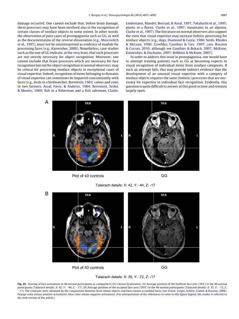
ycholo
dtctaepsacrbvofi&
Fp−Ot
T. Busigny et al. / Neurops
amage occurred. One cannot exclude that, before brain damage,hese processes may have been involved also in the recognition ofertain classes of nonface objects to some extent. In other words,he observation of pure cases of prosopagnosia such as GG, as wells the documentation of the reverse dissociation (e.g., Moscovitcht al., 1997), must not be misinterpreted as evidence of module forrocessing faces (e.g., Kanwisher, 2000). Nonetheless, case studiesuch as the one of GG indicate, at the very least, that such processesre not strictly necessary for object recognition. Moreover, oneannot exclude that brain processes which are necessary for faceecognition but not for object recognition in normal observers, maye critical for processing nonface objects in exceptional cases of
isual expertise. Indeed, recognition of items belonging to domainsf visual expertise can sometimes be impaired concomitantly withaces (e.g., birds in a birdwatcher, Bornstein, 1963; calves and cowsn two farmers, Assal, Favre, & Anderes, 1984; Bornstein, Sroka,Munitz, 1969; fish in a fisherman and a fish salesman, Clarke,
ig. 25. Overlap of face activations in 40 normal participants as compared to GG’s lesionarticipants (Talairach details: X: 42, Y: −44, Z: −17). (B) Average position of the occipita17). The contrasts were obtained by the conjunction between faces minus objects andrange color means positive activations; blue color means negative activations. (For inte
he web version of the article.)
gia 48 (2010) 4057–4092 4087
Lindemann, Maeder, Borruat, & Assal, 1997; Takahashi et al., 1995;plants in a florist, Clarke et al., 1997; mountains in an alpinist,Clarke et al., 1997). The literature on normal observers also supportthe view that visual expertise may increase holistic processing fornonface objects (e.g., dogs, Diamond & Carey, 1986; birds, Rhodes& McLean, 1990; Greebles, Gauthier & Tarr, 1997; cars, Rossion& Curran, 2010; although see Gauthier & Bukach, 2007; McKone,Kanwisher, & Duchaine, 2007; Robbins & McKone, 2007).
In order to address this issue in prosopagnosia, one would haveto attempt training patients such as GG at becoming experts invisual recognition of individual items from nonface categories. Ifsuch an attempt fails, this may provide indirect evidence that the
development of an unusual visual expertise with a category ofnonface objects requires the same (holistic) processes that are nec-essary for expertise in individual face recognition. Evidently, thisquestion is quite difficult to answer at this point in time and remainslargely open.localization. (A) Average position of the fusiform face ares (‘FFA’) in the 40 normall face ares (‘OFA’) in the 40 normal participants (Talairach details: X: 35, Y: −72, Z:faces minus scrambled faces (see Dricot, Sorger, Schiltz, Goebel, & Rossion, 2008).rpretation of the references to color in this figure legend, the reader is referred to

4 ycholo
4i
rntstc(l2CSTdfsitYerteh&e(am&2cb2hrfowtAcwrGpltee(tcWev2taeHR&
088 T. Busigny et al. / Neurops
.4. The neural substrates of prosopagnosia and face recognitionn the human brain
The last theoretical issue for which the present case study iselevant concerns the neural substrates of prosopagnosia. Origi-ally, and for quite a long time, the debate has been centered onhe respective roles of the right and left hemispheres. Following aurvey of twenty-two cases, Hécaen and Angelergues (1962) werehe first to argue that the right hemisphere lesion was dominant inausing prosopagnosia. Contrary to later claims by Damasio et al.1982), it was later clearly established that a right hemisphereesion is sufficient in causing prosopagnosia (e.g., Barton et al.,002; Bouvier & Engel, 2006; De Renzi, 1986b; Landis, Cummings,hristen, Bogen, & Imhof, 1986; Riddoch et al., 2008; Sergent &ignoret, 1992b; Wada & Yamamoto, 2001; Wilkinson et al., 2009).he case report of the patient GG adds up to this evidence, whichoes not mean however that the right hemisphere is sufficientor face recognition. Indeed, one cannot exclude that left hemi-pheric processes potentially important for faces are deprived ofnputs from damaged right hemisphere areas in such patients,hereby contributing to increase their face recognition impairment.et, the right hemispheric dominance in face recognition is anstablished fact, supported by multiple evidence in cognitive neu-oscience: faster and better performance for faces presented inhe left than the right visual field (e.g., Hillger & Koenig, 1991),nhanced face-sensitivity in areas of the right compared to the leftemisphere as found in neuroimaging (e.g., Kanwisher, McDermott,Chun, 1997; Sergent, Otha, & MacDonald, 1992), as well as in
lectrophysiological components recorded from the human scalpe.g., Bentin, Allison, Puce, Perez, & Mccarthy, 1996). Behaviouralnd neural studies performed in non-human primates and otherammals also support this right hemispheric dominance (PeirceKendrick, 2002; Perrett et al., 1988; Zangenehpour & Chaudhuri,
005). In humans, it has been related to configural/holistic per-eption, not only at the basic-level (Parkin & Williamson, 1987),ut also and mainly at the individual level (Jacques & Rossion,009; Schiltz & Rossion, 2006). The present study suggests thatolistic perception of the individual face depends critically on theight occipito-temporal cortex. Regarding GG, in the absence ofunctional neuroimaging data, it is unclear whether his ventralccipito-temporal lesions, which are quite medial, encompass twoell-defined ventral areas showing face preferential response in
he human brain (e.g., Fusiform face area, FFA, and Occipital Facerea, OFA; Fig. 25) or their putative direct or indirect anatomicalonnections. Interestingly, GG’s lesions show almost no overlapith PS’ lesions in the right hemisphere, which concern the ter-
itory of the right OFA in the lateral occipital cortex (see Sorger,oebel, Schiltz, & Rossion, 2007). Moreover, the pure cases ofrosopagnosia referred to above (Table 1) presents with different
ocalization of brain damage: some patients present with quite pos-erior right hemispheric lesions in the occipital lobe (WB, Buxbaumt al., 1996; Anna, De Renzi & di Pellegrino, 1998; PS, Rossiont al., 2003), some have right lesions in the temporal lobe onlyVA, De Renzi et al., 1991; OR, De Renzi et al., 1994) or in bothhe occipital and the temporal lobes (Patient 4, De Renzi, 1986a;ase 3, Takahashi et al., 1995; MT, Schweinberger et al., 1995;
ada & Yamamoto, 2001; 009, Barton et al., 2004; FB, Riddocht al., 2008; DC, Rivest et al., 2009), and one of them also has aery anterior lesion, in the right temporal pole (LR, Bukach et al.,006). These observations are in agreement with the fact that inhe normal human brain there is a whole set of areas distributed all
long the ventral occipital and temporal cortex that respond pref-rentially or even exclusively to faces (Fox, Iaria, & Barton, 2009;axby, Hoffman, & Gobbini, 2000; Ishai, 2008; Pinsk et al., 2009;ajimehr, Young, & Tootell, 2009; Sergent et al., 1992; Tsao, Moeller,Freiwald, 2008; see also Allison, Puce, Spencer, & McCarthy,
gia 48 (2010) 4057–4092
1999). They suggest that all these areas play an important role inface recognition, and/or that they form a tightly connected func-tional network whose full integrity is necessary to carry out facerecognition efficiently (Rossion et al., 2003; Fox, Iaria, & Barton,2008). In this context, one may wonder how patients with differentlesion localization might show a common functional impairment –perhaps to a different extent – at holistic individual face percep-tion. One reason may be that damage to any node of the underlyingdistributed cortical face processing network impinges on the func-tional integrity of other areas of this network (Fox et al., 2008;Rossion, 2008b; Sergent & Signoret, 1992b). In this way, a criti-cal aspect of the face processing function would always be altered,at least to a certain extent, in all prosopagnosic patients. Sup-porting this view, we have previously found that the right middlefusiform gyrus of the patient PS is structurally preserved and showssensitivity to faces over other object categories (‘FFA’; Rossionet al., 2003). However, this area – which is the most sensitive toholistic face perception in the normal brain (Schiltz & Rossion,2006) – does not even present release from adaptation to iden-tity in PS’ brain (Schiltz et al., 2006), presumably lacking criticalinputs from the posteriorly damaged right inferior occipital cortex.This example shows that brain regions which may appear struc-turally intact and thus not considered to be critically associatedwith the impaired function(s) in a prosopagnosic patient may infact be functionally depressed because they do not receive nor-mal inputs from lesioned regions (‘diaschisis’; see Price, Warburton,Moore, Frackowiak, & Friston, 2001). Therefore, if face recognitionis subtended by a highly distributed network of interdependentareas, predominantly in the right hemisphere, it might not be sur-prising that all cases of acquired prosopagnosia, despite differentlocalization of brain damage, present relatively similar functionalimpairments.
5. Conclusion
We report a detailed single-case study of a new prosopag-nosic patient following right unilateral damage which offers newinsights in the comprehension of this clinical syndrome as well asthat of normal face recognition. First, with respect to the issue ofspecificity, we demonstrated that face recognition can be affectedselectively in acquired prosopagnosia, without affecting other cate-gories of objects (even when these objects are very similar). Second,the basis of GG’s deficit can be interpreted in terms of a selec-tive impairment of holistic perception of the individual face, whichis distinct from holistic object and face processing at the basiclevel. We conclude that right unilateral brain damage may lead toa specific impairment of holistic perception of individual faces, afunction that appears critical for normal face recognition but notfor object recognition.
Acknowledgments
We are immensely grateful to GG for the numerous hours hespent to carry out our tasks and for his great kindness and patience.We also are thankful to her spouse and to all the controls thatparticipated in the study. Moreover, we would like to thank inparticular: Aaron Schurger for providing the stimuli of experi-ment 16; Valérie Goffaux for the stimuli of experiment 23; DanielBub, Jim Tanaka and Martha Kaiser for stimuli of experiment 24;Olivier d’Arripe, Coralie Buxant and Mona Spiridon for having col-
lected some of the control data; Laurence Dricot for the fMRI data;Eve Tramoni for the follow-up neuropsychological assessment in2007; Eric Guedj from the department of Nuclear Medicine at theTimone Hospital, Marseille, for providing the SPECT analysis; aswell as two anonymous reviewers for their helpful commentaries
ycholo
opM0Ca
R
A
A
A
A
B
B
B
B
B
B
B
B
B
B
B
B
B
B
B
B
B
B
B
B
B
B
B
B
T. Busigny et al. / Neurops
n a previous version of this manuscript. This research was sup-orted by the Belgian National Fund for Scientific Research (FNRS,andat d’Impulsion Scientifique to BR) and a grant ARC 07/12-
07, Communauté Francaise de Belgique – Actions de Rechercheoncertées. Sven Joubert is supported by a Chercheur boursierward from the Fonds de la recherche en santé du Québec (FRSQ).
eferences
llison, T., Puce, A., Spencer, D. D., & McCarthy, G. (1999). Electrophysiological stud-ies of human face perception. I. Potentials generated in occipitotemporal cortexby face and non-face stimuli. Cerebral Cortex, 9, 415–430.
naki, D., Kaufman, Y., Freedman, M., & Moscovitch, M. (2007). Associative(prosop)agnosia without (apparent) perceptual deficits: A case-study. Neuropsy-chologia, 45, 1658–1671.
rguin, M., Bub, D., & Dudek, G. (1996). Shape integration for visual object recog-nition and its implication in category-specific visual agnosia. Visual Cognition,3(3), 221–275.
ssal, G., Favre, C., & Anderes, J. P. (1984). Non-reconnaissance d’animaux familierschez un paysan. Revue Neurologique, 140(10), 580–584.
ahrick, H. P., Bahrick, P. O., & Wittinger, R. P. (1975). Fifty years of memory fornames and faces: A cross-sectional approach. Journal of Experimental Psychology:General, 104, 54–75.
arton, J. J. S. (2008a). Structure and function in acquired prosopagnosia: Lessonsfrom a series of 10 patients with brain damage. Journal of Neuropsychology, 2,197–225.
arton, J. J. S. (2008b). Prosopagnosia associated with a left occipitotemporal lesion.Neuropsychologia, 46, 2214–2224.
arton, J. J. S. (2009). What is meant by impaired configural processing in acquiredprosopagnosia? Perception, 38, 242–260.
arton, J. J. S., & Cherkasova, M. V. (2005). Impaired spatial coding within objects butnot between objects in prosopagnosia. Neurology, 65, 270–274.
arton, J. J. S., Cherkasova, M. V., Press, D. Z., Intriligator, J. M., & O’Connor, M. (2004).Perceptual functions in prosopagnosia. Perception, 33, 939–956.
arton, J. J. S., Press, D. Z., Keenan, J. P., & O’Connor, M. (2002). Lesions of the fusiformface area impair perception of facial configuration in prosopagnosia. Neurology,58, 71–78.
arton, J. J. S., Zhao, J., & Keenan, J. P. (2003). Perception of global facial geometry inthe inversion effect and prosopagnosia. Neuropsychologia, 41, 1703–1711.
ay, E. (1953). Disturbances of visual perception and their examination. Brain, 76,515–530.
ehrmann, M., Avidan, G., Marotta, J. J., & Kimchi, R. (2005). Detailed explorationof face-related processing in congenital prosopagnosia. 1. Behavioral findings.Journal of Cognitive Neuroscience, 17(7), 1130–1149.
ehrmann, M., & Kimchi, R. (2003). What does visual agnosia tell us about perceptualorganization and its relationship to object perception? Journal of ExperimentalPsychology: Human Perception and Performance, 29(1), 19–42.
ehrmann, M., Peterson, M. A., Moscovitch, M., & Susuki, S. (2006). Independent rep-resentation of parts and the relations between them: Evidence from integrativeagnosia. Journal of Experimental Psychology: Human Perception and Performance,32(5), 1169–1184.
ehrmann, M., & Williams, P. (2007). Impairments in part-whole representationsof objects in two cases of integrative visual agnosia. Cognitive Neuropsychology,24(7), 701–730.
entin, S., Allison, T., Puce, A., Perez, E., & Mccarthy, G. (1996). Electrophysiolog-ical studies of face perception in humans. Journal of Cognitive Neuroscience, 8,551–565.
enton, A. L. (1980). The neuropsychology of facial recognition. The American Psy-chologist, 35(2), 176–186.
enton, A. L. (1986). Reaction time in brain disease: Some reflections. Cortex, 22,129–140.
enton, A. L., Sivan, A. B., Hamsher, K., Varney, N. R., & Spreen, O. (1983). Benton facialrecognition: Stimulus and multiple choice pictures. Lutz: Psychological AssessmentResources Inc.
eyn, E. S., & Knyazeva, G. R. (1962). The problem of prosopagnosia. Journal of Neu-rology, Neurosurgery and Psychiatry, 25, 154–158.
iederman, I. (1987). Recognition-by-components: A theory of human image under-standing. Psychological Review, 94(2), 115–147.
iederman, I., & Kalocsai, P. (1997). Neurocomputational bases of object and facerecognition. Philosophical Transactions of the Royal Society of London. Series B,Biological Sciences, 352(1358), 1203–1219.
lanz, V., & Vetter, T. (1999). A morphable model for the synthesis of 3D faces.In Proceedings of the 26th Annual Conference on Computer Graphics and Inter-active Techniques (pp. 187–194). New York, USA: ACM Press/Addison-WesleyPublishing Co.
obes, M. A., Lopera, F., Garcia, M., Diaz-Comas, L., Galan, L., & Valdes-Sosa, M. (2003).Covert matching of unfamiliar faces in a case of prosopagnosia: An ERP study.
Cortex, 39(1), 41–56.odamer, J. (1947). Die prosopagnosie. Archiv für Psychiatrie und Nervenkrankheiten,179, 6–54. Partial English translation by Ellis, H. D., & Florence, M. (1990). Cog-nitive Neuropsychology, 7, 81–105
ornstein, B. (1963). Prosopagnosia. In L. Halpern (Ed.), Problems of dynamic neurol-ogy (pp. 283–318). Jerusalem: Hadassah Medical School.
gia 48 (2010) 4057–4092 4089
Bornstein, B., Sroka, H., & Munitz, H. (1969). Prosopagnosia with animal face agnosia.Cortex, 5, 164–169.
Boutsen, L., & Humphreys, G. W. (2002). Face context interferes with local partprocessing in a prosopagnosic patient. Neuropsychologia, 40, 2305–2313.
Bouvier, S. E., & Engel, S. A. (2006). Behavioral deficits and cortical damage loci incerebral achromatopsia. Cerebral Cortex, 16(2), 183–191.
Bruce, V., & Young, A. (1986). Understanding face recognition. British Journal of Psy-chology, 77, 305–327.
Bruyer, R., Laterre, C., Seron, X., Feyereisen, P., Strypstein, E., Pierrard, E., et al. (1983).A case of prosopagnosia with some preserved covert remembrance of familiarfaces. Brain and Cognition, 2, 257–284.
Bukach, C., Le Grand, R., Kaiser, M., Bub, D., & Tanaka, J. W. (2008). Preservation ofmouth region processing in two cases of prosopagnosia. Journal of Neuropsychol-ogy, 2, 227–244.
Bukach, C. M., Bud, D. N., Gauthier, I., & Tarr, M. J. (2006). Perceptual expertise effectsare not all or none: Spatially limited perceptual expertise for faces in a case ofprosopagnosia. Journal of Cognitive Neurosciences, 18(1), 48–63.
Busigny, T., Graf, M., Mayer, E., & Rossion, B. (2010). Acquired prosopagnosia asa face-specific disorder: Ruling out the general visual similarity account. Neu-ropsychologia, 48, 2051–2067.
Busigny, T., Robaye, L., Dricot, L., & Rossion, B. (2009). Right anterior temporal lobeatrophy and person-based semantic defect: A detailed case study. Neurocase,15(6), 485–508.
Busigny, T., & Rossion, B. (2010). Acquired prosopagnosia abolishes the face inversioneffect. Cortex, 46, 965–981.
Busigny, T., & Rossion, B. (in press). Holistic processing impairment can be restrictedto faces in acquired prosopagnosia: Evidence from the Global/Local Navon effect.Journal of Neuropsychology, doi:10.1348/174866410X500116.
Busigny, T., Van Belle, G., Jemel, B., Hosein, A., Bub, D., Tanaka, J., et al. (in prepa-ration). Prosopagnosia following right unilateral temporal pole damage can beassociated with face-specific holistic perception difficulties.
Buxbaum, L. J., Glosser, G., & Coslett, H. B. (1996). Relative sparing ofobject recognition in alexia-prosopagnosia. Brain and Cognition, 32, 202–205.
Caldara, R., Schyns, P., Mayer, E., Smith, M. L., Gosselin, F., & Rossion, B. (2005). Doesprosopagnosia take the eyes out from faces? Evidence for a defect in the useof diagnostic facial information in a brain-damaged patient. Journal of CognitiveNeuroscience, 17, 1652–1666.
Caramazza, A. (1986). On drawing inferences about the structure of normal cognitivesystems from the analysis of patterns of impaired performance: The case forsingle-patient studies. Brain and Cognition, 5, 41–66.
Carbon, C. C. (2008). Famous faces as icons. The illusion of being an expert in therecognition of famous faces. Perception, 37, 801–806.
Cavanagh, P. (1991). What’s up in top-down processing? In A. Gorea, Y. Fregnac, Z.Kapoula, & J. Findlay (Eds.), Representations of visions: Trends and tacit assump-tions in vision research (pp. 295–304). Cambridge: Cambridge University Press.
Charcot, J.-M. (1883). Un cas de suppression brusque et isolée de la vision men-tale des signes et des objets (formes et couleurs). Le Progrès Médical, 11, 441–444.
Clarke, S., Lindemann, A., Maeder, P., Borruat, F.-X., & Assal, G. (1997). Facerecognition and postero-inferior hemispheric lesions. Neuropsychologia, 35(12),1555–1563.
Crawford, J. R., & Garthwaite, P. H. (2002). Investigation of the single case in neu-ropsychology: Confidence limits on the abnormality of test scores and test scoredifferences. Neuropsychologia, 40, 1196–1208.
Crawford, J. R., & Howell, D. C. (1998). Comparing an individual’s test scoreagainst norms derived from small samples. The Clinical Neuropsychologist, 12(4),482–486.
Damasio, A. R., Damasio, H., & Van Hoesen, G. W. (1982). Prosopagnosia: Anatomicbasis and behavioural mechanisms. Neurology, 32, 331–341.
Damasio, H., & Damasio, A. R. (1989). Lesion analysis in neuropsychology. New York,Oxford: University Press.
Davidoff, J., & Landis, T. (1990). Recognition of unfamiliar faces in prosopagnosia.Neuropsychologia, 28(11), 1143–1161.
Davidoff, J., Matthews, B., & Newcombe, F. (1986). Observations on a case ofprosopagnosia. In H. Ellis, M. Jeeves, F. Newcombe, & A. Young (Eds.), Aspectsof face processing (pp. 279–290). Dordrecht: Martinus Nijhoff.
De Gelder, B., Frissen, I., Barton, J., & Hadjikhani, N. (2003). A modulatory role forfacial expressions in prosopagnosia. Proceedings of the National Academy of Sci-ences of the United States of America, 100(22), 13105–13110.
De Gelder, B., & Rouw, R. (2000a). Paradoxical configuration effects for faces andobjects in prosopagnosia. Neuropsychologia, 38, 1271–1279.
De Gelder, B., & Rouw, R. (2000b). Structural encoding precludes recognition of faceparts in prosopagnosia. Cognitive Neuropsychology, 17, 89–102.
De Haan, E. H. F., Heywood, C. A., Young, A. Y., Edelstyn, N., & Newcombe, F. (1995).Ettlinger revisited: The relation between agnosia and sensory impairment. Jour-nal of Neurology, Neurosurgery, and Psychiatry, 58, 350–356.
De Haan, E. H. F., Young, A., & Newcombe, F. (1987). Face recognition without aware-ness. Cognitive Neuropsychology, 4, 385–415.
De Renzi, E. (1986a). Current issues on prosopagnosia. In H. D. Ellis, M. A. Jeeves,
F. Newcombe, & A. Young (Eds.), Aspects of face processing (pp. 243–252). Dor-drecht, Netherlands: Martinus Nijhoff.De Renzi, E. (1986b). Prosopagnosia in two patients with CT scan evidence of damageconfined to the right hemisphere. Neuropsychologia, 24(3), 385–389.
De Renzi, E., & di Pellegrino, G. (1998). Prosopagnosia and alexia without objectagnosia. Cortex, 34, 403–415.
4 ycholo
D
D
D
D
D
D
D
E
E
E
E
F
FF
F
F
F
F
F
F
G
G
G
G
G
G
G
G
G
G
G
G
G
HH
H
H
H
090 T. Busigny et al. / Neurops
e Renzi, E., Faglioni, P., Grossi, D., & Nichelli, P. (1991). Apperceptive and associativeforms of prosopagnosia. Cortex, 27(2), 213–221.
e Renzi, E., Faglioni, P., & Spinnler, H. (1968). The performance of patients withunilateral brain damage on face recognition tasks. Cortex, 4, 17–34.
e Renzi, E., Perani, D., Carlesimo, G. A., Silveri, M. C., & Fazio, F. (1994). Prosopagnosiacan be associated with damage confined to the right hemisphere. An MRI andPET study and a review of the literature. Neuropsychologia, 32(8), 893–902.
elvenne, J.-F., Seron, X., Coyette, F., & Rossion, B. (2004). Evidence for perceptualdeficits in associative visual (prosop)agnosia: A single-case study. Neuropsy-chologia, 42, 597–612.
iamond, R., & Carey, S. (1986). Why faces are and are not special: An effect ofexpertise. Journal of Experimental Psychology: General, 115(2), 107–117.
ixon, M. J., Bub, D. N., & Arguin, M. (1998). Semantic and visual determinants of facerecognition in a prosopagnosic patient. Journal of Cognitive Neuroscience, 10(3),362–376.
ricot, L., Sorger, B., Schiltz, C., Goebel, R., & Rossion, B. (2008). The roles of “face” and“non-face” areas during individual face perception: Evidence by fMRI adaptationin a brain-damaged prosopagnosic patient. NeuroImage, 40, 318–332.
imer, M., & McCarthy, R. A. (1999). Prosopagnosia and structural encoding of faces:Evidence from event-related potentials. NeuroReport, 10, 255–259.
tcoff, N. L., Freeman, R., & Cave, K. R. (1991). Can we lose memories of faces? Contentspecificity and awareness in a prosopagnosic. Journal of Cognitive Neuroscience,3(1), 25–41.
ttlin, T. M., Beckson, M., Benson, D. F., Langfitt, J. T., Amos, E. C., & Pineda, G. S.(1992). Prosopagnosia: A bihemispheric disorder. Cortex, 28, 129–134.
ttlinger, G. (1956). Sensory deficits in visual agnosia. Journal of Neurology, Neuro-surgery and Psychiatry, 19, 297–397.
arah, M. J. (1990). Visual agnosia: Disorders of object recognition and what they tellus about normal vision. Cambridge, MA: MIT Press.
arah, M. J. (2004). Visual agnosia (2nd ed.). Cambridge, MA: MIT Press.arah, M. J., Levinson, K. L., & Klein, K. L. (1995). Face perception and within-category
discrimination in prosopagnosia. Neuropsychologia, 33(6), 661–674.arah, M. J., Wilson, K. D., Drain, H. M., & Tanaka, J. R. (1995). The inverted face inver-
sion effect in prosopagnosia: Evidence for mandatory, face-specific perceptualmechanisms. Vision Research, 35(14), 2089–2093.
arah, M. J., Wilson, K. D., Drain, H. M., & Tanaka, J. R. (1998). What is “special” aboutface perception? Psychological Review, 105, 482–498.
aust, C. (1955). Evaluation of posttraumatic organic personality disorders. DeutscheMedizinische Wochenschrift, 80, 1237–1239.
lavell, J. H., & Draguns, J. G. (1957). A microgenetic approach to perception andthought. Psychological Bulletin, 54, 197–217.
ox, C. J., Iaria, G., & Barton, J. J. (2008). Disconnection in prosopagnosia and faceprocessing. Cortex, 44(8), 996–1009.
ox, C. J., Iaria, G., & Barton, J. J. (2009). Defining the face processing network:Optimization of the functional localizer in fMRI. Human Brain Mapping, 30(5),1637–1651.
ainotti, G. (2007). Different patterns of famous people recognition disorders inpatients with right and left anterior temporal lesions: A systematic review.Neuropsychologia, 45, 1591–1607.
alli, G. (1964). Prosopagnosia und normale Gesichtwahrnehmung. WienerZeitschrift für Nervenheilkunde, 22, 28–37.
alton, F. (1883). Inquiries into human faculty and its development. London: Macmil-lan.
authier, I., Behrmann, M., & Tarr, M. J. (1999). Can face recognition really be dis-sociated from object recognition? Journal of Cognitive Neuroscience, 11(4), 349–370.
authier, I., & Bukach, C. (2007). Should we reject the expertise hypothesis? Cogni-tion, 103(2), 322–330.
authier, I., & Tarr, M. J. (1997). Becoming a “Greeble” expert: Exploring mechanismsfor face recognition. Vision Research, 37(12), 1673–1682.
eorge, N., Jemel, B., Fiori, N., & Renault, B. (1997). Face and shape repetitions effectsin humans: A spatio-temporal ERP study. NeuroReport, 8, 1417–1423.
loning, I., Gloning, K., Hoff, H., & Tschabitscher, H. (1966). Zur prosopagnosie. Neu-ropsychologia, 4, 113–132.
offaux, V., & Rossion, B. (2006). Faces are “spatial”-Holistic face perception is sup-ported by low spatial frequencies. Journal of Experimental Psychology: HumanPerception and Performance, 32(4), 1023–1039.
offaux, V., & Rossion, B. (2007). Face inversion disproportionately impairs theperception of vertical but not horizontal relations between features. Journal ofExperimental Psychology: Human Perception and Performance, 33, 995–1002.
ollin, E. S. (1960). Developmental studies of visual recognition of incompleteobjects. Perceptual and Motor Skills, 11, 289–298.
osselin, F., & Schyns, P. G. (2001). Bubbles: A technique to reveal the use of infor-mation in recognition. Vision Research, 41, 2261–2271.
ross, C. G., & Sergent, J. (1992). Face recognition. Current Opinion in Neurobiology,2, 156–161.
arris, A. M., & Aguirre, G. K. (2007). Prosopagnosia. Current Biology, 17(1), R7–8.axby, J. V., Hoffman, E. A., & Gobbini, M. I. (2000). The distributed human neural
system for face perception. Trends in Cognitive Sciences, 4(6), 223–233.écaen, H. (1981). The neuropsychology of face recognition. In G. Davies, H. Ellis, &
J. Shepherd (Eds.), Perceiving and remembering faces (pp. 39–54). London: Aca-demic Press.
écaen, H., & Angelergues, R. (1962). Agnosia for faces. Archives of Neurology, 7,92–100.
egdé, J., Thompson, S., & Kersten, D. (2007). Identifying faces in two-tone ‘Mooney’images: A psychophysical and fMRI study. Journal of Vision, 7, 264.
gia 48 (2010) 4057–4092
Henke, K., Schweinberger, S. R., Grigo, A., Klos, T., & Sommer, W. (1998). Specificity offace recognition: Recognition of exemplars of non-face objects in prosopagnosia.Cortex, 34, 289–296.
Hillger, L. A., & Koenig, O. (1991). Separable mechanisms in face processing. Evidencefrom hemispheric specialization. Journal of Cognitive Neuroscience, 3, 42–58.
Hulten, P. (1987). The Arcimboldo effect: Transformations of the face from the 16th tothe 20th century. New York: Abbeville Press.
Humphreys, G. W., & Riddoch, M. J. (1987). Visual object processing: A cognitive neu-ropsychological approach. London: Lawrence Erlbaum.
Ingvalson, E. M., & Wenger, M. J. (2005). A strong test of the dual-mode hypothesis.Perception and Psychophysics, 67, 14–35.
Ishai, A. (2008). Let’s face it: It’s a cortical network. Neuroimage, 40(2), 415–419.Jacques, C., & Rossion, B. (2009). The initial representation of individual faces in the
right occipito-temporal cortex is holistic: Electrophysiological evidence fromthe composite face illusion. Journal of Vision, 9(6), 1–16.
Jeffreys, D. A. (1989). A face-responsive potential recorded from the human scalp.Experimental Brain Research, 78, 193–202.
Joubert, S., Felician, O., Barbeau, E., Sontheimer, A., Barton, J. J., Ceccaldi, M., et al.(2003). Impaired configurational processing in a case of progressive prosopag-nosia associated with predominant right temporal lobe atrophy. Brain, 126,2537–2550.
Kanizsa, G. (1955). Margini quasi-percettivi in campi con stimolazione omogenea.Rivista di Psicologia, 49, 7–30.
Kanwisher, N. (2000). Domain specificity in face perception. Nature Neuroscience, 3,759–763.
Kanwisher, N., McDermott, J., & Chun, M. (1997). The fusiform face area: A mod-ule in human extrastriate cortex specialized for face perception. The Journal ofNeuroscience, 17, 4302–4311.
Kemelmacher-Shlizerman, I., Basri, R., & Nadler, B. (2008). 3D shape reconstructionof Mooney faces. In IEEE, Computer Vision and Pattern Recognition Alaska, (pp.1–8).
Kimchi, R. (1992). Primacy of wholistic processing and global/local paradigm: Acritical review. Psychological Bulletin, 112, 24–38.
Kimchi, R. (2000). The perceptual organization of visual objects: A microgeneticanalysis. Vision Research, 40, 1333–1347.
Koffka, K. (1963). Principles of Gestalt psychology. New-York: Harcourt, Brace andWorld. (Original work published in 1935)
Kohler, W. (1929). Gestalt psychology. New York: Liveright.Kohler, W. (1971). Human perception. In M. Henle (Ed.), The selected papers of Wolf-
gang Kohler (pp. 142–167). New York: Liveright (Original work published in1930).
Kubovy, M., & Poremantz, J. R. (1981). Perceptual organisation. Hillsdale, NJ: Erlbaum.Landis, T., Cummings, J. L., Christen, L., Bogen, J. E., & Imhof, H.-G. (1986). Are unilat-
eral right posterior cerebral lesions sufficient to cause prosopagnosia? Clinicaland radiological findings in six additional patients. Cortex, 22, 243–252.
Lê, S., Cardebat, D., Boulanouar, K., Hénaff, M.-A., Michel, F., Milner, D., et al. (2002).Seeing, since childhood, without ventral stream: A behavioural study. Brain, 125,58–74.
Le Grand, R., Mondloch, C. J., Maurer, D., & Brent, H. P. (2004). Impairment in holis-tic face processing following early visual deprivation. Psychological Science, 15,762–768.
Leder, H., & Carbon, C. C. (2005). When context hinders! Learn-test compatibilityin face recognition. The Quarterly Journal of Experimental Psychology A: HumanExperimental Psychology, 58, 235–250.
Leder, H., & Carbon, C. C. (2006). Face-specific configural processing of relationalinformation. British Journal of Psychology, 97, 19–29.
Levine, D. N., & Calvanio, R. (1989). Prosopagnosia: A defect in visual configuralprocessing. Brain and Cognition, 10, 149–170.
Levine, D. N., Calvanio, R., & Wolf, E. (1980). Disorders of visual behaviour followingbilateral posterior cerebral lesions. Psychological Research, 41, 217–234.
Lewis, M. B., & Edmonds, A. J. (2003). Face detection: Mapping human performance.Perception, 32, 903–920.
Lhermitte, F., Chain, F., Escourolle, R., Ducarne, B., & Pillon, B. (1972). Etude anatomo-clinique d’un cas de prosopagnosie. Revue Neurologique, 126(5), 329–346.
Lopera, F., & Ardila, A. (1992). Prosopamnesia and visuolimbic disconnection syn-drome: A case study. Neuropsychology, 6(1), 3–12.
Marotta, J. J., McKeeff, T. J., & Behrmann, M. (2002). The effects of rotation andinversion on face processing in prosopagnosia. Cognitive Neuropsychology, 19(1),31–47.
Marr, D. (1982). Vision: A computational investigation into the human representationand processing of visual information. New York: W.H. Freeman and Co.
Maurer, D., Le Grand, R., & Mondloch, C. J. (2002). The many faces of configuralprocessing. Trends in Cognitive Sciences, 6(6), 255–260.
McKone, E. (2004). Isolating the special component of face recognition: Peripheralidentification and a Mooney face. Journal of Experimental Psychology. Learning,Memory, and Cognition, 30, 181–197.
McKone, E., Kanwisher, N., & Duchaine, B. (2007). Can generic expertise explainspecial processing for faces? Trends in Cognitive Sciences, 11(1), 8–15.
McKone, E., Martini, P., & Nakayama, K. (2003). Isolating holistic processing in faces(and perhaps objects). In M. Peterson, & G. Rhodes (Eds.), Perception of faces,
objects, and scenes: Analytic and holistic processes (pp. 92–119). Oxford, UK:Oxford University Press.McKone, E., & Yovel, G. (2009). Why does picture-plane inversion sometimes disso-ciate perception of features and spacing in faces, and sometimes not? Towarda new theory of holistic processing. Psychonomic Bulletin and Review, 16(5),778–797.
ycholo
M
M
M
M
M
M
M
M
M
M
M
N
N
O
P
P
P
P
P
P
Q
R
R
R
R
R
R
R
R
R
R
R
R
R
T. Busigny et al. / Neurops
cNeil, J. E., & Warrington, E. K. (1991). Prosopagnosia: A reclassification. The Quar-terly Journal of Experimental Psychology A, 43(2), 267–287.
cNeil, J. E., & Warrington, E. K. (1993). Prosopagnosia: A face-specific disorder. TheQuarterly Journal of Experimental Psychology A, 46(1), 1–10.
eadows, J. C. (1974). The anatomical basis of prosopagnosia. Journal of Neurology,Neurosurgery, and Psychiatry, 37, 489–501.
ichel, C., Caldara, R., & Rossion, B. (2006). Same-race faces are perceived moreholistically than other-race faces. Visual Cognition, 14(1), 55–73.
ichel, C., Rossion, B., Han, J., Chung, C.-S., & Caldara, R. (2006). Holistic processingis finely tuned for faces of our own race. Psychological Science, 17, 608–615.
ilner, B. (1968). Disorders of memory after brain lesions in man. Neuropsychologia,6, 175–179.
ilner, A. D., Perrett, D. I., Johnston, R. S., Benson, P. J., Jordan, T. R., Heeley, D. W.,et al. (1991). Perception and action in ‘visual form agnosia’. Brain, 114, 405–428.
ooney, C. M. (1956). Closure with negative after images under filtering light. Cana-dian Journal of Psychology, 10, 191–199.
ooney, C. M. (1957). Age in the development of closure ability in children. CanadianJournal of Psychology, 11, 219–226.
oore, C., & Cavanagh, P. (1998). Recovery of 3D volume from 2-tone images ofnovel objects. Cognition, 67, 45–71.
oscovitch, M., Winocur, G., & Behrmann, M. (1997). What is special about facerecognition? Nineteen experiments on a person with visual object agnosia anddyslexia but normal face recognition. Journal of Cognitive Neuroscience, 9(5),555–604.
avon, D. (1977). Forest before trees: The precedence of global features in visualperception. Cognitive Psychology, 9, 353–383.
avon, D. (2003). What does a compound letter tell the psychologist’s mind? ActaPsychologica, 114, 273–309.
rban de Xivry, J. J., Ramon, M., Lefèvre, P., & Rossion, B. (2008). Reduced fixationof the eyes area of personally familiar faces following acquired prosopagnosia.Journal of Neuropsychology, 2, 245–268.
arkin, A. J., & Williamson, P. (1987). Cerebral lateralisation at different stages offacial processing. Cortex, 23, 99–110.
eirce, J. W., & Kendrick, K. M. (2002). Functional asymmetry in sheep temporalcortex. Neuroreport, 13(18), 2395–2399.
errett, D. I., Mistlin, A. J., Chitty, A. J., Smith, P. A., Potter, D. D., Broennimann, R.,et al. (1988). Specialized face processing and hemispheric asymmetry in manand monkey: Evidence from single unit and reaction time studies. BehavioralBrain Research, 29, 245–258.
errett, D. I., Smith, P. A., Potter, D. D., Mistlin, A. J., Head, A. S., Milner, A. D., et al.(1984). Neurones responsive to faces in the temporal cortex: Studies of func-tional organization, sensitivity to identity and relation to perception. HumanNeurobiology, 3, 97–208.
insk, M. A., Arcaro, M., Weiner, K. S., Kalkus, J. F., Inati, S. J., Gross, C. G., et al.(2009). Neural representations of faces and body parts in macaque and humancortex: A comparative fMRI study. Journal of Neurophysiology, 101(5), 2581–2600.
rice, C. J., Warburton, E. A., Moore, C. J., Frackowiak, R. S., & Friston, K. J. (2001).Dynamic diaschisis: Anatomically remote and context-sensitive human brainlesions. Journal of Cognitive Neuroscience, 13, 419–429.
uaglino, A., & Borelli, G. (1867). Emiplegia sinistra con amaurosi-guarigione-perdita totale della percezione dei colori e della memoria della configurazionedegli oggetti. Giornale d’Oftalmologiia Italiano, 10, 106–117.
ajimehr, R., Young, J. C., & Tootell, R. B. (2009). An anterior temporal face patch inhuman cortex, predicted by macaque maps. Proceedings of the National Academyof Sciences of the United States of America, 106(6), 1995–2000.
amon, M., Busigny, T., & Rossion, B. (2010). Impaired holistic processing of unfa-miliar individual faces in acquired prosopagnosia. Neuropsychologia, 48(4),933–944.
amon, M., & Rossion, B. (2010). Impaired processing of relative distances betweenfeatures and of the eye region in acquired prosopagnosia-two sides of the sameholistic coin? Cortex, 46(3), 374–389.
atcliff, G., & Newcombe, F. (1982). Object recognition: Some deductions from theclinical evidence. In A. W. Ellis (Ed.), Normality and pathology in cognitive func-tions (pp. 147–171). New York: Academic Press.
hodes, G., & McLean, I. G. (1990). Distinctiveness and expertise effects with homo-geneous stimuli: Towards a model of configural coding. Perception, 19, 773–794.
iddoch, M. J., & Humphreys, G. W. (1987). A case of integrative visual agnosia. Brain,110(6), 1431–1462.
iddoch, M. J., & Humphreys, G. W. (1993). Birmingham Object Recognition Battery(BORB). Hove: Lawrence Erlbaum.
iddoch, M. J., Johnston, R. A., Bracewell, R. M., Boutsen, L., & Humphreys, G. W.(2008). Are faces special? A case of pure prosopagnosia. Cognitive Neuropsychol-ogy, 25(1), 3–26.
ivest, J., Moscovitch, M., & Black, S. (2009). A comparative case study of face recog-nition: The contribution of configural and part-based recognition systems, andtheir interaction. Neuropsychologia, 47(13), 2798–2811.
obbins, R., & McKone, E. (2007). No face-like processing for objects-of-expertise inthree behavioural tasks. Cognition, 103, 34–79.
ondot, P., & Tzavaras, A. (1969). La prosopagnosie après vingt années d’études clin-
iques et neuropsychologiques. Journal de Psychologie Normale et Pathologique,66, 133–165.ossion, B. (2008a). Picture-plane inversion leads to qualitative changes of faceperception. Acta Psychologica, 128, 274–289.
ossion, B. (2008b). Constraining the cortical face network by neuroimaging studiesof acquired prosopagnosia. NeuroImage, 40(2), 423–426.
gia 48 (2010) 4057–4092 4091
Rossion, B. (2009). Distinguishing the cause and consequence of face inversion: Theperceptual field hypothesis. Acta Psychologica, 128, 274–289.
Rossion, B., Caldara, R., Seghier, M., Schuller, A.-M., Lazeyras, F., & Mayer, E. (2003).A network of occipito-temporal face-sensitive areas besides the right middlefusiform gyrus is necessary for normal face processing. Brain, 126, 2381–2395.
Rossion, B., & Curran, T. (2010). Visual expertise with pictures of cars correlates withRT magnitude of the car inversion effect. Perception, 39(2), 173–183.
Rossion, B., Kaiser, M. D., Bub, D., & Tanaka, J. W. (2009). Is the loss of diagnosticity ofthe eye region of the face a common aspect of acquired prosopagnosia? Journalof Neuropsychology, 3, 69–78.
Rossion, B., Dricot, L., Goebel, R., & Busigny, T. (submitted). Holistic face categoriza-tion in higher-order visual areas of the normal and prosopagnosic brain: towardsa non-hierarchical view of face perception.
Rousselet, G. A., Macé, M. J.-M., & Fabre-Thorpe, M. (2003). Is it an animal? Is it ahuman face? Fast processing in upright and inverted natural scenes. Journal ofVision, 3, 440–455.
Saumier, D., Arguin, M., & Lassonde, M. (2001). Prosopagnosia: A case study involv-ing problems in processing configurale information. Brain and Cognition, 46,255–316.
Scapinello, K. F., & Yarmey, A. D. (1970). The role of familiarity and orientation inimmediate and delayed recognition of pictorial stimuli. Psychonomic Science, 21,329–331.
Schiltz, C., & Rossion, B. (2006). Faces are represented holistically in the humanoccipito-temporal cortex. Neuroimage, 32(3), 1385–1394.
Schiltz, C., Sorger, B., Caldara, R., Ahmed, F., Mayer, E., Goebel, R., et al. (2006).Impaired face discrimination in acquired prosopagnosia is associated withabnormal response to individual faces in the right middle fusiform gyrus. Cere-bral Cortex, 16, 574–586.
Schneider, W., Eschman, A., & Zuccolotto, A. (2002). E-Prime reference guide. Pitts-burgh: Psychology Software Tools Inc.
Schweich, M., & Bruyer, R. (1993). Heterogeneity in the cognitive manifestations ofprosopagnosia: The study of a group of single cases. Cognitive Neuropsychology,10, 529–547.
Schweinberger, S. R., Klos, T., & Sommer, W. (1995). Covert face recognition inprosopagnosia: A dissociable function? Cortex, 31, 517–529.
Sekunova, A., & Barton, J. J. (2008). The effects of face inversion on the perception oflong-range and local spatial relations in eye and mouth configuration. Journal ofExperimental Psychology: Human Perception and Performance, 34(5), 1129–1135.
Sergent, J. (1989). Structural processing of faces. In A. W. Young, & H. D. Ellis (Eds.),Handbook of research on face processing (pp. 57–91). Amsterdam: Elsevier.
Sergent, J., Otha, S., & MacDonald, B. (1992). Functional neuroanatomy of face andobject processing. A positron emission tomography study. Brain, 115, 15–36.
Sergent, J., & Poncet, M. (1990). From covert to overt recognition of faces in aprosopagnosic patient. Brain, 113(4), 989–1004.
Sergent, J., & Signoret, J.-L. (1992a). Varieties of functional deficits in prosopagnosia.Cerebral Cortex, 2, 375–388.
Sergent, J., & Signoret, J.-L. (1992b). Functional and anatomical decomposition offace processing: Evidence from prosopagnosia and PET study of normal subjects.Philosophical Transactions of the Royal Society of London. Series B, 335, 55–62.
Sergent, J., & Villemure, J. G. (1989). Prosopagnosia in a right hemispherectomizedpatient. Brain, 112(4), 975–995.
Shallice, T. (1988). From neuropsychology to mental structure. Cambridge: CambridgeUniversity Press.
Shuttleworth, E. C., Syring, V., & Allen, N. (1982). Further observations on the natureof prosopagnosia. Brain and Cognition, 1, 302–332.
Snodgrass, J. G., & Vanderwart, M. (1980). A standardised set of 260 pictures:Norms for name agreement, image agreement, familiarity and visual complex-ity. Journal of Experimental Psychology: Human Perception and Performance, 6(2),174–215.
Soldan, A, Hilton, H. J., & Stern, Y. (2009). Bias effects in the possible/impossible objectdecision test with matching objects. Memory and Cognition, 37(2), 235–247.
Sorger, B., Goebel, R., Schiltz, C., & Rossion, B. (2007). Understanding the functionalneuroanatomy of prosopagnosia. NeuroImage, 35, 856–862.
Spillmann, L., Laskowski, W., Lange, K. W., Kasper, E., & Schmidt, D. (2000). Stroke-blind for colors, faces and locations: Partial recovery after three years. RestorativeNeurology and Neuroscience, 17, 89–103.
Steeves, J. K., Culham, J. C., Duchaine, B. C., Pratesi, C. C., Valyear, K. F., Schindler,I., et al. (2006). The fusiform face area is not sufficient for face recognition:Evidence from a patient with dense prosopagnosia and no occipital face area.Neuropsychologia, 44(4), 594–609.
Stephan, B. C. M., Breen, N., & Caine, D. (2006). The recognition of emotional expres-sion in prosopagnosia: Decoding whole and part faces. Journal of the InternationalNeuropsychological Society, 12, 884–895.
Street, R. F. (1931). A Gestalt completion test: A study of a cross section of intellect. NewYork: Bureau of Publications, Teachers College, Columbia University.
Takahashi, N., Kawamura, M., Hirayama, K., Shiota, J., & Isono, O. (1995). Prosopag-nosia: A clinical and anatomical study of four patients. Cortex, 31(2), 317–329.
Tanaka, J. W., & Farah, M. J. (1993). Parts and wholes in face recognition. The QuarterlyJournal of Experimental Psychology, 46A(2), 225–245.
Tiberghien, G., & Clerc, I. (1986). The cognitive locus of prosopagnosia. In R. Bruyer
(Ed.), The neuropsychology of face perception and facial expression (pp. 39–62).Hillsdale: Lawrence Erlbaum.Tohgi, H., Watanabe, K., Takahashi, H., Yonezawa, H., Hatano, K., & Sasaki, T. (1994).Prosopagnosia without topographicagnosia and object agnosia associated witha lesion confined to the right occipitotemporal region. Journal of Neurology, 241,470–474.
4 ycholo
T
T
T
V
V
V
W
WW
W
W
of familiar from unfamiliar faces. Cortex, 29, 65–75.
092 T. Busigny et al. / Neurops
ownsend, J. T., & Ashby, F. G. (1983). Stochastic modelling of elementary psychologicalprocesses. London: Cambridge University Press.
reisman, A. (1986). Features and objects in visual processing. Scientific American,255(5), 114B–125.
sao, D. Y., Moeller, S., & Freiwald, W. A. (2008). Comparing faces patch systems inmacaques and humans. Proceedings of the National Academy of Sciences of theUnited States of America, 105(49), 19514–19519.
alentine, T., & Bruce, V. (1986). The effect of race, inversion and encoding activityupon face recognition. Acta Psychologica, 61, 259–273.
an Belle, G., de Graef, P., Verfaillie, K., Busigny, T., & Rossion, B. (2010). Whole nothole: Expert face recognition requires holistic perception. Neuropsychologia, 48,2609–2620.
an Belle, G., de Graef, P., Verfaillie, K., Rossion, B., & Lefèvre, P. (2010). Face inversionimpairs holistic perception: Evidence from gaze-contingent stimulation. Journalof Vision, doi:10.1167/10.5.10.
ada, Y., & Yamamoto, T. (2001). Selective impairment of facial recognition due toa haematoma restricted to the right fusiform and lateral occipital region. Journalof Neurology, Neurosurgery and Psychiatry, 71, 254–257.
arrington, E. K. (1984). Recognition memory test. Windsor, Berkshire: NFER.arrington, E. K., & James, M. (1967). An experimental investigation of facial recog-
nition in patients with unilateral cerebral lesions. Cortex, 3, 317–326.arrington, E. K., & James, M. (1985). The visual object and space perception battery.
Bury St Edmunds (UK): Thames Valley Test Company.ertheimer, M. (1967). Gestalt theory. In W. D. Ellis (Ed.), A source book of Gestalt
psychology (pp. 1–11). New-York: Humanities Press (Original work published in1925).
gia 48 (2010) 4057–4092
Weschler, D. (2001). Echelle clinique de mémoire de Weschler MEM III (WMS-III). Paris:Les éditions du Centre de Psychologie appliquée.
Whiteley, A. M., & Warrigton, E. K. (1977). Prosopagnosia: A clinical, psychological,and anatomical study of three patients. Journal of Neurology, Neurosurgery, andPsychiatry, 40, 395–403.
Wigan, A. L. (1844). A new view of insanity: The duality of the mind. London: Longman.Wilbrandt, H. (1887). Die seelenblindheit als herderscheinung und ihre beziehungen
zur homonymen hemianopsie. Wiesbaden: J.F. Bergmann.Wilkinson, D., Ko, P., Wiriadjaja, A., Kilduff, P., McGlinchey, R., & Milberg, W. (2009).
Unilateral damage to the right cerebral hemisphere disrupts the apprehensionof whole faces and their component parts. Neuropsychologia, 47, 1701–1711.
Williams, P., & Tarr, M. J. (1997). Structural processing and implicit memory for possi-ble and impossible figures. Journal of Experimental Psychology. Learning, Memory,and Cognition, 23(6), 1344–1361.
Yin, R. K. (1969). Looking at upside-down faces. Journal of Experimental Psychology,81, 141–145.
Young, A. W., De Haan, E. H. F., & Newcombe, F. (1990). Unawareness of impairedface recognition. Brain and Cognition, 14, 1–18.
Young, A. W., Flude, B. M., Hay, D. C., & Ellis, A. W. (1993). Impaired discrimination
Young, A. W., Hellawell, D., & Hay, D. C. (1987). Configural information in face per-ception. Perception, 16, 747–759.
Zangenehpour, S., & Chaudhuri, A. (2005). Patchy organization and asymmetric dis-tribution of the neural correlates of face processing in monkey inferotemporalcortex. Current Biology, 15, 993–1005.