homoeopathy in filariasis · in bancroftian filariasis the features are hydrocele, elephantiasis,...
TRANSCRIPT
HOMOEOPATHY IN FILARIASIS
KEYWORDS:
Microfilarie, Lymphoedema, Lymphangitis, Elephantiasis, Tropical, Eosiophilia, W.Bancrofti,
Chyluria, Homoeopathic Medicament, D.E.C.
Introduction:
Filariasis is a group of parasitic infections. They are nematodes dwell in the subcutaneous
tissues and the lymphatics. They share similar life cycle but differ in their vectors.
- the final dwelling place of the adult worms.
- the circadian periodicity of the microfilarae.
- the pathological syndromes they cause (1)
There are eight filarial species infects humans. They are as follows:
Magnitude of the problem.
Filariasis is a global problem. It is found in tropics and subtropics of Africa, Asia, Western
Pacific and parts of America, affecting over 120 million people in 73 countries. More than 1.1 billion
people live in areas where there is risk of infection (3)
It is estimated that about 600 million people are living in areas of endemic for lymphatic
Filariasis in SEAR. There are about 60 million people infected in the region and about 31 million
people have clinical manifestation of the disease (4).
In India 420 million people are living in Zones where lymphatic Filariasis is endemic of which
109 million are living in urban area and rest in rural areas.(5). There are about 6 million attacks of
acute Filarial disease per year and at least 45 million persons currently have one or more chronic
Filarial lesion (6).
As per our D.H.S. Govt. of Orissa 1999 report m.f. rate is 1.51. Disease rate 10.34 and
endemicity rate is 11.85 (7).
Filariasis produce a spectrum of illness ranging from
1. Asymptomatic form - with circulating microfilariae.
2. Acute form - Lymphatic inflammation with streaky tender lymphangitis
and tender lymph nodes.
3. Chronic form - Lymphatic obstruction leading to Lymphoedema,
hydrocele, elephantiasis of the limbs and episodic
adenolymphangitis with filarial fever.
4. Cryptic form - Causing lymphatic and renal pathology and tropical
eosiophilia (TPE) (8)
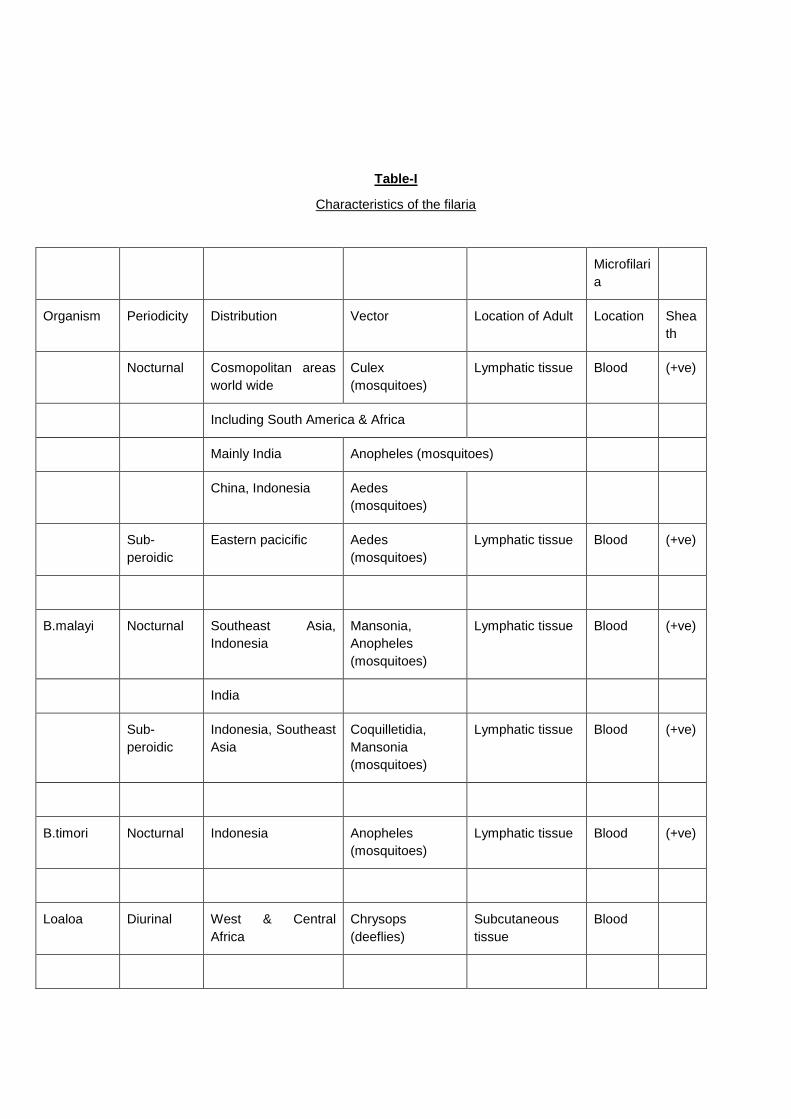
Table-I
Characteristics of the filaria
Microfilari
a
Organism Periodicity Distribution Vector Location of Adult Location Shea
th
Nocturnal Cosmopolitan areas
world wide
Culex
(mosquitoes)
Lymphatic tissue Blood (+ve)
Including South America & Africa
Mainly India Anopheles (mosquitoes)
China, Indonesia Aedes
(mosquitoes)
Sub-
peroidic
Eastern pacicific Aedes
(mosquitoes)
Lymphatic tissue Blood (+ve)
B.malayi Nocturnal Southeast Asia,
Indonesia
Mansonia,
Anopheles
(mosquitoes)
Lymphatic tissue Blood (+ve)
India
Sub-
peroidic
Indonesia, Southeast
Asia
Coquilletidia,
Mansonia
(mosquitoes)
Lymphatic tissue Blood (+ve)
B.timori Nocturnal Indonesia Anopheles
(mosquitoes)
Lymphatic tissue Blood (+ve)
Loaloa Diurinal West & Central
Africa
Chrysops
(deeflies)
Subcutaneous
tissue
Blood
On.volvulu
s
None South & Central
America
Simulium
(blackflies)
Skin, eye
Africa
M.ozzardi None South & Central
America
Culicoides
(midges)
Undetermined
site
Blood
Carribean Simulium
(blackflies)
M.perstans None South & Central
America
Culicoides
(midges)
Body cavities Blood
Africa mesentery
Perirenal tissues
M.streptoc
era
None West & Central
Africa
Culicoides
(midges)
Subcutaneous
tissue
Skin
The Lymphatic Filiariasis covers infection with three closely related nematode worm –
W.bancrofti, B.malayi and B.timori, Lymphatic filariasis is a major public health problem in India. The
parasite causing non-lymphatic filariasis will not be described here as they are not found in India.
Biology
Infection is introduced by the bite of the mosquito. Infected larvae penetrate the feeding
wound in the skin, enters the lymphatics and travel to the lymph node of the definite host (man). After
maturation in a few months they develop into white thread like adult worms (male- 40 X 01mm and
female 100 X 0.25 mm) and survive for several (10-18) years in the lymphnode. Once fertilized the
female discharges, thousand micro filarae (150-300 u long) which dwell in peripheral blood for 5-10
years. There is nocturnal periodicity (between 11 a.m. to 3 a.m.) of microfilarea in the blood stream.
The circulating microfilarae are ingested by the mosquito (intermediate host) the organism develop in
to infective larvae over the next 2 weeks and are ready to repeat the cycle when the mosquito bites.

Incubation period:
a. Pre-patent period – The time interval between inoculation of infected larvae and 1st appearance
of m.f.
b. Clinical Incubation period – The time interval from invasion of infective larvae to the development of clinical manifestation commonly 8 to 16 months (9).
Clinical features:
The disease manifestations can be divided into two district clinical types:
I. Lymphatic Filariasis.
II. Occult Filariasis
I. Lymphatic Filariasis:
The following stages have been described.
a. Asymptomatic amicrofilaraemia:
In all endemic areas a proportion of population does not show m.f. or clinical
manifestations of the disease although they have some degree of exposure to infective larvae
as those who become infected. Presently available diagnostic tools can not determine it.
b. Asymptomatic amicrofilaraelmia:
This group are symptomatic but blood are having (+ve) for m.f., they are an important
source of infection in the community.
c. Stage of acute manifestation:
There are recurrent epsodes of acute inflammation in lymph glands and vessels.
Manifestation are filarial fever, lymphangitis, lymphoedema and epididymo orchitis in male.
Life cycle of Filaria
d. State of chronic obstructive lesions:
It takes 10-15 years to develop. This phase is due to fibrosis and obstruction of
lymphatic vessels causing permanent structural changes.
In Bancroftian Filariasis the features are hydrocele, elephantiasis, chyluria.
Elphantiasis may effect the legs, scrotum, arms, penis, vulval and breast. Brugian filariasis is
similar but rarely involves genitalia.
II. Occult filariasis:
Here classical clinical manifestations are not present and m.f. are not found in blood. It is
believed to result from hypersensitivity reaction to filarial anitgen derived from M.F. But known as
example is tropical pulmonary eosinophilia.
Diagnosis
1. Demonstration of M.F. in human blood.
a. The thick film
b. Membrane filler concentration (MFC) method
c. DEC provocative test.
2. Contrast lymphangiography.
3. Ultrasonography
4. Immune diagnosis using antigen and antibody detection.
Complications of Lympahatic Filariasis
I. Thrombophlebitis
II. Tenosynovitis
III. Nerve palsies
IV. Dermatosis due to lymphangectsis and stasis in popliteal lymphatics.
V. Pericardial fluid.
VI. Glomerulonephritis-immune-mediated.
VII. Vasculitis
VIII. Mono-arthritis involving the knee joint.
IX. Endomyocardial fibrosis due to pericarditis.
X. Ocular filarisis causing raised intracranial tension and iridocyclitis.
National Filaria control programme is launched from 1955 despite all measures, the disease
filariasis is still posing problem in modern medicine. DEC is an effective drug for controlling m.f. but
has no action on the adult worms. On the other hand Homoeopathic system of treatment has wider
scope as the subtle philosophy advocates in favour of it as ti seen in practice that Homoeopathy is
abating fever mitigating the pain and inflammation of lymphatic channel (lymphangitis) and
inflammation of lymphnodes (lymphadenitis) and in some cases reducing the swelling, the
lymphaoedema but delated affect in removing m.f.
All those days from 1979 author has been trying to combat his own way to the disease
filariasis. A study was undertaken from 1979 to 1985 where 83 patients were documented under the
given parameters to assess the positive.

Positive Response
a) Cure – disappearance of subjective and objective symptoms for more than 2 years.
b) Improvement – disappearance of subjective and objective symptoms but period relief is within 2 years.
Negative Response
a) Partial improvement.
b) No Improvement.
c) Dropped out.
The results obtained were as follows:
Positive
1) % of cases cured – 29.5 47.5%
2) % of cases improved – 18
Negative
1) % of cases showed partial improvement- 22.7 52.6 %
2) % of cases dropped out – 22.7%
Most frequently appearing drugs were Bry alb. Apis mel, Rhus, tox. It was observed that a
number of cases showed cure / remarkable improvement with Bry.alb., Rhus. tox., Apis mel. but these
drugs failed to achieve desired affect in many cases too. It was taken for understanding that it might
have occurred due to defect in choosing right medicine / right potency / right repetition schedule.
Therefore the author felt to carry out a prospective study so as to get a reproducible results in
a novel way.
Hence another experiment in vitro was carried out with an object to study the effect of
Bry.alb. – Q, 6, 30
Apis.mel. – Q, 6, 30
Rhus tox. – Q, 6, 30
Methodology adopted and results obtained were as follows:
Methodology:
Known microfilaria positive cases were detected and night blood samples were collected. 10
slides were taken. One was kept for control , other nine slides were impregnated with the above drugs
and were kept separately for study. On each slide iniform quantity of blood was collected and
considerable quantity of blood was added to avoid early drying of blood slides, were seen under
microscope. Time taken by microfilariae to die in each slide was recorded and following results were
obtained.
Results:
Case 1. Sobani Samal (25 H M)
Case-1 Sobani samal (25 H M)

S. N. Name of
the drugs
Time taken by M.F. to
die
1 Apis mel. Q 5’21”
2 Bry.alb. 6 6’07”
3 Bry.alb. 30 7’17”
4 Bry.alb. Q 8’34”
5 Rhus tox. 30 10’0”
6 Apis mel. 30 12’15”
7 Apis mel. 6 13’26”
8 Rhus tox. Q 16’20”
9 Rhus tox. 30 16’20”
10 Apis mel. Q 16’20”
Case-II Sakuntala Debi (30 H F)
S. N. Name of
the drugs
Time taken by M.F. to die
1 Rhus tox. 30 9’5”
2 Apis mel. Q 11’30”
3 Apis mel. 6 12’15”
4 Apis mel. 30 12’55”
5 Rhus tox. 6 14’10”
6 Bry.alb. 6 16’05”
7 Bry.alb. Q 16’05”
8 Rhus tox. Q 16’05”

9 Bry.alb. 30 17’07”
10 Control 17’07”
Case-III Subash Ch.Muduli(12 H M)
S. N. Name of
the drugs
Time taken by M.F. to die
1 Apis mel. 30 7’18”
2 Apis mel. Q 10’07”
3 Bry.alb. Q 10’16”
4 Apis mel. 6 11’24”
5 Bry.alb. 6 12’02”
6 Rhus tox. Q 17’30”
7 Bry.alb. 30 18’10”
8 Rhus tox. 6 18’20”
9 Rhus tox. 30 18’10”
10 Control 18’20”
Case-IV Maguni.Muduli (12 H M)
S.N. Name of the
drugs
Time taken by M.F. to die
1 Rhus tox. 30 5’02”
2 Apis mel. Q 6’22”
3 Apis mel. 6 7’06”
4 Rhus tox. Q 8’45”
5 Apis mel. 30 8’57”
6 Rhus tox. 6 11’11”
7 Bry.alb. 6 14’08”

8 Control 18’45”
9 Bry.alb. 30 20’16”
10 Bry.alb. Q 22’00”
Case-V Sailabala Muduli (35 H F)
S.N. Name of the
drugs
Time taken by M.F. to die
1 Apis mel. 30 8’15”
2 Bry.alb. 6 13’43”
3 Apis mel. 6 16’50”
4 Rhus tox. Q 17’35”
5 Bry.alb. 30 20’10”
6 Rhus tox. 30 21’17”
7 Control 21’17”
8 Rhus tox. 6 21’17”
9 Bry.alb. Q 22’40”
10 Apis mel. Q 25’43”
Case-VI Dambaru Routa (14 H M)
S.N. Name of the
drugs
Time taken by M.F. to die
1 Rhus tox. 30 5’57”
2 Rhus tox. 6 7’01”
3 Rhus tox. Q 8’57”
4 Apis mel. 6 9’40”
5 Apis mel. 30 10’42”
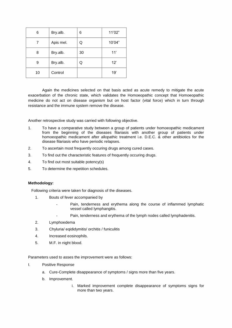
6 Bry.alb. 6 11’02”
7 Apis mel. Q 10’04”
8 Bry.alb. 30 11’
9 Bry.alb. Q 12’
10 Control 19’
Again the medicines selected on that basis acted as acute remedy to mitigate the acute
exacerbation of the chronic state, which validates the Homoeopathic concept that Homoeopathic
medicine do not act on disease organism but on host factor (vital force) which in turn through
resistance and the immune system remove the disease.
Another retrospective study was carried with following objective.
1. To have a comparative study between a group of patients under homoeopathic medicament from the beginning of the diseases filariasis with another group of patients under homoeopathic medicament after allopathic treatment i.e. D.E.C. & other antibiotics for the disease filariasis who have periodic relapses.
2. To ascertain most frequently occuring drugs among cured cases.
3. To find out the characteristic features of frequently occuring drugs.
4. To find out most suitable potency(s)
5. To determine the repetition schedules.
Methodology:
Following criteria were taken for diagnosis of the diseases.
1. Bouts of fever accompanied by
- Pain, tenderness and erythema along the course of inflammed lymphatic vessel called lymphangitis.
- Pain, tenderness and erythema of the lymph nodes called lymphadenitis.
2. Lymphoedema
3. Chyluria/ eqididymitis/ orchitis / funiculitis
4. Increased eosinophils.
5. M.F. in night blood.
Parameters used to asses the improvement were as follows:
I. Positive Response
a. Cure-Complete disappearance of symptoms / signs more than five years.
b. Improvement.
i. Marked improvement complete disappearance of symptoms signs for more than two years.
ii. Moderate improvement – Disappearance of fever, lymphangitis, lymphadenitis, normal eosinophil level.
iii. Mild improvement – Disappearance of fever, lymphangitis, lymphadenitis but no change to m.f. & eosinophils.
II. Negative Response
a. No improvement – There is no reduction of signs / symptoms of the disease inspite of our several days medication.
b. Dropped out – Patient did not stick to our treatment for sufficient period of treatment.
Patients were collected from Dr. A.C.Homoeopathic Medical College & Hospital and author’s
clinic. In each case symptoms were collected from patients in a standardised case recording format
and were repertorised in classical method and medicines were prescribed in 50 millesimal and
centesimal scale.
Results:
204 patents were scanned and by means of above diagnostic features, cases were
diagnosed. As per the fixed parameters the results were documented. They are as follows:
Table-I
Positive Response Negative Response
Types of cases Cure Mark
Impr.
Mod.
Impr.
Mild
Impr.
Total No.
impr.
Dropp. Total
Cases without
allopathic directly with
homoeopathy
9 13 15 35 72 12 20 20
Cases after allopathy
with homoeopathy
40 17 18 5 80 9 11 11
Results on effects of various potency :
Table-II
Types of potencies (+)ve response (-)ve response
50 millesimal 133 37
Centesimal 19 15

Total 152 52
Results on effects of repetition schedule:
Table-III
Types of repetitions (+)ve response (-)ve response
Single dose 14 12
Repeated dose 138 40
Frequency of drugs appeared in cured cases:
Table-IV
14 12
138
40
0
20
40
60
80
100
120
140
160
(+)ve response (-)ve response
Typ
es o
f re
pet
itio
n
Response
Results on effects of repetition schedule
Single dose Repeated dose

Name of the
dugs
frequency of
appearance
Bry a. Ars
a.
Rhus
. t
Apis
m.
Puls. Nat. m. Bell. Sil. Phos
.
Sulp
h.
Calc. c
29 26 26 25 14 8 8 5 4 4 3
Result analysis:
Characterestic features of frequently occuring drugs.
Bry. alb.
1. Lymphoedema < exertion / evening / warm – 29
rest / morning / cold - 28
2. Thirst (+++) with dry tongue – 26
3. Fever with thirst – 26
4. Constipation without desire – 26
5. Bitter vomiting with bitter taste in mouth with thirst – 25
6. Chill with external coldness – 24
7. Sour smelling sweat – 24
8. Fever with headache > pressure – 24
Ars. alb.
1. Lymph oedema < by cold – 26
With burning > warm - 26
2. Fever – periodic < night – 26
< mid night / mid day - 25
3. Chill with thirst - 25
4. Thirst for small quantities of warm water - 25
29
26 2625
14
8 8
54 4
3
0
5
10
15
20
25
30
35
Fre
qu
en
cy o
f ap
pe
ara
nc
e
Name of the dugs
Frequency of drugs appeared in cured cases

5. Restlessness - 25
6. Chilly patient - 24
7. Aversion sweets - 24
8. Desire – warm food / drink – 24
Rhus. tox.
1. Fever < at night - 26
2. Fever with thirst with bitter taste in mouth - 25
3. Restlessness - 25
4. Lymphangitis / Lymphadenitis / Myalgia / Lymphoedema
< rest / cold – 25
motion / warm - 25
5. Pruritus with oedema < cold - 24
6. Chilliness with restlessness with dry tongue - 24
Apis mel.
1. Fever with chilliness with thirst - 24
2. Thirstless with dry tongue in other times - 24
3. Oedema with pruritus < warm, > cold 23
4. Rt. Sided oedema - 23
5. Lymphangitis / Lymphadenitis with itching - 23
Pulsatilla.
1. Fever with chilliness without thirst with dry tongue - 13
2. Lymphangitis / Lymphoedema / Lymphadenitis
3. Bitter vomiting with bitter taste in mouth with thirstlessness – 12
4. Sweat on single parts - 12
5. Weeping disposition – 12
6. Chilliness wants open air – 12
7. Fever with headache > by pressure – 12
Results obtained from comparative study of group of patients with homoeopathic medicines
alone not taking allopathic medicine and after allopathic medicines were processes for reliability test
through chi-square test by using 2X2 contingency table. On referring to chi-square table with 1 degree
of freedom the value of chi-square for a probability of 0.05 is 3.841. Since the calculated value (3.9) is
much above, we conclude that the Null hypothesis is rejected and the result is significant and it is
established statistically that homoeopathic medicine act better after allopathic medicine.
Observation to the results of positive response provides us another inference that large
number of cure and marked improvement are seen when homoeopathy is prescribed after allopathic
treatment. D.E.C. kills the m.f. but no effect on adult worms, wherein homoeopathy the microfilarea
disappears at last. It is perhaps due to Hering’s law of cure the signs / symptoms that appears first will
disappear “last and adult worms die first, which appears last. By this homoeopathic medicine is not
preventing the communicability of the diseases immediately. In other hand D.E.C. is preventing
communicability of the disease, but no effect on adult worm. Therefore both have their merits /
demerits in the treatment of filariasis.
Now it is an urgent need to set up a new principle / a new practice and have new drugs to
combat m.f. first in order to prevent communicability of the disease and followed by constitutional drug
to change the constitutional dyscrasia by which man can be protected to filariasis in future.
Results obtained from effects of various potencies were prescribed for similar test and
calculated value (7. 71) is much above. We conclude that Null hypothesis is rejected and the result is
significant and it is established scientifically that homoeopathic medicine in 50 millesimal scale acts
better than centesimal scale.
N.B.: Exception to the cases of chyluria, who responded to single dose of very high potency i.e. Kali
bichromicum. Similarly Bry alb. 200 single dose to all cases indicating Bry.alb.
Results obtained from effects of repetitive schedules were processed for similar test and
calculated value (0.56) is much less. The Null hypothesis is accepted and the result is non-significant
and it is established scientifically that there is no difference between single dose & repeated doses in
the treatment of filariasis.
Conclusion:
From above study it is envisaged that:
a. Homoeopathic medicines act curatively and provides better response, when it is prescribed after allopathic treatment.
b. 50 millesimal acts better than centesimal scale exception to this is Kali bichromicum 10M in chyluria and Bry alb. 200, when they are indicated.
c. Regarding repetition schedule, it is difficult to opine with this study. Therefore a separate study is needed to be designed to opine on the effect of single dose and repeated doses.
d. While prescribing for Homoepathic purpose characteristic symptoms count more value than common symptoms which validates, the observation of earlier stalwarts of Homoeopathy.
However, it is concluded that to ascertain the results obtained by this retrospective study
needs to be reconfirmed by a prospective study i.e. homoeopathy for adult worms and allopathy for
m.f.
Apart from that homoeopathy needs new principles / new kind of practice and new drugs to
combat m.f. first to prevent the wrath of the communicability of disease filariasis. Thereby
homoeopathy can rise to the zenith in the treatment of filariasis compared to the counter part
allopathy.
Bibliography
1. Dilip Mathai, Humaqn Filariasis & Related infections, API Text Book of Medicine, Editorial in Chief – G.S.Sainani, 6
th Edition, 1999, Page-106.
2. Thomas B.Nutmanm Peter F. Wellen, Filariasis & Related Infections (Loiasis, Onchoceriasis & Dracunculosis, Harrison’s Principles of Internal Medicines Vol-1 exclusive rights by Tata Mc Graw-hill Book co, Singapore.
3. WHO 1998, World Health Report 1998, Life in the 21st Century A vision for all, Report of
Director General WHO.
4. WHO 1999, Health situation in South East Asia Region 1994-97 Region office for SEAR, New Delhi.

5. Govt. of India (1996) Annual Report 1995-96, DGHS, Ministry of Health & Family Welfare, New Delhi. [Epidemiology of Communicable diseases, Park’s Text Book of Preventive & Social Medicine, Park. K, 16
th Edition, 2000, Published by M/S Banarasi Das Bhanot, pages-
204].
6. ICMR, Annual Report of the Director General 1994-95.
7. D.H.S., Report Govt. of Orissa, 1999.
8. Dilip Mathai, Human Filariasis & Related Infections, API Text Book of Medicine, Editorial in Chief G.S. Sainani, 6
th Edition, 1999, Page-106.
9. 14-WHO. Techni Rep. Ser. No.702 (page No.204 K.Park).
10. 15 – Price, E.W. and Henderson, W.J. (1979). Trans R.soc. Med. Hyg., 73:640-7.
11. Sainani G.S., Editorial in Chief, API Test Book of Medicine, 6th Edition 1999, Page-108.