how age of acquisition influences brain architecture in...
TRANSCRIPT

Journal of Neurolinguistics 36 (2015) 35e55
Contents lists available at ScienceDirect
Journal of Neurolinguisticsjournal homepage: www.elsevier .com/locate/
jneurol ing
How age of acquisition influences brainarchitecture in bilinguals
Miao Wei a, Anand A. Joshi b, Mingxia Zhang c, Leilei Mei d,Franklin R. Manis a, Qinghua He a, Rachel L. Beattie e,Gui Xue c, David W. Shattuck f, Richard M. Leahy b, Feng Xue a,Suzanne M. Houston a, Chuansheng Chen g, Qi Dong c,Zhong-Lin Lu e, *
a Department of Psychology, University of Southern California, Los Angeles, CA 90089-1061, USAb Signal and Image Processing Institute, University of Southern California, Los Angeles, CA 90089-2564, USAc National Key Laboratory of Cognitive Neuroscience and Learning, Beijing Normal University, Beijing100875, Chinad Center for Studies of Psychological Application and School of Psychology, South China Normal University,Guangzhou 510631, Chinae Center for Cognitive and Behavioral Brain Imaging and Department of Psychology, The Ohio StateUniversity, Columbus, OH 43210, USAf Ahmanson-Lovelace Brain Mapping Center, Department of Neurology, David Geffen School of Medicine,University of California, Los Angeles, CA 90095-7334, USAg Department of Psychology and Social Behavior, University of California Irvine, Irvine, CA 92697, USA
a r t i c l e i n f o
Article history:Received 10 January 2015Received in revised form 29 April 2015Accepted 1 May 2015Available online 22 May 2015
Keywords:Age of acquisitionBilingualStructural plasticityMRI
* Corresponding author. Department of PsycholoTel.: þ1 614 247 8252.
E-mail address: [email protected] (Z.-L. Lu).
http://dx.doi.org/10.1016/j.jneuroling.2015.05.0010911-6044/© 2015 Elsevier Ltd. All rights reserved
a b s t r a c t
In the present study, we explored how Age of Acquisition (AoA) ofL2 affected brain structures in bilingual individuals. Thirty-sixnative English speakers who were bilingual were scanned withhigh resolution MRI. After MRI signal intensity inhomogeneitycorrection, we applied both voxel-based morphometry (VBM) andsurface-based morphometry (SBM) approaches to the data. VBManalysis was performed using FSL's standard VBM processingpipeline. For the SBM analysis, we utilized a semi-automated sulcidelineation procedure, registered the brains to an atlas, andextracted measures of twenty four pre-selected regions of interest.We addressed three questions: (1) Which areas are more suscep-tible to differences in AoA? (2) How do AoA, proficiency and cur-rent level of exposure work together in predicting structural
gy, The Ohio State University, 1835 Neil Avenue Columbus, OH 43210, USA.
.

M. Wei et al. / Journal of Neurolinguistics 36 (2015) 35e5536
differences in the brain? And (3) What is the direction of the effectof AoA on regional volumetric and surface measures? Both VBMand SBM results suggested that earlier second language exposurewas associated with larger volumes in the right parietal cortex.Consistently, SBM showed that the cortical area of the right su-perior parietal lobule increased as AoA decreased. In contrast, inthe right pars orbitalis of the inferior frontal gyrus, AoA, profi-ciency, and current level of exposure are equally important in ac-counting for the structural differences. We interpret our results interms of current theory and research on the effects of L2 learningon brain structures and functions.
© 2015 Elsevier Ltd. All rights reserved.
1. Introduction
In the field of second language acquisition (SLA), the age of SLA onset attracts much less attentionthan the level of proficiency when researchers try to explore cortical representations of languages inbilinguals. The term “Age of Acquisition (AoA)” has been widely used to denote the age at which amonolingual individual first started learning a new or second language (Kovelman, Baker, & Petitto,2008). The effect of AoA is still entangled with that of the level of proficiency on cerebral organiza-tion in bilinguals (Wattendorf & Festman, 2008). It has been shown that AoA modulates functionalneural activity in several aspects of language processing, for example: phonology (Frenck-Mestre,Anton, Roth, Vaid, & Viallet, 2005), syntax (Mahendra, Plante, Magloire, Milman, & Trouard, 2003),different aspects of grammar (Hernandez, Hofmann, & Kotz, 2007; Waldron & Hernandez, 2013;Wartenburger et al., 2003; Weber-Fox & Neville, 1996) and lexical access (Isel, Baumgaertner, Thr€an,Meisel, & Büchel, 2010; Mahendra et al., 2003; Perani et al., 2003).
Growing evidence indicates that AoA is associated with fMRI BOLD activations in bilingual brains.For instance, in a narration task, Bloch et al. found that later AoAwas associated with greater individualvariations in the local cerebral activation of different languages in Broca's and Wernicke's areas (Blochet al., 2009). Likewise, Kim and colleagues reported that in Wernicke's area, identical regions serveboth the first language (L1) and the second language (L2) in early and late bilinguals, but early bi-linguals share overlapping L1 and L2 regions while late bilinguals have spatially distinct but neigh-boring L1 and L2 regions in Broca's area (Kim, Relkin, Lee, & Hirsch, 1997). Other functionalneuroimaging studies on effects of AoA suggest that late bilingual exposure is linked to a broaderrecruitment of neural tissues in the left inferior frontal gyrus (IFG), and bilateral IFG (Wartenburgeret al., 2003). Taken together, these studies suggest that AoA of L2 may have important effects on thefunctional organization of the language system in bilingual brains.
Functional neuroimaging methods such as PET and fMRI are widely used to study neural mecha-nisms in different cognitive skills. Although the traditional view is that experience and expertise withspecific skills are mediated by functional (rather than structural) plasticity in the brain (Kim et al.,1997), structural brain changes as a result of extensive experience in acquiring certain skills havebeen widely reported. For example, increased bilateral posterior hippocampal grey matter volume hasbeen associated with acquisition of spatial representation in the London taxi driver study (Woollett &Maguire, 2011). Likewise, medical students showed increased grey matter volume in the posterior andlateral parietal cortex bilaterally during their extensive medical examination study period (Draganskiet al., 2006). Structural changes related to bilingualism and multilingualism have also been reported.For example, bilinguals tend to have increased grey matter volume/density in Heschl's gyrus (Resselet al., 2012), the left caudate (Zou, Ding, Abutalebi, Shu, & Peng, 2012), and the left inferior parietalstructures (Della Rosa et al., 2013; Mechelli et al., 2004). Abutalebi et al. (2013) showed that the onlygrey matter volume difference between early multilinguals and monolinguals was found in the leftputamen and related to their proficiency levels of L3. However, upon careful review of the paper, onecannot rule out that the grey matter volume difference is rather due to both proficiency level and AoA.

M. Wei et al. / Journal of Neurolinguistics 36 (2015) 35e55 37
In terms of second language AoA, at the whole brain level, Mechelli and colleagues reported highergreymatter density in the left inferior parietal lobule (IPL) in both early and late bilinguals compared tomonolinguals. The effect was more evident in early bilinguals. These findings demonstrate changes inbrain structures associated with second language learning. However, with regard to L2 AoA, the righthemisphere has not received the same attention as the left hemisphere language regions, and hence, ithas been relatively neglected by neuroscientists working in this field. This is surprising since in themost influential neuroimaging study on AoA by Mechelli et al., a trend of greater grey matter densitywas observed in the right IPL in early bilinguals (Mechelli et al., 2004). Further, in neurosurgery, theright hemisphere structural plasticity has been reported widely (Duffau, 2006, 2014). However,whether and how changes in brain structures accompany variation in AoA has not been sufficientlyinvestigated. Hence, it is evident that the right hemispheric neuroplasticity related to L2 AoA needsfurther study and clarification. In general, as pointed out by Abutalebi et al. (2013), there are twomajorneural differences in L1 and L2 processing that were observed in the literature: one is L2's enhancingand sharing regions with L1 and the other is the specific engagement of areas that are outside of“traditional” left hemisphere language cortex that are recruited by the use of L2.
In the present study, we analyzed high resolution structural MRI images in brain regions implicatedin prior structural and fMRI studies on second language learning in an attempt to gain insights into thenature of bilingual language processing and the impact of AoA on bilingual brains. We first undertookan exploratory whole-brain VBM analysis and then using SBM, we specified a set of ROIs based onexisting literature, and performed statistical tests on the ROIs. Volume measures of 24 ROIs wereobtained using a semi-automated sulci delineation procedure that registered the brains to an atlas todefine the ROIs for each individual subject. Because a relatively small number of ROIs were used in SBM(surface-based morphometry), we only had to perform multi-comparison correction for a relativelysmall number of comparisons, leading to improved statistical power (Poldrack, 2007), which gave usanother opportunity to discover subtle volume differences that may not survive the VBM whole-brainmultiple comparison correction.
We sought to answer three questions. First, which of these brain regions are structurally affected byAoA? The adaptive control hypothesis by Green and Abutalebi (2013) predicted adaptive changes in theneural circuits associated with cognitive control processes in a dual-language context. The authorsprovided a schematic description of the neural structures that were involved in language controlprocesses. The cortical and subcortical “control network” included the prefrontal cortex (PFC), pre-supplementary motor area (pre-SMA), IFG, anterior cingulate cortex (ACC), caudate, putamen, thal-amus and the parietal cortex. The model allows for bilateral structures such as bilateral inferior frontalcortex and bilateral basal ganglia, and predicts that earlier exposure to a bilingual environment wouldintroduce more neural adaptation changes in this circuit. We first applied a whole-brain VBM searchand then conducted a ROI analysis incorporating the regions that could possibly show AoA relatedvolume changes according to the adaptive control hypothesis: the left PFC, left pre-SMA, the left IFGthat is typically involved in effortful semantic retrieval (Fiez, 1997), left ACC, left caudate, left putamen,left thalamus and the left parietal cortex that is important for attention control (Della Rosa et al., 2013).To provide a more detailed analysis, we divided the IFG into three sub-regions [pars opercularis(IFGop), pars triangularis (IFGpt), and pars orbitalis (IFGor)], the posterior parietal cortex into threesub-regions [supramarginal gyrus (SMG), angular gyrus (AG), and superior parietal lobule (SPL)],leading to a total of 12 ROIs in the left hemisphere. The selection of the 12 ROIs are also consistent withother previous studies (Hernandez & Li, 2007; Jeong et al., 2007; Mechelli et al., 2004). Functionally, ithas been generally accepted that more extensive left hemisphere activations are associated with bi-linguals than monolinguals especially in late bilinguals and low proficiency bilinguals. But more andmore studies also demonstrated involvement of right hemisphere homologous structures in bilinguals(Badzakova-Trajkov, Kirk, &Waldie, 2008; Dehaene et al., 1997; Wang et al., 2011). Among the 12 ROIswe chose above, their right homologues, for example, the right IFG (also noted in the adaptive controlhypothesis) and the right parietal cortex were repeatedly reported to have increased activation for L2processing (Musso et al., 2003; Perani et al., 2003; Sebastian, Laird, & Kiran, 2011; Wartenburger et al.,2003). In terms of AoA of L2, a recent meta-analytic review also found that bilinguals who acquiredboth languages by 6 years of age demonstrated bilateral hemispheric involvement for both languages,whereas those who acquired their second language after age 6 showed left hemisphere dominance for

M. Wei et al. / Journal of Neurolinguistics 36 (2015) 35e5538
both languages (Hull & Vaid, 2007). Therefore, we included the right homologous regions of these 12ROIs in our analyses. Structurally, the trend of greater grey matter density was observable in the rightIPL in early bilinguals (Mechelli et al., 2004). However, no other significant effects have been found ineither grey or white matter.
Our second question was: how do AoA, proficiency and current level of exposure work together toshape bilingual brains? Various factors, such as linguistic distance between L1 and L2, nonnative statusof the learner, L2 learner's language environment, years of education in L2, motivation for learning L2,language learning aptitude, age of L2 acquisition, degree of L2 proficiency, and relative L1 and L2exposure, may modulate brain representations of languages in bilinguals (Higby, Kim, & Obler, 2013;Klein et al., 2006; Vaid, 1983). Among these variables, other than AoA, two factors, the degree of L2proficiency (Chee, Hon, Lee, & Soon, 2001; Chee, Soon, Lee, & Pallier, 2004; De Bleser et al., 2003;Elston-Guttler, Paulmann, & Kotz, 2005; Mechelli et al., 2004; Perani et al., 1998, 2003; Vingerhoetset al., 2003; Wartenburger et al., 2003) and the current level of exposure (Perani et al., 2003;Vingerhoets et al., 2003), have often been implicated in their effects on the neural organization oflanguage processing in bilinguals (Consonni et al., 2013). How these factors are related to structuralchanges in bilingual brains is still under debate. The current general consensus seems to be that dif-ferences in AoA, level of proficiency, type of grammatical construction and the amount of dailyexposure to a language are generally reflected in the degree of activation but not in the localization ofactivation in functional imaging (Perani & Abutalebi, 2005). It has been suggested that AoA has moreinfluence on the neural substrate of L2 grammar processing compared to proficiency (Abutalebi, 2008;Wartenburger et al., 2003), but the pattern of brain activity for semantic judgments depends stronglyon proficiency (Indefrey, 2006; Perani & Abutalebi, 2005; Wartenburger et al., 2003). In this study, weevaluated effects of AoA, proficiency and current level of L2 exposure on the structures of bilingualbrains.
The third question concerns the direction of structural brain differences. Do cortical regions becomelarger or smaller as AoA increases? Increased grey matter density in the mid-temporal area and leftposterior intraparietal sulcus has been shown after 3 months of juggling training (Draganski et al.,2004). Other than grey matter density, experience-induced cortical volume and/or thicknesschanges have also been reported. For example, researchers found that after 3 months of languagelearning, the volume of the right hippocampus, and the cortical thickness of the left middle frontalgyrus (MFG), left IFG and left superior temporal gyrus (STG) increased (Mårtensson et al., 2012). Othersreported that the volume of the posterior hippocampus correlated positively with the amount of timespent as a taxi driver, but the volume of the anterior hippocampus correlated negatively with the timespent as a taxi driver (Maguire et al., 2000). As far as “early learning” is concerned, musicians showedincreased grey matter volume in the left IFGop and the volume was positively correlated with years ofmusical performance (Abdul-Kareem, Stancak, Parkes, & Sluming, 2011). It has also been reported thatbilinguals who learned both languages before age 7 have larger Heschl's gyri volume than mono-linguals (Ressel et al., 2012). These results show that experience could either increase or decreasecortical volume and/or thickness. Although Mechelli and colleagues reported that the grey matterdensity in the left IPL decreased when the AoA of second language increased (Mechelli et al., 2004), it isnot completely clear how cortical volume or density varies with AoA. Based on the observation thatskill acquisition alters grey matter volume in task-related areas and that fMRI studies showed broaderactivations in late bilinguals, our hypothesis is that larger greymatter volumewould be associatedwithearlier AoA. Whether similar patterns would occur in the white matter, total volumes, cortical thick-ness and cortical surface area of the same regions is an open question.
2. Materials and methods
2.1. Participants
Thirty-six healthy native English-speaking bilingual adults were recruited from the University ofSouthern California and University of California, Irvine. Within these participants fourteen early bi-linguals (mean age 22.3 ± 2.6, range 19e28) were exposed to their non-native language before theywere 6 years old, four intermediate bilinguals (mean age 20.5 ± 1.3, range 19e22) acquired their non-

M. Wei et al. / Journal of Neurolinguistics 36 (2015) 35e55 39
native language between age 6 and 12, and eighteen late bilinguals (mean age 21.8 ± 2.9, range 18e30)were not exposed to their non-native language until they were 12 years old. The age cut-offs for thethree groups were based on previous research (Canseco-Gonzalez et al., 2010; Frenck-Mestre et al.,2005; Wattendorf & Festman, 2008). Demographics of early and late bilinguals are as follows: 1) Early:6 M and 8 F; 4 Asian and 10 Westerner, and 2) Late: 8 M and 10 F; 4 Asian and 14 Westerner. Bothparents belonged to the same ethnic group. All participants were right-handed according to theEdinburgh Inventory (Oldfield, 1971), had normal or corrected-to-normal vision and no history of headinjury, psychiatric or neurological disorders. Informed consent was obtained from all participants, andall procedures were approved by the IRB on both sites.
2.2. Language history questionnaire
To render our findings independent of the effect of the type of the second language, we includedparticipants with various non-native languages. Their second languages comprised Spanish (15),French (7), Mandarin (3), Cantonese (2), German (2), Vietnamese (2), Hebrew (1), Japanese (1), Por-tuguese (1), Serbian (1), and Urdu (1). In the present study, we controlled the similarity of the par-ticipants' two languages by balancing the second language type in the early and late bilinguals. AoA,language proficiency and current level of exposure of the native (English) and second languages wereevaluated using self-reported measures in a language history questionnaire. Participants self-ratedtheir native and second language proficiency on a seven-point scale (1 ¼ “only knows a little”,7 ¼ “native-level proficiency”) in four different domains: reading, writing, listening, and speaking. Toobtain a compositemeasure of native and second language proficiency, we computed the total scores ofreading, writing, listening and speaking of each language. For both L1 and L2, the current level ofexposure is evaluated by a question in the questionnaire “what is the overall percentage of time thatyou speak this language now?”
Previous studies suggested that bilinguals are able to assess their language proficiency and reporttheir language history in a way that is consistent with behavioral performance (Jia, Aaronson, & Wu,2002). Grogan et al. (2012) and Mayberry, Chen, Witcher, and Klein (2011) used a similar languagehistory questionnaire in their work. All participants reported higher proficiency in English as shown inTable 1.
2.3. Image acquisition
Image data was obtained on a 3T Siemens Magnetom Trio MRI scanner located in the Dana andDavid Dornisife Cognitive Neuroscience Imaging Center at the University of Southern California. Weacquired 3D, T1-weighted images using the Magnetization Prepared Rapid Gradient Echo (MP-RAGE)sequence and a 12-channel head coil. The scan parameters were: TR 2530 ms, TE 3.09 ms, TI 800 ms,flip angle 10 deg, field of view 256 mm, slice thickness 1 mm, voxel dimensions: 1.0 � 1.0 � 1.0 mm,acquisition time 10 min 48 s.
Table 1Summary of descriptive statistics in self-reported language history questionnaire.
Language history measures Native language (English) Non-native language
Mean SD Range Mean SD Range
Age of acquisition (years) 0.97 1.58 0.00e6.00 9.47 7.19 0.00e21.00Self-reported proficiencyReading 6.89 0.40 5.00e7.00 3.28 1.54 1.00e6.00Writing 6.78 0.59 4.00e7.00 2.97 1.42 1.00e6.00Listening 6.92 0.37 5.00e7.00 3.81 1.79 1.00e7.00Speaking 6.83 0.51 5.00e7.00 3.72 1.63 1.00e7.00
Current level of L2 exposure (% of day) 91.33 10.49 40.00e100.00 10.16 12.47 0.00e60.00

M. Wei et al. / Journal of Neurolinguistics 36 (2015) 35e5540
2.4. VBM and SBM analyses
Two well-established techniques were available to detect the correlation between structure dif-ferences and language variables among subjects. VBM is a widely used automated technique whichaligns the cortex of a subject to an atlas and requires no manual input. SBM, on the other hand, usessulcal/gyral anatomy information for co-registration which can also provide additional informationcompared to VBM, such as cortical thickness, area, and curvature. VBM and SBM could generatedifferent information and for the purpose of maximizing the robustness of our results, we choose bothmethods and report convergent findings.
2.4.1. VBMStructural MRI data were analyzed with the Oxford Centre for Functional MRI of the Brain (FMRIB)
software Library voxel-based morphometry (FSL-VBM), a VBM style analysis tool (Good et al., 2001).After MRI signal inhomogeneity correction using the N4 method (http://www.slicer.org), brains werefirst extracted, segmented and aligned to the MNI 152 standard space (Andersson, Jenkinson, &Smith, 2007). Second, the spatially normalized images were then averaged to create a study-specific template, to which the native grey matter images were registered again using both linearand nonlinear algorithms. Third, the modulated grey matter images (to correct for local expansion orcontraction due to the non-linear component of the spatial transformation) were then smoothedwith an isotropic Gaussian kernel with a sigma of 3 mm. The final measure of this procedure wasgrey matter volume.
2.4.2. SBMBrainSuite is a surface-based image analysis tool that has been developed for MRI image and surface
visualization, manual tracing of brain sulci, extraction of the inner and outer surfaces of the cerebralcortex, and segmentation of grey and white matter structures based on the 26 sulci drawn. We chosethis approach because many volumetric-based approaches do not align the cortical anatomy well(Pantazis et al., 2010) and we were interested in the cortical thickness, area and volume in functionalareas of the cortex, which are closely related to the folding pattern.
2.4.2.1. Data pre-processing. Our data analysis pipeline consisted of three steps: pre-processing, seg-mentation and registration. During pre-processing, we performed skull stripping, manual editing ofthe brain masks using BrainSuite11 (Shattuck & Leahy, 2002) and N4 inhomogeneity correction.
We first imported the raw MRI data and used the BrainSuite Brain Surface Extractor (BSE) tocomplete skull and scalp stripping (Shattuck & Leahy, 2002). BSE applied a combination of anisotropicdiffusion filtering, edge detection and mathematical morphological (Dogdas, Shattuck, & Leahy, 2005;Shattuck & Leahy, 2002) operators to remove non-brain tissues in the MRI image. The interactive BSEtool was used to find the suitable parameters for brain extraction: diffusion iterations 5, diffusioncontrast 25, edge constant 0.75, and erosion size 1.We obtained a set of three-dimensional brain masksthat excluded most non-brain tissues while preserving the cerebellum. Shattuck and Leahy reportedthat BSE could on average retain 99% of the white matter and grey matter in the final extracted brainmasks with only 1% of non-brain tissues (Shattuck & Leahy, 2002). To make these masks even moreaccurate, we performed additional manual editing with the mask editing tool in BrainSuite. Theseedited masks were then saved for later intensity inhomogeneity correction purposes.
Imperfections in scanner hardware and variations in brain anatomy introduce intensity in-homogeneity to MR images that could confound the segmentation procedure (Shattuck, Sandor-Leahy, Schaper, Rottenberg, & Leahy, 2001). We performed inhomogeneity correction using theN4ITK MRI Bias Correction module from 3D Slicer. Corrections were performed on the raw brainimages using masks generated from the previous manual editing step since better inhomogeneitycorrection results could be obtained when the bias estimation is limited to a meaningful region.Further, we employed two sets of N4 parameters to optimize individual bias correction. Because N4'sbias correction is a preprocessing step for tissue classification with its quality reflected in the per-formance of the tissue classification routine (Shattuck et al., 2001), we chose between these two sets

Table 2Two sets of parameters used in N4 bias field correction procedure.
Number of iterations Convergence threshold BSpline grid resolution Spline distance Shrink factor
Set 1 150,140,130 0.0001 1, 1, 1 0.00 4Set 2 150,140,130 0.0001 2, 2, 2 0.00 2
M. Wei et al. / Journal of Neurolinguistics 36 (2015) 35e55 41
of parameters (See Table 2) based on the quality of tissue classification results from the next step, theset of parameters that yielded better tissue classification results for a particular participant waschosen for that participant.
To avoid group differences caused by N4 parameter differences, we balanced the number of par-ticipants using different parameters in our sample.
2.4.2.2. Brain tissue segmentation. Next, we applied the Partial Volume Classifier (PVC) module inBrainSuite to perform tissue classification on the intensity-corrected brain. The algorithm labels eachvoxel in the image as background voxel, cerebrospinal fluid, grey matter and white matter. The labelswere used to determine the volumes of each type of tissue.
The cerebrumwas identified and extracted by registering a multi-subject average brain (ICBM452)to the individual brains using Automated Image Registration (AIR; Pantazis et al., 2010;Woods, Grafton,Holmes, Cherry, & Mazziotta, 1998). We performed manual editing on the cerebrum masks and to-pology correction to the cortical surfaces. A graph-based approach was used to remove topologicaldefects (handles and holes) and ensure a tessellationwith spherical topology. Finally, brain pial, middleand inner cortex surfaces were generated. The inner cortex was expanded to form an outer/pialboundary. Results were carefully examined after each step. We returned to the previous step toimprove parameter settings if necessary.
2.4.2.3. Cortical surface and volume registration. After cortical surfaces were generated from the last step,we split the brain into two hemispheres. Each hemisphere was then parameterized into a unit squaremap and the position in the unit square gave the coordinate for each vertex (Joshi et al., 2009). Corticalsurfaces from different participants were aligned in the 2D space and aligned to a template later in theregistration step. Because the cytoarchitectural and functional parcellation of the cortex was intimatelyrelated to the folding of the cortex (Joshi, Shattuck, Thompson, & Leahy, 2007), we aligned corticalsurfaces by aligning meaningful cortical features/sulci first. The curve labeling protocol (Pantazis et al.,2010) was followed in using the Curve Protocol Tool (Joshi et al., 2010; Shattuck et al., 2009) that hasbeen integrated into BrainSuite software package. A thorough description of the sulcal curves withinstructions on how to trace them is available on the website (http://neuroimage.usc.edu/CurveProtocol.html). The Curve Protocol Tool was an interactive tool to semi-automatically identifysurface sulci. The rater was trained on the curve protocol by two experienced neuroanatomists (C.R.and S.Y.C.). Ten training sessions were scheduled (2 h per session) to learn cortical anatomy and thetracing protocol. A set of 26 sulci on each hemisphere were delineated and used as landmarks in thesurface matching procedure. The algorithm used a graph theory approach to identify the path betweenthe start and end points of each sulcus specified by users with the lowest cost, which is defined as acombination of local curvature features and the distance between vertices on the surface represen-tation (Shattuck et al., 2009). We traced the curves with 0.5 stickiness (stickiness is associated withcurvature weighting and it is a parameter to ensure that the paths of curves follow the sulcal depth ofvalleys) on themidcortical surface, midway between the pial surface and the greyewhite interface, andwhen it was necessary to cross gyri, we used zero stickiness. Midcortical surfaces were used becausethey provided better access to the depth of sulci than pial surfaces, allowing more stable tracing(Pantazis et al., 2010).
Finally, SVREG (Surface constrained Volumetric Registration), a set of programs that registers andlabels BrainSuite generated cortical surfaces and volumes (Joshi, Shattuck, & Leahy, 2012), was used tosegment the cortical surfaces and generate labels for each subregion. The program took BrainSuite-generated surfaces and volumes, registered them with the ICBM template, and segmented them into

M. Wei et al. / Journal of Neurolinguistics 36 (2015) 35e5542
subregions with labels (since this data was processed, SVREG has been integrated with and is a part ofnewly released BrainSuite14b). Each hemisphere was divided into 47 cortical and subcortical ROIs(Table 3). The labeled surfaces were displayed in BrainSuite and carefully examined to make sure thatdesirable results were generated.
To work with the language control framework proposed by Green and Abutalebi (2013), threeadditional ROIs that were not included in the original 47 ROIs were manually delineated on the currenttemplate and further registered with SVREG. They were ACC, pre-SMA and PFC. The detailed de-scriptions of demarcation are illuminated here. ACC's caudal boundary as established by themammillary bodies (Desikan et al., 2006), pre-SMAwas defined as the area of the medial frontal cortexin the superior frontal gyrus lying dorsal to the cingulate sulcus, rostral to the vertical commissureanterior (VCA) line, and caudal to the virtual line passing through the genu of the corpus callosum (Kimet al., 2010), whereas the PFC located anteriorly from the first slice that contained brain tissue and theposterior landmark was determined by first locating the most anterior slice that contained the tem-poral stem (thewhitematter tract connecting the temporal and frontal lobes (Wible, Shenton, Hokama,& et al., 1995)).
See Table 3 for all 50 ROIs segmented on each hemisphere.
2.5. Statistical analysis
2.5.1. VBMAwhole brainmultiple regression analysis for all 36 subjects was performedwith the FSL-VBMGLM
model. AoA was the regressor of interest while proficiency and exposure were included as nuisancevariables. Voxel-wise grey matter volume for the whole brain GLM was applied using permutation-based non-parametric testing, correcting for multiple comparisons across space.
2.5.2. SBMThe total volume, grey matter volume, white matter volume, thickness, and cortical surface area of
the ROIs were computed by BrainSuite when available and exported to SPSS (version 17) for further
Table 3Segmented brain regions.
CortexFrontal lobe Superior frontal gyrus Occipital lobe Superior occipital gyrus
Middle frontal gyrus Middle occipital gyrusPars opercularis Inferior occipital gyrusPars triangularis Lingual gyrusPars orbitalis CuneusPrecentral gyrus Limbic lobe HippocampusPrefrontal cortex Parahippocampal GyrusPre-Supplementary motor area Cingulate gyrusTransverse frontal gyrus Anterior cingulate cortexGyrus rectus Subcallosal areaAnterior orbito-frontal gyrus Insular lobe InsulaMiddle orbito-frontal gyrusLateral orbito-frontal gyrus SubcortexPosterior orbito-frontal gyrus Thalamus
Parietal lobe Postcentral gyrus AmygdalaSupramarginal gyrus Caudate nucleusAngular gyrus PutamenSuperior parietal gyrus Globus pallidusPrecuneus Nucleus accumbens
Frontaleparietal junction Paracentral lobule ClaustrumTemporal lobe Temporal pole Basal forebrain
Superior temporal gyrus Lateral geniculate nucleusTransverse temporal gyrus Medial geniculate nucleusMiddle temporal gyrus Superior colliculusInferior temporal gyrus Inferior colliculusFusiform gyrus Mamillary body

M. Wei et al. / Journal of Neurolinguistics 36 (2015) 35e55 43
analysis. The volume measures of each ROI were adjusted for total intracranial volume to account forhead size. The adjustment was performed following a linear regression method (Buckner et al., 2004;Chee, Zheng, Goh, Park, & Sutton, 2011).
Previous research suggests that AoA and L2 proficiency are associated with neuro-functional or-ganization in bilingual brains and are negatively correlated (DeKeyser& Larson-Hall, 2005). To evaluateseparate effects of AoA and proficiency on cortical organization, we applied statistical partialingtechniques (Birdsong, 2005) with AoA, proficiency, and current exposure level of L2 as three factors.Hypothesis-driven analyses were carried out on the set of 24 pre-selected regions of interest (ROI): PFC,pre-SMA, IFGop, IFGpt, IFGob, anterior ACC, caudate, putamen, thalamus, SMG, AG, and SPL in bothhemispheres.
The analysis was performed in two steps. In step one, AoAwas included as the only predictor in theregression (correlation) models for all outcome measures, including grey matter volume, white mattervolume, total volume, thickness, and cortical surface area of each ROI. We then performed multiplecomparison corrections with a false discovery rate (FDR) of 0.05 using Matlab programs written byourselves (Storey, 2002).1 In step two, to further examine the effect of AoA on brain structures whenproficiency and exposure are controlled, we included all three variables (AoA, proficiency and currentexposure level of L2) as predictors in the ROI-based multiple regression models for the outcomemeasures of ROIs that survived the FDR correction. For all three variables, no extremely high corre-lation was found (r > 0.8).
3. Results
3.1. Exploratory VBM analyses
No foci survived the whole-brain permutation-based correction for multiple comparisons. How-ever, as shown in Fig. 1, using a threshold of p ¼ 0.01, we were able to identify a right parietal lobecluster (MNI coordinates: x, y, z ¼ 42, �52, 44; cluster size ¼ 116 voxels; t score ¼ 3.65) of increasedbrain volume when the value of AoA was smaller controlling for both proficiency and exposure. It waslocated in the right AG and extended to the right SPL.
3.2. Hypothesis-driven SBM analyses
For each ROI, we first entered AoA as the only predictor of our outcome variables (grey mattervolume, white matter volume, total volume, thickness and cortical surface area of the ROIs) in distinctregression models. Regions that survived FDR multiple comparison correction are as follows. For totalvolume (the total of grey and white matter volume), three out of 24 ROIs, the right IFGor, the right AGand the right RSPL, survived the multiple testing correction (pcorrected < 0.05). For grey matter volume,two ROIs, the right IFGor and right SPL survived the correction. For white matter volume, two ROIs, theright IFGor and right AG survived the correction. For thickness, none survived the correction. For area,two ROIs, the right IFGor and right SPL survived the correction. The multiple regression model of theleft AG was statistically significant for total, grey and white matter volumes but not strong enough tosurvive the FDR correction. The trend is that the earlier a non-native language was learned, the largerthe volume in left AG. We then used multiple linear regression models for the ROIs that survived FDRcorrection to evaluate the relationship between different measures and AoA, proficiency and currentexposure level (see results in Table 4).
Since all three variables were entered in the predictionmodel, it is possible to evaluate independentcontributions of each variable. Overall, none of the three variables made a significant independent
1 A variety of methods are available for multi-comparison correction. The most commonly used method, the Bonferronicorrection, tends to eliminate both false and true positives in neuroimaging datasets (Genovese, Lazar, & Nichols, 2002). FDR isa more liberal, yet powerful method (Storey, 2002). A recent study on age of language learning also used FDR to correct formultiple comparisons (Klein, Mok, Chen, and Watkins (2014).

Fig. 1. Volume increase with earlier second language acquisition. Increased grey matter volume in the right parietal lobe thresh-olded at p ¼ 0.01 (uncorrected). (a) axial view (b) sagittal view (c) coronal view.
M. Wei et al. / Journal of Neurolinguistics 36 (2015) 35e5544
contribution in the right IFGor. In the right AG and right SPL, AoA did provide a significant independentcontribution to the regression model while proficiency and current L2 exposure level did not.
In the right IFGor, the overall regression model was statistically significant for the total volume [Rsquare¼ 0.302, F (3, 32)¼ 4.610, p¼ 0.009], greymatter [R square¼ 0.299, F (3, 32)¼ 4.545, p¼ 0.009],white matter volume [R square ¼ 0.236, F (3, 32) ¼ 3.292, p ¼ 0.033] and cortical area [Rsquare ¼ 0.308, F (3, 32) ¼ 4.162, p ¼ 0.015]. In other words, these measures of the right IFGor aresignificantly correlated with AoA, L2 proficiency and current level of L2 exposure. However, none of thethree predictors made a statistically significant contribution.

Table 4Multiple regressions of grey matter volume, white matter volume, total volume and cortical area in FDR correction survivedregions.
Outcome variable Predictor variable R2 F Sig. model b t Sig. predictor Variance predictedby AoA
Total volume R.IFGor AoA 0.302 4.610 0.009** 0.229Proficiency 0.865Exposure 0.059
R.AG AoA 0.254 3.639 0.023* �230.971 �2.654 0.012* 16.403%Proficiency 0.092Exposure 0.970
R.SPL AoA 0.333 5.337 0.004** �178.670 �3.008 0.005** 18.836%Proficiency 0.233Exposure 0.583
Grey matter volume R.IFGor AoA 0.299 4.545 0.009** 0.281Proficiency 0.958Exposure 0.053
R.SPL AoA 0.352 5.792 0.003** �120.590 �3.097 0.004** 19.448%Proficiency 0.188Exposure 0.563
White matter volume R.IFGor AoA 0.236 3.292 0.033* 0.171Proficiency 0.873Exposure 0.192
R.AG AoA 0.315 4.900 0.006** �93.862 �3.148 0.004** 21.252%Proficiency 0.122Exposure 0.992
Cortical area R.IFGor AoA 0.308 4.162 0.015* 0.338Proficiency 0.529Exposure 0.127
R.SPL AoA 0.270 3.453 0.030* �45.273 �2.632 0.014* 18.063%Proficiency 0.661Exposure 0.601
Note. AoA, proficiency and exposure were added in themodel as predictors. When none of the three predictors had a statisticallysignificant contribution, we reported each predictor's p value in the model. When any of the three predictors was a significantpredictor, we reported the t, b, p values and variance explained by this predictor (p< 0.05*, p< 0.01**). R¼ right; IFGor¼ inferiorfrontal gyrus pars orbitalsi; AG ¼ angular gyrus; SPL ¼ Superior parietal lobule.
M. Wei et al. / Journal of Neurolinguistics 36 (2015) 35e55 45
In the right AG, the overall regression was statistically significant for the total ROI volume [Rsquare ¼ 0.254, F (3, 32) ¼ 3.639, p ¼ 0.023]. Thus, the total volume of right AG could be predicted atlevels significantly above chance from AoA, L2 proficiency and level of exposure combined (that iswhen AoA, proficiency and exposure were used as predictors, about 25.4% of the variance in right AGvolume could be predicted). In addition, AoA as a predictor variable made a statistically significantcontribution. AoA was significantly predictive of the right AG total volume when proficiency andexposure were statistically controlled: t (32) ¼ �2.654, b ¼ �230.971, p ¼ 0.012. The negative slopeindicated that there was about a 230 mm3 decrease in right AG volume for each 1-year increase in theage of L2 acquisition, controlling for proficiency and exposure. For the predictor variable proficiency(b¼�177.964, t¼�1.738, p¼ 0.092) and level of exposure (b¼�202.164, t¼�0.038, p¼ 0.970), therewere no significant contributions. So AoA was a stronger predictor of the total volume of the right AGthan proficiency and exposure. About 16% of the variance in the total volume of the right AG wasuniquely predicted from AoA (when proficiency and exposure were statistically controlled). In addi-tion, we found proficiency accounted for 7% of the variance by itself (when AoA and current L2exposure level were controlled) and current L2 exposure level did not provide significant additionalcontribution. For white matter volume measures, in right AG, the overall model was statistically sig-nificant, with R square ¼ 0.315, F (3, 32) ¼ 4.900, p ¼ 0.006. AoA made a significant contribution(b¼�93.862, t¼�3.148, p¼ 0.004) and explained about 21% of variance in thewhitematter volume ofthe right AG when proficiency and exposure were controlled. Proficiency accounted for an additional5% of the variance (when AoA and exposure level were controlled) and exposure level did not explainany additional variance.

M. Wei et al. / Journal of Neurolinguistics 36 (2015) 35e5546
In the right SPL, the overall regression was statistically significant for total ROI volume [Rsquare¼ 0.333, F (3, 32)¼ 5.337, p¼ 0.004]. Hence, the total volume of the right SPL could be predictedat levels significantly above chance from AoA, L2 proficiency and level of exposure combined. Inaddition, AoA was significantly predictive of right SPL total volume when proficiency and exposurewere statistically controlled: t (32) ¼ �3.008, b ¼ �178.670, p ¼ 0.005. AoA was a stronger predictorthan proficiency and exposure and contributed about 19% variance to the total volume. Proficiencyaccounted for 3% of the variance and exposure level only explained 0.6% unique variance. Similarly, theoverall model of grey matter volume of the right SPL was reliably significant [R square ¼ 0.352, F (3,32) ¼ 5.792, p ¼ 0.003]. AoA was the only significant predictor [t (32) ¼ �3.097, b ¼ �120.590,p ¼ 0.004] and it contributed about 19% variance for grey matter volume. Proficiency accounted for 4%of the variance and exposure level only explained 0.7% unique variance. Moreover, for the cortical areameasure, AoA was significantly predictive of the right SPL contributing an 18% of the variance whileproficiency accounted for 0.7% of the variance and exposure level explained 0.5% unique variance.
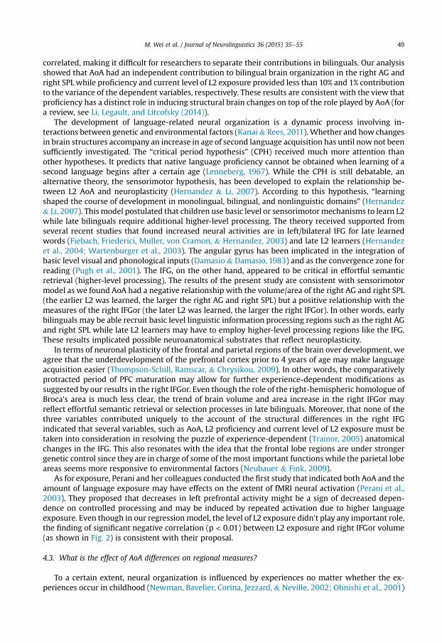
To further elucidate their influence on volume measures, separate scatter plots of volumes vs AoA,proficiency and exposure are provided in Fig. 2.
Fig. 2 shows that, in the right IFGor (a), both AoA and exposure are significantly correlated withtotal, grey andwhite volumemeasures. Positive correlations were found between AoA and the volumeswhile negative correlations occurred between exposure and the volumes. In other words, when the ageof non-native language acquisition increased, the volume measures of right IFGor increased and whenthe exposure to L2 increased, the volumes decreased (p < 0.01). For the two parietal regions, right AGand right SPL (b and c), out of three predictors, AoA played a crucial role in grey matter, white matterand total volumes with only one exception, that is, proficiency was positively associated with greymatter volume of the right SPL. Differently from the right IFGor of the frontal lobe, the two parietalareas showed negative correlations in terms of AoA and volumes measures, that is, when the age of L2acquisition increased, the grey matter, white matter and total volumes were smaller.
4. Discussion
Following the line of investigation of fMRI studies that focused on the AoA effect, we concentratedon structural variations. We were able to find significant and consistent effects using two differentwhole-brain analysis approaches (VBM and SBM). The results suggest that we had adequate statisticalpower to detect reliable effects. We observed that neuroanatomical structures differed as a function ofthe age of second language acquisition in bilingual brains. The analyses generated the following an-swers to our questions: (1) Where?Which brain structures are more susceptible to differences in AoA?Out of the 24 pre-specified cortical regions, we found that volumetric measures of the right AG andright SPL plus the cortical area of right SPL in the parietal lobe were reliably sensitive to the age ofsecond language acquisition. Our VBM analysis was consistent with this SBM finding. (2) How? How doAoA, proficiency and current level of exposure work together to shape bilingual neural representation?The bilingual brain organization is more dependent upon AoA in the right AG and SPL. In the rightIFGor, AoA, proficiency and current level of exposure were related to structural differences but none ofthem was significantly stronger than the other two factors. (3) What? What is the effect of AoA onstructural measures of brain structures? The earlier a non-native language was learned, the larger thevolumes/area of right AG and right SPL were. For the left AG, the trend of the effect was that the earliera non-native language was learned, the larger the left AG volume was. We discuss these findings in thefollowing paragraphs.
4.1. Areas showing AoA effect
In the parietal lobe SBM analysis, we found that the volume measures of the right AG and right SPLwere predicted by AoA, proficiency and exposure combined, and AoA as a predictor variable made astatistically significant contribution. Our cortical area data from SBM also supported the AoA's effect inthe right SPL. These findings are consistent with the VBM analysis which found negative correlationsbetween grey matter volume and AoA in a cluster located at the right AG extended to the SPL. Themultiple regression model of the left AG was statistically significant for total, grey and white matter

Fig. 2. Scatter plots of the association between the volumes of selected regions and AoA, proficiency, exposure. WM ¼ white matter; GM ¼ grey matter. From anterior to posterior regions: (a) Theright pars orbitalis. (b) The right angular gyrus. (c) The right superior parietal lobule (Correlation coefficient significance: p < 0.05*, p < 0.01**).
M.W
eiet
al./Journal
ofNeurolinguistics
36(2015)
35e55
47

M. Wei et al. / Journal of Neurolinguistics 36 (2015) 35e5548
volumes but not strong enough to survive the FDR correction. The angular gyrus is located in theposterior part of the inferior parietal lobule and is posterior to the supramarginal gyrus. The bilateralposterior parietal cortex was found to be involved in the conflict resolution network including the ACC,dorsolateral PFC, IFG, posterior parietal cortex, and anterior insula (Nee, Wager, & Jonides, 2007).Similarly, the bilateral posterior SPL was found to be significantly activated in an attention shifting task(Le, Pardo, & Hu, 1998). Gurd et al. (2002) proposed that the parietal lobe was involved in switchingbetween language tasks. Bilinguals experience switching between their two languages on a daily basis,so it is reasonable to anticipate the need in bilinguals to recruit the parietal lobe. AG's grey mattermaturation peaks between 8.5 and 13 years of age (Seghier, 2013). So thematuration peak of AG arrivesafter early bilinguals started to learn a second language (before 6 years old) and this may give the brainan opportunity to recruit additional resources (e.g., right AG). In early bilinguals, increased GM volume,WM volume, total volume and surface area relative to later bilinguals could correspond to greatercomputational power in the parietal cortex leading to a more efficient language-switching system andthus more efficient processing of two languages for early compared to late learners.
There are at least twoways to interpret the fact that the volumes of these right parietal regionsweremore strongly associated with AoA than the left parietal regions. Yoon, Fahim, Perusse, and Evans(2010) employed fifty-seven MZ (monozygotic) and 35 DZ (dizygotic) twin pairs and did MRI scanswhen they were 8 years old. It was found that genetic and environmental influences on individualhuman brain structural differences were lateralized, with the language-dominant left cerebral cortexunder stronger genetic control than the right. Therefore, the language-related regions in the righthemisphere might be more vulnerable to the type of the environmental changes that might be re-flected in AoA effects, as suggested by our results. Another way to interpret the recruitment of the rightparietal regions is that neural plasticity in the parietal lobes of bilinguals largely depends on brainmaturation. When brain resources are relatively limited during early bilingual learning, the right ho-mologous language structures may be recruited, suggesting a compensatory role for right hemisphereregions while the left regions have to be used under any context. Some researchers have proposed thatbilinguals had more overall right-hemisphere involvement for L2 and therefore weaker languagelateralization compared to monolinguals (Dehaene et al., 1997; Park, Badzakova-Trajkov, & Waldie,2012). Furthermore, as pointed out by Hernandez (2009) “it is very interesting that L1 effects appear asleft lateralized whereas L2 effects appear as right lateralized”. Existing evidence converge on the samepoint that being a bilingual, especially an early bilingual, may recruit more right hemisphere regionsduring language tasks and thus some right cortical regions may readily manifest the AoA effect. Theseexplanations are admittedly post hoc in the case of our results, but merit further investigation.
We found that themultiple regressionmodel of the left AGwas statistically significant for total, greyand white matter volumes but not strong enough to survive the FDR correction. The earlier a non-native language was learned, the larger the volume in left AG. From evolutionary perspective, wespeculated that the reason of a generally weaker correlation of AoA and left AG volumes was becausethat the left hemisphere played a more important role in language and it may have to maintain itsinvariance structurally across various environmental conditions. However, this claim will requirefuture research to support.
4.2. How do AoA, proficiency and current level of L2 exposure work together to shape bilingual brains?
Learning two languages simultaneously is a challenging task and thus may require taking fulladvantage of neural plasticity during childhood in early bilinguals (Peng & Wang, 2011). However, thecerebral representation of languages has been debated for decades. It seems that more researchers areleaning towards the conclusion that L1 and L2 share the same classical cortical regions, although braindifferences have also been found. These differences arise from multiple factors in bilinguals, such asproficiency, AoA, current L2 exposure level, etc. In a meta-analysis study, Sebastian et al. (2011)concluded that highly proficient bilinguals showed similar activation patterns in both languages,while low-medium-proficient bilinguals showed more widely distributed activation patterns whenperforming a language task in L2 compared to L1. Similar to what has been found for low proficiencybilinguals, thosewho acquired L2 later in life showed a greater amount of activation for L2 [for a reviewsee Higby et al. (2013)]. Proficiency and AoA are two important factors that are almost always highly
M. Wei et al. / Journal of Neurolinguistics 36 (2015) 35e55 49
correlated, making it difficult for researchers to separate their contributions in bilinguals. Our analysisshowed that AoA had an independent contribution to bilingual brain organization in the right AG andright SPL while proficiency and current level of L2 exposure provided less than 10% and 1% contributionto the variance of the dependent variables, respectively. These results are consistent with the view thatproficiency has a distinct role in inducing structural brain changes on top of the role played by AoA (fora review, see Li, Legault, and Litcofsky (2014)).
The development of language-related neural organization is a dynamic process involving in-teractions between genetic and environmental factors (Kanai& Rees, 2011). Whether and how changesin brain structures accompany an increase in age of second language acquisition has until now not beensufficiently investigated. The “critical period hypothesis” (CPH) received much more attention thanother hypotheses. It predicts that native language proficiency cannot be obtained when learning of asecond language begins after a certain age (Lenneberg, 1967). While the CPH is still debatable, analternative theory, the sensorimotor hypothesis, has been developed to explain the relationship be-tween L2 AoA and neuroplasticity (Hernandez & Li, 2007). According to this hypothesis, “learningshaped the course of development in monolingual, bilingual, and nonlinguistic domains” (Hernandez& Li, 2007). This model postulated that children use basic level or sensorimotor mechanisms to learn L2while late bilinguals require additional higher-level processing. The theory received supported fromseveral recent studies that found increased neural activities are in left/bilateral IFG for late learnedwords (Fiebach, Friederici, Muller, von Cramon, & Hernandez, 2003) and late L2 learners (Hernandezet al., 2004; Wartenburger et al., 2003). The angular gyrus has been implicated in the integration ofbasic level visual and phonological inputs (Damasio & Damasio, 1983) and as the convergence zone forreading (Pugh et al., 2001). The IFG, on the other hand, appeared to be critical in effortful semanticretrieval (higher-level processing). The results of the present study are consistent with sensorimotormodel as we found AoA had a negative relationship with the volume/area of the right AG and right SPL(the earlier L2 was learned, the larger the right AG and right SPL) but a positive relationship with themeasures of the right IFGor (the later L2 was learned, the larger the right IFGor). In other words, earlybilinguals may be able recruit basic level linguistic information processing regions such as the right AGand right SPL while late L2 learners may have to employ higher-level processing regions like the IFG.These results implicated possible neuroanatomical substrates that reflect neuroplasticity.
In terms of neuronal plasticity of the frontal and parietal regions of the brain over development, weagree that the underdevelopment of the prefrontal cortex prior to 4 years of age may make languageacquisition easier (Thompson-Schill, Ramscar, & Chrysikou, 2009). In other words, the comparativelyprotracted period of PFC maturation may allow for further experience-dependent modifications assuggested by our results in the right IFGor. Even though the role of the right-hemispheric homologue ofBroca's area is much less clear, the trend of brain volume and area increase in the right IFGor mayreflect effortful semantic retrieval or selection processes in late bilinguals. Moreover, that none of thethree variables contributed uniquely to the account of the structural differences in the right IFGindicated that several variables, such as AoA, L2 proficiency and current level of L2 exposure must betaken into consideration in resolving the puzzle of experience-dependent (Trainor, 2005) anatomicalchanges in the IFG. This also resonates with the idea that the frontal lobe regions are under strongergenetic control since they are in charge of some of the most important functions while the parietal lobeareas seems more responsive to environmental factors (Neubauer & Fink, 2009).
As for exposure, Perani and her colleagues conducted the first study that indicated both AoA and theamount of language exposure may have effects on the extent of fMRI neural activation (Perani et al.,2003). They proposed that decreases in left prefrontal activity might be a sign of decreased depen-dence on controlled processing and may be induced by repeated activation due to higher languageexposure. Even though in our regressionmodel, the level of L2 exposure didn't play any important role,the finding of significant negative correlation (p < 0.01) between L2 exposure and right IFGor volume(as shown in Fig. 2) is consistent with their proposal.
4.3. What is the effect of AoA differences on regional measures?
To a certain extent, neural organization is influenced by experiences no matter whether the ex-periences occur in childhood (Newman, Bavelier, Corina, Jezzard, & Neville, 2002; Ohnishi et al., 2001)

M. Wei et al. / Journal of Neurolinguistics 36 (2015) 35e5550
or in adulthood (Draganski et al., 2004; Maguire et al., 2000).We originally hypothesized that when thesecond language was acquired early in life, larger cortical volume in specific regions would beobserved.
However, consistent with by Li et al. (2014), we again found different relationships between AoAand brain regional measures: AoA had a positive relationship with the volume/area of right IFGor but anegative relationship with the volume/area of right AG and right SPL. We know that the brain follows a“back to front” rule when it matures (Gogtay et al., 2004). That is, growth in cortical regions occursearlier in posterior than anterior cortical regions. Peak thickness in occipital and parietal regions isattained at about 7e10 years of age, whereas the peak thickness in frontal and temporal regions isreached at 10e14 years of age (Shaw et al., 2008). Therefore, inspired by a recent paper (Archila-Suerte,Zevin, Bunta, & Hernandez, 2012), we propose that the reason for the different directions of rela-tionship between ROI volume/area and AoA is that when the non-native language is acquired early inlife the regions that reach maturity early in life, such as the AG and SPL in the parietal lobe, are morelikely to be recruited. In contrast, if the non-native language is acquired late in life the regions thatreach maturity late in life such as the pars orbitalis in the IFG may be recruited. In other words, brainregions may become maximally available for learning new skills at different points of time during thematuration process. This may be a way for the brain to maximize learning resources and minimize thecost in terms of energy consumption.
The changes in brain morphometry following training in prior studies are not restricted to greymatter but involve white matter as well (May, 2011). Changes in grey matter volume are in accordancewith white matter volume changes. Draganski et al. (2006) found a decrease of grey matter in thebilateral occipito-parietal lobe accompanied by a proportional increase of white matter in this regionbetween scans taken 3 months before and right after participants took a sophisticated medical exam.The findings of Draganski et al. need to be viewed with caution since the VBMmethod they used is notvery sensitive for detecting changes in white matter.
Additionally, more fine-grained methods such as microscopy are necessary to clarify the under-lying cellular changes that support the macroscopic alternations in cortical volumes/area. Somepossibilities are: changes in neuron size or glial cell size, and changes in synapse formation andelimination (Chklovskii, Mel, & Svoboda, 2004; Draganski et al., 2004; Draganski &May, 2008). Futuremulti-model imaging research needs to address the relationship between cellular changes and thecomputational capacity of cortical regions [see Zatorre, Fields, and Johansen-Berg (2012)]. A detailedexamination of our results in Fig. 2 could reveal two important findings: one is that the relationshipbetween AoA and the grey matter/white matter volume always has the same direction with the totalvolume, meaning that when there is a positive relationship between AoA and total volume of aspecific region, this relationship holds for both grey matter and white matter volume. Another pro-posal is that reduction in grey matter volume reflects synaptic and/or neuronal pruning processes andmay lead to more efficient processing (Kanai & Rees, 2011). However, our results were not consistentwith this hypothesis.
4.4. VBM vs. SBM
Previous studies suggested that VBM and SBM would produce similar results (Cerasa et al., 2011;Lehmann et al., 2011). Even though VBM is rapidly becoming the dominant method, it is not areplacement of ROI based analysis (Giuliani, Calhoun, Pearlson, Francis, & Buchanan, 2005). Given thatmanual ROI delineation is extremely time-consuming, our semi-automatic SBM procedure couldprovide another choice.
We are aware that the volume differences detected by whole-brain VBM analysis would not survivestringent multiple comparison correction. However, it still provided us with useful information. It ispossible that our hypothesis-driven SBM analyses reflect type I errors. Yet, with the prior FSL-VBMresults at uncorrected p ¼ 0.01 level and in light of our hypotheses concerning the language controlnetwork, we have maximized the possibility that these results are rather from legitimate brainstructure and AoA relationships. As manifested in our results, VBM and SBM might provide differenttype of information and should be conducted concurrently (Giuliani et al., 2005).

M. Wei et al. / Journal of Neurolinguistics 36 (2015) 35e55 51
5. Conclusions
In summary, the present results show that volumetric and area measures of the right IFGor arelinked to differences in AoA, proficiency and exposure. In the left counterpart of this region, the left IFG,functional activation is known to be broader when AoA increases. The structural differences in the rightIFG seen here macroscopically using MRI might reflect the recruitment of additional resources to allowprocessing of two languages when the non-native language is acquired relatively late in life. The totalandwhitematter volumes of the right AGwere reliably predicted by AoA, while AoA served as themostsignificant predictor for the total volume, grey matter volume and surface area of the right SPL. Thisfinding, similarly, may reflect the need to recruit additional resources in the right parietal lobe early inlife when presented with two languages. The current study suggests that the structure of the humanbrain is reworked by the experience of acquiring a non-native language and when consideringstructural changes in bilingual brains, AoA, proficiency and exposure level should all be taken intoconsideration.
One important limitation is that the nature of our study design did not allow us to separate effects ofgenetic predisposition on structural changes from structural plasticity induced by bilingual experience.It could be that inherited factors simultaneously account for individual differences in cortical volumes/thickness/area and the ability to learn a second language. Thus, further longitudinal, experimental orgenetically informed studies are necessary to clarify the role of experience-dependent structuralplasticity in second language acquisition (Richardson & Price, 2009).
In terms of future directions, it is often assumed that experience-based brain plasticity is limited tofunctional changes instead of structural changes. Studies have illustrated that structural changesusually correspond to functional task-related activations, which corroborates a close relationship be-tween structure and function (Li et al., 2014; Richardson & Price, 2009). We suggest that linking fMRIfindings with anatomical findings might be a rich area for further research. The cellular basis under-lying structural MRI findings remains to be investigated.
Acknowledgments
This work was supported by grants from the National Science Foundation (BCS-0823495 & BCS-0823624) and the National Institute of Child Health and Development (HD057884). We thank CorianneRogalsky (C.R.) and So Young Choi (S.Y. C.) for their help with brain anatomy and curve delineation.
Appendix
Abbreviation Brain region
ACC Anterior cingulate cortexAG Angular gyrusIFG Inferior frontal gyrusIFGor Inferior frontal gyrus pars orbitalisIFGop Inferior frontal gyrus pars opercularisIFGpt Inferior frontal gyrus pars triangularisIPL Inferior parietal lobuleMFG Middle frontal gyrusPFC Prefrontal cortexPre-SMA Pre-supplementary motor areaSMG Supramarginal gyrusSPL Superior parietal lobuleSTG Superior temporal gyrus

M. Wei et al. / Journal of Neurolinguistics 36 (2015) 35e5552
References
Abdul-Kareem, I. A., Stancak, A., Parkes, L. M., & Sluming, V. (2011). Increased gray matter volume of left pars opercularis in maleorchestral musicians correlate positively with years of musical performance. Journal of Magnetic Resonance Imaging, 33,24e32.
Abutalebi, J. (2008). Neural aspects of second language representation and language control. Acta Psychologica, 128, 466e478.Abutalebi, J., Della Rosa, P. A., Gonzaga, A. K., Keim, R., Costa, A., & Perani, D. (2013). The role of the left putamen in multilingual
language production. Brain and Language, 125, 307e315.Andersson, J. L., Jenkinson, M., & Smith, S. (2007). Non-linear registration aka Spatial normalisation FMRIB technical report
TR07JA2. FMRIB Analysis Group of the University of Oxford.Archila-Suerte, P., Zevin, J., Bunta, F., & Hernandez, A. E. (2012). Age of acquisition and proficiency in a second language
independently influence the perception of non-native speech. Bilingualism: Language and Cognition, 15, 190e201.Badzakova-Trajkov, G., Kirk, I. J., & Waldie, K. E. (2008). Dual-task performance in late proficient bilinguals. Laterality, 13,
201e216.Birdsong, D. (2005). Interpreting age effects in second language acquisition. In Handbook of bilingualism: Psycholinguistic ap-
proaches (pp. 109e127). New York: Oxford University Press.Bloch, C., Kaiser, A., Kuenzli, E., Zappatore, D., Haller, S., Franceschini, R., et al. (2009). The age of second language acquisition
determines the variability in activation elicited by narration in three languages in Broca's and Wernicke's area. Neuro-psychologia, 47, 625e633.
Buckner, R. L., Head, D., Parker, J., Fotenos, A. F., Marcus, D., Morris, J. C., et al. (2004). A unified approach for morphometric andfunctional data analysis in young, old, and demented adults using automated atlas-based head size normalization: reli-ability and validation against manual measurement of total intracranial volume. Neuroimage, 23, 724e738.
Canseco-Gonzalez, E., Brehm, L., Brick, C. A., Brown-Schmidt, S., Fischer, K., & Wagner, K. (2010). Carpet or Carcel: the effect ofage of acquisition and language mode on bilingual lexical access. Language and Cognitive Processes, 25, 669e705.
Cerasa, A., Quattrone, A., Gioia, M. C., Tarantino, P., Annesi, G., Assogna, F., et al. (2011). Dysbindin C-A-T haplotype is associatedwith thicker medial orbitofrontal cortex in healthy population. Neuroimage, 55, 508e513.
Chee, M. W., Hon, N., Lee, H. L., & Soon, C. S. (2001). Relative language proficiency modulates BOLD signal change when bi-linguals perform semantic judgments. Blood oxygen level dependent. Neuroimage, 13, 1155e1163.
Chee, M. W., Soon, C. S., Lee, H. L., & Pallier, C. (2004). Left insula activation: a marker for language attainment in bilinguals.Proceedings of the National Academy of Sciences of the United States of America, 101, 15265e15270.
Chee, M. W., Zheng, H., Goh, J. O., Park, D., & Sutton, B. P. (2011). Brain structure in young and old East Asians and Westerners:comparisons of structural volume and cortical thickness. Journal of Cognitive Neuroscience, 23, 1065e1079.
Chklovskii, D. B., Mel, B. W., & Svoboda, K. (2004). Cortical rewiring and information storage. Nature, 431, 782e788.Consonni, M., Cafiero, R., Marin, D., Tettamanti, M., Iadanza, A., Fabbro, F., et al. (2013). Neural convergence for language
comprehension and grammatical class production in highly proficient bilinguals is independent of age of acquisition.Cortex: A Journal Devoted to the Study of the Nervous System and Behavior, 49, 1252e1258.
Damasio, A. R., & Damasio, H. (1983). The anatomic basis of pure alexia. Neurology, 33, 1573e1583.De Bleser, R., Dupont, P., Postler, J., Bormans, G., Speelman, D., Mortelmans, L., et al. (2003). The organisation of the bilingual
lexicon: a PET study. Journal of Neurolinguistics, 16, 439e456.Dehaene, S., Dupoux, E., Mehler, J., Cohen, L., Paulesu, E., Perani, D., et al. (1997). Anatomical variability in the cortical repre-
sentation of first and second language. Neuroreport, 8, 3809e3815.DeKeyser, R., & Larson-Hall, J. (2005). What does the critical period really mean?. In Handbook of bilingualism: Psycholinguistic
approaches (pp. 88e108).Della Rosa, P. A., Videsott, G., Borsa, V. M., Canini, M., Weekes, B. S., Franceschini, R., et al. (2013). A neural interactive location for
multilingual talent. Cortex, 49, 605e608.Desikan, R. S., Segonne, F., Fischl, B., Quinn, B. T., Dickerson, B. C., Blacker, D., et al. (2006). An automated labeling system for
subdividing the human cerebral cortex on MRI scans into gyral based regions of interest. Neuroimage, 31, 968e980.Dogdas, B., Shattuck, D. W., & Leahy, R. M. (2005). Segmentation of skull and scalp in 3-D human MRI using mathematical
morphology. Human Brain Mapping, 26, 273e285.Draganski, B., Gaser, C., Busch, V., Schuierer, G., Bogdahn, U., & May, A. (2004). Neuroplasticity: changes in grey matter induced
by training. Nature, 427, 311e312.Draganski, B., Gaser, C., Kempermann, G., Kuhn, H. G., Winkler, J., Buchel, C., et al. (2006). Temporal and spatial dynamics of
brain structure changes during extensive learning. The Journal of Neuroscience, 26, 6314e6317.Draganski, B., & May, A. (2008). Training-induced structural changes in the adult human brain. Behavioural Brain Research, 192,
137e142.Duffau, H. (2006). Brain plasticity: from pathophysiological mechanisms to therapeutic applications. Journal of Clinical
Neuroscience, 13, 885e897.Duffau, H. (2014). The huge plastic potential of adult brain and the role of connectomics: new insights provided by serial
mappings in glioma surgery. Cortex, 58, 325e337.Elston-Guttler, K. E., Paulmann, S., & Kotz, S. A. (2005). Who's in control? Proficiency and L1 influence on L2 processing. Journal
of Cognitive Neuroscience, 17, 1593e1610.Fiebach, C. J., Friederici, A. D., Muller, K., von Cramon, D. Y., & Hernandez, A. E. (2003). Distinct brain representations for early
and late learned words. Neuroimage, 19, 1627e1637.Fiez, J. A. (1997). Phonology, semantics, and the role of the left inferior prefrontal cortex. Human Brain Mapping, 5, 79e83.Frenck-Mestre, C., Anton, J. L., Roth, M., Vaid, J., & Viallet, F. (2005). Articulation in early and late bilinguals' two languages:
evidence from functional magnetic resonance imaging. Neuroreport, 16, 761e765.Genovese, C. R., Lazar, N. A., & Nichols, T. (2002). Thresholding of statistical maps in functional neuroimaging using the false
discovery rate. Neuroimage, 15, 870e878.

M. Wei et al. / Journal of Neurolinguistics 36 (2015) 35e55 53
Giuliani, N. R., Calhoun, V. D., Pearlson, G. D., Francis, A., & Buchanan, R. W. (2005). Voxel-based morphometry versus region ofinterest: a comparison of two methods for analyzing gray matter differences in schizophrenia. Schizophrenia Research, 74,135e147.
Gogtay, N., Giedd, J. N., Lusk, L., Hayashi, K. M., Greenstein, D., Vaituzis, A. C., et al. (2004). Dynamic mapping of human corticaldevelopment during childhood through early adulthood. Proceedings of the National Academy of Sciences of the United Statesof America, 101, 8174e8179.
Good, C. D., Johnsrude, I. S., Ashburner, J., Henson, R. N., Friston, K. J., & Frackowiak, R. S. (2001). A voxel-based morphometricstudy of ageing in 465 normal adult human brains. Neuroimage, 14, 21e36.
Green, D. W., & Abutalebi, J. (2013). Language control in bilinguals: the adaptive control hypothesis. Journal of Cognitive Psy-chology, 1e16.
Grogan, A., Parker, J., Ali, N., Crinion, J., Orabona, S., Mechias, M. L., et al. (2012). Structural correlates for lexical efficiency andnumber of languages in non-native speakers of English. Neuropsychologia, 50, 1347e1352.
Gurd, J. M., Amunts, K., Weiss, P. H., Zafiris, O., Zilles, K., Marshall, J. C., et al. (2002). Posterior parietal cortex is implicated incontinuous switching between verbal fluency tasks: an fMRI study with clinical implications. Brain, 125, 1024e1038.
Hernandez, A. E. (2009). Language switching in the bilingual brain: what's next? Brain and Language, 109, 133e140.Hernandez, A. E., Hofmann, J., & Kotz, S. A. (2007). Age of acquisition modulates neural activity for both regular and irregular
syntactic functions. Neuroimage, 36, 912e923.Hernandez, A. E., Kotz, S. A., Hofmann, J., Valentin, V. V., Dapretto, M., & Bookheimer, S. Y. (2004). The neural correlates of
grammatical gender decisions in Spanish. Neuroreport, 15, 863e866.Hernandez, A. E., & Li, P. (2007). Age of acquisition: its neural and computational mechanisms. Psychological Bulletin, 133,
638e650.Higby, E., Kim, J., & Obler, L. K. (2013). Multilingualism and the brain. Annual Review of Applied Linguistics, 33, 68e101.Hull, R., & Vaid, J. (2007). Bilingual language lateralization: a meta-analytic tale of two hemispheres. Neuropsychologia, 45,
1987e2008.Indefrey, P. (2006). A meta-analysis of hemodynamic studies on first and second language processing: which suggested dif-
ferences can we trust and what do they mean? Language Learning, 56, 279e304.Isel, F., Baumgaertner, A., Thr€an, J., Meisel, J. M., & Büchel, C. (2010). Neural circuitry of the bilingual mental lexicon: effect of age
of second language acquisition. Brain and Cognition, 72, 169e180.Jeong, H., Sugiura, M., Sassa, Y., Haji, T., Usui, N., Taira, M., et al. (2007). Effect of syntactic similarity on cortical activation during
second language processing: a comparison of English and Japanese among native Korean trilinguals. Human Brain Mapping,28, 194e204.
Jia, G., Aaronson, D., & Wu, Y. (2002). Long-term language attainment of bilingual immigrants: predictive variables and languagegroup differences. Applied Psycholinguistics, 23, 599e621.
Joshi, A. A., Pantazis, D., Li, Q., Damasio, H., Shattuck, D. W., Toga, A. W., et al. (2010). Sulcal set optimization for cortical surfaceregistration. Neuroimage, 50, 950e959.
Joshi, A. A., Shattuck, D. W., & Leahy, R. M. (2012). A fast and accurate method for automated cortical surface registration andlabeling. In Proc. WBIR LNCS Springer (pp. 180e189).
Joshi, A., Shattuck, D., Pantazis, D., Li, Q. Z., Damasio, H., Leahy, R., et al. (2009). Optimization of landmark selection for corticalsurface registration. In Cvpr: 2009 IEEE Conference on Computer Vision and Pattern Recognition (Vols. 1e4, pp. 699e706). NewYork: IEEE.
Joshi, A., Shattuck, D., Thompson, P., & Leahy, R. (2007). Brain image registration using cortically constrained harmonic map-pings. Information Processing in Medical Imaging, 20, 359e371.
Kanai, R., & Rees, G. (2011). The structural basis of inter-individual differences in human behaviour and cognition. Nature Re-views Neuroscience, 12, 231e242.
Kim, J.-H., Lee, J.-M., Jo, H. J., Kim, S. H., Lee, J. H., Kim, S. T., et al. (2010). Defining functional SMA and pre-SMA subregions inhuman MFC using resting state fMRI: functional connectivity-based parcellation method. Neuroimage, 49, 2375e2386.
Kim, K. H., Relkin, N. R., Lee, K. M., & Hirsch, J. (1997). Distinct cortical areas associated with native and second languages.Nature, 388, 171e174.
Klein, D., Mok, K., Chen, J. K., & Watkins, K. E. (2014). Age of language learning shapes brain structure: a cortical thickness studyof bilingual and monolingual individuals. Brain and Language, 131, 20e24.
Klein, D., Zatorre, R. J., Chen, J. K., Milner, B., Crane, J., Belin, P., et al. (2006). Bilingual brain organization: a functional magneticresonance adaptation study. Neuroimage, 31, 366e375.
Kovelman, I., Baker, S. A., & Petitto, L.-A. (2008). Age of first bilingual language exposure as a new window into bilingual readingdevelopment. Bilingualism: Language and Cognition, 11, 203e223.
Le, T. H., Pardo, J. V., & Hu, X. (1998). 4 T-fMRI study of nonspatial shifting of selective attention: cerebellar and parietal con-tributions. Journal of Neurophysiology, 79, 1535e1548.
Lehmann, M., Crutch, S. J., Ridgway, G. R., Ridha, B. H., Barnes, J., Warrington, E. K., et al. (2011). Cortical thickness and voxel-based morphometry in posterior cortical atrophy and typical Alzheimer's disease. Neurobiology of Aging, 32, 1466e1476.
Lenneberg, E. H. (1967). Biological foundations of language. New York: Wiley (Chapter).Li, P., Legault, J., & Litcofsky, K. A. (2014). Neuroplasticity as a function of second language learning: anatomical changes in the
human brain. Cortex, 58, 301e324.Maguire, E. A., Gadian, D. G., Johnsrude, I. S., Good, C. D., Ashburner, J., Frackowiak, R. S., et al. (2000). Navigation-related
structural change in the hippocampi of taxi drivers. Proceedings of the National Academy of Sciences of the United States ofAmerica, 97, 4398e4403.
Mahendra, N., Plante, E., Magloire, J., Milman, L., & Trouard, T. P. (2003). FMRI variability and the localization of languages in thebilingual brain. Neuroreport, 14, 1225e1228.
Mårtensson, J., Eriksson, J., Bodammer, N. C., Lindgren, M., Johansson, M., Nyberg, L., et al. (2012). Growth of language-relatedbrain areas after foreign language learning. Neuroimage, 63, 240.
May, A. (2011). Experience-dependent structural plasticity in the adult human brain. Trends in Cognitive Sciences, 15, 475e482.

M. Wei et al. / Journal of Neurolinguistics 36 (2015) 35e5554
Mayberry, R. I., Chen, J. K., Witcher, P., & Klein, D. (2011). Age of acquisition effects on the functional organization of language inthe adult brain. Brain and Language, 119, 16e29.
Mechelli, A., Crinion, J., Noppeney, U., Doherty, J., Ashburner, J., Frackowiak, R. S., et al. (2004). Structural plasticity in thebilingual brain e proficiency in a second language and age at acquisition affect grey-matter density. Nature, 431.
Musso, M., Moro, A., Glauche, V., Rijntjes, M., Reichenbach, J., Buchel, C., et al. (2003). Broca's area and the language instinct.Nature Neuroscience, 6, 774e781.
Nee, D. E., Wager, T. D., & Jonides, J. (2007). Interference resolution: insights from a meta-analysis of neuroimaging tasks.Cognitive, Affective, & Behavioral Neuroscience, 7, 1e17.
Neubauer, A. C., & Fink, A. (2009). Intelligence and neural efficiency. Neuroscience & Biobehavioral Reviews, 33, 1004e1023.Newman, A. J., Bavelier, D., Corina, D., Jezzard, P., & Neville, H. J. (2002). A critical period for right hemisphere recruitment in
American Sign Language processing. Nature Neuroscience, 5, 76e80.Ohnishi, T., Matsuda, H., Asada, T., Aruga, M., Hirakata, M., Nishikawa, M., et al. (2001). Functional anatomy of musical
perception in musicians. Cerebral Cortex (New York, N.Y.: 1991), 11, 754e760.Oldfield, R. C. (1971). The assessment and analysis of handedness: the Edinburgh inventory. Neuropsychologia, 9, 97e113.Pantazis, D., Joshi, A., Jiang, J., Shattuck, D. W., Bernstein, L. E., Damasio, H., et al. (2010). Comparison of landmark-based and
automatic methods for cortical surface registration. Neuroimage, 49, 2479e2493.Park, H. R., Badzakova-Trajkov, G., & Waldie, K. E. (2012). Language lateralisation in late proficient bilinguals: a lexical decision
fMRI study. Neuropsychologia, 50, 688e695.Peng, G., & Wang, W. S. Y. (2011). Hemisphere lateralization is influenced by bilingual status and composition of words. Neu-
ropsychologia, 49, 1981e1986.Perani, D., & Abutalebi, J. (2005). The neural basis of first and second language processing. Current Opinion in Neurobiology, 15,
202e206.Perani, D., Abutalebi, J., Paulesu, E., Brambati, S., Scifo, P., Cappa, S. F., et al. (2003). The role of age of acquisition and language
usage in early, high-proficient bilinguals: an fMRI study during verbal fluency. Human Brain Mapping, 19, 170e182.Perani, D., Paulesu, E., Galles, N. S., Dupoux, E., Dehaene, S., Bettinardi, V., et al. (1998). The bilingual brain e proficiency and age
of acquisition of the second language. Brain, 121, 1841e1852.Poldrack, R. A. (2007). Region of interest analysis for fMRI. Social Cognitive and Affective Neuroscience, 2, 67e70.Pugh, K. R., Mencl, W. E., Jenner, A. R., Katz, L., Frost, S. J., Lee, J. R., et al. (2001). Neurobiological studies of reading and reading
disability. Journal of Communication Disorders, 34, 479e492.Ressel, V., Pallier, C., Ventura-Campos, N., Díaz, B., Roessler, A., �Avila, C., et al. (2012). An effect of bilingualism on the auditory
cortex. The Journal of Neuroscience, 32, 16597e16601.Richardson, F. M., & Price, C. J. (2009). Structural MRI studies of language function in the undamaged brain. Brain Structure and
Function, 213, 511e523.Sebastian, R., Laird, A. R., & Kiran, S. (2011). Meta-analysis of the neural representation of first language and second language.
Applied Psycholinguistics, 32, 799e819.Seghier, M. L. (2013). The angular gyrus: multiple functions and multiple subdivisions. Neuroscientist, 19, 43e61.Shattuck, D. W., Joshi, A. A., Pantazis, D., Kan, E., Dutton, R. A., Sowell, E. R., et al. (2009). Semi-automated method for delineation
of landmarks on models of the cerebral cortex. Journal of Neuroscience Methods, 178, 385e392.Shattuck, D. W., & Leahy, R. M. (2002). BrainSuite: an automated cortical surface identification tool. Medical Image Analysis, 6,
129e142.Shattuck, D. W., Sandor-Leahy, S. R., Schaper, K. A., Rottenberg, D. A., & Leahy, R. M. (2001). Magnetic resonance image tissue
classification using a partial volume model. Neuroimage, 13, 856e876.Shaw, P., Kabani, N. J., Lerch, J. P., Eckstrand, K., Lenroot, R., Gogtay, N., et al. (2008). Neurodevelopmental trajectories of the
human cerebral cortex. The Journal of Neuroscience, 28, 3586e3594.Storey, J. D. (2002). A direct approach to false discovery rates. Journal of the Royal Statistical Society: Series B (Statistical Meth-
odology), 64, 479e498.Thompson-Schill, S. L., Ramscar, M., & Chrysikou, E. G. (2009). Cognition without control: when a little frontal lobe goes a long
way. Current Directions in Psychological Science, 18, 259e263.Trainor, L. J. (2005). Are there critical periods for musical development? Developmental Psychobiology, 46, 262e278.Vaid, J. (1983). Bilingualism and brain lateralization. In S. Segalowitz (Ed.), Language function with brain organization (pp.
315e339). New York: Academic Press.Vingerhoets, G., Van Borsel, J., Tesink, C., van den Noort, M., Deblaere, K., Seurinck, R., et al. (2003). Multilingualism: an fMRI
study. Neuroimage, 20, 2181e2196.Waldron, E. J., & Hernandez, A. E. (2013). The role of age of acquisition on past tense generation in Spanish-English bilinguals: an
fMRI study. Brain and Language, 125, 28e37.Wang, Y., Xiang, J., Vannest, J., Holroyd, T., Narmoneva, D., Horn, P., et al. (2011). Neuromagnetic measures of word processing in
bilinguals and monolinguals. Clinical Neurophysiology, 122, 1706e1717.Wartenburger, I., Heekeren, H. R., Abutalebi, J., Cappa, S. F., Villringer, A., & Perani, D. (2003). Early setting of grammatical
processing in the bilingual brain. Neuron, 37, 159e170.Wattendorf, E., & Festman, J. (2008). Images of the multilingual brain: the effect of age of second language acquisition. Annual
Review of Applied Linguistics, 28, 3e24.Weber-Fox, C. M., & Neville, H. J. (1996). Maturational constraints on functional specializations for language processing: ERP and
behavioral evidence in bilingual speakers. Journal of Cognitive Neuroscience, 8, 231e256.Wible, C. G., Shenton, M. E., Hokama, H., Kikinis, R., Jolesz, F. A., Metcalf, D., et al. (1995). Prefrontal cortex and schizophrenia: a
quantitative magnetic resonance imaging study. Archives of General Psychiatry, 52, 279e288.Woods, R. P., Grafton, S. T., Holmes, C. J., Cherry, S. R., & Mazziotta, J. C. (1998). Automated image registration: I. General methods
and intrasubject, intramodality validation. Journal of Computer Assisted Tomography, 22, 139e152.Woollett, K., & Maguire, E. A. (2011). Acquiring “the Knowledge” of London's layout drives structural brain changes. Current
Biology, 21, 2109e2114.

M. Wei et al. / Journal of Neurolinguistics 36 (2015) 35e55 55
Yoon, U., Fahim, C., Perusse, D., & Evans, A. C. (2010). Lateralized genetic and environmental influences on human brainmorphology of 8-year-old twins. Neuroimage, 53, 1117e1125.
Zatorre, R. J., Fields, R. D., & Johansen-Berg, H. (2012). Plasticity in gray and white: neuroimaging changes in brain structureduring learning. Nature Neuroscience, 15, 528e536.
Zou, L., Ding, G., Abutalebi, J., Shu, H., & Peng, D. (2012). Structural plasticity of the left caudate in bimodal bilinguals. Cortex, 48,1197e1206.