hum. reprod.-2003-kroon-2323-7
DESCRIPTION
Hum. Reprod.-2003-Kroon-2323-7TRANSCRIPT

The value of transvaginal ultrasound to monitor theposition of an intrauterine device after insertion. Atechnology assessment study
C.D.de Kroon, J.C.van Houwelingen, J.B.Trimbos and F.W.Jansen1
Departments of Gynaecology and Medical Statistics, Leiden University Medical Center, P.O.Box 9600, 2300 RC Leiden,
The Netherlands
1To whom correspondence should be addressed. E-mail: [email protected]
BACKGROUND: The intrauterine device (IUD) is an effective contraceptive method. The contraceptive power as
well as the side-effects of IUD are thought to relate to the position of the IUD in the uterine cavity. We assessed the
accuracy of clinical evaluation of IUD position. METHODS: A prospective comparative study was performed. The
clinical evaluation was compared with the TVU measurement of IUD position both immediately after insertion and
6 weeks after insertion. The primary outcome measures were the positive and negative predictive values (PPV and
NPV) of the clinical evaluation of IUD position. RESULTS: 195 women were included consecutively, 181 women
(92.8%) were available for follow-up. The PPV and NPV of clinical evaluation of IUD position immediately after
insertion were respectively 0.60 (95% CI: 0.39±0.81) and 0.98 (95% CI: 0.96±1.0). The prevalence of an abnormally
positioned IUD was 7.7% (95% CI: 3.9±11.4). The PPV and NPV of the clinical evaluation at follow-up were
respectively 0.54 (95% CI: 0.26±0.81) and 1.0 (95% CI: 0.98±1.0). The prevalence of abnormal position was 4.0%
(95% CI: 1.7±7.1). CONCLUSION: Clinical evaluation is an excellent test for the evaluation of the position of an
IUD and routine TVU is not indicated for this purpose.
Key words: contraception/follow-up/gynaecological examination/intrauterine device/transvaginal ultrasound
Introduction
The intrauterine position of an intrauterine contraceptive
device (IUD) is thought to be closely related to its contracep-
tive power (Bernaschek et al., 1981; Tadesse et al., 1985;
Anteby et al., 1993). IUD located more cervically may prevent
conception to a lesser degree compared to adequately localized
IUD (Anteby et al., 1993). Therefore proper insertion of an
IUD is of utmost importance. Moreover, follow-up after the
®rst menses is recommended since the ®rst month is suggested
to be the period with the highest risk of downwards migration
and spontaneous expulsion (Anteby et al., 1993). The
gynaecological interview and pelvic examination, in combin-
ation with self-examination, can be used to ensure adequate
localization of the IUD (American College of Obstetricians and
Gynecologists, 1992). For the above reasons, insertion and
follow-up of IUD has been routine in general practice
(Harrison-Woolrych et al., 2002).
Currently, transvaginal ultrasound (TVU) is used increas-
ingly to complete the gynaecological examination in hospital
settings (Valentin, 1999). The routine use of TVU to monitor
the position of the IUD after insertion has been advocated
(Bonilla-Musoles et al., 1996). There is much evidence that
shows TVU to be highly accurate in monitoring the location of
any type of IUD (Petta et al., 1996; FauÂndes et al., 1997, 2000;
Palo, 1997; Aleem et al., 1992). The IUD±endometrium
distance (IUD±ED) seems to be the most relevant measure-
ment, especially for copper IUD (Petta et al., 1996; FauÂndes
et al., 1997, 2000). However, the maximum IUD±ED to ensure
adequate contraception is under debate (Petta et al., 1996;
FauÂndes et al., 1997), especially since T-shaped IUD tend to
accommodate in their position during the ®rst 3 months after
insertion (FauÂndes et al., 2000). Therefore removal of all
`abnormally located' IUD at TVU may result in a high number
of unnecessary removals (Petta et al., 1996; FauÂndes et al.,
2000).
The silent incorporation of routine TVU in gynaecology and
especially in IUD follow-up needs urgent evaluation (Valentin,
1999; Rivera and Best, 2002). Before adopting the routine use
of TVU for this purpose, and consequently transfer the
insertion of IUD from general practice to the gynaecological
outpatient department, the clinical value of this phenomenon
should be evaluated and guidelines adjusted accordingly. The
aim of this prospective study was to evaluate the clinical
relevance of the routine use of TVU immediately after and 6
weeks after the insertion of an IUD.
Human Reproduction Vol.18, No.11 pp. 2323±2327, 2003 DOI: 10.1093/humrep/deg433
Human Reproduction 18(11) ã European Society of Human Reproduction and Embryology 2003; all rights reserved 2323
by guest on Novem
ber 14, 2015http://hum
rep.oxfordjournals.org/D
ownloaded from

Materials and methods
All women who attended the outpatient Department of Gynaecology
of the Leiden University Medical Center for the insertion of an IUD
between July 1, 2001 and January 1, 2003 were eligible for this study,
regardless of type of IUD. After informed consent, women were
included and the IUD inserted according to our protocol. Pain
medication was not prescribed routinely while insertion was
preferably performed during or just after menses.
The clinician who inserted the IUD had to ®ll out a standardized
form with questions about the procedure: (i) whether the IUD was
thought to be located properly (adequate position) or erroneously
(indadequate position) inside the uterine cavity; and (ii) whether the
procedure was with or without complications and/or dif®culties.
Clinicians were divided into groups according to their experience
(respectively <25, 25±75 and >75 inserted IUD).
Immediately after the insertion of an IUD, an independent
sonographer who was unaware of the clinician's answers to the
above-mentioned questions performed TVU. A 5±7.5 MHz multi-
frequency vaginal probe and a Power Vision 6000 ultrasound machine
(Toshiba Medical Systems, The Netherlands) were used. With TVU,
the distance between the top of the vertical arm of the IUD and the
junction between the endometrium and the uterine cavity (IUD±ED)
was measured in the mid-longitudinal plane (Figure 1). Whenever this
junction could not be identi®ed clearly, the IUD±ED was calculated by
subtracting half of the double endometrial thickness from the distance
from the top of the IUD until the endo-myometrial junction (Figure 2)
(Petta et al., 1996; FauÂndes et al., 1997; 2000). The mean IUD±ED
was calculated from three independent measurements. In 20 non-
selected consecutive women, a reliability analysis was performed by
means of calculation of the intra-class correlation of two independent
IUD±ED measurements (Khan and Chien, 2001; Sackett and Haynes,
2002).
According to our protocol, all women had a follow-up visit 6 weeks
after insertion. At this visit, a standardized interview was taken
concerning complaints of the IUD with emphasis on abdominal pain
and cramping, bleeding disorders and complaints possibly due to the
threads of the IUD. A gynaecological examination was performed to
monitor the position of the IUD by estimating the length of the threads.
By this means, the position of the IUD was evaluated clinically.
Thereafter TVU was performed and the IUD±ED measured as
described above. The sonographer again was unaware of the results
of the interview and the gynaecological examination. Whenever the
IUD±ED was >5.0 mm, removal of the IUD was advised. All further
follow-up was independent of participation in the trial.
The predictive values and likelihood ratios with 95% con®dence
intervals (95% CI) of the clinical evaluation of IUD localization were
calculated using the IUD±ED as the gold standard. A multiregression
analysis was used to calculate the multivariate odds ratios (OR with
95% CI) of risk factors for inadequate insertion and inadequate IUD
position at follow-up. Multivariate OR (with 95% CI) were also
calculated for risk factors for improper clinical evaluation of the
position of the IUD both immediately after insertion and at follow-up.
Figure 1. A systematic representation and transvaginal scandisplaying the distance between the top of the intrauterine device(IUD) and the end of the uterine cavity: the IUD±endometriumdistance (IUD±ED).
Figure 2. A systematic representation and transvaginal scandisplaying the intrauterine device±endometrium distance (IUD±ED)calculated by subtracting half of the double endometrial thicknessfrom the distance between the IUD and the endo-myometrialjunction.
C.D.de Kroon et al.
2324
by guest on Novem
ber 14, 2015http://hum
rep.oxfordjournals.org/D
ownloaded from

Student's t-test (continuous data), c2-test and Fisher's exact test
(categorical data) were used to compare groups; the level of
signi®cance was set at P < 0.05.
Results
A total of 195 consecutive women were eligible and included
in the study. The mean age was 33.2 years (SD 7.3; range 17±
49 years). Seventy-nine women were nulliparous (40.5%)
while the others had delivered at least once. Most women
(73.8%) had never had an IUD before. The levonorgestrel-
releasing intrauterine system (Mirenaâ) was inserted 114 times
(58.5%), all other women had a copper IUD inserted:
Multiloadâ 63 (32.3%) and Nova-Tâ and 18 (9.2%) times.
Women who had a Nova-Tâ were signi®cantly younger (28.2
versus 33.8 years, P < 0.01) and more often nulliparous (89
versus 30%, P < 0.01) compared with the other women.
Validity analysis
After insertion, the clinician was certain about adequate
position of the IUD in 175 cases (90%). According to the
IUD±ED measurements, 172 of these IUD were positioned
adequately [negative predictive value (NPV) of clinical
evaluation: 0.98; 95% CI: 0.96±1.00]. According to the
clinician, the IUD was positioned inadequately in 20 cases
(10%), in 12 of these the IUD±ED was indeed >5.0 mm
[positive predictive value (PPV) of clinical evaluation: 0.6;
95% CI: 0.39±0.81]. The 232 table and likelihood ratios (with
95% CI) are shown in Table I. In a total of 15 women, the IUD±
ED was >5.0 mm. In eight of these women, the IUD was
removed because of this erroneous location.
The data from 181 women (92.8%) were available for
follow-up 6 weeks after insertion. Fourteen women were lost to
follow-up: in seven cases (3.6%) the sonographer was not
available at the scheduled follow-up and seven women (3.6%)
did not attend at their scheduled follow-up appointment. In all
14 women who were lost to follow-up, the adequacy of the
position of the IUD had been ascertained by TVU immediately
after insertion. The characteristics (age, parity and type of IUD)
of these women did not differ signi®cantly from the rest of the
women. The IUD had been removed immediately after
insertion in eight women (4.1%), who were excluded from
further analysis.
Of the remaining 173 women, the clinician was certain about
the adequate position of the IUD in 160 (91.9%) women. In all
these women, the IUD±ED was <5.0 mm (NPV of the clinical
evaluation at follow-up: 1.0). In 13 women (8.1%) the clinician
concluded, according to the results of the gynaecological
interview and pelvic examination, that the IUD was located
inadequately. In six of these, the IUD±ED was indeed located
>5.0 mm (PPV of the clinical evaluation at follow-up: 0.54;
95% CI: 0.26±0.81). In seven women, the IUD had been
inserted inadequately; in two of these women, the IUD was still
located inadequately at follow-up. In the remaining ®ve
women, the IUD had migrated upwards to an adequate
intrauterine position. The 232 tables and likelihood ratios of
the clinical evaluation of IUD position are shown in Table II.
Multiregression analysis: prediction of IUD position
The two signi®cant variables for the prediction of inadequate
position of the IUD immediately after insertion (n = 15) were
the type of IUD (Nova-T versus other types respectively: 5/18
versus 10/177; OR: 5.93; 95% CI: 1.89±18.6; P < 0.01) and
complicated insertion (complicated versus uncomplicated
respectively: 6/12 versus 13/171; OR: 5.47; 95% CI: 1.99±
14.9; P < 0.01). Experience of the inserting clinician was did
not signi®cantly in¯uence adequacy of insertion (threshold <25
and <75 inserted IUD respectively; OR: 1.67; 95% CI: 0.31±
9.2, P = 0.56; and OR: 2.27; 95% CI: 0.61±12.6, P = 0.19). In
contrast to women with an adequately positioned IUD, all
women with an inadequately positioned IUD had complaints at
follow-up (8.4 versus 100%, OR: 11.9, 95% CI: 7.2±19.6).
`Complaints at follow-up' was the only signi®cant variable for
inadequate position at follow-up (OR: 96.61; 95% CI: 6.34±
1471, P < 0.01). Due to the low prevalence of complaints at
follow-up, it was not possible to differentiate which of the
complaints was the single most signi®cant predictor of
inadequate position of the IUD.
Multiregression analysis: correctness of clinical evaluation
Immediately after insertion, the clinician evaluated the position
of the IUD to be incorrect in 11 cases (5.6%; eight false
Table I. The results of the clinical evaluation of the position of the intrauterine device (IUD) immediatelyafter insertion compared with transvaginal ultrasound (TVU) measurement of the IUD±endometrial distance(>5.0 mm = inadequate position)
Inadequate position IUDaccording to TVU
Adequate position of IUDaccording to TVU
Inadequate position of IUD accordingto clinical evaluation
12 8
Adequate position of IUD accordingto clinical evaluation
3 173
Prevalence of inadequate position 7.7%PPV of the clinical evaluation 0.60 (0.39±0.81)NPV of the clinical evaluation 0.98 (0.96±1.00)LR +ve test 18 (8.7±37.1)LR ±ve test 0.21 (0.08±0.57)
PPV = positive predictive value; NPV = negative value; LR = likelihood ratio.
No need for TVU to monitor IUD position after insertion
2325
by guest on Novem
ber 14, 2015http://hum
rep.oxfordjournals.org/D
ownloaded from
negative evaluations and three false positive evaluations).
Complicated insertion was the only signi®cant predictor of
incorrect clinical evaluation (OR: 2.89; 95% CI: 1.25±6.67,
P = 0.01). Inexperience (<25 inserted IUD) was not of
signi®cant in¯uence in the incorrect clinical evaluation imme-
diately after insertion (OR: 1.81; 95% CI: 0.36±9.2; P = 0.47).
At follow-up, the clinical evaluation was incorrect in six cases
(3.5%; all false negative evaluations). `Complaints at follow-
up' was the only signi®cant predictive variable (OR: 11.09;
95% CI: 1.51±81.39, P = 0.02). Again, inexperience was not a
signi®cant variable for improper clinical evaluation (OR: 0.71;
95% CI: 0.11±4.7; P = 0.71). At follow-up, highly experienced
clinicians did not signi®cantly more often evaluate the position
of the IUD incorrectly (OR: 1.39; 95% CI: 0.22±8.91,
P = 0.69).
Reliability analysis
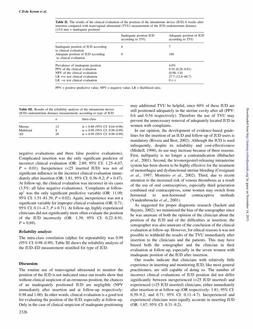
The intra-class correlation (alpha) for repeatability was 0.99
(95% CI: 0.98±0.99). Table III shows the reliability analysis of
the IUD±ED measurement strati®ed for type of IUD.
Discussion
The routine use of transvaginal ultrasound to monitor the
position of the IUD is not indicated since our results show that
without clinical suspicion of an erroneous position, the chances
of an inadequately positioned IUD are negligible (NPV
immediately after insertion and at follow-up respectively:
0.98 and 1.00). In other words, clinical evaluation is a good test
for evaluating the position of the IUD, especially at follow-up.
Only in the case of clinical suspicion of inadequate positioning
may additional TVU be helpful, since 60% of these IUD are
still positioned adequately in the uterine cavity after all (PPV:
0.6 and 0.54 respectively). Therefore the use of TVU may
prevent the unnecessary removal of adequately located IUD in
women with complaints.
In our opinion, the development of evidence-based guide-
lines for the insertion of an IUD and follow-up of IUD users is
mandatory (Rivera and Best, 2002). Although the IUD is used
infrequently, despite its reliability and cost-effectiveness
(Mishell, 1998), its use may increase because of three reasons.
First, nulliparity is no longer a contraindication (Hubacher
et al., 2001). Second, the levonorgestrel-releasing intrauterine
system has been shown to be highly effective for the treatment
of menorrhagia and dysfunctional uterine bleeding (Crosignani
et al., 1997; Monteiro et al., 2002). Third, due to recent
attention to the increased risk of venous thrombosis as a result
of the use of oral contraceptives, especially third generation
combined oral contraceptives, some women may switch from
hormonal to non-hormonal contraceptive methods
(Vandenbroucke et al., 2001).
As suggested for proper diagnostic research (Sackett and
Haynes, 2002), we minimized the bias of the sonographer since
he was unaware of both the opinion of the clinician about the
position of the IUD and of the dif®culties at insertion; the
sonographer was also unaware of the conclusion of the clinical
evaluation at follow-up. However, for ethical reasons it was not
possible to withhold the results of the TVU immediately after
insertion to the clinicians and the patients. This may have
biased both the sonographer and the clinician in their
evaluation at follow-up, especially in the seven women with
inadequate position of the IUD after insertion.
Our results indicate that clinicians with relatively little
experience in inserting and monitoring IUD, like most general
practitioners, are still capable of doing so. The number of
incorrect clinical evaluations of IUD position did not differ
signi®cantly between inexperienced (<25 IUD inserted) and
experienced (>25 IUD inserted) clinicians, either immediately
after insertion or at follow-up (OR respectively: 1.81; 95% CI:
0.39±9.2; and 0.71; 95% CI: 0.11±4.7). Inexperienced and
experienced clinicians were equally accurate in inserting IUD
(OR: 1.67; 95% CI: 0.31±9.2).
Table III. Results of the reliability analysis of the intrauterine device(IUD)±endometrium distance measurements according to type of IUD
n Intra-class
Mirena 12 a = 0.88 (95% CI: 0.64±0.96)Multiload 8 a = 0.99 (95% CI: 0.99±0.99)All 20 a = 0.99 (95% CI: 0.98±0.99)
Table II. The results of the clinical evaluation of the position of the intrauterine device (IUD) 6 weeks afterinsertion compared with transvaginal ultrasound (TVU) measurement of the IUD±endometrium distance(>5.0 mm = inadequate position)
Inadequate position IUDaccording to TVU
Adequate position of IUDaccording to TVU
Inadequate position of IUD accordingto clinical evaluation
6 7
Adequate position of IUD accordingto clinical evaluation
0 160
Prevalence of inadequate position 4.0%PPV of the clinical evaluation 0.54 (0.26±0.81)NPV of the clinical evaluation (0.98±1.0)LR +ve test clinical evaluation 27.7 (12.6±60.7)LR ±ve test clinical evaluation 0 (±)
PPV = positive predictive value; NPV = negative value; LR = likelihood ratio.
C.D.de Kroon et al.
2326
by guest on Novem
ber 14, 2015http://hum
rep.oxfordjournals.org/D
ownloaded from

TVU has been shown to be able to monitor the position of all
different types of IUD (Bonilla-Musoles et al., 1996; Petta
et al., 1996; FauÂndes et al., 1997, 2000; Palo 1997); however,
this is the ®rst report on the excellent reliability of the IUD±ED
measurement by TVU (intra-class coef®cient 0.99). We choose
5.0 mm as threshold for abnormal IUD position as consensus
between earlier reports. First FauÂndes showed that in 90% of
women without complaints of their copper IUD, the IUD±ED
was <7.0 mm (FauÂndes et al., 1997). Petta showed that if IUD
were removed whenever the IUD±ED was >3.0 mm, this
resulted in a signi®cant reduction in spontaneous expulsion
(Petta et al., 1996). Since there is no evidence concerning the
optimal IUD±ED for levonorgestel-releasing intrauterine sys-
tems, we also decided to use 5.0 mm, although there are
reasons to believe that this particular IUD might be as
effective, both in the prevention of pregnancy as well as for
the treatment of menorrhagia, if it is located more cervically
(Andersson and Rybo, 1990; Andersson et al., 1994). Whether
5.0 mm is the most effective IUD±ED to ensure adequate
contraception remains to be elucidated. However, investigation
of the most effective IUD±ED is dif®cult because of the large
number of women needed.
In conclusion, the results of our prospective study show that
TVU is not necessary on a routine basis to monitor the position
of the IUD, neither immediately after insertion nor at the
recommended follow-up after 6 weeks. Clinicians with little
experience are equally capable of inserting and monitoring
IUD when compared with more experienced clinicians.
Therefore there is no need to refer women from primary to
secondary care for insertion of an IUD. This is, especially from
the cost-effectiveness point of view, highly favourable. Only in
the case of clinical suspicion of inadequate location of the IUD,
as a result of the gynaecological interview and examination,
may the use of TVU be bene®cial and cost-effective since this
decreases the number of unnecessarily removed IUD.
References
Aleem, H.A., Kamel, H.S. and Aboul-Oyoun, E.M. (1992) Role ofultrasonography in managing IUD-related complaints. Contraception, 46,211±220.
American College of Obstetricians and Gynecologists (1992) The intrauterinedevice. [Technical Bulletin 164.] Int. J. Gynaecol. Obstet., 41, 189±195.
Andersson, K. and Rybo, G. (1990) Levonorgestrel-releasing intrauterinedevice in the treatment of menorrhagia. Br. J. Obstet. Gynecol., 97, 690±694.
Andersson, K., Odlind, V. and Rybo, G. (1994) Levonorgestrel-releasing andcopper-releasing (Nova-T) IUD's during ®ve years of use: a randomisedcomparative trial. Contraception, 49, 56±72.
Anteby, E., Revel, A., Ben-Chetrit, A., Rosen, B., Tadmor, O. and Yagel, S.(1993) Intrauterine device failure: relation to its location within the uterinecavity. Obstet. Gynecol., 81, 112±114.
Bernaschek, G., Spernol, R. and Beck, A. (1981) The position of the IUD andintrauterine pregnancies. Geburtshilfe Frauenkheilk, 41, 645±647.
Bonilla-Musoles, F., Raga, F., Osborne, N.G. and Blanes, J. (1996) Control ofintrauterine device insertion with three-dimensional ultrasound: is it thefuture? J. Clin. Ultrasound, 24, 263±267.
Crosignani, P.G., Vercellini, P., Mosconi, P., Oldani, S., Cortesi, I. and DeGiorgi, O. (1997) Levonorgestrel-releasing intrauterine device versushysteroscopic endometrial resection in the treatment of dysfunctionaluterine bleeding. Obstet. Gynecol., 90, 257±263.
FauÂndes, D., Bahamondes, L., FauÂndes, A., Petta, C.A., Diaz, J. and Marchi,N. (1997) No relation between the IUD position evaluated by ultrasoundand complaints of bleeding and pain. Contraception, 56, 43±47.
FauÂndes, D., PerdigaÄo, A., FauÂndes, D., Bahamondes, L. and Petta, C.A.(2000) T-shaped IUDs accomodate in their position during the ®rst 3months after insertion. Contraception, 62, 165±168.
Harrison-Woolrych, M., Ashton, J. and Coulter, D. (2002) Insertion of themultiload Cu375 intrauterine device; experience in over 16000 NewZealand women. Contraception, 66, 387±391.
Hubacher, D., Lara-Ricalde, R., Taylor, D.J., Guerra-Infante, F. and Guzman-Rodrigues, R. (2001) Use of copper intrauterine devices and the risk of tubalinfertility among nulligravid women. N. Engl. J. Med., 345, 561±567.
Khan, K.S. and Chien, P.F.W. (2001) Evaluation of a clinical test. I:assessement of reliability. Br. J. Obstet. Gynecol., 108, 562±567.
Mishell, D.R. (1998) Intrauterine devices: mechanisms of action, safety andef®cacy. Contraception, 58, 45S±53S.
Monteiro, I., Bahamondes, L., Diaz, J., Perrotti, M., and Petta, C.A. (2002)Therapeutic use of levonorgestrel releasing system in women withmenorrhagia: a pilot study (1). Contraception, 65, 325±328.
Palo, P. (1997) Transabdominal and transvaginal ultrasound detection oflevonorgestrel IUD in the uterus. Acta Obstet. Gynecol. Scand., 76, 244±247.
Petta, C.A., FauÂndes, D., Pimentel, E., Diaz, J. and Bahamondes, L. (1996)The use of vaginal ultrasound to identify copper T IUD's at high risk ofexpulsion. Contraception, 54, 287±289.
Rivera, R. and Best, K. (2002) Consensus statement on intrauterinecontraception. Contraception, 65, 385±388.
Sackett, D.L. and Haynes, R.B. (2002) The architecture of diagnostic research.Br. Med. J., 324, 539±541.
Tadesse, E., van Brandenburg, W.J.A. and Exalto, N. (1985) Intrauterinedevices: results of a separate clinical and ultrasound follow-up study. Eur. J.Obstet. Gynecol. Reprod. Biol., 19, 289±295.
Valentin, L. (1999) High-quality gynecological ultrasound can be highlybene®cial but poor-quality gynecological ultrasound can do harm.Ultrasound Obstet. Gynecol., 13, 1±7.
Vandenbroucke, J.P., Rosing, J., Bloemenkamp, K.W., Middeldorp, S.,Helmerhorst, F.M., Bouma, B.N. and Rosendaal, F.R. (2001) Oralcontraceptives and the risk of venous thrombosis. N. Engl. J. Med., 344,1527±1535.
Submitted on May 6, 2003; accepted on July 9, 2003
No need for TVU to monitor IUD position after insertion
2327
by guest on Novem
ber 14, 2015http://hum
rep.oxfordjournals.org/D
ownloaded from