human color vision
TRANSCRIPT

HUMAN COLOR VISION
Md. Abdullah al MamunAssistant Professor
Department of Textile EngineeringMawlana Bhashani Science and Technology University
Santosh, Tangail-1902E-mail: [email protected]

Sketch of horizontal section of the Human eye

The Cornea
The cornea is the outer covering of the eye. Thislayer protects your eye from elements that couldcause damage to the inner parts of the eye. Thereare several layers of the cornea, creating a toughlayer that provides additional protection. Theselayers regenerate very quickly, helping the eye toeliminate damage more easily. The cornea alsoallows the eye to properly focus on light moreeffectively. Those who are having troublefocusing their eyes properly can have theircorneas surgically reshaped to eliminate thisproblem
The Sclera/ Sclerotic coat
The sclera is commonly referred to as the"whites" of the eye. This is a smooth, whitelayer on the outside, but the inside is brownand contains grooves that help the muscles ofthe eye attach properly. The sclera providesstructure and safety for the inner workings ofthe eye, but is also flexible so that the eye canmove to seek out objects as necessary.
The Pupil
The "hole" or circular opening in the iristhrough which light passes to the lens and theretina; located slightly to the nasal side of thecenter of the iris; lies behind the anteriorchamber of the eye and the cornea and infront of the lens; diameter changes withcontraction and relaxation of the muscularfibers of the iris as the eye responds tochanges in light, emotional states and otherkinds of stimulation.
The Irish
The iris is the area of the eye that contains thepigment which gives the eye its color. Thisarea surrounds the pupil, and uses the dilatorpapillae muscles to widen or close the pupil.This allows the eye to take in more or less lightdepending on how bright it is around you. If itis too bright, the iris will shrink the pupil sothat they eye can focus more effectively.

The Retina
The light focuses by the lens will be transmitted ontothe retina. This is made of rods and cones arranged inlayers, which will transmit light into chemicals andelectrical pulses. The retina is located in the back of theeye, and is connected to the optic nerves that willtransmit the images the eye sees to the brain so theycan be interpreted. The back of the retina, known asthe macula, will help interpret the details of the objectthe eye is working to interpret. The center of themacula, known as the foveal pit will increase the detailof these images to a perceivable point.

The Choroid Coat
A brownish black pigmented layer just behindthe retina(between the retina and the sclera)that absorbs light transmitted through theretina.
It also provides blood supply to the eye. Justlike any other portion of the body, the bloodsupply gives nutrition to the various parts ofthe eye.

The vitreous humor
A viscous transparent fluid filling the interior ofthe eye. It helps it hold its shape and maintain anearly constant distance between the lense andretina. This gel takes in nutrients from thegastronomic body, aqueous humor and theretinal vessels so the eye can remain healthy.When debris finds its way into the vitreoushumor, it causes the eye to perceive "floaters," orspots that move across the vision area thatcannot be attributed to objects in theenvironment.

The aqueous humor
The aqueous humor is a watery substance
between the cornea and the lens. It is split into
two chambers. The anterior chamber is located in
front of the iris, and the posterior chamber is
directly behind it. The pressure of these layers
maintain the shape of the cornea.

The lens
A transparent, colorless, firm structure of the eye,
enclosed in a capsule, located between the iris
and the vitreous humor; refracts light to focus
images onto the retina at the back of the eye; in
old age the lens becomes flattened, more dense,
slightly opaque, and amber-tinted.
Foveal Pit
An area of the retina that consists of entirelyof cone cells.
This rod free region is about one squaremillimeter in area and contains about 50,000cone cells.
It is the highest color sensitive area of theretina.

Yellow Spot
The nerve layer of the retina in the vicinity ofthe foveal pit is colored with a yellowpigment.
This serves to protect the fovea from overstimulation by blue light.

Blind Spot
• The spot where the nerve connections leavethe eye to form the optic nerve which trasmitsthe signals to the brain.
• This region contains very few light sensitivecells and that part of an image on this area isinvisible.
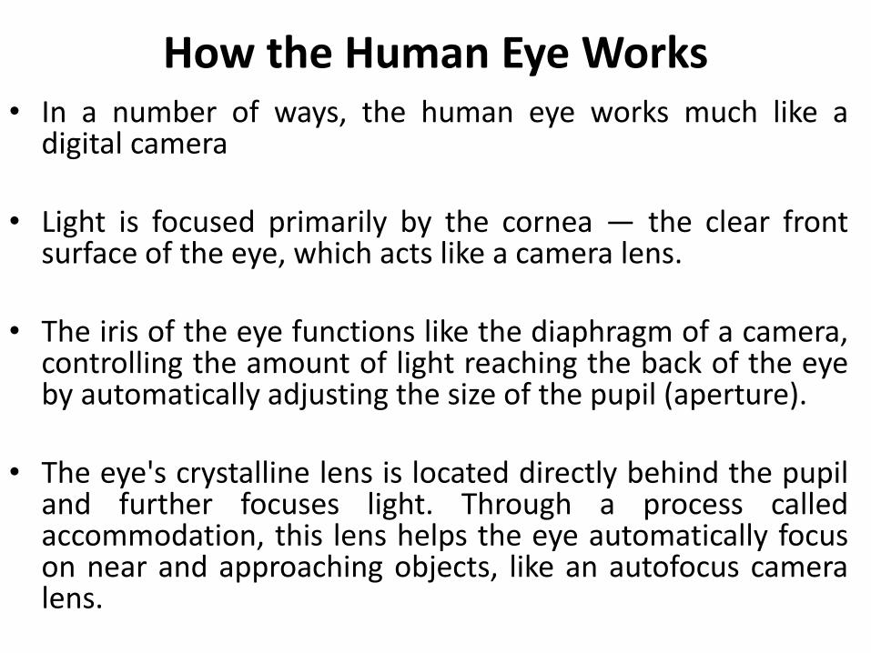
How the Human Eye Works• In a number of ways, the human eye works much like a
digital camera
• Light is focused primarily by the cornea — the clear frontsurface of the eye, which acts like a camera lens.
• The iris of the eye functions like the diaphragm of a camera,controlling the amount of light reaching the back of the eyeby automatically adjusting the size of the pupil (aperture).
• The eye's crystalline lens is located directly behind the pupiland further focuses light. Through a process calledaccommodation, this lens helps the eye automatically focuson near and approaching objects, like an autofocus cameralens.

How the Human Eye Works
Light focused by the cornea and crystallinelens (and limited by the iris and pupil) thenreaches the retina — the light-sensitive innerlining of the back of the eye. The retina actslike an electronic image sensor of a digitalcamera, converting optical images intoelectronic signals. The optic nerve thentransmits these signals to the visual cortex —the part of the brain that controls our sense ofsight.
The Rods
• All rods are of similar type.
• Very sensitive to small amount of light andresponsible for our vision at low levels ofilluminations ( Night vision)
• Unable to provide color vision and provides amonochromatic view of the world (light-dark).
• Contains photosensitive pigment ( rhodopsin). Itbleaches out in high level of illuminations and rodcell looses its sensitivity power. It takes fewminutes to become the rod cells active again.
The Rods
• Not individually connected to the brain viaoptic nerve rather groups of cells togetherform a connection to the optic nerve.
• So Scotopic vision is thus lower in resolutionthan vision under high level of illuminations.


The Cone Cells
• The cones are active at higher light levels(photopic vision),
• These are capable of color vision and areresponsible for high spatial acuity.
• The central fovea is populated exclusively bycones.
• Each cone cell is connected to the single opticnerve thus photopic vision has higherresulation.

The cone cell

Types of Cones
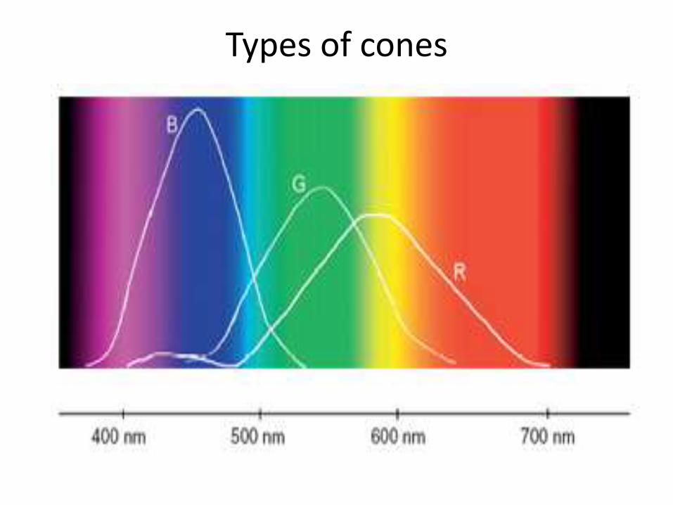
• There are 3 types of cones which are referredto as the short-wavelength sensitive cones,the middle-wavelength sensitive cones andthe long-wavelength sensitive cones or S-cone, M-cones, and L-cones for short.
Types of cones

Spectral sensitivitySpectral sensitivity :is the relative efficiency of detection, of light or other signal, as a
function of the frequency or wavelength of the signal

Defective color vision/ Color blindness
• Color blindness causes patients, particularlymen, to have difficulty distinguishing betweencolors.
• Color blindness is not actually a form ofblindness, but a vision problem that causespeople to have difficulty distinguishing colors.
• The most common type of color blindness is red and green color blindness, though some will experience difficulty distinguishing blue and yellow.

• This condition is largely inherited genetically withmen experiencing it around eight times morefrequently than women.
Symptoms of Color Blindness
• Those that have color blindness may find thatthey have difficulty telling the differencebetween two colors such as red and green.
• Color blinds actually see some colors quitevividly while others appear washed out,making it difficult to tell which color they areseeing.
• If any one has been able to see a variety ofcolors previously and suddenly developdifficulty seeing colors, should contact doctorright away.

Causes of Color BlindnessColor blindness is caused by cells in the retina failingto respond to the varying light wavelengths the eyeuses to detect colors. These cells are known as thephotoreceptors or rods and cones. Rods are sensitiveto light while cones are responsible for detectingcolors. Apart from genetic reasons, some conditionscan cause a deficiency in the cones or cause sometypes of cones to be absent.
(i) Genetic ( inherited by birth)
(ii) Illness (severe long lasting deases)
(iii) Damage ( blow on head, may happen to the kids by accident.

All people cab be classified into four classes on the basis of their color vision
Trichromates
Majority of the people (92%) are trichromates.
Trichromates have the normal color vision.
They use three of the primaries for color vision.

Dichromates
• About 2-3% people are dichromates.
• The dichromates respond to only two of the
primaries.
• Discrimination capacity of the dicromates is less
than the trichromates.
• Dichromates may not have sensitivity to any one
of the three primaries.

Monochromates
• Very less number people 0.003% in case of male
and 0.002% in case of female are
monochromates.
• Monochromates have no color vision.
• Monochromates respond to only one of the
primaries.
• This may happen due to excess lack of nutrition
mainly to the aged person.
Anomalous Trichromates
• Out of thrichromates 4-6% are called
anomalous trichromates.
• They are no perfectly trichromates or could be
fall into dichromates.
• Their eyes do not respond equally to the three
primaries.
• They can watch three colors but not perfectly.

Types of Dichromates
Dichromates
Ignore red primary
Male: 1%Female:1 %
Ignore green primary
Male: 0.01%Female: 0.01%
Ignore blue primary
Male: 0.02%Female: 0.001%

The End