human retina based identification system using gabor
TRANSCRIPT

Abstract—A biometric authentication system provides an
automatic person authentication based on some characteristic
features possessed by the individual. Among all other biometrics,
human retina is a secure and reliable source of person
recognition as it is unique, universal, lies at the back of the eye-
ball and hence it is unforgeable. The process of authentication
mainly includes pre-processing, feature extraction and then
features matching and classification. Also authentication systems
are mainly appointed in verification and identification mode
according to the specific application. In this paper, pre-
processing and image enhancement stages involve several steps to
highlight interesting features in retinal images. The feature
extraction stage is accomplished using a bank of Gabor filter with
number of orientations and scales. Generalized Discriminant
Analysis (GDA) technique has been used to reduce the size of
feature vectors and enhance the performance of proposed
algorithm. Finally, classification is accomplished using k-nearest
neighbor (KNN) classifier to determine the identity of the genuine
user or reject the forged one as the proposed method operates in
identification mode. The main contribution in this paper is using
Generalized Discriminant Analysis (GDA) technique to address
‘curse of dimensionality’ problem. GDA is a novel method used
in the area of retina recognition.
Index Terms— biometrics, human retina, Gabor filters, GDA,
nonvascular, KNN.
I. INTRODUCTION
any types of systems require reliable personal
recognition methods to ascertain and determine the
identity of individuals accessing various areas or services and
data. These schemes ensure that the services and data are
accessed only by authorized personnel. Systems that may use
biometric recognition schemes include private areas of
buildings, computer systems, government and military
facilities and automated teller machines (ATM). Such areas
are vulnerable to security violation and require high level of
security to control such situations. Biometric Recognition is
Manuscript received March 10, 2020; revised September 1, 2020. Date of
publication September 8, 2020. Date of current version September 8, 2020.
This research was supported by Computer Engineering Department, College
of Engineering, University of Mosul, Mosul, Iraq.
Authors are with the Computer Engineering Department, College of
Engineering, Mosul, Iraq (e-mails: [email protected],
Digital Object Identifier (DOI): 10.24138/jcomss.v16i3.1031
defined as the automated authentication of individuals based
on their physiological or behavioral characteristics. A high
security level can be achieved by using this scheme as
mentioned in references [1- 11]. In this paper, the research
concentrates on the use of retinal scans as means of
authentication and human retina as the biometric. Proposed
method improves the performance of retina based
identification system by using Generalized Discriminant
Analysis (GDA) technique as a solution for ‘curse of
dimensionality’ problem which usually presents in the feature
vector of biometrics. GDA is a novel method used in the area
of retina recognition. It reduces the time complexity of
classifier and increases the accuracy rate of the classification
process. Since it only selects the most discriminant features
from the feature vector. Improvement of the proposed method
has been demonstrated by doing a comparison with other
published research works. These works were in the area of
retina recognition and used the same databases as shown later
in Section V.
The retina is a layer of blood vessels that located at the back
of human eye-ball. Since retina is not easily accessible, it is
impossible to forge. Also it is not exposed to threats from the
external environment, unlike other biometrics such as
fingerprint, hand geometry, ear shape, face etc. The retina has
a very distinct blood vessels form so that, every eye has its
own totally unique pattern of this form. In addition, the eyes of
identical twins are not identical in their retinal pattern. So, it is
called ‘Eye Print’ [5, 12], these unique blood vessels form the
foundation of the retinal recognition system. Blood vessels of
retina remain stable over a person's lifetime unless the person
subjects to a severe eye surgical operation or injury [1] [6–9]
[13-16]. So, among all other biometrics, retina has the most
fixed features for individual recognition. It is one of the most
accurate and robust sources of authentication that can be used
for highly sensitive areas.
Proposed method is designed to operate in identification
mode. It uses Gabor filtering to extract nonvascular features
from retinal image (to consume less computation time and
build a real time identification system). During feature
extraction phase; each retinal image is processed with Gabor
filter bank. This bank consists of several two-dimensional
(2D) Gabor filters with number of scales and orientations.
Human Retina Based Identification System
Using Gabor Filters and GDA Technique
Shahad A. Sultan and M. F. Ghanim
M
JOURNAL OF COMMUNICATIONS SOFTWARE AND SYSTEMS, VOL. 16, NO. 3, SEPTEMBER 2020 243
1845-6421/09/1031 © 2020 CCIS

Using Gabor filter bank, a robust three-dimensional (3D)
feature vector is extracted from the retinal image. Since
biometrics have a large number of features, dimensionality
reduction techniques are needful to minimize the size of
feature vectors. Generalized discriminant analysis (GDA) is
used for this purpose. Finally, a simple k-nearest neighbor
(KNN) classifier is used for classification. The overall
proposed algorithm is shown in Fig. 1.
The rest of the paper is organized as the follows: Section II
states a summary of relevant literature. Section III describes
the anatomy of human retina. Section IV illustrates the
proposed method. Section V discusses the experimental
results. Section VI gives the conclusion of this research.
II. RELEVANT LITERATURE
With retinal authentication based on non-vascular features
after preprocessing the features of the retinal image are
extracted from retina without undergoing blood vessel
segmentation, hence they are known as nonvascular features
as these do not depend on blood vessels of retina only. It
decreases the execution time of the system while preserving its
good performance. Some of these methods are mentioned
below:
Sabaghi et al. [17] have compensated the rotation of retinal
image for a robust system. They have used FT (Fourier
Transform) of the retinal image to extract features and called
this feature vector as FSPF, and then they have used Euclidean
distance to perform matching process. Their method is robust
to noise. In [18] Sabaghi et. al. have proposed a second
method, which composes wavelet transform with Fourier
transform to extract FSPF. Also they have used distance in
matching phase. They have obtained accuracy rate of 95.4%
with authentication system based on Fourier transform
features and 97.2% with authentication system that based on
wavelet transform features but they have obtained accuracy
rate of 99.1% by using both techniques. Dehghani et. al. [2]
have extracted features using Harris corner detector. They
have used phase correlation method to determine the rotation
of retinal image. Finally, they have evaluated a similarity
function for matching process. Their method is more efficient
because they have obtained accuracy rate of 100%. Also their
method consumes very low processing time. In [13] Ong et al.
have proposed a graphical method for feature extraction where
scale-invariant feature transform (SIFT) has been used. A sub-
graph matching algorithm has been used for matching process.
They have generated DRIVERA (DRIVE for Retinal
Authentication) database which contained 280 images that
were created from 20 images of DRIVE database. They have
used false acceptance rate (FAR) and false rejection rate
(FRR) to evaluate the performance of their proposed method
which are 0% and 3.169% respectively. They have showed
that their system outperforms two of the high performance
methods [4] and [19] when they evaluated their method on the
same database. Waheed et al. [3] have proposed method that
reads two images, to perform comparison at the same time,
then they have evaluated Luminance and Contrast functions.
They have incorporated luminance and contrast functions to
get structure measurement. To evaluate a similarity value, an
empirically optimized function is evaluated between two
selected images. To evaluate their method, they have used
only 34 subjects. Their method has got an identification rate of
92.5%. In [8], Modarresi et al. have proposed retinal
identification algorithm based on Shearlets transform feature
extraction. They have used Radial Tchebichef Moments to
eliminate the rotation effect from retinal image. They have
also extracted the region of interest from retinal images in
order to acquire similar parts from images that are belonging
to the same person. In this method, Mahalanobis distance is
used to estimate the degree of similarity between biometric
patterns. Experimental results of this method show an equal
error rate (EER) of 0.0024. In [20], B.M.S.Rani and A.Jhansi
Rani have proposed biometric retinal security system for user
authentication in smartphones. They have segmented blood
vessels from retinal images and then measured the angle of
bifurcation points in these vessels and the width of them. RNN
(Recursive Neural Network) is used for matching process.
Their method shows good performance and decreases the error
rates.
III. RETINAL ANATOMY
Human retina has three main components: OD, macula and
blood vessels as shown in fig 2-a. OD is brighter than other
portions and is mainly circular in shape with a diameter of
about 3 mms. It is also the entree and the exit point where
nerves enter and quit the retina to and from the brain.
Macula or the “yellow spot” is the part of the retina that is
most sensitive to light where it is responsible for our sharp
central sight. It is located near the center of the retina about 2
Fig. 1. Proposed algorithm for retinal based identification system
244 JOURNAL OF COMMUNICATIONS SOFTWARE AND SYSTEMS, VOL. 16, NO. 3, SEPTEMBER 2020

ω + β
θ
(a)
(c)
Fig. 3. Retinal image problems a) Retinal image without rotation b) Retinal image with rotation c) Retinal image before translation (d) Retinal image
after translation
(b)
(d)
OD diameter (2DD) temporal to the OD. The darkest part of
the macula called fovea, which is a very small region, forms
the center of it. In the typical retinal image, the mean angle
(𝜔) between fovea and center of the OD with the horizon is
about -5.6˚, ±3.3˚ as shown in fig. 2-2-b. Blood vessels are
patterns that have tree shape, branch from OD and continue on
the surface of the retina. The mean diameter of the vessels is
about 250 μm [9][17][18].
IV. RESEARCH METHOD
Any biometric authentication system has three main phases:
pre-processing, feature extraction, and feature matching. The
proposed method deals with these phases as shown in Fig. 1
which represents the complete algorithm of this research.
A. Pre-processing
This stage prepares the retinal image to correctly extract
features later. The main problem related to the identification
system based on human retina is that the retinal image can be
affected by anatomic movement of eyeball or user head in
front of the fundus camera during the acquisition process. This
problem causes three different situations: scaling, rotation and
translation in the retinal image. These three situations produce
different images coming from the same person as shown in
Fig. 3. So, to build a robust identification system it should be
invariant to these three situations. Hence, pre-processing phase
in the proposed method composes the following steps:
1. Rotation compensation
During this step, rotation angle of the retinal image is
determined and then compensated to generate a rotation
invariant template later in the feature extraction process. In the
proposed method rotation angle has been estimated by
localizing the center of the OD and the center of the macula
and then determining the angle of the line that connects these
two points (center of the OD and center of the macula) with
the horizon. The measured angle represents 𝜔 + β and can be
compensated by applying rotation in opposite direction on the
input image. Compensation angle can be equal to 𝜔 or 𝜔 + β.
Whether rotation is compensated with 𝜔 or 𝜔 + β, at the last
all input images will be rotated to the same point. Rotation
compensation process is illustrated in Fig. 4.
In the proposed method, template matching algorithm is
used to localize the OD and determine the center point of it.
Source image in the proposed method is the retinal image and
template is the image of optic disc as shown in Fig 5.a and 5.c
× ×
2DD
𝛚
Optic disk Blood vessels
Macula
Fig. 2. Retinal Anatomy (a) Retinal main components (b) Position of macula and mean angle of fovea and center of the OD with the
horizon
Fovea
(a) (b)
S. A. SULTAN et al.: HUMAN RETINA BASED IDENTIFICATION SYSTEM USING GABOR FILTERS AND GDA TECHNIQUE 245

respectively.
In the proposed method, area-based matching approach is
used and matching score is calculated using Normalized Cross
Correlation (NCC). NCC is a matching score of two images
with different sizes (source and template). Cross correlation
process is analogous to convolution of two signals and NCC is
a modern version of the cross correlation. It is invariant to
changes in image intensities or noise level. It has a fast
implementation which makes it widely used for template
matching issues within real-time applications. Equation 1
illustrates the mathematical form of the NCC method [19, 20]:
𝑁𝐶𝐶 = ∑ [𝑓(𝑥,𝑦)− 𝑓�̅�,𝑣 ][𝑡(𝑥−𝑢,𝑦 − 𝑣)− 𝑡̅]𝑥,𝑦
{∑ [𝑓(𝑥,𝑦)− 𝑓�̅�,𝑣]2 ∑ [𝑡(𝑥−𝑢,𝑦 − 𝑣)− 𝑡̅]2 𝑥,𝑦𝑥,𝑦 }0.5 (1)
𝑓(𝑥, 𝑦) represents the source image; 𝑓 ̅ is the mean value of
source image intensities within the sliding window; 𝑡
represents the template image; 𝑡̅ is the mean of template image
intensities; 𝑥, 𝑦 and 𝑢, 𝑣 represents pixel coordinates [20].
In the proposed method, template matching algorithm is
implemented using green channel of the retinal image and
properly picked template image. This channel is used because
it shows the pre-eminent contrast between blood vessels in
retinal image and the retina itself. Template image is picked as
a rectangular region around the OD. The result of
implementing algorithm is illustrated in Fig 5. It determines a
point that represents almost the center of the optical disc
(COD).
After localizing the OD, it is the time of localizing macula
center point. Scientifically, fovea located at the Center of
Mass (CoM) of retinal image. To find CoM of retinal image, it
should be gone through pre-processing steps to remove the
noise from this image and enhance its contrast thereby get
more accurate results. These steps are as the following:
1) Each retinal image is resized and saved in JPEG
format.
2) Since the noise is only arised in the intensity
component of the RGB images, this component is separated
from the image. Separation is accomplished by converting the
retinal image from RGB colour space to the HSV colour
space. Then, the only value channel (V) is used in the
following step.
3) Morphological structuring element processing is used
to remove background noise from the V channel. Where, a flat
structuring element is generated with disk shape and radius of
3 pixels. Open morphological operation eliminates, using this
structuring element, snowflakes that have radius less than 3
pixels.
4) V component is filtered using the adaptive median
filter to remove the noise from over this entire channel.
5) The contrast of V component is then enhanced using
Contrast-limited adaptive histogram equalization (CLAHE)
function.
6) The enhanced V channel is recombined with the H and
S channels and then turned back to the RGB colour space.
7) RGB colour space image is converted to Lab colour
space image. Lab is a wide colour space and more similar to
the human eye system. Lab colour space provides a more
convenient representation of colours compared to the RGB
colour space which is designed specifically for display
purposes. By trying several hypotheses on Lab colour space
image and inspect its channels. Concatenation of (L and b)
channels gives better results than using (L with a) channels
and even than using the entire Lab colour space.
After implementing pre-processing steps on retinal image,
the result is shown in Fig 6, where Fig 6.c and 6.d represents
retinal images after implementing pre-processing steps on
images in Fig 6.a and 6.b respectively. At this point,
calculating the CoM point is still affected by the remaining
𝛉 + 𝛃
Fig. 4. Rotation compensation process (a) Determining the center of the optic
disc and the center of the macula (b) Estimating the rotation angle (c) retinal
image after compensating the rotation angle.
(a) (b) (c)
Fig. 5. Localization of optic disc using template matching algorithm (a) Retinal image from RIDB database (b) Green channel from
retinal image (c) Template image (d) Green channel from template image (e)Result of localization.
(a) (b) (e)
(c)
(d)
246 JOURNAL OF COMMUNICATIONS SOFTWARE AND SYSTEMS, VOL. 16, NO. 3, SEPTEMBER 2020
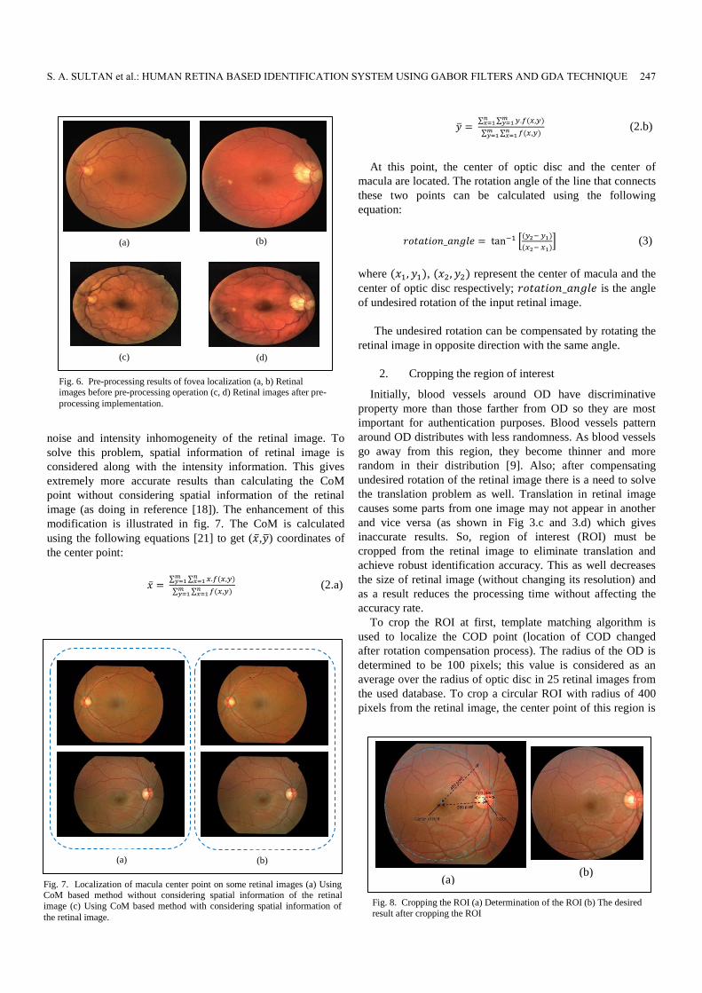
noise and intensity inhomogeneity of the retinal image. To
solve this problem, spatial information of retinal image is
considered along with the intensity information. This gives
extremely more accurate results than calculating the CoM
point without considering spatial information of the retinal
image (as doing in reference [18]). The enhancement of this
modification is illustrated in fig. 7. The CoM is calculated
using the following equations [21] to get (�̅�,�̅�) coordinates of
the center point:
�̅� = ∑ ∑ 𝑥.𝑓(𝑥,𝑦)𝑛
𝑥=1𝑚𝑦=1
∑ ∑ 𝑓(𝑥,𝑦)𝑛𝑥=1
𝑚𝑦=1
(2.a)
�̅� = ∑ ∑ 𝑦.𝑓(𝑥,𝑦)𝑚
𝑦=1𝑛𝑥=1
∑ ∑ 𝑓(𝑥,𝑦)𝑛𝑥=1
𝑚𝑦=1
(2.b)
At this point, the center of optic disc and the center of
macula are located. The rotation angle of the line that connects
these two points can be calculated using the following
equation:
𝑟𝑜𝑡𝑎𝑡𝑖𝑜𝑛_𝑎𝑛𝑔𝑙𝑒 = tan−1 [(𝑦2− 𝑦1)
(𝑥2− 𝑥1)] (3)
where (𝑥1, 𝑦1), (𝑥2, 𝑦2) represent the center of macula and the
center of optic disc respectively; 𝑟𝑜𝑡𝑎𝑡𝑖𝑜𝑛_𝑎𝑛𝑔𝑙𝑒 is the angle
of undesired rotation of the input retinal image.
The undesired rotation can be compensated by rotating the
retinal image in opposite direction with the same angle.
2. Cropping the region of interest
Initially, blood vessels around OD have discriminative
property more than those farther from OD so they are most
important for authentication purposes. Blood vessels pattern
around OD distributes with less randomness. As blood vessels
go away from this region, they become thinner and more
random in their distribution [9]. Also; after compensating
undesired rotation of the retinal image there is a need to solve
the translation problem as well. Translation in retinal image
causes some parts from one image may not appear in another
and vice versa (as shown in Fig 3.c and 3.d) which gives
inaccurate results. So, region of interest (ROI) must be
cropped from the retinal image to eliminate translation and
achieve robust identification accuracy. This as well decreases
the size of retinal image (without changing its resolution) and
as a result reduces the processing time without affecting the
accuracy rate.
To crop the ROI at first, template matching algorithm is
used to localize the COD point (location of COD changed
after rotation compensation process). The radius of the OD is
determined to be 100 pixels; this value is considered as an
average over the radius of optic disc in 25 retinal images from
the used database. To crop a circular ROI with radius of 400
pixels from the retinal image, the center point of this region is
(a)
(c)
(b)
(d)
Fig. 6. Pre-processing results of fovea localization (a, b) Retinal images before pre-processing operation (c, d) Retinal images after pre-
processing implementation.
(a) (b)
Fig. 8. Cropping the ROI (a) Determination of the ROI (b) The desired
result after cropping the ROI
Fig. 7. Localization of macula center point on some retinal images (a) Using CoM based method without considering spatial information of the retinal
image (c) Using CoM based method with considering spatial information of
the retinal image.
(a) (b)
S. A. SULTAN et al.: HUMAN RETINA BASED IDENTIFICATION SYSTEM USING GABOR FILTERS AND GDA TECHNIQUE 247

determined at 350 pixels distance from the COD point to
obtain a circular ROI as shown in Fig. 8. The resulting image
contains mutual points of different images of the same
individual; this meaning is illustrated in Fig. 9 which shows
execution results of the proposed method (Fig. 9 (e & j)). By
cropping the ROI, accuracy rate of the proposed method
improved from 97.05% to 100%, which is considered a good
improvement.
3. Image resizing
Image resizing represents the last step in the pre-processing
phase. It is used to minimize the size of retinal image to enable
processing of digital image to be accomplished with less
computational time.
B. Image enhancement
This phase highlights the parts of interest from retinal
image. It involves smoothing the intensity of different parts of
the retinal image, sharpening, and removing the background
noise that may be caused during the image acquisition process.
Image enhancement makes the features of retinal image more
visible and prepares it for features extraction phase.
1. Morphological structuring
Morphological structuring in the proposed algorithm is
used to remove the background noise and some unwanted
parts (features) from the retinal image. During this step, a flat
structuring element is generated with disk shape and radius of
3 pixels. Morphological processing (open Morphological
operation) uses this structuring element to eliminate
snowflakes that have radius of less than 3 pixels. This
eliminates thin bumps and small points that may be interfered
with the major blood vessels in the retinal image.
2. Image Sharpening
This step performs process of de-blurring and increase the
focus of the retinal image. This in turn highlights blood
vessels and improves the contrast of the image.
3. Histogram Equalization
This process enhances the retinal image using its histogram.
In the proposed algorithm, adaptive histogram equalization is
used. Unlike histogram equalization, it processes small parts in
the image in lieu of the entire image. It enhances each region
in the retinal image separately then, the neighboring regions
are combined. Using this process the contrast of the
homogeneous parts can be restricted to obviate magnifying the
noise that may be appeared in the retinal image.
4. Noise Removal and Filtering
Retinal image is liable to a wide range of noise. In retinal
image, noise may be caused during the image acquisition
process, during the transmission of image from acquisition
hardware or as a result of how the image being transmitted.
Linear filtering process is used to remove the noise from
retinal image. It involves averaging filter and Gaussian filter.
Averaging filter is used to remove grain noise and Gaussian
filter which is a type of low pass filters used to remove the
Speckle Noise that generally present in digital image. Image
enhancement steps are illustrated briefly in Fig 10.
After implementing pre-processing and image enhancement
steps, retinal image becomes as shown in Fig. 11.
C. Feature extraction
By implementing pre-processing and image enhancement
processes on the retinal image, it becomes prepared for the
feature extraction phase. During this phase, Gabor filter is
used to extract non-vascular features from the retinal image.
2D Gabor filter is a Gaussian kernel function modulated by a
complex sinusoidal wave. Gabor filters have two special
merits which are not shared with other filters or methods of
feature extraction. These merits are: frequencies and
orientations depiction of Gabor filters are similar to those of
(a) (b) (c) (d) (e)
(f) (g) (h) (i) (j)
Fig. 9. Cropping the region of interest. (a) and (f) Retinal images from the same person. (b) and (g) Green channel from retinal images. (c) and (h) Determination of the ROI. (d) and (i) Final desired result. (e) and (j) Execution result after cropping the ROI
using proposed method.
248 JOURNAL OF COMMUNICATIONS SOFTWARE AND SYSTEMS, VOL. 16, NO. 3, SEPTEMBER 2020

mammals’ optical system. So, they are widely used to model
the attitude of the primary visual cortex of mammals’ optical
system. The second reason of depending Gabor filters is their
optimal space-frequency resolution [22]. Gabor filters have the
ability to hold responses from texture at different scales and
directions; this feature can be exploited to extract scale
invariant features vector from the image [23]. Also, they are
stable to change in photometric sensations like noise in the
image and illumination effect. It is found that Gabor filters are
particularly suitable for texture representation and pattern
recognition [14], [22]. So, they are used in the proposed
algorithm to extract features from retinal image. 2D Gabor
filter is defined in the following equations [23].
𝐺(𝑥, 𝑦; 𝜆, 𝜙, 𝜓, σ, γ ) = 𝑒(−
𝑥′2+𝛾2𝑦′2
2𝜎2 )𝑒𝑗(2𝜋
𝑥′
λ+𝜓) (4)
𝑥′ = 𝑥 cos 𝜙 + 𝑦 sin 𝜙 (4.a)
𝑦′ = −𝑥 sin 𝜙 + 𝑦 cos 𝜙 (4.b)
where, 𝑥, 𝑦 represent spatial coordinates of the 2D Gabor filter
frequency; λ represents the wavelength (inverse of spatial
frequency) of the sinusoidal wave; 𝜙 represents the orientation
of the parallel strips of Gabor function; 𝜓 represents the phase
offset of the sinusoidal wave; 𝜎 represents the standard
deviation of the Gaussian envelope; γ represents the spatial
aspect ratio.
In the proposed algorithm, Gabor filter bank is composed of
forty bandpass filters (in 8 directions and 5 scales) as shown in
Fig. 12-a. Gabor filters with different orientations are used to
extract features from retinal blood vessels which are presented
at different directions. Different scales are used to eliminate
scaling problem in retinal images.
This bank is applied to each input retinal image by
convolving that image with each filter from the bank as shown
in Fig 12-b. The results of the convolution process are then
concatenated into a robust 3D feature vector. Feature
extraction process is illustrated by using the following
expressions:
𝑣(𝑥, 𝑦, 𝑧) ≡ 𝑉𝜙(𝑧),𝑄(𝑧)(𝐼(𝑥, 𝑦)) (5)
𝑉𝜙(𝑧),𝑄(𝑧)( 𝐼 (𝑥, 𝑦)) = 𝐼(𝑥, 𝑦)⨂ 𝐺𝜙(𝑧),𝑄(𝑧)(𝑥, 𝑦) (6)
𝑄(𝑧) = 𝑓(𝑧) (7)
𝑓(𝑧) ∈ {𝑓𝑚𝑎𝑥 ,𝑓𝑚𝑎𝑥
√2,
𝑓𝑚𝑎𝑥
(√2)2 ,
𝑓𝑚𝑎𝑥
(√2)3 ,
𝑓𝑚𝑎𝑥
(√2)4 ,
5𝜋
(√2)5} (8)
𝜙(𝑧) ∈ {0,𝜋
8,
𝜋
4,
3𝜋
8,
𝜋
2,
5𝜋
8,
3𝜋
4,
7𝜋
8} (9)
where, 𝑣(𝑥, 𝑦, 𝑧) represents 3D feature vector; 𝐼 represents the
Fig. 10. Image enhancement process
Fig. 11. Effect of preprocessing and image enhancement on retinal image (a) Retinal image from RIDB database (b) Pre-processing effect (c) Image
enhancement effect
(a) (b) (c)
(a) (b)
Fig. 12. Convolving retinal image with a bank of Gabor filters (a) Forty Gabor filters (in 8 directions and 5 scales) (b) Resultant images
from Gabor filters
S. A. SULTAN et al.: HUMAN RETINA BASED IDENTIFICATION SYSTEM USING GABOR FILTERS AND GDA TECHNIQUE 249

input retinal image; x, y represent pixel coordinates, x ∈ [1, n];
y ∈ [1, m]; 𝑓(𝑧) represent frequencies of scaled versions from
Gabor function. Q (z) represent scales of Gabor filters.
𝐺𝛳(𝑧),𝑠(𝑧)(𝑥, 𝑦) represents Gabor filter bank. 𝜙 (z) Represent
orientations of Gabor filters.
The size of the resultant feature vector is related to the size
of the retinal image, and the number of filters used in the
Gabor filter bank. The size of the retinal image after image
pre-processing and image enhancement phases becomes 120 x
120 pixels. By convolving this image with forty filters, the
size of feature vector becomes (120 * 120 * 40 = 576000).
Neighboring pixels in an image have high degree of
correlation, so there is a need to reduce these redundant pixels
and minimize the size of the feature vector. This can be
accomplished by downsampling each of the resultant images
from bank filters by a factor of 6. After the downsampling
process, the size of the feature vector becomes (576000/ (6*6)
= 16000).
As illustrated above, the size of the feature vector is large. It
is very important to optimize the size of feature vector to
guarantee response within deadline and generate a real time
identification system. Also, there is an essential need to
eliminate the overlap among classes in a given dataset which
greatly affects the accuracy rate of the designed system. One
of the dimensionality reduction techniques can be used for this
purpose.
1. Dimensionality reduction of retina feature vector
As shown in section 3.3, feature vector has a major length
and contains ineffective information (those belong to the
background of retinal image); this increases the computational
time and reduces the accuracy rate of classification process.
This problem is called ‘curse of dimensionality’ and so, there
is a real need to remove the redundant features from the high
dimensional feature vector and reduce the size of it.
Dimensionality reduction techniques project features of
high dimensional onto a new generated axis. The most
commonly used techniques, in the field of biometrics based
authentication, are: Principle Component Analysis (PCA), and
linear discriminant analysis (LDA). Such techniques are
considered as classical dimensionality reduction techniques
and each of them has its own drawbacks. PCA technique
investigates in the data for directions that have greatest
variance and then project the features into it to produce lower
dimensional features vectors. PCA technique suffers from a
number of problems which make it not the best solution for
“curse of dimensionality” problem in the biometrics field.
These problems are [23]: Linearity, Information packing
transform, and it does not care about classes of a given dataset.
LDA increases between class scatter and minimizes within
class scatter which makes it shows a better classification
performance than PCA technique but it also suffers from two
essential problems: the linearity problem and the Small
Sample Size (SSS) problem. These problems make it
impossible to be implemented in the proposed system [24].
In this research, the main contribution is using Generalized
Discriminant Analysis (GDA) technique to address ‘curse of
dimensionality’ problem. GDA is a novel method used in the
area of retina recognition. It is produced for non-linear
classification and multi-class dataset. GDA is a kernel version
of LDA where the original data space may not accept a linear
separation. It is the more general case and used to eliminate
any shortcomings of both the PCA and LDA techniques. With
the GDA space the most valuable information is preserved
which makes show high classification efficiency and reduces
the training time of the used classifier [21][22][25][26]. Due to
above reasons GDA is adopted, as a dimensionality reduction
technique, in the proposed method.
D. Classification process
K-NN classifier is used in the proposed system for the
classification purpose. Euclidean distance (ED), given by the
equation (10), is determined to measure the similarity score in
K-NN algorithm. K value is selected experimentally to be 4.
𝐸𝐷 = √∑ (𝐹𝑉𝑖𝑘 − 𝐹𝑉𝑗𝑘)2𝑀𝑘=1
2 (10)
V. EXPERIMENTAL RESULTS AND DISCUSSION
All experiments in this research work were conducted in the
same environment which is composed of: Windows 10 Pro
operating system, Intel (R) Core (TM) CPU @ 1.8 GHz, 8 GB
RAM, and Matlab (R2019b). Then, the system was tested
using the following two databases:
1) Retinal Identification DataBase (RIDB) which was
designed by Joddat Fatima, Adeel M. Syed, and M. Usman
Akram during their research work in reference [5]. RIDB
database contains 100 images of resolution 1504 × 1000
(collected from 20 different individuals with 5 images per
individual).
2) Digital Retinal Images for Vessel Extraction (DRIVE)
database. DRIVE database were acquired in the Netherlands
from a diabetic retinopathy checking program. The checking
people consisted of 400 diabetic persons between 25-90 years
old. Forty images of resolution 768 × 584 pixels have been
randomly opted from them.
To check the system in terms of rotation invariance, DRIVE
database has been rotated. Rotation angles applied to retinal
images are: ±10˚, ±15˚, ±20˚, ±25˚, ±30˚, ±35˚. After these
rotation processes, number of images becomes 500. Fig. 13
shows some different images from the used databases.
Performance of the system has been evaluated using the
following performance criteria:
False Acceptance Rate (FAR): is the ratio of persons
which are incorrectly accepted. Feature vectors of these
individuals are matched to templates of non-matching users in
the database. It can be expressed as:
𝐹𝑉𝑖𝑘, 𝐹𝑉𝑗𝑘 Represent ith and jth feature vectors
respectively, each with length M.
250 JOURNAL OF COMMUNICATIONS SOFTWARE AND SYSTEMS, VOL. 16, NO. 3, SEPTEMBER 2020

𝐹𝐴𝑅 = 𝑛𝑢𝑚𝑏𝑒𝑟 𝑜𝑓 𝑎𝑐𝑐𝑒𝑝𝑡𝑒𝑑 𝑖𝑚𝑝𝑜𝑠𝑡𝑒𝑟𝑠
𝑛𝑢𝑚𝑏𝑒𝑟 𝑜𝑓 𝑖𝑚𝑝𝑜𝑠𝑡𝑒𝑟 𝑐𝑜𝑚𝑝𝑎𝑟𝑖𝑠𝑜𝑛𝑠× 100% (11)
False Rejection Rate (FRR): is the ratio of persons which
are incorrectly rejected. Feature vector of these individuals are
not matched to their samples which already exist in the system
database. It can be expressed as:
𝐹𝐴𝑅 = 𝑛𝑢𝑚𝑏𝑒𝑟 𝑜𝑓 𝑟𝑒𝑗𝑒𝑐𝑡𝑒𝑑 𝑔𝑒𝑛𝑢𝑖𝑛𝑒 𝑖𝑛𝑑𝑖𝑣𝑖𝑑𝑢𝑎𝑙𝑠
𝑛𝑢𝑚𝑏𝑒𝑟 𝑜𝑓 𝑔𝑒𝑛𝑢𝑖𝑛𝑒 𝑐𝑜𝑚𝑝𝑎𝑟𝑖𝑠𝑜𝑛𝑠× 100% (12)
Receiver Operating Characteristic (ROC): is the curve
that represents the relation between FAR and FRR. It
represents a function of threshold value and abstracts the
performance of the biometric authentication system. The
tradeoff between FAR and FRR can easily be achieved using
this curve.
Equal Error Rate (EER): is the ratio at which both FAR
and FRR have equal values. EER can easily be found out from
the Receiver Operating Characteristic (ROC) curve. The most
accurate system has the lowest EER value. EER can be
computed at the point (on the ROC curve) where:
FAR(t) = FRR(t) (13)
But practically, similarity score distributions are not
continuous and the crossover point may not exist within these
distributions. In this case ERR can be calculated as:
𝐸𝐸𝑅 = {
𝐹𝐴𝑅(𝑡1)+𝐹𝑅𝑅(𝑡1)
2 𝑖𝑓 𝐹𝐴𝑅 (𝑡1) − 𝐹𝑅𝑅 (𝑡1) ≤ 𝐹𝑅𝑅 (𝑡2) − 𝐹𝐴𝑅 (𝑡2)
𝐹𝐴𝑅(𝑡2)+𝐹𝑅𝑅(𝑡2)
2 𝑜𝑡ℎ𝑒𝑟𝑤𝑖𝑠𝑒
(14)
𝑡1 = 𝑚𝑎𝑥𝑡∈𝑠{𝑡|𝐹𝑅𝑅(𝑡) ≤ 𝐹𝐴𝑅(𝑡)} (14.a)
𝑡2 = 𝑚𝑖𝑛𝑡∈𝑠{𝑡|𝐹𝑅𝑅(𝑡) ≥ 𝐹𝐴𝑅(𝑡)} (14.b)
𝑠 is the set of threshold values used in the score distribution.
Accuracy: is the rate of individuals that are correctly
classified, accuracy of a given system is calculated as:
𝐴𝑐𝑐𝑢𝑟𝑎𝑐𝑦 (%) = [100 − (𝐹𝐴𝑅(%)+𝐹𝑅𝑅(%)
2)] (13)
So, accuracy of the system improves if FAR, FRR decreases.
In biometric systems, there is a tradeoff between FAR and
FRR values. Values of these rates depend on the selected
threshold value. If threshold value is brought down, FRR will
increase (since the similarity score in the proposed system is
the distance between two feature vectors) and FAR will
decrease. When FAR and FRR for a given biometric system
are computed against different threshold values (score
distributions), the desired Operation Point (OP) of it can be
easily selected [5, 6, 27].
Fig. 14.a illustrates FAR and FRR values against threshold
for the proposed identification system using RIDB database.
This figure shows that the proposed system is not sensitive to
threshold values in the range between 24 and 34. In this
region, FAR = FRR = ERR = zero, which represents the ideal
performance for high security level application. Hence, the OP
of the proposed system was determined to be here by making
the threshold value of the classification process in the range of
[24 to 34]. Fig. 14.b shows the ROC curve of the proposed
system using RIDB database, it shows a good separation
distance between the genuine and imposter class. The
performance of the proposed identification system using
DRIVE database is shown in fig. 14 (c and d). EER for both
databases (RIDB and DRIVE) is zero. This value indicates the
excellent performance of the proposed system. Finally, table 1
states comparison among the proposed method and other state
TABLE I
COMPARISON AMONG RESULTS OF PROPOSED METHOD AND OTHER
IDENTIFICATION METHODS
(a) (b)
Fig. 13. Samples from used databases (a) Samples from RIDB database
(b) Samples from DRIVE database.
S. A. SULTAN et al.: HUMAN RETINA BASED IDENTIFICATION SYSTEM USING GABOR FILTERS AND GDA TECHNIQUE 251

of the art published research works. To achieve a fair
comparison, these works used the same databases presented in
this research. From this table it can be seen that the proposed
method outperforms other methods in terms of some criteria
like accuracy and time consuming.
VI. CONCLUSION
In This research a new algorithm is proposed for retinal
identification system. A bank of Gabor filter with different
scales and directions is used for feature extraction process and
then GDA technique is used to diminish the length of feature
vectors. This algorithm shows efficient performance as the
accuracy of the proposed system is 100% for both RIDB and
DRIVE databases. It outperforms many state of the art
authentication systems.
ACKNOWLEDGEMENT
The authors are grateful to University of Mosul and
Computer Engineering department for their support in
carrying out this research.
REFERENCES
[1] Jarina B, S. R. Nirmala. Retina based Biometric Authentication System. International Journal of Advanced Research in Computer Science. DOI:
10.26483/ijarcs.v9i1.5322. 2018; 9(1): 711-718.
[2] A. Dehghani, Z. Ghassabi, H. A. Moghddam, M. S. Moin. Human
recognition based on retinal images and using new similarity function.
EURASIP Journal on Image and Video Processing. 2013; 2013(1): 1–10.
[3] Z. Waheed, A. Waheed, M. U. Akram. A robust nonvascular retina recognition system using structural features of retinal image. 13th
International Bhurban Conference on Applied Sciences and Technology
(IBCAST). Bhurban. 2016: 101–105. [4] H. Oinonen, H. Forsvik, P. Ruusuvuori, O. Yli-Harja, V. Voipio, H.
Huttunen. Identity verification based on vessel matching from fundus
images. IEEE International Conference on Image Processing. Hong Kong. 2010: 4089– 4092.
[5] J. Fatima, A. M. Syed, M. U. Akram. A secure personal identification
system based on human retina. 2013 IEEE Symposium on Industrial Electronics and Applications (ISIEA). Kuching. DOI:
10.1109/ISIEA.2013.6738974. 2013: 90–95.
[6] Sheela S, V.R Udupi, Rahul D. Biometric Verification, Security Concerns and Related Issues. I.J. Information Technology and Computer
Science. DOI: 10.5815/ijitcs.2016.04.06. 2016: 42-51.
[7] Parth P, Ronak B, Tejendra P. An Algorithm for Retinal Feature Extraction using Hybrid Approach. Procedia Computer Science. 7th
International Conference on Communication, Computing and
Virtualization. Gujarat. DOI: 10.1016/j.procs.2016.03.009. 2016: 61-68. [8] Modarresi M, Oveisi IS, Janbozorgi M. Retinal Identification using
Shearlets Feature Extraction. Journal of Austin Biometrics and
Biostatistics. 2017; 4(1): 1-8.
(a) (b)
(c) (d)
Fig. 14. Performance of the proposed system (a) FAR and FRR as functions of threshold values using RIDB database (b) ROC curve
for the RIDB database (c) FAR and FRR as functions of threshold values using DRIVE database (d) ROC curve for the DRIVE
database.
252 JOURNAL OF COMMUNICATIONS SOFTWARE AND SYSTEMS, VOL. 16, NO. 3, SEPTEMBER 2020

[9] Rostom Kachouri, Mohamed Akil, Yaroub Elloumi. Retinal image
processing in Biometrics. In: Amine Nait-ali. Hidden Biometrics When Biometric Security Meets Biomedical Engineering.1. Singapore:
Springer, Singapore. 2019: 1- 22.
[10] T.S. Sasikala , K. Siva Sankar. Unimodal Biometric Based Security Application by Exploiting Retina. International Journal of Engineering
and Advanced Technology (IJEAT). 2018; 8(2S): 344-353.
[11] S.sravan Kumar, N.Anand Ratnesh. A Review on Different Biometric Techniques: Single and Combinational. IOSR Journal of Electronics and
Communication Engineering (IOSR-JECE). DOI: 10.9790/2834-
1104012530. 2016; 11(4): 25-30. [12] C. K¨ose, C. ˙Iki, et al. A personal identification system using retinal
vasculature in retinal fundus images. Expert Systems with Applications.
DOI:10.1016/j.eswa.2011.04.141. 2011: 13670–13681. [13] E. P. Ong, Y. Xu, D. W. K. Wong, J. Liu. Retina verification using a
combined points and edges approach. Image Processing (ICIP). IEEE
International Conference. Singapore. DOI: 10.1109/ICIP.2015.7351297. 2015: 2720–2724.
[14] Mohamed A, M. Hassaballah, Mohammed A. Identity Verification of
Individuals Based on Retinal Features Using Gabor Filters and SVM. Journal of Signal and Information Processing.
http://dx.doi.org/10.4236/jsip.2016.71007. 2016: 49-59.
[15] Shaydyuk, N. K., Cleland T. Biometric Identification via Retina Scanning With Liveness Detection Using Speckle Contrast Imaging.
2016 IEEE International Carnahan Conference on Security Technology
(ICCST). Orlando. 2016: 1-5. [16] M. Suganya, K. Krishnakumari. A Novel Retina based Biometric
Privacy using Visual Cryptography. IJCSNS International Journal of
Computer Science and Network Security. 2016; 16(9): 76-80. [17] Masoud Sabaghi, S. Reza Hadianamrei, Ali Zahedi, Maziyar Niyakan
Lahiji. A New Partitioning Method in Frequency Analysis of the Retinal
Images for Human Identification. Journal of Signal and Information Processing. DOI:10.4236/jsip.2011.24039. 2011; 2(4): 274-278.
[18] Masoud Sabaghi, S. Reza Hadianamrei, Mehdi Fattahi, Mohammad Reza
Kouchaki, Ali Zahedi. Retinal Identification System Based on the Combination of Fourier and Wavelet Transform. Journal of Signal and
Information Processing. http://dx.doi.org/10.4236/jsip.2012.31005.
2012; 3(1): 35-38. [19] A. P. Condurache, J. Kotzerke, A. Mertins. Robust retina-based person
authentication using the sparse classifier. Signal Processing Conference (EUSIPCO). Bucharest. 2012: 1514–1518.
[20] B. M. S. Rani, A. J. Rani. Biometric Retinal Security System for user
Identification and Authentication in Smartphones. International Journal of Pure and Applied Mathematics. 2018; 119(14): 187- 202.
[21] G.R. Prashantha, Chandrashekar M. Patil. An Approach for the Early
Detection of Retinal Disorders and Performing Human Authentication. Proceedings of International Conference on Cognition. DOI: DOI
10.1007/978-981-10-5146-3_16. Singapore. 2017: 157-173.
[22] S. N. Kayte. Design and Development of Non-Proliferative Diabetic Retinopathy Detection Technique using Image Features Extraction
Techniques. Master's thesis. Aurangabad: Dr. Babasaheb Ambedkar
Marathwada University. Department of Computer Science and Information Technology; 2013.
[23] Nazanin Sadat Hashemi, Roya Babaei Aghdam, Atieh Sadat Bayat
Ghiasi, and Parastoo Fatemi. Template Matching Advances and Applications in Image Analysis. American Scientific Research Journal
for Engineering, Technology, and Sciences (ASRJETS). 2016; 26(3):
91-108. [24] A. Grinshpan and J. Golabek. Locating Centers of Mass with Image
Processing. Undergraduate Journal of Mathematical Modeling: One +
Two. DOI: https://doi.org/10.5038/2326-3652.10.1.4906. 2019; 10(1): 1-24.
[25] Rached Belgacem, Hédi Trabelsi, Ines Malek, Imed Jabri. Applying a Set
of Gabor Filter to 2D-Retinal Fundus Image to Detect the Optic Nerve Head (ONH). Annals of Medical and Health Sciences Research. 2018; 8:
48-58.
[26] Mohammad Haghighat, Saman Zonouz, Mohamed Abdel-Mottaleb. CloudID: Trustworthy cloud-based and cross-enterprise biometric
identification. Expert Systems with Applications.
http://dx.doi.org/10.1016/j.eswa.2015.06.025. 2015; 42(21): 7905–7916. [27] A. Tharwat, Linear discriminant analysis: A detailed tutorial, AI
Communications. DOI 10.3233/AIC-170729. 2017; 30(2): 169–190.
[28] D. J. Bora, A. K. Gupta, and F. A. Khan. Comparing the Performance of L*A*B* and HSV Color Spaces with Respect to Color Image
Segmentation. International Journal of Emerging Technology and
Advanced Engineering. 2015; 5(2): 192-203. [29] Y. Zhang, D. Yeung. Semi-Supervised Generalized Discriminant
Analysis. IEEE Transactions on Neural Networks. 30 Jun. 2011; 22(8):
1-11. DOI: 10.1109/TNN.2011.2156808. [30] A. J. Mansfield, J. L. Wayman / Dave Rayner. Best Practices in Testing
and Reporting Performance of Biometric. National Physical Laboratory.
14/02. 2002. [31] T.S. Sasikala, K. S. Sankar. Cascaded Biometric System Based on
Fingerprint and Retina for User Identity Recognition. International
Journal of Innovative Technology and Exploring Engineering (IJITEE). Apr. 2019; 8(6S4): 1357- 1363.
Shahad Ali Sultan is presently working as
researcher to get master degree in computer
engineering from Mosul University. She did her bachelor’s degree in computer engineering/
University of Mosul, 2014. She presented
“Implementation of Voice Equalizer using Tms320c6711” as final year project. Her area of
interest is computer security.
Mayada Faris Ghanim has experiences in wireless
and mobile communications and computer security.
Her educational attainments are BSc and MSc from Computer Engineering Department, College of
Engineering at University of Mosul, Mosul, Iraq,
her PhD. from Faculty of Electrical and Electronic Engineering at University Tun Hussein Onn
Malaysia, Malaysia.
S. A. SULTAN et al.: HUMAN RETINA BASED IDENTIFICATION SYSTEM USING GABOR FILTERS AND GDA TECHNIQUE 253