hyperglycemic crises in adult patients with diabetes: a consensus statement from the american...
TRANSCRIPT
Hyperglycemic Crises in Adult Patients With Diabetes: A consensus statement from the American Diabetes
Association
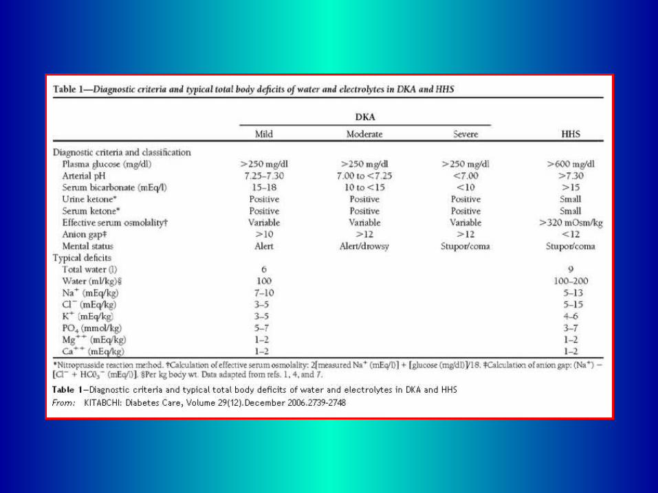
Diabetic ketoacidosis (DKA) and hyperosmolar hyperglycemic state (HHS) are the two most serious acute metabolic complications of diabetes. Most patients with DKA have autoimmune type 1 diabetes; however, patients with type 2 diabetes are also at risk during the catabolic stress of acute illness such as trauma, surgery, or infection. Table 1 outlines the diagnostic criteria and electrolyte and fluid deficits for both disorders.
The mortality rate in patients with DKA is <5% in experienced centers, whereas the mortality rate of patients with HHS still remains high at ~11%. Death in these conditions is rarely due to the metabolic complications of hyperglycemia or ketoacidosis but rather relates to the underlying precipitating illness. The prognosis of both conditions is substantially worsened at the extremes of age and in the presence of coma and hypotension.
This consensus statement will outline precipitating factors and recommendations for the diagnosis, treatment, and prevention of DKA and HHS in adult subjects. It is based on a previous technical review and more recently published peer-reviewed articles since 2001, which should be consulted for further information.
PATHOGENESIS
Although the pathogenesis of DKA is better understood than that of HHS, the basic underlying mechanism for both disorders is a reduction in the net effective action of circulating insulin coupled with a concomitant elevation of counterregulatory hormones, such as glucagon, catecholamines, cortisol, and growth hormone. DKA and HHS can fall anywhere along the disease continuum of diabetic metabolic derangements. At one extreme, pure DKA without significant hyperosmolarity typically indicates the total or relative absence of insulin (seen in type 1 diabetes).
At the other extreme, HHS without ketoacidosis typically occurs with lesser degrees of insulin deficiency, as seen in type 2 diabetes. However, in most circumstances, a mixed presentation occurs depending on the duration of symptoms, coexisting medical illnesses, or underlying precipitating cause. In one study, 123 DKA laboratory admission profiles were reviewed, and 37% demonstrated an elevated total osmolality.
Hormonal alterations in DKA and HHS lead to increased gluconeogenesis and hepatic and renal glucose production and impaired glucose utilization in peripheral tissues, which results in hyperglycemia and hyperosmolality of the extracellular space. The combination of insulin deficiency and increased counterregulatory hormones in DKA also leads to the release of free fatty acids into the circulation from adipose tissue (lipolysis) and to unrestrained hepatic fatty acid oxidation to ketone bodies ([beta]-hydroxybutyrate ([[beta]-OHB] and acetoacetate), with resulting ketonemia and metabolic acidosis.
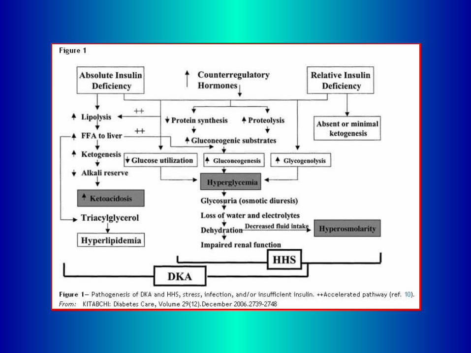
On the other hand, HHS may be caused by plasma insulin concentrations that are inadequate to facilitate glucose utilization by insulin-sensitive tissues but adequate (as determined by residual C-peptide) to prevent lipolysis and subsequent ketogenesis. Both DKA and HHS are associated with glycosuria, leading to osmotic diuresis, with loss of water, sodium, potassium, and other electrolytes. The pathogenic pathways of DKA and HHS are depicted in Fig. 1. The diagnostic criteria and typical total deficits of water and electrolytes in DKA and HHS are summarized in Table 1. As can be seen, DKA and HHS differ in the magnitude of dehydration, ketosis, and acidosis.
DKA is a proinflammatory state producing reactive oxygen species that are indicative of oxidative stress. A recent study has shown elevated levels of proinflammatory cytokines and lipid peroxidation markers, as well as cardiovascular risk factors (plasminogen activator inhibitor-1) and C-reactive protein, which return to normal levels with insulin therapy and remission of hyperglycemia.
PRECIPITATING FACTORS
The two most common precipitating factors in the development of DKA or HHS are inadequate or inappropriate insulin therapy or infection. Other precipitating factors include pancreatitis, myocardial infarction, cerebrovascular accident, and drugs. In addition, new-onset type 1 diabetes or discontinuation of insulin in established type 1 diabetes commonly leads to the development of DKA. Underlying medical illness such as stroke or myocardial infarction that provokes the release of counterregulatory hormones and/or compromises the access to water is likely to result in severe dehydration and HHS.
In most patients, restricted water intake is due to the patient being bedridden or restrained and is exacerbated by the altered thirst response of the elderly. Because 20% of these patients have no history of diabetes, delayed recognition of hyperglycemic symptoms may have led to severe dehydration. Elderly individuals with new-onset diabetes (particularly residents of chronic care facilities) or individuals with known diabetes who become hyperglycemic and are unaware of it or are unable to take fluids when necessary are at risk for HHS.
Drugs that affect carbohydrate metabolism, such as corticosteroids, thiazides, and sympathomimetic agents (e.g., dobutamine and terbutaline) and second-generation antipsychotics agents may precipitate the development of HHS or DKA. In young patients with type 1 diabetes, psychological problems complicated by eating disorders may be a contributing factor in 20% of recurrent ketoacidosis. Factors that may lead to insulin omission in younger patients include fear of weight gain with improved metabolic control, fear of hypoglycemia, rebellion from authority, and the stress of chronic disease.
Before 1993, the use of continuous subcutaneous insulin infusion devices had also been associated with an increased frequency of DKA, but with improvement in technology and better education of patients, the incidence of DKA appears to have reduced in pump users. However, additional prospective studies are needed to document reduction of DKA incidence with the use of continuous subcutaneous insulin infusion devices.
During the past decade, an increasing number of DKA cases without precipitating cause have been reported in children, adolescents, and adult subjects with type 2 diabetes. Observational and prospective studies indicate that over half of newly diagnosed adult African-American and Hispanic subjects with unprovoked DKA have type 2 diabetes. In such patients, clinical and metabolic features of type 2 diabetes include a high rate of obesity, a strong family history of diabetes, a measurable pancreatic insulin reserve, low prevalence of autoimmune markers of [beta]-cell destruction, and the ability to discontinue insulin therapy during follow-up.
This variant of type 2 diabetes has been referred to in the literature as idiopathic type 1 diabetes, atypical diabetes, Flatbush diabetes, type 1.5 diabetes, and more recently as ketosis-prone type 2 diabetes. At presentation, they have markedly impaired insulin secretion and insulin action, but aggressive management with insulin significantly improves [beta]-cell function, allowing discontinuation of insulin therapy within a few months of follow-up. Recently, it was reported that the near-normoglycemic remission is associated with a greater recovery of basal and stimulated insulin secretion and that 10 years after diabetes onset, 40% of patients with ketosis-prone type 2 diabetes are still non–insulin dependent.
Furthermore, a novel genetic mechanism related to the high prevalence of glucose-6-phosphate dehydrogenase deficiency has been linked with ketosis-prone diabetes.
DIAGNOSIS
History and physical examination
The process of HHS usually evolves over several days to weeks, whereas the evolution of the acute DKA episode in type 1 diabetes or even in type 2 diabetes tends to be much shorter. Although the symptoms of poorly controlled diabetes may be present for several days, the metabolic alterations typical of ketoacidosis usually evolve within a short time frame (typically <24 h). The classic clinical picture of patients with DKA includes a history of polyuria, polydipsia, weight loss, vomiting, abdominal pain, dehydration, weakness, mental status change, and coma. Physical findings may include poor skin turgor, Kussmaul respirations, tachycardia, hypotension, alteration in mental status, shock, and ultimately coma.
Up to 25% of DKA patients have emesis, which may be coffee-ground in appearance and guaiac positive. Mental status can vary from full alertness to profound lethargy or coma, with the
latter more frequent in HHS. Although infection is a common precipitating factor for both DKA and HHS, patients can be normothermic or even hypothermic primarily because of peripheral vasodilation. Severe hypothermia, if present, is a poor prognostic sign. Abdominal pain, sometimes mimicking an acute abdomen, is present in 50–75% of DKA cases. The abdominal pain usually resolves with correction of hyperglycemia and metabolic acidosis.
The most common clinical presentation in patients with HHS is altered sensorium. Physical examination reveals signs of dehydration with loss of skin turgor, weakness, tachycardia, and hypotension. Fever due to underlying infection is common, and signs of acidosis (Kussmaul breathing, acetone breath) are usually absent. In some patients, focal neurologic signs (hemiparesis, hemianopsia) and seizures (partial motor seizures more common than generalized) may be the dominant clinical features.
Laboratory findings
The initial laboratory evaluation of patients with suspected DKA or HHS should include determination of plasma glucose, blood urea nitrogen, creatinine, serum ketones, electrolytes (with calculated anion gap), osmolality, urinalysis, urine ketones by dipstick, as well as initial arterial blood gases and complete blood count with differential. An electrocardiogram, chest X-ray, and urine, sputum, or blood cultures should also be obtained, if clinically indicated. HbA1c may be useful in determining whether this acute episode is the culmination of an evolutionary process in previously undiagnosed or poorly controlled diabetes or a truly acute episode in an otherwise well-controlled patient. The diagnostic criteria for DKA and HHS are shown in Table 1.
DKA consists of the biochemical triad of hyperglycemia, ketonemia, and metabolic acidosis. Accumulation of ketoacids results in an increased anion gap metabolic acidosis. The anion gap is calculated by subtracting the sum of chloride and bicarbonate concentration from the sodium concentration [Na+ - (Cl- + HCO3-)]. The normal anion gap has been historically reported to be <12 ± 2 mEq/l. Most laboratories, however, currently measure sodium and chloride concentrations using ion-specific electrodes, which measure plasma chloride concentration 2–6 mEq/l higher than with prior methods.
Thus, the normal anion gap using the current methodology is between 7 and 9 mEq/l, and an anion gap >10–12 mEq/l indicates the presence of increased anion gap acidosis. The severity of DKA is classified as mild, moderate, or severe based on the severity of metabolic acidosis (blood pH, bicarbonate, ketones) and the presence of altered mental status (1). Significant overlap between DKA and HHS has been reported in more than one-third of patients. Although most patients with HHS have an admission pH >7.30, a bicarbonate level >20 mEq/l, mild ketonemia may be present.
The majority of patients with hyperglycemic emergencies present with leukocytosis proportional to blood ketone body concentration. However, leukocytosis >25,000 may designate infection and require further evaluation. The admission serum sodium is usually low because of the osmotic flux of water from the intracellular to the extracellular space in the presence of hyperglycemia. An increase in serum sodium concentration in the presence of hyperglycemia indicates a rather profound degree of water loss. Unless the plasma is cleared of chylomicrons, pseudonormoglycemia and pseudohyponatremia may occur in DKA. Serum potassium concentration may be elevated because of an extracellular shift of potassium caused by insulin deficiency, hypertonicity, and acidemia.
Patients with low normal or low serum potassium concentration on admission have severe total-body potassium deficiency and require very careful cardiac monitoring and more vigorous potassium replacement, because treatment lowers potassium further and can provoke cardiac dysrhythmia. The classic work of Atchley et al. established that the total body deficit of sodium and potassium might be as high as 500–700 mEq.
Studies on serum osmolality and mental alteration have established a positive linear relationship between osmolality and mental obtundation. The occurrence of stupor or coma in diabetic patients in the absence of definitive elevation of effective osmolality (320 mOsm/kg) demands immediate consideration of other causes of mental status change. In the calculation of effective osmolality {2[measured Na (mEq/l)] + [glucose (mg/dl)]/18}, the urea concentration is not taken into account because it is freely permeable and its accumulation does not induce major changes in intracellular volume or osmotic gradient across the cell membrane.
Amylase levels are elevated in the majority of patients with DKA, but this may be due to nonpancreatic sources, such as the parotid gland. A serum lipase determination may be beneficial in the differential diagnosis of pancreatitis; however, lipase could also be elevated in DKA. Finally, abnormal acetoacetate levels may falsely elevate serum creatinine if the clinical laboratory uses a colorometric method for the creatinine assay.
Differential diagnosis
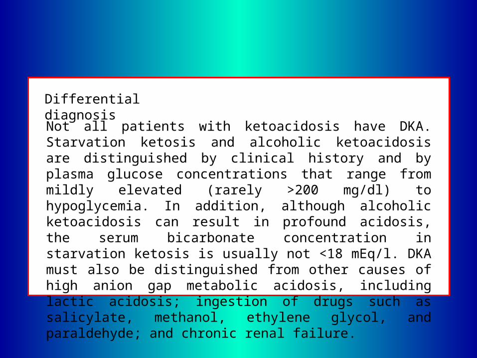
Not all patients with ketoacidosis have DKA. Starvation ketosis and alcoholic ketoacidosis are distinguished by clinical history and by plasma glucose concentrations that range from mildly elevated (rarely >200 mg/dl) to hypoglycemia. In addition, although alcoholic ketoacidosis can result in profound acidosis, the serum bicarbonate concentration in starvation ketosis is usually not <18 mEq/l. DKA must also be distinguished from other causes of high anion gap metabolic acidosis, including lactic acidosis; ingestion of drugs such as salicylate, methanol, ethylene glycol, and paraldehyde; and chronic renal failure.
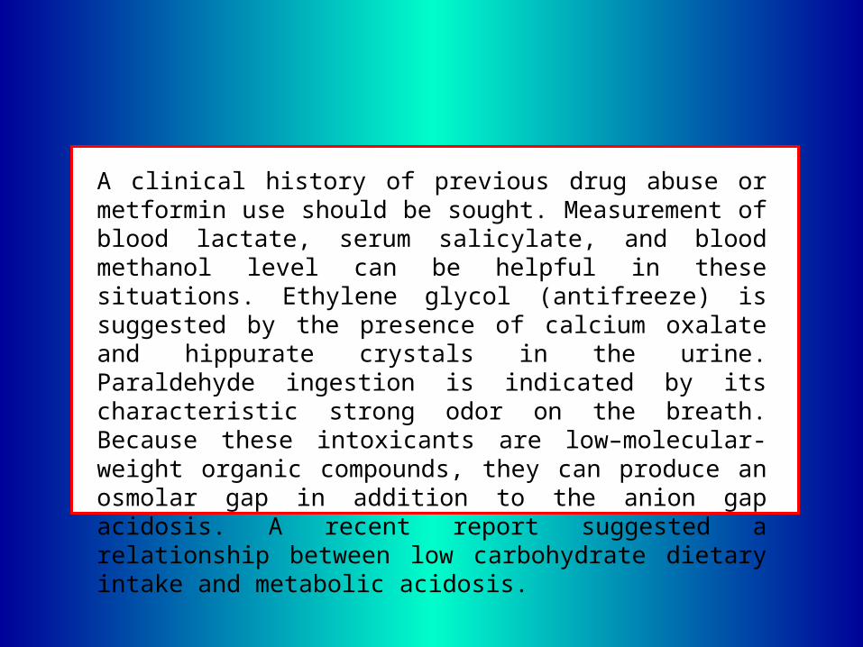
A clinical history of previous drug abuse or metformin use should be sought. Measurement of blood lactate, serum salicylate, and blood methanol level can be helpful in these situations. Ethylene glycol (antifreeze) is suggested by the presence of calcium oxalate and hippurate crystals in the urine. Paraldehyde ingestion is indicated by its characteristic strong odor on the breath. Because these intoxicants are low–molecular-weight organic compounds, they can produce an osmolar gap in addition to the anion gap acidosis. A recent report suggested a relationship between low carbohydrate dietary intake and metabolic acidosis.

Finally, four case reports have shown that patients with undiagnosed acromegaly may present with DKA as the primary manifestation of their disease.
TREATMENT
Successful treatment of DKA and HHS requires correction of dehydration, hyperglycemia, and electrolyte imbalances; identification of comorbid precipitating events; and above all, frequent patient monitoring. Protocols for the management of patients with DKA and HHS are summarized in Figs. 2 and 3.
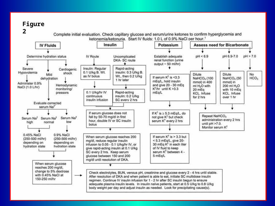
Figure 2
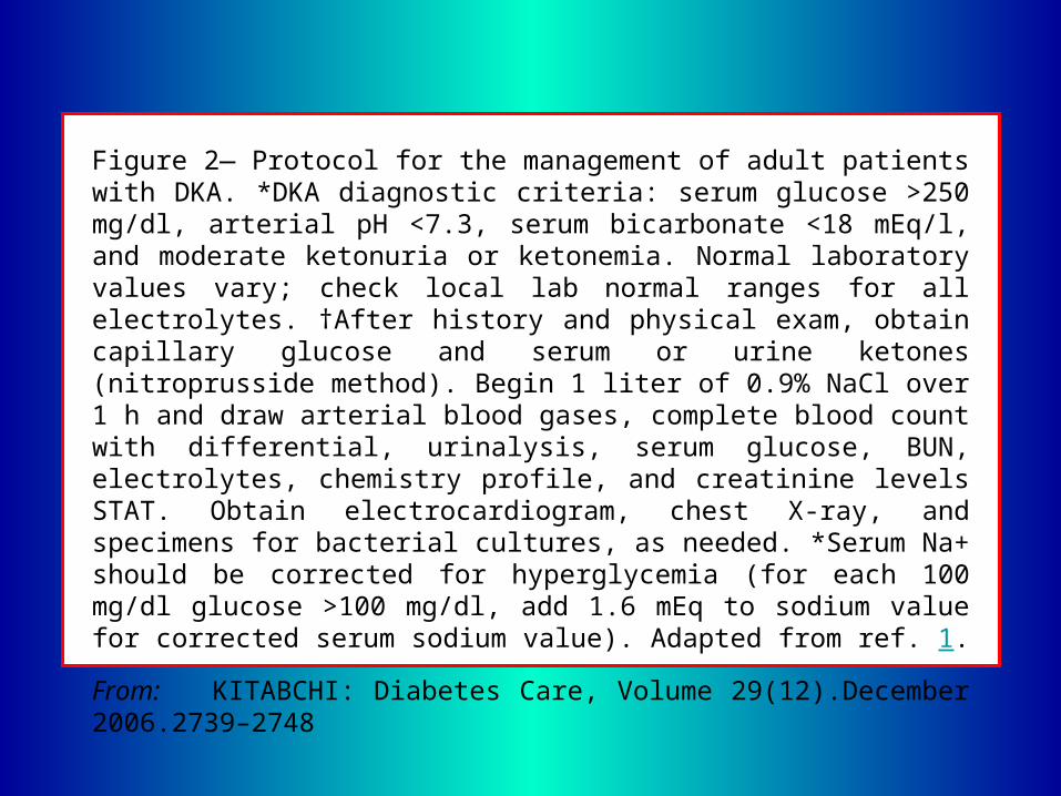
Figure 2— Protocol for the management of adult patients with DKA. *DKA diagnostic criteria: serum glucose >250 mg/dl, arterial pH <7.3, serum bicarbonate <18 mEq/l, and moderate ketonuria or ketonemia. Normal laboratory values vary; check local lab normal ranges for all electrolytes. †After history and physical exam, obtain capillary glucose and serum or urine ketones (nitroprusside method). Begin 1 liter of 0.9% NaCl over 1 h and draw arterial blood gases, complete blood count with differential, urinalysis, serum glucose, BUN, electrolytes, chemistry profile, and creatinine levels STAT. Obtain electrocardiogram, chest X-ray, and specimens for bacterial cultures, as needed. *Serum Na+ should be corrected for hyperglycemia (for each 100 mg/dl glucose >100 mg/dl, add 1.6 mEq to sodium value for corrected serum sodium value). Adapted from ref. 1.
From: KITABCHI: Diabetes Care, Volume 29(12).December 2006.2739–2748
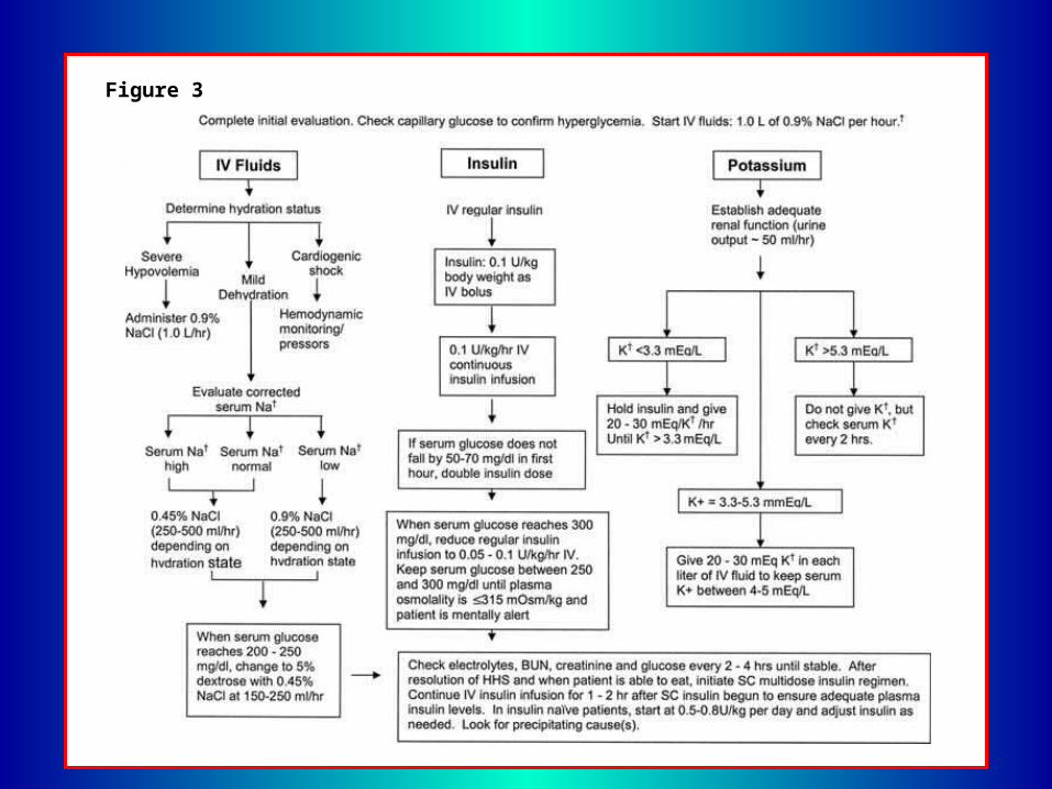
Figure 3
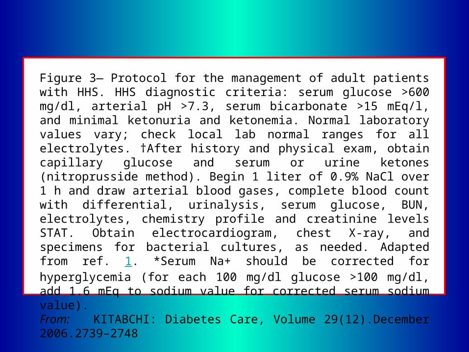
Figure 3— Protocol for the management of adult patients with HHS. HHS diagnostic criteria: serum glucose >600 mg/dl, arterial pH >7.3, serum bicarbonate >15 mEq/l, and minimal ketonuria and ketonemia. Normal laboratory values vary; check local lab normal ranges for all electrolytes. †After history and physical exam, obtain capillary glucose and serum or urine ketones (nitroprusside method). Begin 1 liter of 0.9% NaCl over 1 h and draw arterial blood gases, complete blood count with differential, urinalysis, serum glucose, BUN, electrolytes, chemistry profile and creatinine levels STAT. Obtain electrocardiogram, chest X-ray, and specimens for bacterial cultures, as needed. Adapted from ref. 1. *Serum Na+ should be corrected for hyperglycemia (for each 100 mg/dl glucose >100 mg/dl, add 1.6 mEq to sodium value for corrected serum sodium value). From: KITABCHI: Diabetes Care, Volume 29(12).December 2006.2739–2748
Fluid therapy
fluid therapy is directed toward expansion of the intravascular and extra vascular volume and restoration of renal perfusion. In the absence of cardiac compromise, isotonic saline (0.9% NaCl) is infused at a rate of 15–20 ml · kg-1 body wt · h-1 or 1–1.5 l during the first hour. The subsequent choice for fluid replacement depends on the state of hydration, serum electrolyte levels, and urinary output. In general, 0.45% NaCl infused at 4–14 ml · kg-1 body wt · h-1 is appropriate if the corrected serum sodium is normal or elevated; 0.9% NaCl at a similar rate is appropriate if corrected serum sodium is low (Fig. 2).
Successful progress with fluid replacement is judged by hemodynamic monitoring (improvement in blood pressure), measurement of fluid input and output, laboratory values, and clinical examination. Fluid replacement should correct estimated deficits within the first 24 h. In patients with renal or cardiac compromise, monitoring of serum osmolality and frequent assessment of cardiac, renal, and mental status must be performed during fluid resuscitation to avoid iatrogenic fluid overload. Adequate rehydration with subsequent correction of the hyperosmolar state has been shown to result in a more robust response to low-dose insulin therapy.
Insulin therapy
Unless the episode of DKA is uncomplicated and mild/moderate (Table 1), regular insulin by continuous intravenous infusion is the treatment of choice. In adult patients, once hypokalemia (K+ < 3.3 mEq/l) is excluded, an intravenous bolus of regular insulin at 0.1 unit/kg body wt, followed by a continuous infusion of regular insulin at a dose of 0.1 unit · kg-1 · h-1 should be administered. This low dose of insulin usually decreases plasma glucose concentration at a rate of 50–75 mg · dl-1 · h-1, similar to a higherdose insulin regimen. If plasma glucose does not decrease by 50–75 mg from the initial value in the first hour, the insulin infusion may be doubled every hour until a steady glucose decline is achieved.
When the plasma glucose reaches 200 mg/dl in DKA or 300 mg/dl in HHS, it may be possible to decrease the insulin infusion rate to 0.05–0.1 unit · kg-1 · h-1, at which time dextrose may be added to the intravenous fluids. Thereafter, the rate of insulin administration or the concentration of dextrose may need to be adjusted to maintain the above-glucose values until acidosis in DKA or mental obtundation and hyperosmolality in HHS are resolved.
Prospective and randomized studies have reported on the efficacy and cost effectiveness of subcutaneous rapid-acting insulin analogs in the management of patients with uncomplicated DKA. Patients treated with subcutaneous rapid-acting insulin received an initial injection of 0.2 units/kg followed by 0.1 unit/kg every hour or an initial dose of 0.3 units/kg followed by 0.2 units/kg every 2 h until blood glucose was <250 mg/dl, then the insulin dose was decreased by half to 0.05 or 0.1 unit/kg, respectively, and administered every 1 or 2 h until resolution of DKA.
There were no differences in length of hospital stay, total amount of insulin administration until resolution of hyperglycemia or ketoacidosis, or number of hypoglycemic events among treatment groups. In addition, the use of insulin analogs allowed treatment of DKA in general wards or in the emergency department, avoiding admission to an intensive care unit. By avoiding intensive care admissions, these investigators reported a reduction of 30% in the cost of hospitalization.
Ketonemia typically takes longer to clear than hyperglycemia. Direct measurement of [beta]-OHB in the blood is the preferred method for monitoring DKA and has become more convenient with the recent development of bedside meters capable of measuring whole-blood [beta]-OHB. The nitroprusside method, which is used in clinical chemistry laboratories, measures acetoacetic acid and acetone; however, [beta]-OHB, the strongest and most prevalent acid in DKA, is not measured by the nitroprusside method. During therapy, [beta]-OHB is converted to acetoacetic acid, which may lead the clinician to believe that ketosis has worsened.
Therefore, assessments of urinary or serum ketone levels by the nitroprusside method should not be used as an indicator of response to therapy. During therapy for DKA or HHS, blood should be drawn every 2–4 h for determination of serum electrolytes, glucose, blood urea nitrogen, creatinine, osmolality, and venous pH (for DKA). Generally, repeat arterial blood gases are unnecessary during the treatment of DKA in hemodynamically stable patients. Since venous pH is only 0.02–0.03 units lower than arterial pH, it is adequate to assess venous pH response to therapy, thus avoiding the pain and potential complications associated with repeated arterial punctures.
Criteria for resolution of DKA include glucose <200 mg/dl, serum bicarbonate >=18 mEq/l, and venous pH >7.3. When the patient is able to eat, a multiple-dose insulin schedule should be started that uses a combination of short- or rapid-acting and intermediate- or long-acting insulin as needed to control plasma glucose. Intravenous insulin infusion should be continued for 1–2 h after the subcutaneous insulin is given to ensure adequate plasma insulin levels. An abrupt discontinuation of intravenous insulin coupled with a delayed onset of a subcutaneous insulin regimen may lead to hyperglycemia or recurrence of ketoacidosis.
If the patient is to remain n.p.o., it is preferable to continue the intravenous insulin infusion and fluid replacement. Patients with known diabetes may be given insulin at the dose they were receiving before the onset of DKA or HHS. In insulin-naïve patients, a multidose insulin regimen should be started at a dose of 0.5–0.8 units · kg-1 · day-1, including regular or rapid-acting and basal insulin until an optimal dose is established. However, good clinical judgment and frequent glucose assessment are vital in initiating a new insulin regimen in insulin-naïve patients.
Potassium
Despite total-body potassium depletion, mild to moderate hyperkalemia is not uncommon in patients with hyperglycemic crises. Insulin therapy, correction of acidosis, and volume expansion decrease serum potassium concentration. To prevent hypokalemia, potassium replacement is initiated after serum levels decrease to <5.3 mEq/l, assuming the presence of adequate urine output at 50 ml/h). Generally, 20–30 mEq potassium in each liter of infusion fluid is sufficient to maintain a serum potassium concentration within the normal range of 4–5 mEq/l. Rarely, DKA patients may present with significant hypokalemia. In such cases, potassium replacement should begin with fluid therapy, and insulin treatment should be delayed until potassium concentration is restored to >3.3 mEq/l to avoid arrhythmias or cardiac arrest and respiratory muscle weakness.
Bicarbonate
Bicarbonate use in DKA remains controversial. At a pH >7.0, administration of insulin blocks lipolysis and resolves ketoacidosis without any added bicarbonate. However, the administration of bicarbonate may be associated with several deleterious effects including an increased risk of hypokalemia, decreased tissue oxygen uptake, and cerebral edema. A prospective randomized study in 21 patients failed to show either beneficial or deleterious changes in morbidity or mortality with bicarbonate therapy in DKA patients with an admission arterial pH between 6.9 and 7.1. This study was small and limited to those patients with an admission arterial pH of >6.9. The average pH in the bicarbonate group was 7.03 ± 0.1 and for the nonbicarbonate group was 7.0 ± 0.02.
Therefore, if the pH is 6.9–7.0, it seems prudent to administer 50 mmol bicarbonate in 200 ml of sterile water with 10 mEq KCL over 1 h until the pH is >7.0. No prospective randomized studies concerning the use of bicarbonate in DKA with pH values <6.9 have been reported. Given that severe acidosis may lead to a myriad of adverse vascular effects, adult patients with a pH <6.9 should receive 100 mmol sodium bicarbonate (two ampules) in 400 ml sterile water (an isotonic solution) with 20 mEq KCl administered at a rate of 200 ml/h for 2 h until the venous pH is >7.0. Bicarbonate as well as insulin therapy lowers serum potassium; therefore, potassium supplementation should be maintained in the intravenous fluid as described above and carefully monitored. (See Fig. 2 for guidelines.) Thereafter, venous pH should be assessed every 2 h until the pH rises to 7.0, and treatment should be repeated every 2 h if necessary. See reference 1 for further review.
Phosphate
Despite whole-body phosphate deficits in DKA that average 1.0 mmol · kg-1 · body wt-1, serum phosphate is often normal or increased at presentation. Phosphate concentration decreases with insulin therapy. Prospective randomized studies have failed to show any beneficial effect of phosphate replacement on the clinical outcome in DKA, and overzealous phosphate therapy can cause severe hypocalcemia. Therefore, the routine use of phosphate in the treatment of DKA or HHS has resulted in no clinical benefit to the patient. However, to avoid cardiac and skeletal muscle weakness and respiratory depression due to hypophosphatemia, careful phosphate replacement may sometimes be indicated in patients with cardiac dysfunction, anemia, or respiratory depression and in those with a serum phosphate concentration <1.0 mg/dl. When needed, 20–30 mEq/l potassium phosphate can be added to replacement fluids.

COMPLICATIONS
The most common complications of DKA and HHS include hypoglycemia and hypokalemia due to overzealous treatment with insulin. Low potassium may also occur as a result of treatment of acidosis with bicarbonate. Hyperglycemia may occur secondary to interruption/discontinuance of intravenous insulin therapy after recovery from DKA but without subsequent coverage with subcutaneous insulin. Commonly, patients recovering from DKA develop a transient hyperchloremic non–anion gap acidosis. The hyperchloremic acidosis is caused by the loss of large quantities of ketoanions that occur during the development of DKA. Because ketoanions are metabolized with regeneration of bicarbonate, the prior loss of ketoacid anions in the urine hinders regeneration of bicarbonate during treatment. Other mechanisms include the administration of intravenous fluids containing chloride that exceeds the plasma chloride concentration and the intracellular shifts of NaHCO3 during correction of DKA.
Cerebral edema is a rare but frequently fatal complication of DKA, occurring in 0.7–1.0% of children with DKA. It is most common in children with newly diagnosed diabetes, but it has been reported in children with known diabetes and in young people in their twenties. Fatal cases of cerebral edema have also been reported with HHS. Clinically, cerebral edema is characterized by deterioration in the level of consciousness, lethargy, decreased arousal, and headache. Neurological deterioration may be rapid, with seizures, incontinence, pupillary changes, bradycardia, and respiratory arrest. These symptoms progress as brain stem herniation occurs. The progression may be so rapid that papilledema is not found. Once the clinical symptoms other than lethargy and behavioral changes occur, mortality is high (>70%), with only 7–14% of patients recovering without permanent morbidity.

Although the mechanism of cerebral edema is not known, it may result from osmotically driven movement of water into the central nervous system when plasma osmolality declines too rapidly with the treatment of DKA or HHS. However, a recent study using magnetic resonance imaging to assess cerebral water diffusion and cerebral vascular perfusion during the treatment of 14 children with DKA found that the cerebral edema was not a function of cerebral tissue edema but rather a function of increased cerebral perfusion. There is a lack of information on the morbidity associated with cerebral edema in adult patients; therefore, any recommendations for adult patients are based on clinical judgment rather than scientific evidence.
Preventive measures that might decrease the risk of cerebral edema in high-risk patients are gradual replacement of sodium and water deficits in patients who are hyperosmolar and the addition of dextrose to the hydrating solution once blood glucose reaches 200 mg/dl in DKA and 300 mg/dl in HHS. In HHS, a glucose level of 250–300 mg/dl should be maintained until hyperosmolarity and mental status improves and the patient becomes clinically stable. Hypoxemia and, rarely, noncardiogenic pulmonary edema may complicate the treatment of DKA. Hypoxemia is attributed to a reduction in colloid osmotic pressure that results in increased lung water content and decreased lung compliance. Patients with DKA who have a widened alveolo-arteriolar oxygen gradient noted on initial blood gas measurement or with pulmonary rales on physical examination appear to be at higher risk for the development of pulmonary edema.
PREVENTION
Many cases of DKA and HHS can be prevented by better access to medical care, proper education, and effective communication with a health care provider during an intercurrent illness. The observation that stopping insulin for economic reasons is a common precipitant of DKA in urban African Americans and Hispanics underscores the need for our health care delivery systems to address this problem, which is costly and clinically serious. Sick-day management should be reviewed periodically with all patients. It should include specific information on 1) when to contact the health care provider, 2) blood glucose goals and the use of supplemental short- or rapid-acting insulin during illness, 3) means to suppress fever and treat infection, and 4) initiation of an easily digestible liquid diet containing carbohydrates and salt. Most importantly, the patient should be advised to never discontinue insulin and to seek professional advice early in the course of the illness.
Successful sick-day management depends on involvement by the patient and/or a family member. The patient/family member must be able to accurately measure and record blood glucose, urine, or blood ketone determination when blood glucose is >300 mg/dl; insulin administered; temperature; respiratory and pulse rates; and body weight, and must be able to communicate all of this to a health care professional. Adequate supervision and help from staff or family may prevent many of the admissions for HHS due to dehydration among elderly individuals who are unable to recognize or treat this evolving condition. Better education of caregivers as well as patients regarding signs and symptoms of new-onset diabetes; conditions, procedures, and medications that worsen diabetes control; and the use of glucose monitoring could potentially decrease the incidence and severity of HHS.
The annual incidence rate for DKA from population-based studies ranges from 4.6 to 8 episodes per 1,000 patients with diabetes, with a trend toward an increased hospitalization rate in the past 2 decades. The incidence of HHS accounts for <1% of all primary diabetic admissions. Significant resources are spent on the cost of hospitalization. DKA episodes represent more than $1 of every $4 spent on direct medical care for adult patients with type 1 diabetes and $1 of every $2 in those patients experiencing multiple episodes. Based on an annual average of 100,000 hospitalizations for DKA in the U.S., with an average cost of $13,000 per patient, the annual hospital cost for patients with DKA may exceed $1 billion per year. Many of these hospitalizations could be avoided by devoting adequate resources to apply the measures described above.
Because repeated admissions for DKA are estimated to drain approximately one of every two health care dollars spent on adult patients with type 1 diabetes, resources need to be redirected toward prevention by funding better access to care and educational programs tailored to individual needs, including ethnic and personal health care beliefs. In addition, resources should be directed toward the education of primary care providers and school personnel so that they can identify signs and symptoms of uncontrolled diabetes and new-onset diabetes can be diagnosed earlier. This has been shown to decrease the incidence of DKA at the onset of diabetes.
NOTE ADDED IN PROOF
A recent study from a city hospital reports that active cocaine use is an independent risk factor for recurrent DKA.

Acknowledgments
Studies cited by the authors were supported in part by USPHS grants RR00211 (to the General Clinical Research Center) and AM 21099, training grant AM 07088 of the National Institutes of Health, and grants from Novo-Nordisk, Eli Lilly, the American Diabetes Association, and the Abe Goodman Fund.