ideas you have met before
TRANSCRIPT

12 AQA GCSE Biology: Student Book
Biology
IDEAS YOU HAVE MET BEFORE:
ALL LIVING ORGANISMS ARE MADE OF CELLS.
•Cellsarethebuildingblocksoflife.•Cellscontainspecialisedstructures.•Organismssuchasbacteriaareunicellular.•Mostplantsandanimalsaremulticellular.
ORGANISMS OBTAIN ENERGY BY THE PROCESS OF RESPIRATION.
•Theenergythatisreleaseddrivesalltheprocessesnecessaryforlife.
•Mostorganismsrespirebyaerobicrespiration,usingoxygen.•Somecellsororganismscansurvivewithoutoxygen.They
respireanaerobically.
CELL BIOLOGY
MICROORGANISMS CAN HELP TO KEEP US HEALTHY AND PROVIDE US WITH FOOD.
•Microorganismsproduceimportantfoodproductsbyfermentation.
•Bacteriainthegutareimportantinkeepingushealthy.
IN MULTICELLULAR ORGANISMS CELLS BECOME SPECIALISED.
•Specialisedcellshaveaparticularjobtodo.•Specialisedcellsareorganisedintotissues,tissuesinto
organs,andorgansintobodysystems.
thyroid
trachea
lungsheart
liverstomachlarge intestine
small intestine
brain
bladder
58750_P012_053.indd 12 07/05/16 8:29 AM

13Cell Biology
IN THIS CHAPTER YOU WILL FIND OUT ABOUT:
1
HOW HAVE SCIENTISTS DEVELOPED THEIR UNDERSTANDING OF CELL STRUCTURE AND FUNCTION?
• Thestructuresinsidecellsdodifferentjobswithinthecell.•Cellscanbestudiedusingdifferenttypesofmicroscopes.•Thecellsofbacteriaaredifferentfromthecellsofplantsand
animals.
WHY IS IT IMPORTANT TO STUDY MICROORGANISMS, AND HOW DO WE GROW THEM IN THE LAB AND COMMERCIALLY?
•Thebiochemistryoffermentationisinvolvedintheproductionofalcoholicdrinksandbread.
•Labtechniquesareusedtogrow,orculture,microorganisms.•Microorganismsreproduce,andthenumberofbacteria
producedcanbeestimated.•Testscanshowhoweffectiveantibiotics,antisepticsand
disinfectantsareatinhibitingthegrowthofbacteria.
HOW DO WE DEVELOP INTO A COMPLEX ORGANISM FROM JUST A FERTILISED EGG CELL?
•Thebody’scellsdivideandthenewlyformedcellsareidenticaltotheexistingcells.
•Cellsdifferentiatetobecomespecialised,andspecialisedcellsareorganised.
•Whencelldivisionacceleratesoutofcontrol,cancerdevelops.•Cellsthatareunspecialisedintheembryo,andcellsthat
remainunspecialisedinusasadults,arecalledstemcells.•Stemcellscouldbeusedtotreatcertainconditionsand
diseasesthatarecurrentlyuntreatable.
HOW DO ORGANISMS OBTAIN THEIR ENERGY FROM FOOD?
•Anaerobicrespiration:whensomeorganismsrunoutofoxygen,theycanrespirewithoutit.
•Manymicroorganismscanrespireanaerobically,ascanthemusclesofmammalsforshortperiods.
58750_P012_053.indd 13 07/05/16 8:29 AM
14 AQA GCSE Biology: Student Book
Biology
Learning objectives:
• describe the structure of eukaryotic cells• explain how the main sub-cellular structures are related to
their functions.
Looking at cells
Cell biology helps us to understand how parts of the cell function and interact with each other. It also helps us to learn how we develop, and about our relationships with other organisms.Biomedical scientists use cells to look for signs of disease and in new drug development.
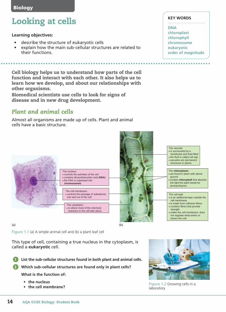
Plant and animal cellsAlmost all organisms are made up of cells. Plant and animal cells have a basic structure.
This type of cell, containing a true nucleus in the cytoplasm, is called a eukaryotic cell.
List the sub-cellular structures found in both plant and animal cells.
Which sub-cellular structures are found only in plant cells?
What is the function of:
• the nucleus• the cell membrane?
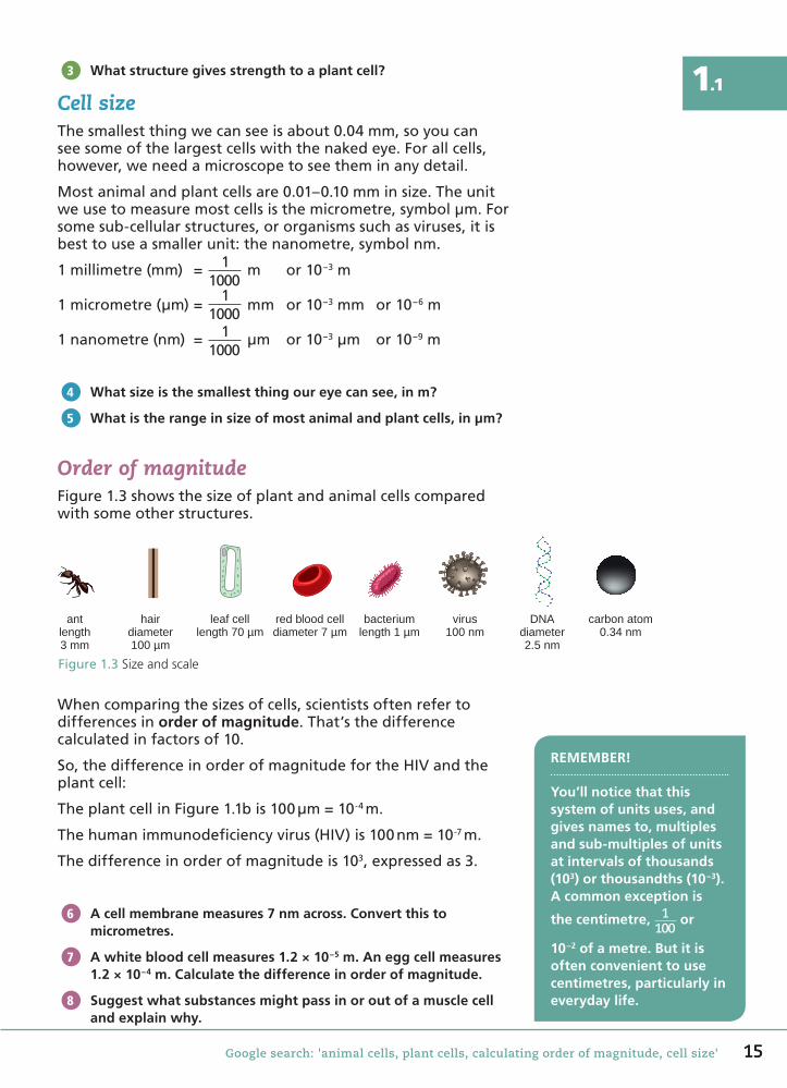
Figure 1.2 Growing cells in a laboratory
1
2
KEY WORDS
DNAchloroplastchlorophyllchromosomeeukaryoticorder of magnitude
(b)
The vacuole:• is surrounded by a membrane and fluid filled• the fluid is called cell sap• vacuoles are permanent structures in plants.
The chloroplasts:• are found in plant cells above ground• contain chlorophyll that absorbs the light the plant needs for photoynthyesis.
The cell wall:• is an additional layer outside the cell membrane• is made from cellulose fibres• contains fibres that provide strength• unlike the cell membrane, does not regulate what enters or leaves the cell.
The nucleus:• controls the activities of the cell• contains deoxyribonucleic acid (DNA)• the DNA is organised into chromosomes.
The cell membrane:• controls the passage of substances into and cut of the cell.
The cytoplasm:• is where most of the chemical reactions in the cell take place.
(a)
Figure 1.1 (a) A simple animal cell and (b) a plant leaf cell
14 AQA GCSE Biology: Student Book
58750_P012_053.indd 14 5/7/16 12:15 PM
1515Google search: 'animal cells, plant cells, calculating order of magnitude, cell size'
1.1What structure gives strength to a plant cell?
Cell sizeThesmallestthingwecanseeisabout0.04mm,soyoucanseesomeofthelargestcellswiththenakedeye.Forallcells,however,weneedamicroscopetoseetheminanydetail.
Mostanimalandplantcellsare0.01–0.10mminsize.Theunitweusetomeasuremostcellsisthemicrometre,symbolμm.Forsomesub-cellularstructures,ororganismssuchasviruses,itisbesttouseasmallerunit:thenanometre,symbolnm.
1millimetre(mm) = 11000
m or10−3m
1micrometre(μm)=1
1000mm or10−3mm or10−6m
1nanometre(nm) = 11000
μm or10−3μm or10−9m
What size is the smallest thing our eye can see, in m?
What is the range in size of most animal and plant cells, in µm?
Order of magnitudeFigure1.3showsthesizeofplantandanimalcellscomparedwithsomeotherstructures.
Figure1.3Size and scale
antlength3 mm
hairdiameter100 µm
leaf celllength 70 µm
red blood celldiameter 7 µm
bacteriumlength 1 µm
virus100 nm
DNAdiameter2.5 nm
carbon atom0.34 nm
Whencomparingthesizesofcells,scientistsoftenrefertodifferencesinorder of magnitude.That’sthedifferencecalculatedinfactorsof10.
So,thedifferenceinorderofmagnitudefortheHIVandtheplantcell:
TheplantcellinFigure1.1bis100μm=10-4m.
Thehumanimmunodeficiencyvirus(HIV)is100nm=10-7m.
Thedifferenceinorderofmagnitudeis103,expressedas3.
A cell membrane measures 7 nm across. Convert this to micrometres.
A white blood cell measures 1.2 × 10⁻⁵ m. An egg cell measures 1.2 × 10⁻⁴ m. Calculate the difference in order of magnitude.
Suggest what substances might pass in or out of a muscle cell and explain why.
3
4
5
REMEMBER!
You’ll notice that this system of units uses, and gives names to, multiples and sub-multiples of units at intervals of thousands (103) or thousandths (10−3).A common exception is
the centimetre, 1100
or
10−2 of a metre. But it is often convenient to use centimetres, particularly in everyday life.
6
7
8
58750_P012_053.indd 15 07/05/16 8:30 AM

16 AQA GCSE Biology: Student Book
KEY WORDS
magnificationresolving powermicrographs
The light microscopeLearning objectives:
• observeplantandanimalcellswithalightmicroscope• understandthelimitationsoflightmicroscopy.
The type of light microscope you have used in the school laboratory is called a compound microscope. Microscopes magnify the specimen you are looking at, making them look bigger than they are.
Magni�cationThemagnifiedimageisproducedbytwolenses,aneyepieceandanobjectivelens.Thereisusuallyachoiceofobjectivelenses.
Total magnification = magnification of eyepiece × magnification of objective lens
Forinstance,iftheeyepiecehasamagnificationoften,whichiswritten×10,andtheobjectivelenshasamagnificationof×40,thetotalmagnificationis×400.
Calculate the total magnification with an eyepiece magnification of × 15 and an objective lens magnification of × 40.
What magnification would the objective lens need to be to give a total magnification of × 300 with an eyepiece of × 15?
1
2
DID YOU KNOW?
British scientist Robert Hooke first used the term ‘cell’. He recorded the first drawings of cells using a compound microscope in his book Micrographia, which was 350 years old in 2015.
You may also have heard of Hooke for his law of elasticity, Hooke’s law, in physics.
Figure1.4A light microscope
Biology
Some early microscopes had just a single lens. The compound microscope has two.
As lens-making techniques improved, microscopes were developed with higher magnifications and resolutions.
58750_P012_053.indd 16 07/05/16 8:30 AM

17Google search: 'magnification, resolving power'
1.2Magni�cation of imagesThemagnificationdescribedonthepreviouspageisthemagnificationusedtoviewanimage.Microscopeimages,ormicrographs,inbooks,scientificpapersorexampapersmustshowthemagnificationinordertobemeaningful.
magnificationoftheimage=size of the imagesize of real object
ThecellinFigure1.5is50mmacrossonthepage.Inreallife,itmeasures40μm.
Tocalculatethemagnification,firstconvertthe50mmintomicrometres(orconvert40μmtomillimetres).
50mm=50000μm
Thecellmeasures40μm
Therefore,themagnificationoftheimage=50 000
40=×1250.
A micrograph of a plant cell in a book is 150 mm long. The plant cell measures 120 µm long. Calculate the magnification.
Why is it essential to state the magnification of an image of a cell in a book but of little value on a website?
The limits of the light microscopeVeryhighmagnificationsarenotpossiblewiththelightmicroscope.Thisisbecauseofthelight-gatheringabilityofthemicroscopeandtheshortworkingdistancesofhigh-powerlenses.Thehighestmagnificationpossibleisaround×1500.
Usinghighermagnificationdoesnotalwaysmeanthatyoucanseegreaterdetailinanimage.Thisdependsontheresolving power,orresolution.Thisistheabilitytodistinguishbetweentwopoints.Inotherwords,whetheryouseethemastwopoints,orone.
Theresolvingpowerofalightmicroscopeisaround0.2μm,or200nm.Thismeansthatyoucouldnotseparatelypickouttwopointscloserthan200nmapart.
What is the maximum resolving power of the light microscope?
What is the maximum magnification possible with a light microscope?
Make a table to show the pros and cons of using a light microscope.
3
4
COMMON MISCONCEPTIONS
Do not confuse magnification, which is how much bigger we can make something appear, with resolving power, which is the level of detail we can see.
Think about a digital photo. You can make it as big as you like, but at a certain point you will not be able to see any more detail.
5
6
7
40 µm
Figure1.5A drawing of a micrograph of a cell
Figure1.6 A micrograph of the cross section of a root. Magnification x100
58750_P012_053.indd 17 07/05/16 8:30 AM

18 AQA GCSE Biology: Student Book
Biology
KEY WORDS
scanning electron microscope (SEM)transmission electron microscope (TEM)
Learning objectives:
• identifythedifferencesinthemagnificationandresolvingpoweroflightandelectronmicroscopes
• explainhowelectronmicroscopyhasincreasedourunderstandingofsub-cellularstructures.
Looking at cells in more detail
The transmission electron microscope (TEM) uses an electron beam instead of light rays.
Some of the electrons are scattered as they pass through the specimen. Those able to pass through it are focused TEMs using electromagnetic coils instead of lenses.
Electron microscopesTEMsareusedforlookingatextremelythinsectionsofcells.Thehighestmagnificationthatcanbeobtainedfromatransmissionelectronmicroscopeisaround×1000000,butimagescanalsobeenlargedphotographically.
Thelimitofresolutionofthetransmissionelectronmicroscopeisnowlessthan1nm.
Thescanning electron microscope(SEM)worksbybouncingelectronsoffthesurfaceofaspecimenthathashadanultra-thincoatingofaheavymetal,usuallygold,applied.Anarrowelectronbeamscansthespecimen.Imagesareformedbythesescatteredelectrons.
SEMsareusedtorevealthesurfaceshapeofstructuressuchassmallorganismsandcells.Becauseofthis,resolutionislowerandmagnificationsusedareoftenlowerthanforTEM.
Electronsdonothaveacolourspectrumlikethevisiblelightusedtoilluminatealightmicroscope.Theycanonlybe‘viewed’inblackandwhite.Here,falsecolourshavebeenadded.
What is the maximum resolution of an electron microscope?
What types of samples would a TEM and an SEM be used to view?
How has electron microscopy improved our understanding of cells?
Figure1.8A scanning electron micrograph of a cancer cell
1
2
3
Figure1.7A transmission electron microscope. The electrons are displayed as an image on a fluorescent screen
58750_P012_053.indd 18 07/05/16 8:30 AM

19
1.3
Google search: 'scanning electron microscopy, transmission electron microscopy'
Cell ultrastructureTheTEMrevealstinysub-cellularstructuresthatarenotvisiblewiththelightmicroscope.Italsoshowsfinedetailinthosestructures.
DID YOU KNOW?
Three scientists won the Nobel Prize in 2014 for the development of super-resolved fluorescence microscopy. It allows a much higher resolution than normal light microscopy. And, unlike electron microscopy, it has the advantage of allowing scientists to look at living cells.
Wecanseemitochondriaandchloroplastswiththelightmicroscope,buttheelectronmicroscoperevealstheirinternalstructure.
COMMON MISCONCEPTIONS
Don’t assume that we always use electron microscopes in preference to light microscopes, or that electron microscopes are always used at high magnifications. Confocal microscopy is used in a lot of biomedical research. It can give high resolution images of live cells. And SEM is often used at low magnifications.
cell membrane
nucleus
cytoplasm
ribosomes
mitochondria
Figure1.9A white blood cell, as seen with a light microscope and a transmission electron microscope
Mitochondria are where
aerobic respiration
takes place in the cell. A
mitochondrion has a double
membrane. The internal
membrane is folded.
Chloroplasts are the
structures in the plant
cell where photosynthesis
takes place. Like
mitochondria, they also
have a complex internal
membrane structure.
Ribosomes are tiny
structures where protein
synthesis takes place.
You can see them as dots
in the micrograph. They
can either lie free in the
cytoplasm or may be
attached to an internal
network of channels
within the cytoplasm.
(a) (b) (c)
Figure1.10Viewing (a) mitochondria, (b) chloroplasts and (c) ribosomes by transmission electron microscopy
Thesizeofsub-cellularstructuresisimportant.Mitochondriaandchloroplastsvaryinsizeandshape.Thecomplexityofamitochondrionindicateshowactiveacellis.Chloroplastsizevariesfromonespeciestoanother.Scientistssometimesinvestigatetheratiooftheareaofthecytoplasmtothatofthenucleusinmicrographs.Ahighratioofcytoplasmic:nuclearvolumecanindicatethatthecellisabouttodivide.Alowonecanbecharacteristicofacancercell.
Name one structure visible to the electron microscope, but not the light microscope.
What process happens in ribosomes?
Which type of microscope would be best suited to viewing the 3D structure of a cell? Explain why.
4
5
6
58750_P012_053.indd 19 07/05/16 8:32 AM
20 AQA GCSE Biology: Student Book
Biology
KEY WORDS
fi eld of viewscale bar
Many scientists use electron microscopes to observe � ne detail in cells. But much of the microscope work carried out – including in hospital and forensic science labs – is done with the light microscope.
Preparing cells for microscopyLivecellscanbemountedinadropofwaterorsalineonamicroscopeslide.
Mostcellsarecolourless.Wemuststainthemtoaddcolourandcontrast.Intheschoollaboratory,youmayhaveusedmethylenebluetostainanimalcellsoriodinesolutiontostainplantcells.
Write an equipment list for looking at cheek cells with a microscope. State why each piece of equipment is used.
Suggest why it’s better to mount the cells in saline than in water.
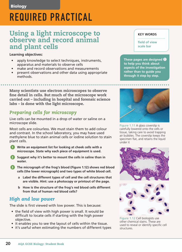
The micrograph of the frog’s blood (Figure 1.12) shows red blood cells (the lower micrograph) and two types of white blood cell.
a Label the different types of cell and the cell structures that are visible. Hint: use a photocopy or printout of the page.
b How is the structure of the frog’s red blood cells different from that of human red blood cells?
High and low powerTheslideisfirstviewedwithlowpower.Thisisbecause:
• thefieldofviewwithhighpowerissmall.Itwouldbedifficulttolocatecellsifstartingwiththehighpowerobjective.
• itenablesyoutoseethelayoutofcellswithinthetissue.• it’susefulwhenestimatingthenumbersofdifferenttypes
1
2
3
Learning objectives:
• applyknowledgetoselecttechniques,instruments,apparatusandmaterialstoobservecells
• makeandrecordobservationsandmeasurements• presentobservationsandotherdatausingappropriate
methods.
Using a light microscope to observe and record animal and plant cells
REQUIRED PRACTICAL
Figure1.11A glass coverslip is carefully lowered onto the cells or tissue, taking care to avoid trapping air bubbles. The coverslip keeps the specimen fl at, and retains the liquid under it
These pages are designed to help you think about aspects of the investigation rather than to guide you through it step by step.
20 AQA GCSE Biology: Student Book
Figure1.12Cell biologists use other chemical stains. These are used to reveal or identify specifi c cell structures.
58750_P012_053.indd 20 07/05/16 8:32 AM
2121
REQUIRED PRACTICAL
1.4ofcellontheslideorinatissue(thoughhere,highpowermaybeneeded).
Alowpowerdigitalimage(ordrawing)canbeusedtoshowthearrangementofcellsinatissue.Thisincludesregionsofthetissuebutnotindividualcells.
Ifrequired,thecellsortissuecanthenviewedwithhighpowertoproduceadetailedimageofapartoftheslide.
Why is a slide viewed with low power first?
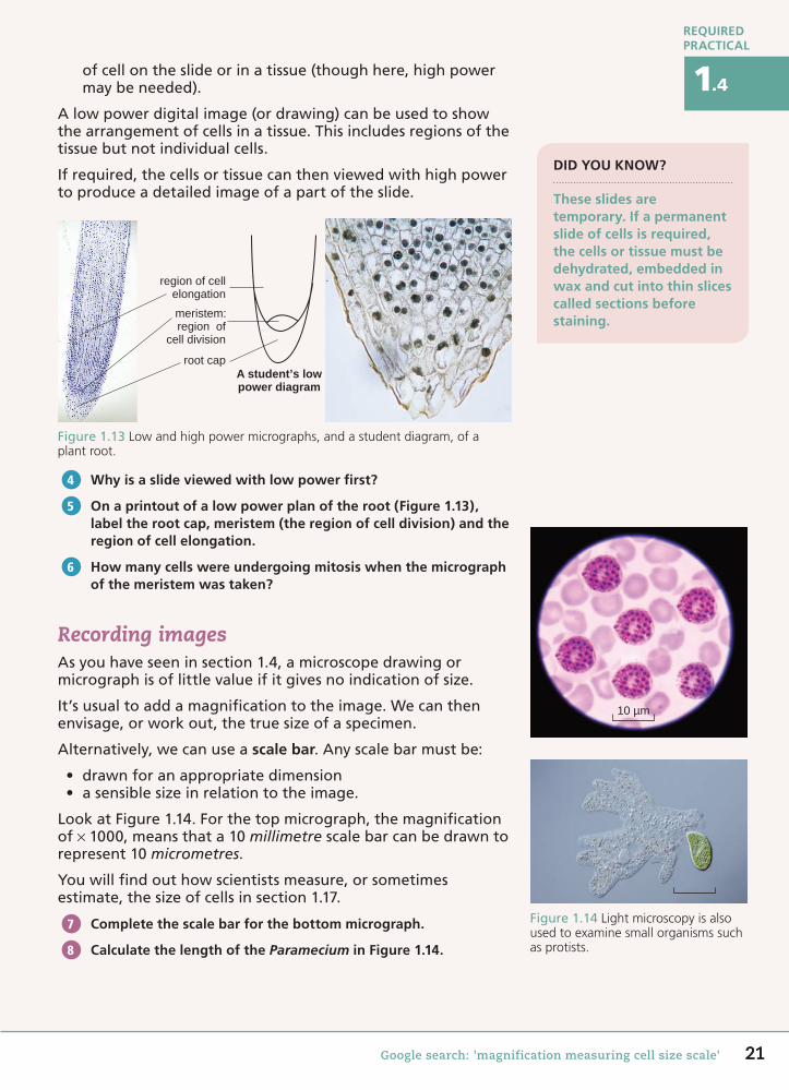
On a printout of a low power plan of the root (Figure 1.13), label the root cap, meristem (the region of cell division) and the region of cell elongation.
How many cells were undergoing mitosis when the micrograph of the meristem was taken?
Recording imagesAsyouhaveseeninsection1.4,amicroscopedrawingormicrographisoflittlevalueifitgivesnoindicationofsize.
It’susualtoaddamagnificationtotheimage.Wecanthenenvisage,orworkout,thetruesizeofaspecimen.
Alternatively,wecanuseascale bar.Anyscalebarmustbe:
• drawnforanappropriatedimension• asensiblesizeinrelationtotheimage.
LookatFigure1.14.Forthetopmicrograph,themagnificationof× 1000,meansthata10millimetrescalebarcanbedrawntorepresent10micrometres.
Youwillfindouthowscientistsmeasure,orsometimesestimate,thesizeofcellsinsection1.17.
Complete the scale bar for the bottom micrograph.
Calculate the length of the Paramecium in Figure 1.14.
4
5
6
10 µm
Figure1.14Light microscopy is also used to examine small organisms such as protists.
7
8
Google search: 'magnification measuring cell size scale'
DID YOU KNOW?
These slides are temporary. If a permanent slide of cells is required, the cells or tissue must be dehydrated, embedded in wax and cut into thin slices called sections before staining.
A student’s lowpower diagram
region of cellelongation
meristem:region of
cell division
root cap
Figure1.13Low and high power micrographs, and a student diagram, of a plant root.
58750_P012_053.indd 21 07/05/16 8:33 AM
22 AQA GCSE Biology: Student Book
Biology
Primitive cells
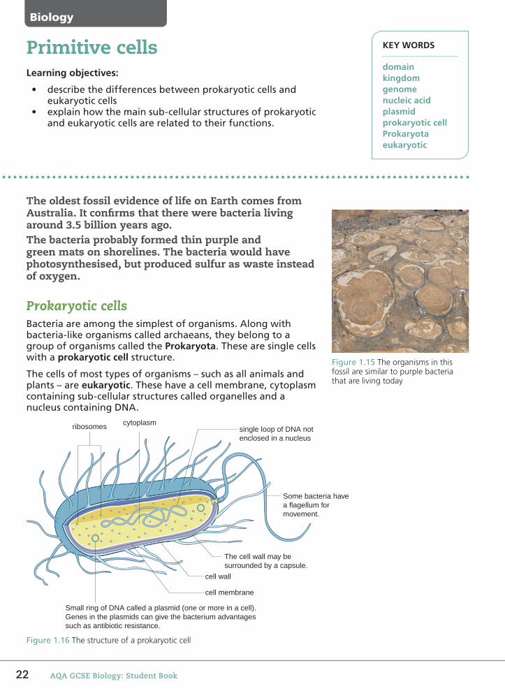
Figure1.16The structure of a prokaryotic cell
The oldest fossil evidence of life on Earth comes from Australia. It con�rms that there were bacteria living around 3.5 billion years ago.The bacteria probably formed thin purple and green mats on shorelines. The bacteria would have photosynthesised, but produced sulfur as waste instead of oxygen.
Prokaryotic cellsBacteriaareamongthesimplestoforganisms.Alongwithbacteria-likeorganismscalledarchaeans,theybelongtoagroupoforganismscalledtheProkaryota.Thesearesinglecellswithaprokaryotic cell structure.
Thecellsofmosttypesoforganisms–suchasallanimalsandplants–areeukaryotic.Thesehaveacellmembrane,cytoplasmcontainingsub-cellularstructurescalledorganellesandanucleuscontainingDNA.
KEY WORDS
domainkingdomgenomenucleic acidplasmidprokaryotic cellProkaryotaeukaryotic
Learning objectives:
• describethedifferencesbetweenprokaryoticcellsandeukaryoticcells
• explainhowthemainsub-cellularstructuresofprokaryoticandeukaryoticcellsarerelatedtotheirfunctions.
cytoplasm
The cell wall may besurrounded by a capsule.
cell wall
cell membrane
Small ring of DNA called a plasmid (one or more in a cell).Genes in the plasmids can give the bacterium advantagessuch as antibiotic resistance.
ribosomes single loop of DNA notenclosed in a nucleus
Some bacteria havea flagellum formovement.
Figure1.15The organisms in this fossil are similar to purple bacteria that are living today
58750_P012_053.indd 22 07/05/16 8:33 AM
23
1.5
Google search: 'Archaea, bacteria, eukaryotic, plasmid, prokaryotic, Carl Woese'
Prokaryotic cells are much smaller than eukaryotic cells, around 1 µm across. Their DNA is not enclosed in a nucleus. It is found as a single molecule in a loop. They may also have one or more small rings of DNA called plasmids.
List the differences between prokaryotic and eukaryotic cells.
Where is DNA found in prokaryotic cells?
A new classi�cation systemBy the 1970s, biologists had classified living organisms into five kingdoms.
Very small, microscopic organisms called archaeans were originally grouped in a kingdom with bacteria. But in 1977, American microbiologist Carl Woese suggested that certain types of organisms that lived in extreme environments or produced methane gas should be placed in a separate group.
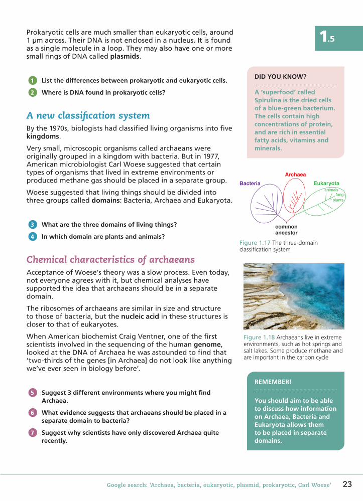
Woese suggested that living things should be divided into three groups called domains: Bacteria, Archaea and Eukaryota.
What are the three domains of living things?
In which domain are plants and animals?
Chemical characteristics of archaeansAcceptance of Woese’s theory was a slow process. Even today, not everyone agrees with it, but chemical analyses have supported the idea that archaeans should be in a separate domain.
The ribosomes of archaeans are similar in size and structure to those of bacteria, but the nucleic acid in these structures is closer to that of eukaryotes.
When American biochemist Craig Ventner, one of the first scientists involved in the sequencing of the human genome, looked at the DNA of Archaea he was astounded to find that ‘two-thirds of the genes [in Archaea] do not look like anything we’ve ever seen in biology before’.
Suggest 3 different environments where you might find Archaea.
What evidence suggests that archaeans should be placed in a separate domain to bacteria?
Suggest why scientists have only discovered Archaea quite recently.
1
2
DID YOU KNOW?
A ‘superfood’ called Spirulina is the dried cells of a blue-green bacterium. The cells contain high concentrations of protein, and are rich in essential fatty acids, vitamins and minerals.
Figure 1.17 The three-domain classification system
commonancestor
animalsfungi
plants
Eukaryota
Archaea
Bacteria
3
4
Figure 1.18 Archaeans live in extreme environments, such as hot springs and salt lakes. Some produce methane and are important in the carbon cycle
REMEMBER!
You should aim to be able to discuss how information on Archaea, Bacteria and Eukaryota allows them to be placed in separate domains.
5
6
7
58750_P012_053.indd 23 09/05/16 6:16 PM

24 AQA GCSE Biology: Student Book
Biology
As an adult, we are made up of 37 trillion (3.7 × 10¹³) cells. To produce these cells, the fertilised egg needs to undergo many cell divisions.
ChromosomesAswegrow,thecellsproducedbycelldivisionmustallcontainthesamegeneticinformation.
Thegeneticinformationofallorganismsiscontainedinchromosomes,madeofDNA.TheDNAinrestingcellsisfoundinthenucleusaslong,thinstrands.Forcelldivision,thesestrandsformcondensedchromosomes.
Humanbodycellshave46chromosomes,or23pairs.Eachchromosomeinapairhasthesametypeofgenesalongitslength.
How many chromosomes are found in human body cells?
How are the chromosomes arranged in a karyotype?
MitosisNewcellshavetobeproducedforgrowthanddevelopment,andtoreplacewornoutanddamagedbodycells.
Whennewcellsareproducedtheymustbeidenticaltotheparentcell.Cellsdividetoproducetwonewones.Thistypeofcelldivisioniscalledmitosis.Twodaughter cellsareproducedfromtheparentcell.
Forsomecelltypes,newcellsareproducedbythedivisionofstem cells(discussedlaterinthechapter).
When are new cells produced?
In this type of cell division:
• how many chromosomes do daughter cells have?• how many daughter cells are produced?
1
2
3
4MAKING CONNECTIONS
To come up with a figure for how many cells there are in the human body, scientists must estimate the number by adding up cell counts from different organs.
KEY WORDS
mitosisstem cellsdaughter cell
Learning objectives:
• describetheprocessofmitosisingrowth,andmitosisaspartofthecellcycle
• describehowtheprocessofmitosisproducescellsthataregeneticallyidenticaltotheparentcell.
Cell division
1 2 3 4 5
6 7 8 9 10 11 12
13 14
19 20 21 22 23
15 16 17 18
The chromosomes in the photograph arecut out and arranged into pairs.The pairs are arranged so that Pair 1 has thelongest chromosomes; Pair 22 the shortestPair 23 is the sex chromosomes.
A photograph is taken of a dividing cell.
1 2 3 4 5
6 7 8 9 10 11 12
13 14
19 20 21 22 23
15 16 17 18
The chromosomes in the photograph arecut out and arranged into pairs.The pairs are arranged so that Pair 1 has thelongest chromosomes; Pair 22 the shortestPair 23 is the sex chromosomes.
A photograph is taken of a dividing cell.
Figure1.19A profile of a set of chromosomes, called a karyotype
58750_P012_053.indd 24 07/05/16 8:33 AM

25Google search: 'cell division, mitosis, stem cells'
1.6So that the daughter cells produced are identical to the parent cell, the DNA must first copy itself. Each of the 46 chromosomes then consists of two molecules of DNA.
DID YOU KNOW?
Using radioactive carbon (14C) dating of a cell’s DNA, researchers in Sweden have been able to estimate the lifespan of different types of cells.
During mitosis, the double chromosomes are pulled apart as each new set of 46 chromosomes moves to opposite ends of the cell (Figure 1.21). Two nuclei then form. The cytoplasm and cell membrane then divides and two cells are produced.
Why do chromosomes appear double, or X-shaped, in micrographs?
The cell cycleA cell that is actively dividing goes through a series of stages called the cell cycle. The cycle involves the growth of the cell and the production of new cell components and division.
5
2
3
4
5
6
1
5. The cytoplasm divides into two and the new cell membrane separates off two new cells.
6. Temporary cell resting period, or the cell no longer divides, e.g. a nerve cell.
3. Further growth occurs and the DNA is checked for errors and any repairs made.
4. Mitosis – the chromosomes move apart and two nuclei form.
1. The cell grows. The number of sub-cellular structures, e.g. mitochondria and ribosomes, increases.
2. The DNA replicates.
Figure 1.22 The cell cycleIn actively dividing human cells, the whole cell cycle lasts 1 hour
DNAcondensesto form a
chromosome.
DNAreplicatesto form a
double chromosome.Each half has an
identical set of genes.
DNA moleculein the nucleus.
Figure 1.20 It is these ‘double’ chromosomes that we always see in micrographs or illustrations of chromosomes
Figure 1.21 Mitosis in an onion cell
Using Figure 1.22, calculate the proportion of the cell cycle spent in mitosis. You will need a protractor.
If the cell cycle lasts 2 hours, estimate the time spent in mitosis.
Mitosis occurs rapidly in a newly formed fertilised egg. Suggest another situation in the body where you might expect cells to be actively dividing by mitosis.
6
7
8
58750_P012_053.indd 25 5/7/16 12:16 PM

26 AQA GCSE Biology: Student Book
Biology
KEY WORDS
differentiationorganorgan systemspecialisedtissue
Cell division makes up only part of our growth and development.
For the �rst four or �ve days of our lives, the cells produced as the fertilised egg divides are identical. Then, some of our cells start to become specialised to do a particular job.
Cell adaptationsIn a multicellular organism, many different types of cell take on different roles to ensure that the organism functions properly and as a whole.
As cells divide, new cells acquire certain features required for their specific function. This is differentiation. A cell’s size, shape and internal structure are adapted for its role. Most animal cells differentiate at an early stage.
Figure 1.24 The function of a sperm cell is to swim in the female reproductive system with the aim of fertilising an egg. (Cell length 55 µm; width at widest point 3 µm)
cell membranetail for
movement
middlepiece
headnucleus containing23 chromosomes
acrosome – contains enzymesto penetrate the egg
many mitochondria for energy,arranged in a spiral
Figure 1.23 By this stage in its de-velopment, this human embryo has developed many of the 200 different cell types in the human body
Learning objectives:
• explain the importance of cell differentiation• describe how cells, tissues, organs and organ systems are
organised to make up an organism• understand size and scale in relation to cells, tissues,
organs and organ systems.
Cell differentiation
Figure 1.25 Nerve cells carry messages, or electrical impulses, from one part of the body to another. This type of cell brings about movement of the skeleton. (Motor nerve cell length: up to 1 m or more; diameter 1–20 µm)
main part of cell(cell body)
cytoplasm
nucleus
fatty covering – the myelin sheath
branches that connect witha muscle
gap in myelin sheath – the nerveimpulse jumps from one gap tothe next, making it quicker
extensions that communicatewith other nerve cells
58750_P012_053.indd 26 5/7/16 12:16 PM
27
1.7
Google search: 'differentiation, specialised cells, human body tissues'
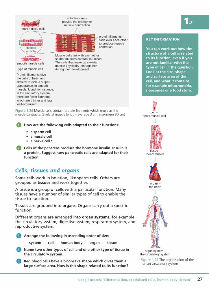
Figure 1.26 Muscle cells contain protein filaments which move as the muscle contracts. (Skeletal muscle length: average 3 cm; maximum 30 cm)
mitochondria –provide the energy for
muscle contraction
protein filaments –slide over each otherto produce musclecontration
Muscle cells link with each otherso that muscles contract in unison.The cells that make up skeletalmuscle physically join togetherduring their development.
heart muscle cells
skeletalmuscle
smooth muscle cells
Type of muscle cell
Protein filaments givethe cells of heart andskeletal muscle a stripedappearance. In smoothmuscle, found, for instance,in the circulatory system,there are fewer filaments,which are thinner and lesswell-organised.
How are the following cells adapted to their functions:
• a sperm cell• a muscle cell• a nerve cell?
Cells of the pancreas produce the hormone insulin. Insulin is a protein. Suggest how pancreatic cells are adapted for their function.
Cells, tissues and organsSome cells work in isolation, like sperm cells. Others are grouped as tissues and work together.
A tissue is a group of cells with a particular function. Many tissues have a number of similar types of cell to enable the tissue to function.
Tissues are grouped into organs. Organs carry out a specific function.
Different organs are arranged into organ systems, for example the circulatory system, digestive system, respiratory system, and reproductive system.
Arrange the following in ascending order of size:
system cell human body organ tissue
Name two other types of cell and one other type of tissue in the circulatory system.
Red blood cells have a biconcave shape which gives them a large surface area. How is this shape related to its function?
1
2
KEY INFORMATION
You can work out how the structure of a cell is related to its function, even if you are not familiar with the type of cell in the question. Look at the size, shape and surface area of the cell, and what it contains, for example mitochondria, ribosomes or a food store.
3
Figure 1.27 The organisation of the human circulatory system
cell –heart muscle cell
tissue –heart muscle
organ –the heart
organ system –the circulatory system
4
5
58750_P012_053.indd 27 5/7/16 12:19 PM
28 AQA GCSE Biology: Student Book
Biology
KEY WORDS
benigncarcinogenmalignantmutationsecondary tumour
Every year, over 300 000 people in the UK are diagnosed with cancer. It is estimated, however, that four in ten cases of cancer could be prevented by lifestyle changes.
What is cancer?Normally, cells grow and divide by mitosis when the body needs new cells to replace old or damaged cells. When a cell becomes cancerous, it begins to divide uncontrollably. New cells are produced even though the body does not need them.
The extra cells produced form growths called tumours. Most tumours are solid, but cancers of the blood, for instance leukaemia, are an exception.
What is cancer?
Name one type of cancer that does not form a solid tumour.
Types of tumour
Type of tumour Characteristics
Benign
• slow growing• often have a capsule around them, so can be removed easily• not cancerous and rarely spread to other parts of the body• they can press on other body organs and look unsightly.
Malignant
• grow faster• can spread throughout other body tissues• as the tumour grows, cancer cells detach and can form secondary
tumours in other parts of the body.
Figure 1.29 The growth and spread of a tumour
Blood vessels are stimulatedto grow around the tumour;the blood vessels supply thetumour with food and oxygen.
Malignant cells detachfrom the tumour andare transported away
in the blood.
The tumour secreteshormone-like
chemicals.
The malignant cellsqueezes throughthe capillary wall.
The cell dividesto produce a
secondary tumour.
Malignant cells can detach fromthe tumour and spread to other
parts of the body.
The malignant cells divide andcan invade normal tissues.
Malignant cells develop.
1
2
Learning objectives:
• describe cancer as a condition resulting from changes in cells that lead to their uncontrolled growth, division and spread
• understand some of the risk factors that trigger cells to become cancerous.
Cancer
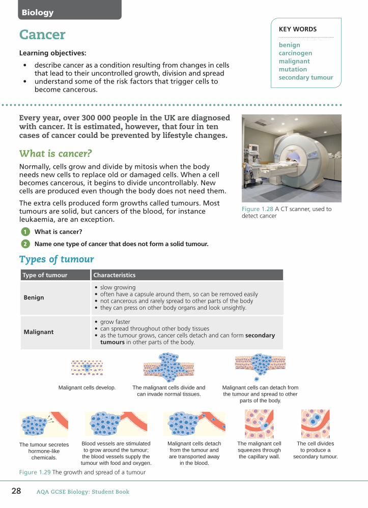
Figure 1.28 A CT scanner, used to detect cancer
58750_P012_053.indd 28 5/7/16 12:22 PM
29
1.8
Google search: 'causes of cancer, carcinogen, detection of cancer, tumour'
Name two types of tumour.
Explain why a tumour needs a blood supply.
What is the name of the type of tumour formed when a cancer spreads?
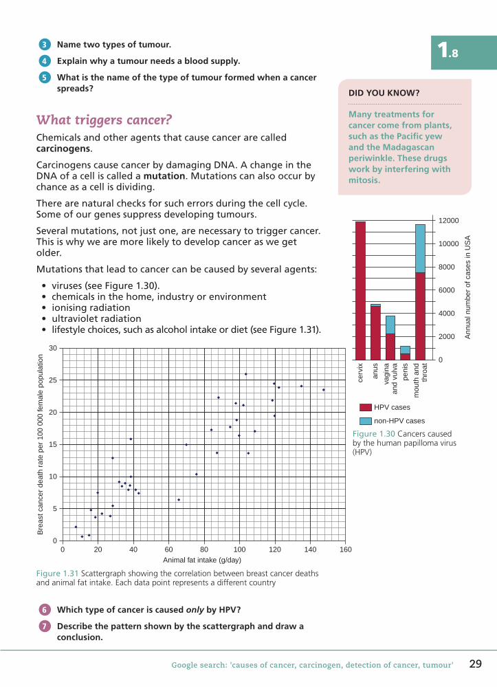
What triggers cancer?Chemicalsandotheragentsthatcausecancerarecalledcarcinogens.
CarcinogenscausecancerbydamagingDNA.AchangeintheDNAofacelliscalledamutation.Mutationscanalsooccurbychanceasacellisdividing.
Therearenaturalchecksforsucherrorsduringthecellcycle.Someofourgenessuppressdevelopingtumours.
Severalmutations,notjustone,arenecessarytotriggercancer.Thisiswhywearemorelikelytodevelopcanceraswegetolder.
Mutationsthatleadtocancercanbecausedbyseveralagents:
• viruses(seeFigure1.30).• chemicalsinthehome,industryorenvironment• ionisingradiation• ultravioletradiation• lifestylechoices,suchasalcoholintakeordiet(seeFigure1.31).
Figure1.31Scattergraph showing the correlation between breast cancer deaths and animal fat intake. Each data point represents a different country
30
25
20
15
10
5
00 20 40 60 80
Animal fat intake (g/day)
Bre
ast c
ance
r de
ath
rate
per
100
000
fem
ale
popu
latio
n
100 120 140 160
Which type of cancer is caused only by HPV?
Describe the pattern shown by the scattergraph and draw a conclusion.
3
4
5
DID YOU KNOW?
Many treatments for cancer come from plants, such as the Pacific yew and the Madagascan periwinkle. These drugs work by interfering with mitosis.
Figure1.30Cancers caused by the human papilloma virus (HPV)
cerv
ix
anus
vagi
naan
d vu
lva
peni
s
mou
th a
ndth
roat
0
2000
4000
Ann
ual n
umbe
r of
cas
es in
US
A
6000
8000
10000
12000
HPV cases
non-HPV cases
6
7
58750_P012_053.indd 29 07/05/16 8:34 AM
30 AQA GCSE Biology: Student Book
Learning objectives:
• describe the function of stem cells in embryonic and adult animals
• discuss potential benefits and risks associated with the use of stem cells in medicine.
Stem cells KEY WORDS
adult stem cellculturecell linesembryonic stem cellethicalin-vitro fertilisation
The UK has a shortage of blood donors.
In the summer of 2015 the NHS announced that it planned to start giving people blood transfusions using arti�cial blood by 2017.
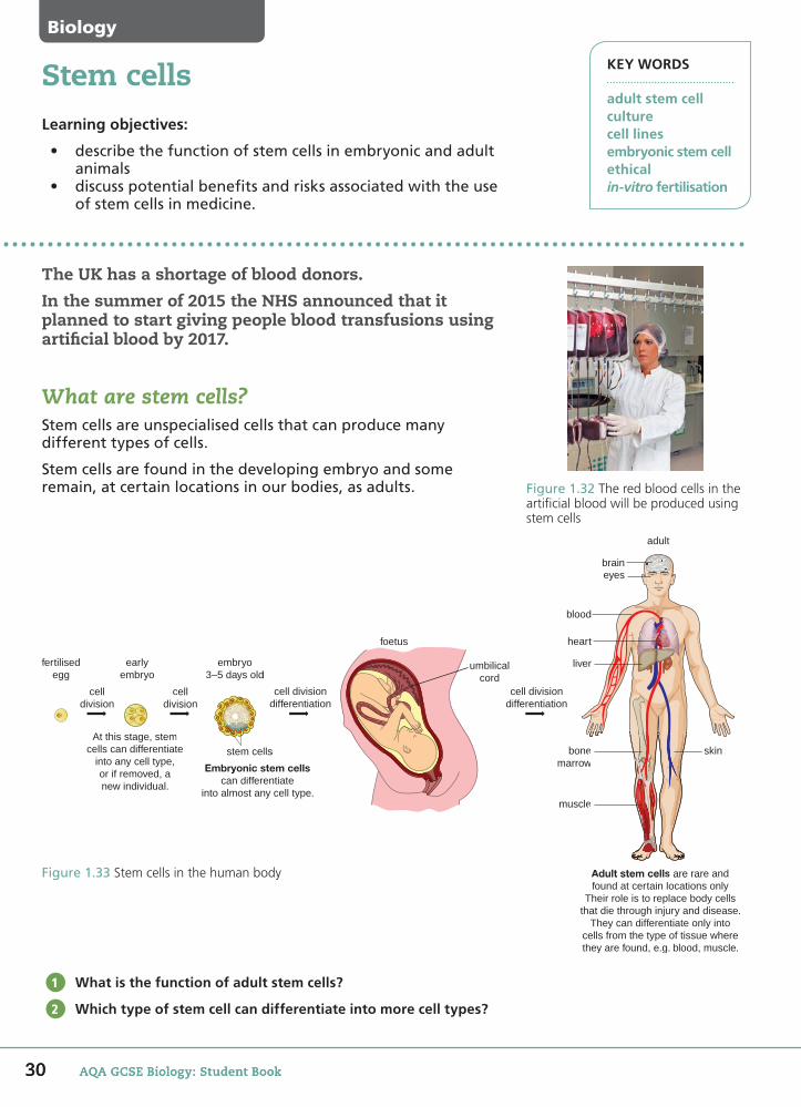
What are stem cells?Stem cells are unspecialised cells that can produce many different types of cells.
Stem cells are found in the developing embryo and some remain, at certain locations in our bodies, as adults.
What is the function of adult stem cells?
Which type of stem cell can differentiate into more cell types?
1
2
Figure 1.33 Stem cells in the human body
fertilisedegg
earlyembryo
embryo3–5 days old
foetus
umbilicalcord
adult
braineyes
blood
heart
liver
bonemarrow
skin
muscle
Adult stem cells are rare andfound at certain locations only
Their role is to replace body cellsthat die through injury and disease.
They can differentiate only intocells from the type of tissue wherethey are found, e.g. blood, muscle.
celldivision
At this stage, stemcells can differentiate
into any cell type,or if removed, anew individual.
Embryonic stem cellscan differentiate
into almost any cell type.
celldivision
stem cells
cell divisiondifferentiation
embryo3–5 days old
matee,
Embryonic stem celcan differentiate
cellvision
stem cells
cell divdifferen
cell divisiondifferentiation
ew
e
Adult stem cells are rarefound at certain locations
Figure 1.32 The red blood cells in the artificial blood will be produced using stem cells
Biology
30 AQA GCSE Biology: Student Book
58750_P012_053.indd 30 5/7/16 12:23 PM

31Google search: 'adult stem cells, bone marrow stem cells, embryonic stem cells, umbilical cord blood cells'
1.9Stem cell transplantsTransplantingstemcells,ortransplantsofspecialisedcellsgrownfromstemcells,couldhelppeoplewith:
• injuries,e.g.spinalinjuriesleadingtoparalysis• conditionsinwhichcertainbodycellsdegenerate,e.g.
Alzheimer’sdisease,diabetesandmultiplesclerosis• cancers,orfollowingtreatmentsforcancersuchas
chemotherapyorradiation,e.g.peoplewithleukaemia.
Stemcelltransplantsalsoenablechemotherapypatients,whohavehadtheirbonemarrowdestroyed,toproduceredbloodcells.
Thehopeisthatwewillbeabletoculturestemcellsinlimitlessnumbers.Stemcell linesproducedfrompatientswithrareandcomplexdiseasescouldtransformthehealthservice.
Name two conditions that could be treated with stem cell transplants.
Why are stem cell transplants important for people who have had chemotherapy?
Stem cell research and therapy is controversialStemcellresearchisnecessarytofindoutmoreaboutstemcelldevelopment,andthebesttypestouseintreatments.
Theuseofembryonic stem cells,whichareremovedfromalivinghumanembryo,isespeciallycontroversial.
Untilrecently,theembryosprovidingthestemcellswereusuallythoseleftoverfromfertilitytreatmentsinvolvingin-vitro fertilisation (IVF).SpareembryoswouldbedestroyediftheyhadnotbeendonatedbytheIVFcouplesforresearch.
Britishlawnowallowsembryostobecreatedpurelyforscientificresearch.Somepeopleobjecttothis.Somereligiousbeliefsarguethatnewlifebeginsatthepointofconception,soanembryohasrights.Andwhoshoulddecidewhenahumanlifeends?
Thesearemoralandethical questions.Amoralquestionlooksatwhethersomethingisrightorwrong.Anethicalquestiondiscussesthereasonswhysomethingmightberightorwrong.
Why do some people object to stem cell transplants?
Write down one ethical objection to stem cell research.
What are the potential benefits and drawbacks of using stem cells in medicine?
DID YOU KNOW?
Stem cell transplants are not new. Transplants of bone marrow, which contain stem cells, have been carried out since 1968. But there are very few stem cells in bone marrow (only 1 in 10 000 bone marrow cells). We currently isolate these from blood, rather than bone marrow.
3
4
KEY INFORMATION
Current potential for adult stem cell use in therapies is restricted to certain cell lines, but it may be greater than once thought. Scientists are trying to induce them to differentiate into a wider range of tissues, a process called transdifferentiation.
5
6
7
Figure1.34Embryonic stem cells
31
58750_P012_053.indd 31 07/05/16 8:34 AM

32 AQA GCSE Biology: Student Book
KEY WORDS
donorgenemutationtherapeutic cloningumbilical cord
Stem cell banksLearning objectives:
• discusspotentialbenefitsandrisksassociatedwiththeuseofstemcellsinmedicine.
Scientists predict that, in the future, vast banks of stored stem cells will be available to treat many medical conditions.
Rejection of stem cell transplantsThestemcellsfromabankoriginatefrommanydifferentpeople.Rejectionofstemcelltransplantsbyapatient’simmunesystemis,therefore,aproblem.
Onecurrentsolutionistofindascloseamatchaspossiblebetweendonorandpatientcells.Anotheristogivethepatientdrugstosuppresstheirimmunesystem.Scientistsarelookingforotherwaystoavoidtransplantrejection.
Onepossiblesourceofstemcellsisbloodleftintheumbilical cordandplacentaafterababyisborn.Cordbloodiseasytocollectandstore.
Suggest sources of stem cells that would give the best match between donor and patient.
Suggest some possible advantages and disadvantages of having a baby’s blood stored to treat possible disease or injury in later life.
1
2
Figure1.35Stem cells can be stored in liquid nitrogen
Biology
32 AQA GCSE Biology: Student Book
58750_P012_053.indd 32 07/05/16 8:34 AM

33
1.10Therapeutic cloningTheideaoftherapeutic cloningistoproducestemcellswiththesamegenesasthepatient.Theywouldnotberejectedbythepatient’simmunesystem.
Theprocessinvolvesnucleartransfer.Thenucleusofabodycellfromthepatientistransferredtoaneggcellthathashaditsnucleusremoved.
Figure1.36Therapeutic cloning
Thestemcellsareusedtotreatthepatient.Theembryoisdiscarded.
What is therapeutic cloning?
When are stem cells removed from the embryo?
Scienti�c, ethical and social questionsManyquestionsarisefromtherapeuticcloningandstemcelltherapy.
Thefirstscientificquestionishowsuccessfulmightthesetherapiesbe?Othersconsidersafety:stemcellskeptinculturecanshowsimilaritiestocancercells.Afterabout60celldivisions,mutationshavebeenobserved.Itisalsopossibleforvirusestobetransferredwithstemcells,leadingtoinfection.
Therearealsoethicalquestions:
• Isitmorallyrighttocreateanembryowiththeintentofdestroyingit?
• Couldanembryosimplybecomearesourceforresearchers?
Therearealsoimportantsocialquestions.Whatarethepotentialbenefitsfromsuccessfulstemcelltreatmentanddotheseoutweightheobjections?Publiceducationonthisissueisimportant.
Thereisnoevidencethathumanembryoshave,sofar,beenproducedfortherapeuticcloning.
Give two questions scientists might have about therapeutic cloning.
Evaluate the risks and benefits as well as the ethical concerns associated with therapeutic cloning.
3
4
Figure1.37Blind patients have had their sight restored by stem cells. It has been possible to safely treat the part of the eye responsible for central vision
5
6
MAKING CONNECTIONS
You should be able to evaluate information from a variety of sources regarding practical, social and ethical issues relating to stem cell research and treatment.
DID YOU KNOW?
Scientists have succeeded in removing human skin cells and reprogramming them to become cells similar to embryonic stem cells. This removes some of the ethical concerns over stem cell transplants.
nucleus isremoved
humanegg cell
cell frompatient
nucleus frompatient’s cell
Cell is stimulatedto divide.
Embryoproducedis grown.
After 4–5 daysstem cells are
removed.
Stem cells fromthe embryo are
cultured.
Google search: 'embryonic stem cells, therapeutic cloning, umbilical cord blood cells' 33
58750_P012_053.indd 33 07/05/16 8:34 AM
Cell developmentLearning objectives:
• giveexamplesofwheremitosisisnecessarytoproduceidenticaldaughtercells
• understandtheneedforthereductiondecision,meiosis• describetheuseandpotentialofclonedcellsin
biologicalresearch.
KEY CONCEPTBiology
KEY WORDS
asexual reproductiondifferentiationgametemeiosismitosisplacentazygote
Cell development involves the processes of cell growth, division and differentiation. These processes are closely linked, and are a key focus for current biological research.
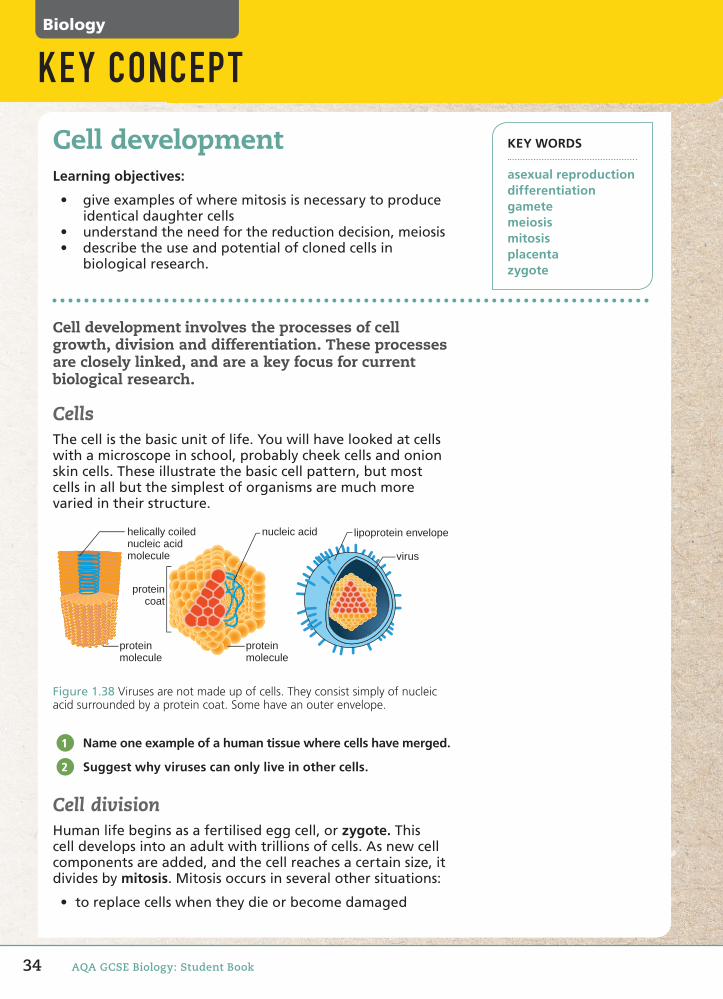
CellsThecellisthebasicunitoflife.Youwillhavelookedatcellswithamicroscopeinschool,probablycheekcellsandonionskincells.Theseillustratethebasiccellpattern,butmostcellsinallbutthesimplestoforganismsaremuchmorevariedintheirstructure.
proteincoat
nucleic acidhelically coilednucleic acidmolecule
proteinmolecule
virus
lipoprotein envelope
pproteincoat
nuucleic molecu
proteinmolecule
Figure1.38Viruses are not made up of cells. They consist simply of nucleic acid surrounded by a protein coat. Some have an outer envelope.
Name one example of a human tissue where cells have merged.
Suggest why viruses can only live in other cells.
Cell division Humanlifebeginsasafertilisedeggcell,orzygote.Thiscelldevelopsintoanadultwithtrillionsofcells.Asnewcellcomponentsareadded,andthecellreachesacertainsize,itdividesbymitosis.Mitosisoccursinseveralothersituations:
• toreplacecellswhentheydieorbecomedamaged
1
2
34 AQA GCSE Biology: Student Book
58750_P012_053.indd 34 07/05/16 8:35 AM
Google search: 'cell development, cell division, cell differentiation'
• whensingle-celled,eukaryoticorganismsreproducebyasexual reproduction,forexampleyeast
• whencancercellsdivide• wheneukaryoticcellsarecloned.
Whenanorganismreproducessexually,thesexcells,orgametes,cannotbeproducedbymitosis.Iftheywere,thenumberofchromosomesinourcellswoulddoubleeverygeneration!Weneedanothertypeofcelldivision,calledmeiosis.
Give three examples of situations in which mitosis occurs.
Name one type of cell that does not divide by mitosis.
Cell differentiationCellsmustbecomespecialisedforthedevelopmentofcomplex,multicellularorganisms.
Cellsthatcandifferentiateintoothercelltypesarecalledstemcells.Embryonicstemcells,foundafterfourtofivedays,candevelopintoalmostanycelltype.Wecan’tsay‘all’,astheycan’tdevelopintocellsoftheplacenta.
Stemcellshavethepotentialtoproduceanunlimitedamountoftissuefortransplants.Theyarealsoimportantinmedicalresearchsuchasonhowcellsdifferentiate,andinthetestingofdrugs.
Stemcellresearchandtreatmentswillrequirethecloningofcells.Somepeopleobjecttotheideaofthesetechniquesformoralandethicalreasons.
Cancercellsdivideuncontrollablybymitosisanddonotdifferentiateintomature,specialisedcells.Cancercellsearlyinthedevelopmentofthediseasecanlookalmostnormal,butinadvancedcancers,differentiationinmostcellsisverylimited.
Why are some news articles that suggest that embryonic stem cells can differentiate into all cell types, strictly speaking, incorrect?
Stem cells are being used to test new drugs. What are the advantages of using human stem cells over using rats to test drugs?
3
4
5
6
KEY SKILLS
For each chapter in the book, map out how different concepts you have learnt link with each other. Use a large sheet of paper or computer software.
1.11
KEY CONCEPT
35
58750_P012_053.indd 35 07/05/16 8:35 AM
36 AQA GCSE Biology: Student Book
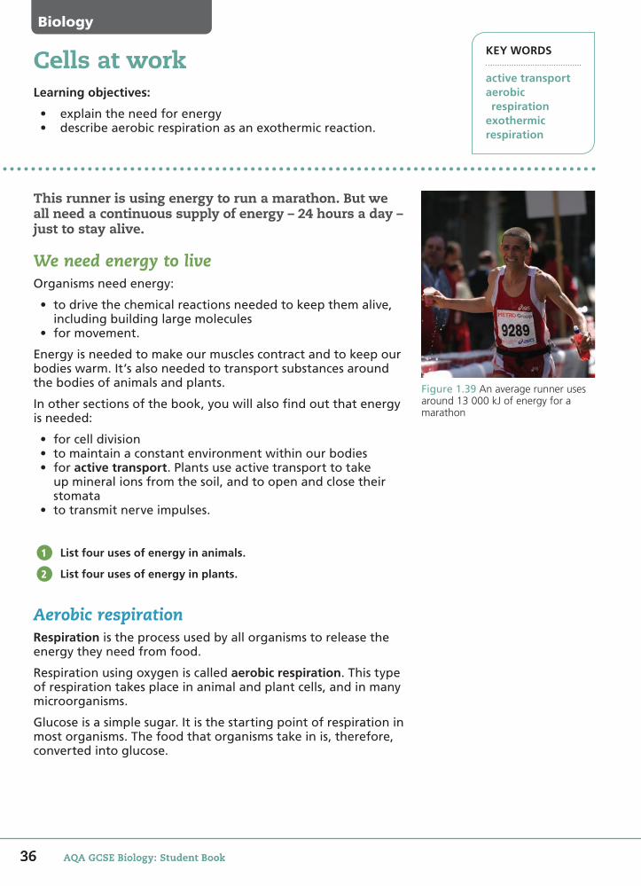
This runner is using energy to run a marathon. But we all need a continuous supply of energy – 24 hours a day – just to stay alive.
We need energy to live Organismsneedenergy:
• todrivethechemicalreactionsneededtokeepthemalive,includingbuildinglargemolecules
• formovement.
Energyisneededtomakeourmusclescontractandtokeepourbodieswarm.It’salsoneededtotransportsubstancesaroundthebodiesofanimalsandplants.
Inothersectionsofthebook,youwillalsofindoutthatenergyisneeded:
• forcelldivision• tomaintainaconstantenvironmentwithinourbodies• foractive transport.Plantsuseactivetransporttotake
upmineralionsfromthesoil,andtoopenandclosetheirstomata
• totransmitnerveimpulses.
List four uses of energy in animals.
List four uses of energy in plants.
Aerobic respirationRespirationistheprocessusedbyallorganismstoreleasetheenergytheyneedfromfood.
Respirationusingoxygeniscalledaerobic respiration.Thistypeofrespirationtakesplaceinanimalandplantcells,andinmanymicroorganisms.
Glucoseisasimplesugar.Itisthestartingpointofrespirationinmostorganisms.Thefoodthatorganismstakeinis,therefore,convertedintoglucose.
1
2
Learning objectives:
• explaintheneedforenergy• describeaerobicrespirationasanexothermicreaction.
Cells at work KEY WORDS
active transportaerobic respirationexothermicrespiration
Figure1.39An average runner uses around 13 000 kJ of energy for a marathon
Biology
36 AQA GCSE Biology: Student Book
58750_P012_053.indd 36 07/05/16 8:35 AM
37Google search: 'aerobic respiration'
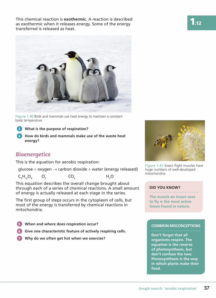
1.12Thischemicalreactionisexothermic.Areactionisdescribedasexothermicwhenitreleasesenergy.Someoftheenergytransferredisreleasedasheat.
Figure1.40Birds and mammals use heat energy to maintain a constant body temperature
What is the purpose of respiration?
How do birds and mammals make use of the waste heat energy?
BioenergeticsThisistheequationforaerobicrespiration:
glucose+oxygen→carbondioxide+water(energyreleased)
C6H12O6 O2 CO2 H2O
Thisequationdescribestheoverallchangebroughtaboutthrougheachofaseriesofchemicalreactions.Asmallamountofenergyisactuallyreleasedateachstageintheseries.
Thefirstgroupofstepsoccursinthecytoplasmofcells,butmostoftheenergyistransferredbychemicalreactionsinmitochondria.
When and where does respiration occur?
Give one characteristic feature of actively respiring cells.
Why do we often get hot when we exercise?
3
4
Figure1.41Insect flight muscles have huge numbers of well-developed mitochondria
COMMON MISCONCEPTIONS
Don’t forget that all organisms respire. The equation is the reverse of photosynthesis, but don’t confuse the two. Photosynthesis is the way in which plants make their food.
DID YOU KNOW?
The muscle an insect uses to fly is the most active tissue found in nature.
5
6
7
37
58750_P012_053.indd 37 07/05/16 8:35 AM
38 AQA GCSE Biology: Student Book
Biology
KEY WORDS
anaerobic respirationfermentation
Living without oxygenLearning objectives:
• describetheprocessofanaerobicrespiration• comparetheprocessesofaerobicandanaerobic
respiration.
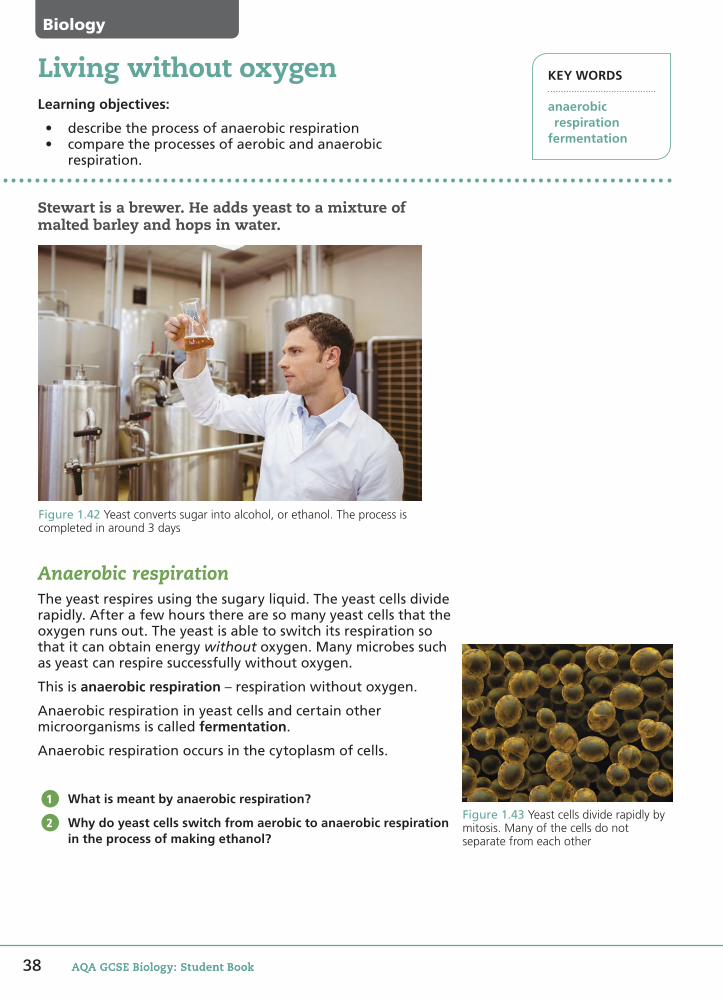
Stewart is a brewer. He adds yeast to a mixture of malted barley and hops in water.
Figure1.42Yeast converts sugar into alcohol, or ethanol. The process is completed in around 3 days
Anaerobic respirationTheyeastrespiresusingthesugaryliquid.Theyeastcellsdividerapidly.Afterafewhourstherearesomanyyeastcellsthattheoxygenrunsout.Theyeastisabletoswitchitsrespirationsothatitcanobtainenergywithoutoxygen.Manymicrobessuchasyeastcanrespiresuccessfullywithoutoxygen.
Thisisanaerobic respiration–respirationwithoutoxygen.
Anaerobicrespirationinyeastcellsandcertainothermicroorganismsiscalledfermentation.
Anaerobicrespirationoccursinthecytoplasmofcells.
What is meant by anaerobic respiration?
Why do yeast cells switch from aerobic to anaerobic respiration in the process of making ethanol?
1
2Figure1.43Yeast cells divide rapidly by mitosis. Many of the cells do not separate from each other
AQA GCSE Biology: Student Book
58750_P012_053.indd 38 07/05/16 8:36 AM
39
1.13
Google search: 'anaerobic respiration in yeast, anaerobic respiration in muscle, fermentation'
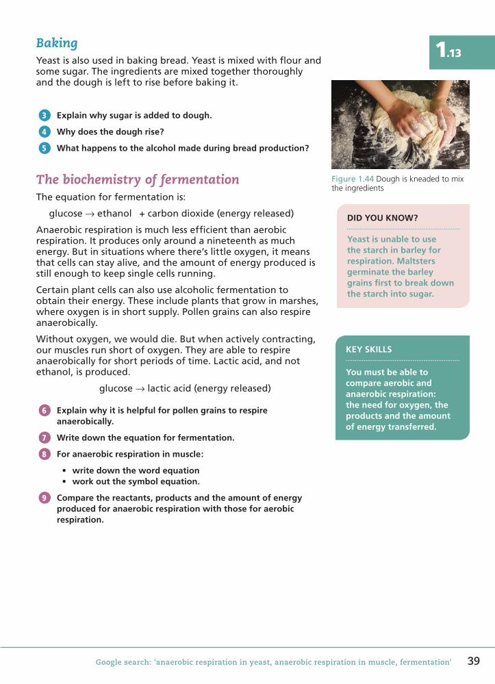
BakingYeastisalsousedinbakingbread.Yeastismixedwithflourandsomesugar.Theingredientsaremixedtogetherthoroughlyandthedoughislefttorisebeforebakingit.
Explain why sugar is added to dough.
Why does the dough rise?
What happens to the alcohol made during bread production?
The biochemistry of fermentationTheequationforfermentationis:
glucose→ethanol +carbondioxide(energyreleased)
Anaerobicrespirationismuchlessefficientthanaerobicrespiration.Itproducesonlyaroundanineteenthasmuchenergy.Butinsituationswherethere’slittleoxygen,itmeansthatcellscanstayalive,andtheamountofenergyproducedisstillenoughtokeepsinglecellsrunning.
Certainplantcellscanalsousealcoholicfermentationtoobtaintheirenergy.Theseincludeplantsthatgrowinmarshes,whereoxygenisinshortsupply.Pollengrainscanalsorespireanaerobically.
Withoutoxygen,wewoulddie.Butwhenactivelycontracting,ourmusclesrunshortofoxygen.Theyareabletorespireanaerobicallyforshortperiodsoftime.Lacticacid,andnotethanol,isproduced.
glucose→lacticacid(energyreleased)
Explain why it is helpful for pollen grains to respire anaerobically.
Write down the equation for fermentation.
For anaerobic respiration in muscle:
• write down the word equation• work out the symbol equation.
Compare the reactants, products and the amount of energy produced for anaerobic respiration with those for aerobic respiration.
Figure1.44Dough is kneaded to mix the ingredients
3
4
5
DID YOU KNOW?
Yeast is unable to use the starch in barley for respiration. Maltsters germinate the barley grains first to break down the starch into sugar.
KEY SKILLS
You must be able to compare aerobic and anaerobic respiration: the need for oxygen, the products and the amount of energy transferred.
6
7
8
9
58750_P012_053.indd 39 07/05/16 8:36 AM
40 AQA GCSE Biology: Student Book
Learning objectives:
• describe the techniques used to produce uncontaminated cultures of microorganisms
• describe how bacteria reproduce by binary fission• calculate the number of bacteria in a population.
Growing microorganisms
We’re most familiar with bacteria through the tiny minority of species that cause disease.
But harmless bacteria help us to live healthily. In our digestive system, they prevent harmful bacteria from gaining a foothold in our bodies and also produce essential nutrients.
Culturing bacteriaOwing to their size, it’s best to grow bacteria in large numbers to study them. Bacteria are grown in culture, in or on a culture medium.
The culture medium is a liquid, such as nutrient broth, or a gel called agar. Different nutrients can be added to the agar. Because it’s a gel, the agar contains the water required for the bacteria to grow.
All the equipment used must be sterilised. To make sure cultures and samples are kept uncontaminated by other microorganisms, and do not contaminate the environment:
• The inoculating loop must be sterilised by passing it through a Bunsen flame before and after use.
• The lid of the agar plate must be secured, but not sealed using adhesive tape.
After an investigation, agar plates are sterilised in an autoclave before disposal.
Explain why scientists need to work with uncontaminated cultures.
What piece of equipment is used to transfer bacteria from a culture to an agar plate?
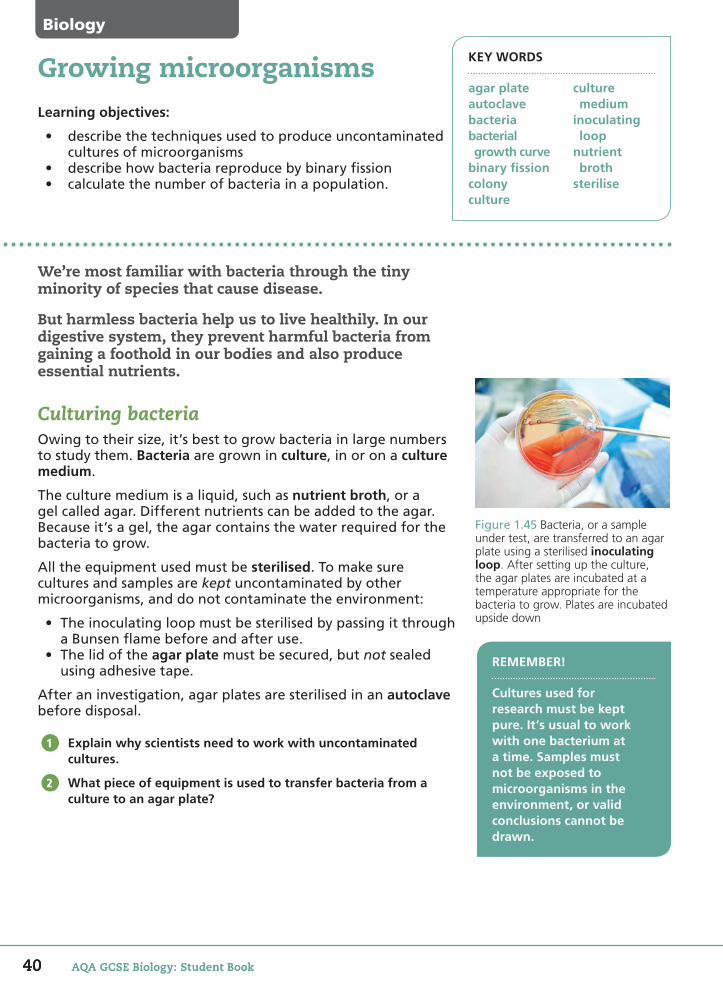
Figure 1.45 Bacteria, or a sample under test, are transferred to an agar plate using a sterilised inoculating loop. After setting up the culture, the agar plates are incubated at a temperature appropriate for the bacteria to grow. Plates are incubated upside down
1
REMEMBER!
Cultures used for research must be kept pure. It’s usual to work with one bacterium at a time. Samples must not be exposed to microorganisms in the environment, or valid conclusions cannot be drawn.
2
KEY WORDS
agar plateautoclavebacteriabacterial growth curvebinary fissioncolonyculture
culture medium inoculating loopnutrient brothsterilise
Biology
40 AQA GCSE Biology: Student Book
58750_P012_053.indd 40 5/7/16 12:28 PM

41Google search: 'antibiotic sensitivity testing'
1.14Quantitative studies with microorganismsWhensuppliedwithnutrientsandasuitabletemperature,bacteriawillmultiply.Theydothisbydividingintotwo.Theprocessiscalledbinary fission.Thisisnotthesameasmitosisineukaryoticcells.Binaryfissioninvolvesprokaryoteswithasinglechromosome.
Alivebacteriumlandingonthesurfaceofagarwilldividerepeatedlytoformacolony.Acolonycontainsmillionsofbacteria.
For a bacterium where the mean division time is 20 minutes:0 minutes 20 minutes 40 minutes 60 minutes
1bacterium
2bacteria
4bacteria
8bacteria
Figure1.46Binary fission of a bacterium
A bacterium has a mean division time of 20 minutes. Starting with one bacterium, how long would it take to produce a million bacteria?
If the mean mass of a bacterium is 1 × 10–12 g, estimate the mass of bacteria produced in Question 3. Suggest why the true mass is likely to be lower.
Bacterial growth curvesWiththeoptimumconditions,somebacteriacandivideintotwoasoftenasevery20minutes.Estimatesofcellsincultureareplottedtoproduceabacterial growth curve.
Valuesonthey-axisarethelogarithmsofthenumbersinthepopulation.Otherwise,therangeofnumberswouldbetoolargetofitappropriatelyontothescale.
Afteracertaintime,theculturemayreachitsstationaryphase.Binaryfissionslowsasfoodbeginstorunoutandwasteproductsbuildup.
What is meant by exponential growth? Name a process which causes exponential growth.
Some cultures enter a death phase. Suggest the possible causes.
Predict what would happen if you introduced more food during the stationary phase.
DID YOU KNOW?
The Human Microbiome Project is cataloguing the genes of the microorganism population in our intestines. There are 100 times the number of species originally thought to be present.
3
4
5
6
7Figure1.47The growth curve of a bacterium
During the initial ‘lag phase’ there is no reproduction. The bacteria are copying DNA and proteins
within their single cells.
Exponential growth phase followed by ‘stationary phase’ where resources
become scarce and bacteria die at same rate
as being produced.
The ‘death phase’ occurs as bacteria are
poisoned by the build-up of toxins in
the culture.
Num
ber
of b
acte
ria
Time
exponentialgrowth phase
41
58750_P012_053.indd 41 07/05/16 8:36 AM

42 AQA GCSE Biology: Student Book
Bacteria are becoming resistant to antibiotics. A 2015 government report suggested that by 2050, 10 million people worldwide may die every year from diseases we can no longer cure.Scientists are looking for new antibiotics to treat antibiotic-resistant bacteria.
Antibiotic sensitivity testingThemethodusedtotesttheeffectivenessofanantibioticisthedisc-diffusiontechnique.
Figure1.48An agar plate is inoculated with the bacterium being tested and spread evenly across the plate. It is not incubated at this stage
Adiscoffilterpaperisimpregnatedwiththeantibiotic.Severalconcentrationsoftheantibioticaretested.
Youcandothisintheschoollabbyimmersingthefilterpaperdiscinasolutionoftheantibiotic,andallowingtheantibiotictodrainoff.
Thediscisplacedonthesurfaceofanagarplatecontainingthebacteriumbeingtested.
The metal spreader is heated in a Bunsen flame before use and allowed to cool before spreading the bacteria. Suggest why.
When setting up the agar plate, explain why it is inoculated but not incubated.
1
2
Learning objectives:
• useappropriateapparatustoinvestigatetheeffectofantibioticsonbacterialgrowth
• usemicroorganismssafely• applysamplingtechniquestoensurethatsamplesare
representative.
Testing new antibiotics KEY WORDS
antibioticpathogensampling techniques
Biology
42 AQA GCSE Biology: Student Book
58750_P012_053.indd 42 07/05/16 8:36 AM

43Google search: 'antimicrobial resistance (AMR), antibiotic sensitivity testing (AST)'
1.15
Selecting the most appropriate apparatus and techniquesTheapparatusandsampling techniquesaprofessionalscientistwouldusetocarryoutthisinvestigationarealmostidenticaltothoseyouwoulduse.However,therearetwoimportantdifferences:
• ThestandardmediumfortestingantibioticsisMueller–Hintonagar,oftenwithaddedblood.Itcontainsbeefandmilkproteinandisidealforculturinghumanpathogens.
• Agarplatesareincubatedat37°C–humanbodytemperature.Samplesinschoolmustneverbeincubatedabove25°Cbecauseoftheriskofgrowingpathogens.
Name three ingredients of Mueller–Hinton agar.
Suggest why scientists use no more than 12 discs per plate.
Ensuring the investigation is validOneofthemostdangerousbacteriashowingantibioticresistanceismethicillin-resistantStaphylococcus aureus(MRSA).Asampleofbacteriafortestingmustberepresentativeofthepopulationofbacteria.
Ifthebacteriaarespreadappropriately,theclearzoneswillbeuniformlycircularandtherewillbecontinuousgrowthofbacteriaacrosstheremainderoftheplate.Measurementsaremadewitharulerorcallipers.Anyplateswherezonesarenotcircular,orwherethereispoorgrowthofthebacterium,shouldbediscarded.
What is meant by a representative sample? Why is it important that the sample is representative?
How is a representative sample of the bacterial culture taken?
Why is MRSA considered such a dangerous bacteria?
3
4
REMEMBER!
You should be able to describe the apparatus and techniques used when testing the effects of antibiotics, antiseptics and disinfectants.
5
6
DID YOU KNOW?
Two main strains of MRSA have caused problems in British hospitals since the 1990s. EMRSA16 is the most common form.
7
disc of filter paperwith antibiotic
antibiotic diffuses into andthrough the agar
The bacterium in the agar plate on theleft is sensitive to the antibiotic.
The bacterium in the agar plate on theright shows resistance to the antibiotic.
incubation
Figure1.49The larger the clear area, the more effective the antibiotic
Figure1.50The sample of Staphylococcus aureus transferred for testing must be from a colony that looks identical to others on the plate
43
58750_P012_053.indd 43 07/05/16 8:36 AM

Biology
KEY WORDS
antisepticdiffusionincubation
For use in a hospital, choosing the right disinfectant or antiseptic to achieve the appropriate hygiene levels is essential. The correct dilution is also important: a concentration high enough to work, but not so high as to be wasteful.
Setting up a disc-diffusion investigationScientists need a number of different skills to carry out this investigation. This section looks at some of those skills.
The method used to test the effectiveness of a disinfectant (or an antiseptic or antibiotic) is the disc-diffusion technique.
In this experiment, different concentrations of the disinfectant sodium hypochlorite are investigated.
In the investigation, which is the independent variable and which is the dependent variable?
Suggest the other possible variables that need to be controlled.
Health and safetyBefore scientists can begin a disc-diffusion investigation, they must carry out a risk assessment.
Hazard Type of hazard Risk Safety precautions
Ethanol
Sodium hypochlorite
Bacteria
Agar plate
Add more rows to include the activities involved, e.g. � aming an inoculating loop.
Complete the risk assessment table.
Suggest why:• scientists would use Mueller–Hinton blood agar; in the
school lab, you would use nutrient agar• you would incubate the plate at 25°C; the scientists at 37°C.
1
2
3
4
Learning objectives:
• carry out experiments with due regard to health and safety• present and process data, identifying anomalous results• evaluate methods and suggest further investigations.
Investigating disinfectants
paper discsoaked indisinfectant
bacteriagrowing onagar plate
paper discsoaked indisinfectant
clear zone –area wherebacteria didnot grow
dimension measured
agar plateinoculated
with bacteria
Figure 1.51 Disc-diffusion technique
These pages are designed to help you think about aspects of the investigation rather than to guide you through it step by step.
REQUIRED PRACTICAL
44 AQA GCSE Biology: Student Book
58750_P012_053.indd 44 5/7/16 12:29 PM
REQUIRED PRACTICAL
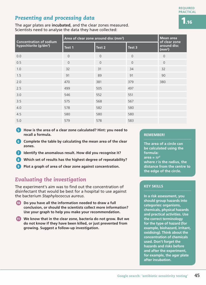
1.16Presenting and processing dataTheagarplatesareincubated,andtheclearzonesmeasured.Scientistsneedtoanalysethedatatheyhavecollected:
Concentration of sodium hypochlorite (g/dm3)
Area of clear zone around disc (mm2) Mean area of clear zone around disc (mm2)Test 1 Test 2 Test 3
0.0 0 0 0 0
0.5 0 0 0 0
1.0 32 31 34 32
1.5 91 89 91 90
2.0 470 381 379 380
2.5 499 505 497
3.0 546 552 551
3.5 575 568 567
4.0 578 582 580
4.5 580 580 580
5.0 579 578 583
How is the area of a clear zone calculated? Hint: you need to recall a formula.
Complete the table by calculating the mean area of the clear zones.
Identify the anomalous result. How did you recognise it?
Which set of results has the highest degree of repeatability?
Plot a graph of area of clear zone against concentration.
Evaluating the investigationTheexperiment’saimwastofindouttheconcentrationofdisinfectantthatwouldbebestforahospitaltouseagainstthebacteriumStaphylococcus aureus.
Do you have all the information needed to draw a full conclusion, or should the scientists collect more information? Use your graph to help you make your recommendation.
We know that in the clear zone, bacteria do not grow. But we do not know if they have been killed, or just prevented from growing. Suggest a follow-up investigation.
5
6
7
8
9
10
11
KEY SKILLS
In a risk assessment, you should group hazards into categories: organisms, chemicals, physical hazards and practical activities. Use the correct terminology for the type of hazard (for example, biohazard, irritant, oxidising). Think about the concentration of chemicals used. Don’t forget the hazards and risks before and after the experiment, for example, the agar plate after incubation.
Google search: 'antibiotic sensitivity testing'
REMEMBER!
The area of a circle can be calculated using the formula:area = πr2
where r is the radius, the distance from the centre to the edge of the circle.
45
58750_P012_053.indd 45 07/05/16 8:36 AM
MATHS SKILLS
KEY WORDS
calibrate graticule haemocytometerstandard form
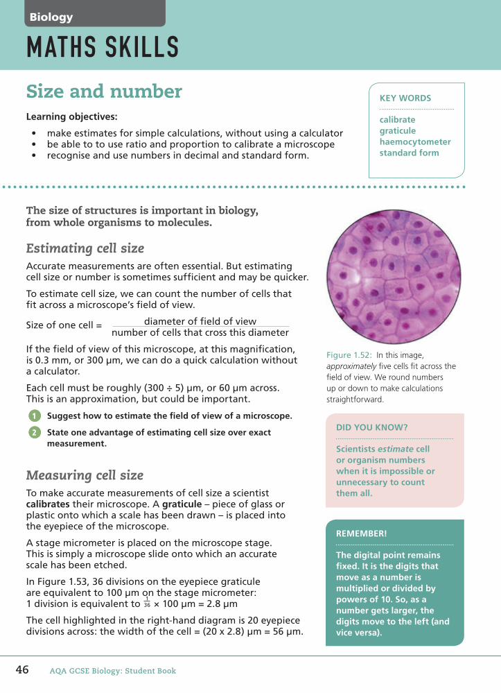
Figure 1.52: In this image, approximately fi ve cells fi t across the fi eld of view. We round numbers up or down to make calculations straightforward.
The size of structures is important in biology, from whole organisms to molecules.
Estimating cell sizeAccurate measurements are often essential. But estimating cell size or number is sometimes suffi cient and may be quicker.
To estimate cell size, we can count the number of cells that fi t across a microscope’s fi eld of view.
Size of one cell = diameter of fi eld of view number of cells that cross this diameter
If the fi eld of view of this microscope, at this magnifi cation, is 0.3 mm, or 300 µm, we can do a quick calculation without a calculator.
Each cell must be roughly (300 ÷ 5) µm, or 60 µm across. This is an approximation, but could be important.
Suggest how to estimate the fi eld of view of a microscope.
State one advantage of estimating cell size over exact measurement.
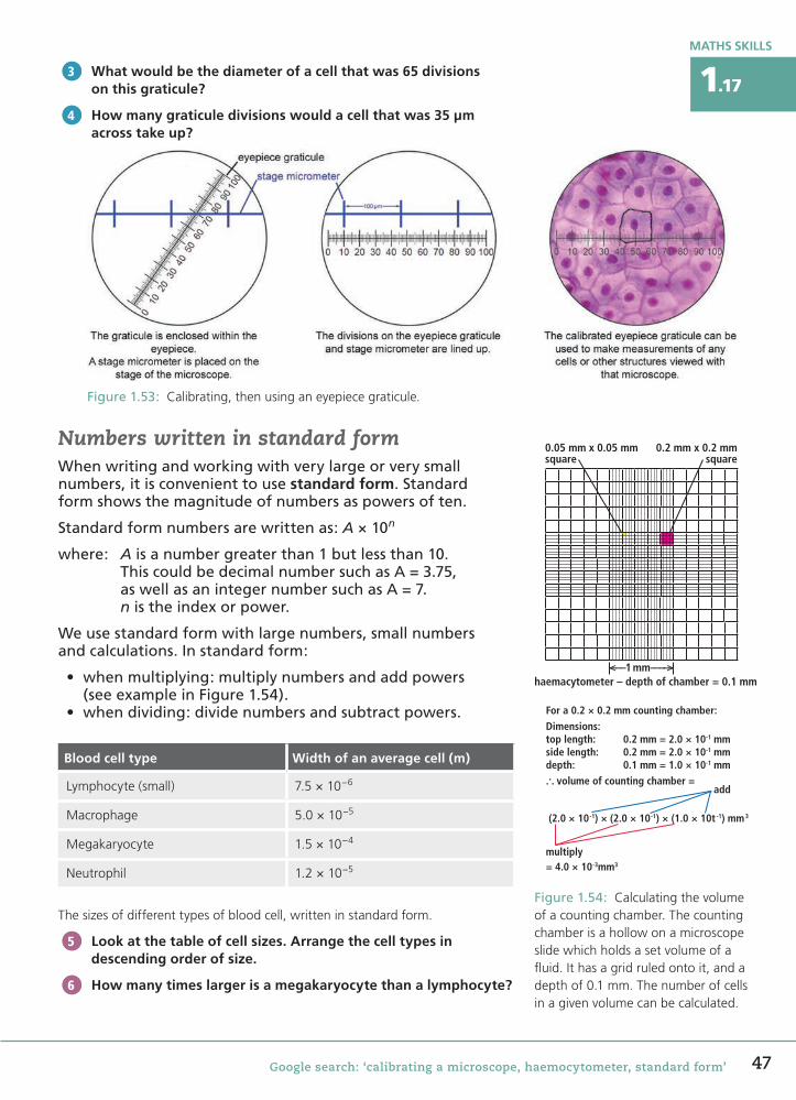
Measuring cell sizeTo make accurate measurements of cell size a scientist calibrates their microscope. A graticule – piece of glass or plastic onto which a scale has been drawn – is placed into the eyepiece of the microscope.
A stage micrometer is placed on the microscope stage. This is simply a microscope slide onto which an accurate scale has been etched.
In Figure 1.53, 36 divisions on the eyepiece graticule are equivalent to 100 µm on the stage micrometer:1 division is equivalent to
136 × 100 µm = 2.8 µm
The cell highlighted in the right-hand diagram is 20 eyepiece divisions across: the width of the cell = (20 x 2.8) µm = 56 µm.
11
12
Size and numberLearning objectives:
• make estimates for simple calculations, without using a calculator• be able to to use ratio and proportion to calibrate a microscope• recognise and use numbers in decimal and standard form.
REMEMBER!
The digital point remains fi xed. It is the digits that move as a number is multiplied or divided by powers of 10. So, as a number gets larger, the digits move to the left (and vice versa).
DID YOU KNOW?
Scientists estimate cell or organism numbers when it is impossible or unnecessary to count them all.
Biology
46 AQA GCSE Biology: Student Book
Biology Maths Skills.indd 46 5/7/16 8:06 AM
MATHS SKILLS
1.17
Google search: ‘calibrating a microscope, haemocytometer, standard form’
What would be the diameter of a cell that was 65 divisions on this graticule?
How many graticule divisions would a cell that was 35 µm across take up?
Numbers written in standard formWhen writing and working with very large or very small numbers, it is convenient to use standard form. Standard form shows the magnitude of numbers as powers of ten.
Standard form numbers are written as: A × 10n
where: A is a number greater than 1 but less than 10. This could be decimal number such as A = 3.75, as well as an integer number such as A = 7. n is the index or power.
We use standard form with large numbers, small numbers and calculations. In standard form:
• when multiplying: multiply numbers and add powers (see example in Figure 1.54).
• when dividing: divide numbers and subtract powers.
Blood cell type Width of an average cell (m)
Lymphocyte (small) 7.5 × 10⁻⁶
Macrophage 5.0 × 10⁻⁵
Megakaryocyte 1.5 × 10⁻⁴
Neutrophil 1.2 × 10⁻⁵
The sizes of different types of blood cell, written in standard form.
Look at the table of cell sizes. Arrange the cell types in descending order of size.
How many times larger is a megakaryocyte than a lymphocyte?
13
14
15
16
0.05 mm x 0.05 mmsquare
0.2 mm x 0.2 mmsquare
<–––1 mm–––––>haemacytometer – depth of chamber = 0.1 mm
For a 0.2 × 0.2 mm counting chamber:Dimensions:top length: 0.2 mm = 2.0 × 10⁻¹ mmside length: 0.2 mm = 2.0 × 10⁻¹ mmdepth: 0.1 mm = 1.0 × 10⁻¹ mm... volume of counting chamber =
add
(2.0 × 10-1) × (2.0 × 10-1) × (1.0 × 10t -1) mm3
multiply= 4.0 × 10⁻³mm3
Figure 1.53: Calibrating, then using an eyepiece graticule.
Figure 1.54: Calculating the volume of a counting chamber. The counting chamber is a hollow on a microscope slide which holds a set volume of a fluid. It has a grid ruled onto it, and a depth of 0.1 mm. The number of cells in a given volume can be calculated.
47
Biology Maths Skills.indd 47 5/7/16 1:40 PM

48 AQA GCSE Biology: Student Book
Biology
Check your progress
You should be able to:
� describe the functions of the sub-cellular structures found in eukaryotic cells �
� understand the size and scale of cells and be able to use and convert units �
� carry out order of magnitude calculations when comparing cell size; calculate with numbers in standard form
� calculate magnification used by a light microscope using eyepiece and objective lens magnifications
� � calculate the magnification of a light or electron micrograph �
� explain limitations of light microscopy and advantages of electron microscopy
� describe the structure of a prokaryotic cell
�
� describe the differences between eukaryotic and prokaryotic cells �
� explain why scientists have now separated organisms into three domains using evidence from chemical analysis
� recall that cells must divide for growth and replacement of cells
�
� describe how chromosomes double their DNA and are pulled to opposite ends of the cell, before the cytoplasm divides, during mitosis
�
� describe the events of the cell cycle and explain the synthesis of new sub-cellular components and DNA
� recall that organism development is based on cell division and cell specialisation
� � explain the importance of differentiation and explain how cells are specialised for their functions
� � understand size and scale in the components of organ systems
� recall where stem cells are found �
� understand the potential of stem cell therapies �
� evaluate scientific and ethical issues involved with stem cell therapies
� recall that organisms can respire with oxygen (aerobic respiration) or without oxygen (anaerobic respiration)
�
� use word equations to describe the processes of aerobic and anaerobic respiration
�
� use symbol equations for aerobic and anaerobic respiration and be able to compare the two processes
� describe equipment, materials and procedures required to work with microorganisms
� � describe the process of binary fission
� � be able to calculate numbers of microorganisms produced given the mean generation time
58750_P012_053.indd 48 07/05/16 8:36 AM
49
Worked example
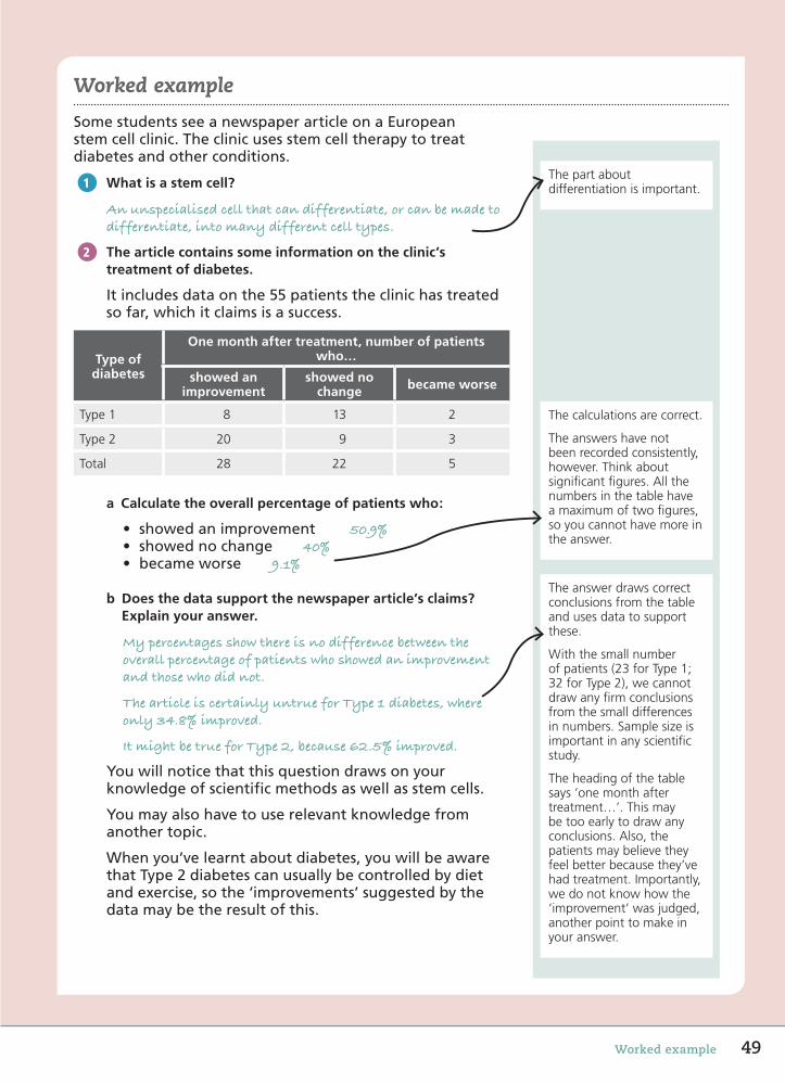
The part about differentiation is important.
The calculations are correct.
The answers have not been recorded consistently, however. Think about significant figures. All the numbers in the table have a maximum of two figures, so you cannot have more in the answer.
The answer draws correct conclusions from the table and uses data to support these.
With the small number of patients (23 for Type 1; 32 for Type 2), we cannot draw any firm conclusions from the small differences in numbers. Sample size is important in any scientific study.
The heading of the table says ‘one month after treatment…’. This may be too early to draw any conclusions. Also, the patients may believe they feel better because they’ve had treatment. Importantly, we do not know how the ‘improvement’ was judged, another point to make in your answer.
Worked example
SomestudentsseeanewspaperarticleonaEuropeanstemcellclinic.Theclinicusesstemcelltherapytotreatdiabetesandotherconditions.
What is a stem cell?
An unspecialised cell that can differentiate, or can be made to differentiate, into many different cell types.
The article contains some information on the clinic’s treatment of diabetes.
Itincludesdataonthe55patientstheclinichastreatedsofar,whichitclaimsisasuccess.
Type of diabetes
One month after treatment, number of patients who…
showed an improvement
showed no change became worse
Type 1 8 13 2
Type 2 20 9 3
Total 28 22 5
a Calculate the overall percentage of patients who:
• showed an improvement 50.9%• showed no change 40%• became worse 9.1%
b Does the data support the newspaper article’s claims? Explain your answer.
My percentages show there is no difference between the overall percentage of patients who showed an improvement and those who did not.
The article is certainly untrue for Type 1 diabetes, where only 34.8% improved.
It might be true for Type 2, because 62.5% improved.
Youwillnoticethatthisquestiondrawsonyourknowledgeofscientificmethodsaswellasstemcells.
Youmayalsohavetouserelevantknowledgefromanothertopic.
Whenyou’velearntaboutdiabetes,youwillbeawarethatType2diabetescanusuallybecontrolledbydietandexercise,sothe‘improvements’suggestedbythedatamaybetheresultofthis.
1
2
58750_P012_053.indd 49 07/05/16 8:36 AM
End of chapter questions
Biology
50 AQA GCSE Biology: Student Book
Getting started
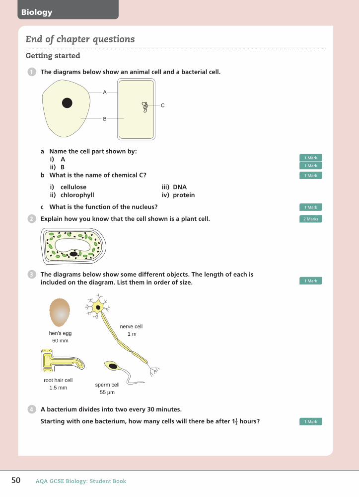
The diagrams below show an animal cell and a bacterial cell.
a Name the cell part shown by:i) Aii) B
b What is the name of chemical C?
i) celluloseii) chlorophyll
iii) DNAiv) protein
c What is the function of the nucleus?
Explain how you know that the cell shown is a plant cell.
The diagrams below show some different objects. The length of each is included on the diagram. List them in order of size.
hen’s egg60 mm
root hair cell1.5 mm sperm cell
55 µm
nerve cell 1 m
A bacterium divides into two every 30 minutes.
Starting with one bacterium, how many cells will there be after 1 hours?
1
C
A
B
1Mark
1Mark
1Mark
1Mark
2 2Marks
31Mark
4
12 1Mark
58750_P012_053.indd 50 07/05/16 8:36 AM

51End of chapter questions
Gemma is using Feulgen stain to stain some dividing cells.
a The label on the bottle of stain is shown.
IRRITATING TO EYES,RESPIRATORY SYSTEMAND SKIN
Explain two safety precautions she should take when using the stain.
Going further
Draw lines to match the two cell structures with their function.
Cell structure Function
Controls what enters and leaves cells
Mitochondrion
Respiration
Ribosome
Protein synthesis
Use words from the box to complete the word equation for aerobic respiration.
alcohol carbon dioxide glucose lactic acid
…………….. + oxygen → …………….. + water (+ energy)
Explain how the features of a sperm cell help it to fertilise an egg cell.
A scientist is investigating the effect of different sugars on the anaerobic respiration of yeast.
She measures the volume of carbon dioxide produced over a two hour period.
She investigates two types of sugar, sucrose and lactose.
plunger
plunger is pushed out
carbon dioxide collectsin gas syringe
5 cm3 of yeast suspension+ 25 cm3 of 10% sugar solution
5
2Marks
6 1Mark
71Mark
8 2Marks
9
58750_P012_053.indd 51 07/05/16 8:36 AM
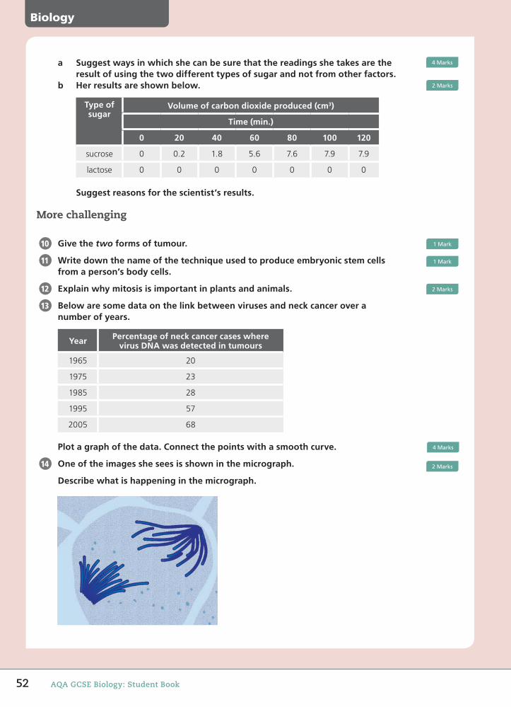
Biology
52 AQA GCSE Biology: Student Book
a Suggest ways in which she can be sure that the readings she takes are the result of using the two different types of sugar and not from other factors.
b Her results are shown below.
Type of sugar
Volume of carbon dioxide produced (cm3)
Time (min.)
0 20 40 60 80 100 120
sucrose 0 0.2 1.8 5.6 7.6 7.9 7.9
lactose 0 0 0 0 0 0 0
Suggest reasons for the scientist’s results.
More challenging
Give the two forms of tumour.
Write down the name of the technique used to produce embryonic stem cells from a person’s body cells.
Explain why mitosis is important in plants and animals.
Below are some data on the link between viruses and neck cancer over a number of years.
Year Percentage of neck cancer cases where virus DNA was detected in tumours
1965 20
1975 23
1985 28
1995 57
2005 68
Plot a graph of the data. Connect the points with a smooth curve.
One of the images she sees is shown in the micrograph.
Describe what is happening in the micrograph.
4Marks
2Marks
10 1Mark
11 1Mark
12 2Marks
13
4Marks
14 2Marks
58750_P012_053.indd 52 07/05/16 8:36 AM

53End of chapter questions
Most demanding
Explain how anaerobic respiration differs from aerobic respiration.
Some companies offer to store the stem cells found in the umbilical cord of a new born baby. These stem cells can be used for transplants or therapy for blood diseases. Suggest two possible advantages of using stem cells from an umbilical cord over using stem cells from bone marrow.
The graph below shows the correlation between colon cancer incidence and daily meat consumption. What conclusions can you draw from the graph? Evaluate the evidence for meat consumption affecting the incidence of colon cancer in females.
15 2Marks
16
2Marks
17
New Zealand
USA
Canada
UKDenmark
SwedenNetherlands
Iceland
Norway
IsraelGermany
Hungary
PolandPuerto RicoFinland
Jamaica
Yugoslavia
JapanChile
Romania
Colombia
Female Colon Cancer Incidence and Daily Meat Consumption
Per Capita Daily Meat Consumption (grams)
Col
on C
ance
r In
cide
nce/
100,
000
Wom
en
00
10
30
50
40 80 120 160 200 240 280 3206Marks
Total:40Marks
58750_P012_053.indd 53 07/05/16 8:36 AM