identification of the adipocyte acid phosphatase as a paoâ€sensitive
TRANSCRIPT

Protein Science (1992), I, 710-721. Cambridge University Press. Printed in the USA. Copyright 0 1992 The Protein Society
_ _ _ __
Identification of the adipocyte acid phosphatase as a PAO-sensitive tyrosyl phosphatase
~~ ~~~ .~ ~~ . ~. ~~
LAURIE L. SHEKELS,' ANNE J. SMITH,' ROBERT L. VAN ETTEN,* AND DAVID A. BERNLOHR' I Department of Biochemistry, University of Minnesota, St. Paul, Minnesota 55108
Department of Chemistry, Purdue University, West Lafayette, Indiana 47907
(RECEIVED November 20, 1991; REVISED MANUSCRIPT RECEIVED February 5 , 1992)
Abstract
We have partially purified an 18-kDa cytoplasmic protein from 3T3-Ll cells, which dephosphorylates pNPP and the phosphorylated adipocyte lipid binding protein (ALBP), and have identified it by virtue of kinetic and im- munological criteria as an acid phosphatase (EC 3.1.3.2). The cytoplasmic acid phosphatase was inactivated by phenylarsine oxide (PAO) (K,,,, = 10 pM), and the inactivation could be reversed by the dithiol, 2,3-dimercap- topropanol (K,,,,, = 23 pM), but not the monothiol, 2-mercaptoethanol. Cloning of the human adipocyte acid phosphatase revealed that two isoforms exist, termed HAAPa and HAAPP (human adipocyte acid phosphatase), which are distinguished by a 34-amino acid isoform-specific domain. Sequence analysis shows HAAPa and HAAPP share 74% and 90% identity with the bovine liver acid phosphatase, respectively, and 99% identity with both isoenzymes of the human red cell acid phosphatase but no sequence similarity to the protein tyrosine phos- phatases (EC 3.1.3.48). HAAPP has been cloned into Escherichia coli, expressed, and purified as a glutathione S-transferase fusion protein. Recombinant HAAPP was shown to dephosphorylate pNPP and phosphoALBP and to be inactivated by PA0 and inhibited by vanadate (K, = 17 pM). These results describe the adipocyte acid phos- phatase as a cytoplasmic enzyme containing conformationally vicinal cysteine residues with properties that sug- gest it may dephosphorylate tyrosyl phosphorylated cellular proteins.
Keywords: acid phosphatase; phenylarsine oxide; tyrosine phosphatases
Although it has been appreciated for several years that the insulin receptor functions as a ligand-activated pro- tein tyrosyl kinase, the exact steps in the insulin signal- ing pathway have remained elusive. The insulin signaling pathway initiates with the specific binding of insulin to the extracellular CY subunit of the insulin receptor and the subsequent activation of the insulin receptor tyrosyl ki-
~- ~ ~
Reprint requests to: David A. Bernlohr, Department of Biochemis- try, University of Minnesota, 1479 Gortner Avenue, St. Paul, Minne- sota 55108.
Abbreviations: PAO, phenylarsine oxide; HAAP, human adipocyte acid phosphatase; PTPase, protein tyrosine phosphatase; 2,3-DMP, 2,3- dimercaptopropanol; ALBP, adipocyte lipid binding protein; WGA, wheat germ agglutinin; IRK, insulin receptor kinase; BHAP, bovine heart acid phosphatase; BLAP, bovine liver acid phosphatase; pNPP, p-nitrophenyl phosphate; GST, glutathione S-transferase; GST-K- PTPU323, GST-rat brain PTPase fusion protein; BLOTTO, bovine
acid; PMSF, phenylmethylsulfonyl fluoride; HEPES, 4-(2-hydroxy- lacto transfer technique optimizer; EDTA, ethylenediamine tetraacetic
ethyl)-l-piperazine ethane sulfonic acid; SDS, sodium dodecyl sulfate; PAGE, polyacrylamide gel electrophoresis; SSC, standard saline citrate (0.15 M NaCI, 0.0015 M sodium citrate).
nase through autophosphorylation of one or more tyro- syl residues on the cytoplasmic domain of the /3 subunit (Ellis et al., 1986; Rosen, 1987). Following activation of the receptor, a cascade of regulatory phosphorylation events occurs that culminates in alterations in cellular amino acid, protein, carbohydrate, and lipid metabolism. Efforts to dissect the individual steps in this cascade have been concentrated on the identification of physiologically relevant substrates for the receptor kinase and their cor- responding phosphatases.
In 3T3-Ll adipocytes, PA0 inhibits insulin-stimulated but not basal hexose transport (Frost & Lane, 1985). Concomitant with insulin addition to PAO-treated cells, a cytoplasmic 15-kDa phosphorylated protein accumu- lates (Bernier et al., 1987). Reversal of P A 0 action with 2,3-DMP results in the loss of the phosphorylated 15-kDa protein and the reacquisition of insulin sensitivity. The 15-kDa protein has been identified as the ALBP (also referred to as 422 and aP2) (Hresko et al., 1988), a cy- toplasmic fatty acid binding protein belonging to a multi-
710

Adipocyte acid phosphatase 71 1
gene family of hydrophobic ligand binding proteins. The phosphorylation of ALBP occurs with identical proper- ties both in situ and in vitro (Chinander & Bernlohr, 1989; Buelt et al., 1991) suggesting that ALBP is a pri- mary substrate of the insulin receptor kinase. Together the in situ and in vitro observations suggest that follow- ing insulin addition to cultured 3T3-Ll adipocytes, ALBP is phosphorylated by the insulin receptor and that its subsequent dephosphorylation proceeds via the action of a PAO-sensitive tyrosyl phosphatase.
Approximately 90% of the phosphoALBP phospha- tase activity found in adipocytes is membrane associated (Liao et al., 1991). However, the existence of a soluble ty- rosine phosphatase that is responsible for phosphoALBP dephosphorylation cannot be ruled out based on several lines of evidence. Firstly, in situ, phosphoALBP from 3T3-Ll cells resides cytoplasmically, and, in vitro, puri- fied ALBP has no measurable affinity for membranes or artificial vesicles. Secondly, the bulk of the phosphatase activity in these cells is P A 0 insensitive, suggesting that if phosphoALBP were to associate with membranes it would be dephosphorylated readily. Lastly, Frost and Schwalbe (1990) have shown that in 3T3-Ll cells approx- imately half of the proteins modified by a tritium-labeled derivative of P A 0 were cytoplasmic. In light of these ob- servations, we considered whether any known cytoplas- mic phosphatases exhibited properties (i.e., active site sulfhydryl[s] and sensitivity to PAO) that would suggest that it may function as a phosphoALBP phosphatase.
Currently, there are two rapidly growing classes of pro- tein tyrosyl phosphatases. The first class (to be referred to here as the PTPases; for review see Hunter [1989]) shares sequence similarity within a 25-kDa catalytic do- main. They are sulfhydryl enzymes and include both re- ceptorlike and nonreceptor PTPases. The receptorlike PTPases such as CD45 (Tonks et al., 1990) are membrane spanning proteins and often possess two catalytic regions on the intracellular domain. The nonreceptor PTPases frequently reside in the cytosol and contain only a single catalytic domain. Recently, Guan et al. (1990) have re- ported the cloning of a rat brain PTPase and the charac- terization of its catalytic properties. The expression of the catalytic domain in Escherichia coli as a soluble GST fu- sion protein, GST-K-PTPU323, has greatly facilitated its molecular analysis (Guan & Dixon, 1991).
A second class of phosphatases consists of low molec- ular mass enzymes termed acid phosphatases. Although these 18-kDa enzymes have an acidic pH optimum, par- ticularly when measured with the acidic substrate pNPP, they are reasonably active on phosphotyrosyl protein sub- strates at pH 6-7 (Chernoff & Li, 1985; Boivin et al., 1987; Waheed et al., 1988). The acid phosphatases are also sulfhydryl enzymes (Laidler et al., 1982; Zhang & Van Etten, 1990; Wo et al., 1992); however, the sequences of the bovine liver protein (Camici et al., 1989) and the human red cell enzymes (Dissing et al., 1991) show no
similarity to the catalytic domain of the PTPases (Char- bonneau et al., 1989). The acid phosphatases contain eight cysteine residues all of which exist in the free thiol form despite the small size of the protein. The abundance of free sulfhydryls suggests that these enzymes may be sen- sitive t o P A 0 modification. Additionally, the cytosolic location of the acid phosphatases may also allow these enzymes to act as soluble phosphoALBP phosphatases.
The purpose of this study was to determine if adipo- cytes express a low molecular mass cytoplasmic acid phosphatase, to investigate its inactivation with PAO, and to examine its phosphoALBP dephosphorylation ac- tivity. We characterize here the cytoplasmic low molec- ular weight acid phosphatase from 3T3-Ll adipocytes and demonstrate that it is a PAO-sensitive phosphatase. Furthermore, the cloning and sequencing of the human adipocyte acid phosphatase (HAAP) has revealed the presence of two isoforms, HAAPa and HAAPP, both of which possess numerous cysteine residues similar to the liver, red cell, and heart enzymes.
Results
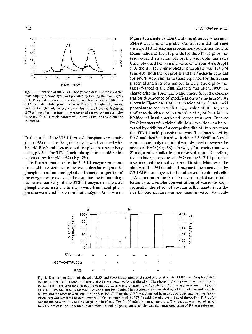
To test the hypothesis that adipocytes express an acid phosphatase capable of dephosphorylating phospho- ALBP, we have partially purified a soluble low molec- ular weight phosphatase from 3T3-Ll adipocytes using a modification of the procedures described by Taga and Van Etten (1982). In the purification, a soluble protein extract was collected following digitonin permeabilization of the adipocyte plasma membranes. Previous studies have shown that the acid phosphatase is most stable at pH 5- 6; consequently, a pH 5.0 soluble extract was obtained and used throughout the isolation. As shown in Figure 1, the phosphatase activity largely partitions with the low molecular weight fraction of the soluble adipocyte ex- tract. The 3T3-Ll phosphatase was then examined for its ability to utilize phosphoALBP as a substrate and for its sensitivity to PAO. When the partially purified 3T3-Ll phosphatase (specific activity, 5 unitslmg) was incubated with phosphoALBP, the substrate was dephosphorylated (Fig. 2A). To compare the dephosphorylation properties of the 3T3-Ll phosphatase to those of a member of the PTPase family of enzymes, the catalytic fragment of the rat brain phosphatase (Guan & Dixon, 1991) was utilized. The rat brain PTPase identified by Dixon and colleagues had previously been subcloned, expressed, and purified as a soluble GST fusion protein termed GST-K-PTPU323. As shown, when homogeneous GST-K-PTPU323 (spe- cific activity, 29 units/mg) was assayed for phosphatase activity, it was also capable of dephosphorylating phos- phoALBP (Fig. 2A). These results indicated that in vitro little specificity for dephosphorylation was evident. Con- sistent with this was our observation that a variety of membrane extracts contained active phosphoALBP phos- phatase activity (results not shown and Liao et al. [1991]).
712
.- 1 I -
, 0 1 0 20 30 4 0 5 0 6 0 7 0
Fraction Number
Fig. 1. Purification of the 3T3-Ll acid phosphatase. Cytosolic extract from adipocyte monolayers was prepared by treating the monolayers with 50 pg/mL digitonin. The digitonin releasate was acidified to pH 5.0 and the soluble protein recovered by centrifugation. Following delipidation, the soluble protein was fractionated over a Sephadex G-75 column. Column fractions were assayed for phosphatase activity using pNPP (0). Protein content was estimated by the absorbance at 280 nm ( 0 ) .
To determine if the 3T3-Ll tyrosyl phosphatase was sub- ject t o PA0 inactivation, the enzyme was incubated with 100 pM P A 0 and then assessed for phosphatase activity using pNPP. The 3T3-Ll acid phosphatase could be in- activated by 100 pM P A 0 (Fig. 2B).
To further characterize the 3T3-Ll enzyme prepara- tion and its relatedness to the low molecular weight acid phosphatases, immunological and kinetic properties of the enzyme were assessed. To examine the immunolog- ical cross-reactivity of the 3T3-Ll enzyme to the acid phosphatases, antisera to the bovine heart acid phos- phatase were used in western blot analysis. As shown in
L. L . Shekels et al.
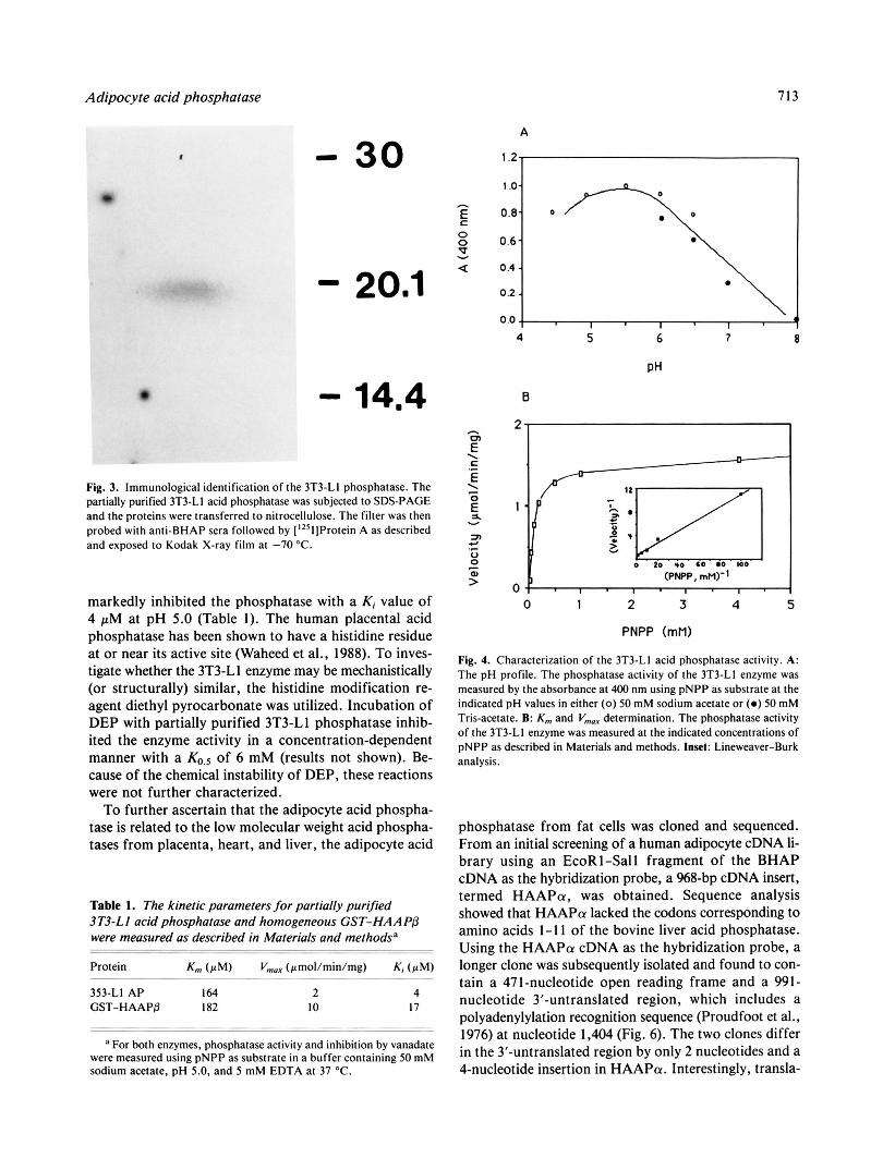
Figure 3, a single 18-kDa band was observed when anti- BHAP was used as a probe. Control sera did not react with the 3T3-Ll enzyme preparation (results not shown). Examination of the pH profile for the 3T3-Ll phospha- tase revealed an acidic pH profile with optimum rates being obtained between pH 4.5 and 7.5 (Fig. 4A). At pH 5.0, the K,,, for p-nitrophenyl phosphate was 164 pM (Fig. 4B). Both the pH profile and the Michaelis constant for pNPP were similar to those reported for the human placental and liver low molecular weight acid phospha- tases (Waheed et al., 1988; Zhang & Van Etten, 1990). To characterize the P A 0 inactivation more fully, the concen- tration dependence of modification was measured. As shown in Figure 5A, P A 0 inactivation of the 3T3-Ll acid phosphatase occurs with a K,,,,., value of 10 pM, very similar to the observed in situ value of 7 pM for P A 0 in- hibition of insulin-activated hexose transport. Because PA0 interacts with vicinal dithiols, its action can be re- versed by addition of a competing dithiol. In vitro when the 3T3-Ll acid phosphatase was first inactivated by P A 0 and then incubated with either 2,3-DMP or 2-mer- captoethanol only the dithiol was observed to reverse the action of P A 0 (Fig. 5B). The K,,,,, for reactivation was 23 pM, a value similar to that observed in situ. Therefore, the inhibitory properties of P A 0 on the 3T3-Ll phospha- tase mirrored the results observed in situ. Moreover, the ability of the PAO-inhibited enzyme to be reactivated by 2,3-DMP is analogous to that observed in cultured cells.
A common property of tyrosyl phosphatases is inhi- bition by micromolar concentrations of vanadate. Con- sequently, the effect of sodium orthovanadate on the 3T3-Ll phosphatase was examined in vitro. Vanadate
A B 100- - - - -100 ~
80- - 80 3.
60 - -60 2
- 4 0 rc 40 -
c -
z -.
20 - - 2 0 2 0 . . 7 0
3T3-Ll AP - + - + + - -
GST-K-PTPU323 - - + - - + +
PA0 - - - - + - +
Fig. 2. Dephosphorylation of phosphoALBP and P A 0 inactivation of the acid phosphatase. A: ALBP was phosphorylated by the soluble insulin receptor kinase, and ATP was removed by gel filtration. The phosphorylated proteins were then incu- bated in the presence or absence of 1 p g of the 3T3-Ll acid phosphatase (specific activity = 5 units/mg) for 60 rnin or 1 f i g of GST-K-PTPU323 (specific activity = 29 units/mg) for 40 min. The reactions were quenched by addition of Laemmli sample buffer, and the proteins were separated by SDS-PAGE. PhosphoALBP was visualized by autoradiography and the phosphory- lation level was measured by densitometry. B: One microgram of the 3T3-LI acid phosphatase or l pg of the GST-K-PTPU323 was incubated with 100 pM P A 0 at pH 8.8 in 10 mM Tris for 30 min at room temperature. The reaction was then adjusted to pH 5.0 as described in Materials and methods and the phosphatase activity was then measured using pNPP as a substrate.
Adipocyte acid phosphatase
- 30
- 20.1
Fig. 3. Immunological identification of the 3T3-LI phosphatase. The partially purified 3T3-LI acid phosphatase was subjected to SDS-PAGE and the proteins were transferred to nitrocellulose. The filter was then probed with anti-BHAP sera followed by ['2Sl]Protein A as described and exposed to Kodak X-ray film at -70 "C.
markedly inhibited the phosphatase with a Ki value of 4 pM at pH 5.0 (Table I ) . The human placental acid phosphatase has been shown to have a histidine residue at or near its active site (Waheed et al., 1988). To inves- tigate whether the 3T3-Ll enzyme may be mechanistically (or structurally) similar, the histidine modification re- agent diethyl pyrocarbonate was utilized. Incubation of DEP with partially purified 3T3-Ll phosphatase inhib- ited the enzyme activity in a concentration-dependent manner with a of 6 mM (results not shown). Be- cause of the chemical instability of DEP, these reactions were not further characterized.
To further ascertain that the adipocyte acid phospha- tase is related to the low molecular weight acid phospha- tases from placenta, heart, and liver, the adipocyte acid
Table 1. The kinetic parameters for partially purified 3T3-LI acid phosphatase and homogeneous GST-HAAPP were measured as described in Materials and methodsa
Protein Km ( rM) Vmar (rmol/min/mg) Ki (aM)
353-LI AP 164 2 4 GST-HAAPP 182 I O 17
a For both enzymes, phosphatase activity and inhibition by vanadate were measured using pNPP as substrate in a buffer containing 50 mM sodium acetate, pH 5.0, and 5 mM EDTA at 37 "C.
713
A
1 .o-
0.8-
0.6-
0.4 - 0.2 -
I 8
r J
(PNPP, mM)" 0 1 ' 1 . 1 - 1 .
0 20 *o 60 00 IO0
0 1 2 3 4 5
PNPP (mM)
Fig. 4. Characterization of the 3T3-LI acid phosphatase activity. A: The pH profile. The phosphatase activity of the 3T3-LI enzyme was measured by the absorbance at 400 nm using pNPP as substrate at the indicated pH values in either ( 0 ) 50 mM sodium acetate or (0 ) 50 mM Tris-acetate. B: K,,, and V,, determination. The phosphatase activity of the 3T3-LI enzyme was measured at the indicated concentrations of pNPP as described in Materials and methods. Inset: Lineweaver-Burk analysis.
phosphatase from fat cells was cloned and sequenced. From an initial screening of a human adipocyte cDNA li- brary using an EcoRl-Sal1 fragment of the BHAP cDNA as the hybridization probe, a %8-bp cDNA insert, termed HAAPol, was obtained. Sequence analysis showed that HAAPa lacked the codons corresponding to amino acids 1-1 1 of the bovine liver acid phosphatase. Using the HAAPol cDNA as the hybridization probe, a longer clone was subsequently isolated and found to con- tain a 471-nucleotide open reading frame and a 991- nucleotide 3"untranslated region, which includes a polyadenylylation recognition sequence (Proudfoot et al., 1976) at nucleotide 1,404 (Fig. 6). The two clones differ in the 3"untranslated region by only 2 nucleotides and a 4-nucleotide insertion in HAAPa. Interestingly, transla-

714 L.L. Shekels et al.
A
I 1
I 0 10 20 30 40 50 60 70 80 90 100
PA0 (pM)
6
$ 2 1 pM 10pM 2 0 p M 50pM I O O p M
Fig. 5. P A 0 inactivation and DMP reactivation of the 3T3-LI acid phosphatase. A: P A 0 inactivation. The 3T3-LI acid phosphatase was modified with the given concentrations of P A 0 at pH 8.8 for 30 min at room temperature. Following modification, the phosphatase activ- ity was measured at pH 5.0, using pNPP as described in Materials and methods. B: Reactivation of the 3T3-LI phosphatase. After modifica- tion with 50 p M PAO, the 3T3-LI acid phosphatase was incubated with the indicated concentrations of either 2-mercaptoethanol (filled bars) or 2,3-DMP (open bars) for 60 min. The phosphatase activity on pNPP was then assayed at pH 5.0.
tion of the open reading frame of the second clone, termed HAAPP, revealed that it encoded an isoform of HAAPa (Fig. 7). Overall the protein coding region of HAAPa shares 81% sequence similarity to HAAPP. A region of 34 amino acids constitutes the isoform-specific domain in which HAAPa and HAAPP share only 43% sequence similarity.
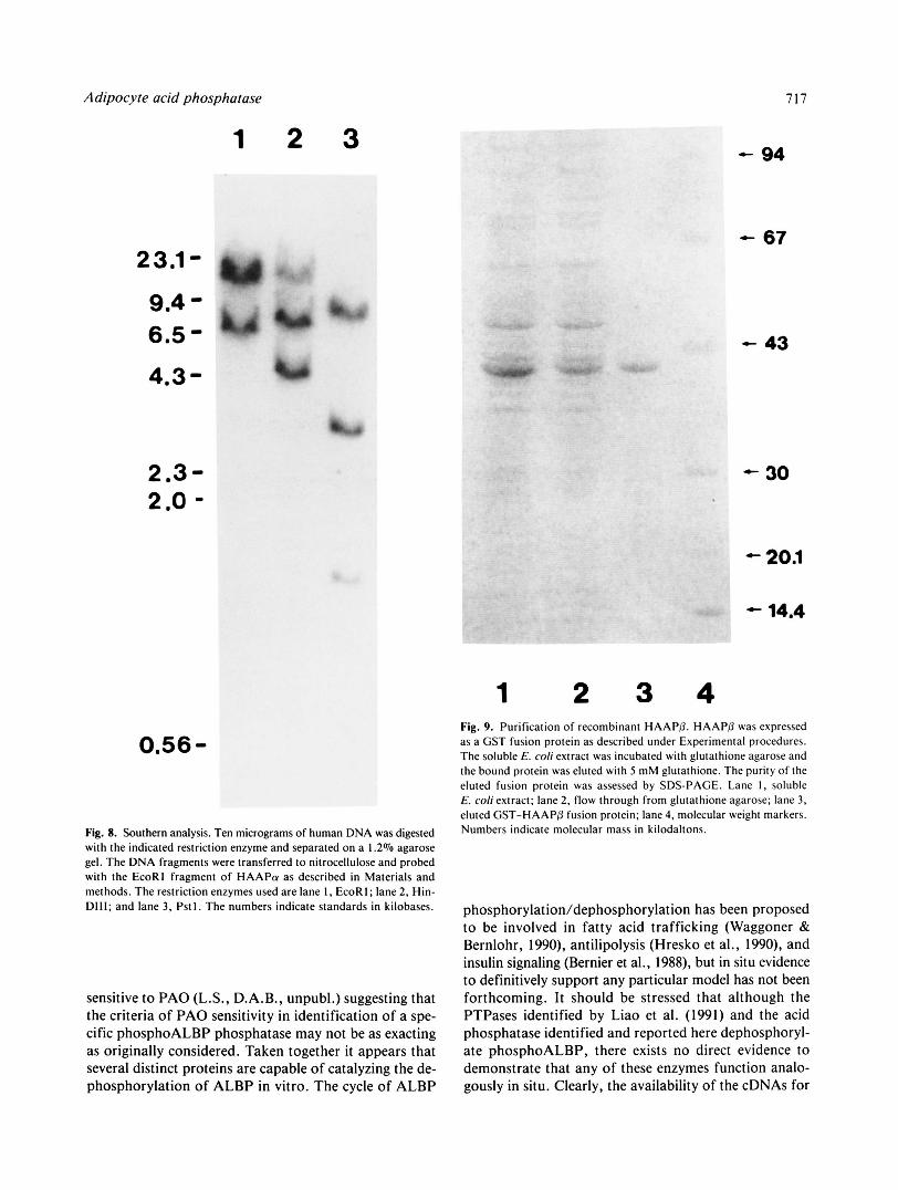
To examine the adipocyte acid phosphatase gene struc- ture, Southern analysis of human DNA with the cDNA of HAAPa (Fig. 8) was performed. When HAAPa was used as the hybridization probe, multiple bands were ob- tained in every restriction digest. Identical results were obtained using HAAPP as the hybridization probe. Mul- tiple bands in Southern analysis typically suggest multiple genes. However, given the virtually identical 3"untrans- lated region of HAAPa and HAAPP, it is reasonable to suggest that the isoenzymes arise from differential splic- ing of a single gene and that there may be pseudogenes present in the human genome.
To definitively demonstrate that the human adipocyte acid phosphatases were capable of phosphoALBP de- phosphorylation and that our protein preparations were not contaminated with another tyrosine phosphatase, the phosphatases were expressed in E. coli as fusion proteins with GST. The GST-HAAPa fusion protein was found to be insoluble when expressed in E. coli and refractory to analysis, consequently no further characterization was performed. In contrast, the HAAPP fusion was ex- pressed as a soluble protein and readily purified from E. coli by glutathione agarose affinity chromatography. The purified phosphatase exhibited a single 44-kDa Coomas- sie staining band after SDS-PAGE (Fig. 9) correspond- ing to 26 kDa of GST and 18 kDa of HAAPP. The fusion protein had a specific activity of 20 unitslmg when assayed with pNPP. The K,,, for pNPP was 182 pM, which is similar to the value measured for the 3T3-Ll acid phosphatase (Table 1). Vanadate inhibited the GST- HAAPP fusion with a K j of 17 pM, a value similar to that for vanadate inhibition of the bovine heart acid phosphatase ( K , = 29 pM) (Zhang & Van Etten, 1990). When assessed for its PA0 sensitivity, the phosphatase activity of GST-HAAPP was completely abolished by 100 pM PAO. Finally, HAAPP catalyzed the dephos- phorylation of phosphoALBP and the phosphorylated insulin receptor as shown in Figure 10.
Discussion
The present investigation was undertaken to address the hypothesis that there exists in adipocytes a member of the acid phosphatase family, that it should possess tyrosine phosphatase activity, and by virtue of conformationally vicinal essential cysteine(s) that it should be inactivated by the arsenical PAO. Based on pH dependence, Michaelis constant, inhibition by orthovanadate and di- ethyl pyrocarbonate, and immunological cross-reactivity with anti-BHAP antiserum, we have shown that our par- tially pure protein preparation is indeed the adipocyte acid phosphatase. Definitive evidence that adipocytes ex- press a low molecular weight acid phosphatase was pro- vided by the cloning, sequencing, and expression of the adipocyte isoenzymes.
Comparison of the HAAP protein sequences with the amino acid sequence of the bovine liver acid phosphatase indicates that BLAP shares 90% similarity to HAAPP and 74% similarity to HAAPa with no sequence similar- ity to the catalytic domain of the PTPases. The BLAP differs from the isoform-specific region of HAAPP by only two amino acids. Additional differences in the amino-terminal sequences of the two adipocyte enzymes are also present. Due to the abundance of proline residues in the amino-terminus of HAAPa and the lack of simi- larity between the adipocyte enzymes and BLAP in this region, it is possible that the differences in the sequences

Adipocyte acid phosphatase
A E P I I
715
HAAPa
SX E I I I
B HAAPa : HAAPD :
HAApn: HAAPR :
HAAPa : HAAPR :
HAAPa : HAAPR :
HAAPa : HAAPR :
HAAPa : HAAPR :
HAAPff : HAAPR :
HAAPa : HAAPR :
HAAPa : HAAPD :
HAAPa : HAAPD:
m P a : HAAPR:
HAAPa : HAAPD:
HRAPa: HAAPB:
HAApl3:
HAAPR E I
P I
sx I I
t t t t
E I
5 ’ I 3 ‘
4
4
4
GAA TTC CCG CGC AGA GGC CGC AAG TCC GTG CTG T T T GTG TGT CTG GGT AAC ATT TGT CGA TCA CCC G AAT TCC CCC GCG CGC AGA GAG GCC GCG AGA CCC
ATT GCA GAA GCA G T T T T C AGG AAA CTT GTA ACC GAT CAA AAC ATC TCA GAG AAT TGG AGG GTA GAC ATT GCA GAA GCA GTT TTC AGG AAA CTT GTA ACC GAT CAA AAC ATC TCA GAG AAT TGG GTC ATT GAT
AGC GGT GCT GTT TCT GAC TGG AAC GTG GGC CGG TCC CCA GAC CCA AGA GCT GTG AGC TGC CTA AGA AGC GCG GCA ACT TCC GGG TAT GAG ATA GGG AAC CCC CCT GAC TAC CGA GGG CAG AGC TGC ATG AAG
AAT CAT GGC ATT CAC ACA GCC CAT AAA GCA AGA CAG ATT ACC AAA GAA GAT TTT GCC ACA TTT GAT AGG CAC GGC ATT CCC ATG AGC CAC GTT GCC CGG CAG ATT ACC AAA GAA GAT TTT GCC ACA T T T GAT
TAT ATA CTA TGT ATG GAT GAA AGC AAT CTG AGA GAT TTG AAT AGA AAA AGT AAT CGA GTT AAA ACC TAT ATA CTA TGT ATG GAT GAA AGC AAT CTG AGA GAT TTG AAT AGA AAA AGT AAT CGA GTT AAA ACC
TGC AAA GCT AAA ATT GAA CTA CTT GGG AGC TAT GAT CCA CAA AAA CAA CTT ATT ATT GAA GAT CCC TGC AAA GCT AAA ATT GAA CTA CTT GGG AGC TAT GAT CCA CAA AAA CAA CTT ATT ATT GAA GAT CCC
TAT TAT GGG AAT GAC TCT GAC TTT GAG ACG GTG TAC CAG CAG TGT GTC AGG TGC TGC AGA GCG TTC TAT TAT GGG AAT GAC TCT GAC T T T GAG ACG GTG TAC CAG CAG TGT GTC AGG TGC TGC AGA GCG TTC
TTG GAG AAG GCC CAC tgaggcaggttcgtgccctqctgcqgccaqcctgactagaccccaccctqaqqtcctqcatttctcagtcqqtgtgtaatc TTG GAG AAG GCC CAC tgaggcaggttcgtqccctqctgcgqccagcctqactagaccccaccctqag~tcctqcatttctcagtcqqtgtgtaatc
acgttccagggcccaaagccagctctttgttcagttgacttactqtttcttaccttaaaaagtaattgtaqatqqaaatcagttqtgtttggcaggaqaatcaata acgttccaqggcccaaagccagctctttgttcagttqacttactqtttcttaccttaaaaaqtaattqtaqatggaaatcaqttqtqtttggcagqaqaatcaata
aaaatctttqattcagacagcttatqgqqtattttaagcattcttaqactaqttqaacatctcactttqccccaqttacaaaaataqtagaacaaqcaacataaaa aaaatctttgattcagacagcttatggggtattttaaqcattcttaqactaqttgaacatctcacttagctccagttacaaaaatagtagaacaaqcaacataaaa
caatgaaggaaaacctcacttgaaggcccaqgtcaacatctaagcctqttqagacttaqataatcgagtctacctcttcaqtaggtttgtgtqgatggcct~qagg caatgaaggaaaacctcacttgaaggcccaqgtcaacatctaaqcctqttgaqacttaqataatcgaqtctacctcttcaqtaggtttgtgtgqatqgcctggagg
caggtgccctctgctccccagtgctacctctctcttccctagggccttttqtqqattgaca~taqtcccctccgtaqagctcacagtctaqattagaaqtgtttta caggtgc----tgctccccagtgctacctctctcttccctaqggccttttqtqqattqacaqtaqtcccctccgtaqaqctcacagtctaqattaqaaqtgtttta
atttctacacaccg- ‘Oo7 atttctacacacccatagtgcacacttgtatattgaaaaqataqqgaagaqagaaacatttatqgaatcagtcqttggcaccttcaatacttcatgatttttgtcg
agtttacttcatqaggaggtcagcccattqqctcccatctqaaccactttgcctctqaaacttaattacatccaqaa~qaaqqacacttgtatgctaqtctatqqt cagttqaggaatatqactgtttttatatgcacatqtaacccaaatgtccaatataaattqqcttattttttaaaataattttaaaagttgggaaaaqtgttattat ttggcatqcttaaatattgaataagtattcttcatcagcatttaataaatgtataggcaqatgtaaggtaatttct t tattttgagataatgtcaaaatcatqa atatttcaa~ctggggagttataaaaatacaactagaqatataaaaaaaaaaaaaaaaaaag~ ‘4’1
66
132
198
2 64
330
396
462
553
659
765
871
977
1083
1189 1295 1401
Fig. 6. Nucleotide sequences of HAAPo and HAAPP. A: Sequencing strategy and restriction sites. The coding regions of HAAPa and HAAPP are depicted by the open boxes followed by the 3’-untranslated region. The sequencing strategy is illustrated by the arrows. Selected restriction sites are indicated; E, EcoR1; P, Pstl ; S, Sacl; and X, Xbal. B: Nucleotide sequences of HAAPa and HAAPP were determined by the dideoxy sequencing method. The EcoRl cloning sites are underlined and the polyadenyly- lation recognition site is marked by a double underline.

716 L.L . Shekels et ai.
n u p a : Pro Ala Arg Arg Glu A l a A l a Arg Pro I l e Ala 2 2
HAAPD: Pro Arg A r g Gly Arg Lys S e r V a l Leu Phe V a l Cys Leu Gly Asn I le Cys Arg S e r P r o I l e Ala BLAP: Ala Glu Gln Val Thr Lys S e r V a l Leu Phe Val Cys Leu Gly Asn I le Cys Arg S e r P r o I le Ala
H A A P ~ : G l u A l a V a l Phe A r g ~ y s Leu V a l Thr ASP Gln Asn I le Se r G lu Asn T r p A r g V a l ASP S e r A l a 4 4 BLAP: Glu A l a V a l Phe A r g Lys Leu V a l Thr Asp Gln Asn I le S e r Asp Asn T r p V a l I l e Asp S e r G l y
HAAPD: Glu Ala V a l Phe Arg Lys Leu Val Thr Asp Gln Asn I le Se r G lu Asn Trp Va l I l e Asp S e r G l y
HAAPa: A l a Thr Se r G ly Tyr Glu I le Gly Asn Pro Pro Asp Tyr Arg Gly Gln Ser Cys Met Lys Arg H i s 66
HAAPD: A l a Val S e r Asp T r p Asn Val Gly A r g S e r P r o Asp P r o A r g A l a V a l Ser Cys Leu Arg Asn H i s BLAP: A l a Val S e r Asp T r p Asn Val Gly A r g S e r P r o Asn P r o A r g A l a V a l S e r Cys Leu Arg Asn H i s
HAAPa: Gly I l e P r o Met S e r H i s V a l Ala A r g Gln I l e Thr Lys G l u Asp Phe A l a Thr Phe Asp Tyr I l e BLAP: Gly I l e Asn Thr Ala H i s Lys Ala A r g Gln V a l T h r Lys G l u Asp Phe V a l Thr Phe Asp Tyr I l e
HAAPA: Gly I le H i s Thr A l a H i s Lys A l a Arg Gln I le Thr Lys Glu Asp Phe A l a Thr Phe Asp Tyr I l e
HAAPa: l e u Cys Met Asp G l u S e r Asn Leu A r g Asp Leu Asn A r g Lys Ser Asn A r g V a l Lys Thr Cys Lys ‘Io
HAAPD: Leu Cys Met Asp Glu Ser Asn Leu A r g Asp Leu Asn A r g Lys Ser Asn A r g Val Lys Thr Cys Lys BLAP: Leu Cys Met Asp G l u S e r Asn Leu A r g Asp Leu Asn A r g Lys Se r Asn G l n V a l Lys Asn Cys Arg
HAAPa: A l a Lys I l e Glu Leu Leu Gly Ser Tyr Asp Pro Gln Lys G l n Leu I le I l e Glu Asp P ro Tyr Tyr 132 BLAP: A l a Lys I l e Glu Leu Leu Gly Ser Tyr Asp Pro Gln Lys G l n Leu Ile I le Glu A s p P r o T y r T y r
HAAPA: A l a Lys I l e Glu Leu Leu Gly Ser Tyr Asp Pro Gln Lys G l n Leu I le I le Glu Asp Pro Tyr Tyr
HAAPa: Gly Asn Asp S e r Asp Phe Glu Thr V a l Tyr Gln Gln Cys Val A r g Cys Cys Arg Ala Phe Leu Glu 154
HAAPA: Gly Asn Asp Ser Asp Phe Glu Thr V a l Tyr Gln Gln Cys Val A r g Cys Cys A r g Ala Phe Leu Glu BLAP: Gly Asn Asp Ala Asp Phe Glu Thr V a l Tyr G l n Gln Cys Val Arg Cys Cys Arg A l a Phe Leu Glu
HAAPa: Lys A l a H i s 157
HAAPB: LyS A l a H i s BLAP: Lys V a l Arg
Fig. 7. Amino acid sequences of the low molecular weight acid phosphatases. The nucleotide sequences of HAAPa and HAAPp were translated into the amino acid sequence and are shown in comparison to the amino acid sequence of the bovine liver acid phosphatase. The region from amino acids 40 to 73 is the isoform-specific region.
at the amino-termini represent errors from the reverse transcriptase used in cloning rather than true sequence differences. Despite repeated attempts, longer cDNA clones containing additional 5’ sequences have not been obtained. Molecular cloning of the acid phosphatase gene@) is in progress to clarify this issue.
Recently, the protein sequences of two isozymes of the human red cell acid phosphatase have been determined (Dissing et al., 1991). The human acid phosphatase locus is highly polymorphic with three common alleles in the population each encoding two isozymes, designated f and s for fast and slow electrophoretic migration. Inter- estingly, the sequences of the red cell isozymes, ACPl Bf and ACPl Bs, show that they differ from each other in a region of 34 amino acids analogous to that found in the adipocyte acid phosphatases. Comparing the human adipocyte sequences to the red cell acid phosphatase se- quences indicates that HAAPa and the red cell Bf iso- zyme are identical in the isoform-specific region as are HAAPP and the red cell Bs enzymes. Overall, the adipo- cyte acid phosphatases share 99% sequence similarity to the respective red cell isoenzymes. Outside the isoform- specific region the adipocyte enzymes differ from the red cell enzymes by only two amino acids at positions 4 and 105. Whereas residue 4 resides in a region of questionable sequence integrity as discussed above, the difference at position 105 of the red cell and adipocyte enzymes may reflect a polymorphism. The observation that a single al- lele gives rise to two isoforms has led Dissing et al. (1991)
to suggest that the two isoenzymes are the products of differential splicing of a single primary transcript.
Further examination of the primary sequences of HAAPa and HAAPP along with that of liver and red cell acid phosphatases reveals the presence of eight con- served cysteinyl residues. Studies utilizing sulfhydryl- directed reagents have indicated that the acid phosphatases possess at least one essential cysteine in the active site (Laidler et al., 1982; Zhang & Van Etten, 1990). Addi- tionally, the observation that P A 0 inactivates the en- zyme@) indicates that at least two sulfhydryl groups are conformationally vicinal. Two cysteine residues, C145 and C148, are positioned in a Cys-X-X-Cys motif near the carboxyl-terminus with the potential to be conforma- tionally vicinal by analogy to that motif in a variety of protein disulfide isomerases and prokaryotic thioredox- ins (Pigiet & Conley, 1978). Alternatively, chemical mod- ification with thiol-directed reagents (Camici et al., 1989) has pointed to cysteine residues near the amino-terminus as being essential for activity. Further structure-function analysis will be necessary to resolve the issue of essential cysteine residues.
Recently, Liao et al. (1991) have reported the identifi- cation of two PAO-sensitive membrane-associated tyro- syl phosphatases capable of dephosphorylating ALBP. The properties of these enzymes suggest that they are members of the PTPase family of protein tyrosyl phos- phatases. Consistent with this we have noted that the ac- tivity of the GST-K-PTPU323 fusion protein is also
Adipocyte acid phosphatase
1
9.4 6.5 -
2.3- 2.0 -
2 3
1
717
- 94
- 67
- 43
- 30 - 20.1
2 3 4
Om56
Fig. 8. Southern analysis. Ten micrograms of human DNA was digested with the indicated restriction enzyme and separated on a 1.2% agarose gel. The DNA fragments were transferred to nitrocellulose and probed with the EcoRl fragment of HAAPa as described in Materials and methods. The restriction enzymes used are lane I , EcoRI; lane 2, Hin- DIII; and lane 3, Pstl. The numbers indicate standards in kilobases.
sensitive to PA0 (L.S., D.A.B., unpubl.) suggesting that the criteria of PA0 sensitivity in identification of a spe- cific phosphoALBP phosphatase may not be as exacting as originally considered. Taken together it appears that several distinct proteins are capable of catalyzing the de- phosphorylation of ALBP in vitro. The cycle of ALBP
Fig. 9. Purification of recombinant HAAPP. HAAPP was expressed as a GST fusion protein as described under Experimental procedures. The soluble E. coli extract was incubated with glutathione agarose and the bound protein was eluted with 5 mM glutathione. The purity of the eluted fusion protein was assessed by SDS-PAGE. Lane 1. soluble E. coli extract; lane 2, flow through from glutathione agarose; lane 3, eluted GST-HAAPP fusion protein; lane 4, molecular weight markers. Numbers indicate molecular mass in kilodaltons.
phosphorylation/dephosphorylation has been proposed to be involved in fatty acid trafficking (Waggoner & Bernlohr, 1990), antilipolysis (Hresko et al., 1990), and insulin signaling (Bernier et al., 1988), but in situ evidence to definitively support any particular model has not been forthcoming. It should be stressed that although the PTPases identified by Liao et al. (1991) and the acid phosphatase identified and reported here dephosphoryl- ate phosphoALBP, there exists no direct evidence to demonstrate that any of these enzymes function analo- gously in situ. Clearly, the availability of the cDNAs for

718
HAAPB - + - +
I R K - ALBP -
1 2 3 4 Fig. 10. Dephosphorylation of phosphoALBP and IRK by GST- HAAPP. PhosphoALBP or phosphorylated insulin receptor kinase was incubated with 7 pg of fusion protein GST-HAAPP for 2 h at 37 "C. The reactions were quenched by addition of Laemmli sample buffer and the proteins were separated by SDS-PAGE followed by autoradiogra- phy at -70 "C. Lane 1, phosphoALBP alone; lane 2, phosphoALBP in the presence of GST-HAAPP.
the adipocyte acid phosphatases as well as the ability to easily purify active enzyme from expressing E. coli will aid us in the delineation of the metabolic role(s) of these enzymes.
Materials and methods
Materials
[ T ~ ~ P ] A T P (3,000 Ci/mmol) was obtained from Amer- sham. [1251]insulin (111 pCi/pg) was from DuPont- NEN. WGA-Sepharose and N-acetylglucosamine were purchased from Sigma. Insulin was purchased from GIBCO. The human abdominal fat cDNA library was
L.L. Shekels et at.
prepared as previously described (Baxa et al., 1989). All other chemicals were reagent grade.
Isolation of ALBP and the 3T3-LI insulin receptor
ALBP was isolated from either cultured 3T3-Ll adipo- cytes (Matarese & Bernlohr, 1988) or expressing E. coli cultures (Xu et al., 1991). Quantitation of ALBP was based upon an extinction coefficient of 1.55 x lo4 M" an-'. Insulin receptor was partially purified from 3T3- L1 adipocytes as previously described (Chinander & Bernlohr, 1989). The yield of active insulin receptor was quantitated by measuring the specific binding of [12SI]in- sulin using the polyethylene glycol precipitation method as originally described by Cuatrecasas (1 972) and modi- fied by Kohanski and Lane (1983). The insulin receptor preparation was stored at 4 "C and used within 2 weeks. The soluble 48-kDa cytoplasmic domain of the human in- sulin receptor was obtained from Dr. Leland Ellis, Dal- las, Texas.
In vitro phosphorylation assays and isolation of phosphoALBP
ALBP was phosphorylated with either partially purified 3T3-Ll insulin receptor or the cloned 48-kDa soluble ki- nase domain of the human insulin receptor (Cobb et al., 1989). When using the partially purified 3T3-Ll insulin receptor, phosphorylation of the insulin receptor 0 sub- unit and ALBP was conducted at room temperature with 2 pg of the WGA-Sepharose insulin receptor preparation in a buffer containing 25 mM HEPES, pH 7.5, 10 mM MgCI2, 4 mM MnC12, and 100 nM insulin. Following a 30-min preincubation of the insulin receptor with insulin, 3 mM MgC12, and 0.2 mM MnC12, autophosphorylation was initiated by the addition of 100 pM ATP. Insulin- stimulated autophosphorylation was rapid, reaching a maximum level within 30 min. ALBP was added 30 min after the initiation of autophosphorylation, and the reac- tions were allowed to proceed for the times indicated. In phosphorylation reactions utilizing the soluble insulin re- ceptor kinase, the kinase (2 pg) was incubated with ALBP in a buffer containing 25 mM HEPES, pH 7.5, 5 mM MnC12, and 100 pM ATP. Homogeneous phosphoALBP was then isolated by a combination of anion exchange chromatography arid affinity chromatography on anti- phosphotyrosine agarose. Details of the purification and characterization of phosphoALBP will be published else- where (Buelt et al., 1992).
Preparation of the 3T3-LI acid phosphatase
The 3T3-Ll acid phosphatase was partially purified from a cytoplasmic extract of 3T3-LI adipocytes. All steps of the purification were assayed using pNPP according to

Adipocyte acid phosphatase 719
the method of Taga and Van Etten (1982). Cultured 3T3- L1 cells were differentiated into adipocytes as previously described (Matarese & Bernlohr, 1988) and used within 10 days of acquiring the adipocyte phenotype. To prepare a cytoplasmic extract, monolayers of adipocytes were washed three times with 10 mM NaPO, buffer, pH 7.0, containing 150 mM NaCl and 1 mM EDTA. The mono- layers were then incubated for 15 min at room temper- ature with wash buffer containing 50 pg/mL digitonin (2 mL/lO-cm plate). The digitonin releasate was collected and the pH was immediately adjusted to 5.0 with 2 M so- dium acetate pH 5.0. Following acidification, the sample was incubated overnight at 4 "C to allow insoluble pro- teins to precipitate. The soluble protein fraction was re- covered by centrifugation (100,000 x g , 30 min, 4 "C), and soluble lipid was removed by chromatography at 37 "C on Lipidex 1000 resin. The delipidated soluble ex- tract was concentrated by ultrafiltration (Amicon YM5 membrane) and applied to a calibrated 5 X 100-cm Seph- adex G-75 column equilibrated at pH 4.85 with 25 mM sodium acetate. The column fractions containing active phosphatase were pooled and applied to a Beckman Spherogel TSK CM-3SW high performance liquid chro- matography (HPLC) column equilibrated in 25 mM so- dium acetate, pH 4.85. Elution of active phosphatase was accomplished using a linear NaCl gradient from 0 to 0.4 M in 25 mM sodium acetate, pH 4.85. Acid phospha- tase activity eluted in 300 mM NaC1, and, when analyzed by SDS-PAGE, an 18-kDa band (phosphatase) was ob- served together with several lower molecular weight pro- teins. Phosphatase was stored in column elution buffer at 4 "C. The specific activity of partially purified 3T3-Ll adipocyte acid phosphatase was 1-5 units/mg. One unit of activity is defined as the amount of enzyme required to hydrolyze 1 pmol of pNPP per minute at 37 "C.
Phosphatase assays
The phosphatase activity was measured at 37 "C using ei- ther pNPP or phosphorylated ALBP as substrate. In as- says using pNPP as substrate, the indicated amount of acid phosphatase was incubated with pNPP in 50 mM sodium acetate, pH 5.0, at 37 "C. The reactions were quenched at the given times by addition of 1 mL of 1 N NaOH and the amount of product p-nitrophenol released was measured from the absorbance at 405 nm using a molar extinction coefficient of 18,000 M-l cm" (Zhang & Van Etten, 1990).
The phosphoALBP phosphatase activity was deter- mined in a similar manner. Isolated phosphorylated ALBP was incubated at either pH 5.0 in sodium acetate, 0.15 M NaCl, or at pH 7.0 in 50 mM Tris-acetate, 0.15 M NaCl, with the indicated amounts of 3T3-Ll phosphatase at 37 "C. The concentration of phosphorylated protein
in most reactions was 2 pM, which was determined to be 10-fold greater than the apparent K,. The dephosphor- ylation was monitored by removing an aliquot of the reaction and immediately adding SDS to a final concen- tration of 1 .O% followed by heating to 95 "C for 3 min. The extent of dephosphorylation was determined by SDS- PAGE and autoradiography. The dephosphorylation was quantitated by densitometric analysis of the correspond- ing autoradiogram using a Hoefer GS300 scanning den- sitometer and by direct scintillation counting of the radioactive protein band excised from the dried poly- acrylamide gel.
Immunochemical methods
Total antiserum raised in rabbits against the bovine heart acid phosphatase was dialyzed into 50 mM sodium phos- phate, pH 7.0, and applied to a 1 .O-mL column of Pro- tein A Sepharose (Pharmacia) equilibrated in the same buffer. The unbound material was discarded, and the bound immunoglobulin was eluted with 0.1 M glycine HCI, pH 3.0. The eluted antibody was neutralized with 1 M Tris-HC1, pH 8.0, dialyzed into 50 mM sodium phosphate, pH 7.0, and stored at a concentration of 1 mg/mL at -20 "C until use. Immunoblotting was per- formed as described by Baxa et al. (1989) using anti- BHAP sera at a dilution of 1 : 1,000 and detection with '251-labeled Protein A (Amersham).
Inhibition of phosphatase activity by P A 0 and reactivation by 2,3-DMP
The acid phosphatase was incubated at pH 8.0 in 1 mM Tris-HC1 with the indicated concentrations of P A 0 for 30 min. Following modification, either 1 M Tris-acetate, pH 7.0, or 1 M acetic acid, pH 5.0, was added to a final concentration of 50 mM. Activity was assayed as de- scribed previously using either phosphoALBP or pNPP as substrate. A time course of modification indicated that the reaction was complete after 20 min under all condi- tions utilized.
To demonstrate reactivity with 2,3-DMP, the 3T3-Ll phosphatase was first modified with P A 0 as described at pH 8.0. Following modification, the reaction mixture was applied to a 1.0 x 20-cm Sephadex G-50 column equili- brated in 10 mM Tris-HC1, pH 8.0. The phosphatase- containing fractions were identified by the absorbance at 280 nm, pooled, and concentrated using Centricon-10 centrifugation (Amicon). The modified protein was incu- bated with the indicated concentrations of either 2-mer- captoethanol or 2,3-DMP for 60 min. The pH of the reaction was adjusted to 5.0 with 1 M acetic acid buffer and the activity was assayed using pNPP as the substrate.

720 L. L . Shekels et al.
Kinetic measurements
Kinetic parameters were measured at either pH 5.0 or 7.0 in 50 mM Tris-acetate buffer using pNPP as substrate. Duplicate points at 8-12 substrate concentrations were utilized. Apparent K,,, values were corrected for ioniza- tion of substrate by the method of Lawrence and Van Et- ten (1981). Inhibition by orthovanadate was measured at three concentrations, and K, values were calculated as described by Taga and Van Etten (1982).
Isolation of HAAP cDNA clone
To identify the human adipocyte acid phosphatase, the 580-bp EcoR1-Sal 1 fragment containing the bovine heart acid phosphatase coding region (Wo et al., 1992) was ra- diolabeled and used to screen a human adipocyte cDNA library. The hybridization was performed at 42 "C in 50% formamide, 0 . 0 5 ~ BLOTTO, and 6x SSC (Mania- tis et al., 1989). Following hybridization, the nitrocellu- lose filters were washed at 60 "C in 0 . 5 ~ SSC and 0.1% SDS and exposed to film at -70 "C. From approximately 20,000 plaques screened, a single hybridizing plaque was obtained, and the clone, HAAPa, was sequenced by the dideoxy method (Sanger et al., 1977). The cDNA library was rescreened a second time using a 400-bp EcoR1-Pst 1 fragment from HAAPa as the hybridization probe. The hybridization was carried out in 50% formamide, 0.2X BLOTTO, 6x SSC, and 10 pg/mL sonicated salmon sperm DNA at 42 "C. The filters were then washed in 0.1 x SSC and 0.1 Yo SDS at 65 "C and exposed to Kodak X-ray film at -70 "C. A second clone, HAAPP, was iso- lated and also sequenced by the dideoxy method.
Southern analysis of human DNA
Human DNA (10 pg) was digested with the appropriate restriction endonuclease and subsequently separated on a 1.2% agarose gel. The DNA fragments were transferred to nitrocellulose and hybridized at 42 "C in 4x SSC, 50% formamide, and 0.1% SDS. The EcoRl fragment from HAAPa or HAAPP was labeled using the random- primer method of Feinberg and Vogelstein (1983) for use as the hybridization probe. The hybridized filter was washed two times at 65 "C with 0.1 X SSC and 0.1070 SDS and exposed to Kodak X-ray film at -70 "C.
Expression and purification of PTPase and HAAPP
HAAPP and the rat brain PTPase, PTPU323, were ex- pressed in E. coli and purified as GST fusion proteins, GST-HAAPP and GST-K-PTPU323, as described by Smith and Johnson (1988) and Guan and Dixon (1991). Briefly, HAAPP was subcloned into the EcoRl site of the
expression vector pGEX-1, and the resulting plasmid was used to transform E. coli strain JM109. The recombinant plasmid pGEX-KG containing the rat brain PTPase cDNA, designated PTPU323, was used to transform competent E. coli strain JM109. Successful transformants were screened for expression of fusion proteins by
To purify the GST fusion proteins, overnight cultures of E. coli harboring the recombinant plasmid were di- luted 1 : 10 and allowed to grow for 1 h at 37 "C. Expres- sion of the fusion protein was induced by addition of IPTG to a final concentration of 0.1 mM. The culture was allowed to grow 3-6 h at which time the cells were collected by centrifugation and resuspended in solubili- zation buffer containing 20 mM sodium phosphate, pH 7.3, 150 mM NaCl, and 1% Triton X-100. The cells were lysed by sonication, and the bacterial debris was removed by centrifugation. The supernatant was mixed with glu- tathione agarose, and the fusion protein was allowed to bind at 4 "C for 15 min. The resin was collected in a col- umn and washed with solubilization buffer. The GST- HAAPP fusion protein was eluted with 5 mM glutathione in 10 mM sodium acetate, pH 5, 5 mM EDTA, and 10% glycerol and the GST-K-PTPU323 fusion protein was eluted in the same buffer at pH 6. Protein concentration was determined by the method of Bradford (1976), and phosphatase activity was followed throughout the puri- fication using pNPP as substrate as described before. The purity of the eluted fusion proteins was assessed by SDS- PAGE and Coomassie blue staining. The specific activity of the purified fusion GST-HAABP was typically 10-20 units/mg and 20-30 units/mg for GST-K-PTPU323.
SDS-PAGE.
Acknowledgments
We thank Dr. Jack Dixon, University of Michigan, for kindly providing the pGEX-KG-PTPase plasmid and Dr. Leland Ellis, Dallas, for his gift of the soluble insulin receptor kinase. We ap- preciate the generosity of Dr. Brian Van Ness for providing the human DNA. We also thank Dr. Melissa Buelt for the prepara- tion of phosphoALBP. This work was supported by NSF grant DMB8552942 (D.A.B.) and NIH grant GM27003 (R.L.V.E.).
References
Baxa, C.A., Sha, R.S., Buelt, M.K., Smith, A.J . , Matarese, V., Chinander, L.L., Boundy, K.L., & Bernlohr, D.A. (1989). Human adipocyte lipid binding protein: Purification of the protein and clon- ing of its complementary DNA. Biochemistry 28, 8683-8690.
Bernier, M., Laird, D.M., & Lane, M.D. (1987). Insulin-activated ty- rosine phosphorylation of a 15-kilodalton protein in intact 3T3-Ll adipocytes. Proc. Nutl. Acud. Sci. USA 84, 1844-1848.
Bernier, M., Laird, D.M., & Lane, M.D. (1988). Effect of vanadate on the cellular accumulation of pp15, an apparent product of insulin receptor tyrosine kinase action. J. Biol. Chem. 263, 13626-13634.
Boivin, P., Galand, C., & Bertrand, 0. (1987). Protein band 3 phospho- tyrosyl phosphatase. Purification and characterization. J. Biochem.
Bradford, M. (1976). A rapid and sensitive method for the quantitation 19, 613-618.

Adipocyte acid phosphatase 72 1
of microgram quantities of protein utilizing the principle of protein- dye binding. Anal. Biochem. 72, 248-254.
Buelt, M.K., Shekels, L.L., Jarvis, B.W., & Bernlohr, D.A. (1991). In vitro phosphorylation of the adipocyte lipid binding protein (p15) by the insulin receptor: Effects of fatty acid on receptor kinase and substrate phosphorylation. J. Bioi. Chem. 266, 12266-12271.
Buelt, M.K., Xu, Z . , Banaszak, L., & Bernlohr, D.A. (1992). Structural and functional characterization of the phosphorylated adipocyte lipid-binding protein (pp15). Biochemistry (in press).
Camici, G., Manao, G., Cappugi, G., Modesti, A., Stefani, M., & Ram- poni, G. (1989). The complete amino acid sequence of the low mo- lecular weight cytosolic acid phosphatase. J. Bioi. Chem. 264,
Charbonneau, H., Tonks, N.K., Kumar, S., Diltz, C.D., Harrylock, M., Cool, D.E., Krebs, E.G., Fischer, E.H., & Walsh, K.A. (1989). Hu- man placenta protein-tyrosine-phosphatase: Amino acid sequence and relationship to a family of receptor-like proteins. Proc. Nazi. Acad. Sci. USA 86, 5252-5256.
Chernoff, J. & Li, H.-C. (1985). A major phosphotyrosyl-protein phos- phatase from bovine heart is associated with a low molecular weight acid phosphatase. Arch. Biochem. Biophys. 240, 135-145.
Chinander, L.L. & Bernlohr, D.A. (1989). Cloning of murine adipocyte lipid binding protein in Escherichia coli: Its purification, ligand bind- ing properties and phosphorylation by the adipocyte insulin recep- tor. J. Bioi. Chem. 264, 19564-19572.
Cobb, M.H., Sang, B.-C., Gonzalez, R., Goldsmith, E., & Ellis, L. (1989). Autophosphorylation activates the soluble cytoplasmic do- main of the insulin receptor in an intermolecular reaction. J. Biol. Chem. 264, 18701-18706.
Cuatrecasas, P. (1972). Isolation of the insulin receptor of liver and fat cell membranes. Proc. Natl. Acad. Sci. USA 69, 318-322.
Dissing, J., Johnsen, A.H., & Sensabaugh, G.F. (1991). Human red cell acid phosphatase (ACPI): The amino acid sequence of the two iso- zymes Bf and Bs encoded by the ACPI *B allele. J. Bioi. Chem. 266,
Ellis, L., Clauser, E., Morgan, D.O., Edery, M., Roth, R.A., & Rut- ter, W.J. (1986). Replacement of insulin receptor tyrosine residues 1162 and 1163 compromises insulin-stimulated kinase activity and uptake of 2-deoxyglucose. Cell 45, 721-732.
Feinberg, A.P. & Vogelstein, B. (1983). A technique for radiolabeling DNA restriction endonuclease fragments to high specific activity. Anal. Biochem. 132, 6-1 3.
Frost, S.C. & Lane, M.D. (1985). Evidence for the involvement of vic- inal sulfhydryl groups in insulin-activated hexose transport by 3T3- L1 adipocytes. J. Bioi. Chem. 260, 2646-2652.
Frost, S.C. & Schwalbe, M.S. (1990). Uptake and binding of radiola- belled phenylarsine oxide in 3T3-LI adipocytes. Biochem. J. 269,
Guan, K. & Dixon, J.E. (1991). Eukaryotic proteins expressed in Esch- erichia coli: An improved thrombin cleavage and purification pro- cedure of fusion proteins with glutathione S-transferase. Anal. Biochem. 192, 262-267.
Guan, K., Haun, R.S., Watson, S.J., Geahlen, R.L., & Dixon, J.E. (1990). Cloning and expression of a protein-tyrosine phosphatase. Proc. Natl. Acad. Sci. USA 87, 1501-1505.
Hresko, R.C., Bernier, M., Hoffman, R.D., Flores-Riveros, J.R., Liao, K., Laird, D.M., & Lane, M.D. (1988). Identification of phosphor- ylated 422 (aP2) protein as pp15, the 15-kilodalton target of the in- sulin receptor tyrosine kinase in 3T3-LI adipocytes. Proc. Nuti.
Hresko, R.C., Hoffman, R.D., Flores-Riveros, J.R., & Lane, M.D. (1990). Insulin receptor tyrosine kinase-catalyzed phosphorylation
2560-2567.
2061 9-20625.
589-595.
ACUd. Sei. USA 85, 8835-8839.
of 422 (aP2) protein: Substrate activation by long chain fatty acid. J. Bioi. Chem. 265, 21075-21085.
Hunter, T. (1989). Protein-tyrosine phosphatases: The other side of the coin. Ce1158, 1013-1016.
Kohanski, R.A. & Lane, M.D. (1983). Homogeneous functional insu- lin receptor from 3T3-LI adipocytes: Purification using NmB'-(bi- otinyl-6-aminocaproyl) insulin and avidin-Sepharose. J. Biol. Chem. 258, 7460-7468.
Laidler, P.M., Taga, E.M., &Van Etten, R.L. (1982). Human liver acid phosphatases: Cysteine residues of the low molecular weight enzyme. Arch. Biochem. Biophys. 216, 512-521.
Lawrence, G.L. & Van Etten, R.L. (1981). The low molecular weight acid phosphatase from bovine liver: Isolation, amino acid compo- sition and chemical modification studies. Arch. Biochem. Biophys.
Liao, K., Hoffman, R.D., &Lane, M.D. (1991). Phosphotyrosyl turn- over in insulin signaling: Characterization of two membrane-bound pp15 protein tyrosine phosphatases from 3T3-LI adipocytes. J. Biol. Chem. 266, 6544-6553.
Maniatis, T., Fritsch, G.F., & Sambrook, J. (1989). Molecular Cloning: A Laboratory Manual. Cold Spring Harbor Laboratory, Cold Spring Harbor, New York.
Matarese, V. & Bernlohr, D.A. (1988). Purification of murine adipo- cyte lipid binding protein: Characterization as a fatty acid and ret- inoic acid binding protein. J. Bioi. Chem. 263, 14544-14551.
Pigiet, V. & Conley, R.R. (1978). Isolation and characterization of phos- phothioredoxin from Escherichia coli. J. Biol. Chem. 253,
Proudfoot, N.J. & Brownlee, G.G. (1976). 3' Non-coding region se- quences in eukaryotic messenger RNA. Nature 263, 21 1-214.
Rosen, O.M. (1987). After insulin binds. Science 237, 1452-1458. Sanger, F., Nicklen, S., & Coulson, A.R. (1977). DNA sequencing with
chain-terminating inhibitors. Proc. Null. Acad. Sci. USA 74,
Smith, D.B. &Johnson, K.S. (1988). Single step purification of poly- peptides expressed in Escherichia coli as fusions with glutathione S- transferase. Gene 67, 31-40.
Taga, E.M. & Van Etten, R.L. (1982). Human liver acid phosphatases: Purification and properties of a low molecular weight isoenzyme. Arch. Biochem. Biophys. 214, 505-515.
Tonks, N.K., Diltz, C.D., & Fischer, E.H. (1990). CD45, an integral membrane protein tyrosine phosphatase: Characterization of enzyme activity. J. Biol. Chem. 265, 10674-10680.
Waggoner, D.W. & Bernlohr, D.A. (1990). In situ labeling of the adipo- cyte lipid binding protein with 3-[ Iz5I] iodo-4-azido-N-hexadecyl- salicylamide: Evidence for a role of fatty acid binding proteins in lipid uptake. J. Biol. Chem. 265, 11417-11420.
Waheed, A., Laidler, P.M., Wo, Y.-Y.P., &Van Etten, R.L. (1988). Pu- rification and physicochemical characterization of a human placen- tal acid phosphatase possessing phosphotyrosyl protein phosphatase activity. Biochemistry 27, 4265-4273.
Wo, Y.-Y.P., Zhou, M.-M., Stevis, P., Davis, J.P., Zhang, Z.-Y., &Van Etten, R.L. (1992). Cloning and expression of the low molecular weight phosphotyrosyl protein phosphatase from bovine heart. Bio- chemistry (in press).
Xu, Z., Buelt, M.K., Banaszak, L., & Bernlohr, D.A. (1991). Expres- sion purification and crystallization of the adipocyte lipid binding protein. J. Biol. Chem. 266, 14367-14370.
Zhang, Z.-Y. & Van Etten, R.L. (1990). Purification and characteriza- tion of a low molecular weight acid phosphatase-A phosphotyrosyl protein phosphatase from bovine heart. Arch. Biochem. Biophys. 282, 39-49.
206, 122-131.
1910-1920.
5463-5467.