iiiieieeeeeeee - apps.dtic.mil · r. kuttan, m. lafranconi, i.g. sipes, e. leezan and k. brendel...
TRANSCRIPT
AD-AI0G 702 ARIZONA UNIV HEALTH SCIENCES CENTER TUCSON F/B 6/20OXYGEN TOXICITY AND LUNG COLLAGENOUS PROTEIN. (U)FEB 81 K BRENDEL, I G SIPES N00014-77-C-0506
UNCLASSIFIED ML
mEEllEEEllE~lIEIElllEEEEllEE
IIIIEIEEEEEEEE

FINAL :ECHNICAL REPUTO
OXYGEN TOXICITY' AND L.JNG !'01 .CEN'OUS PROTEIN
Contract No. N00014-7-C-50 ~Office of Naval Research
7=4Dates: 1 September 1977 -23 February 1981
44i
Principal Investigators: Maus*Brendel,' M.-D. and I- Glenn ipesl Ph.D.-Ueo artments of ?tlarmacology and Toxicology
-- Arizona .Health Sciences CenterUniversity of ArizonaTucson, AZ 85724
A~~~ce~~~dstrbuio i______________ unlimited
[ f c s i n F o r .- -T h is d o c u m e n t h a s b e e n a p p r o v e d
S P 2 2 1 8
fl A
Dist GaI
~~c00,
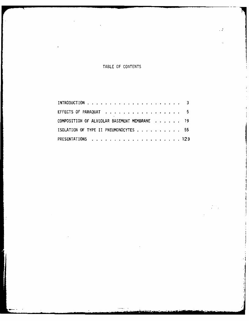
TABLE OF CONTENTS
INTRODUCTION .. .. .. ... ..... . .... ..... 3
EFFECTS OF PARAQUAT .. .. ..... . ... ....... 5
COMPOSITION OF ALVEOLAR BASEMENT MEMBRANE. .. .. ... 19
ISOLATION OF TYPE II PNEUMONOCYTES. .. .. .. . .... 55
PRESENTATIONS. .. .. .... .... ..... .... 1231
INTRODUCTION
The major objective of this proposal was to investigate the chemical
composition and subunit structure of the basement membrane and collagenous
components of the normal lung and of lungs exposed to agents thought to prcmote
the formation of singlet oxygen and thereby produce oxidative damage to tissu
components. The pathological and functional changes attributable to singlet
oxygen production can be induced experimentally by paraquat (l,l-dimethyl-4,
4-dipyridilium dichloride) which is thought to produce toxicity comparable to
that produced by hyperoxia.
Connective tissue makes up about 25% of the adult human lung and plays an
important role in the mechanical and physiological functions of the lung. The
alveolar basement membrane, because it lies in the primary pathway traversed by
ventilated gases, is of particular interest and, therefore, the determination
of the structure and composition of this component of the extracellular matrix
of the lung is of considerable importance. In the last few years, it has become
increasingly obvious from the study of extracellular matrix from a variety of
tissues that a complex population of proteins make up the extracellular matrix.
Several new collagen types have been described as well as high molecular weight
non-colla gens that appear to be associated with normal basement membranes. In
view of the new and somewhat unanticipated information accumulating in this area,
the characterization of putative changes in the nature of matrix components in
pathologic states must, of necessity, lag behind a more thorough characteriza-
tion of normal basement membrane and extracellular components.. .
The lung is also made up of a complicated population of different cell types,
each of which may respond differently to toxic substances. The interpretation of
toxicological studies of relevance to pulmonary function would be greatly
facilitated by the development of an in vitro system allowing the use of defined
-3-
pulmonary cell type of major physiological importance such as the type II
pneumocyte. Accordingly, this aspect of the project was pursued vigorously.
This report summarizes the work performed during this contract by providing
a brief introduction to the various experiments followed by the pertinent data
in the form of the resulting publication or manuscript.
-4-

EFFECTS OF PARAQUAT
Par3quat is a broad spectrum herbicide that has a very toxic affinity
for the lung, although with some indications of toxicity for the kidney as
well. Single oral or injected doses result in hemorrhagic pulmonary edema
within a few hours followed by an irreversible inflammation and fibrosis
detectable morphologically. The effects of paraquat treatment on prolyl
hydroxylase activation and collagen synthesis were determined in lung and
kidney slices and cultured lung cells taken from rats exposed to paraquat
nine days prior to sacrifice. The results are presented in the following
manuscri pt:
Kuttan, R., Lafranconi, M., Sipes, I.G., Meezan, E.and Brendel, K. (1979). Effect of paraquat treatmenton prolyl hydroxylase activity and collagen synthesisof rat lung and kidney. Res. Comm. Path. Pharm. 25, 257-268.
-5-

VOL.25, NO.2 Research Communications inAUGUST 1979 Chemical Pathology and Pharmacology
E:ECT OF PA,.nUAT TREATME,;T 10: :LYL HY -nxi.SE ACTI'ITY.' CLLAGEN SyTHESIS 1:' D,7 ,:D*'
R. Kuttan, M. Lafranconi, I.G. Sipes, E. leezanand K. Brendel
Deoartments of Pharmacology and AnesthesiologyThe University of Arizona Health Sciences Center,
Tucson, Arizona 85724
ABSTRACT
Morphological evidence has shown that the herbicide oaraquat pro-duces severe lung fibrosis in experimental animals. In order toinvestigate the biochemical basis for this fibrosis, prolyl hydroxy-lase activity, total hydroxyproline content and collaqen and proteinsynthesis were estimated in rats (200 gm) which were oretreated withparaquat (25 mc/Kq; I.P.) nine days before sacrifice. Prolylhydroxylase activity in the treated rats was sionificantly elevatedin the lunqs. However, no such increase was seen in the kidney. Therewas no sionificant chanae in the total hydroxyoroline content or collagenformation in the treated animals. When paraquat was added to the culturemedium and incubated with lung slices, there was a marked decrease incollagen synthesis alona with a decrease in protein synthesis. Whenparaquat was added to luno cells in culture, it was found that lungexplants were more sensitive to paraquat toxicity than cells obtainedby trypsinization. In explants which were sensitive to paraquat, therewas a marked decrease in both protein synthesis and collagen synthesis.
INTRODUCTION
Morphological evidence suggests that the herbicide oaraquat produces
severe lung fibrosis in human beings who have ingested this chemical
accidently as well as in experimental animals (Vijeyaratnam and Corrin,
1971; Smith, 1976). Even though the lung has a capacity to concentrate
paraquat (Rose et al., 1974), pathological changes have also been seen
in kidney and liver (Gibson and Caqen, 1977). The primary effect of
paraquat in animals has been shown to be the destruction of type I alveo-
lar epithelial cells (Kimbrough and Gaines, 1970). Damage to granular
257

VOL.25, NO.2 Research Communications inAUGUST 1979 Chemical Pathology and Pharmacology
cneurnocytes (tyoe II cells) and endotnelial 2"s -as ,:-inim'l (,je arat-
ham and Corin, 1971; Kirnbrouch ana Lincer,l?']. s suests a
selective action of oaraauat on dilferent cll line, Jeoeninc :on the
rate of absorption and metabolism of oaraouat by the cells. However,
the effect of Paraquat on culture systems has not been studied. Such
an approach can yield interesting data on the action of oaraouat.
Paraquat induced lung fibrosis has been shown to result in deposi-
tion of collagen (Smith et al., 1974). Studies on other sy~terrs have
shown that prolylhydroxylase can be used as a biochemical mi.rker for
collagen synthesis (Cardinale and Udenfriend, 197-). Collaaen synthesis
can also be estimated by the incorporation of radioactive Droline into
radioactive hydroxyproline or into collagenase degradable protein. Using
these biochemical markers, the effect of Daraquat on collagen synthesis
in the lung and kidneys of whole animals was measured nine days after
administration of an acute dose of paraquat t23 mg/Kg), At this time
the lung tissue is in a proliferative phase of repair of the acute injury
induced shortly after paraquat administration. A tissue culture system
has been employed to study the effect of oaraquat on collaqen synthesis
of isolated lung and kidney slices and isolated explants and cells ob-
tained by trypsinization from neonatal lungs.
MATERIALS AND IETHODS
ParaquiJ dihydrochloride (Imperial Chemicals) was a gift from Dr.L. Smith. 'C-proline was obtained from New England Nuclear. Tissueculture media were obtained from GIBCO.
Both neonatal and adult rats were used in the exoeriments. Para-quat was administered to the adult rats (200 am) by intraperitonealinjection (25 mg/Kg). The rats were sacrificed after nine days byexsanguination.
258

VOL.25, NO.2 Research Communications inAUGUST 1979 Chemical Pathology and Pharmacology
Lung cells were Prepared from neonatal rats by two technicues, anex~lant 7ethc and by trycsinization. To start the cells from explants,tin slices of the neor3tal rat iunos were placed on nylon crids in apetri dish containing Oulbecco's modified Eaales medium with 10" fetalcalf serum and antibiotics (penicillin 100 units/mil, streptomycin 100ug/ml and fungizone 0.25 ug/ml). Trypsinization of the lung cells wasdone using 0.1* trypsin for 1 hr as described by Hance et al. (1976).After trypsinization, the cells were washed in ohosphate buffered salinepH 7.4 and were qrown with an initial cell density of 0.2 million/ml inthe same medium described before.
Collagen synthesis was measured using lung and kidney slices (100mg) employing a procedure involving treatment with a highly purifiedform of bacterial collagenase (Peterkofsky and Diegelmann, 1971).This was done as follows: medium was changed to a fresh medium (5 ml)without serum but containing antibiotics. The fojlowing mixture wasadded to the medium; sodium ascorbate (l "M), '4C-proline (2 uc) andB-amino propionitrile (20 ug/ml) which inhibits cross-linking of thenewly formed collagen. The incubation was performed at 27*C for 48 hrs.The tissue was seoarated from the medium and was homoqenized in Trisbuffer (0.05 M, pH 7.6) containing ethylene diamine tetracetic acid(10-0M). dithiothreitol (10- M) and Triton X-100 (0.1%). Part of thehomogenate was dialyzed against Tris buffer (0.01 M, pH 7.6) overnight.Incorporation into collagen was determined by initial treatment witha highly purified preiaration of collagenase.(Advanced Biofactures)which is free from protease contamination. The reaction mixture afterincubation with collagenase was treated with 5% trichloroacetic acid.Radioactivity which remained in the supernatant was considered to becollagenase degradable orotein. A blank was always carred out to whichno collagenase was added, but which was otherwise treated under thesame conditions. Radioactivity which was precivitated with trich-loroacetic acid was taken as a measure of non-collagen protein synthe-sis. Prolylhydroxylase was measured accordino to the method of Huttonet al. (1966) using chick embryo substrate. (We are grateful to Dr.George Fuller, University of Rhode Island for giving us the substrate).The reaction was done in half the original volume (0.5 ml). After thereaction, tritiated water was separated from the reaction mixture bypassing through a 1 ml Dowex 50 x4H 4+ column and subsequent treatmentwith charcoal. The background counts were 60 C.P.M. Hydroxyprolinein the tissues was determined after acid hydrolysis by the method ofNeuman and Logan (1950). 14C-hydroxyoroline was determined by amethod described by Peterkofsky and Prockop (1962). Protein wasdetermined by the method of Lowry et al. (1951).
RESULTS AND DISCUSSION
Effect of Paraquat on Collagen Synthesis in the Lungs of Adult Rats
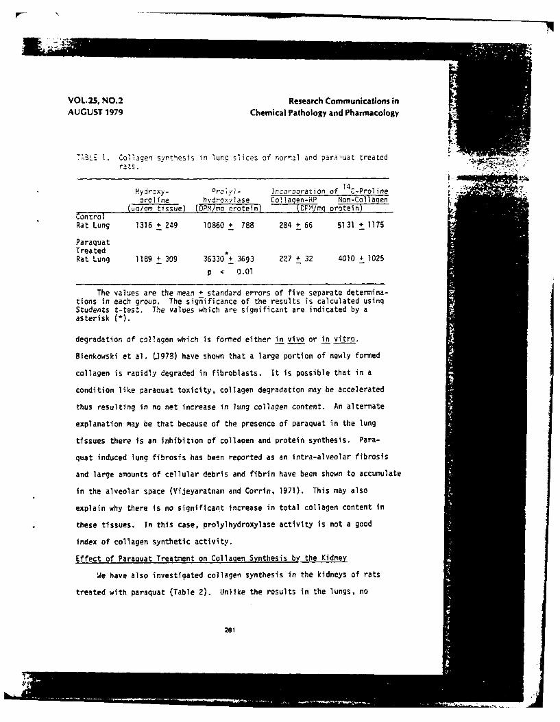
Table I shows the effect of oaraquat treatment on collagen synthe-
sis in adult rat lungs. Prolylhydroxylase activity, which is a marker
259

VOL.2S, NO.2 Research Communications in
AUGUST 1979 Chemical Pathology and Pharmacology
af collagen synthesis, has been siqnific3ntly elevated. However, the
total hydroxyproline conterc, which is an indizator of total Coiiagen
content in the lungs of the treated and control animals. was almost the
same. There was also no difference in the incorooration of lC-Droline
14into C-hydroxyproline and into non-collagenous orotein in the lungs
of the paraquat treated and control animals.
Previous studies have produced conflictinq data on the effects of
paraquat treatment on collagen synthesis in rat lungs. Hollinger and
Chvapil (1977) reported that there is an increase of prolyihydroxylase
activity in rat lungs seven days after acute paraquat treatment but no
increase in total collagen content. However, further studies were not in
agreement with this finding and indicated that paraauat is not a qood
model for experimentally induced lung fibrosis (Hollinger et al, 1978).
This conclusion was also supported by another group of workers (Autor
and Schmitt, 1977). However, other reports indicate that paraquat pro-
duces substantial increases in prolylhydroxylase activity and collagen
synthesis after a few days of treatment (Thompson and Patrick, 1978;
Greenberg et a]., 1978a & b). There is much less data on the effects of
paraquat treatment on the total collagen content of the lungs. AltNough
a small increase in the total hydroxyproline content of the lungs of
paraquat treated animals was observed at 21 days after an acute dose,
no change was seen at seven days (Hollinger and Chvapil, 1977). Our
data indicates that there is a significant increase of prolylhydroxylase
activity in the lungs of paraquat treated animals nine days after an
acute dose (25 mg/Kg). However, we could not demonstrate any increase
in total hydroxyproline content or increased hydroxyoroline synthesis
in the lungs of these animals. These results might be explained by a rapid*
260
VOL.25, NO.2 Research Communications inAUGUST 1979 Chemical Pathology and Pharmacology
T-BLE 1. Collagen synthesis in lung slices of norral and paraluat treated , 4krats. I
14Hydroxy- Prvl- Incorooration of C-Proline .oroline hvdroxvlase Collagen-HP Non-Collaqen
(uo/cm tissue) (DPMl/ma protein) DPM/ g protein)ControlRat Lung 1316 + 249 10860 + 788 284 + 66 5131 + 1175
ParaquatTreatedRat Lung 1189 4 309 36330 + 3693 227 + 32 4010 + 1025
p < 0.01
The values are the mean + standard errors of five separate determina-tions in each grouo. The significance of the results is calculated usinqStudents t-test. The values which are significant are indicated by aasterisk (*).
degradation of collagen which is formed either in vivo or in vitro.
Bienkowski et al. L1978) have shown that a large portion of newly formed
collagen is rapidly degraded in fibroblasts. It is possible that in a
condition like paracuat toxicity, collagen degradation may be accelerated
thus resulting in no net increase in lung collagen content. An alternate
explanation may be that because of the presence of paraquat in the lung
tissues there is an inhibition of collaaen and protein synthesis. Para-
quat induced lung fibrosis has been reported as an intra-alveolar fibrosis
and large amounts of cellular debris and fibrin have been shown to accumulate
in the alveolar space (Vijeyaratnam and Corrin, 1971). This may also
explain why there is no significant increase in total collagen content in
these tissues. In this case, prolyihydroxylase activity is not a good
index of collagen synthetic activity.
Effect of Paraquat Treatment on Collagen Synthesis by the Kidney
We have also investigated collagen synthesis in the kidneys of rats
treated with paraquat (Table 2). Unlike the results in the lungs, no
261

VOL. 25, NO.2 Research Communications inAUGUST 1979 Chemical Pathology and Pharmacology
" l Cagen snthesis in , slices of ncr-al and pardoua. trectE4
rits.
Hydroxy- Prolyl- 1proline hydroxylase 4C-Proline Incorporation
(Gg/gm tissue) (DPM/mq Protein) Collagen-HP Non-Collaqen
Control 381 + 87 2555 + 636 71 + 6 922 + 79
Paraquat 393 + 40 1581 + 428 70 + 4 1220 + 53
o < .05
The values are the mean + standard error of five separate determinationsin each group. The significance of the results is calculated using Studentst-test. The values which are significant are indicated by an asterisk (*).
difference in the specific activity of Drolyl hydroxylase in the treated
and control rats was seen. There was also no difference in the total
hydroxyproline content of the kidney or on collagen hydroxyproline synthe-
sis. Morphological evidence suggests that there is kidney damage associated
with many paraquat poisonings. Paraquat has been described as a "hit and
run" type of toxic agent. After paraquat ingestion, nearly 80-90% of the
compound is excreted in the urine within 24 hrs. Hence, there is an
increased total concentration of paraquat in the kidney during this period.
Necrosis of proximal tubules has also been shown in kidneys affected by
paraquat (Fowler and Brooks, 1971). The transient presence of high concen-
trations of paraquat in the kidney, however, did not result in increased
prolylhydroxylase activity as was the case for the lung where paraquat
is concentrated and may remain for longer periods of time.
Effect of Paraquat on Lung Tissues in Culture.
In order to find out whether there is any direct effect of paraquat
262

VOL.25, NO.2 Research Communications inAUGUST 1979 Chemical Pathology and Pharmacology
on collacen synthesis, paraquat was added to culiture medium containir, lung
sIices fr= youni rats. Addition of nr.uLt -3r.edly decreased prolyl-
nydroxyl~se activity and coli3zen synthesis in these cultured tissues
(Table 3). There was also a concomitant decrease in total protein synthe-
sis. Although the values shown were obtained after 48 hrs of treatment,
decreases in prolyihydroxylase activity were detected as soon as 3 hrs
after treatment. Similar results were obtained with neonatal rat lungs
and adult rat kidneys. Thus, paraquat produces an acute general
toxicity in the organ culture system unlike that observed in vivo and
does not have any specific direct effect on these tissues.
TABLE 3. Effect of paraquat addition on collagen synthesis of lung slicesobtained from young rats.
Prolyl- Collagen ProteinHydroxylase Synthesized Synthesized
Control 11838 5062 88689
Paraquat(I0-MM) 5768 527 89821
Paraguat(10-3M) 3505 670 22304
All values are expressed as cpm/mg protein and are the mean of threedeterminations. Estimations were done 48 hrs after the addition ofparaquat.
Effect of Paraquat on Neonatal Lung Cells in Culture
Since the lung is composed of 40 different cell types and paraquat
may have a specific effect on only some of these which would not be
apparent in an oroan culture system, we examined the effects of paraquat
on cells proliferated by an explant technique and on those derived by
trypsinization of lung tissue. We observed that cells proliferated by
the explant technique are different from those obtained by the
283
1

VOL.25, NO. 2 Research Communications inAUGUST 1979 Chemical Pathology and Pharmacology
tryps'nizat~on 7etr*d. The cells r':I nt rcC
were hetercc enecus in nature dc 3t 'eJ 'Ojr ,*r.s of ceis were
observed, small round cells, large cilited ce,7s, fibroblists and
epithelia! cells. Cells obtained by the trypsinization method were round,
non-ciliated, and smaller in size, and divided more rapidly than
those derived from explants. Paraquat had different effects on both
cell populations (Table 4). In cells obtained by the explant method,
paraquat addition markedly decreased orolyl hydr-xyase activity and
collagen synthesis, similar to the effects sezn in whole slice organ
culture. However, addition of paraquat to ceils obtained by the
trypsinization method did not decrease prolyl hydroxylase activity but
showed inhibition of collagen synthesis, indicating again that in the
lung the effect of paraquat on the activity of this enzyme was not a
consistent indicator of its effects on collagen synthesis.
The selective action of paraquat on these cell Populations is
interesting. It is possible that cells derived from the explant
procedure may take up more paraouat than those obtained by trypsinization
TABLE 4. Effect of addition of paraquat to the lung cells in culture.
Prolyl Hydroxylase Collagen Synthesis
Explant Trypsin Explant Trypsin
Control 113878 37916 17041 6304
Paraquat (10-4M) 43577 40794 13950 4269
Paraquat (10 3M) 22105 43084 1182 2951
All values are expressed as CPM/mg protein. Determinations weredone 48 hrs after the addition of paraquat.
264
VOL.2S, NO.2 Research Communications inAUGUST 1979 Chemical Pathology and Pharmacology
thus contributing to their higher sensitivity to this acent. For examole,
it n:s been c:ser.ed that macrohcaces are rcre sensitive to oaracv.at
than fibroblasts because -acrooha.,es take ; ' -ore oDracuat than fibroblasts
(Styles, 1974). Similarly prolyl hydroxylase and collagen synthetic
activity may have different thresholds of sensitivity to paraquat in
vitro, the latter beina affected at concentrations which do not inhibit
activity of this specific enzyme. Further investigation using specific
established cell lines will be necessary to clarify-whether the paraquat
resistant cells obtained hy trypsinization have any relation to the cells
which proliferate in pararuat toxicity, namely, tyne II cells.
Effect of Paracuat on the Activation of Prolyl Hydroxylase
It has been shown in many cell lines in their early-log phase, that
prolyl hydroxylase activity could be increased by the addition of small
amounts of sodium ascorbate (Cardinale et al., 1975). In order to see
whether paraquat has any effect on this activation process, lung cells
from early log phase were treated with paraquat in the presence and absence
of sodium ascorbate. As seen in Table 5, the specific activity of prolyl
hydroxylase in the lung cells was increased by short incubation with
sodium ascorbate. However, it was found that the addition of paraquat
prevented the ". qation of prolyl hydroxylase induced by sodium ascorbate.
Addition of paraquat alone decreased the specific activity of prolyl
hydroxylase, to a lesser degree than the combination of paraquat and
ascorbate.
Ascorbate is needed for collagen synthesis. This data indicates
ascorbate is not active in the presence of paraquat. The significance
265

VOL.2S, NO.2 Research Communications inAUGUST 1979 Chemical Pathology and Pharmacology
TABLE 5. Effect of paraquat on oro.vl hydroxylase 2ctivation by sodiumascorbate in fun, cells at early-eop phase.
Condition Proljl Hydroxylase Activity(CPM/mg Protein)
Control Lung Cells 39481Lung cells + Sodium Ascorbate
(1O-4M) 72715Lung cells + Paraquat (10 4M) 32632Lung cells + Paraquat (10-4m) +
Sodium Ascorbate (lO-4M) 18577
Lunq cells (early-log phase) which were obtained from explants wereincubated in medium containing ascorbate, paraquat or both for a periodof 3 hrs. The cells were then sonicated and assayed for prolyl hydroxy-lase.
of the apparent synergistic action of ascorbate in increasina paraquat
inhibition of prolyl hydroxylase is not known at present. An attempt to
prevent or reduce paraquat toxicity with sodium ascorbate in adult and
neonatal rats in this lab was unsuccessful (Kuttan, unpublished observa-
tions.
The results presented here show that paraquat induces its damage
by an acute toxic effect on lung cells and the resulting fibrosis does
not biochemically resemble a process in which collagen deposition plays
a major role. This finding is in agreement with morphological evidence
which indicates that at ten days after an acute dose of paraquat similar
to that used in these studies, the pulmonary fibrosis induced in the
rat is typically cellular with the lung architecture being obliterated
by a dense mass of fibroblastic tissue containing only small quantities
of collagen (Smith and Heath, 1976). The observed increase in prolyl
hydroxylase activity in vivo after paraquat administration appears
specific to the lung and is a secondary event which does not correlate
266

VOL25, NO.2 Research Communications inAUGUST 1979 Chemical Pathology and Pharmacology
well with the acute inhibition of this enzyme by paraquat in vitro or
with collagen synthetic activity.
ACK!NOI;LEDGEP:ENTS
This investigation was supported by a contract from the Office of
Naval Research.
REFERENCES
Autor, A.P. and Schmitt, S.L. (1977). Pulmonary fibrosis and paraquattoxicity. In A.P. Autor (Ed.), Biochemical Mechanisms of ParaquatToxicity. Academic Press, New York, pp. 175-182.
Bienkowski, R.S., Baum, B.J. and Crystal, R.G. (1978). Fibroblasts degradenewly synthesized collagen within the cell before secretion. Nature 276,413-416.
Cardinale, G.J., Stassen, F.L.H., Kuttan, R. and Udenfriend, S. (1975).Activation of orolyl hydroxylase in fibroblasts by ascorbic acid. Ann.N.Y. Acad. Sci. 258, 278-287.
Cardinale, G.J. and Udenfriend, S. (1974). Prolyl hydroxylase. Adv.Enzymol. 41, 245-300,
Fowler, B.A. and Brooks, R.E. (1971). Effects of the herbicide paraquaton the ultrastructure of mouse kidney. Am. J. Path. 63, 505-520.
Gibson, J.E. and Cagen, S.Z. (1977). Paraquat induced functional changesin kidney and liver. In A.P. Autor (Ed.), Biochemical Mechanisms ofParaquat Toxicity. Academic Press, New York, pp. 117-135.
Greenberg, D.B., Lyons, S.A. and Last, J.A. (1978a). Paraquat-inducedchanges in the rate of collagen biosynthesis by rat lung explants. J.Lab Clin. Med. 92, 1033-1042.
Greenberg, D.B., Reiser, K.M. and Last, J.A. (1978b). Correlation ofbiochemical and morpholoqic manifestations of acute pulmonary fibrosisin rats administered paraquat. Chest 74, 421-425.
Hance, A.J., Bradley, K. and Crystal, R.G. (1976). Lung collagen heter-ogeneity. Synthesis of type I and type III collagen by rabbit and humanlung cells in culture. J. Clin. Invest. 57, 102-111.
Hollinger, M.A. and Chvapil, M. (1977). Effect of paraquat on rat lungprolyl hydroxylase. Res. Commun. Chem. Path. Pharm. 16, 159-162.
267
VOL.25, NO.2 Research Communications inAUGUST 1979 Chemical Pathology and Pharmacology
H:1inaer, I.IA., Zuckerman, J.E. and Giri, S.N. (1973). Effect of acuteand c-r-onic paracluat on rat luna collanen content. Res. Commun. Chem.
2 an.?h~n.21 , 295-30S.
Hut-on, 3.1., Ta:oe1 A.L. and I.;cenfriend, S. (1966). A raoid assay for.:,aqen proline hycroxylase. Znal. 34ochem.. 16, 38-1-394.
Kimbrcuqh, R.D. dnd Gaines, T.B. (1970). Toxicity of Daraquat and itseffect on rat lungs. Tuxicol. Apol. Pharmacol. 17, 67§-690.
Kimbrough, R.D. and Linder, R.E. (1973). The ultrastructure of the para-quat lung lesion in the rat. Environ. Res. 6, 265-273.
Lowry, 0.H., Rosebrough, N.J., Farr, A.L. and Randall, R.J. (1951).Protein measurement with the folin phenol reaqent. J. Biol. Chem. 193,265-275.
Neuman, R.E. and Loqan, M.A. (1950). The determination of hydroxyproline.J. Biol. Chem. 134, 299-306.
Peterkofsky, B. and Diegelinann, R. (1971). Use of a mixture of proteinase-free collagenases for the specific assay of radioactive collagen in thepresence of other proteins. Biochemistry 10, 988-994.
Peterkofsky, B. and Prockop, 0.3. (1962). A pthod for the simultan ?usmeasurement of the radioactivity of proline-C" and hydroxyproline-Cin biological materials. Anal. Biochem. 4, 400-406.
Rose, M.S., Smith, L.L. and W4yatt, 1. (1974). Evidence for energy-dependent accumulation of paraquat into rat lunq. Nature 252, 314-315.
Smith, P. and Heath, D. (1976). Paraquat. CRC Crit. Rev. Toxicol. 4, 411-444.
Smith, P., Heath, D. and Kay, J.M. (1974). The pathogenesis and structureof paraquat-induced pulmonary fibrosis in rats. J. Pathol. 114, 57-67.
Styles, J.A. (1974). Studies on the effects of paraquat and diquat oncells in culture. Viability of macrophages and fibroblasts incubatedwith paraquat and diquat. Br. J. Exp. Path. 55, 71-77.
Thomp~son, W.D. and Patrick, R.S. (1978). Collagen prolyl hydroxylaselevels in experimental paraquat poisoning. Br. J. Exp. Path. 59, 288-291.
Vljeyaratnam, G.S.. and Corrin, B. (1971). Experimental paraquat poisoning:A histological and electron-optical study of the changes in the lung. J.Pathol. 103, 123-219.
Copyrighit0 197 9 ByPJD Publications Ltd., Box 966, Westbury, N.Y. 1! 590
268

COMPOSITION OF ALVEOLAR BASEMENT MEMBRANE
To obtain baseline data on the connective tissue components of
normal lungs, basement membrane-containing lung extracellular matrix was
obtained from several species (rat, rabbit, cow and dog) of various ages.
The amino acid compositions of the preparations from all species tested were
similar to each other and to extracellular matrix prepared similarly from
other tissues such as the kidney and the retina. The results are
presented in the following manuscript.
Kuttan, R., Spall, R.D., Duhamel, R.C., Sipes, I.G.,Meezan, E. and Brendel, K. Preparation and compositionof alveolar extracellular matrix and incorporated base-ment membrane. Submitted to Lung.
-19-

PREPARATIJN AND COMPOSITION OF ALVEOLAR
EXTRACELLULAR MATRIX AND PNCORPORATED BASEMENT MEMBRANE
Running Head: Alveolar Extracellular Matrix
R. Kuttan1 , R. D. Spall r. C Duhamel,I. G. Sipes, E. Meezan, K. Brendel
Departments of Pharmacology and AnesthesiologyUniversity of Arizona Health Sciences Center
Tucson, Arizona 85724
IPresent Address: Department of Biochemistry, Connective Tissue ResearchLaboratory, Baylor College of Medicine, Texas MedicalCollege, Houston, TX 77030
2Present Address: Department of Pharmacology, University of Alabama inBirmingham, Birmingham, Alabama 35294
This investigation was supported by a contract from the Office ofNaval Research.
Send proofs and reprints to Dr. Meezan
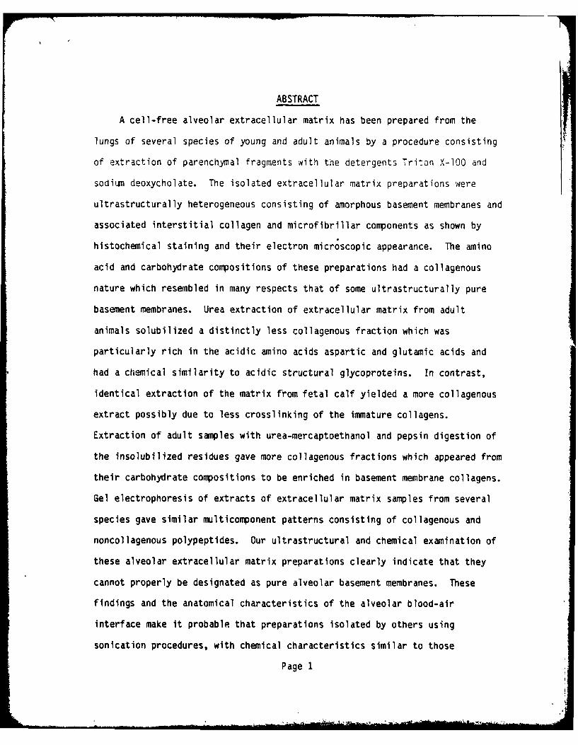
ABSTRACT
A cell-free alveolar extracellular matrix has been prepared from the
lungs of several species of young and adult animals by a procedure consisting
of extraction of parenchymal fragments with the detergents Triton X-100 and
sodium deoxycholate. The isolated extracellular matrix preparations were
ultrastructurally heterogeneous consisting of amorphous basement membranes and
associated interstitial collagen and microfibrillar components as shown by
histochemical staining and their electron microscopic appearance. The amino
acid and carbohydrate compositions of these preparations had a collagenous
nature which resembled in many respects that of some ultrastructurally pure
basement membranes. Urea extraction of extracellular matrix from adult
animals solubilized a distinctly less collagenous fraction which was
particularly rich in the acidic amino acids aspartic and glutamic acids and
had a chemical similarity to acidic structural glycoproteins. In contrast,
identical extraction of the matrix from fetal calf yielded a more collagenous
extract possibly due to less crosslinking of the immature collagens.
Extraction of adult samples with urea-mercaptoethanol and pepsin digestion of
the insolubilized residues gave more collagenous fractions which appeared from
their carbohydrate compositions to be enriched in basement membrane collagens.
Gel electrophoresis of extracts of extracellular matrix samples from several
species gave similar multicomponent patterns consisting of collagenous and
noncollagenous polypeptides. Our ultrastructural and chemical examination of
these alveolar extracellular matrix preparations clearly indicate that they
cannot properly be designated as pure alveolar basement membranes. These
findings and the anatomical characteristics of the alveolar blood-air
interface make it probable that preparations isolated by others using
sonication procedures, with chemical characteristics similar to those
Page 1
... .. . . ... ., ,,, .. ... . .. .. .... " - '. ... iF.. . *0 "....... .. . . . . i l

INTRODUCTION
The alveolar-capillary barrier is the morphological and functional
mediator of gas exchange in the lung. An integral component of this barrier
is the alveolar basement membrane which lies between the capillary endothelium
and alveolar epithelium and serves as a supporting structure upon which these
cells rest, as well as a boundary layer which defines the separation between
the blood and air spaces of the lung. [18,30]. The juxtaposition of the
alveolar basement membrane between two different cell types and its
morphological features by which it appears alternately as a double layered
structure separated by an intervening interstitial space, or as a single layer
which appears to be the fusion of two membranes which may be heterogeneous in
nature [12] make it a complex structure not easily accessible to
isolation and biochemical investigation. For this reason it has been one of
the least studied basement membranes in regard tu chemical composition and
structure.
Kefalides and Denduchis [15] reported on the composition of an alveolar
basement membrane fraction obtained from dog lung which was prepared by a
combination of exhaustive extraction of thin sections of peripheral lung
tissue with O.3M acetic acid at 4C and sonication in physiological saline.
Bray and LeRoy [1] isolated an alveolar basement membrane preparation
from human lungs using a sieving and sonication procedure analogous to that
previously employed for the preparation of bovine glomerular basement membrane
[29]. Although both of these preparations had a collagenous composition
similar to those obtained from the morphologically pure and chemically well
characterized basement membrane of the lens capsule and renal glomerulus, no
electron microscopic examination of their structure was given to reveal the
Page 3

reported here, were not ultrastructurally pure alveolar basement membranes as
reported but were actually heterogeneous mixtures of basement membranes,
interstitial collagens and microfibrils. It would appear that the alveolar
basement membrane cannot be isolated in a form comparable to ultrastructuraily
pure basement membranes such as that of the renal glomerulus.
Key Words:
Lung extracellular matrixalveolar extracellular matrixalveolar basement membranelung basement membranelung collagen
Page 2
reported here, were not ultrastructurally pure alveolar basement membranes as
reported but were actually heterogeneous mixtures of basement membranes,
interstitial collagens and microfibrils. It would appear that the alveolar
basement membrane cannot be isolated in a form comparable to ultrastructjraMl
pure basement membranes such as that of the renal glomerulus.
Key Words:
Lung extracellular matrixalveolar extracellular matrixalveolar basement membranelung basement membranelung collagen
Page 2

possible extent of contamination by other connective tissue components. In
view of the known intimate association of the basement membranes of the
alveolar dali with striated interstitial collagen and microfibrillary
:-rponent- as revealed by several electron microscopic examinations of lung
" :sue 'l6,19,28], it Nas difficult to see how simple sieving, sonication and
acid extraction procedures could result in the isolation of a basement
membrane fraction substantially free of these components. Madri and Furthmayr
[20] have recently demonstrated the collagen polymorphism of the lung by
immiunochemical techniques and have shown that lung parenchyma contains at
least four distinct types of collagen (I, III, IV and V (AB2)).
We have therefore isolated alveolar extracellular matrix from the lungs
of several species of animals of different ages using a detergent extraction
procedure which has proved valuable in the isolation of ultrastructurally pure
and intact basement membranes from renal glomeruli and tubules and retinal and
brain microvessels [5,21,22], as well as in characterizing the more complex
extracellular matrix obtained from human retina, brain, peripheral nerve and
placenta [4,23-26]. Our results indicate that detergent extraction of lung
parenchymal tissue yields an extracellular matrix consisting of amorphous
basement membrane material as well as enclosed and associated interstitial
collagen fibers and microfibrils, which although having a chemical similarity
to ultrastructurally pure basement membranes, are morphologically more similar
to the ultrastructurally heterogeneous extracellular matrix previously
characterized from human placenta [24,25]. Selective extraction of the
alveolar extracellular matrix with urea solutions under reducing and
nonreducing conditions followed by pepsin treatment of the insoluble residue
yielded markedly collagenous fractions and those which more closely resembled
acidic structural glycoproteins [3,6] in their composition.
Page 4
--------------------------
MATERIALS AND METHODS
Preparation of Alveolar Extracellular Matrix
Alveolar extracellular matrix was prepared from the lungs of several
species of animals of different ages by a modification of a procedure developed
in this laboratory for the isolation of basement membranes and extracellular
matrix from other tissues and tissue fractions [4,5,21-25]. Lungs of dog,
rabbit, adult rat and newborn rats were obtained fresh and processed on the same
day. Fetal calf lung which was obtained frozen from Pelfreeze was processed as
soon as it arrived. The parenchymatous material from the lung was separated
from the large vessels using a blade at 4C. Care was taken to exclude large
airways, blood vessels and fibrous tissue. The rest of the isolation was done at
room temperature (20C). The tissue was washed well in isotonic saline and
suspended in 20 times its volume of saline. It was homogenized using a Polytron
(Brinkmann Instruments) at an intermediate setting for 30 sec for soft tissues
and 2 min for tougher tissues. The homogenate was filtered through a 400 Pm
opening nylon sieve to remove large particles. These consisted mainly of blood
vessels and portions of bronchi which are not homogenized to small fragments
under the conditions used as are the more 'ragile alveoli. The filtrate was put
through a 110 Pm opening sieve and this filtrate was discarded. The material
collected on the sieve was washed with saline and after examination by phase
contrast microscopy to assure the absence of large airways and blood vessels was
treated with a solution of 4% Triton X-lO0 in phosphate buffer (0.2 M, pH 8.0).
The tissue weight to Triton volume ratio was usually 1:10. All extractions were
done in the presence of a proteinase inhibitor (phenylmethylsulfonylfluoride 50
pg/ml), a bacteriostat (sodium azide 50 Ug/ml) and a fungizide (caprylic acid 50
ug/ml). In order to further reduce the possibility of proteolysis, the Triton
mixture was changed after 1/2 h, 2 h and 15 h. During each change the tissue
was washed with saline. After 24 h the tissue was filtered and washed. It was
Page 5

then stirred with sodium deoxycholate (4%). After 24 h of stirring, the tissue
was filtered and washed first with 0.1% sodium bicarbonate solution, and then
with distilled water. This material was lhen treated with deoxyribonuclease
(lO0 g,/:nl, 1 h, 37C) in the prese;ce j- calciun and magnesium ions. Ahen the
amount of the tissue was large, this treatment was repeated. The material after
this treatment was washed well in \vater and preserved with sodium azide.
Histology
Samples of extracellular matrix preparations were embedded for routine
light microscopy and periodic-acid-Schiff reagent counterstained with
hemotoxylin was used to assess gross cellular ccntamination with every sample
which was analyzed. Jones silver stain which specifically stains basement
membrane material black was used to distinguish basement membrane materials.
Interstitial collagen (Type I) was detected by staining with Van Gieson's
solution which stains collagen red. In order to determine the extent of fibrin
contamination, preparations were stained with phosphotungstic acid-hemotoxylin
which gives a blue color when fibrin is present.
Immunofluorescence Reaction
The antigen was a preparation of basement membrane purified from rabbit
renal tubules by extraction with detergents as described previously [5,21,22].
Hyperimmune antiserum was raised in a young female goat by an initial
injection of an emulsified suspension of tubular basement membrane in complete
Freund's adjuvant followed by booster injections at monthly intervals in
incomplete adjuvant. An indirect immunofluorescence procedure was performed
on cryostat sections (5 um) of lung tissue or on basement membrane suspensions
which were dried onto glass slides. The slides were incubated with antiserum
for 30 min at room temperature. The goat antiserum was diluted 30 fold in 33%
Page 6
(v/v) normal rabbit serum in buffered saline. The control serum was a
pre-immune serum from the same goat used in similar dilution. There was no
detectable fluorescence with the control, pre-immune goat serum diluted 30
fold in 33% rabbit serum as was the antiserum nor when diluted only 5 fold.
After incubation with the antiserum or the control serum, the slides were
washed and incubated for 15 min with the secondary antiserum,
fluorescein-conjugated rabbit antigoat IgG purchased from Miles Laboratories,
Inc. After washing with phosphate buffered saline, the slides were mounted in
buffered glycerol and examined with a Zeiss microscope equipped with
epiillumination and filters for fluorescence detection.
Electron Microscopy
The alveolar extracellular matrix preparations were examined by electron
microscopy. Matrix preparations were washed exhaustively with water and fixed
in cold (4*C) Karnovsky's solution followed by washing and storage in 0.15 M,
pH 7.4 cacodylate buffer. After post fixation in 0.5% osmium tetroxide
solution, the samples were prepared for transmission electron microscopy.
Ultrathin sections were stained with uranylacetate or bismuth subnitrate and
examined on a Hitachi HS-7 transmission electron microscope.
Extraction of Alveolar Extracellular Matrix
A portion of the alveolar extracellular matrix was dried in a lyophilizer
and 20 mg of this dried material was first extracted with 8 M urea in
phosphate buffer (0.1 M, pH 7.7) for 24 hours at room temperature to
solubilize non-covalently bound non-collagenous proteins and non-crosslinked
collagens. After centrifugation of the extracted material, the residue was
extracted with 8 M urea containing 3% mercaptoethanol to solubilize any
proteins linked to the matrix by disulfide bonds. The remaining insoluble
residue was washed with 0.5 M acetic acid and digested with pepsin (100 pg/ml,
Page 7

40C) in acetic acid for 24 hours to solubilize crosslinked collagens. The
pepsin digest was neutralized with 10 N NaOH and then dialyzed against
phosphate buffer (0.1 M, pH 7.2).
Amino Acid and Carbohydrate Analysis
Samples for amino acid analysis qera hydrolyzed at 110C f)r 22 h
constant boiling HCI under nitrogen. Samples were driea at room tereratre
over NaOH in a desicator and analyzed on a Beckman 121 C amino acid analyzer
using a modification of the method of Guire, et al [9].
Carbohydrate analysis was performed by a gas chromatographic procedure
modified from the method of Grimes and Greegor [8]. Samples were
hydrolyzed in 2 N trifluoroacetic acid for 6 hrs at 110*C in sealed tubes
using arabitol as an internal standard, followed by deamination, reduction ana
acetylation of the released sugars as described previously [25].
Analysis of the derivitized samples was carried out on a Hewlett-Packard
automated gas-liquid chromatograph using an OV-225 column and an automatic
integrator for the quantitation of peaks.
Gel Electrophoresis
Gel electrophoresis was done by the method of Furtuayr and Timpl [7]
using 5% acrylamide gels in phosphate buffer (0.1 M, pH 7.2) with accompanying
internal standards of type I collagen. Gels were stained with Coomassie Blue
R-250 (0.025%) and destained in acetic acid (7%).
Materials
All chemicals were analytical reagent grade. The following chemicals
were obtained from the respective manufacturers: Deoxycholate (sodium salt)
from Sigma, U.S.A., Triton X-100 from Rohm and Haas, deoxyribonuclease
(pancreatic) from Sigma, pepsin (twice crystallized) from Worthington and
Page 8
'41
collagenase type III from Advanced Biofactures. Fetal calf lung (frozen) was
obtained from Pelfreeze.
Results
Isolation Procedures
The scheme for preparing and characterizing alveolar extracellular matrix
is outlined in Figure 1. After removal of large blood vessels, bronchi and
other unwanted connective tissue of the lung, the parenchymatous tissue was
disrupted in a Polytron homogenizer for different times depending upon whether
it was derived from fetal or adult lungs. Initial filtration through a nylon
sieve with 400 ,,m openings served to remove large undisrupted tissue fragments
consisting mainly of remaining blood vessels and bronchi which were discarded.
The filtrate consisting mainly of alveolar fragments and smaller cell debris
was passed through a 110 um nylon sieve which retained most of the alveolar
pieces and let the cell debris pass through to be discarded. The retained
alveolar fragments were freed of cellular material in a graded fashion by
stepwise treatment with Triton X-100, sodium deoxycholate and pancreatic
deoxyribonuclease yielding a cell-free alveolar extracellular matrix as shown
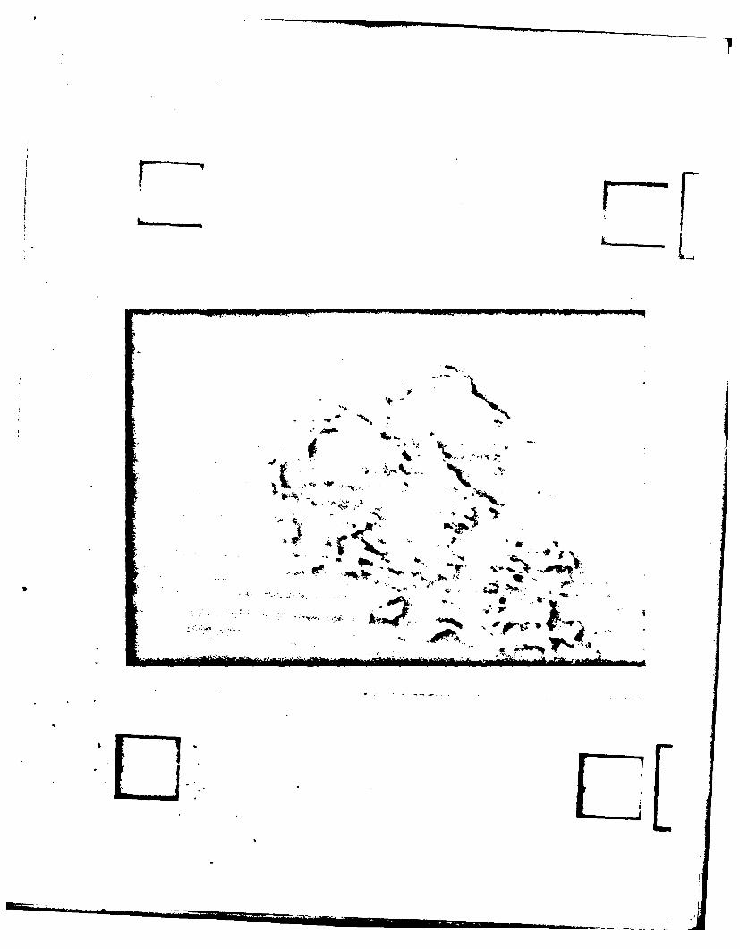
by periodic acid-Schiff hematoxylin staining of samples (Fig. 2). Analyses of
the matrix for DNA and phosphate gave minimal values (<0.2% dry weight)
comparable to those obtained for glomerular and tubular basement membranes
isolated by an analogous detergent procedure [17]. Jones silver staining of
this material yielded a pattern of black staining indicative of the presence
of basement membranes (not shown). Van Gieson's stain gave both blue and
light pink staining indicating the presence of basement membrane and
associated collagens. Phosphotungstic acid-hemotoxylin stain which was used
to monitor fibrin contamination showed that these samples do not have any
contamination from fibrin.
Although free of cells, these preparations probably contain a mixture of
Page 9

interstitial collagens, microfibrillar components and elastin in addition to
alveolar baseme.t membrane, as indicated by their appearance in transmission
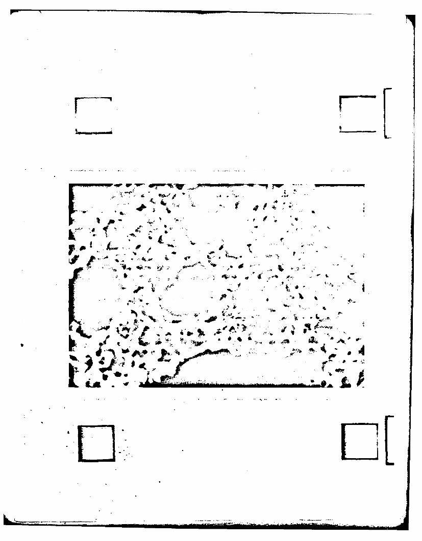
electron microscopy (Fig. 3).
Isolatad fracments of ewborn rat alveolar basement membrane-containing
extracediular matrix exhibited substantial fluorescence in an indirect
imunofluorescent procedure using a goat antiserum raised against rabbit
tubular basement membrane (Fig. 4a). Reaction of the antiserum with cryostat
sections of intact fetal rabbit lung tissue results in marked fluorescence of
the alveolar wall (Fig. 4b). In this case, a control slide using pre-immune
goat serum showed no detectable fluorescence (not shown).
Chemical Analyses
The amino acid analyses of alveolar extracellular matrix prepared from
the lung parenchyma of dog, rabbit, fetal and adult cow, and newborn and adult
rat are shown in Table 1. The compositions from all species are typically
collagenous in nature with a high content of glycine, proline, and alanine and
appreciable amounts of 4-hydroxyproline and hydroxylysine. Sulfur-containing
and aromatic amino acids were low in all samples and there was a virtual
absence of the 3-isomer of hydroxyproline. In comparison with the matrix from
adult animals, that from fetal or newborn animals showed a lower content of
many of the amino acids abundant in collagen, glycine, proline, alanine, and
hydroxyproline and a higher content of the acidic amino acids, glutamic and
aspartic acid, the hydroxylated amino acids serine and threonine and the basic
amino acids lysine and histidine. Enough samples were not analyzed, however,
to allow a quantitative statistical analysis of these results.
Carbohydrate analyses of the preparations (Table 2) indicated that
galactose and glucose were the most abundant sugars in each sample with lesser
amounts of mannose and minor amounts of fucose. In each case the galactose
Page 10
content was somewhat in excess of that found for glucose. Fetal and newborn
animals showed lower amounts of glucose and galactose in the extracellular
matrix than adult animals, but somewhat higher levels of mannose and fucose,
although the limited number of samples analyzed did not allow a statistical
comparison of these results.
Selective Extraction of Alveolar Extracellular Matrix
Initial extraction of alveolar extracellular matrix from fetal calf or
adult cow lungs with 8 M urea alone solubilized about one-third of the matrix.
Further extraction of the residue with 8 M urea in the presence of 3%
mercaptoethanol solubilized another 40% of the material and controlled
digestion of the final residue with pepsin at 4C brought another one-sixth
into solution leaving about 10-15% of the material intractable to denaturing
solvents or enzyme digestion. Similar results were obtained by analogous
extraction and pepsin treatment of the extracellular matrix from adult rat
lung alveoli.
Amino acid analyses of the urea solubilized fractions obtained under
non-reducing and reducing conditions and of the pepsin solubilized fractions
are given for fetal calf and bovine lung alveolar extracellular matrix in
Table 3. While the urea solubilized fraction from fetal calf lung has a
composition which is markedly collagenous in nature and more collagenous than
the unextracted matrix, the comparable fraction from bovine lung is distinctly
less collagenous than the starting material. The fraction solubilized by
urea-mercaptoethanol from bovine lung is more collagenous than that obtained
with urea alone, while the analogous fraction from fetal calf lung is less
collagenous than the urea fraction obtained under non-reducing conditions.
Pepsin digestion of the residues remaining after urea-mercaptoethanol
Page 11
extraction of fetal calf or bovine lung alveolar extracellular matrix
liberated markedly collagenous materials in both cases as can be seen by the
high content of hydroxyproline, glycine, proline 3nd alanine in these
fractions. The least collagenous fraction :btained in Iiis series of
extractions was that solubilized by urea alone froi bovine lung alveolar
extracellular matrix. This fraction was notable for its markedly lower
content of glycine, hydroxyproline, proline and hydroxylysine and its
enrichment in the acidic amino acids glutamic and aspartic acid and the
hydroxylated amino acids threonine and serine.
Amino acid analyses of the extracts obtained from adult rat alveolar
extracellular matrix (Table 4) yielded a pattern analogous to that seen for
the comparable fractions from adult bovine lung (Table 3). With both tissues
the urea extracts were the least collagenous and the richest in the acidic and
hydroxylated amino acids and the pepsin-liberated materials most resembled
collagen in their amino acid compositions.
Comparison of the carbohydrate compositions of the fractions obtained by
selective extraction of the extracellular matrix from adult rat lungs (Table
4) revealed the urea-mercaptoethanol and the pepsin-derived fractions to be
the richest in their content of the neutral sugars glucose and galactose while
the urea-extracted fraction was lowest in the content of these sugars and
higher in its content of mannose and the amino sugars. Only qualitative
comparisons of these amino acid and carbohydrate patterns were attempted since
variations in extent of extractability and the limited number of samples
analyzed did not allow for statistically quantitative comparisons of these
results.
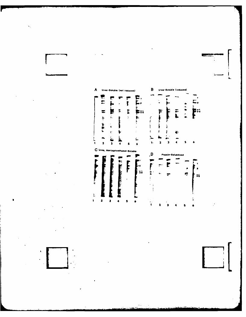
SDS Gel Electrophoresis
The SOS-polyacrylamide gel electrophoretic patterns of the urea,
Page 12
urea-mercaptoethanol and pepsin-derived fractions of the alveolar
extracellular matrix from the lungs of several species are shown in Figure 5.
The patterns are similar for each species. The urea extract of the alveolar
extracellular matrix gave two bands near the a position of collagen. Although
the nature of these bands has not been fully studied, preliminary evidence in
the case of the fetal calf urea extracts indicates that these bands do not
correspond in their mobility with the al(I) and a2 bands of type I collagen,
but instead have mobilities which resemble that of procollagen chains. The
top band has been found to be partially digestible with pepsin (Kuttan, et
al., unpublished observation). The nature of these bands in other species has
not been studied. It is interesting to note here that the adult rat gave only
one prominent band at the a position. There were also other bands in the urea
extracts. The remarkable ones are the fast moving bands that appear after
reduction, and the bands between the a and s chains. A large portion of the
applied material did not enter the gel until reduced, a behavior typical of
this type of sample [20]. The urea-mercaptoethanol extracts of several
alveolar extracellular matrix preparations also gave bands at the a position.
There were very strong bands between the a and s chains. There were some
differences in the mobility of these bands in different species. Pepsin
extracts yielded two bands at the 1 position and a band between the a and a
position. The nature of these bands has not been investigated, but they are
presumed to be collagenous in nature. Interpretation of these results is
complicated by uncertainties about the pepsin susceptibility of matrix
residues previously extracted with denaturing solvents (urea and SOS), and
the difficulty of comparing the mobility of bands from extracted
non-enzymatically treated samples to standards obtained by controlled pepsin
digestion.
Page 13
DISCUSSION
In this report we describe the isolation of alveolar extracellular matrix
from the lungs of several species of young and adult animals. Although
alveolar basement membrane is a principal coaponent of this extracellular
matrix preparation, the preparation did not consist solely of a
morphologically-homogeneous, amorphous boundary membrane as is the case for
the basement membranes obtained by analogous treatment of renal glomeruli and
tubules, and brain and retinal microvessels [5,17,21,22]. Whereas detergent
treatment [5,17,21,22] or sonication, and differential centrifugation [29] of
renal glomeruli or tubules yield a preparation in which basement membranes
devoid of interstitial striated collagen fibers, elastin or fibrin comprise
the entire extracellular matrix of these organ subfractions, similar treatment
of placental villi [24,25] or lung alveoli yield preparations which, although
having colpositional and immunological similarities to well characterized
basement membranes, are ultrastructurally heterogeneous.
Several electron microscopic examinations of the alveolar-capillary
barrier have shown it to contain numerous collagen fibers which appear to be
incorporated with the basement membrane between alveoli and capillary lumina
[18,19,28). The intimate structural association of microfibrillar components
of the extracellular space with the basement membranes of the alveolar wall
have also been described in detail [19]. Therefore, in the absence of a
careful morphological analysis of the isolated sample, one should be cautious
in describing a preparation as that of alveolar basement membrane. In fact,
of the two preparations previously described denoted as alveolar basement
membrane, one was subjected to exhaustive extraction with acetic acid at 48C
in order to remove contaminating interstitial collagen [15], while the other
showed evidence of contamination with elastin and had a chemical
Page 14
composition markedly different from that of glomerular basement membrane
isolated by an analogous procedure [I]. No electron microscopic
evidence of the ultrastructural purity of either preparation was presented.
We have previously characterized, both morphologically and chemically, an
extracellular matrix preparation from human placental villi, and shown that
although the amino acid composition of this material bore a strong resemblance
to that of human glomerular basement membrane, its electron microscopic
appearance was that of amorphous basement membranes associated with
interstitial collagen fibers and scattered patches of fibrin [24,25]. The
preparation of alveolar extracellular matrix described in this paper bears a
superficial resemblance to ultrastructurally pure basement membranes.
Although there are similarities in the amino acid compositions, there are no
distinctive chemical characteristics by which one can unequivocally label this
preparation as alveolar basement membrane and ultrastructural examination
clearly precludes this label. The amino acid compositions of our alveolar
extracellular matrix preparations from cow and adult rat lungs are similar to
that of the human alveolar basement membrane preparation reported by Bray and
LeRoy £1], and it is probable that these are comparable materials prepared by
detergent extraction and sonication, respectively.
The carbohydrate compositions of the samples of alveolar extracellular
matrix examined also have a pattern similar to that seen in pure basement
membraqes, being richest in glucose and galactose, which are present in nearly
equimolar quantities, with lesser amounts of mannose and traces of fucose [5,
16,21,22,29]. Similar proportions of these carbohydrates were present in the
alveolar preparation of Kefalides and Denduchis [15]. The glucose
content of the preparations is probably a good index of their collagenous
content in general and their content of basement membrane collagens in
Page 15
I.

particular, since this sugar is not a common constituent of glycoproteins and
is present in appreciable amounts only in type II collagen [10] and collagens
of basement membrane origin [16,29]. Since in the lung, Type II collagen has
onny een reported as a constituent of trachea [10], this would not be a
likely source of the glucose in our preparations.
The increased collagen-like profile of the amino acid compositions of
alveolar extracellular matrix preparations from adult rat and cow compared
with those from their fetal and newborn conterparts are similar to the age
related changes in amino acid composition reported in rat glomerular basement
membrane [11,14], and may reflect an increased deposition or reduced
degradation of the collagenous components of the matrix with age. Sequential
extraction, with urea under non-reducing and reducing conditions, of the
alveolar extracellular matrix preparations from adult bovine lung and fetal
calf lung also revealed an interesting age-related possible difference between
the two preparations. While the urea extract of the adult sample was markedly
less collagenous than the starting material, as was also seen for adult rat
lung extracellular matrix and reported for glomerular basement membrane [13],
the comparable extract of the fetal sample was highly collagenous in
composition. This probably reflects a greater degree of non-crosslinked
collagens in the fetal than in the mature matrix. The urea extracts of the
samples from adult bovine and rat lungs were also notable for their increased
content of the acidic amino acids, glutamic and aspartic acids, the polar
amino acids, threonine and serine, and the neutral amino acids, leucine and
isoleucine. In this respect their composition is similar to that of acidic
structural glycoproteins isolated from lung parenchyma by extraction with
urea-sodium borohydride [6] or acetic acid [3), although in contrast to the
latter, our extracts still contained some collagenous components as evidenced
Page 16
by the continued presence of hydroxyproline and hydroxylysine. The
urea-mercaptoethanol extracts of the alveolar matrix from adult bovine and rat
lungs were decidedly more collagenous than the urea extracts alone, indicating
that an appreciable amount of collagenous material was bound to the matrix by
disulfide bonds. The material obtained from the extracted residues of both
fetal and adult matrix samples by controlled pepsin digestion was almost
completely collagenous and similar in amino acid composition to the insoluble
residues obtained after complete extraction of glomerular basement membrane
under comparable dissociating and reducing conditions [13,14].
Of particular interest in this collagenous material derived from the core
of insoluble material in the matrix from adult rat lung is its relatively high
content of glucose and galactose. The high content of these sugars in this
sample makes it likely that basement membrane collagens are a significant
component of the insoluble alveolar matrix core, possibly bound by
non-reducible covalent crosslinks. A significant amount of the basement
membrane collagen is also bound in the matrix by disulfide bonds and can be
extracted with urea-mercaptoethanol as can be seen by the high
glucose-galactose content of this fraction derived from rat alveolar
extracellular matrix.
The multicomponent nature of the alveolar extracellular matrix
preparations and their qualitative similarity from species to species is
indicated by the patterns obtained after SDS gel electrophoresis of urea and
urea-mercapto-ethanol extracts of the preparations. The results confirm the
importance of reducible.disulfide cross-links in the organization of the
matrix and its comprisal of several polypeptide chains of differing molecular
weights of collagenous and non-collagenous characteristics. A similar complex
organization has been reported for both pure basement membranes such as that
Page 17
of the renal glomerulus +&Qul4,15] and for placental chorionic villar extra-
cellular matrix [24-26] which is a complex of connective tissue components of
which the trophoblast basement membrane. is just one part. The multiple
banding patterns given by these exzracts of lunc extracellular matrix were
reproducible and were the same wheter iiatrix isolation was carried out in the
presence or absence of proteolytic inhibitors, indicating that they were not
the result of in vitro proteolysis during isolation.
Because of the likely impossibility of separating a truly ultrastruc-
turally-pure alveolar basement membrane preparation from the collagenous and
other connective tissue components with which it is intimately associated in
the alveolar matrix, future work on this basement membrane will have to
concentrate on the fractionation of components from the matrix and their
identification as basement membrane constituents by comparison with analogous
components obtained from ultrastructurally pure basement membranes such as
those of lens capsule and renal glomeruli. Until then it would appear
prudent not to designate the clearly ultrastructurally heterogeneous
preparations, which are derived by sonication or detergent treatment of tissue
subfractions such as those from lung or placenta, as 'basement membranes'
[1,3], nor to ascribe components present in such preparations such as
fibronectin [2] as being basement membrane constituents.
Page 18

LUNG4
REMOVE LARGE VESSELS AND DISSECT OUTPARENCHYMATOUS TISSUE
FETAL TISSUES ADULT TISSUES
AILD HOMOGENIZATION IN STRONG HOMOGENIZATION IN0.85% NaC1 0.85% NaCl
(30 SEC POLYTRON) (2 MIN POLYTRON)
PASSED THROUGH 400 um SIEVE
PASSED THROUGH FRACTION
+
FILTER ON 110 pm SIEVE
RETAINED MATERIAL
REMOVE CELLULAR MATERIAL BYSTIRRING IN 4% TRITON X-lO0, 24 HRS
REMOVE NUCLEAR AND OTHER MEMBRANES BYSTIRRING IN 4% DEOXYCHOLATE, 24 HRS
4
REMOVE DNA BY DIGESTING WITHDNAase (100 pg/ml, 1 h, 37*C)
PURIFIED ALVEOLAR EXTRACELLULAR MATRIX
HISTOLOGY FLUORESCENT ELECTRON DRIED INANTIBODY MICROSCOPY LYOPHILIZER
Amino Carbohydrate SelectiveAcid Analysis Extraction
Analysis andGel Electrophoresis
FIGURE I
Page 19

Figure 1. Scheme for preparation and characterization of lung alveolar
extracellular matrix. All steps were performed in presence of 50
ig/ml phenylmethylsulfonylfluoride, sodium azide and caprylic cJ.
See text for details.
Figure 2. Light micrograpn of section of isolated alveolar extracellu!_ r
matrix stained with periodic acid-Schiff hematoxylin stain. Note
absence of cells and staining of matrix (X 90).
Figure 3. Transmission electron micrograph of isolated alveolar extracellular
matrix. Preparations consisted of amorphous basement membrane
material, free and closely associated fibrils of interstitial
collagen and other microfibrillar components (X 8250).
Figure 4A.Immunofluorescence reaction of newborn rat lung alveolar extra-
cellular matrix with goat anti-rabbit renal basement membrane
antiserum (X 90). Control goat serum at the same concentration
produced no detectable fluorescence.
4B.Immunofluorescence reaction of cryostat section of fetal rabbit lung
with renal basement membrane antibody (X 150). Control goat at the
same concentration produced no detectable fluorescence.
Figure 5. SOS-polyacrylamide gel electrophoresis patterns of extracts from
lung alveolar extracellular matrix preparations of various species.
lDog, 2=Rabbit, 3=Fetal Calf, 4=Bovine, 5=Adult Rat, 6=Newborn Rat.
A. Urea extracts, B. Urea extracts reduced with mercaptoethanol,
C. Urea-mercaptoethanol extracts, D. Pepsin extracts.
The mobilities of collagen chains derived from Type I collagen are
as indicated.
Page 20
REFERENCES
1. Bray BA, LeRoy EC (1976) Human alveolar basement membranes.Chemical and immunologic comparisons with glomerular basement membraneand trophoblast basement membrane. Microvasc Res 12:77-89.
2. 3ray BA (1973) Presence of fibronectin in basement membranes andacidic structural glycoproteins from human placenta and lung. Ann NYAcad Sci 142-150.
3. Bray BA (1978) Cold-insoluble globulin (fibronectin) in connectivetissues of adult human lung and in trophoblast basement membrane. J ClinInvest 62:745-752.
4. Brendel K, Meezan E (1980) Vascular basement membranes: Preparationand properties of material isolated with the use of detergents. InEisenberg HM, Suddith RL (eds) Cerebral Microvasculature: Investigationof the Blood-Brain Barrier, Plenum Press, New York, pp. 89-103.
5. Carlson EC, Brendel K, Hjelle JT, Meezan E (1978) Ultrastructural andbiochemical analyses of isolated basement membranes from kidney glomeruliand tubules, and brain and retinal microvessels. J Ultrastruct Res 62:26-53.
6. Francis G, Thomas J (1975) Isolation and characterization of acidicstructural glycoproteins in pulmonary tissues, Biochem J 145:299-304.
7. Furthmayr H, Timpl R (1971) Characterization of collagen peptides bysodium dodecylsulfate-polyacrylamide gel electrophoresis. Anal Biochem41:510-516.
8. Grimes WJ, Greegor S (1976) Carbohydrate compositions of normal,spontaneously transformed and virally transformed cells derived fromBALB/c mice. Cancer Res 36:3905-3910.
9. Guire P, Riquetti P, Hudson BG (1974) An accelerated single columnprocedure for the programmed analysis of naturally-occurring amino acidsof collagen and basement membrane. J Chromatog 90:350-353.
10. Hance AJ, Crystal RG (1976) Collagen. In Crystal RG (ed) The BiochemicalBasis of Pulmonary Function, Dekker, New York, pp. 215-271.
11. Hoyer JR, Spiro RG (1978) Studies on the rat glomerular basementmembrane: Age-related changes in composition. Arch Biochem Biophys185:496-503.
12. Huang TW (1978) Composite epithelial and endothelial basal laminas inhuman lungs. Am J Path 93:681-691.
13. Hudson BG, Spiro RG (1972) Studies on the native and reduced alkylatedrenal glomerular basement membrane. Solubility, subunit size andreaction with cyanogen bromide. J Biol Chem 247:4229-4238.
Page 21
14. Kalant H, Satomi S, White R, Tel E (1977) Changes in renal glomerularbasement membrane with age and nephritis. Can J Biochem 55:1197-1206.
15. Kefalides NA, Denduchis B (1969) Structural components of epithelial andendothelial basement membranes. Biochemistry 8:4613-4621.
16. Kefalides NA (1978) Current status of chemistry and structure of basement:embranes. In Kefalides NA (ed) Biology and Chemistry of Basement1,embranes, Academic Press, New YorK, pp. 215-223.
-7L,• Langel'd JPM, Veerkamp JH, Monnens LAH, Van Haelst VJG (1973) Chemical
characterization of glomerular and tubular basement membranes of cattleof different ages. Biochim Biophys Acta 514:225-238.
18. Low FN (1961) The extracellular portion of the human blood-air barrierand its relation to tissue space. Anat Rec 139:105-124.
19. Low FN (1962) Microfibrils: Fine filamentous components of the tissuespace. Anat Rec 142:131-137.
20. Madri JA, Furthmayr H (1980) Collagen polymorphism in the lung. Animmunohistochemical study of pulmonary fibrosis. Human Path 11:353-366.
21. Meezan E, Hjelle JT, Brendel K, Carlson EC (1975) A simple, versatilenon-aisruptive method for the isolation of morphologically and chemicallypure basement membranes from several tissues. Life Sci 17:1721-1732.
22. Meezan E, Brendel K, Hjelle JT, Carlson EC (1978) A versatile method forthe isolation of ultrastructurally and chemically pure basement membraneswithout sonication. In Kefalides NA (ed) Biology and Chemistry ofBasement Membranes, Academic Press, New York, pp. 17-30.
23. Meezan E, Nagle RB, Johnson P, Wagner C, White R, Brendel K (1979)Structural and functional properties of acellular histoarchitecturallyintact basement membranes. In Robert AM, Robert L (eds) Frontiers ofMatrix Biology, Karger, Basel, pp. 101-119.
24. Ohno M, Nagle RB, Meezan E, Brendel K (1979) Isolation andcharacterization of human placental chorionic villar extracellularmatrix. J Supramol Struct 12:457-466.
25. Ohno M, Spall R, Nagle RB, Brendel K, Meezan E (1980) Human placentalchorionic villar extracellular matrix 1. Preparation and chemicalcomposition from villar fragments fractionated according to their size.Int J Biol Res Preg 1:38-47.
26. Ohno M, Meezan E, Brendel K (1980) Human placental chorionic villarextracellular matrix 2. Solubilization and characterization from villarfragments fractionated according to their size. Int J Biol Res Preg 1:79-89.
Page 22

27. Sato, T, Spiro RG (1976) Studies on the native and reduced alkylated renalglomerular basement membrane. Solubility, subunit size and reaction withcyanogen bromide. J Biol Chem 247:4229-4238.
28. Sobin SS, Bernick S, Tremer HM, Rosenquist TH, Lindal R, Fung YC (1974)The fibroprotein network of the pulmonary interalveolar wall. Chest65:45-55.
29. Spiro RG (1967) Studies on the renal glomerular basement membrane.Preparation and chemical composition. J Biol Chem 242:1915-1922.
30. Weibel ER (1963) Morphology of the Human Lung, Academic Press, New York.
Page 23

TABLE I
Amino Acid Composition of AlveolarExtracellular Matrix Preparations
Residues/lO00 Anino Acids
New-Young Fetal born Adult
Amino Acids 009 Rabbit Calf Cow Rat Rat
3-hydroxyproline trace trace trace trace trace trace4-hydroxyproline 68.7 43.2 50.7 64.8 41.0 65.2Aspartic Acid 42.2 54.3 59.0 48.2 49.0 52.1Threonine 24.3 32.1 31.0 24.4 32.7 25.6Serine 32.3 40.5 40.5 35.2 41.0 38.0Glutamic Acid 62.7 75.7 83,3 72.1 69.5 68.4Proline 101.2 89.7 88.0 95.4 87.8 98.9Glycine 296.5 237.4 228.8 265.7 263.7 272.8Alanine 130.1 117.5 107.0 120.3 124.8 118.11/2 Cystine 9.2 14.2 15.4 8.2 10.1 8.0Valine 54.2 63.8 54.3 61.9 59.3 56.0Methionine 8.3 10.6 11.1 8.7 6.1 7.2Isoleucine 23.4 28.2 36.3 24.7 28.7 26.0Leucine 42.0 59.9 53.3 50.2 59.3 51.4Tyrosine 18.1 22.5 16.0 13.0 22.6 15.9Phenylalanine 22.7 26.1 25.1 24.6 22.6 17.6Hydroxylysine 10.7 7.7 7.9 9.4 8.2 7.7Lysine 18.6 29.2 34.4 24.2 26.6 23.7Histidine 8.1 11.9 11.7 8.5 10.1 9.5Arginine 26.7 35.5 46.4 40.6 36.7 38.3
Duplicate samples of each preparation were analyzed except for adult rat where 4 sampleswere analyzed.
Page 24

TABLE 2
Carbohydrate Composition of AlveolarExtracellular Matrix Preparations
ug Sugar/mg Extracellular Matrix
New-Young Fetal born Adult
Dog Rabbit . Calf Cow Rat Rat
Fucose 0.55 0.85 0.93 0.68 0.83 0.71
Mannose 3.59 4.51 4.13 3.58 5.33 3.81
Galactose 18.03 10.81 11.88 14.37 12.03 12.39
Glucose 15.00 9.20 10.03 12.27 9.98 12.00
The results are the mean of duplicate samples from all species except for adult ratwhere 4 samples were analyzed.
Page 25

TABLE 3
Amino Acid Composition of Urea and Pepsin Extractsof Bovine Alveolar Extracellular Matrix
Residues/lO00 Amino Acids
Fetal Calf Lung 3 ,ineLur
Urea Urea-ME Pepsin Urea Urea-hiE Pepsin
3-hydroxyproline 2.7 trace 0.9 0 trace 3.84-hydroxyproline 82.9 58.5 85.4 23.2 53.0 56.0Aspartic Acid 54.2 69.2 51.2 86.4 74.0 43.4Threonine 23.3 32.5 21.4 43.5 38.1 23.0Serine 40.6 47.0 41.1 54.6 53.0 32.5Glutamic Acid 80.2 92.4 71.5 120.7 102.1 60.9Praline 107.8 91.0 109.5 65.3 73.6 96.7Glycine 290.2 235.0 295.6 146.3 202.7 287.5Alanine 106.9 92.1 116.8 79.7 74.7 129.01/2 Cystine 2.7 9.8 1.8 13.2 17.3 4.9Valine 30.9 43.4 41.1 53.6 48.3 74.6Methionine 5.6 8.2 5.5 12.7 9.1 6.2Isoleucine 16.1 25.6 20.1 38.6 30.8 25.7Leucine 36.3 49.0 32.5 77.7 62.1 55.1Tyrosine 8.3 12.7 7.9 19.6 17.8 14.2Phenylalanine 18.6 24.9 18.3 32.8 30.1 31.6Hydroxylysine 7.0 8.5 7.0 6.6 15.4 15.3Lysine 28.5 33.6 24.7 46.2 34.8 12.4Histidine 7.2 12.9 6.6 18.0 15.9 5.8Arginine 50.2 53.6 41.3 62.0 47.2 21.7
The results are the means of duplicates from all samples.
Page 26

TABLE 4
Amino Acid and Carbohydrate Compositionof Urea and Pepsin Extracts of Adult Rat
Alveolar Extracellular Matrix
Residues/lO00 Amino Acids
Extra-cellular Urea Urea-MEMatrix Extract Extract Pepsin
3-hydroxyproline 0 0 0 04-hydroxyproline 65.2 35.3 59.0 60.9Aspartic Acid 52.1 85.8 77.9 60.5Threonine 25.6 43.5 36.7 27.8Serine 38.0 47.6 44.4 37.9Glutamic Acid 68.4 98.1 96.9 50.3Proline 98.9 71.1 78.7 104.3Glycine 272.8 185.6 209.0 315.3Alanine 118.1 78.8 85.2 100.51/2 Cystine 8.0 20.4 24.6 8.5Valine 56.0 55.8 43.4 59.5Methionine 7.2 10.7 12.4 5.2Isoleucine 26.0 38.4 31.0 26.4Leucine 51.4 68.3 57.3 50.8Tyrosine 15.9 17.4 15.6 10.1Phenylalanine 17.6 24.1 24.6 17.0Hydroxylysine 7.7 8.3 18.6 12.7Lysine 23.7 37.8 26.3 18.1Histidine 9.5 12.3 15.0 5.9Arginine 38.3 61.0 43.5 28.3
4g Sugar/mg Extracellular Matrix
Fucose 1.60 1.28 1.20 1.07Mannose 4.15 7.62 6.99 5.29Galactose 11.94 11.11 24.71 19.66Glucose 11.45 9.09 22.33 25.51Glucosamine 4.13 12.77 10.44 6.56Galactosamine 1.19 2.18 1.82 2.59
The results are the means of duplicates of all samples.
Page 27
.............................

rnn~~L
' , .~ .I. .. " ,
I11 " P ")pe+. ++ -, .. 'X A - . . _ . -r ", -". .e+ .. ., -= -" - - .d.,-
. ', , .. -..C. -'-- ;. -;- , _s ,- ,- ---- , .. - -r -
!I' , ". n P ,,.':..; ;. ,. . . j m - - ..ti L9- .

mrL
~ $'-. - r --- . ~ -j..*~
~' ~ -
A
-K>~~- ,d-~- ~ 4
-'1 4~ \A -.
.. ~.' 2
.4 ~ ~4*~***
r ~ ~
~ *A
- / ~ -" I..~ f
A ~ -~-.t U.4
4~V'~. " -~
* 'I ~- *
,~ ~*
~ i~~
~
I
* EJI~ .. JSfAt A
161
roN o- -
,, . •
F -' nI-4-4mflrr~w'
A *p
- - a
~4I~ tr ~ r ft
40b ~t-1w:it#
El
A Urs*-SOluble (not Fgduced) B Urea-Soluble (reduced)
C UrO&- Usap409tSenolblolubd
ID Pi- sobielowoF
ISOLATION OF TYPE II PNEUMONOCYTES
In the past, methods to assess toxicity in the lung have used whole
organ preparations or lung homogenates, but were relatively insensitive
at detecting subtle biochemical alterations which may be present in differ-
ent cell types. Therefore, it would be advantageous to conduct toxicity
studies utilizing a homogeneous cell population from the lung. One parti-
cular cell type in the lung that has been of interest is the type II or
alveolar septal cell. Pulmonary injury and repair from oxidative type
insults have been shown to be mediated by these cells (1-4). A method
was developed for the separation of pneumonocytes that is 50% faster than
current methods and that has proven more reliable at removing macrophages
than prior methods which rely upon size or density differences. The studies
resulting in the development of the method and metabolic characterization
of the cell preparation are described in the following manuscripts.
Lafranconi, W.M., Spall, R.D., Sipes, I.G., Duhamel, R.C.,Meezan, E. and Brendel, K. A method for removal of macro-phages to rapidly prepare lung cell fractions enriched inalveolar type II pneumonocytes. To be submitted to Experi-mental Lung Research.
Lafranconi, W.M., Brendel, K. and Sipes, I.G. (1981) Iso-lation and metabolic characteristics of lung cell types.Fed. Proc. FASEB (in press).
-55-

ABSTRACT
A method is described for the rapid preparation of lung cell fractions
enriched in type II alveolar pneumonocytes. Isolated perfused rabbit lungs
are exposed to Fe304 by tracheal lavage which permits pulmonary alveolar
macrophages to phagocytize the particles. Alveolar epthelial cells are then
selectively freed from the basement membrane matrix by critical placement of
collagenase and elastase. Detached cells are harvested either by repeated
tracheal lavage or by mincing the lobes and filtering freed cells through a
series of nylon mesh sieves. Iron oxide containing macrophages are then
removed from the harvested cells by a strong magnetic field. A final sizing
of the macrophage depleated suspension yields a preparation enriched in
alveolar type II cells. Eight million viable cells (95% type I) were
obiained per rabbit lung when harvested by lavage, while 32 X 10 6 (88% type
II) cells were obtained from mince lungs. These values for cell yield and
relative purity are comparable to previously described separation methods
which depend upon differences in cell density or size. A major advantage of
the magnetic separation procedure is the substantially shortened preparation
time, typically 2 hours instead of 4. The viability (90-95%), oxygen
consumption (88 nmoles/10 6 cells/h) and incorporation of 14Iacetate and
JlACcholine (0.44 and 0.115 nmoles/l0 cells/h respectively) indicate these
cells will be suitable for pharmacological of toxicological investigations.
2
A METiCD FOR REMOVA-L OF YACROPIIAGES TO0 RAPIDLY PREPARE LUNG
CELL FRACTIONS ENRICHED IN ALVEOLAR TYPE II PNELUMONOCYTES
W. Mark Lafrancon,-, Richard D. Spall, I. Glenn Sipes,Raymond C. Duhamel;, Elias Meezan* and Klaus Brendel
Department of PharmacologyCollege of Medicine
University of ArizonaTucson, Arizona 85724
*Department of PharmacologyUniversity of Alabama in Birmingham
Birmingham, Alabama 35294
Running Title: Preparati.on of Alveolar Type II Cells
Corresponding Author: Dr. Klaus BrendelDepartment of PharmacologyArizona Health Sciences CenterTucson, Arizona 85724(602) 626-7729
INTRODUCTION
The lung presents a major target organ for toxic insult. It receives 100%
of the cardiac output, and therefore maximur, exposure to systemic toxins.
Airborne toxins reach the lung through large volumes of inhaled materials
which routinely pass over the delicate alveolar surfaces. Evaluation of
specific pulmonary toxic responses have been difficult because of the extreme
heterogeneity of cell types. Under toxic influences, these cells may change
qualitatively, quantitatively or both. In the past, methods to assess
toxicity in the lung have used whole organ preparations or lung homogenates,
but were relatively insensitive at detecting subtle biochemical alterations
which may be present in different cell types. Therefore, it is advantageous
to conduct toxicity studies utilizing a homogeneous cell population from the
lung to assess the specific biochemical events of toxic response.
One particular cell type in the lung that has been of interest to
investigators is the type II or alveolar septal cell. Pulmonary injury and
repair from oxidative type insults have been shown to be mediated by these
cells (1-4). Further, type II cells produce pulmonary surfactant which is
largely responsible for maintaining the low surface tension at the alveolar
air-liquid interface (5-7). Alterations in surfactant production have been
suggested in a number of disease states and in response to certain pulmonary
toxins (8).
A number of procedures have been described fro isolating type II cells
from the lungs of both rats and rabbits (9-15). Most of these methods rely on
density differences to separate cell types from a monodispersed lung digest.
More recently procedures which use centrifugal elutriation for the separation
3
of cells on the basis of size have been described (12-16). The same
disadvantage is apparent for both procedures. Type II cells have densities
and sizes similar to macrophages and lymphocytes. Therefore, separation of
cells based on size or density may very well be incomplete because of
contamination from other cells of similar density or size.
Consequently a method to separate pneumonocytes based on techniques other
than density centrifugation or elutriation was developed. It was reasoned
that if macrophages could be induced to internalize magnetic particles they
could be effectively removed from the suspension with a strong magnetic
field. Other cell types could be removed from the lung preparation by
perfusion and sizing techniques as well as by critical enzyme placement.
These methods provide good yield, purity and reproducibility. Additionally,
this method can be completed in less than two hours compared to over four
hours required for other methods.
4

MATERIALS AND METHODS
An ima l s
White New Zealand male rabbits (2 Kg) were obtained from a local vendor
(Blue Ribbon Ranch). Rabbits were fed standard PurinaR Rabbit Chow ad
libitum and maintained in a temperature controlled room (220C) with 12 hrs
of light and 12 h of darkness in the University of Arizona Division of Animal
Resources facilities.
Perfusion Medium
Perfusion medium was composed of 128 mM NaCI, 5 mM K2S04, 5 mM KCI,
2.5 mM sodium phosphate buffer pH 7.4, 17 mM HEPES (n-2-hydroxyethyl
piperazine n-2-ethane sulfonic acid), 5/5 mm glucose. This was adjusted to pH
7.4 with 2N HCI, (during later stages of cell harvesting this buffer was
brought to 10% fetal calf serum with GIBCO F.C.S.). The buffer solution was
prepared no more than 24 h in advance, filtered through 0.45 um milipore
filters and oxygenation with 95% 02, 5% CO2 20 minutes prior to use.
Magnetite Preparation
Magnetic iron oxide (magnetite) was freshly prepared for each experiment.
A mixture of two ml of 250 mM FeSO4 and 2 ml 250 mM FeCl 3 were added
dropwise to 100 ml of boiling 0.2 N NaOH. A precipitate formed immediately
and the mixture was alloied to boil 10 min. The precipitate was removed from
the beaker by decanting the liquid phase and then resuspending the precipitate
in 10 ml of distilled water. The suspension was transferred to 50 ml
centrifuge tubes and centrifuged at 1000 x g. The supernatant was removed by
suction and the precipitate washed three additional times in the same manner.
The washed magnetite was then suspended in 10 ml of fetal calf serum (GIBCO)
5

and dialyzed overnight against distilled water. The dialysate was sonicated
under cooling at 50% pulsation mode - 50% maximal output with a Sonifier model
350 (Branson Sonic Power Co.). The preparation was transferred to a 20 ml
glass vial and kept at -20'C until used. NOTE: The colloidal iron oxide
must not be allowed to come into contact with strong magnetic fields (e.g.,
magnetic stirrer) at any time as this will cause the particles to magnetize
and aggregate the suspensio
Dispersing Enzymes
One hundred ml of dispersing enzyme solutions containing 0.5 mg/ml
collagenase CLS type II (Worthington Biochemical Corp) and 11 ug/ml elastase
(11 mg/ml Sigma) were freshly prepared for each experiment.
Preparation of Isolated Lungs and Instillation of Fe 4
Animals were anesthetized with 50 mg/Kg sodium pentobarbitol and treated
with 2500 units/Kg sodium heparin by a single intraperitoneal injection.
Fifteen min later, the ventral side of the animal was incised and the aorta
transected. Both the trachea and the pulmonary artery wre cannulated with 18
gage steel cannulas. After the cannulas were in place, the heart was trimmed
away from pulmonary tissue. The pulmonary artery cannula was attached to an
elevated i liter aspiration bottle containing perfusion medium and the system
perfused until 1 liter of buffer was used. Total hydrostatic pressure was 1
meter of water.
To remove free macrophages, 20 ml of fresh buffer was instilled into the
lung via the tracheal cannula. After 1 min, this buffer was removed and
replaced with 25 ml of fresh buffer. The tracheal cannula was then closed
with hemostats. The second instillation of buffer remained in the lung until
after vascular perfusion.
6
Thus prepared, the lung system was removed from the rabbit en bloc and
attached to a continuous perfusion apparatus through the pulmonary artery
cannula. Warmed and oxygenated buffer was perfused continuously through the
lung in a recirculatory manner at a flow of 35 ml/min. (See Fig. 1).
Once continuous perfusion began, hemostats were removed from the trachea
and the intra-tracheal lavage fluid removed. With a 25 ml syringe, 20 ml of
colloidal iron oxide were instilled into the lungs via the tracheal cannula.
The perfusion buffer was replaced at this time with 100 ml of fresh buffer.
After 20 min, the perfusion apparatus buffer was again replaced. The lung was
then lavaged through the tracheal cannula three additional times with 20 ml of
buffer each time. Lung prepared in this manner were then ready for enzymatic
dispersion.
Perfusion with Collagenase and Elastase
Enzyme solutions (collagenase 0.5 mg/ml, elastase 1.1 mg%) were instilled
into the lungs via the cannulated trachea. After instillation of collagenase
and elastase, the trachea was closed with hemostats to prevent material from
leaking back through the cannula. At this time, the buffer in the perfusion
aparatus reservoir was replaced with fresh buffer containing enzymes, 3 mM
CaCd 2 and 3 mM MgC1 2 , and lung perfusion continued through the pulmonary
artery. In this way, proteolytic enzymes were supplied to the alveolar
regions via tracheal instillation as well as to the endothelial side through
perfusion medium via the pulmonary artery. Lungs were perfused with the
enzymes for 10 min. This completed the first portion of the dispersion which
was followed by either of the following two methods for harvesting cells.
Harvesting of Cells by Lavage
After enzymatic digestion, 100 ml of perfusate was removed through the
trachea in small 10 ml aliquots. To avoid depletion of buffer, 10 ml of fresh
7
buffer were added to the perfusion apparatus reservoir when each 10 ml was
removed from the lungs. After removing each sample, the lungs were gently
massaged to free more cells. The material recovered in this way contained
mainly alveolar epithelial cells. The recovered fluid was centrifuged at 50 x
g for 10 min and the resulting pellet was resuspended in 2 ml of cold (4 0 C)
fresh buffer supplemented with 10% fetal calf serum.
Harvesting of Cells by Mincing
The lung was removed from the perfusion apparatus after the 10 minute
incubation with enzymes and placed on a 15 cm siliconized watch glass. Both
cannulas were removed after the trachea and visible connective structures were
cut away and the lungs minced with scissors until the pieces were
approximately 0.5 cm in diameter. The pieces and fluid from mincing were
transferred to a 250-mi siliconized Erlenmeyer flask and brought to 200-ml
volume with HEPES buffer supplemented with 10% fetal calf serum (Gibco). The
flasks were shaken for 10 min in a gyrotory shaker bath (New Brunswick model
G-76) at a setting of 8 (approximately 100 rotations/min). The temperature
was maintained at 37 0 C. Minces and free cells vere consecutively passed
through 180-um, 100-urn, and 15-urn nylon mesh screens (TETCO Inc., Elmsford,
NY) and the filtrate collected in centrifuge tubes. The filtrate was
centrifuged at 50 x g and the resulting pellet resuspended in 20 ml of cold
(40C) fresh buffer supplemented wth 10% fetal calf serum. After
centrifuging, gentle vortexing of the pellet helped to resuspend the cells.
Removal of Macrophages by Magnetic Retention
The final steps in purification for both preparations I and 2 was
conducted in the cold (4 0 C) and involved passing the cells through a strong
magnetic field. To do this, a chamber was constructed with a narrow (2--)
8

diameter. Celi, flowed through the chamber at a rate of 1.5 ml/min with a
linear velocity of 2 mm/sec. (See Figures 2 and 3). Effluent from the
chamber contained a population of cells enriched in granular type II
pneumonocytes. Cells thus collected were washed once in buffer, centrifuged
at 100 x g, then resuspended in fresh buffer.
Cell Identification
Cells were identified by the modified Papanicolaou method on the baJis of
their characteristically dark staining inclusion bodies as described by
Kikkawa and Yoneda (13). Cells were further identified by fluorescence
microscopy according to the method of Mason and Williams (17) modified by
using 1 ug/ml acridine orange instead of phosphine 3-R (18). With this
method, type II cells fluoresce intensely yellowish green while other cells
fluoresce diffusely green. For electron microscopic evaluation, cells were
fixed for 3 h in 4% Karnovsky's fixative, then postfixed with 2% osmium
tetroxide, dehydrated in ethanol, embedded in Spurs and thin-sectioned by
standard techniques. Sections were stained wtih 5% uranyl acetate and were
examined with a Hitachi model HS-75 electron microscope.
Centrifugal Elutriation
Centrifugal elutriation of the purified cells was performed in order to
further characterize the cell population. Elutriation techniques described by
Devereux et al (16) and Greenleaf et al (12) were used. Purified cell
preparations were introduced into a Beckman JE-6 rotor on a 221-C centrifuge
(Beckman Instruments, Inc., Palo Alto, Calif.). Rotor speed was maintained at
2000 rpm with a loading flow rate of 7 ml/min. Cells were eluted and 10 ml
fractions collected at stepwise flow rates of 15, 22, 35 and 44 ml/min. The
fraction collected at each flow rate was centrifuged at 100 x g for 5 min and
9

the pellet resuspended in 2 ml of buffer supplemented with 10% fetal calf
serum. Cells in each fraction were identified by modified Papanicolaou
staining, fluorescent microscopy with acridine orange, and by electron
microscopy.
Oxygen Uptake
Oxygen uptake experiments were conducted with a Clark oxygen electrode
with 5 x 10⁶ cells suspended in glucose free HEPES buffer in a 5 ml
chamber. Sodium succinate was added to a final concentration of 10 mM as
described (19,12).
Lipogenesis Experiments
Radiolabeled precursor studies for lipogenesis were conducted with (14C)
acetate (2.0 uCi/uMole, New England Nuclear) and (14C)choline chloride (5.37
uCi/uMole, New England Nuclear). Ten x 106 cells in 2 ml buffer were
incubated with 2 uMoles sodium acetate or 2 uMoles choline chloride for each
time point and lipids extracted and washed according to the procedure of Folch
et al (20). Saturated phospholipids were isolatd from the lipid fraction
according to the procedure of Mason -et al (21) and submitted to scintillation
counting. One hundred ul of using Aquasol as the scintillation fluid in a
Beckman LS-330 liquid scintillation counter.
Viability
Cell viability was determined by trypan blue (0.20%) exclusion. Cells
were counted on a standard hemocytometer.
10

RESULTS
Cell Yield and Purity
A summary of cell yield per rabbit is presented in Table 1. Large
variations in yield were seen from one experiment to another which is
* reflected in the large standard deviations presented. It was found that the
best cell recovery occurred in experiments in which initial pulmonary
perfusion rapidly cleared blood elements from the lung. When clearance of
blood elements was slow the recovery of cells decreased. The results of yield
reported in Table 1 were compiled from 20 consecutive experiments and included
data points from animals that did not perfuse rapidly.
Fig. 4 compares the relative purity and yield of cells obtained by lavage
and minced lung methods. Lavage harvesting produced cells of higher purity
but low cell numbers. Minced lung preparation yielded high cell numbers but
lesser purity than lavage harvesting.
In both preparations, the high initial amounts of type II cells (55-68%)
obtained indicate the enzyme preferentially releases these pneumonocytes from
the lung matrix. This suggests that the infusion of enzymes in the perfused
lung constitutes in itself an enrichment procedure since application of
enzymes to the minced lung releases type II cells in initial concentrations of
only 10-30% (data not shown) Other groups have reported preferential release
of type II cells using trypsin (13,15).
Table 2 describes the nature of cellular contaminants in type II cell
preparations. Counts were made of macrophages, ciliated epithelial cells,
Clara cells, lymphocytes and eosinophils. Qualitatively, contamination by
these cell types was similar in lavage or minced lung preparations; however,

contamination from the ciliated epithelial cells was significantly greater
from minced lungs. This difference was consistent in each series of
experiments.
Cell Viability
Evaluation of cell viability revealed that greater than 90% of the
isolated cells from both procedures excluded trypan blue. Since exclusion of
trypan blue is not the sole indicator of viability other means were also used
to evaluate cells.
Oxygen Consumption
Oxygen consumption of experiments were conducted on both isolated type II
cells and the macrophages retained in the magnetic field. Ten mM Na succinate
did not significantly alter the rate of 02 consumption by cells when resuts
were evaluated by the paired student t test. This indicated cell membranes
were intact and impermeable to sodium succinate supporting the maintenance of
good cell viability in the preparation (19).
Lipogenesis
The viability of cells were further evaluated by their ability to
incorporate radiolabeled choline and acetate into the saturated lecithin
components of chloroform-methanol extractable lipids. Fig. 6 shows the
results fom these experiments. Incorporation of both 14C acetate and 14C
choline was linear for over two hours. Cells for the precursor studies were
used umediately after purification without allowing time for recovery from
the isolation procedure. A comparison of acetate and choline incorporation
into macrophages was conducted as a control for the saturated lecithin assay.
When expressed on a per cell basis, the incorporation of acetate and choline
into saturated lecithins was greater in the type II cells.
12

Morphology
Isolated cells appoared normal upon examination by transmission electron
microscopy. M'itochondria and cell membranes did not appear swollen or
damaged. With minced lung harvesting a number of cells were observed which
presented a slightly swollen appearance. All cells resembled those found in
the intact lung.
Fractional Elutriation of Isolated Type II Cells
When cells wexe further separated on the elutriator, 86% of the recovered
cells eluted at 16 or 24 ml/min flow rate. Ninety percent of the cells
reoccured at 24 ml/min were type II cells. Lesser amounts of type II cells
were found at other flow rates as seen in Fig. 11. The use of elutriation
helped confirm the identity of the cells isolated since the distribution of
cells obtained at various flow rates correspond to distributions of type II
cells reported by other groups (16,12).
Preparation Time
Isolation of cells according to the described procedure required less than
two hours for completion. Preparation of the lung, including surgery, could
be completed in 25 min while 20 miin were required for perfusion and incubation
after instillation of Fe304. Enzymatic dispersion, mincing and shaking of
the lung and sizing of freed cells were completed in 40 min. Twenty-five
additional minutes were required to separate the cells magnetically and to
wash the final cell preparation. Total time was 110 min.
13
DISCUSS ION
These results show that a method for producing lung cell fractions
enriched to greater than 85% purity for type II cells has been developed.
This method can produce 32 X 106 type II cells per rabbit lung with similar
values for viability (90%), oxygen consumption (88 nMole/106 cells/h) and
lipogenic potential (0.115 and 0.44 nMole/106 cells/h choline and acetate
incorporation respectively) as previously published isolation procedures. The
one distinct advantage of this procedure is the rapid preparation time,
typically 2 hours instead of 4 to 8 with other methods.
This method includes three basic techniques (i.e., critical enzyme
placement, magnetic removal of macrophages and cell sizing through sieves) to
remove contaminating cell fractions from mono-dispersed lung cell preparations
without using time consuming differential gradient centrifugation. Past
methods of isolating alveolar type II cells relied on density or size
differences between cells to achieve separations (16,12,22). When large size
or density differences exist between cells, these procedures afford good
separation. In the lung however, type II and macrophage cell sizes and
densities are too similar for complete separation. To overcome this problem,
other investigators (13,15,22) developed procedures for type II pneumonocyte
isolation based on addition of colloidally suspended dense materials such as
BaS04 or fluorocarbon albumin which became phagocytized by the macrophages
and increased their densities. Even though this manipulation of macrophages
-improved separation from other cell types, contamination still occurred.
The substitution of Fe30 4 for BaSO4 or fluorocarbon albumin
increases macrophage density but, in addition, it confers a unique property to
14

the cells prepared in the presence of colloidally suspended magnetic iron
oxide (Fe304 ), in that macrophages containing iron oxide could be removed
in the presence of a magnetic field. Electron microscopic examination of type
II enriched fractions indicated that there was no conramination from
macrophages which contained visible iron oxide. However, a swall percentage
of cells (5-10%) could be identified as macrophages which had not internalized
Fe304 particles. It was assumed that these cells were non-viable at the
time of iron oxide incubation or did not have access to the particles. Of the
other contaminating cell types found in the effluent from the magnetic
chamber, lymphocytes contribute most heavily and this contamination is
persistent. Endothelial cells are partially removed early in the lung
perfusion with collagenase and elastase, and appear in the circulating
perfusate. A significant number of endothelial cells are also found on the 15
um filter. Perfusion of enzymes through the pulmonary artery provided
discrete application of enzymes to the alveolar areas. Other contaminating
cells (type I and ciliated epithelial cells) are also partially removed upon
sieving or are destroyed during the dispersion procedure (23,24). They do not
readily pass through the filter while smaller cells, including type II, can.
Throughout the cell separation procedures it was important to maintain
cells in a mono-dispersed suspension. Clumping of cells interfered not only
with the effectiveness of filtering and magnetic separation of cells but also
reduced yields greatly. As a partial solution to this problem, separation
procedures were conducted at 40C, since cell aggrt ation has been shown to
be a function of temperatue (25). A further measure which reduced clumping
was the addition of fetal calf serum to the buffer used during cell
harvesting. The combination of separation in the cold and the addition of
15

protein to the buffers prevented most clumping and helped to improve cell
yield.
The results of this study demonstrate the isolation of an alveolar type II
cell preparation whose viability and purity as well as yield per animal were
similar to those reported by other groups (9,12-16,22,26,27), but with the
advantage of decreased preparation time. Staining with trypan blue was used
as a gross screen for viability in all cell populations. Oxygen uptake
experiments show that cells isolated with this procedure consume oxygen at a
rate comparable to that reported in the literature (12,14,15,22) and
radiolabeled lipid precursor experiments fi.rther demonstrated specific
cellular lipogenic functions.
Other groups (12,15,28) reported cells required 30 minutes to 1 hour of
incubation after isolation before linear uptake of radiolabeled precursors
could be demonstrated. This incubation period was postulated as being
necessary to allow cells to recover normal metabolic function after disruption
t - the isolation procedure. With the isolation procedure described in this
report, linear uptake of radiolabeled precursors could be shown imediately
after isolation of the cells eliminating the need for a lengthy recovery
period. This may, in part, be due to the rapidity with which the cells were
isolated. Other procedures require 3-8 h for separation and in that time some
reversible metabolic alterations could occur. The method described in this
report takes less than two hours to complete and does not require the cells to
be subjected to lengthy centrifugations, thereby reducing the possibility of
cell damage or other alterations.
Furthermore, differences in enzymes used for dispersing the pneumonocytes
may be responsible for recovery time differences among procedures. Dobbs and
16

Mason (29) suggested alterations in membrane receptors may occur after
treatment with trypsin. Further, Finkelstein and Mavis (30) reported release
of NADPH cytochrome C reductase from intercellular membranes upon treatment
with trypsin or elastase. Since our method primarily uses collagenase and
elastase to remove cells from the extracellular matrix, it is conceivable that
the cellular damage caused by these enzymes was different than methods which
use trypsin (12,13,14,15,22) hence the decreased recovery time.
Disadvantages of this procedure obviously include its lack of generality;
that is, while it might be useful in the isolation of type II epithelial
cells, it does not provide adequate material for the isolation of type I or
endothelial cell preparations.
The main potential advantage of this procedure is the fact that it takes
less than 2 hours from sacrificing the animal to having an enriched type II
cell suspension in a tissue culture media. Previously reported methods for
isolating type II cells required 4-6 hrs of preparation time
(9,12-16,22,26,27). It is conceivable that within this time frame metabolic
alterations may be taking place which might contribute to cellular responses
which differ from those in the intact animal. Numerous groups have reported
biochemical and morphological alterations in type II cells in culture
(9,11,26). Since it is not known what causes these alterations or even when
and how they are initiated, it becomes important to isolate the cells rapidly.
17

REFERENCES
1. Adamson, T., Bowden, D.H.: The Type II Cell as a Progenitor of AlveolarEpithelial Regeneration. A Cytodynamic Study in Mice after Exposure toOxygen. Lab Invest 30:35-42, 1974.
2. Cross, C.E., DeLucia, A.J., Reddy, A.K., Hussain, M.Z., Chow, C.: OzoneInteractions with Lung Tissue (Biochemical Approaches). Am J Med60:929-935, 1976.
3. Evans, M.J., Stephens, R.J., Cabral, L.J., Freeman, G.: Cell Renewal inthe Lungs of Rats Exposed to Low Levels of NO2 . Arch Environ Health24:180-188, 1972.
4. Evans, M.J., Cabral, L.J., Stephens, R.J., Freeman, G.: Transformation ofAlveolar Type II Cells to Type I Cells Following Exposure to NO2 . Expand Molec Pathol 22:142-150, 1975.
5. Massaro, G.D., Massaro, D.: Adaptation to Hyperoxia: Influence on ProteinSynthesis by the Lung and on Granular Pneumocyte Ultrastructure. J ClinInvest 53:705-709, 1974.
6. Morgan, T.E.: Pulmonary Surfactant. New Eng J Med 284:1185-1196, 1971.
7. Young, S.L., Tierney, D.F.: Dipalmitoyl Lecithin Secretion and Metabolismby the Rat Lung. Am J Physiol 222:1539-1544, 1972.
8. Balint, J.A., Kyriakides, F.C., Van Gunavardhane, G.O., Rissenberg, H.:Surfactant Lecithin Fatty Acid composition and its Relation to theInfantile Respiratory Distress Syndrome. Pediat Res 12:715719, 1978.
9. Diglio, C.A., Kikkawa, Y.: Type II Epithelial Cells of the Lung:Adaptation and Behavior of Isolated Type II Cells in Culture. Lab Invest37:622-631, 1977.
10. Douglas, W.H.J., Farrel, P.M.: Isolation of Cells that RetainDifferentiated Functions in Vitro: Properties of Clonally Isolated Type IIAlveolar Pneumonocytes. Env Health Perspectives 16:83-88, 1976.
11. Frosolono, M.F., Kress, Y., Wittner, M., Rosenbaum, R.M.: CultureCharacteristics of Cells Derived from Type II Pneumonocyte EnrichedFractions from Rabbit and Rat. In Vitro 12:708-717, 1976.
12. Greenleaf, R.D., Mason, R.J., Williams, M.D.: Isolation of Alveolar TypeII Cells by Centrifugal Elutriation. In Vitro 15:673-684, 1979.
13. Kikkawa, Y., Yoneda, K.: The Type II Epithelial Cell of the Lung: Methodof Isolation. Lab Invest 30:76-84, 1974.
14. Mason, R.J., Dobbs, L.G., Greenleaf, R.D., Williams, M.C.: Alveolar TypeII Cells. Fed Proc 36:2697-2702, 1977a.
18
15. Mason, R.J., Williams, M.C., Greenleaf, R.D., Clements, J.A.: Isolationand Properties of Type II Alveolar Cells from Rat Lung. Am Rev Reap Dis115:1015-1026, 1977b.
1
16. Devereux, T.R., Hook, G.E.R., Fouts, J.R.: Foreign Compound Metabolism by
Isolated Ce11s from Rabbit Lung. Drug Metab Disp 7:70-75, 1979.
17. Mason, R.J., Williams, M.C.: Identification of Type II Alveolar Cells
with Phosphine 3R. Am Rev Resp Dis 113:47, 1976.
18. Mason, R.J.: Private Communication 1980.
19. Berry, M.N.: Metabolic Properies of Cells Isolated From Adult MouseLiver. I Cell Biol 15:1-8, 1962.
20. Folch, J., Lees, M., Sloan-Stanley, G.H.: A Simple Method for theIsolation and Purification of Total Lipids from Animal Tissues. 3 BiolChem 226:497-509, 1957.
21. Mason, R.J., Nellenbogen, J., Clements, J.A.: Isolation of DisaturatedPhosphatidylcholine with Osmium Tetroxide. J Lipid Res 17:281-284, 1976.
22. Fisher, A.B., Furia, L.: Isolation and Metabolism of Granular Pneumocytesfrom Rat Lungs. Lung 154:155-165, 1977.
23. Sorokin, S.P.: The Cells of the Lungs. In Morphology of Experimental
Respiratory Carcinogenesis. Atomic Energy Commission Symposium Series21:3-43, 1970.
24. Picciano, P., Rosenbaum, R.M.: The Type I Alveolar Lining Cells of theMa-malian Lung. Am I Path 90:99-115, 1978.
25. Moscona, A.: Rotation-Mediated Histogenetic Aggregation of DissociatedCells. Exp Cell Res 22:455-475, 1961.
26. Mason, R.J., Williams, M.C.: Phospholipid Composition and Ultrastructureof A-549 Cells and Other Cultured Pulmonary Epithelial Cells of PresumedType II Cell Origin. Biochim Biophys Acta 617:36-50, 1980.
27. Smith, B.T.: Cell Line A-549: A Model System for the Study of AlveolarType II Cell Function. Am Rev Resp Disease 115:285-293, 1977.
28. Kikkawa, Y., Yoneda, K., Smith, F., Packard, B., Suzuki, K.: The Type IIEipthelial Cells of the Lung: II. Chemical Composition and PhospholipidSynthesis. Lab Invest 32:295-302, 1975.
29. Dobbs, L.G., Mason, R.J.: Pulmonary Alveolar Type II Cells Isolated fromRats - Release of Phosphatioylcholine in Response to AdrenergicStimulation. 3 Clin Invest 63:378-387, 1979.
30. Finklestein, J.N., Mavis, R.D.: Biochemical Evidence for InternalProteolytic Damage During Isolation of Type II Alveolar Epithelial Cells.Lung 156:243-254, 1979.
19

LEGENDS TO FIGURES
FIG. 1
Lungs were removed from anesthetized animals and attached to the recirculatory
perfusion apparatus. Warmed and oxygenated buffer was continuously perfused
through the isolated organ. Free macrophages were removed by lavage through
the cannulated trachea. Colloidal Fe30 4 was introduced to the alveolar
regions via the cannulated trachea.
FIG. 2
To separate cells the loading chamber and separating chamber were filled with
buffer and air bubbles removed from the lines and chambers. Monodispersed
cells were introduced into the loading chamber and the stopcock rotated to
allow the pump to move fresh buffer through the loading chamber, carrying
cells through the separating chamber and into the collecting tube.
FIG. 3
Enlarged figure of separating chamber: Monodispersed cells were introduced to
the chamber and past baffles which reduced turbulent flow. Cells moved
through the magnetic field with a linear velocity of 2 -m/sec which allowed
retention of Fe304 containing cells while moving non-magnetic cells to the
collection tube. The narrow magnetic gap between the two opposed horseshoe
magnets provided a strong field in which to retain magnetic cells.
20
FIG. 4
Lungs from. white New Zealand male rabbits (2-3 kg) were perfused with
collagenase (50 mg%) and elastase (1.1 mg%) through the cannulated pulmonary
artery. Cells were removed from the lungs either by mincing and shaking the
lung to dislodge cells or by harvesting the cells directly through the trachea
from intact lungs. Viable cells were counted with a standard hemocytometer in
0.1% trypan blue and identified with fluorescence microscopy, modified
Papanicolaou staining and electron microscopy both before and after magnetic
removal of macrophages and sizing of cells on nylon filters. n-4
FIG. 5
5a. Type II cells were obtained by methods described in this report. 10 X
I6 cells were placed in a 1.8 ml Clark type 02 electrode with glucose
free HEPES buffer and 200 ul fetal calf serum. After a 5 minute equilibrium
period 02 consumption was measured during 10 minute periods. During one of
these measurements the total chamber concentration of Na succinate was brought
to 10 mM and 02 consumption measured. 5b. Values for 02 consumption also
obtained from Fe 3 04 containing macrophages which were taken from the
separation chamber after type II cell enrichment. Values were expressed as n
moles 02 consumed/10 6 cells/hr. n-20.
FIG. 6
6a. Type II cells were isolated by methods described in this report. 10 X
106 cells were incubated with either 4uCi 14C Na acetate (20 Ci/ Mole)
6a or 10.7 uCi 14C Choline Chloride (5.37uCi/uMole) 6b for 3 hours. The
reaction was stopped by adding 20 volumes of Folch reagent and the lipids
21
separated according to methods described. Rates of incorporation were
determined and expressed as nM/lO 6 cells/hr. Rates were compared with
non-paired student T. n=6.
FIG. 7a & b
7a shows a preparation of type II cells stained with a modified Papanicolaou
stain. Cells appeared granular with darkly staining inclusion bodies thought
to be the surfactant-containing lamellar bodies characteristic of type II
cells. 7b is a mixed preparation of cells, containing many pulmonary alveolar
macrophages. They appear larger and stain more uniformly blue. 250X
FIG. 8a & b
8a cells were isolated according to procedures described in the Materials and
Methods section. For identification with fluorescence microscopy, cells were
incubated for 5 min. at room temperature with lug/ml Acridine Orange and
viewed through a Nikon fluorescence microscope with excitation energy at 466
nm and emission at 530 nm. Type 11 cells were identified by their intensely
fluorescing appearance as compared to macrophages which displayed more
granular diffusely fluorescing properties. A number of macrophages can be
observed in this preparation. 8b. For comparison a phase contrast micrograph
is shown in Fig. Both show the same .field of cells. 250X
FIG. 9
Electron micrograph of isolated type II cell from rabbit lung. Inclusion
bodies appear vaccuolated but do contain various amounts of osmiophilic
material. Membranes appear normal and the cytoplasm is stained uniformly.
22

FIG. 10
Electron micrograph of magnetite containing alveolar macrophage obtained from
rabbit lung and retained in magnetic separation chamber. The granular
appearance of the plasma membrane is due to non-specific binding of Fe304
particles. Round inclusion bodies are phagosome vacuoles of surfactant-like
material which were cleared from the alveolar region by the macrophage.
FIG. 11
25 x 106 cells recovered from the isolation procedure described in this
report were further separated on a Beckman JE-6 elutriator rotor and each
fraction evaluated by electron microscopy, modified Papanicolaou stain #and
fluorescence microscopy as indicated in materials and methods. Recovery from
elutriator head was 68%. Figures abeut error bars indicates % Type II cells
in each fraction. A 2.
23
. . . - .. . ,, -- * . . . .. . .. , . . . ..f., .. , - - - " -. - __ . . li:j _._ . .. " _ , '. -" C., . .

. .~ ...........
00 00)C1 0 < '5 v0 4rt2 tS 0 t-. 4. ms 0
o 0 00 > C .. 0%- Vt4- :r r 0C n- :3 (
'0 m~r 00 Ft00. 0A. VUrv 04 0
0' 0' M 0 L
P0 4 0J'0
1. 0
to Cr-
00t%0 V " .
1+ aq :100 rm OQ 00
Ft (U..I- CAr
-4A vi m. 0% PIU4 C(0o (a 0- 0 P
+ N+ '1 m0D 0 C6 0
lb 0 0 4
OO
(bU
'-.OQ
0 '4m0
03 h3
coO CAOo+ 1+ 0
+ 03
0 n+0 0 0
0 0 m

Table 2. Contaminants in Type II Cell Preparations
Relative Contribution of Non Type II Cells to the Total Contaminants (100%)
Ciliated Clara Eosinophils andMacrophages Epithelial Cells* Cells Lymphocytes Neutrophils
Harvestedby Lavage 9 +6 7 +3 5 +4 75 +14 4 +2
Harvestedby Minc ing 8 +8 12 +5 5 +4 73 +20 2+ 2
Cell types were identified by transmission electron microscopy. With eachpreparation, 100 contaminant cells were counted.
*Mnn-.Whitney U Test comparison between Methods 1 and 2 demonstratedsignificant differences in populations of non type II cells in ciliatedepithelial cell contamination only. N -20. *Signif. p 0.01.
25

Acknowledgements
This work was supported in part by a grant from the office of Naval ResearchN00014-77-C-0506 and an NIH training grant fellowship 1-I-32-ESO-7091 to WMLand a Research Services Award from the NIAMOD to RCD. We gratefullyacknowledge Dr. Eugene Gerner and Mr. David Holmes of the Department ofRadiation Biology University of Arizona for their generosity in helping toconduct the elutriation experiments.
26

SyringeBubble Trap1
Trachea / Pulmonary4, Artery
Perfusion BufferReservoir //
,.Si lasticTubing
Oxygenator
Thermostat-(370)
StopcockRecircuiated
Drain
Perfusion Apparatus
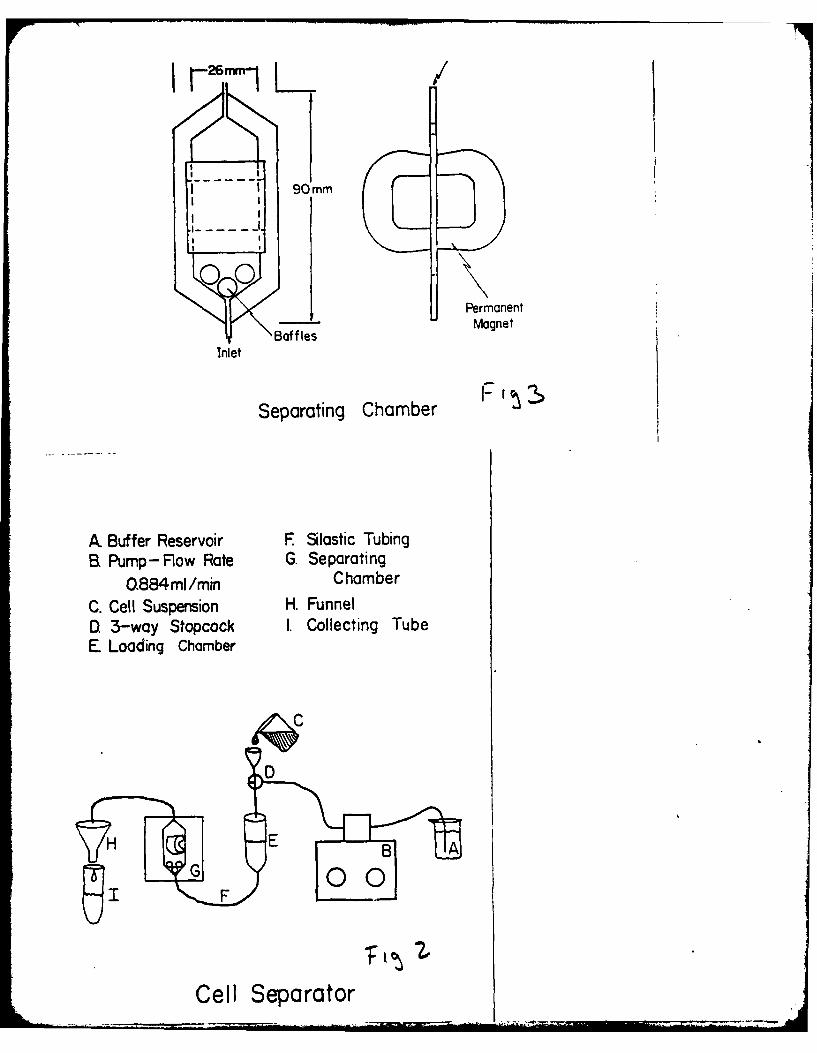
26 mm1L
I90 mm
00 PermanentBafflesMagnet
Inlet
Separating Chamber F'
A Buffer Reservoir F Silastic TubingB. Pump- Rlow Rate G. Separating
0.884ml/min ChamberC. Cell Suspension H. FunnelD. 3-way Stopcock I. Collecting TubeE Loading Chamber
H E
Cell Separator

Figure 4
Macrophaqes and Non-Type T Cells
(CUaro, O~ated, Lym~hocyuc. Endolat Cells)
140t4 Type 31 Cells
140
fir 0 True heghfts given2- in numbers, error
bars in standarddeviation
38*
~40-
;z 30-
20- 1217
10 4,t 2 0
Before After Before AfterP urifi caton Purification
Minced Lung Trocheal LovageCollagense Digest After Collogenase
..... . .. I ... I ... I ....... ' ....... .....Ft% ....I...... i ....... "..
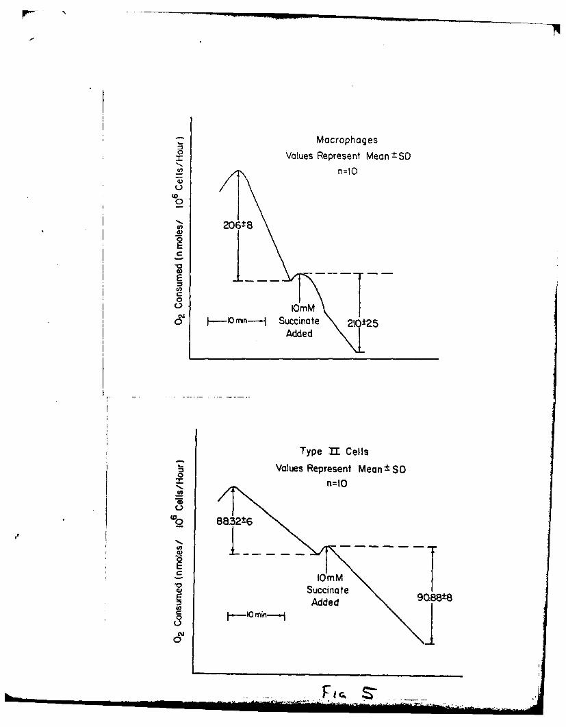
-z MacrophagesValues Represent Mean ±SD
U, n=10
0
206±80EV
0 ~ .- Imn--- Succinate 260±5Added
Type 11 CellsValues Represent Mean± SD
0=1
88.32±6
E
Succmnate
0cu
0
G

0
1- Ou 0O0O
00 0
00
C-)j
CL0
22
0
A0
NN
4)o
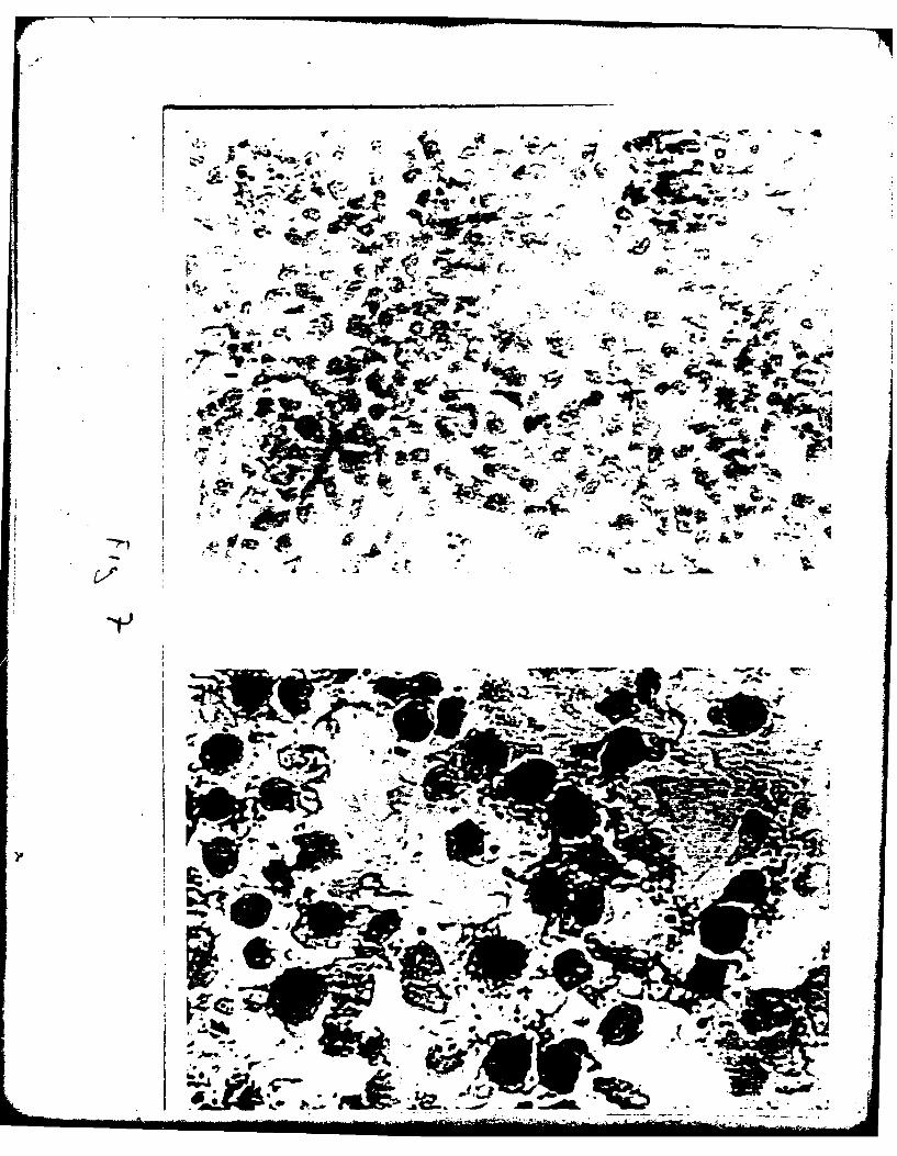
01
I w 1%* wV
Ar-
4 It
414
I4 4r
* ., ~ fq
- 4=LA
- -I a
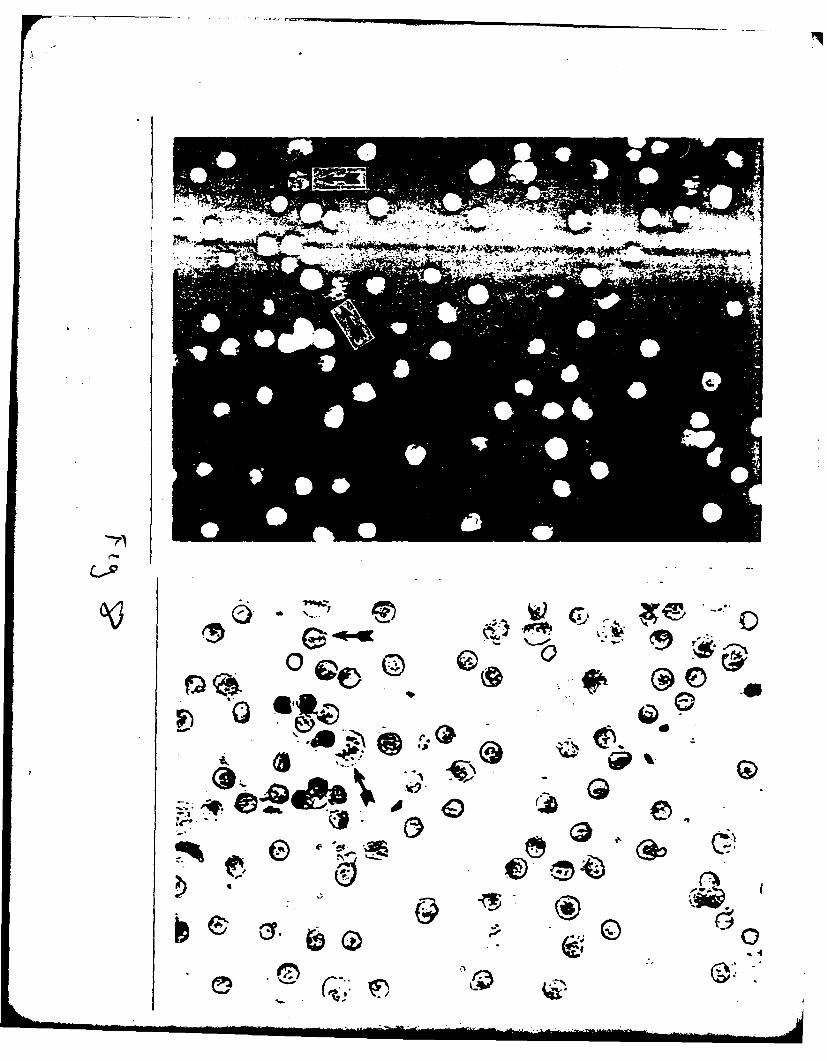
CIOO
Itse Q6

* r,
55.

ELUTRIATION SEPARATION OF TYPE It CELLS30
85%
ROTOR SPEED 2000 RPM
20.
- 90%
10 50%
.... ... . 20%. . ..
toad 7 12 1624 44
COUNTER CURRENT FLOW mi/min
Isolation and Metabolic Characteristics of Lung-Cell Types
M. Lafranconi, K. Brendel, and I.G. Sipes
Department of Pharmacology
College of MedicineUniversity of Arizona
Tucson, Arizona 85724
Running Title: Isolated Lung Cells
Corresponding Author: Mark LaFranconiDepartment of PharmacologyArizona Health Sciences CenterTucson, Arizona 85724(602) 626-7843

Abstract
A nu-Mber of pulmonary toxins have a surprisingly specific site of
action within the lung. Silica particles cause lysis of pulmonary alveolar
macrophages, paraquat administration results in lipid peroxidation and type I
cell death, urethan can cause adenomas of type II cell origin, pyrrolizidine
alkaloids specifically damage pulmonary endothelium while 4-ipomeanol or
carbon tetrachloride results in focal necrotic lesion of clara cells.
Studying these events with isolated cell types would provide good specificity
of respone without interference from other cell types. Unfortunatel few
cell types in the lung have been successfully isolated. Methods available
to isolate pneumonocytes generally take advantage of one or a combination
of 4 basic differences in cells. 1. Availability 2. Density 3. Size 4.
Specific cellular functions Macrophages have been isolated based on
availability by lavage. Cell dispersal techniques become important in
cell isolation procedures relying on density, size or specific cellular
function differences. Because of the extensive connective tissue matrix
in the lung, obtaining cells in a monodispersed from for separation has
proven to be difficult. Density differences between pneumonocytes is slight
but has been used to isolate type II cells. Size differences either through
unit gravity velocity sedimentation or elutriation techniques have been used
to isolate type I and 1I cells as well as partial enrichment of clara cells
and macrophages. Specific cellular functions such as differential adherence
or culture techniques or surface charge differences have been used to
prepare cell fraction rich in macrophages, type II cells, endothelial cells
and clara cells. Combinations of techniques have been used successfully
but all are time consuming, requiring up to 8 hours. A technique using
. ....

critical enzyism* ,pluceine~nt, magnetic removal of macrophages and sizing of
cells on nylon rrn-th scre~ens results in a preparation of type II cells of
over 85% purity. The prime advantage of the method is the short preparittion
time of less tha-- 2 hours.
2
Isolating specific cell types is a promising area of lung toxicology. For
some time now evidence has been building to suggest that cells within the lung
may respond uniquely to noxious stimuli. Silica particles of less than 5
microns administered intratracheally to rodents will cause lysis specifically
of pulmonary alveolar macrophages (3,10,45). When urethane is administered to
mice IP within a matter of weeks proliferation of Type II cells ensues leading
to the formation of adenomas in the lung of primarily Type II cell origin
(53). Paraquat in rats can cause free radical formation and lipid
peroxidation in Type I cells causing destruction of these cells (52).
Endothelial cells are the first cells of the lung to encounter systemically
applied agents and so are readily damaged. But a pyrrolizidine alkaloid
called monocrotaline when administered in low doses for extended periods of
time can alter uptake of serotonin by these cells while ehancing the activity
of angiotensin converting enzyme (30). Carbon tetrachloride or 4-Ipomeanol
administered parentally will cause focal lesions within the clara cells of the
lung (6,7). Isolated cell types from the lung would provide the opportunity
to study toxic responses from the cell specific agents without possible
biochemical contribution from other cells types therby allowing good
quantiation. In addition, normal metabolism and physiology could be studied
using isolated cells and these agents as possible chemical probes.
While there are over 40 cell types within the lung (54) only a few have
been successfully isolated. The impetous for most of these isolations was the
desire to study normal morphology and physiology. For example, pulmonary
aveolar macrophages were initially isolated to study the mechanisms of
phagocytosis (44). Type II cells were isolated to study sufactant synthesis
(34). Only recently have efforts been made to isolate cell types from the
j lung to specifically study toxic responses (12,13).
3
The purpose of paper then, is to briefly outline the methods currently
available for isolating cells from the lung and to discuss some of the
problems associated with each.
Methods developed to isolate cells as they were required by
investigators. Naturally, the simplest methods were exploited first, followed
by methods of increased complexity. Despite the various methods available in
the literature to prepare enriched fractions of lung cells they all rely on
one or a combination of 4 basic properties to separate cells. These are
differences in: 1) Availability; 2) Density; 3) Size; 4)
Specific cellular functions. This last is a catch all category we will
discuss later.
Availabili ty
Isolating cells on the basis of availability is the simplest method to
date and is one of the few success in cell isolation attempts from the lung.
Pulmonary alveolar macrophages (PAMS) are free roaming cells in the lung
alveolar areas. Thirty - 100 X 106 cells can be removed from a single
rabbit lung by simple lavage (44) (See Fig. 1). Cells obtained in this way
are greater than 90% macrophages with a viability greater than 90%. Further
enrichment can be achieved by culture techniques (11). When cells are plated
in tissue culture medium such as minimal essential medium supplemented with
10% fetal calf serum macrophages will adhere within 2 hours. The non adherent
cells include non-viable macrophages, lymphocytes, red blood cells, some Type
II cells and others.
Cells isolated in this way have been used, in studies investigating
mechanisms of phagocytosis (44). However, other experimenters have used these
4
cells to study the toxicity of a variety a airborne contaminants because the
cells were easily obtained and because, presuambly, the pulmonary alveolar
macrophages were representative of all cells in the alveolus to airborne
exposure (1,9,32). Isolated pulmonary alveolar macrophages were used to show
toxic effects of silica particles. These cells showed extensive in vitro
degredation and lysis within 8 hours of application of silica particles of
less than 5 microns (3,4,6). Using a similar system other groups were able to
show elevated collagen synthesis in cultured fibroblasts when incubated in
medium obtained from silicotic macrophages (2,8). In other experiments
Cd+ , Hg ++ , Pb++ and Ni++ inhibited release of reactive oxygen species
during phagocytosis as well as decreasing oxygen consumption and glucose
metabolism both at rest and during phagocytosis in isolated pulmonary alveolar
macrophages (9).
Cell Removal
Unfortunately, the remainder of the cells in the lung are not as easily
obtained. The cells of the lung are, for the most part, immobilized through a
variety of attachments - the most important being connections to an extensive
collagen containing matrix (25,28). Removing the cells from this matrix is
the most recalcitrant problem in isolating cells from the lung. For a
comprehensive review of this subject see Gould (25). To conduct any of the
remaining procedures for isolating cells, they must be put into a mondispersed
form - irividual cells without attached basement membranes or clumping to
form cell to cell attractions and all this while maintaining the cells in a
viable state.
There are 3 primary methods available to disperse cells: mechanical,
chemical or enzymatic means (25). Mechanical procedures provides a physical

5
disruption and shearing process which interupts the cell-matrix bonds as well
as cell to cell connections but unfortunately these methods are damaging to
the cells and results in poor yields when used alone. Chemical means have not
been used exclusively in removing cells from the lung either. The concept
used in these procedures is to chemically chelate divalent cations such as
Ca++ or Mg+ + with EDTA or EGTA, thereby disrupting the Ca++ dependent
desmisomes (25,58). Enzymatic means have been used with some success although
cellular damage does occur as suggested by changes in surface receptors (15).
Intercellular alterations have also been reported. Proteases caused a 4 fold
reduction in NADPH cytochrome C reductase activity in rat lungs (19).
Cell isolation from lungs is dependent primarily on the success of
obtaining individual dispersed cells. In other organs such as the liver, this
step has been readily achieved using a combination of the above methods of
cell removal. Proteases perfused through the portal vein, Ca*+ chelation,
mincing and pressing the liver through 160 um nylon meshes can provide
approximately 500 million cells from the liver of a 200 gram rat (29). In
contrast the lungs from a 200 gram animal will yield less than 10 million
cells (unpublished observation). The difficulty in obtaining cells from
mammalian lungs lies in the structure of the organ.
From the liver section shown in Fig. 2 it can be seen that cells are
arranged in sinusiods with little structural support from extracellular
materials. The parenchyma are arranged through cell to cell junctions via
desmisomes. In the lung however parenchyma are arranged about a collagen
extracellular matrix as well as cell to cell junctions in zonas occludens
(25,28) (see Fig. 3). In addition, there are large liquid gas interfaces from
the alveolar aspect. Attempts to dissociate the lung then are hampered by
6
tight cellular junction which inhibit acess of enzymes, the recalcitrant
nature of the cell Junction to basement membranes, and the high surface
tensions at air - liquid interfaces. However, through extensive lavage,
mechanical disruption by mincing, and enzymatic dissociation some success has
been achieved in removing cells from the lung (12,13,15,21,22,24,25,26,34,
35,36,40,48).
Specific and non-specific enzymes in various combination or separately
have been used to dissociate the lung. The non-specific enzymes such as
trypsin or the pronases have good proteolytic activity and release cells from
the lung effectively (12,34,40). In rabbit lungs, 0.1% pronase Type I can
release up to 300 million cells from a single rabbit lung (12,13). Because of
the non-specificty of action of both trypsin and 742 pronases certain cellular
damage does occur and viability of the preparation falls with time (19,22).
It therefore becomes a trade-off between viability and cell yield that
investigators must deal with when trying to remove cells from the lung. For
example, rat lungs treated with thermolysin, one of the commercially available
pronases will yield 10 million cells from a single lung within 25 minutes, but
viability begins to fall after 10 minutes (see Fig. 4).
Specific enzymes such as collagenase, elastase or others have the
advantage of being less damaging to the cells but the effectiveness of the
agents in freeing cells from the lung is diminished (15,25). In our lab less
than 100 X 106 cells can be obtained from rabbit lungs when collagenase Type
II is used. This is compared to over 300 million obtained with pronase
(12,13).
Removing cells from the lung with the combination of techniques described
earlier leads to as yet another problem. The enzymatic action on cells by the
F AD-AlON 702 ARIZONA UNIV HEALTH SCIENCES CENTER TUCSON Ff/B 1/20OXYGEN TOXICITY AND LUNG COLLAGENOUS PROTEXN.CU)FEB 81 K BRENOEL, 1 6 SIPES N0001IG77-C-050
UNCLASSIFIED NL
7
proteases can cause altered surface proteins and change surface charges which
contributes to cell aggregation. It is critical for successful cell isolation
procedures that the cells are in a monodispersed form. Clumping can be
avoided by adding protein to the medium such as bovine serum albumin up to 1%
or fetal calf serum. Another technique used is to separate cells in the cold
since clumping has been shown to be a function of temp (43). Divalent cations
such as Ca+ , or Mg+ + will contribute to cell clumping so chelation of
these materials is also used (25). Sometimes damaged cells will release DNA
and the hgh viscocity of this material causes cell clumping which is prevented
by adding DNAase to the medium, coimmonly in concentration of 0.05% (12,25).
With these precautions monodispersed cells can be obtained from the lung and
separation of cell types based on differences in density, size, or specific
cellular functions can be achieved.
Density Separations
The first reported methods for isolation of Type II cells from the lung
were based on density separations (21,34,40). With these method lungs were
perfused to remove blood elements then lavaged to remove free macrophages.
The lungs were then dissociated into monodispersed cells, primarily with
trypsin, and the cells separated according to density differences on either
albumin gradients (40), ficol gradients (21,34), or metrizamide (13).
The basis of this separation is derived from Stokes Law (42). The cells
develop a velocity by centrifugal forces as a function of their radii. As
long as the density of the cell is greater than density of the medium the cell
continues to move. However, once the cell reaches a gradient with a density
equal to or greater than that of the cell, the density function becomes 0 and
the cell ceases to move.
8
With this method a number of investigators have enriched Type I cell
fractions to greater than 50% (13,21,34,35,40). The process however is time
consuming requiring 4-6 hours for completion. Furthermore, there is little
within lung cells to provide much of a difference in density. Nucleic acids
or protein concentrations may alter densities but levels of these components
are not dramatically different between cell types in the lung. So therefore
purification by density gradients has proven to be difficult. For instance
Type II cells and smaller Pans layer on the same density gradient (34,40). A
clever manipulation used by investigators to enhance macrophage and Type II
cell separation is to provide a dense material such as BaSO4 or fluorocarbon
albumin mixtures which can be taken up into macrophages by phagocytosis,
increasing their density enough to cause them to layer at a different zone
than the Type II cells (21,34,40).
Using this method for isolating Type II cells aid in conjunction with in
vivo experiments mechanisms of surfactant synthesis and synthetic pathways
with Type II cells have been investigated (For review see 4,35).
Size Separations
It has been proposed that size is the most accurate way of separating
cells since lung cell sizes may vary from 10 microns for lymphocytes to 50 or
60 microns for Type I cells or macrophages (49). A number of methods have
been published to separate lung cells based on size.
One of the procedures based on size is unit velocity sedimentation
(31,48). In this instance velocity is developed as a function of the radius
of the cell and the acceleration of gravity. The density term is always
greater than 1. Monodispersed cells are obtained then placed in a chamber and
allowed to separate according to velocities generated.

9
With this method of separation Type I cells of the lung have been prepared
and enriched to nearly 70% homogeneity (48). This method, perhaps more than
any other, is sensitive to cell clumping. It is also time consuming,
requiring 6-8 hours of preparation to obtain cells in an enriched form.
This method has also been used to partially purify Type II cell
fractions. After density separation of Type II cells the enriched fraction is
then further subjected to unit gravity sedimentation which results in an
enrichment of nearly 90% (40).
Another procedure to separate cells based on size is centrifugal
elutriation. The same concepts apply here as for unit velocity sedimentation
except the force for acceleration is centrifugal. Cells are introduced into
the spinning rotor and pelleted to the outside of the chamber by centrifugal
acceleration. By precisely increasing flow rates of the counter current
buffer, cells are separated from the pellet according to their size.
Using this method fractions can be obtained which are enriched in
pulmonary alveolar macrophages, Type II cells, clara cells, and others
(12,13,26). It is faster than other methods mentioned so far for isolating
cells, but when used alone only a limited enrichment is obtained - 60% Type II
cells for an example (12).
Using a combination of centrifugal elutriation and density gradient
centrifugation Type II cells have been enriched to nearly 90% purity
(12,13,26). These methods of isolation have been used to study drug metabolic
capabilities of cell types from the lung (12,13). They have also been used to
study surfactant synthesis (24).
. ... . . . . ... . ..
10
Specific Cellular Functions
As I mentioned earlier this last classification, specific cellular
functions, is a mixed bag for separation techniques. It includes functions
such as cell culturing, cloning, induction, differential adherence, surface
charge differences, differences in membrane affinities, etc.
One of these methods, differential adherence has been used to isolate
macrophages which we discussed already (11). But Type II cells have also been
prepared from differential adherence procedures (15). With this procedure a
preparation partially enriched in Type II cells, by some other means such as
density gradi-.nt centrifugation, is plated in culture medium such as
Dulbecco't modified eagle medium for 1 hour. At this point macrophages and
some Type LI cells adhere. The non-adherent cells are removed and plated
again but for longer periods of time - 22 hours. In this second plating Type
II cells adhere while non-adherent cells such as lymphocytes and red blood
cells are removed with a fresh change of medium. With this method greater
than 80% Type II cells con be obtained. Unfortunately, it is a time consuming
process and in this preparation time and through the stresses of adapting to
culture conditions cells might be altered physiologically since Type II cells
reportedly rapidly dedifferentiate into some type of precurser cell (14,39).
With this method however, it was demonsted that adrenergic stimulation of
Type II cells caused an enhanced release of surfactant synthesis (15).
A number of methods for culturing cells from the lung have been reported.
Pulmonary alveolar macrophages have been maintained in cultures for weeks
(11). Pulmonary endothelial cells harvested from lungs by retrograde
perfusion of the pulmonary circulation with collagnease have been successfully
cultured (27).

11
Limited success has been achieved with attempts to culture other cell
types from the lung (14,16,17,18,23,51). Although a number of groups report
successful cultures of Type II cells, recent reports have provided evidence
that these cultured cells have alterd phospholipid content and morphology
compared to freshly isolated Type II cells or cells from the intact lung
'(41,50). None the less, cultured Type II cells have been used to examine
surfactant synthesis (16) and recent reports demonstrate the existence of Aryl
hydrocarbon hydroxylase activity in Type II cells in culture (55,56).
An innovative method has recently been developed to isolate clara cells
based in part on size by elutriation separation then further separated on the
basis of surface charge (13). When clara cell rich fraction are placed at the
interface between a 2 phase aqueous solution with differnt charges the clara
htells separate from a majority of the contaminating cells and appeared in the
postively charged phase. Enrichments up to 70% were reported.
Another method which relies on a combination of techniques for isolating
Type II cells has recently been developed by our lab (manuscript in
preparation). This method employs critical enzyme placement, magnetic removal
of macrophages and cell sizing to rapidly prepared Type II cells.
As with most of the procedures previously mentioned the initial step is to
prepare a monodispersed suspension of pneumonocytes. To accomplish this cold
isotonic saline is perfused through the cannulated pulmonary artery to remove
free blood elements. Once the lungs have perfused white, they are attached to
a continuous perfusion apparatus and pefused in a recirculatory manner with a
HEPES buffer containing 128 mM NaCl, 5mM K S04, 5 mM Kcl, 2.5 mM Sodium
Phosphate buffer pH 7.4, 17 mM HEPES (N-2-hydroxyethyl piperazine N-2-ethane
sulfonic acid), 5.5 mM Dextrose and 10% V/V fetal calf serum (Fig. 9).
12
At this time the lungs are lavaged 3 times through the cannulated trachea
to remove free macrophages. Subsequently 20 ml of colloidal magnetic iron
oxide (Fe 04) are instilled via the trachea. With this the lungs take on a
blac. appearance. This provides for phagocytosis of magnetic Fe3 04
particles by remaining macrophages. After 20 minutes of incubation at 370 C
the lungs are again lavaged to remove excess iron oxide. The lungs are then
trated with a mixture of enzymes to dissociate cells from the matrix.
Collagenase Type II (Worthington Biochemical Corp) 50mg/l00 ml and 1.1 mg/100
ml elastase (11 mg/ml Sigma) are added to the perfusion resevoir in fresh
HEPES buffer without fetal calf serum and perfused through the pulmonary
artery in a recirculatory manner. At the same time collagenase and elastase
containing buffer is introduced to the alveolar areas through the trachea. In
this way the enzymes necessary for dispersing type II cells are in contact
with the cells from both the epithelial and endothelial aspects.
Following a 10 min incubation at 37 0 C freed cells are removed frm the
lung by one of two methods. In one procedure calls are recovered from the
lung by lavage with fresh buffer. This results in a small yield of cells per
lung - often less than 20 X I06, but the initial purity is almost 60% type
II cells. When more cells are required another method of harvesting cells
developed by other workers (40) is used. The lungs are removed from the
perfusion apparatus, minced then shaken in fresh buffer supplemented with 10%
fetal calf serum for 10 min. Cells are then separated from the tissue blocks
by filtering the mixture through a 200 um nylon mesh screen. With this
procedure approximately 180 X 106 cells are obtained from a single rabbit
lung but with only 21% type II cells.
Irregardless of the method used to harvest cells from the lung, the
preparations are then partially enriched by consecutive sizing on nylong mesh

13
screens of 160, 40, and 20 um. The 160 um grid removes cell clumps and broken
pieces of matrix that may be present. The 40 um and 20 um sieves retain large
macrophages, type I cells as well as some ciliated and non ciliated bronchial
epithelial cells and endothelial cells.
At this point the cell prepartion contains mostly type II cells, small
macrophages and lymphocytes. The macrophages are then removed by passing the
preparation through a magnetic separation chamber. The cells are slowly
pumped past a narrow gap contained in a strong magnetic field. Macrophages
contaning magnetic iron oxide are retained in the chamber while non-retained
cells, including type II pneumonocytes are collected.
With this system cells obtained by lavage can be enriched to greater than
90% while cells obtained through minced lung preparations can be enriched to
greater than 85% type II cells. The prime advantage of the minced lung
preparation is the increased number of type II cells obtained, 32 X 106 from
a single rabbit lung compared to 8 X 10⁶ from lavage. The unique feature of
this preparation is the time savings. Commonly type II cell isolations
require 4-8 hours for completion while this method can be used to prepare
cells in less than 2 hours.
Identifying cells obtained from various isolation procedures is an
important but difficult task. Because of the array of cell types in the lung
it becomes important to be able to identify which cells are present in the
preparation. In situ cell identification is, in part, accomplished by
landmarks and location or by iimunohistochemical techniques. But with
isolated cells there are no landmarks or location clues and the proteolytic
activities of the isolation procedure may alter the antigenic characteristics
of the cell. When cells have unique surface receptors these receptors can be
14
bound to an antigenic ligand then visualized with immunohistochemical
techniques. Unfortunately few surface receptors are unique in the lung that
can be exploited at this time. One, the angiotensin converting enzyme on the
surface membranes of endothelial cells has been used to identify endothelial
cell preparations (27).
Other techniques used to identify cells take advantage of specific'
biochemical anomolies the cell. Clara cells have been histochemically
identified by nitro blue tetrazolium which is reduced in clara cells to a
purple formazon (13). Type 11 cells have been identified by a number of
techniques. When incubated with phosphine 3R or acridine organ for 5 minutes
Type II cells with fluoresce intensely yellow green while macrophages
fluoresce diffusely (38,40). Type II cells are also identified by a modified
papanicolaou staining in which Type II cells show darkly staining inclusion
bodies while other cell types stain more uniformly (34). Still another method
is electron microscopy. Type II cells are identified in this way by lamellar
inclusion bodies (34,40). However, the morphology of the lamellar bodies may
be significantly different from species to species (33). The micrograph in
Fig. 4 is a Type II cell obtained from a rabbit, while Type II cells from rats
have a more layered appearance.
Still another means of identifying cells is based on their separation
properties observed in other methods of isolation. For example, Type II cells
isolated by our lab with an apparent purity of 85% were further separated by
centrifugal elutriation. The pattern of distribution of cells obtained at
various flow rates corresponds to distributions of Type I cells reported by
other groups with the majority of the cells recovered collected from fractions
separated with 16 and 24 ml/mmn counter current flow rates (12,13,26).

15
Viability of cells obtained have been determined by trypan blue exclusion
or other vital dyes such as erythrosin B (47) but other indexes of viability
have been used also. Oxygen consumption or glucose utilization are both
sensitive indices of viability (20,57). Here in Fig. 5 is a trace obtained
from Type II cells of the rate of oxygen consumption expressed as
nmoles/million cells/hour. The rate is linear over the ten minute time
course. In addition, there was no sigificant changes in rate of utilization
after addition of 10 mM sodium succinate, a mitochrondrial substrate. This
procedure is used to demonstrate intact plasma membranes since sodium
succinate will normally not gain access to mito-chondria unless plasma
membranes are damaged (5). The second trace is obtained from the macrophages
which were collected in the chamber. Note that these cells have a much
greater rate of oxygen consumption than do Type II cells.
Another index of viability is measurement of synthesis functions. Type II
cells, because of their role in sufactant synthesis, contain high levels of
dissaturated phosphatidyl choline, a phospholipid relatively uncommon in other
cell types (35,37,39). By supplying radio labeled precursors for phospholipid
synthesis it is possible to monitor rates of synthesis of phosphatidyl choline
(35,40). In Fig. 6 incorporation rates are linear for over 2 hours. This
same experiment can also be used as a crude means of identifying the cell
preparation. Type II cells have elevated rates of incorporation of
radiolabeled precursors into disaturated phosphatidyl choline compared to
other cell within the lung. Note the low rate of synthesis in macrophages.
Thus, with a high rate of synthesis there is added support that the cells are
sufficiently enriched in Type II cells.

16
Conclusions
There are a wide array of techniques used to isolate cells from :hc i ' "
Most methods rely on obtaining viable monodispersed cells from the lung matrix
then separating these cells into various fractions either based on differences
in density, size or some particular cellular function. Most of the successful
procedures employed have been obtained through a combination of these
techniques.
Perhaps with the existing techniques other cell types in the lung could be
purified as needed for investigations. With isolated cell type and specific
chemical or biologic probes normal physiology and biochemistry could be
studies in depth. In addition, mechanisms of action of toxins, drugs, or
pathogens could be further investigated.
Future techniques for isolating cell types may rely on electorphoretic
separations, affinity chromatography, flow microfluorimetry or improved
culture techniques. Objectives of new techniques would be improved yield,
viability, purity and reproducability along with perhaps a shorter preparation
time.
- iI
17
References
1. Adkins, B; Lunginbuhl, G.H.; Gardner, D.E. Biochemical Changes inPulmonary Cells Following Manganese Oxide Inhalation. J. Tox andEnviron Health 6:445-454; 1980.
2. Aho, S.; Kulonen, E. Effects of Silica Liberated Macrophage Factors onProtein Synthesis in Cell Free Systems. Exp Cell Res 104:31-38; 1977.
3. Allison, A.C.; Harrington, J.S.; Birbeck, M. An Examination of theCytotoxic Effects of Silica on Marcophages. J Exp Med 124:141-54;1966.
4. Batenburg, J.J. Isolated type II Cells from Fetal Lung as a Model inStudies on the Synthesis and Secretion of Pulmonary Surfactant.Lung. 158:177-192; 1980.
5. Berry, M.N. Metabolic Properties of Cells Isolated from Adult MouseLiver. J Cell Biol 15:1-8; 1962.
6. Boyd, M.R. Evidence For the Clara Cell as a Site of Cytochrome P-450Dependent Mixed Function Oxidase Activity in Lung. Nature.269:713-715; 1977.
7. Boyd, M.R.; Statham, C.N.; Longo, N.S. The Pulmonary Clara Cell as aTarget for Toxic Chemicals Requiring Metabolic Activation; Studieswith Carbon Tetrachloride. J Pharmacol and Exp Therap 212:109-114;1980.
8. Burrell, R.; Anderson, M. Induction of Fibrogenesis by Silica TreatedAlveolar Macrophages. Environ Res 6:389-394; 1974.
9. Castranova, V.; Bowman, L.; Reasor, M.J.; Miles, P.R. Effects of HeavyMetal Ions on Selected Oxidative Metabolic Processes in Rat AlveolarMacrophages. Tox and Appl Pharmacol 53:14-23; 1980.
10. Chvapil, M. Old Problems of Silicotic Fibrosis. Bendz, G.; Lindqvist, I.eds. Biochemistry of Silicon and Related Problems. New York:Plenum Press; 1978; 381-392.
11. Cohen, A.B.; Cline, M.J. The Human Alveolar Macrophage: Isolation,Cultivation in vitro and Studies of Morphologic and FunctionalCharacteristics. J Clin Inves 50:1390-1398; 1971.
12. Devereux, T.R.; Hook, G.E.R.; Fouts, J.R. Foregin Compound Metabolism byIsolated Cells from Rabbit Lung. Drug Metab and Disp 7:70-75; 1979.
13. Devereux, T.R.; Fouts, J.R. Isolation and Identification of Clara Cellsfrom Rabbit Lung. In Vitro. 16:958-968, 1980.
14. Diglio, C.A.; Kikkawa, Y. Type II Epithelial Cells of the Lung IVAdaptation and Behavior of Isolated Type II Cells in Culture. LabInves 37:622-631; 1977.

18
15. Dobbs, L.G.; Mason, R.J. Pulmonary Alveolar Type II Cells Isolated from.a ts: Release of Phsophatidylcholine in Response to B AdrenergicSJulation. J Clin Inves 63:378-387; 1979.
i6. Douglas, W.H.J.; Teel, R.W. An Organotypic in vitro Model System forStudying Pulmonary Surfactant Production by Type Il AlveolarPneumonocytes. Am Rev Resp Dis 113:17-23; 1976.
17. Douglas, W.H.J.; Farrel, P.M. Isolation of Cells that RetainDifferentiated Functions in vitro: Properties of Clonally IsolatedType II Alveolar Pneumonocytes. Environ Health Perspec 16:83-88;1976.
18. Douglas, W.J.H.; Del Vecchio, P.; Teel, R.W.; Jones, R.M.; Farrell, R.M.Culture of Type II Alveolar Lung Cells. Bouhuys, A. ed. Lung Cellsin Disease. New York: Elsevier North Holland; 1976; 53-68.
19. Finkelstein, J.M.; Mavis, R.D. Biochemical Evidence for InternalProteolytic Damage During Isolation of Type II Alveolar EpithelialCells. Lung. 156:243-254; 1979.
20. Fisher, A.B. Oxygen Utilization and Energy Production. Crystal, R.G. ed.Biochemical Basis of Pulmonary Function. New York: Marcel DekkerInc; 1976:75-104.
21. Fisher, A.B.; Furia, L. Isolation and Metabolism of GranularPneumonocytes From Rat Lungs. Lung. 154:155-165; 1977.
22. Frazier, M.E.; Hadley, J.G.; Andrews, T.K.; Drucker, H. Use ofThermolysin for the Dissociation of Lung Tissue into CellularComponents. Lab Inves 33:231-238; 1978.
23. Frosolono, M.F.; Kress, Y.; Wittner, M.; Rosenbaum, R.M. CultureCharacteristics of Cells Derived from Type II Pneumocyte EnrichedFractions from Rabbit and Rat. In Vitro 12:708-717; 1976.
24. Gould, K.G.; Clements, J.A.; Jones, A.L.; Felts, J.M. Dispersal ofRabbit Lung Into Individual Viable Cells: A New Model for the Studyof Lung Metabolism. Science. 178:1209-1210; 1972.
25. Gould, K.G. Dispersal of Lung into Individual Viable Cells. Crystal,R.G. ed. Biochemical Basis of Pulmonary Function. New York: MarcelDekker Inc.; 1976:49-71.
26. Greenleaf, R.D.; Mason, R.J.; Williams, M.C. Isolation of Alveolar TypeII Cells by Centrifugal Elutriation. In Vitro 15:673-684; 1979.
27. Habliston, D.L.; Whitaker, C.; Hart, M.A.; Ryan, U.S.; Ryan, J.W.Isolation and Culture of Endothelial Cells from the Lungs of SmallAnimals. Am Rev of Resp Disease 119:853-868; 1979.

19
28. Hance, A.J.; Crystal, R.G. Connective Tissue of the Lung: Collgen.Crystal, R.G. ed. Biochemical Basis of Pulmonary Function. NewYork: Marcel Dekker Inc.; 1976:215-271.
29. Hayes, S.J.; Brendel, K. N-Demethylation as an Example of DrugMetabolism in Isolated Rat Hepatocytes. Biochem Pharm. 25:1495-1500;1976.
30. Huxtable, R.; Ciaramitaro, D.; Eisenstein, D. The Effect of aPyrrolizidine Alkaloid, Monocrotaline, nd a Pyrrole,Dehydromonocrota line, on the Biochemical Functions of the PulmonaryEndothelium. Mol Pharmacol 14:1189-1203; 1978.
31. Hymer, W.C.; Evans, W.H.; Kraicer, J.; Mastro, A.; Davis, J.; Griswold, E.Enrichment of Cell Types from Rat Adenohypophysis by Sedimentation atUnit Gravity. Endocrinology 92:275-287; 1973.
32. Johanssom, A.; Lundborg, M.; Hellstrom, P.; Camner, P.; Keyser, T.R.;Kirton, S.E.; Natusch, D.F.S. Effect of Iron, Cobalt, and ChromiumDust on Rabbit Alveolar Macrophages: A Comparison with the Effectsof Nickel Dust. Environ Res 21:165-176; 1980.
33. Kikkawa, Y.; Spitzer, R. Inclusion Bodies of Type II Alveolar Cells:Species Differences and Morphogenesis. Anat Rec 163:525-542; 1968.
34. Kikkawa, Y.; Yoneda, K. Type II Epithelial Cells of the LUng I. Methodof Isolation. Lab Inves 30:76-84; 1974.
35. King, R.J. Utilization of Alveolar Epithelial Type II Cells for theStudy of Pulmonary Surfactant. Federation Proc. 38:2637-2643; 1979.
36. Mason, R.J.; Williams, M.C.; Greenleaf, R.D. Isolation of Lung Cells.Bouhuys, A. ed. Lung Cells in Disease. New York: Elsevier NorthHolland; 1976:39-52.
37. Mason, R.J. Lipid Metabolism. Crystal, R.G. ed. Biochemical Basis ofPulmonary Function. New York: Marcel Dekker Inc.; 1976:127-169.
38. Mason, R.J.; Williams, M.C. Identification of Type II Alveolar Cells WithPhosphine 3R. AM Rev Resp Dis: Supplement. 113:47;1976.
39. Mason, R.J.; Dobbs, L.G.; Greenleaf, R.D.; Williams, M.C. Alveolar TypeII Cells. Federation Proc. 36:2697-2702; 1977.
40. Mason, R.J.; Williams, M.C.; Greenleaf, R.D.; Clements, J.A. Isolationand Properties of Type II Alveolar Cells from Rat Lung. Am Rev RespDis 115:1015-1026; 1977.
41. Mason, R.J.; Williams, M.C. Phospholipid Composition and Ultrastructureof A549 Cells and Other Cultured Pulmonary Epithelial Cells ofPresumed Type II Cell Origin. Biochemica et Biophysica Acts617:36-50; 1980.
20
42. Mel, H.C.; Ross, D.W. Biotlhysics of Cell Separations. Quart. RevBiophys 8:421-438; 1975.
43. Moscona, A. Rotation Mediated Histogenic Aggregation of DissociatedCells: A Quantifiable Approach to Cell Interaction in vitro. ExpCell REs 22:455-475; 1961.
44. Myrvik, Q.N.; Leake, E.S.; Fariss, B. Studies on Pulmonary AlveolarMacrophages From the Normal Rabbit: A Technique to Procure them in aHigh State of Purity. J Immunol 86:128-132; 1961.
45. Nash, T.; Allison, A.C.; Harrington, J.S. Physico-Chemical Properties ofSilica in Relation to its Toxicity. Nature. 210:259-261; 1966.
46. Oh, T.H.; Naidoo, S.S.; Jaques, R.C. The Uptake and Disposition of 35SHeparin by Macrophages in vitro. Res J. Rticuloendothel Soc13:134-142; 1973.
47. Phillips, H.J. Dye Exclusion Tests for Cell Viability. Kruse, P.F.;Patterson, M.K. eds. Tissue Culture Methods and Applications. NewYork: Academic Press Inc.; 1973:406-421.
48. Picciano, P.; Rosenbaum, R.M. The Type I Alveolar Lining Cells of theMammalian Lung. Am J of Path 90:99-115; 1978.
49. Pretlow, T.G.; Weir, E.E.; Zettergren, J.G. Problems Connected with theSeparation of Different Kinds of Cells. Internat Rev Exp Path14:92-204; 1975.
50. Schmidt-Somerfield, E.; Borer, R.C. Biochemical Characterization ofClonally Isolated and Continuously Cultured Type II Cells from AdultRat Lung. Am Rev Resp Disease 120:1145-1148; 1979.
51. SmitL, S.T. Cell Line A549: A Model System for the Study of AlveolarType II Cells Function. Am Rev Resp Disease 115:285-293; 1977.
52. Smith, P.; Heaht, D. The Ultrastructure and Time Sequence of the EarlyStages of Paraquat Lung in Rats. J Pathol 114:177-181; 1974.
53. Snyder, C.; Malone, B.; Nettesheim, P.; Snyder, F. Urethan-InducedPulmonary Adenoma as a Tool for the Study of SurfactantBiosynthesis. Cancer Res 33:2437-244; 1973.
54. Sorokin, S.P. The Cells of the Lungs. Nettesheim, P.; Hanna, M.G.;Deathrage, J.W. eds. Morphology of Experimental RespiratoryCarcinogenesis; Conf 700501. U.S. Atomic Energy Comm; 1970:3-43.
55. Teel, R.W. Induction of Aryl Hydrocarbon Hydroxylase in Primary Culturesof Type II Alveolar Lung Cells and Binding of Metabolically ActivatedBenzo (a) pyrene to Nuclear Macromolecules. Cancer Letters.7:349-355; 1979.

211
56. Teel, L.W.; Douglas, W.H.J. Aryl Hydrocarbon Hydroxylase Activity in Type11 Alveolar Lung Cells. Experientia 36:107; 1980.
57. Tierney, D.F.; Levy, S.E. Glucose Metabolism. Crystal, R.G. ed.Biochemical Basis of Pulmonary Function. New York: Marcel DekkerInc.; 1976:105-125.
58. Zewifach, B.W. The Structural Basis of Permeability and Other Functionsof Blood Capillaries. Cold Spring Harbor Symp Quant Biol 8:216-223;1940.

LEGENDS TO FIGURES
Example of preparation of cells based in part on availability - a
modification of the method of Myrvik et al (44). Lungs are lavaged
with tissue culture medium, centrifuged at 300 x g for 5 min then washed
with fresh medium which results in a preparation of pulmonary alveolar
macrophages of greater than 80% purity. Further enrichment is achieved
by plating cells under tissue culture conditions. Adherent cells are
greater than 99% pulmonary alveolar macrophages (11).
Fig. 2
200 x H & E stained liver section. Cells are arranged in sinusoidal
pattern with cell to cell junctions occuring via desmisomes. Little or
no connective tissue is appearent. Using 50 mg type II collagenase/100 ml
buffer, mincing, sizing and EDTA chelation of Ca+ + and Mg++ 500 x 106
hepatocytes can be obtained from a 200 g rat (29).
Fig. 3
200 x H & E stained lung section showing alveolar areas. Cells are arranged
in tight zonas occludens on an extensive collagen conatining basement memrane.
There are also large areas of air-liquid interfaces which also hinder attempts
to remove cells. Methods available to remove cells from this structure have
used extensive lavage, protease treatment and mincing. With powerful
proteases such as trypsin (up to 1% w/v) or thermolysin (up to 20 ug/ml)
only 10 x 106 cells could be obtained from a 200 g rat.

Fig. 4
Male Sprague-Dawley rats weighing 200 g were anesthetized and the pulmonary
artery cannulated as well as the trachea. Lungs were lavaged 3 times with
cold 0.9% saline and perfused through the pulmonary artery cannula to
remove blood elements. Lungs were removed en bloc and perfused with
buffer containing 20 ug/ml thermolysin (Sigma) for times indicated.
Cells were counted on a standard hemocytometer and viability checked
by trypan blue exclusion. (n-3 for each time point).
Fig. 5
Differences in cell densities have been used to separate pneumonocytes
into fractions rich in alveolar type II cells. Monodispersed cells are
placed upon gradients of either ficol, albumin or metrizamide and centrifuged.
Cells move through the gradients with a velocity proportional to the square
of their radii until they reach a density that is greater than or equal to
their own at which point V falls to 0. Cells are then recovered at the
appropriate density layer and washed. With this method type II cells have
been prepared with greater than 50% purity.
Fig. 6
Seperation by size via unit gravity velocity sedimentation has been used
to seperate type I cells from lungs of rabbits (48). Lung cells are
prepared in .a monodispersed form then placed on a shallow gradient (3% to
6% ficols in MEM) and allowed to settle through the system with velocities
proportional to the square of their radii under I G acceleration. Cells
are then removed in discrete layers by displacment with 10% sucrose.
,j,
Elutriation mehtods have also been used to partially enrich fractions in
type II cells (12,13,26) and clara cells (13) based on differences in their
size. Cells are introduced to a spinning elutriator rotor and are pelleted
to *he outside of the chamber via centrifugal acceleration. A counter
current of buffer which opposes the centrifugal forces can be increased in
discrete units until forces generated by angular velocity and radii of the
cells are less than forces resulting from counter current flow, at which
point cells begin to migrate with the counter current buffer out of the
system according to size.
One specific cellular function that has been used to seperate pneumonocytes
is differential adherence to culture vessels. Type II cells have been
prepared in this way (15). Cells previously partially enriched in type II
cells by density seperations are plated under tissue culture conditions.
After 1 h non-adherent cells, primarily type II cells are plated into
fresh vessels for 22 h. During this time type II cells adhere while non-
adherent cells such as lymphocytes, RBCs and non-viable cells are removed
with a change of medium.
Lungs were removed from anesthetized animals and attached to the recirculatory
perfusion apparatus. Warmed and oxygenated buffer was continuously perfused
through the isolated organ. Free macrophages were removed by lavage through
the cannulated trachea. Colloidal Fe304 was introduced to the alveolar
regions via the cannulated trachea.
~~ U
Fig. 10
After enzymatic dissociation with collagnease and elastase cells are harvested
either by simple lavage in which case cell yield is less than 12 x 106 cells
per rabbit lung, or alternatively by mincing lungs, shaking minced preparation
in a 37 C water bath for 10 min to free dissociated cells in which case
140 x 106 cells can be obtained. In either case cells are then sized on
nylon mesh screens of 160, 40 and 20 um, opening size.
Fig. 11
To separate cells the loading chamber and separating chamber were filled with
buffer and air bubbles removed from the lines and chambers. Monodispersed
cells were introduced into the loading chamber and the stopcock rotated to
allow the pump to move fresh buffer through the loading chamber, carrying
cells through the separating chamber and into the collecting tube.
Fig. 12
Enlarged figure of separating chamber: Monodispersed cells were introduced to
the chamber and past baffles which reduced turbulent flow. Cells moved
through the magnetic field with a linear velocity of 2 -m/sec which allowed
retention of Fe304 containing cells while moving non-magnetic cells to the
collection tube. The narrow magnetic gap between the two opposed horseshoe
magnets provided a strong field in which to retain magnetic cells.
Fig. 13.
Lungs from white New Zealand male rabbits (2-3 kg) were perfused with
collagenase (50 mg%) and elastase (1.1 mg%) through the cannulated pulmonary
artery. Cells were removed from the lungs either by mincing and shaking the
lung to dislodge cells or by harvesting the cells directly through the trachea
from intact lungs. Viable cells were counted with a standard hemocytometer in
0.1% trypan blue and identified with fluorescence microscopy, modified
Papanicolaou staining and electron microscopy both before and after magnetic
removal of macrophages and sizing of cells on nylon filters. n-4
Fig. 14
Electron micrograph of isolated type II cell from rabbit lang. Inclusion
bodies appear vaccuolated but do contain various amounts of osmiophilic
material. Membranes appear normal and the cytoplasm is stained uniformly.
Fig. 15
25 x 106 cells recovered from the isolation procedure described in this
report were further separated on a Beckman JE-6 elutriator rotor and each
fraction evaluated by electron microscopy, modified Papanicolaou stain*#and
fluorescence microscopy as indicated in materials and methods. Recovery from
elutriator head was 68%. Figures. about error bars indicates % Type II cells
in each fraction. n 2.

Fig. 16
IX<a)Type II cells were obtained by methods described in this report. 10 X
106 cells were placed in a 1.8 ml Clark type 02 electrode with glucose
free HEPES buffer and 200 ul fetal calf serum. After a 5 minute equilibrium
period 02 consumption was measured during 10 minute periods. During one of
these measurements the total chamber concentration of Na succinate was brought
to 10 mM and 02 consumption measured.6(b- Values for 02 consumption also
obtained from Fe304 containing macrophages which were taken from the
separation chamber after type II cell enrichment. Values were expressed as n
moles 02 consumed/10 6 cells/hr. n=20.
Fig. 17
Type 11 cells were isolated by methods described in this report. 10 X
106 cells were incubated with either 4uCi 14C Na acetate (20 Ci/ Mole)
6a or 10.7 uCi 1 4 C Choline Chloride (5.37uCi/uMole) 6b for 3 hours. The
reaction was stopped by adding 20 volumes of Folch reagent and the lipids
separated. Rates of incorporation were determined and expressed as
nMole/106 cells/h. Rates were compared with non-paired student T.
n-6.
.°.

Table I, Contaminants in Type II Cell Preparations
Relative Contribution of Non Type II Cells to the Total Contaminants (100%)
Ciliated Clara Eosinophils andMacrophages Epithelial Cells* Cells Lymphocytes Neutrophils
Harvestedby Lavage 9 + 6 7 + 3 5 + 4 75 + 14 4 + 2
Harvestedby Mincing 8 + 8 12 + 5 5 + 4 73 + 20 2 + 2
Cell types were identified by transmission electron microscopy. With eachpreparation, 100 contaminant cells were counted.
*Mann-Whitney U Test comparison between Methods 1 and 2 demonstrated
significant differences in populations of non type II cells in ciliatedepithelial cell contamination only. N 20. *Signif. p 0.01.
25

22
Acknowledgements
This work was supported inpart by a grant from the Office of Naval ResearchN00014-77-C-0506 and an NMIH training grant fellowship 1-I-32-ESO-7091 toWML. We gratefully acknowledge Dr. Eugene Gerner and Mr. David Holmes of theDepartment of Radiation Biology University of Arizona for their generosity inhelping to conduct the elutriation experiments.
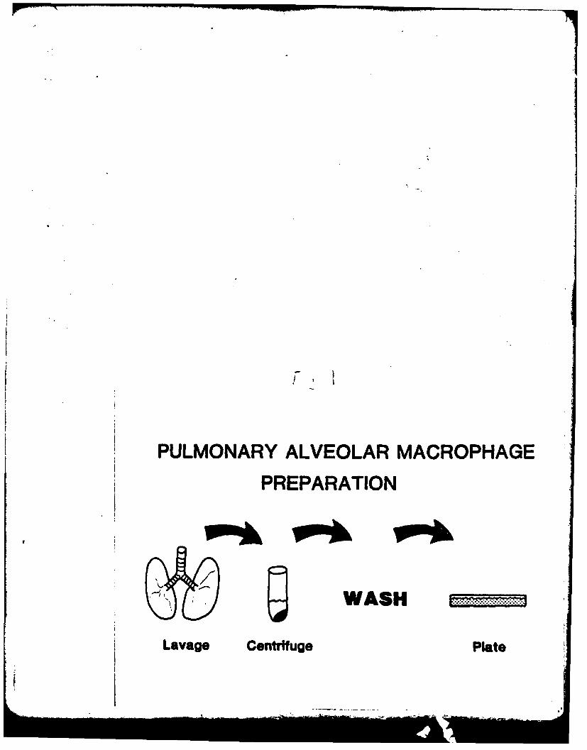
PULMONARY ALVEOLAR MACROPHAGE
PREPARATION
WASH
Lavage Centrifuge Plate

IL
-Aa6
PdNOW&
(**a 4. d~t-ft
bin-I S -~ 'Owl
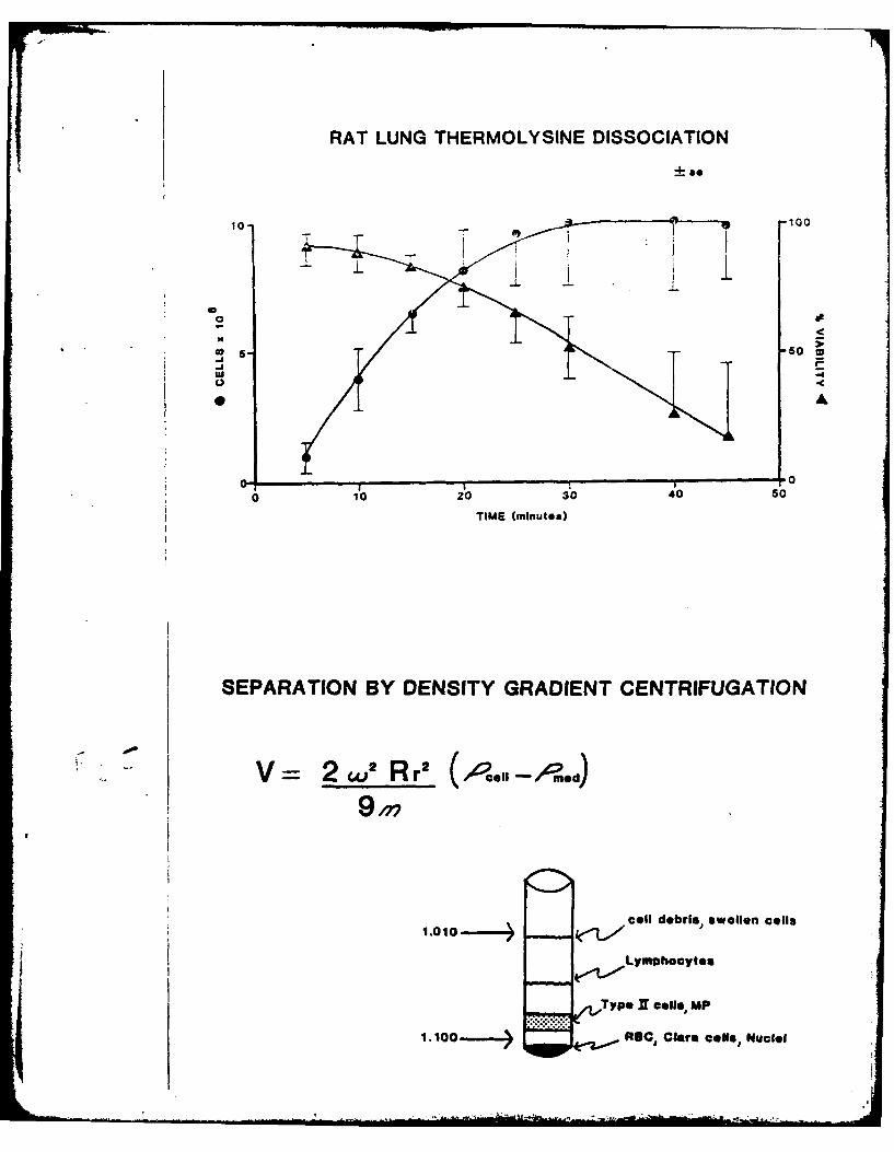
RAT LUNG THERMOLYSINE DISSOCIATION
10100
"5- so
06
-J Po -(
2 2
9 I
V 2,2R 2 ",p -<A.d
cell debris, swollen cells
Type N Cells, MP
1.0 " 8C, Clara cell*, Nuclei
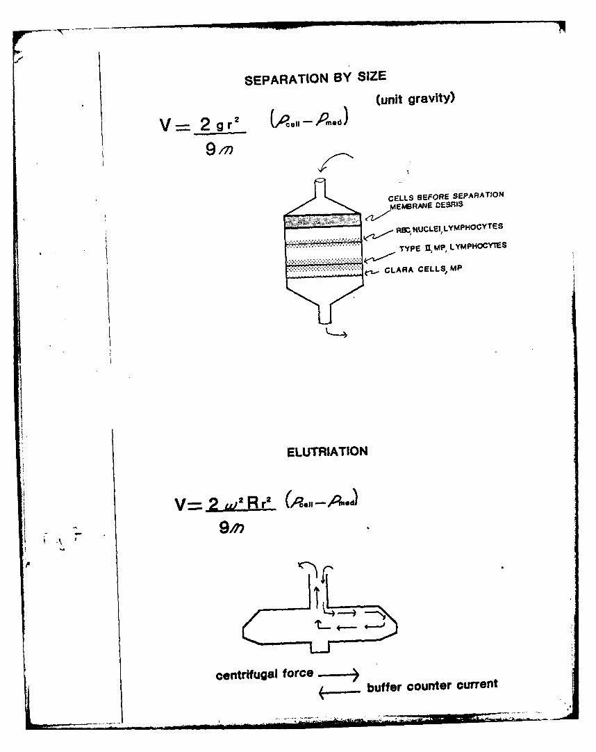
SEPARATION BY SIZE
(unit gravity)
V 2 g r' (P.P m ed)
CELLS BEFORE SEPARATION
.*.*,*, ~ MEMB6RANE DEBRIS
.. .~..~ .* .RSZNUCLE1, LYMpmOCYTES
....... TYPE U, MP, LYMPHOCYES
~.CLARA CELLS, MP
h ELUTRIATION
V=.aL~.Br (,q.0 Aod
centrifugal forcebuffer counter current

DIFFERENTIAL ADHERENCE
PARTIALLY PLATS 1 HOUR PLATE NON-ADHERENT CELLS
PURIFIED 22 HOURS
PP4EUMONOCYTES
z9
-- h4c:A
0 1- r" 1
0_
o v!
CD -0 s2a
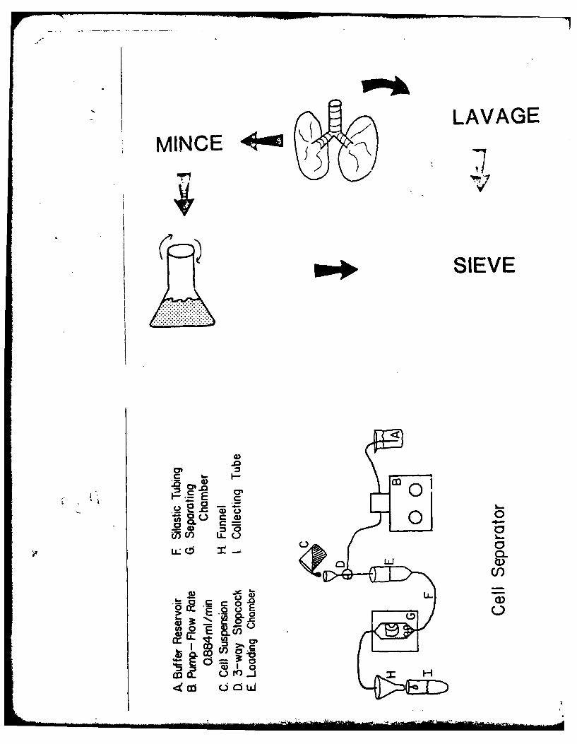
LAVAGEMINCECE
SIEVE
m 0
u = _ 7.r 4
05C/) LLC
'°-
.02).
C/U)
a
Ccz O l
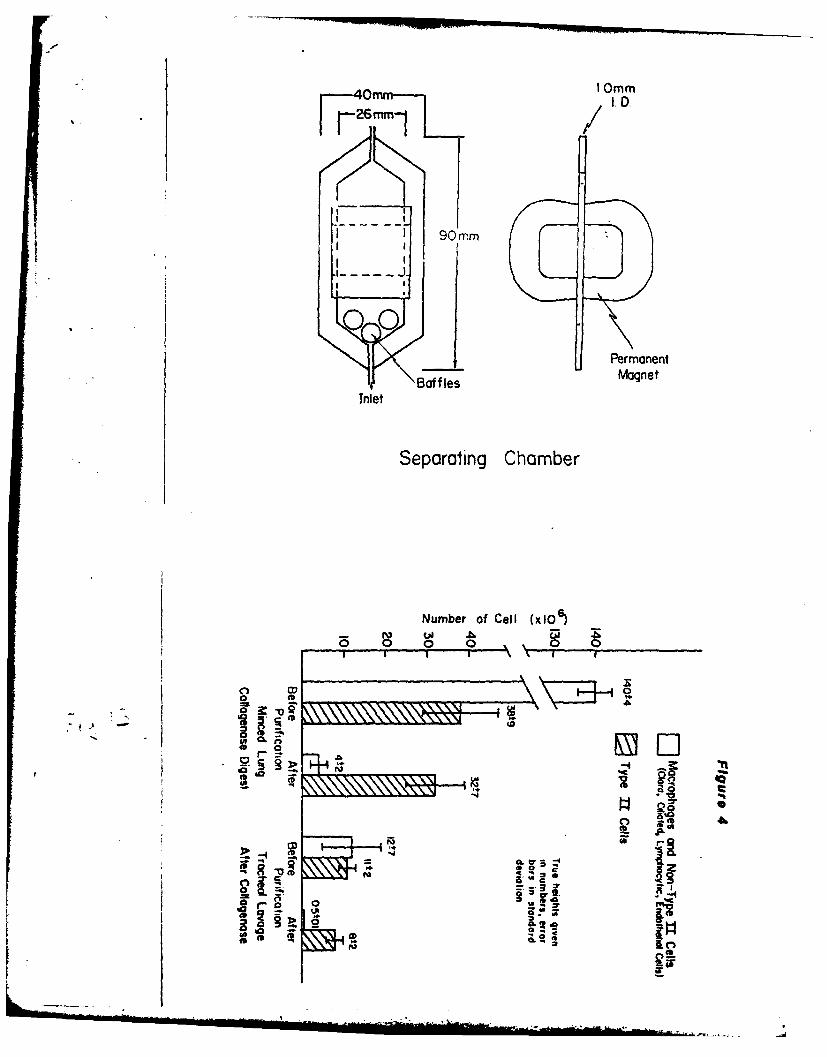
FF40mm--, I 0MM
26m 1 I.
1 90 m
FPermanentBaffles Mgnet
Inlet
Separating Chamber
Number of Cell (x106
r ro 1,
*0 L-0
-'I
S06
0L 0 :6
CD ff-v

ns
K, A
ELUTRIATION SEPARATION OF TYPE 11 CELLS30-
as'ROTOR SPEED 2000 RPM
200
to 50%
2004
load 7 12 1624 44
COUNTER CURRENT PLOW milmin

Type JI Cells
Values Represent Mean± S D
10 88,32t6mi-j988
0
0
0*
10mM Added 10

14C Sodium Acetate Incorporated (nM)
0 0 0 0N CD0
0
o . 0
0/0
00
--4
0 0 0
CDl
0 0
00 0
0- CL
3a
00 00
0 w i0
0
.. ..oI I I . . . . . . . .@ 0'. . . . . ,, +. ". . a. . . 'Qe" (.. .-3''I + . . .. . . . nTv- -.. ... . I

PRESENTATIONS
In addition to the publications and manuscripts cited in this report,
several presentations were also made with support from this project. The
abstracts from these presentations are included on the next three pages.
-123-

Kuttan, R., Spall, D., Sipes, I.G., Meezan, E. and Brendel, K.(1979) Effect of paraquat treatment on rat kidney basementmembrane. Toxicol. and Appl. Pharmacol. 48, A177.
355. EFFECT OF PARAQUAT TREATMENT ON RAT KIDNE " BASEMENT MEN%-B. AKES. R. Kuttan, R. Spall, I.G. Sipes, E. Meezan and K.Brendel, Dept. Pharmacol., AHSC, Univ. of AZ, Tucson, AZ.
The herbicide paraquat (methyl-viologen) has been shown to
produce severe lung fibrosis in both laboratory animals andhuman subjects. Its effect on the kidney has been much lessstudied. A method has been developed in this laboratory toproduce "acellular kidneys" which depends on exhaustiveextraction with detergents. The superstructure composed ofbasement membrane remains intact in these preparations. Us-ing this method, changes in kidney basement membrane obtain-ed 9 days after a single dose of paraquat (I.P.25 mg/Kg)were studied. Electron microscopy showed numerous "spiky"protrusions in the luminal side of tubular basement mem-branes of the affected kidneys, while glomerular basementmembrane remained intact. Chemical analysis of the damagedbasement membrane showed increased levels of hydroxylysine,3-hydroxyproline and cysteine, possibly indicating increasedbasement membrane synthesis. Parallel to this finding, thesynthesis of collagenous proteins of the kidney was increas-ed which was reflected by both elevated prolyl hydroxylaselevels and incorporation of proline into extracellularmatrix. (Supported by a grant from ONR N00014-77-C-0506.)
Presented at the 18th Annual Meeting of the Toxicology Society,New Orleans, 1979.
70

Lafranconi, E.., e ~an, B, Sipes, I.G. and Brendel, K. (1980)A rapia :neciod fir preparation of lung cell fractionse.riciiea in aiv'.olar type 11 pneumocyres, Fed. Proc.,FASEB 39, 3b6.
302 V%*.G
A MAtW JrrKD FOR FREARION 7 LIZG CELL TRLC-,tOS r_1IC_IN X~vo" Uvzt ?:EZA.CYhS, ',. Mar LafranconO. ;-!LAS
mea"a, 1. alenn Stpe and IrJauV Scandal. (SPONI: Thomas 1.Linde.l), L niv. of arizona, Tucson. Arizona 83724.
A method was d.oelnod !or the :a1.d isolation Of Ctype Itaran'dlz pn&l=Gct!t. -ations from 1%m~g of wihite %eSw Zealandrabbits oy tbe *se o mntic. Leon oxide to Afford sepagrat'nfrom macrophagoa. in zhis procedure, a colloidal. 'n;oetitaSuspeSiO t is inc Ubtae. with t'.s crude ceLL suopesion uartissuft culture conditions. Alternatively iOitad lung Par-fused with the tissue L:ur madium are lavSed with colloi-dal magnetite ta~oe rtros o .iation. ."acrophages havimg ha;-acycoved the mnetite ,,articles can then be quLled out oi -
1ature in a stro"n rasnluoiic field. Sizing of the macr ,p atdepleastd pnaeuwyze suspension yields an enriched type :1call praparatoan. Viability of the cell suspension was 90-95'%s detemined by tryan blue axelusion. Identification of t'eZ-anular ponecytes t* I) was accomplished by modified?37*anlcoaoo stain, .Uorst.%ctnc microscopy and electronticrascopy. CoLlular respiration was determned with theClak t ype oxv~en tIectrode. 'Ield of granular type Ii ; ---o-cye** in this procedurt as uell as their relative puri y "ereco parabl.s to other methods daslibed in the literature, F:o
pharracoogta.r Loxicalogical invstigations, this nachcdmight offer advantages over others Sint it does not requirethe use of costly OT unoon equipment. (Supported iA -artby the OfMice of Nav&l Research 4NOO,14-7-C-O0O6 and atraining grant by ~f1.h -1-32-ESO 7091-01).
Presented at the 64th Annual Meeting of the Federation ofAmerican Societies for Experimental Biology, Anaheim, California,
April 13-18, 1980.

Kuttan, R., Lafranconi, M., Spall, D., Sipes, I.G. Meezan, E.and K. Brendel (1979) Selective extraction of basementmembrane from lung extracellular matrix. Fed. Proc. 38,1338.
"LANOLOGY
MELEC7VE EXTRACTION4 OF ASCIMT MORAZ FROM LUNG EXTRA-c aL Aa '.11tRIX. R. Kuttan*. .1. Lalvanconi*. D. SPall. 1I.G.Sines. E. iezan ad K. B-end l (Span: D.C. Johnson). Dept.of Pharmacology. Arizona Halth Sciances Cancer, Tucson, AZ85724.
For the preparation. of hlug eztracehlular matrix the tiss-ue was scraped with a spatula to remove Large vascular andbronchial material. UoMoPeizatioU and a sieving procedureafforded additional prepurification. An extraction procedureinvolving several decergents and the action of D.Y'sa reov-ad all cellular material from the lung parenchymacous piecesand left th. extracllular mat ix. thought to be copoed ofbasemnt wabramv proteins as veil as soluble and insolublecollagens. To extract from chi* ixture the baseme
"nt m r mne
proceins, it was pretreatoed with 8 M urea which extracted"salt and acid soluble" collaegns. The residue was then ex-eracted wich uroa-eercapcoschamal which solubilized basementumbrane and left a residue of insoluble coLleagens of vari-
ous types and origins. In addition, Lung baseant membranecollagen ae prepared from the mrcaptoethamol-urea extractby pepsin digestion. Ilectron microscopy. gel electrophore-eis and amino acid as well as carbohydrate analyses wereused to characterize the various fractions. There Is a remark-able simiLarity in the analysis of baamnt neambrane proteinsfrom various species and becwen youmger mid older animals.The basemt mrane collages also showed siailarities withgloerular basemant mmbrane. Supported by OSPHS Grant No.HD-10781 ad OUR Grant 14o. OOL4-77-C-0506.
Presented at the 63rd Annual Meeting of the Federation ofAmerican Societies for Experimental Biology, Dallas, Texas,April 1-10, 1979.
