imaging assessment of urogenital emergencies fileurogenital tract in abdominal trauma has been...
TRANSCRIPT

Page 1 of 24
Imaging assessment of urogenital emergencies
Poster No.: C-0887
Congress: ECR 2014
Type: Educational Exhibit
Authors: A. Moffa1, R. Del Vescovo2, G. Frauenfelder2, F. Giurazza1, R. L.
Cazzato1, C. L. Piccolo1, R. F. Grasso2, B. B. Zobel1; 1Rome/IT,2Roma/IT
Keywords: Trauma, Obstruction / Occlusion, Acute, Imaging sequences,Cystography / Uretrography, Contrast agent-intravenous,Ultrasound, MR, CT, Urinary Tract / Bladder, Genital /Reproductive system female, Emergency
DOI: 10.1594/ecr2014/C-0887
Any information contained in this pdf file is automatically generated from digital materialsubmitted to EPOS by third parties in the form of scientific presentations. Referencesto any names, marks, products, or services of third parties or hypertext links to third-party sites or information are provided solely as a convenience to you and do not inany way constitute or imply ECR's endorsement, sponsorship or recommendation of thethird party, information, product or service. ECR is not responsible for the content ofthese pages and does not make any representations regarding the content or accuracyof material in this file.As per copyright regulations, any unauthorised use of the material or parts thereof aswell as commercial reproduction or multiple distribution by any traditional or electronicallybased reproduction/publication method ist strictly prohibited.You agree to defend, indemnify, and hold ECR harmless from and against any and allclaims, damages, costs, and expenses, including attorneys' fees, arising from or relatedto your use of these pages.Please note: Links to movies, ppt slideshows and any other multimedia files are notavailable in the pdf version of presentations.www.myESR.org

Page 2 of 24
Learning objectives
Diagnostic Imaging play an important role in the assessment of urogenital emergenciesand is crucial for a rapid and effective management. We propose to:
1) Illustrate the causes of urogenital emergencies
2) Illustrate, for any emergency, different imaging approaches and imaging features
Background
The most important causes of urogenital emergencies are abdominal trauma and acutegynecologic diseases. For any emergency, an accurate imaging work-up is crucial foraddressing an accurate diagnosis and reducing comorbidities.
1) ABDOMINAL TRAUMA
Urogenital tract in abdominal trauma has been reported to be involved in 8-10% of allcases. Wide-impact blunt abdominal trauma is responsible for most of closed injuries ofthe genitourinary organs, with motor vehicle crashes being the most common cause inthe Western hemisphere. The incidence of penetrating trauma is also increasing, whichis seen particularly in inner city trauma centers, and is becoming a major cause of renalinjury. Closed abdominal trauma represent the cause of almost 80% of these injuries.
Adrenal trauma: Trauma to the adrenal glands is unusual because of their relativelywell-protected position deep in the retroperitoneum. The gold standard is CT and typicalfeatures are:
- expansile, hyperattenuating, round or oval hematoma (Fig.1a).
- irregularity or obliteration of the gland by hemorrhage
- periadrenal fat stranding
- enlargement of the gland due to edema or contusion.
- active adrenal hemorrhage may be seen
Sonography is used to evaluate children who have sustained trauma. The adrenal glandmay be enlarged and may show hypoechoic areas of hemorrhage. (Fig.1b)

Page 3 of 24
Renal trauma:Contrast enhanced MDCT is the imaging technique of choice in renaltrauma and the role of IV urography (IVU) is currently relegated to situations in which CTmay not be available. The severity of renal injuries is graded from 1 to 5 according to aclassification system developed by the Organ Injury Scaling Committee of the AmericanAssociation for the Surgery of Trauma (AAST) and is called the organ injury scale (OIS):
-Grade 1 injuries: renal contusion without a parenchymal laceration and a nonexpandingsubcapsular hematoma.
-Grade 2 injuries: superficial cortical lacerations < 1 cm deep (and thus do not involvethe collecting system) and a nonexpanding perinephric hematoma (Fig.2)
-Grade 3 injuries: deeper lacerations, > 1 cm deep, that do not extend into the collectingsystem, and nonexpanding perinephric hematoma.
-Grade 4 injuries: lacerations that extend into the collecting system and injury to the mainand segmental renal vessels (Fig.3)
-Grade 5 injuries: show shattering of the kidney and dispersion of the avulsed portions,avulsion, laceration or thrombosis of the main renal vessels, hilar injury, and ureteropelvicjunction (UPJ) avulsion.
Ureteral trauma: Ureteral injuries caused by external trauma are atypical but whenthey occur are generally related to penetrating trauma, primarily gunshot wounds. Blunttrauma usually affects the UPJ and is related to rapid deceleration injury. One-shotpreoperative or intraoperative IVU may show contrast extravasation, but it is often notperformed because of poor diagnostic performance and indeed the delay in performingthe examination in an unstable patient. Once the patient is able to undergo this exam,complete IVU or contrast-enhanced CT with imaging in the delayed phase may showcontrast extravasation from the ureter (Fig.4) or partial or complete ureteral obstruction.Furthermore, CT may show urinary ascites or urinoma. In patients with blunt trauma andsuspected UPJ injury, CT with excretory phase imaging is a reliable tool for evaluation.
Urinary bladder trauma: The most frequent causes of bladder trauma are motor vehiclecrashes, falls, crush injuries, and blows to the lower abdomen. CT is now considered to bethe diagnostic procedure of choice in the evaluation of abdominal and pelvic injury afterblunt trauma. The conventional CT protocol for abdominal trauma may or may not showthe presence of bladder trauma because visualization of bladder rupture on CT requiresthe bladder to be filled with fluid and under pressure. Therefore, if CT examinationfails to show bladder rupture and bladder rupture is suspected, especially when water-dense fluid is seen in the peritoneal cavity, CT cystography is indicated. A classificationsystem approved by a consensus panel of the Societe Internationale D'Urologie classifiesbladder injury into four types:

Page 4 of 24
-Type 1: bladder contusion
-Type 2: intraperitoneal rupture (Fig.5)
- Type 3: extraperitoneal rupture (Fig.6)
- Type 4: combined injury
Urethral trauma: Blunt urethral trauma traditionally has been classified anatomically aseither anterior or posterior. Posterior urethral injury usually is caused by a crushing forceto the pelvis and is associated with pelvic fractures. The frequency of anterior urethralinjuries is one third that of posterior urethral injuries. Retrograde urethrography is thediagnostic procedure of choice to evaluate patients with suspected urethral injury. CT,MRI and ultrasonography (US) are useful for evaluating periurethral structures. Urethralinjuries are classified into five types (Goldman Classification):
-Type I: posterior urethra stretched but intact
-Type II: urethra disrupted at the membranoprostatic junction above the urogenitaldiaphragm
-Type III: membranous urethra disrupted, with extension to the proximal bulbous urethraor disruption of the urogenital diaphragm (most common type) (Fig.7)
-Type IV: bladder neck injury with extension into the urethra (Fig.8)
-Type IVa: injury of the base of the bladder and periurethral extravasation simulating atrue type IV urethral injury
-Type V: partial or complete pure anterior urethral injury
Testicular trauma: The testes can be injured with sporting activities, which accountfor more than half of all cases of testicular injury. Motor vehicle collisions are alsoan important cause, particularly two-wheeled motorized vehicles, when the testes arecrushed between the bony pelvis and the fuel tank. High-frequency US with a linear-array transducer is the first option to take into consideration for the evaluation of testiculartrauma. Heterogeneous echotexture in the testis, testicular contour abnormality due toextrusion of the testes through a tunical defect, and disruption of the tunica albuginea(Fig.9) are indicative of testicular rupture. Testicular fractures (Fig.10) are surgicallymanaged with débridement of extruded seminiferous tubules and closure of the tunicaldefect. Testicular hematomas may also cause the echotexture to be heterogeneous(Fig.11). The appearance of hematomas varies with their age, but they show nointernal vascularity. Other findings that may accompany testicular trauma are a scrotalhematocele, scrotal wall hematoma, and traumatic epididymitis. MRI is helpful in patientswith equivocal findings.

Page 5 of 24
2) ACUTE GYNECOLOGIC DISEASES
An acute gynecologic condition is characterized by sudden onset of lower abdominalpain, fever, genital bleeding, intraperitoneal bleeding, or symptoms of shock. It isimportant to differentiate diseases such as ovarian torsion, rupture of an ectopicpregnancy, and uterine arteriovenous malformations, which require an immediatesurgical or interventional approach, from conditions such as pelvic peritonitis and ovarianhemorrhage, which can be treated with conservative therapy. Physical examination has alimited role in diagnosis of acute gynecologic conditions. US is a useful imaging modalityfor evaluation of patients suspected to have acute gynecologic diseases. However, USfindings are not always conclusive and MRI is a valuable complement to US.
Ovarian torsion: Ovarian torsion is a well-known complication of ovarian tumors andcysts. Sonography is the diagnostic modality of choice. Typical sonographic features oftorsion are:
-enlarged ovaries (>4 cm)
-ovarian cyst or mass
-free fluid
-tenderness
-an abnormal ovarian position (affected ovary in the pouch of Douglas, anterior to theuterus, or on the contralateral side)
-presence of a pedicle
-whirlpool sign
-abnormal Doppler flow (absence of venous or arterial flow)
-presence of follicular rings (Fig.12)
Hemorrhagic necrosis due to torsion can be established by MR imaging evaluationwith a combination of fat-suppressed T1-weighted images and contrast-enhanced fatsuppressed T1-weighted images. High signal intensity of a tumor on fat-suppressed T1-weighted images suggests hemorrhage or vascular congestion (Fig.13). MR imagingfeatures indicative of ovarian torsion are:
- lack of enhancement of the solid component
- a thickened cyst wall or a mural nodule

Page 6 of 24
Ectopic pregnancy: Ectopic pregnancy usually cause lower abdominal pain andhemoperitoneum. Currently, diagnosis in unruptured ectopic pregnancy is achievedusing a combination of transvaginal ultrasonography and measurement of serum #-hCGconcentrations. 95% of ectopic pregnancies occur within the fallopian tube; other sitesare the ovary, cervix, and peritoneal cavity. Effectively, in the absence of an intrauterinegestation sac, an ectopic pregnancy can be diagnosed by the presence of an adnexalmass, often visible within the Fallopian tube (Fig.14). Massive hemorrhage from aruptured tubal or ovarian pregnancy leads to symptoms of shock.
MR imaging findings in ectopic pregnancy include:
- hemosalpinx
-bloody ascites
-heterogeneous adnexal mass composed of a hematoma
-gestational sac
The hemosalpinx and bloody ascites are slightly hyperintense if compared to urineon fat-suppressed T1-weighted images. The heterogeneous mass has mixed signalintensity on fat-suppressed T1-and T2-weighted images (Fig.15). The bleeding point canbe revealed with contrast-enhanced dynamic subtraction MR imaging. Extravasation ofcontrast material can be seen as a hyperintense spot in the hematoma.
Uterine arteriovenous Malformations: Uterine arteriovenous malformations (AVMs)are broadly classified as congenital or acquired. Congenital AVMs are rare and result fromabnormal embryologic development of primitive vascular structures whereas acquiredor traumatic AVMs are being increasingly diagnosed. Acquired uterine AVMs areusually traumatic, resulting from prior dilation and curettage, therapeutic abortion, uterinesurgery, or direct uterine trauma. The most common symptom of AVMs is genitalbleeding. Initial evaluation of AVMs is made with ultrasonography, at which they appeareither as masses with multiple hypo/anechoic tubular like structures of varying sizesor as focal endometrial and myometrial thickenings. US-Doppler adds the possibility ofrecognizing vessels within malformations. Spectral analysis permits evaluating the flowwithin vessels, recognizing high flow and low resistive index within arteries and veins(Fig.16).
Confirmation of the diagnosis traditionally comes from angiography. Tortuous and tubularsignal voids in the myometrium, in the parametrium, and protrusions into the endometrialcavity are seen on both T1-and T2-weighted images (Fig.17). A vascular lake withsluggish flow is demonstrated as a hyperintense structure mimicking a tumor or placentalpolyp on T2-weighted images. Contrast-enhanced dynamic subtraction imaging revealsthat the lesion enhances as intensely as vessels. Dynamic subtraction MR angiography

Page 7 of 24
clearly demonstrates vascular anatomy, abnormal vessels, and hemodynamics andenables noninvasive confirmation of a diagnosis of uterine arteriovenous malformations.
Hemorrhagic Ovarian Cyst: is an abdominal mass formed by the bleeding intoa follicular ovarian cyst or corpus luteum cyst. It is detected as pelvic mass byultrasonography, but is often misdiagnosed with other organic masses because of itsvariable clinical and sonographic findings and in some cases may lead to laparotomy.Main sonographic features observed are the presence of fibrin strands or a retractingclot , with the absence of suspected septations and wall irregularity as secondary helpfulfindings (Fig.18).
MR imaging shows hemorrhagic cysts as hyperintense on both T1- and T2-weightedimages or hyperintense on T1-weighted images and hypointense on T2-weighted images(Fig.19)
Pelvic Inflammatory Disease: is described as a spread of inflammation from theendometrial cavity and fallopian tubes into the pelvis. It is an umbrella term, whichencompasses endometritis, salpingitis and tubo-ovarian abscesses. Patients often havelower abdominal pain, fever, leukocytosis, an elevated blood level of C-reactive proteinand adnexal tenderness. Endometritis and myometritis can be treated conservativelywith antibiotic therapy. However, tubo-ovarian abscess and pyosalpinx often need tobe treated surgically. Transabdominal Ultrasound can demonstrate uterine enlargementand thickening of the endometrium and can also show the loss of tissue plainsand an ill-defined uterus. Hydrosalpinx or pyosalpinx is a common complication ofsalpingitis. Ultrasound can identify dilated fallopian tubes containing heterogenous fluidwith echogenic debris; features typical of pyosalpinx. The fallopian tubes may be foldedand demonstrate areas of tube tapering, and intraluminal small linear echogenic foci maybe visualised (Fig.20). As pyosalpinx develops into tubo-ovarian abscesses, echogenicdebris can be seen in the fallopian tubes and ovaries, representing inflammatoryexudates, blood and pus. On MR Imaging, inflammation in the parametrium may beseen as ill-defined hyperintense areas on T2-weighted fat-suppressed images, in additionto enhancement on gadolinium-enhanced T1-weighted images. A pyosalpinx can bevisualised as a dilated, fluid-filled, tortuous C or S-shaped structure. Thickwalled fluid-filled abscesses and pyosalpinx may have heterogeneous signal intensity on both T1 andT2 weighting due to mixtures of pus, haemorrhage and debris. The thick-walled masstypically demonstrates marked enhancement following iv gadolinium administration (Fig.21).
Images for this section:
Page 8 of 24
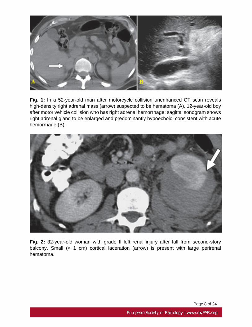
Fig. 1: In a 52-year-old man after motorcycle collision unenhanced CT scan revealshigh-density right adrenal mass (arrow) suspected to be hematoma (A). 12-year-old boyafter motor vehicle collision who has right adrenal hemorrhage: sagittal sonogram showsright adrenal gland to be enlarged and predominantly hypoechoic, consistent with acutehemorrhage (B).
Fig. 2: 32-year-old woman with grade II left renal injury after fall from second-storybalcony. Small (< 1 cm) cortical laceration (arrow) is present with large perirenalhematoma.

Page 9 of 24
Fig. 3: 43-year-old man with grade IV left renal injury after being struck by car whilewalking. Wedge-shaped perfusion defect (arrow) is present in interpolar region of leftkidney with surrounding hematoma.

Page 10 of 24
Fig. 4: A 29-year-old man involved in a highway motor vehicle collision. CT revealscontrast retention in the perirenal retroperitoneum bilaterally (arrows) caused by bilateralcomplete ureteral disruption.
Fig. 5: 34-year-old woman with intraperitoneal bladder rupture after motor vehiclecollision. A-C, CT cystography images (A, axial; B, coronal; C, sagittal) show site ofbladder rupture at dome (arrows) with contrast extravasation into peritoneal space(arrowheads). Note enlarged uterus with fibroids (F).
Fig. 6: 19-year-old man with extraperitoneal bladder rupture after motor vehicle collision.A-C, CT cystography images (A, axial; B, coronal; C, sagittal) show site of bladderrupture at right lateral wall (arrows) with contrast extravasation into extraperitoneal space(arrowheads).

Page 11 of 24
Fig. 7: Posterior urethral rupture extending through the urogenital diaphragm toinvolve the bulbous urethra following blunt trauma (type III urethral injury). Retrogradeurethrogram reveals contrast material extravasation at the membranous urethra (arrow).The contrast material extends below the urogenital diaphragm and surrounds theproximal bulbous urethra.

Page 12 of 24
Fig. 8: Bladder neck urethral injury (type IV) in a 23-year-old woman. (A) Cystogramshows extraperitoneal contrast material extravasation (arrow) that extends fromthe bladder neck to the left underneath the balloon of a Foley catheter. (B)Cystogram obtained 2 minutes later shows progressive extraperitoneal contrast materialextravasation.

Page 13 of 24

Page 14 of 24
Fig. 9: Testicular rupture. Longitudinal color Doppler US image of the left testis in apatient with scrotal trauma depicts tunica albuginea disruption (arrowhead) with a loss ofvascularity in the ruptured portion of the testis (*).
Fig. 10: Testicular fracture. Longitudinal color Doppler US image in a patient withscrotal trauma depicts a hypoechoic avascular band (arrows) that crosses the testicularparenchyma.

Page 15 of 24
Fig. 11: Intratesticular hematoma. Transverse color Doppler US image in a patient withscrotal trauma demonstrates an avascular hypoechoic area (arrow) within the testis. Thehematoma was managed conservatively.

Page 16 of 24
Fig. 12: Follicular rings (arrow) visible on transabdominal scans. The torsed ovary isimpacted in the pouch of Douglas with the uterus lying anterior.

Page 17 of 24
Fig. 13: Twisted left ovarian cystic teratoma with hemorrhagic necrosis in a 10-year-oldgirl. (A, B) Axial T1-weighted (A) and fat-suppressed T1-weighted (B) MR images show acystic tumor (solid arrows) with a markedly thickened wall, which is slightly hyperintense.A small fat component (open arrow) within the tumor is hyperintense on the T1-weightedimage (A) and hypointense on the fat-suppressed T1-weighted image (B).
Fig. 14: Transvaginal gray-scale US image of left adnexa reveals a 25mm hyperechoicmass with a central hypoechoic shadow, suggestive of a tubal pregnancy. ES: ectopicsac; CL: corpus luteum.

Page 18 of 24
Fig. 15: Ovarian pregnancy in a 22-year-old woman with pelvic pain. (A, B) Axial fat-suppressed T1-weighted (A) and T2-weighted (B) MR images show a heterogeneous,hyperintense mass (long arrows) on the right side of the uterus (*). The margin of themass has irregular low signal intensity (short arrows) on the T2-weighted image (B).
Fig. 16: Spectral analysis of the right uterine artery showing high-velocity flow with lowresistance index and pulsatility index. References:

Page 19 of 24
Fig. 17: AVMs in a 28-year-old woman with massive genital bleeding 2 months afterdilation and curettage. (A) Sagittal fat-suppressed T2-weighted MR image shows tangledsignal voids protruding into the endometrial cavity (arrows). (B) Sagittal contrast-enhanced dynamic subtraction MR image (arterial phase) shows that the signal voids(arrows) enhance as intensely as the abdominal aorta (*), a finding indicative of a vascularlesion.

Page 20 of 24
Fig. 18: Sonogram showing fibrin strands (long arrow) in a portion of a mass, whichproved to be a hemorrhagic cyst (resolved 4 weeks later). Note the apparent thickseptation (short arrow) in the mass caused by a clot, which resolved.
Fig. 19: Hemorrhagic ovarian cyst with hemoperitoneum in a 20-year-old woman withpelvic pain. Axial fat-suppressed T2-weighted (A) and fat-suppressed T1-weighted

Page 21 of 24
(B) MR images show a complex left adnexal mass (solid arrows). The mass ismostly hypointense with a small hyperintense portion on the T2-weighted image (A);it is hyperintense and isointense on the T1-weighted image (B). There is a fluidcollection (open arrow), which is hyperintense on the T2 weighted image (A) andslightly hyperintense relative to urine (±) on the T1-weighted image (B), an appearancesuggestive of bloody ascites. Note the layering low signal intensity (arrowhead) on theT2-weighted image (A); this finding is considered to represent fibrin debris or clots. large* = right ovary, small * = uterus.
Fig. 20: PID with pyosalpinx on Ultrasound. This patient presented to the emergencydepartment with lower abdominal pain, pyrexia and vomiting. A-B Transvaginalultrasound of both adenexa. There are bilateral adenexal cysts that contain low-levelechogenic material and have a tubular configuration (white arrows). The appearance isin keeping with bilateral pyosalpinges, a complication of PID.
Fig. 21: PID with pyosalpinx on MRI. This patient presented to the emergency departmentwith pyrexia, lower abdominal pain and diarrhea. (A) Sagittal T2 image of the pelvisdemonstrates multiple fluid-filled cystic structures within the right adnexa (red arrows).The complex cyst is thick walled and there is adjacent fat stranding. (B) Axial T2 imagedemonstrates bilateral tubo-ovarian abcesses. (C)Axial T1 fat-saturated image following

Page 22 of 24
gadolinium administration demonstrates low signal intensity within the pus-filled cavitiesand marked enhancement of the inflammatory walls.

Page 23 of 24
Findings and procedure details
We used contrast-enhanced Computer Tomography for most series of emergencies.Furthermore, we used Cystography, Urethrography, Ultrasonography, Angiography andMR imaging.
Conclusion
Imaging is crucial in the evaluation of the urogenital emergencies. Contrast-enhancedCT is usually the first-line imaging technique used to evaluate upper and lower urinarytract. Cystography and Urethrography remain useful techniques in the initial evaluationand follow-up of trauma to the urinary bladder and urethra. Ultrasonography is auseful imaging modality for evaluation of acute gynecologic diseases, but is not alwaysconclusive; effectively MRI represents appropriate complement to US.
Personal information
References
1. Ramchandani P,Buckler PM. Imaging of genitourinary traumaAm JRoentgenol. 2009 Jun;192(6):1514-23
2. Stawicki SP,Hoey BA,Grossman MD,Anderson HL 3rd,Reed JF 3rdAdrenalgland trauma is associated with high injury severity and mortality.CurrSurg.2003 Jul-Aug; 60(4):431-6
3. Martínez-Piñeiro L EAU Guidelines on Uretheral Trauma Eur Urol. 2010May; 57(5):791-803
4. Gomez RG, Ceballos L, Coburn M, et al. Consensus Consensus statementon bladder injuries. BJU Int 2004; 94:27-32
5. Chan DP,Abujudeh HH,Cushing GL Jr,Novelline RACT cystography withmultiplanar reformation for suspected bladder rupture: experience in 234casesAJR Am J Roentgenol.2006 Nov; 187(5):1296-302
6. Kawashima A,Sandler CM,Wasserman NF,LeRoy AJ,King BF Jr,GoldmanSMImaging of urethral disease: a pictorial reviewRadiographics .2004Oct;24 Suppl 1:S195-216
7. Dohke M,Watanabe Y,Okumura A,Amoh Y,Hayashi T,Yoshizako T,YasuiM,Nakashita S,Nakanishi J,Dodo YComprehensive MR imaging of acutegynecologic diseasesRadiographics.2000 Nov-Dec; 20(6):1551-66

Page 24 of 24
8. Goldman SM, Sandler CM, Corriere JN Jr, McGuire EJ. Blunt urethraltrauma: a unified, anatomical mechanical classification. J Urol 1997;157:85-89
9. Bhatt S,Dogra VSRole of US in testicular and scrotaltraumaRadiographics.2008 Oct; 28(6):1617-29
10. Dunfee BL,Lucey BC,Soto JADevelopment of renal scars on CT afterabdominal trauma: does grade of injury matter?AJR Am J Roentgenol.2008May;190(5):1174-9
11. Sibal M. Follicular ring sign: a simple sonographic sign for early diagnosis ofovarian torsionJ Ultrasound Med.2012 Nov;31(11):1803-9.
12. Sivalingam VN,Duncan WC,Kirk E,Shephard LA,Horne AW. Diagnosis andmanagement of ectopic pregnancyJ Fam Plann Reprod Health Care.2011Oct;37(4): 231-40.
13. Nemoto Y,Ishihara K,Sekiya T,Konishi H,Araki T. Ultrasonographic andclinical appearance of hemorrhagic ovarian cyst diagnosed by transvaginalscanJ Nippon Med Sch.2003 Jun; 70(3):243-9
14. Patel MD,Feldstein VA,Filly RA. The likelihood ratio of sonographic findingsfor the diagnosis of hemorrhagic ovarian cysts.J Ultrasound Med.2005May;24(5):607-14
15. O'Brien P., Amir Neyastani, Anne R. Buckley, Silvia D. Chang, Gerald M.Legiehn, Uterine arteriovenous malformations: from diagnosis to treatmentJUltrasound Med.2006 Nov; 25(11):1387-92
16. Alessandrino F,Di Silverio E,Moramarco LP. Uterine arteriovenousmalformationJ Ultrasound.2013 Feb 23;16(1):41-4
17. Roche O,Chavan N,Aquilina J,Rockall A. Radiological appearances ofgynaecological emergenciesInsights Imaging.2012 Jun; 3(3):265-75