imaging & oncology...imaging & oncology is a publication of the society and college of...
TRANSCRIPT

For imaging and therapy professionalsFF ii i dd th f i l
IMAGING &ONCOLOGY

VOLUMES WITHOUT ARTIFACTS, IT’S IN THE SCIENCEAcquiring an entire volume at a single moment in time without artefacts delivers a step change in CT capability. A ten-year research programme resulted in the development of our patented cone beam algorithm that accurately reconstructs higher resolution volumetric data sets without distortion artefacts. An innovation that produces true dynamic volume imaging for accurate diagnosis, demonstrating our commitment to science in diagnostic imaging, building integrity into everything we do.

IMAGING & ONCOLOGYCONTENTS
Editor: Hazel Edwards, Consultant Sonographer, Norwich Radiology Academy, Norfolk & Norwich University Hospital
Publisher: Dominic Deeson
Designers: Deeson Create
Display advertising: Rob Aspin Tel: 01795 542402
Published by: Deeson Group Ltdwww.deeson.co.uk
Printed by: MWL Print Group, Pontypool
Imaging & Oncology is a publication of The Society and College of Radiographers207 Providence Square, Mill Street, London SE1 2EWTel: 020 7740 7200 • Fax: 020 7740 7204 • E-mail: [email protected]
ISBN 9871 871101 58 1
All correspondence relating to Imaging & Oncology should be addressed to:Hazel Edwards at the Society and College of Radiographers, or to [email protected]
06 The value and future of proton radiotherapy in the UKCarl Rowbottom
14 The impact of stereotactic body radiotherapy on UK oncology servicesAngela Baker, Lynda Appleton, Alison Scott and Pooja Jain
22 Cyberknife technology and its future contribution to oncology Alexandra Aitken, Nihal Shah, Pete Ostler and Peter Hoskin
30 Advances in the imaging of sentinel nodes in breast cancer: the role of nuclear medicineJohn Thompson, Andrew Tootell, Joanne Sil, Judith Kelly, Randeep Kulshrestha and Peter Hogg
38 Globalisation of higher education in radiologic sciences: a new paradigmSarah Baker
42 Radiology accreditation in the UK: the theory and the realityMelanie Hiorns
48 Educating the radiography workforce: a diverse challengeIan Henderson
54 Imaging for stroke and transient ischaemic attack: meeting the standards of the national stroke strategyAndy Beale and Damian Jenkinson
58 Too many hysterectomies in the era of fi broid embolisation?Ram Kasthuri and Jon Moss
64 A picture of the future: cardiac CTJohn Curtin
Disclaimer©The Society of Radiographers 2011Unless otherwise indicated, views expressed are those of the editorial staff, contributors and correspondents. They are not necessarily the views of The Society and College of Radiographers (SCoR), its offi cers, or Council. The publication of an advertisement does not imply that a product is recommended by The Society. Material may only be reproduced by prior arrangement and with due acknowledgement to Imaging & Oncology.

2011IMAGING &ONCOLOGY
4 EDITORIAL
Hazel Edwards
A change for the betterWelcome to the 2011 issue of Imaging & Oncology, which features 10 articles on some of the hottest topics affecting our professions. In addition to their contemporary relevance, each one describes, calls for, or predicts change.
We begin with three cutting-edge radiotherapy topics describing revolutionary advances in technique. Two papers by leading academics from the United Kingdom and the United States discuss the changing face of radiographic education. There are also articles discussing how imaging services for stroke/TIA must be improved and how uterine artery embolisation must become more widely available.
Hiorns takes us on a step-by-step guide through the new Imaging Services Accreditation Scheme, and provides helpful tips for others who will undoubtedly be preparing to embark on the same process. From nuclear medicine, a team describes the importance of sentinel node detection in breast cancer, and fi nally, Curtin predicts the likely impact recently published cardiac guidelines may have on CT departments.
Some years ago, Queen Elizabeth II commented, “I sometimes sense the world is changing too fast for its inhabitants, at least for us older ones.” Nowhere is this more obvious than within the world of imaging and oncology. But radiographers, radiologists, physicists, and educationalists have not only kept pace with change but are, of course, responsible for accelerating it, as demonstrated in the following pages.
Whether in terms of improving patient survival rates, developing better treatment outcomes, or setting higher practice standards, all describe change for the better.
See what you think please and read on.

2011IMAGING &ONCOLOGY
5Back to the futureMy term as President of the Society and College of Radiographers has enabled me to look backwards and forwards on the development of our profession. In August 2010 we celebrated 90 years since the founding of the Society of Radiographers. It is not until you look back to where and when we started do you realise how far we have come in such a short time.
Now in 2011 we are celebrating the Year of Radiotherapy, which is an initiative to raise public awareness of the value of radiotherapy in the treatment of cancer. This is a project where a multi-professional group including the Society of Radiographers, the Royal College of Radiologists, Cancer Care UK, the Institute of Physics and Engineering in Medicine, the National Health Service, and representatives from all of the four countries within the United Kingdom have worked together towards a common goal.
An excellent example of what can be achieved when everyone works together for the good of our patients.
Both events encouraged me to refl ect on the advances of both the technology and the professional practice which enables radiographers both diagnostic and therapeutic to contribute greatly to increased patient care and service delivery. In addition, both celebrations have been set against challenging times, economically and politically – but never forget challenges present great opportunities.
In the delivery of a quality, patient centred service, no profession can work in isolation; there is so much to be learned from each other. Working in multi-professional teams is the way ahead: each valuing the others’ contribution to the overall package of care.
We have chosen dynamic careers in imaging...ever changing and moving forward. This annual publication, Imaging and Oncology 2011,
now in its seventh year, always challenges our thoughts and perceptions, and this year is no exception.
I congratulate the editor for gathering together such an interesting and varied selection of papers covering education and professional practice, future trends, and challenges from both a UK and international perspective. And all this not only for diagnostic and therapeutic radiographers, but also for our fellow colleagues in the imaging and oncology professions.
So take time, sit down and savour this cornucopia of papers, all written for you by leaders in their respective fi elds. Please read the articles which are on topics which are new to you: there is nothing like new knowledge to challenge your established ways of thinking.
What ever your experience, I hope the papers provide food for thought, and inspire you for the future.
FOREWORD
Sandie MathersPresidentThe Society and College of Radiographers

THE VALUE AND FUTURE OF PROTON RADIOTHERAPY IN
THE UK
CARL ROWBOTTOM

2011IMAGING &ONCOLOGY
7Should proton therapy be available in the UK? What are the barriers that must be overcome fi rst?
INTRODUCTIONThe concept of treating cancer patients with protons was fi rst suggested by Robert R Wilson in a paper published in 19461. Initially, patient treatments were performed with particle accelerators built for physics research, most notably at the Berkeley Laboratory in the USA and Uppsala in Sweden in the mid 1950s. It took another 35 years before a purpose built hospital proton facility was built with the Loma Linda University Medical Center in Loma Linda, California in 1990. Recent years have seen a growth in the number of proton radiotherapy treatment centres either opening or in the planning stage with nearly 30 centres worldwide in operation at the end of 2010.
The potential advantage of protons for radiotherapy treatment can clearly be seen from the depth dose characteristics of photons and protons using a spread-out Bragg peak (Figure 1). Proton radiotherapy treatments have the potential to reduce the integral radiation dose received by the patient, and to deliver lower doses to normal tissue proximal to the tumour compared to traditional photon radiotherapy treatments.
For proton radiotherapy a high energy accelerator capable of accelerating protons to at least 230MeV is required to provide suffi ciently energetic protons to penetrate to the centre of body. The particle accelerator will either be a cyclotron or synchrotron and the choice has an effect on the performance parameters of the clinical beam. A cyclotron can produce a high current beam of one energy, the maximum required. For clinical treatments, the beam energy must be degraded to provide the range of energies required to produce a spread-out Bragg peak for treatment.
The degrading can be performed very quickly but reduces the current of the proton beam, which can be a problem when treating at lower energies. Synchrotrons generally operate at a lower current but can accelerate to different energies and so beams do not
require degrading. As a result, they can produce beams with a better energy resolution but the time to switch between energies is longer. They are generally more complex machines and so more expensive and operate at a lower beam current.
The high cost of the particle accelerator for proton radiotherapy means treatment centres run multiple treatment rooms off a single accelerator. Typically, between three and fi ve rooms can be serviced. The limiting factor is that currently only one patient at a time can be treated and it takes time to switch the proton beam between rooms. Achieving effi cient throughput and avoiding patients waiting too long for the treatment beam is dependent on several
100 -
80 -
60 -
40 -
20 -
- - - - - -
Rela
tive
dose
(%
)
Depth in tissue (cm)
4 8 12 160
Spread-outBragg peak
Tumor
10 MV X-rays
Bragg peak (protons)
A UNIQUE OPPORTUNITY FORCONSISTENT PROTON TREATMENTS
Figure 1. Depth dose characteristics in tissue of a single Bragg peak (pink), a spread-out Bragg Peak (red), which consists of different energy Bragg peaks added together, and a 10MV x-ray beam (dashed). Taken from Technology Insight: proton beam radiotherapy for treatment in pediatric brain tumors Torunn I Yock and Nancy J Tarbell, Nature Clinical Practice Oncology (2004) 1, 97-103 (reproduced with permission).

2011IMAGING &ONCOLOGY
8 factors. First, not running too many rooms off one accelerator. Second, on the equipment being able to deliver a high dose rate so that individual fi elds can be treated in a short time and, fi nally, on being able to switch the beam between rooms quickly.
Treatment rooms will generally be a mix of fi xed beam rooms where the beam can be directed at the patient only from one or two directions, and gantry rooms where the beam can be rotated around the treatment couch. Gantries for proton radiotherapy need to incorporate large and powerful magnets to bend the high energy protons. The resulting gantry structure can be three stories high, taking up the fl oors below and above the treatment fl oor level. These major pieces of engineering increase the initial cost, leading centres to treat with fi xed beams where possible. For simpler treatments, a fi xed beam, in combination with a robotic couch, can be used to deliver proton radiotherapy.
A good example is prostate treatment, where two lateral beams can be delivered by a fi xed beam. A variant on the fi xed beam is the dual inclined beam, where two beams at different angles can provide greater fl exibility.
In some centres more complex plans may contain beams that can be delivered by fi xed beams and beams that can only be delivered by gantries. In this situation the patient will receive treatment in different treatment rooms on alternate days with not all beams treated daily. This is possible with proton treatments as each fi eld can deliver a uniform dose to the whole tumour.
PASSIVE SCATTERING VERSUS SCANNING TECHNOLOGIESNearly all of the treatments that have been delivered so far use a scattered proton beam to treat the patient. Passive scattering uses a range shifter wheel, or ridge fi lter, to modulate the energy of a narrow beam to create the spread out Bragg peak (SOBP) to produce a high dose to the target from a single beam direction. The narrow proton beam is scattered to produce a broad beam that can be used to target the complete treatment fi eld in a similar fashion to a photon beam (Figure 2). The scattered beam then uses a custom collimator to defi ne the fi eld size and a compensator to alter depth of penetration across the beam aperture.
The compensator and collimator used for passive scattered proton radiotherapy are made individually for each patient and are unique for each treatment beam used in the proton treatment plan (Figure 3). Each collimator/compensator pair for every treatment beam needs to be manually inserted into the treatment nozzle prior to the irradiation of the patient. Although an effective solution, this can lead to manual handling and radiation protection issues for staff at the proton radiotherapy centre, as well as increasing the cost of proton treatments.
Modulator wheelsor ridge filters
ScattererScatterer
(contoured)
Compensator
Collimator
PatientTarget
Extra dose
Figure 2. A passive scattering treatment system.
Figure 3. Individual collimator and compensator used in passive scattering proton systems.
Magnets
Trteatment volume
Figure 4. A scanning proton treatment system.
Proton scanning systems are seen as the next generation of commercial proton technology. Scanning was fi rst implemented at the Paul Scherrer Institute in Villigen, Switzerland. Firstly, we should consider why scanning beams are seen as being so important to proton radiotherapy. Scanning has the potential to both simplify and improve proton radiotherapy treatments. It has several advantages over passive scattering technology:• It has the potential to reduce commissioning times. • It does not require the manufacture of compensators or collimators for individual
patients’ treatment beams. • It allows the delivery of more conformal treatments. • It has the potential to increase patient throughput.
Scanning technology does not require the custom collimator or compensator because the beam can be controlled to irradiate the desired treatment positions by changing the position in the XY plane using magnets, and Z (depth) by adjusting the incident proton energy (Figure 4). Scanning reduces the cost of producing these customised devices. It also can increase throughput because less radiographer intervention is needed to insert a patient specifi c collimator and compensator combination for every treatment fi eld.
Apart from avoiding patient specifi c compensators and collimators, scanning opens up the possibility of intensity modulated proton therapy (IMPT). The Paul Scherrer Institute has defi ned two main treatment modes of scanning treatments: First, single fi eld uniform dose (SFUD), where each fi eld delivers a homogeneous fi eld across the volume. And second, IMPT, where the Bragg peaks from all fi elds are optimised such that each treatment beam provides a non-uniform dose delivery but, when combined, all treatment beams produce a uniform dose distribution. Both of these modes allow increased conformity with respect to passive scattering.
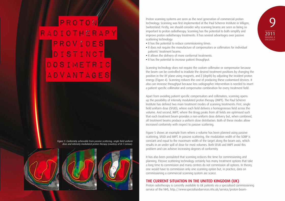
Figure 5 shows an example from where a volume has been planned using passive scattering, SFUD and IMPT. In passive scattering, the modulation width of the SOBP is constant and equal to the maximum width of the target along the beam axis, which results in an under spill of dose for most volumes. Both SFUD and IMPT avoid this problem and can achieve increasing degrees of conformity.
It has also been postulated that scanning reduces the time for commissioning and planning. Passive scattering technology certainly has many treatment options that take a long time to commission and many centres do not commission all options. In theory, one would have to commission only one scanning option but, in practice, data on commissioning a commercial scanning system are scarce.
THE CURRENT SITUATION IN THE UNITED KINGDOM (UK)Proton radiotherapy is currently available to UK patients via a specialised commissioning service of the NHS, http://www.specialisedservices.nhs.uk/service/proton-beam-
2011IMAGING &ONCOLOGY
9PROTONRADIOTHERAPYPROVIDESDISTINCT
DOSIMETRICADVANTAGES
Figure 5. Conformity achievable from passive scattering, single fi eld uniform dose and intensity modulated proton therapy (courtesy of Dr T Lomax).
2011IMAGING &ONCOLOGY
10 therapy. An expert reference panel receives patient referrals for treatment abroad at one of three centres: the Paul Scherrer Institute, Villigen, Switzerland; the Centre-Protontherapie, Orsay, France; or the University of Florida Proton Therapy Institute, Jacksonville, USA. This national service has been available since April 2008 and has resulted in more than 70 UK patients being treated overseas. Although providing a necessary and important clinical service, treatment abroad for several weeks can provide signifi cant challenges for patients and their carers.
There is a clear emphasis on the treatment of paediatric patients in the current list of approved diagnoses, with an expected patient benefi t from the reduction in the integral dose delivered for this group. The UK population receiving proton radiotherapy therefore has a signifi cantly different profi le from the international standard, with approximately a quarter of patients receiving proton radiotherapy presenting with prostate cancer2. In the USA, prostate cancer patients are the predominant referral for proton radiotherapy.
The treatment of patients abroad provides a service only for those who will benefi t most from this form of treatment. However, in its current form, the number of patients receiving this form of radiotherapy is unlikely to meet fully the UK demand. An early estimate of the number of patients that could benefi t from proton radiotherapy in England alone is in excess of 1700 cases per annum3. Treating this number of patients at facilities overseas would be a signifi cant logistical challenge associated with substantial costs. It is therefore highly likely that UK based proton radiotherapy facilities will be required in the next fi ve years.
CLINICAL AND COST-EFFECTIVENESS OF PROTON RADIOTHERAPYOpponents of proton radiotherapy often point to a lack of clinical evidence and the high relative cost of the treatment facilities to question the need for proton radiotherapy in the UK. Systematic reviews are unlikely to provide any defi nite answers on the effectiveness of proton radiotherapy for a number of reasons: The lack of good quality randomised trials; the lack of comparative studies in general; the use of different defi nitions of acute and late effects of treatment; and the emphasis on single institution reported series of proton only treatments. What is clear is that proton radiotherapy provides distinct dosimetric advantages over photon treatments, including IMRT, in terms of reduced dose to normal tissues, particularly distant to the tumour4. This reduction in normal tissue dose may provide the potential for dose escalation, or
morbidity reduction for patients receiving proton radiotherapy.
Ultimately, the clinical effectiveness of proton treatment must be demonstrated by clinical trials. However, one diffi culty in this approach is that the main difference between proton and photon radiotherapy dose distributions lies in the low to medium radiation dose range delivered to the patient, with protons affecting smaller volumes of healthy tissues in this range. This difference is likely to be demonstrated clinically in the late effects of radiotherapy treatment, whose frequency can be diffi cult to measure and requires lengthy follow-up of the patients. The length of follow-up negates the ability of clinical trials to guide health policy in the short term.
As discussed by Zeitman et al5, even those opposed to proton radiotherapy in general, accept that their application to paediatric tumours is desirable, if not clinically proven, and should be supported without further proof. This is due to the fact that normal tissues in a growing child are particularly radiosensitive and the morbidity from conventional radiotherapy treatments can be substantial. The demonstrated improvements in the dose distribution by proton radiotherapy are highly likely to be advantageous for this patient group. Given the high cost of proton radiotherapy, it should perhaps be reserved for patients in whom great benefi ts over best quality photon radiotherapy are to be expected.
DRIVERS FOR TECHNOLOGICAL CHANGEOne of the most important areas of progress for proton radiotherapy is the ongoing development of technology. It is well known that proton radiotherapy treats far fewer patients than photon based radiotherapy and that commercially, proton radiotherapy is a much smaller worldwide market. As a result, proton radiotherapy equipment is currently less mature. However, since proton radiotherapy moved from the realm of the particle physics laboratory to dedicated clinical facilities either attached or affi liated to hospitals, the uptake of proton radiotherapy has accelerated and, along with it, the interest in developing improved technology and integrated solutions. The result is that compared to photon based radiotherapy, proton radiotherapy technology will undergo major developmental changes over the next few years.
The major hindrance to the greater use of proton radiotherapy centres is the substantial
SIGNIFICANT INVESTMENT INSTAFFTRAINING IS REQUIRED

2011IMAGING &ONCOLOGY
11Figure 6. a) A new proton radiotherapy system designed in a vertical arrangement
with a compact type rotating gantry and a 230MeV cyclotron (reproduced with
permission from Sumitomo Heavy Industries).
b) A single room proton facility designed with a rotating cyclotron (reproduced with
permission from Still River Systems).

2011IMAGING &ONCOLOGY
12 size and cost of the cyclotron or synchrotron equipment necessary for patient treatments. Several companies are currently working on developments to reduce the size of clinical systems to deliver proton radiotherapy to patients. Figure 6 shows compact designs from two separate proton manufacturers.
CHALLENGES AND OPPORTUNITIES FOR THE UKFor a UK based proton radiotherapy centre to be operational, there will need to be a signifi cant investment in staff training and education. The inclusion of proton radiotherapy in the educational syllabi for medical physicists, therapeutic radiographers and clinical oncologists, should be considered in the near future. This would provide the necessary background knowledge of the technologies and clinical applications to staff entering the profession over the next few years.
In addition, due to the high capital cost of the required equipment, and the relatively slow throughput of patients in proton radiotherapy centres, the standard hours of operation of hospital based proton centres is usually in excess of 10 hours a day, requiring multiple staff shifts and patient treatments potentially from 8am to 10pm. A UK proton service will therefore be required to provide an appropriate patient service for a routinely extended day, beyond the normal experience of radiotherapy departments. Servicing and medical physics support is also likely to take place outside of the extended working day, producing a major change in working practice for this staff group. These major changes for staff need to be considered in the context of providing an appropriate proton radiotherapy service for the benefi t of patients in the UK.
In conclusion, there is a clinical need for proton radiotherapy in the UK although, ultimately, the clinical effectiveness of proton treatments should be demonstrated by clinical trials. The UK would be well placed to perform multi-institutional trials with close co-operation between UK institutions, with all proton treatments performed according to agreed protocols. Providing such evidence will lead to knowledge of the real proportion of patients for whom proton radiotherapy would be advantageous. It would also provide greater information about the cost effectiveness of proton radiotherapy, and allow for the future development of proton radiotherapy centres in the UK to be truly evidence based.
A UK national proton service, whether delivered within two or three treatment centres, provides a unique opportunity for consistent proton treatments according to nationally agreed protocols, with reliable and standardised long term follow-up of patients.
Carl Rowbottom leads the radiotherapy physics group at the Christie NHS Foundation Trust, Manchester. He is a fellow of the Institute of Physics in Engineering in Medicine and is the Institute’s representative on the National Radiotherapy Implementation Group.
Adult
Base of Skull & Spinal Chordoma
Base of Skull Chondrosarcoma
Spinal & Paraspinal Bone and Soft Tissue Sarcomas (Non Ewing’s)
Paediatric
Base of Skull & Spinal Chordoma
Base of Skull Chondrosarcoma
Spinal & Paraspinal ‘adult type’ Bone and Soft Tissue Sarcomas
Rhabdomyosarcoma Orbit Parameningeal & Head & Neck Pelvis
Ependymoma
Ewing’s Sarcoma
Retinoblastoma
Pelvic Sarcoma
Optic Pathway and other selected Low Grade Glioma
Craniopharyngioma
Pineal Parenchymal Tumours (not Pineoblastoma)
Esthesioneuroblastoma
Table 1: List of approved diagnoses from the specialised commissioning service of the NHS.
REFERENCES1. Wilson R. Radiological Use of Fast Protons,
Radiology, 1946; 47:487-491.
2. J. Sisterson, Ion beam therapy in 2004, Nuclear Instruments and Methods in Physics Research B 2005; 241 713-716.
3. Department of Health. Improving Outcomes: A strategy for cancer. HMSO. London, January 2011.
4. Palm A, Johansson K A. A review of the
impact of photon and proton external beam radiotherapy treatment modalities on the dose distribution in fi eld and out-of-fi eld; implications for the long term morbidity of cancer survivors. Acta Oncologica, 2007, 46(4):462-73.
5. Zietman A, Goitein M, Tepper J E. ‘Technology evolution: is it survival of the fi ttest?’ J Clin Oncol. 2010 Sep 20; 28(27):4275-9.
At Elekta, we believe establishing or growing a particle therapy practice requires as much attention to the components forming the integrated solution as it does to selecting an appropriate treatment machine. From treatment planning, patient positioning and immobilization, to imaging and information management technology, Elekta solutions work in concert to create the best possible workflow – even in multi-vendor, multi-therapy environments.
Visit elekta.com for more information.
Human care makes the future possible

THE IMPACT OF STEREOTACTIC BODY RADIOTHERAPY ON
UK ONCOLOGY SERVICES
ANGELA BAKER, LYNDA APPLETON,
ALISON SCOTT AND POOJA JAIN
2011IMAGING &ONCOLOGY
15An overview of the current status of SBRT in the UK and the impact of this technique on future oncology services.
Stereotactic Body Radiotherapy (SBRT) was developed in the 1990s at the Karolinska Institute, Sweden and gained worldwide momentum after the phase I/II dose escalation studies by Timmerman1. The SBRT treatment technique is similar to that used for intracranial lesions, employing multiple radiation beams to target a tumour with high precision, delivering an ablative dose of radiation, made possible by limiting the treatment volume.
The radiobiological rationale for SBRT is that by delivering a few large fractions in a relatively short overall treatment time, a more potent biological effect is achieved1. Using high dose per fraction for extra-cranial lesions (especially in lung tumours) poses greater challenges due to tumour and organ at risk (OAR) motion both inter and intra-fractionally2. The advantage of this technique in lung cancer is that patients with early stage tumours, who are unfi t for radical surgery, appear to have improved local control and disease specifi c survival than conventional radiotherapy3-5. The serious toxicity (≥ grade 3) reported in the literature is below 5 per cent6.
It is important to consider that a number of the published studies were done prior to the era of on-line image guidance equipment. The introduction of image guidance techniques has the potential to enhance target localisation and the safety of SBRT treatments. With the increased availability of volumetric imaging on linacs, most centres are now delivering extra-cranial stereotactic radiotherapy in a frameless context, enabling a greater fl exibility in the types of patients who can benefi t from SBRT.
The implementation of SBRT is a multidisciplinary team effort and needs a clearly defi ned pathway. The practice of SBRT requires a high level of confi dence in the accuracy of the entire treatment delivery process due to the delivery of large doses in a few fractions, and the minimisation of normal tissue toxicity with rapid dose fall off away from the target.
The UK SBRT Consortium was founded in 2008 to ensure safe, consistent implementation of this technique, for lung cancer patients initially, across the UK. Comprehensive guidelines which detail key publications, patient selection criteria, quality assurance recommendations, planning guidelines and dose/fractionation schedules have been issued. The Consortium has played a vital role in ensuring implementation is achieved safely without the infrastructure provided by a clinical trial process. The model used for lung SBRT treatments will be extended to other sites as experience is gained.
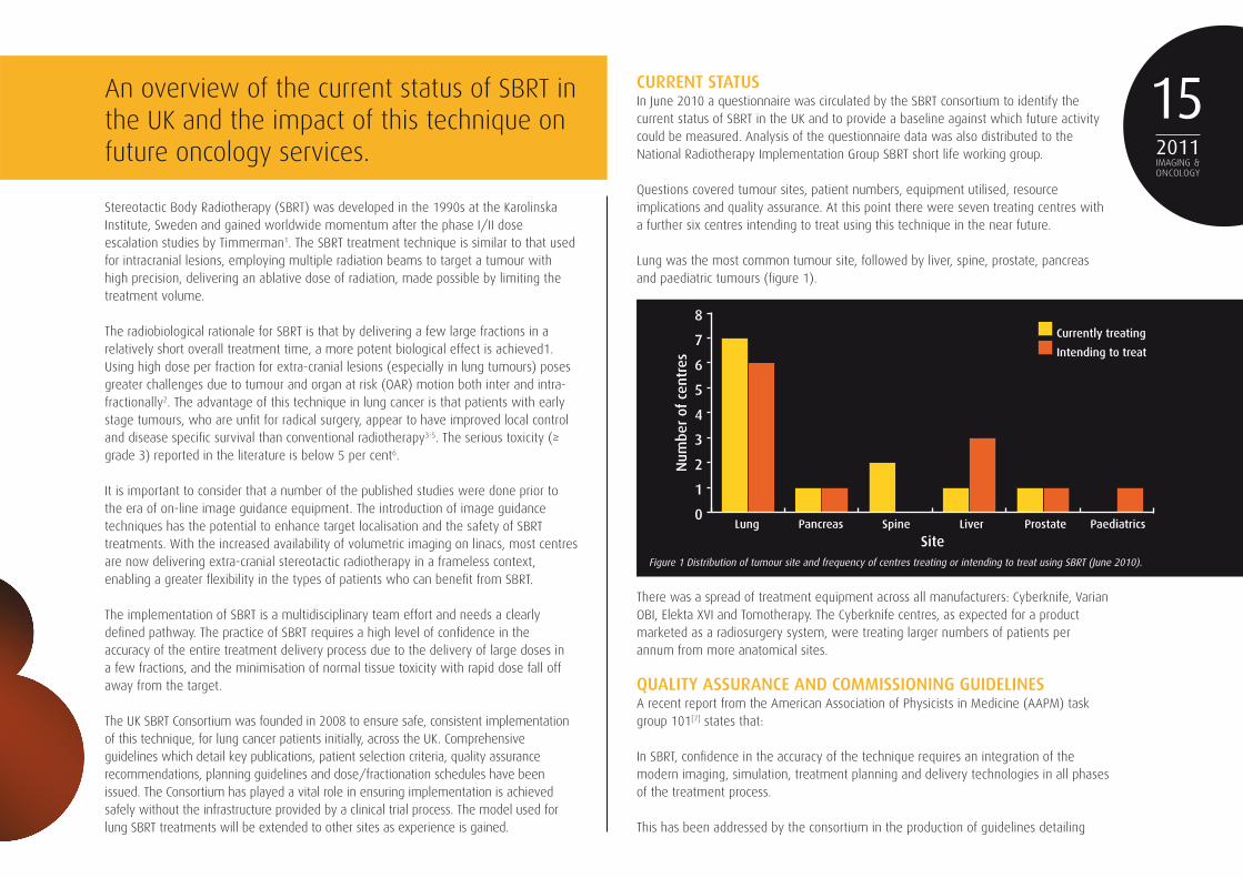
CURRENT STATUSIn June 2010 a questionnaire was circulated by the SBRT consortium to identify the current status of SBRT in the UK and to provide a baseline against which future activity could be measured. Analysis of the questionnaire data was also distributed to the National Radiotherapy Implementation Group SBRT short life working group.
Questions covered tumour sites, patient numbers, equipment utilised, resource implications and quality assurance. At this point there were seven treating centres with a further six centres intending to treat using this technique in the near future.
Lung was the most common tumour site, followed by liver, spine, prostate, pancreas and paediatric tumours (fi gure 1).
There was a spread of treatment equipment across all manufacturers: Cyberknife, Varian OBI, Elekta XVI and Tomotherapy. The Cyberknife centres, as expected for a product marketed as a radiosurgery system, were treating larger numbers of patients per annum from more anatomical sites.
QUALITY ASSURANCE AND COMMISSIONING GUIDELINESA recent report from the American Association of Physicists in Medicine (AAPM) task group 101[7] states that:
In SBRT, confi dence in the accuracy of the technique requires an integration of the modern imaging, simulation, treatment planning and delivery technologies in all phases of the treatment process.
This has been addressed by the consortium in the production of guidelines detailing
0
1
2
3
4
5
6
7
8
Num
ber
of c
entr
es
SiteLung Pancreas Spine Liver Prostate Paediatrics
Currently treating
Intending to treat
Figure 1 Distribution of tumour site and frequency of centres treating or intending to treat using SBRT (June 2010).

2011IMAGING &ONCOLOGY
16 each of these areas.The Quality Assurance (QA) subgroup has written recommendations on the commissioning of SBRT which details:• Dosimetry requirements• Immobilisation methods • Assessment of tumour motion methods• Treatment planning techniques • Types of algorithms which should be used• Linac QA• Image guidance • Plan delivery techniques
All of these areas are also covered in the AAPM task report7.
The lack of funding for a nationwide QA programme is a signifi cant issue and the consortium is currently exploring avenues where a QA programme could be established, outside a trial setting.
It is recommended that each centre should measure the systematic and random errors relating to their own systems of immobilisation and image guidance before using any new technique8. This should be undertaken before any new tumour site is treated using SBRT.
Traditionally, lung SBRT was delivered using fi xed frames or vacuum immobilisation devices with diaphragmatic pressure to reduce breathing motion4. With the introduction of four-dimensional computed tomography planning and 3D volumetric image guidance on linear accelerators, it is now possible to deliver SBRT without rigid frames with greater accuracy9.
Locally we have evaluated our standard lung immobilisation equipment using cone beam CT data from our Varian OBI linacs and have quantifi ed the pre and post treatment uncertainties. The post treatment systematic ∑ and random σ errors were less than 2mm in all directions (table 1) representing the residual displacement error, plus the intrafraction patient movement. These are the important values as we use an on-line imaging protocol with an action level of 2mm, and support the GTV to PTV (gross tumour volume to planning treatment volume) margins of 5mm. This gives confi dence in the immobilisation equipment and the treatment accuracy.
Similar results have been shown by a number of UK centres using standard immobilisation; and they are equivalent to values reported by centres using vacuum bag or frame devices.
PHYSICS (PLANNING) ISSUESAlthough the fi eld sizes used are smaller than for conventional lung radiotherapy, they are not generally small enough to prove a challenge for most modern treatment planning systems (unlike SRS in the cranial setting). However, the combination of relatively small
Pre-treatment CBCT (mm) Post-treatment CBCT (mm)
Set-up error VERT LONG LAT VERT LONG LAT
Mean -0.2 -0.9 -0.4 0.8 0.2 0.5
SD 2.7 3 3 1.4 1.1 1.4
∑ setup 1.8 4.1 3.3 0.9 0.6 0.9
σ setup 3.0 3.3 3.2 1.7 1.3 1.8
Table 1 Pre- and post-treatment systematic and random error margins (in mm)
0
5
10
15
20
25
Hou
rs
Centre1 2 3 4 5 6 7
1# treat
verification
planning
outlining
Scanner
Schedule In-room time (min)
No. of hospital visits
Total time (min)
SBRT (5 x 11 Gy) 24 5 120
SBRT (3 x 18 Gy) 24 3 72
Standard (20 x 2.75 Gy) 12 20 240
Figure 2 Time taken at different UK centres for stages in the process for delivering SBRT.
Table 2 Summary of in-room time and hospital visits for SBRT treatment and standard treatment.

2011IMAGING &ONCOLOGY
17currently being treated. Quality of life data are being collected by nurse specialists. There will be additional follow up clinic visits and appropriate diagnostic imaging dependent on the treatment site where this technique is used.
The UK Consortium means to facilitate consistent data collection across the country and aims to establish a national database of SBRT toxicities and outcomes.
QUALITY OF LIFEReduced treatment visits as a consequence of SBRT may impact positively on the daily life of individual patients. However, the treatment planning and delivery process is prolonged. It is important to manage patient and carer expectations, stressing that they have early disease and the necessary time and care is essential to deliver the best possible treatment.
SBRT appears to be associated with minimal acute and late toxicity. Fatigue is one of the most commonly reported symptoms. The relatively good tolerability of SBRT may result in improved quality of life outcomes which should be formally evaluated.
WORKFORCE DEVELOPMENTDue to the high technical demands of SBRT, the American Society for Radiation Oncology and the American College of Radiology have jointly published practice guidelines that detail recommendations for staffi ng levels and staff responsibilities for this technique12.
Currently at CCO, the SBRT clinician will be present at each treatment fraction to approve the soft tissue match on the CBCT and any necessary isocentre corrections. However, we are working towards the SBRT treatment being a radiographer/physicist led Image Guided Radiotherapy process. The SBRT clinician would be available at the verifi cation (day 0) appointment and then would hand over to the SBRT radiographer/physicist team for subsequent treatments. This team would provide expert knowledge of the treatment plan (physicist) and the image guidance method (radiographer) and could therefore make the appropriate decision for required isocentre corrections.
This is a logical progression following the recent increase in use of soft tissue registration as routine practice in the UK, especially for lung cancer patients. The experience gained for routine conformal treatments will lead to a safe delegation of responsibility to competent radiographers. We are working with academic institutions to produce Masters level modules in advanced imaging techniques, which will incorporate a competency assessment for SBRT treatments image evaluation.
The next step would be the establishment of consultant radiographer posts specialising in SBRT with the technical expertise to take responsibility for soft-tissue match during treatment and to co-ordinate the SBRT radiographer team of advanced imaging practitioners required for this technique. They would have their own workload of SBRT patients to provide on-treatment review and follow-up clinics and would assist in the
fi elds (~4-6cm) and low density lung does introduce uncertainties in the dose distribution. Therefore using a type B algorithm (eg Anisotropic Analytical Algorithm, or collapsed cone), which accurately assesses lack of lateral scatter, is strongly recommended.
To keep OAR doses as low as possible, many beams are utilised (nine or more) and it may be advantageous to use a non-coplanar beam arrangement. The use of dynamic arc techniques will produce a more conformal dose distribution, whilst treating a larger volume of the lung to a low dose bath10. Before implementing such a technique, consideration must be given to the interplay effects between the moving target and the moving leaves11.
RESOURCE IMPLICATIONSAside from the tumour, a number of key organs at risk need to be outlined, so that intended radiation dose to the latter can also be accurately ascertained. If available, help from radiology colleagues is invaluable but this increases signifi cantly the outlining time compared to standard treatments. The treatment planning must consider the multiple OARs and the effect of reduced lateral scatter in the lung surrounding a small tumour. As such, it is relatively complex. At Clatterbridge Centre for Oncology (CCO) it is performed by physicists only.
Figure 2 shows a breakdown of the components of the process from seven UK SBRT centres.
In-room timings showed this SBRT technique to be an effi cient use of linac time. It also gives the additional advantage of a reduced number of hospital visits for the patient. This is shown in table 2 using data from Clatterbridge Centre for Oncology.
As experience increases, there is a decrease in the time for each step in the process, especially the planning stage, and this is similar to the reductions seen when intensity modulated radiotherapy (IMRT) planning techniques were fi rst implemented. It is expected that when any new site is treated with SBRT, these values may increase during the initial implementation phase.
In-room times are reduced with the use of automatic couch corrections from the treatment consoles. An increased use of rotational techniques such a Volumetric Modulated Arc Therapy and RapidArc will also reduce the treatment time - current nine static fi eld technique 12 min beam on time, if two arcs are used, this could be reduced to two minutes.
DATA COLLECTION When introducing a new technique to your centre, even if the use of the technique has been proven in the literature, we appreciate that it is good practice to collect toxicity and outcome data. This data collection needs to be prospective rather than retrospective. However, the NHS does not provide the funding to support this.
This has been addressed locally by involving the clinical effectiveness team to produce a database of treatment toxicities but this is based on the small number of SBRT patients

2011IMAGING &ONCOLOGY
18
development of national SBRT guidelines, training and research projects.
SERVICE DEVELOPMENTOne of the barriers to implementation of SBRT is the lack of an appropriate tariff to recognise the extra planning effort required for this technique. How do we convince commissioners to ensure correct payment? Tariffs are based usually on number of treatment fractions rather than complexity of the planning and treatment process. The use of image guidance, for example, is not currently recognised within the tariff system. This issue has also been highlighted in the UK provision of inverse planned IMRT treatments13. Mayles discusses the need for fi nancial recognition of the additional planning effort required for IMRT, an argument equally applicable to SBRT treatments. Unless a compensating increase in payment for the treatment preparation and data collection methods is implemented, it is unlikely that the desired level of SBRT provision will be achieved in the UK.
At a recent ‘Britain Against Cancer’ conference, the Health Secretary, Andrew Lansley, outlined plans to develop a range of tariffs to reward high-quality, cost-effective services14. These may help to encourage innovation and the early adoption of new techniques, such as SBRT.
The NRIG short term SBRT working party report (due early 2011) will provide guidance for commissioners, providers and clinicians for the provision of SBRT for all anatomical sites. It is anticipated the report will assist in solving this problem and enable more centres to offer this technique.
SBRT RESEARCH IN THE UKThere is a vast number of published papers on SBRT, however there is a lack of level 1 evidence, with a striking absence of multicentre randomised controlled trials. Most of the literature on SBRT consists of cohort studies, phase I and a few phase II studies [15]. Radiobiology predicts that a high biological equivalent dose (≥100Gy) is needed for good local control (>80%)16. Published outcomes from SBRT also support this, with dose/fractionation regimen delivering less than 100Gy being associated with poor local control17. This observation makes it diffi cult to randomise patients between the standard UK regimen of 55Gy/20 fraction (BED of 70Gy) against SBRT dose fractionation with a biologically equivalent dose higher than 100Gy. A couple of international phase III trials began in 2008, but are slow to recruit.
SPECIALISED TEAMS MUST BEESTABLISHED WITH CREATION OFCONSULTANT RADIOGRAPHER ROLES
This does not mean that there is no place for research in SBRT. On the contrary, there are many unknowns. Even though local control rates are equivalent to surgery, this does not translate into a survival advantage. This phenomenon may be explained by the fact that patients selected for SBRT (over surgery) often have signifi cant comorbidities, which may also affect their overall survival. Relapse patterns following SBRT differ signifi cantly from conventional radiotherapy; with delivery of ablative doses, patients appear to relapse at distant sites with further treatment options limited, given that patients are usually medically inoperable. Both these factors could be responsible for the lack of survival advantage despite good local control.
Due to these factors, it is crucial that research in SBRT moves away from the medically inoperable. Certainly there are trials in Europe and America comparing SBRT to surgery, but due to the vastly differing trial arms they are slow to recruit18. The UK Consortium is also planning a trial of SBRT against surgery. A multicentre UK trial will help budding UK centres to start SBRT. In addition, the quality assurance associated with the trial will ensure best practice across centres delivering SBRT.
FUTURE IMPACTThere is an increased body of evidence for the use of SBRT for oligometastases, liver, paraspinal, pancreas, prostate and kidney tumours2. Some of these sites are already being treated with SBRT in the UK. Each will have different issues to consider in terms of immobilisation, planning technique and image guidance. Further training may be required to evaluate the image guided images as the new sites may not be currently treated using radiotherapy and soft tissue matching.
As all new linacs are recommended to be capable of delivering image guided 4D adaptive radiotherapy19, this gives the potential for an increased number of SBRT treatments.
The number of stereotactic systems available in the UK with the facility for tumour tracking is expanding. The Cyberknife system provides continuous image guidance, target tracking and real time corrections. The Novalis Tx system provides marker based tumour tracking with gating techniques.
Answers for life.
Welcome to syngo.plaza. Reading, any dimension.
syngo®.plaza is the first Siemens PACS to offer 2D, 3D and 4D reading in one place. Let it boost your reading speed in combination with syngo.via. See yourimages open automatically in the applications that fit your case. Enjoy your preferred working environment thanks to easy adaption of tools and layouts.Experience flexibility in hardware and storage, which makes syngo.plaza your solution - today and tomorrow. www.siemens.com/ReadingAnyDimension
Can I have my imagesautomatically openedin 2D, 3D and 4D?
Depending on thecase complexity?

2011IMAGING &ONCOLOGY
20 This ability to accurately track tumours during respiratory motion solves one of the main challenges of utilising SBRT for lung and abdominal tumours.
New equipment currently being developed provides real time imaging, together with increased fl exibility for beam arrangement. This is called the Vero system, which has a gimbals-based mechanism designed to anticipate tumour mobility during treatment.
There is an obvious need for specialisation of staff due to the complex nature of the technique. Each department should establish a stereotactic team, dedicated to stereotactic treatments and associated issues. This would include clinicians, physicists and radiographers and may be appropriate for both extra-cranial and cranial sterotactic treatments. There may also be a requirement for a SRT dedicated linac in each department. This is already happening in the UK with Cyberknife equipment at a number of centres.
A current informal referral system enables patients to have SBRT treatment if thought to be benefi cial (even if not offered in their local centre). This might usefully be developed into a centralised national referral system to SBRT centres across the UK, considering the specialisation in different anatomical sites of each hospital.
CONCLUSIONTeamwork, national integration and collaboration are fundamental for the implementation of the complex, continuously evolving technique of SBRT.
The provision of SBRT must be a patient focused service with a clear vision to offer all patients the most appropriate treatment for them. This should be equitable across the UK.
SBRT for small NSCLC (non-small cell lung cancer) tumours is increasing local control rate and signifi cantly improving the patient’s quality of life. It seems likely that similar advantages are possible for other tumour sites and therefore we expect a growth in hypofractionated techniques in the UK.
Arguably, if the UK radiotherapy workforce is to be able to meet this challenge, two areas require urgent development: Firstly, additional funding must be provided to support implementation of new technologies, both in terms of quality assurance and data collection. Secondly, specialised teams must be established with the creation of consultant radiographer roles specifi cally in this area.
REFERENCES1. Timmerman R, An overview of
hypofractionation and introduction to this issue of seminars in radiation oncology. Semin Radiat Oncol. 2008; 18, 4, 215-222.
2. Martin A, Gaya A. Stereotactic body radiotherapy: a review. Clinical Oncology. 2010 (22) 157-172.
3. Lagerwaard F, Haasbeek C, Smit E, et al., Outcomes of risk-adapted fractionated stereotactic radiotherapy for stage I non-small-cell lung cancer. IJROBP, 2008. 70(3): 685-92.
4. Nagata Y, Takayama K, Matsuo Y, et al., Clinical outcomes of a phase I/II study of 48 Gy of stereotactic body radiotherapy in 4 fractions for primary lung cancer using a stereotactic body frame. IJROBP, 2005. 63(5): 1427.
5. Timmerman R, Papiez L, McGarry R, et al., Extracranial Stereotactic Radioablation: Results of a Phase I Study in Medically Inoperable Stage I Non-small Cell Lung Cancer. Chest, 2003. 124(5):1946.
6. Haasbeek C, Senan S, Smit E, et al., Critical review of nonsurgical treatment options for stage I non-small cell lung cancer. Oncologist, 2008. 13(3): 309-19.
7. Benedict et al. Stereotactic body radiotherapy: the report of TG101. Medical Physics, 2010, 37, 8, 4078-4101.
8. The Royal College of Radiologists, Society and College of Radiographers, Institute of Physics and Engineering in Medicine. On target: ensuring geometric accuracy in radiotherapy. 2008, London: RCR.
9. Sonke J, Rossi M, Wolthaus J, et al., Stereotactic body radiotherapy for lung cancer using 4-D conebeam CT guidance. Int J Radiat Oncol Biol Phys. 2009;74(2):567-74.
10. Verbakel W, Senan S, Cuijpers J P, Slotman B, Lagerwaard F. Rapid delivery of stereotactic radiotherapy for peripheral lung tumors using volumetric intensity-
Angela Baker is the lead research and SBRT radiographer. Lynda Appleton is a research nurse. Dr Alison Scott is the lead stereotactic physicist. Dr Pooja Jain is a consultant oncologist with special interest in technical radiotherapy for lung cancer. All practice at the Clatterbridge Centre for Oncology. CCO was one of the fi rst UK centres to implement SBRT in lung patients and was a founding member of the UK SBRT consortium.
AcknowledgementsWe would like to thank the UK SBRT consortium members for their input to this article.
modulated arcs. Radiother Oncol. 2009 ;93(1),122-4. Epub 2009 Jun 22.
11. Ong C, Verbakel W, Cuijpers J, Slotman B, Senan S. Dosimetric impact of interplay effect on RapidArc lung stereotactic treatment delivery. Int J Radiat Oncol Biol Phys. 2011; 79(1):305-11. Epub 2010 Jul 12.
12. ASTRO and ACR Practice guideline for the performance of stereotactic body radiotherapy. Int J Radiat Oncol Biol Phys. 2010; 76(2): 326-332.
13. Mayles W. Survey of the availability and use of advanced radiotherapy technology in the UK. Clinical Oncology. 2010; 22, 636-642.
14. Anon. Health secretary sets out plans for speedier cancer diagnosis. Synergy News. January, 2011, 3.
15. Ball D, Kron T. A phase III randomised trial of hypofractionated stereotactic radiotherapy vs chemoradiation for inoperable stage I non-small cell lung cancer. TROG 2008 Meeting.
16. Martel M, Ten Haken R, Hazuka M et al. Estimation of tumor control probability model parameters from 3-D dose distributions of non-small cell lung cancer patients. Lung Cancer.1999. 24:31-37.
17. Guckenberger M, Wulf J, Mueller G, Krieger T, et al. Dose response relationship for image guided stereotactic body radiotherapy of pulmonary tumours: relevance of 4D dose calculation. Int J Radiat Oncol Biol Phys. 2009 May 1;74(1):47-54.
18. A randomized phase III trial of radiosurgery or surgery for operable early stage (stage 1A) non-small cell lung cancer. ROSEL, ClinicalTrials.gov ID = NCT00687986.
19. National Radiotherapy Advisory Group Report. Radiotherapy: developing a world class service for England. London: Department of Health, 2007.
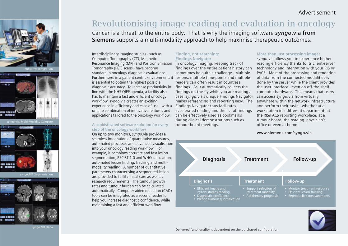
Revolutionising image reading and evaluation in oncologyCancer is a threat to the entire body. That is why the imaging software syngo.via fromSiemens supports a multi-modality approach to help maximise therapeutic outcomes.
Advertisement
Innovative integration
syngo.via, Multi-Modality Compare
syngo.PET Segmentation
syngo.MR Onco
Finding, not searching: Findings NavigatorIn oncology imaging, keeping track offindings over the entire patient history cansometimes be quite a challenge. Multiplelesions, multiple time-points and multiplereaders can often result in countlessfindings. As it automatically collects thefindings on-the-fly while you are reading acase, syngo.via’s unique Findings Navigatormakes referencing and reporting easy. TheFindings Navigator thus facilitatesaccelerated reading and the list of findingscan be effectively used as bookmarksduring clinical demonstrations such astumour board meetings.
Delivered functionality is dependent on the purchased configuration
More than just processing images syngo.via allows you to experience higherreading efficiency thanks to its client-servertechnology and integration with your RIS orPACS. Most of the processing and renderingof data from the connected modalities isdone by the server while the client providesthe user interface - even on off-the-shelfcomputer hardware. This means that userscan access syngo.via from virtuallyanywhere within the network infrastructureand perform their tasks - whether at aworkstation in a different department, atthe RIS/PACS reporting workplace, at atumour board, the reading physician’soffice or even at home.
www.siemens.com/syngo.via
Diagnosis
Interdisciplinary imaging studies - such asComputed Tomography (CT), MagneticResonance Imaging (MRI) and Positron EmissionTomography (PET) scans - have becomestandard in oncology diagnostic evaluations.Furthermore, in a patient centric environment, itis essential to obtain the highest possiblediagnostic accuracy. To increase productivity inline with the NHS QIPP agenda, a facility alsohas to maintain a fast and efficient oncologyworkflow. syngo.via creates an excitingexperience in efficiency and ease of use - with aunique combination of innovative features andapplications tailored to the oncology workflow.
A sophisticated software solution for everystep of the oncology workflowOn up to two monitors, syngo.via provides aseamless integration of quantitative measures,automated processes and advanced visualisationinto your oncology reading workflow. Forexample, it combines accurate and fast lesionsegmentation, RECIST 1.0 and WHO calculation,automated lesion finding, tracking and multi-modality reading. A number of quantitativeparameters characterising a segmented lesionare provided to fulfil clinical care as well asresearch requirements. The tumour growthrates and tumour burden can be calculatedautomatically. Computer-aided detection (CAD)tools can be integrated as a second reader tohelp you increase diagnostic confidence, whilemaintaining a fast and efficient workflow.
Follow-upTreatment
Diagnosis Treatment Follow-up
• Efficient image and • Support selection of • Monitor treatment responsehybrid studies reading treatment modality • Efficient lesion tracking
• Diagnostic confidence • Aid therapy prognosis • Reproducible measurements• Precise tumour quantification
• • •

CYBERKNIFE TECHNOLOGY AND ITS FUTURE CONTRIBUTION
TO ONCOLOGY
ALEXANDRA AITKEN,NIHAL SHAH, PETE OSTLER AND PETER HOSKIN

2011IMAGING &ONCOLOGY
23CyberKnife has been hailed by the media as the ‘must have’ radiotherapy technology. But what are its true capabilities?
INTRODUCTIONIntracranial stereotactic ‘radiosurgery‘ was developed in 1951 by a Swedish neurosurgeon, Lars Leksell1. The term ‘radiosurgery’ was initially defi ned as the delivery of an extremely high radiation dose to an often critically located small intracranial lesion during a single session. In order to minimise dose to normal tissues, multiple non-coplanar beams entering the patient at different locations were used to deliver the high target dose.
Since 1951, radiosurgery has undergone signifi cant technical and clinical developments and, in more recent years, stereotactic radiotherapy has been investigated, developed and extended to extra cranial treatment sites with the intent to deliver a very high dose over a small number of fractions in order to have an ablative effect on the tumour2. When delivered over several sessions, it is commonly referred to as fractionated stereotactic radiotherapy (SRT) and is currently used to treat both benign and malignant tumours.
Most stereotactic platforms available currently consist of a dedicated stereotactic delivery system and an image guidance system - an essential requirement for SRT delivery. Existing radiosurgery systems include CyberKnife, Gammaknife and Tomotherapy. In addition, several linear accelerators are now equipped with stereotactic beam collimators and head frames.
One such system is the Novalis Tx radiosurgery platform. This system consists of a Varian Trilogy linear accelerator (Varian Medial Systems, Palo Alto, California, USA) with a micro multi leaf collimator in conjunction with the ExacTrac (Brainlab, Munich, Germany) image guidance system, offering real time imaging and corrections in six degrees of freedom. The Elekta Axesse (Elekta, Stockholm, Sweden) also offers a similar integrated stereotactic system.
The Gammaknife (Elekta, Stockholm, Sweden) is a well established radiosurgery system
for intra-cranial work requiring a fi xed head frame for treatment delivery purposes, whilst the Tomotherapy Hi-Art system (Tomotherapy, Madison, USA) utilises a ring gantry and delivers helical intensity modulated radiotherapy (IMRT) by means of thousands of small beamlets. This system utilises on-board image guidance with megavoltage computed tomography. The CyberKnife system (Accuray, Sunnyvale, California, USA) is an image guided robotic radiosurgery system featuring a robotic couch with six degrees of freedom and continual real-time kV image guidance (see fi gure 1). The robotic arm on which the linear accelerator is mounted enables the delivery of non-isocentric and non coplanar treatment beams with a high degree of precision. It is suitable for lesions anywhere in the body including structures that move with respiration.
CYBERKNIFEThe CyberKnife system has been under technical development for almost 20 years, whilst the basic concept has remained unchanged. However, signifi cant improvements and additions were implemented more recently. CyberKnife is routinely used to treat brain,3,4 head and neck,5,6 spine,7,8 lung,9,10 liver,11 pancreas and prostate12,13 tumours, in addition to nodal or other tumour recurrences.
The treatment delivery system for the CyberKnife includes an X-band cavity magnetron, a standing wave and side-coupled waveguide, which produces 6MV x-ray beams at a dose rate of 1000cGy/min14. Bending magnets and beam fl attening fi lters are not required and secondary collimation is provided by either fi xed circular collimators ranging in size from 0.5mm-60mm, or a variable aperture collimator, which enables the same selection of collimator sizes without a physical change of collimator. Small collimator fi elds can be complex to calibrate due to steep dose gradients and electronic disequilibrium.
The robotic arm or manipulator, (Kuka Roboter, Augsburg, Germany) on which the linear accelerator is mounted, has the ability to move in six degrees of freedom and moves around the patient with a high degree of precision providing sub-millimetre position reproducibility. The increased geometric fl exibility does, however, require more extensive primary barriers than those needed for conventional gantry mounted linear accelerators14.
Treatment planningAs with all current radiotherapy systems, a 3D CT dataset is required for planning purposes, from which a 3D patient model is generated. Treatment beams are defi ned by
IT MAY BECOME THE TREATMENT OFCHOICE FOR PROSTATE CANCER

2011IMAGING &ONCOLOGY
24
ROBOTIC DELIVERY SYSTEM
X-RAY SOURCES
IMAGE DETECTORS
Figure 1. CyberKnife robotic delivery system

a vector linking a direction point, found usually within the target volume, and a source point, which correlates with the position of the linear accelerator’s focal point. Each source point forms a ‘node’ with a set of ‘nodes’ forming a path set. Path sets provide non-coplanar beam directions and can be achieved without repositioning the patient.
Treatment deliveryPrior to and during treatment delivery, the digitally reconstructed radiographs (DRRs) generated from the 3D CT are automatically registered to the live images acquired using the X-ray imaging system, which determines the beam alignment. Internal radiographic reference points based on bony anatomy or implanted fi ducial markers, in or around the tumour, are required for target localisation.
Before each fraction is delivered, the patient can be positioned using the adjustable treatment couch; this reduces the corrections required from the robotic manipulator. Any additional translational or rotational adjustments during treatment, identifi ed from the real-time imaging, are relayed to the robotic manipulator and corrected, ie the alignment adjustments required are applied by correcting the beam position and orientation relative to the patient.
The unique tracking system featured on CyberKnife enables compensation for all small translations and rotations obtained from the latest image acquired. With the real time image guidance system and alignment correction, a very high dose conformality and steep dose gradient can be achieved. The ability to deliver non-isocentric and non-coplanar beams without repositioning the patient provides additional benefi t.
Respiratory motion trackingA respiratory tracking system (Synchrony, Accuray, Sunnyvale, California, USA) is used for treatment delivery to tumours that move with respiration. The beam moves during treatment ensuring continuous alignment of the beam with the moving target volume. The concept of this system is based on a correlation model between tumour position and external marker position. Prior to treatment delivery, the tumour position is determined by acquiring x-ray images at multiple time points and a correlation model is generated by relating the tumour position at different phases of the respiratory cycle to the simultaneous external marker position.
Throughout treatment delivery the position of the tumour is determined by the position of the external markers using the correlation model. Optical markers are used to provide the external signal. Separate correlation models are built for each marker with each model providing an estimate of target position. An average of all three estimates is then calculated resulting in a fi nal position estimate. The model is continually updated throughout treatment, adapting to any changes in target position or motion accordingly. There is however no fi xed or constant relationship between external contour and tumour position for extra cranial sites between fractions and
during treatment delivery. Therefore the correlation model is regularly adjusted using the x-ray imaging data14.
Fiducial InsertionFiducial markers are required for most extra cranial lesions when using the CyberKnife system. This enables the system to track the tumour in six degrees of freedom (ie all translations and rotations) throughout treatment delivery. A minimum of three fi ducials are required for the system to accurately track the lesion, however it is recommended that four to six fi ducials are inserted due to the uncertainties in localisation of the individual fi ducials15.
Fiducials should be 0.7 to 1.2mm in diameter and 3mm to 6mm in length. Several requirements need to be met when undertaking fi ducial insertion. These include a minimum distance of 2.0cm between fi ducials, a 15 degree angle between three fi ducials, non-collinear placement and maximum distance of 5-6cm from the lesion. If these requirements are not met, some fi ducials may not be suitable for tracking purposes and consequently tracking accuracy may be compromised. An additional tracking algorithm does enable lung tumours to be tracked without markers, provided the tumour diameter is greater than 15mm in all directions and positioned in the peripheral region of the lung.
The fi ducial placement procedure is very similar to a CT guided biopsy with comparable complications and challenges16. Potential complications include the risk of pneumothorax, haemoptysis, haemorrhage and perforation of bowel, depending on the location and position of the tumour.
2011IMAGING &ONCOLOGY
25A HIGH DEGREEOF PRECISION ISACHIEVED THROUGHSUB-MILLIMETREPOSITIONING
2011IMAGING &ONCOLOGY
26 INTRA-CRANIAL STEREOTACTIC RADIOTHERAPYRadiosurgery is an established form of treatment for both benign and malignant intra-cranial lesions and has shown good results with minimal side-effects. Surgical resection remains the gold standard for treatment and aggressive resection offers the best results. However, in many cases surgery may be contraindicated. Radiosurgery has been used as both a primary and adjuvant treatment modality following surgical excision. Whole brain radiation therapy and/or chemotherapy are used in many cases following surgical resection. As whole brain radiotherapy and chemotherapy have signifi cant side-effects, stereotactic radiosurgery may be used as an alternative adjuvant treatment. Moreover, for inoperable patients, stereotactic radiosurgery is the only available ablative procedure targeted directly at the tumour, whilst sparing surrounding healthy tissue. Phase III randomised clinical trials comparing stereotactic radiotherapy alone with stereotactic radiotherapy, plus whole brain radiotherapy, had comparable overall survival rates17. However, local relapse was higher in patients treated with stereotactic radiotherapy alone. Neurologic preservation was similar between both groups.
EXTRA-CRANIAL STEREOTACTIC RADIOTHERAPYInvasive immobilisation frames are used with many stereotactic systems. The CyberKnife system however, does not require the use of any invasive frames due to its unique ability to track throughout treatment. Similarly, alternative methods such as abdominal compression, are not required to assist in minimising motion.
LUNGStereotactic radiotherapy is not the primary treatment option for lung cancer and surgery remains the gold standard for early stage, non-small cell lung cancer NSCLC, providing excellent local control and survival outcomes2. However, radiotherapy is an alternative for patients who are poor surgical candidates, or who are inoperable due to the stage or the location of the tumour. Conventional radiotherapy has proved to be a poor alternative. Dose escalation is limited when using conventional external beam radiotherapy, mainly because large margins are applied routinely, which result in large treated volumes and increased side effects. Hence, results with external beam radiotherapy have been disappointing with long-term survival rates of just 15-30 per cent, and local failure in excess of 50 per cent in stage I NSCLC18,19. Complication rates are also high 20.
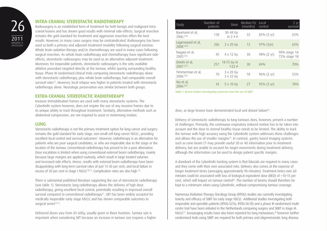
There is substantial published literature supporting the use of stereotactic radiotherapy (see table 1). Stereotactic lung radiotherapy allows the delivery of high dose radiotherapy, giving excellent local control, potentially resulting in improved overall survival compared to conventional radiotherapy21. SRT has been widely accepted for medically inoperable early stage NSCLC and has shown comparable outcomes to surgical series22,23.
Delivered doses vary from 45-60Gy, usually given in three fractions. Tumour size is important when considering SRT because an increase in tumour size requires a higher
Study Number of patients Dose Median FU
(months)Local control
2 yr survival
Baumann et al, 2006 [33] 138
30-48 Gy in 2-4 #
33 85% (3 yr) 65%
Lagerwaard et al, 2008 [34] 206 3 x 20 Gy 12 97% (3yr) 65%
Nagata et al, 2005 [35] 45 4 x 12 Gy 30 98% (2 yr)
90% stage 1A72% stage 1B
Onishi et al,2007 [21] 257
18-75 Gy in 1-22 #
38 84% 70%
Timmerman et al,2006 [36] 70
3 x 20 Gy3 x 22 Gy
18 96% (2 yr) 55%
Xia et al,2006 [37] 43 5 x 10 Gy 27 95% (3 yr) 78%
Table 1: Recent studies investigating outcomes from the use of SBRT
dose, as large lesions have demonstrated local and distant failure24.
Delivery of stereotactic radiotherapy to lung tumours does, however, present a number of challenges. Primarily, the continuous respiratory induced motion has to be taken into account and the dose to normal healthy tissue needs to be limited. The ability to track the tumour with high accuracy using the CyberKnife system addresses these challenges and allows the use of smaller margins25. In contrast, gantry based imaging systems such as cone-beam CT may provide useful 3D or 4D information prior to treatment delivery, but are unable to account for target movements during treatment delivery, although the information can be used to design patient specifi c margins.
A drawback of the CyberKnife tracking system is that fi ducials are required in many cases and they come with their own associated risks. Delivery also comes at the expense of longer treatment times (averaging approximately 90 minutes). Treatment times over 60 minutes could be associated with loss of biological equivalent dose (BED) of >10-15 per cent, which will impact on tumour control26. The number of beams should therefore be kept to a minimum when using CyberKnife, without compromising tumour coverage.
Numerous Radiation Therapy Oncology Group (RTOG) studies are currently investigating toxicity and effi cacy of SBRT for early stage NSCLC. Additional studies investigating both inoperable and operable patients (RTOG 0236, RTOG 0618) and a phase III randomised multi-centre trial have been initiated in the Netherlands comparing surgery and SBRT in stage IA NSCLC27. Encouraging results have also been reported for lung metastases,28 however further randomised trials using SBRT are required for both primary and oligometastatic lung disease.

2011IMAGING &ONCOLOGY
27Several trials using radiofrequency ablation (RFA) have reported clinical effectiveness in early stage inoperable NSCLC. As larger tumours or centrally located tumours are more likely to recur following either RFA or SRT, further studies are currently underway investigating the use of combined RFA and SRT to improve local control.
LIVERSurgery is the treatment of choice for both primary liver tumours and limited liver metastases. However, for patients who are not suitable for surgery due to the extent of disease or medical condition, alternative strategies have been investigated. Conventional radiotherapy techniques can offer only palliation as the radiosensitivity of normal liver tissue limits the dose that can be delivered. SRT techniques enable ablative doses to be delivered to metastatic liver lesions, but long term published data with respect to outcomes are still lacking. SRT is generally used to treat primary liver lesions if other treatment modalities are not suitable, or in the event of recurrence.
RFA is now widely used for smaller lesions (<3cm) and has shown local control rates comparable to surgery29. RFA is, however, unsuitable for tumours situated close to the diaphragm, or large vessels and, in these cases, SRT would be the treatment of choice.
PROSTATEHypofractionated radiotherapy is emerging as an alternative treatment for early stage prostate cancer, potentially offering an increase in tumour control and patient survival compared to conventional radiotherapy30. Conventional radiotherapy is an accepted treatment option for a growing number of patients diagnosed with prostate cancer who are medically inoperable. However, the effectiveness is limited by the negative effects of the radiation on surrounding normal tissue.
The low alpha/beta ratio for prostate cancer suggests high dose hypofractionated radiotherapy will result in a favourable biological response. Data from the use of high dose rate (HDR) brachytherapy in prostate cancer support this theory and have shown positive results31. However, the HDR procedure is invasive, requires anaesthetic, the use of a catheter and hospitalisation. Stereotactic radiotherapy is the optimum external beam technique to deliver large doses per fraction, minimising the risks to surrounding normal tissue, and can offer a non-invasive alternative while applying the same dosimetric and biological considerations32. Furthermore, several recent published studies have demonstrated improved effectiveness and reduced toxicity with focused, high dose radiation treatments delivered in three to four treatment sessions with compensation for tumour motion, using stereotactic radiosurgery.
While improvements in treatment delivery techniques, such as Intensity modulated radiotherapy (IMRT), Intensity modulated arc therapy (IMAT) and SRT have enabled an increased dose per fraction to be delivered without increasing toxicity to normal tissue, and IGRT technologies including CBCT offer pre-treatment image guidance,
these technologies do not address the issue of intrafractional motion. Due to the tracking ability that the CyberKnife system offers, target motion can be identifi ed and corrections applied accordingly, ensuring accurate target coverage despite recurrent and unpredictable prostate motion.
Long term follow up data are still required to confi rm effectiveness and late toxicities of stereotactic radiotherapy. If these data demonstrate that stereotactic radiotherapy is as effective as other modalities such as surgery, brachytherapy and cryotherapy, then stereotactic radiotherapy may become the treatment of choice for prostate cancer because it offers a non-invasive alternative with short treatment duration.
CONCLUSIONIn the future, we are likely to see continued technological advancements and developments further improving the radiotherapy delivery systems that have contributed to the success of stereotactic radiotherapy. Conventional radiotherapy will continue to be suitable for many indications. However, effectiveness will always be limited by the negative effects of the radiation on the volume of surrounding normal tissue. In some circumstances, stereotactic radiotherapy may prove benefi cial when used as a boost following radiotherapy.
The benefi ts offered by stereotactic radiotherapy include accuracy, potentially reduced incidence of treatment related toxicity due to its high conformity, and improved outcomes with dose escalation. It also offers a highly effi cient treatment delivery in a small number of fractions. Patient selection is important and stereotactic radiotherapy is most suitable for smaller localised tumour volumes. Patients with distant active metastatic disease may not be appropriate, unless good local control is important for palliation.
Further studies are required in both the curative and palliative settings to investigate quality of life, symptom control, disease free interval, late effects and survival. Direct comparisons with alternative methods of treatment are also necessary to clarify the relative role of SRT in the overall management of malignant disease.
In conclusion, CyberKnife is a unique stereotactic platform and published data so far are encouraging with good local control being achieved in numerous tumour sites. However, large phase III studies are required if the true potential of stereotactic radiotherapy is to be established in both a palliative and curative setting.
Alexandra Aitken is principal radiographer, and Dr Nihal Shah, Dr Pete Ostler and Professor Peter Hoskin are consultant clinical oncologists at Mount Vernon Cancer Centre, Northwood, Middlesex, United Kingdom. Mount Vernon was the fi rst National Health Service centre with CyberKnife.

2011IMAGING &ONCOLOGY
28 REFERENCES1. Leksell L. The stereotactic method and
radiosurgery of the brain. Acta Chir Scand 1951; 102:316-319.
2. Martin A, Gaya A. Stereotactic body radiotherapy: A review. J Clin Oncol; 2010; 22: 157-172.
3. Colombo F, Casentini L, Cavedon C, Scalchi P, Cora s, Francescon P. Cyberknife radiosurgery for benign meningiomas: short term results in 199 patients. Neurosurgery; 2009;64:A7-13.
4. Soltys S G, Adler J R, Lipani J D, Jackson P S, Choi C Y, Puataweepong P, White S, Gibbs I C, Chang S D. Stereotactic radiosurgery of the postoperative resection cavity for brain metastases. Int J Radiat Oncol Biol Phys; 2008; 70:187-193.
5. Roh K W, Jang J S, Kim M S, Sun D I, Kim B S, Jung S L, Kang J H, Yoo E J, Yoon S C, Jang H S, Chung S M, Kim Y S. Fractionated stereotactic radiotherapy as reirradiation for local recurrent head and neck cancer. Int J Radiat Oncol Biol Phys; 2009; 74:1348-1355.
6. Rwigema J C, Heron D E, Ferris R L, Gibson M, Quinn A, Yang Y, Ozhasoglu C, Burton S. Fractionated stereotactic body radiation therapy in the treatment of previously-irradiated recurrent head and neck carcinoma: updated report of the University of Pittsburgh experience. Am J Clin Oncol; 2010, 33, 3, 286-293).
7. Gangnon G J, Nasr N M, Liao J J, Milzahn I, Marsh D, McRae D, Henderson F C. Treatment of spinal tumours using Cyberknife fractionated stereotactic radiosurgery: pain and quality of life assessment after treatment in 200 patients. Neurosurgery; 2009; 64:297:306.
8. Gerszten P C, Burton S A, Ozhasoglu C, Welch W C. Radiosurgery for spinal metastases: clinical experience in 500 cases from a single institution. Spine; 2007; 32:193:199.
9. Collins B T, Vahdat S, Erickson K, Collins S P, Suy S, Yu X, Zhang Y, Subramaniam D, Reichner C A, Sarikaya I, Esposito G, Yousefi S, Jamis-Dow C, Banovac F, Anderson E D. Radical cyberknife radiosurgery with tumour tracking: an effective treatment for inoperable small peripheral stage I non-small cell lung cancer. J Hematol Oncol; 2009; 2:1.
10. Van der Voort van Zyp N C, Prevost J B,
Hoogeman M S, Praag J, van der Holt B, Levendag P C, van Klaveren R J, Pattynama P, Nuyttens J J. Stereotactic radiotherapy with real-time tumour tracking for non-small cell lung cancer: clinical outcome. Radiother Oncol; 2009; 91:296:300.
11. Stintzing S, Hoffmann R T, Heinemann V, Kufeld M, Muacevic A. Frameless single session robotic radiosurgery of liver metastases in colorectal cancer patients. Eur J Cancer;2010; 46:1026-1032.
12. King C R, Brooks J D, Gill H, Pawlicki T, Cotrutz C, Presti J C. Stereotactic body radiotherapy for localized prostate cancer: Interim results of a prospective phase II clinical trial. Int J Radiat Oncol Biol Phys; 2009; 73:1043-1048.
13. Katz A J, Santoro M, Ashley R, Diblasio F, Witten M. Stereotactic body radiotherapy for organ- confi ned prostate cancer. BMC Urol; 2010; 10:1.
14. Kilby W, Dooley J R, Kuduvalli G, Sayeh S, Maurer C R. The Cyberknife robotic radiosurgery system in 2010. Technology in cancer research and treatment; 2010; 9:5:433-452.
15. Murphy M J. Fiducial-based targeting accuracy for external beam radiotherapy. Med Phy; 2002; 29:334-344.
16. Kothary N, Heit J J, Louie J D, Kuo W T, Loo B W, Koong A, Chang D T, Hovespian D, Sze D Y, Hofmann L U. Saftey of percutaneous fi ducial marker implantation for image- guided radiation therapy. J Vasc Interv Radiol; 2009; 20:235-239.
17. Aoyama H, Shirato H. Stereotactic radiosurgery plus whole brain radiation therapy vs stereotactic radiosurgery alone for treatment of brain metastases. JAMA; 2006; 295: 2483-2491.
18. Jeremic B, Classen J, Bamberg M. Radiotherapy alone in technically operable medically inoperable, early stage (I/II)non-small-cell lung cancer. Int J Radiat Oncol Biol Phys; 2002; 54:119-130.
19. Qiao X, Tullgren O, Lax I, Sirzén F, Lewensohn R. The role of radiotherapy in treatment of stage I non-small cell lung cancer. Lung Cancer; 2003; 41:1-11.
20. Wagner T, Yang G. The role of chemotherapy and radiation in the
treatment of locally advanced non-small cell lung cancer (NSCLC). Curr Drug Targets; 2010; 11:1:67-73.
21. Onishi H, Shirato H, Nagata Y , Nagata Y, Hiraoka M, Fujino M, Gomi K, Niibe Y, Karasawa K, Hayakawa K, Takai Y, Kimura T, Takeda A, Ouchi A, Hareyma M, Kokubo M, Hara R, Itami J, Yamada K, Araki T. Hypofractionated stereotactic radiotherapy (HypoFXSRT) for stage I non-small cell lung cancer: updated results of 257 patients in a Japanese multi-institutional study. J Thorac Oncol; 2007; 2: S94-100.
22. Timmerman R D, Park C, Kavanagh B D. The North American experience with stereotactic body radiation therapy in non-small cell lung cancer. J Thorac Oncol; 2007; 2:s101-112.
23. Chang J Y, Roth J A. Stereotactic body radiation therapy for stage I non-small cell lung cancer. Thorac Surg Clin; 2007; 17:251-259.
24. Willner J, Baiee K, Caragiani E, Tschammler A, Flentje M. Dose, volume, and tumour control predictions in primary radiotherapy of non small cell lung cancer. Int J Radiat Oncol Biol Phys; 2002; 52:382-389.
25. Seppenwoolde Y, Berbeco R I, Nishioka S, Shirato H, Heijmen B. Accuracy of tumour motion compensation algorithm from a robotic respiratory tracking system: a simulation study. Med Phy; 2007; 34:2774:2784.
26. Kavanaugh B D, Timmerman R D. Editors. Stereotactic body radiation therapy; 2005;Philadelphia, PA:Lippincott.
27. Hurkmans C W, Cuijpers J P, Lagerwaard F J, Widder J, van de Heide U A, Schuring D, Senan S. Recommendations for implementing stereotactic radiotherapy in peripheral stage IA non-small cell lung cancer: report from the Quality Assurance Working party of the randomised phase III ROSEL study. Radiat Oncol; 2009; 4:1.
28. Brown W T, Wu X, Fayad F, Fowler J F, Amendola B E, García S, Han H;, de la Zerda A, Bossart E, Huang Z, Schwade J G. Cyberknife radiosurgery for stage I lung cancer: Results at 36 months. Clinical lung cancer; 2007; 8:8.
29. Mulier S, Ruers T, Jamart J, Michel L, Marchal G, Ni Y. Radiofrequency ablation versus
resection for resectable colorectal liver metastases: time for a randomized trial? An update. Dig Surg; 2008; 25(6):445-460.
30. Vargas C, Martinez A, Boike T, Spencer W, Goldstein N, Gustafson G, Krauss D, Gonzalez J. High-dose irradiation for prostate cancer via a high-dose-rate brachytherapy boost: Results of a Phase I to II study. Int J Radiat Oncol Biol Phys 2006;66:416–423.
31. Fowler J F. The radiobiology of prostate cancer including new aspects of fractionated radiotherapy. Acta Oncol; 2005; 44:265-276.
32. Morgia G, De Renzis C. Cyberknife in the treatment of prostate cancer: A revolutionary system. European Urology; 2009; 56:40-42.
33. Baumann P, Nyman J, Lax I, Friesland S, Hoyer M, Rehn Ericsson S, Johansson KA, Ekberg L, Morhed E, Paludan M, Wittgren L, Blomgren H, Lewensohn R. Factors important for effi cacy of stereotactic body radiotherapy of medically inoperable stage I lung cancer. A retrospective analysis of patients treated in the Nordic countries. Acta Oncol; 2006;45(7):787-95.
34. Lagerwaard F, Haasbeek C, Smit E, Slotman B, Senan S. Outcomes of risk-adapted fractionated stereotactic radiotherapy for stage I non-small-cell lung cancer. Int J Radiat Oncol Biol Phys; 2008; 1;70(3):685-92.
35. Nagata Y, Takayama K, Matsuo Y, Norihisa Y, Mizowaki T, Sakamoto T, Sakamoto M, Mitsumori M, Shibuya K, Araki N, Yano S, Hiraoka M. Clinical outcomes of a phase I/II study of 48 Gy of stereotactic body radiotherapy in 4 fractions for primary lung cancer using a stereotactic body frame. Int J Radiat Oncol Biol Phys; 2005; 63(5):1427-31.
36. Timmerman R, McGarry R, Yiannoutsos C, Papiez L, Tudor K, DeLuca J, Ewing M, Abdulrahman R, DeRosiers C, Williams M, Fletcher J. Excessive toxicity when treating central tumors in a phase II study of stereotactic body radiation therapy for medically inoperable early-stage lung cancer. J Clin Oncol; 2006; 24:4833-4839.
37. Xia T, Li H, Sun Q, Wang Y, Fan N, Yu Y, Li P, Chang JY. Promising clinical outcome of stereotactic body radiation therapy for patients with inoperable Stage I/II non-small-cell lung cancer. Int J Radiat Oncol Biol Phys; 2006; 66(1):117-25.

Varian Medical Systems UK Ltd., Crawley, UK
Phone +44 - 1 293 601 200
www.varian.com/rapidarc [email protected]
Stereotactic body radiation therapy (SBRT) is a technique
where high doses of radiation are precisely delivered
from many directions to a focused target. This results in
an ablative treatment with curative intent and spares
surrounding critical structures.
RapidArc radiotherapy technology delivers sophisticated
SBRT treatments faster than previously possible and opens
up new treatment options for your patients.
RapidArc® for SBRT. Simply Revolutionary.

ADVANCES IN THE IMAGING OF SENTINEL NODES IN
BREAST CANCER; THE ROLE OF NUCLEAR MEDICINE
JOHN THOMPSON, ANDREW TOOTELL,
JOANNE SIL, JUDITH KELLY,
RANDEEP KULSHRESTHA AND PETER HOGG

2011IMAGING &ONCOLOGY
31Assessing sentinel nodes is highly valuable in the early period of cancer, facilitating accurate staging and dictating the correct treatment pathway. What are the imaging techniques used in nuclear medicine to detect the sentinel nodes associated with breast cancer and what are the benefi ts of SPECT/CT?
INTRODUCTIONThe evaluation of the sentinel node by nuclear medicine and subsequent removal by surgical intervention has applications in several different forms of cancer (penile, breast, multiple myeloma)1. Assessing sentinel nodes is highly valuable in the early period of cancer, facilitating accurate staging and dictating the correct treatment pathway. This review will focus on the imaging techniques used in nuclear medicine to detect the sentinel nodes associated with breast cancer, detailing the benefi ts of single photon emission computed tomography/computed tomography (SPECT/CT) in aiding surgery.
CANCERStatistics show that rates of morbidity and mortality are reducing for the majority of cancers2. Early detection, raised public awareness, better screening programmes, and improved technology and treatment regimes, are all contributing factors. Breast cancer is a good example, where sentinel lymph node biopsy (SLNB) and SPECT/CT have enhanced the patient experience through an improved technique to allow reduced morbidity.
Cancer rates per annum have stabilised but detection rates can vary with the introduction of new diagnostic tools and variation in population sizes2. Survival rates have also improved in the prevalent population (those with cancer), but increased life expectancy raises the possibility of both disease recurrence and complications related to any treatment of the cancer. Therefore, consideration of the long-term effects of treatment is important to both the patient and those involved in their care. However, treatment strategies are not the same for every patient and are highly dependent on accurate staging of the cancer, where the presence or absence of secondary disease (lymph nodes local to the primary tumour or distant metastases) could determine whether the treatment was conservative or aggressive.
In the United States, the incidence of breast cancer is high, with 1 in 8 females affected. In the United Kingdom, the incidence of breast cancer is slightly lower, with 1 in 9 females affected; despite improvements in mortality rates only lung cancer is responsible for more deaths3. Although large numbers of patients survive the disease, it is clear that breast cancer still constitutes a great challenge in both diagnosis and treatment.
THE SENTINEL NODEThe lymph nodes local to, and on the drainage pathway of, the primary tumour are a common site for initial metastasis4. It had been accepted that the removal of all lymph nodes surrounding the primary tumour (the axilla in breast cancer) could be potentially curative, but it has since been discovered that spread of this nature is more akin to the advanced stages of disease and is not necessarily the case in the early disease, prompting a reconsideration of treatment strategies. Lymph nodes have been found to be disease free in 60 per cent of early breast cancer sufferers. Breast cancer treatment has seen a dramatic change, where lumpectomy can replace mastectomy, and SLNB has replaced total axillary lymph node dissection (ALND) unless lymph nodes are proven to be cancerous.
ALND was typically associated with high morbidity (painful lymphoedema, reduced arm motion and parasthesia) but SLNB may reduce these complications, thus improving the patient experience. There are also associated cost-saving benefi ts to long-term care if morbidity is reduced. Correct identifi cation and evaluation of the sentinel node is, therefore, highly valuable.
Predictable lymphatic drainage was fi rst identifi ed in a 1977 study of penile cancer5. Lymphatic drainage was said to be predictable, involving a chain of lymph nodes (Figure 1)6 where the fi rst node in this chain, the sentinel node, indicates the spread of disease. A disease-free node indicates that the lymphatic system is clear of disease, but anatomical variation of lymphatic drainage can be a complicating factor and care must be taken to accurately determine the sentinel node(s)6. On occasion, there can be more than one sentinel node, depending on the lymphatic drainage pathway.
SPECT/CT ISREDUCING
MORBIDITY ANDOPERATING TIMES

2011IMAGING &ONCOLOGY
32 Histology results achieved from SLNB have been used to plan surgery for a range of cancers, including breast, producing an optimal method for reducing morbidity in the node negative portion of the prevalent population7. Evidence of nodal metastatic spread would lead to node removal.
LOCATING THE SENTINEL NODEUltrasound can be used to locate sentinel nodes, but when used alone it is inhibited by an inability to confi dently establish whether a lymph node is sentinel (fi rst in the lymphatic chain) or not. A more suitable role for ultrasound is in guiding lymph node fi ne needle aspiration (FNA) or core biopsy, and is standard in some countries. This method has
been shown to have good correlation with histological results of surgically removed nodes8. Ultrasound does, of course, have the benefi ts of no radiation burden, low cost and good availability, helping to maintain the established role it holds in the management of nodal involvement in cancer.
A variety of techniques for localising the sentinel node in relation to breast cancer exist in current practice, using various combinations of the following methods:• ‘Blue dye’ injection intra-operatively• Nuclear medicine 99Tcm-colloid + planar imaging• Nuclear medicine 99Tcm-colloid + planar imaging + SPECT• Nuclear medicine 99Tcm-colloid + planar imaging + SPECT/CT
• Nuclear medicine 99Tcm-colloid (No images acquired)• Intra-operative gamma probe
The combination of blue dye, initially introduced for identifying the sentinel node in patients with malignant melanoma9, and a radionuclide injection have proved to be optimal for sentinel node identifi cation, showing great success in guiding surgeons in intra-operative biopsy (prior to histology), node dissection and surgical resection.
Despite the advantage of a visual stimulus provided by the blue dye, there was still no indication that the node was sentinel. Consequently, a non-imaging radionuclide technique was introduced to aid localisation and it became apparent that the two techniques were complementary10 and successful in 81-100% of cases of melanoma11.
Pre-operative sentinel node evaluation allows biopsy, followed by histology. Intra-operative evaluation allows biopsy (± excision) with histology results returned during the procedure. In both cases histology results dictate whether the node is removed.
Pre-operative localisation gives surgeons accurate numbers and locations of nodes, with the potential to perform less invasive surgery because less exploration is required. Operating time can also be reduced, which in turn may reduce complications and improve throughput of patients.
SENTINEL NODE
PRIMARY TUMOUR
Figure 1. Lymph node location for breast cancer. An anterolateral view of the right breast. Illustra-tion depicting cancer in glandular tissue and lymph nodes of the breast. Courtesy of Medical RF / Phototake http://www.phototakeusa.com/results.asp?Image=ZEME027027775-01 Accessed January 2011. Annotated by the authors.

2011IMAGING &ONCOLOGY
33IMPORTANT ADVANCES IN NUCLEAR MEDICINEDespite the ability of magnetic resonance imaging (MRI) to demonstrate physiology without radiation burden, nuclear medicine remains the modality of choice for capturing functional information. Since the introduction of 2-dimensional imaging to nuclear medicine, there has been a steady improvement in gamma camera technology due to the development of equipment and computer software. Planar imaging has been highly valuable for a wide range of pathology, although there have always been limitations such as the highly specifi c tracer uptake (no anatomical landmarks) associated with colloid imaging for sentinel nodes12. This type of diffi culty has driven the advancement in technology to improve imaging.
SPECT was introduced in the mid to late 1980s to produce cross-sectional images of radionuclide uptake, allowing improved specifi city, due in part to the ability of SPECT to differentiate between overlying structures. The popularity of SPECT led to the introduction of dual-headed gamma cameras and the technique became the standard for some investigations. Methods were developed to correct for radionuclide attenuation within the patient (eg Gd-153) to help quantify uptake and improve image quality.
SPECT/CT is the most recent notable advancement in nuclear medicine technology and is already popular for certain techniques. SPECT (emission data) is combined with CT (transmission data) on a single scanner gantry (Figure 2)13 to provide an accurately fused data set of images showing functional and anatomical data.
The CT component of SPECT/CT has three distinct roles: attenuation correction (AC), lesion localisation and diagnosis. There are two different types of CT system available to support these roles: low-resolution (most suitable for AC) and diagnostic quality with the same functionality as a stand-alone CT scanner.
ROLES OF SPECT/CT IN LYMPHOSCINTIGRAPHYAttenuation Correction (AC)Photons emanating from radioactive foci deep within the body are susceptible to absorption by the body before they can be detected by the gamma camera. Consequently, deep sited foci, such as a sentinel node, can go undetected in both planar and SPECT imaging negatively affecting the sensitivity and specifi city of the diagnostic procedure.
CT (low-dose) can generate an attenuation map that, when applied to SPECT data, can correct for photon attenuation. This technique is now commonplace for certain patients and techniques in SPECT imaging; indeed, AC has been shown to have value for obese patients undergoing lymphoscintigraphy, with improved identifi cation and a greater number of nodes being marked as sentinel.14
Lesion localisation and diagnosisWhether using low-dose CT or diagnostic quality CT, foci of radionuclide
CT SCANNER
GAMMA CAMERA 2
GAMMA CAMERA 1
Figure 2. SPECT-CT Unit: CT Studies with SPECT. CT = computed tomography; SPECT = single photon emis-sion CT. Courtesy of Siemens press pictures http://www.siemens.com/press/pool/de/pp_med/2005/sc_upload_fi le_mednm200504048_03_300dpi_1456207.jpg Accessed January 201113. Annotated by the authors.

2011IMAGING &ONCOLOGY
34 uptake can be more accurately localised when using SPECT/CT. Hybrid imaging has value when a SPECT acquisition fails to provide adequate anatomical detail. Lymphoscintigraphy is synonymous with this type of dilemma since the radionuclide uptake is highly specifi c. Precise registration of CT and SPECT data can overcome this where foci of uptake from SPECT data are superimposed upon the anatomical detail provided by CT. Acquiring CT data at a ‘diagnostic quality’ still permits a fused image; but the CT data are diagnostic in their own right and could be used to determine lesion/node position in isolation of SPECT data.
IMAGING THE SENTINEL NODEPreoperative localisation of the sentinel node(s) involves the injection of a radiopharmaceutical, followed by imaging. Imaging can be 2D planar ± SPECT or ± SPECT/CT with the intention of determining:• The number of lymph nodes• The sentinel node(s)• The location of the sentinel nodes(s)
Planar, SPECT and SPECT/CT imagingDespite the recent development of SPECT/CT, many papers have described the usefulness of hybrid imaging for sentinel node detection. Warncke et al found that SPECT/CT better equipped surgeons to plan pre-surgical sentinel node identifi cation, resulting in reduced operation times15. Accurate determination of node positions by SPECT/CT lymphoscintigraphy is highly valuable to the surgeon because it can facilitate a minimally invasive biopsy, thus reducing morbidity; it is also highly valuable for defi ning the involvement of supraclavicular lymph nodes, since this sub-group is associated with a poorer prognosis and may prompt an alternative treatment strategy16. Non-identifi cation of the sentinel node, due to obesity and scattered radiation from the injection site, is a problem associated with planar imaging that SPECT/CT is able to resolve14,17. Additional hot nodes have been found in 13 per cent of patients compared to planar imaging, where the quality of planar imaging was reduced due to scattered radiation or the nodes had been on a less common drainage pathway18. The success of SPECT/CT is not limited to breast cancer; Even-Sapir et al report improved detection rates in cases of trunk melanoma17.
Sentinel node technique – nuclear medicineMany different combinations of injection and imaging technique exist for sentinel node identifi cation and, despite a lack of consensus on the optimal method, there appears to be a robustness of these techniques, yielding a high success rate (>96%) for breast cancer19. Somasundaram et al outline a well considered technique, providing good justifi cation and explanation20.
Radiopharmaceutical administration99Tcm-colloid is injected intradermally over the tumour or at the peri-areolar margin. The precise site is dependent on the nature of the primary cancer and which path of lymphatic drainage is the focus (internal mammary, extra-axillary or axillary). The
Figure 3. Static Planar image of axillary and internal mammary sentinel nodes. Planar scan of axillary (left) and internal mammary (right) sentinel nodes. Reprinted with permission from Aarsvold and Alazraki. Semin Nucl Med. 2005;35:116-128.19
Acquisition parameters
Static planar imaging SPECT imaging
High-resolution, low energy collimator256 x 256 matrix140keV energy peak, suitable windowAcquired for set counts or set time
Low-energy, all purpose collimator64 x 64 or 128 x 128 matrix140keV energy peak, suitable window360° rotationStep and shoot (20-30 seconds per frame)
Table 1. Example of static planar imaging and SPECT acquisition parameters.
IDENTIFICATIONAND EVALUATIONOF THE SENTINEL
NODE ISHIGHLY VALUABLE
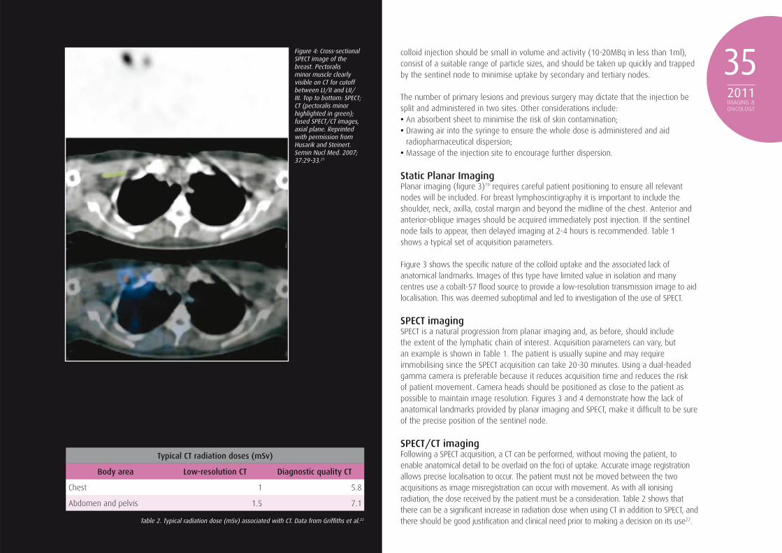
colloid injection should be small in volume and activity (10-20MBq in less than 1ml), consist of a suitable range of particle sizes, and should be taken up quickly and trapped by the sentinel node to minimise uptake by secondary and tertiary nodes.
The number of primary lesions and previous surgery may dictate that the injection be split and administered in two sites. Other considerations include:• An absorbent sheet to minimise the risk of skin contamination;• Drawing air into the syringe to ensure the whole dose is administered and aid
radiopharmaceutical dispersion;• Massage of the injection site to encourage further dispersion.
Static Planar ImagingPlanar imaging (fi gure 3)19 requires careful patient positioning to ensure all relevant nodes will be included. For breast lymphoscintigraphy it is important to include the shoulder, neck, axilla, costal margin and beyond the midline of the chest. Anterior and anterior-oblique images should be acquired immediately post injection. If the sentinel node fails to appear, then delayed imaging at 2-4 hours is recommended. Table 1 shows a typical set of acquisition parameters.
Figure 3 shows the specifi c nature of the colloid uptake and the associated lack of anatomical landmarks. Images of this type have limited value in isolation and many centres use a cobalt-57 fl ood source to provide a low-resolution transmission image to aid localisation. This was deemed suboptimal and led to investigation of the use of SPECT.
SPECT imagingSPECT is a natural progression from planar imaging and, as before, should include the extent of the lymphatic chain of interest. Acquisition parameters can vary, but an example is shown in Table 1. The patient is usually supine and may require immobilising since the SPECT acquisition can take 20-30 minutes. Using a dual-headed gamma camera is preferable because it reduces acquisition time and reduces the risk of patient movement. Camera heads should be positioned as close to the patient as possible to maintain image resolution. Figures 3 and 4 demonstrate how the lack of anatomical landmarks provided by planar imaging and SPECT, make it diffi cult to be sure of the precise position of the sentinel node.
SPECT/CT imagingFollowing a SPECT acquisition, a CT can be performed, without moving the patient, to enable anatomical detail to be overlaid on the foci of uptake. Accurate image registration allows precise localisation to occur. The patient must not be moved between the two acquisitions as image misregistration can occur with movement. As with all ionising radiation, the dose received by the patient must be a consideration. Table 2 shows that there can be a signifi cant increase in radiation dose when using CT in addition to SPECT, and there should be good justifi cation and clinical need prior to making a decision on its use22.
2011IMAGING &ONCOLOGY
35
Typical CT radiation doses (mSv)
Body area Low-resolution CT Diagnostic quality CT
Chest 1 5.8
Abdomen and pelvis 1.5 7.1
Figure 4: Cross-sectional SPECT image of the breast. Pectoralis minor muscle clearly visible on CT for cutoff between LI/II and LII/III. Top to bottom: SPECT; CT (pectoralis minor highlighted in green); fused SPECT/CT images, axial plane. Reprinted with permission from Husarik and Steinert. Semin Nucl Med. 2007; 37:29-33.21
Table 2. Typical radiation dose (mSv) associated with CT. Data from Griffi ths et al.22

2011IMAGING &ONCOLOGY
36
Low-resolution SPECT/CTWhen performing a low-resolution CT, the tube current is often fi xed (1-2.5mA), allowing for no adjustment; but it does allow the operator to select the appropriate scan fi eld of view. As with all acquisitions, this should include the entire lymphatic chain of interest, encompassing all foci of uptake identifi ed on SPECT. A low-resolution CT provides an attenuation map for AC and a traditional CT image; and, despite its low quality, it can still be used to aid pre-surgical localisation23.
Diagnostic quality SPECT/CTScanning patients at diagnostic quality is a more complicated scenario. Protocols vary greatly between both manufacturers and imaging departments and may also require adapting to suit patient needs. Due consideration should be given to each individual’s clinical need24. A wide range of exposure parameters can be adjusted to optimise protocols for each patient (kV, mA, pitch, detector confi guration (slice thickness), rotation time and image reconstruction). Axillary and internal mammary lymph nodes have an average size of 1cm and consequently a 5mm slice thickness is adequate for detection and localisation. For staging of breast cancer, the whole chest and abdomen are frequently scanned to assess for metastases within the lung and liver; narrower slice widths (0.5mm to 2mm on contemporary multi-slice CT) have been shown to increase detection rates for pulmonary lesions <1cm in size25.
Low-resolution or diagnostic quality SPECT/CT?In most centres, this choice would be limited by equipment availability. Buck et al considered the merits of each technique and favoured using a low-resolution technique for accurate localisation, unless there was a clinical need to perform a diagnostic quality scan24. Localising sentinel nodes and pulmonary lesions associated with breast cancer is also more acceptable with a low dose technique due to the low attenuating potential of
the thorax and the high inherent contrast this provides. Figure 5 illustrates the benefi t of a fused image for the localisation of sentinel nodes in a patient with breast cancer.26
Future developmentsPromising results are beginning to emerge from pioneering work exploring the use of ‘freehand SPECT’27. Using a higher dose than previously quoted (60-80MBq), an intra-operative handheld device yielded discovery of at least one node in 49/50 patients, sensitivity 98 per cent, whereas planar imaging achieved a sensitivity of only 84 per cent for the same 91 lymph nodes27. It was concluded that ‘freehand SPECT’ aided the surgeon with accurate 3D localisation detail for axillary nodes28. Whilst more studies are needed to support the validity of this technique, these initial results bode well for future developments.
CONCLUSIONHistological techniques play a critical role in lymph node evaluation for the planning of minimally invasive surgical procedures for the resection of cancer. For this to occur, the sentinel node(s) needs to be identifi ed and assessed; if necessary, the node(s) may require accurate localisation and removal. Various combinations of pre-surgical and intra-operative localisation are available to surgeons, including blue-dye, ultrasound, hand-held gamma probes, and nuclear medicine imaging. Imaging in nuclear medicine has advanced dramatically in recent times and SPECT/CT now allows accurate fusion of physiological (radionuclide uptake) and anatomical data to aid the localisation process. SPECT/CT is thought to allow a more accurate pre-operative biopsy, reducing morbidity, while it is also reducing operating times by allowing the surgeon to localise nodes of interest more quickly.
For further reading, please see The role and value of nuclear medicine in the imaging of sentinel nodes and breast cancer. eRADIMAGING.com website. Available at: http://www.eradimaging.com/site/article.cfm?ID=748&mode=ce. Published 10 February 2010.
Figure 5. Fused SPECT-CT images of sentinel nodes. Sentinel node mapping using 3D SPECT/CT. The yellow arrows indicate the nodes: subpectoral; parasternal; interpectoral. Reprinted with permission from Ibusuki et al. Surg Oncol. 2009. [Epub ahead of print.]26
2011IMAGING &ONCOLOGY
37
REFERENCES1. Mariani G, Moresco L, Viale G, et al.
Radioguided Sentinel Lymph Node Biopsy in Breast Cancer Surgery. J Nucl Med. 2001; 42:1198-1215.
2. Hayat M, Howlader N, Reichman M, Edwards B. Cancer statistics, trends, and multiple primary cancer analyses from the Surveillance, Epidemiology, and End Results (SEER) Program. Oncologist. 2007;12:20-37.
3. Breast Cancer: Little Change in Incidence. Available at: http://www.statistics.gov.uk/cci/nugget.asp?id=575 Accessed: 13 September 2010.
4. Schauer AJ, Becker W, Reiser M, Possinger, K. The sentinel lymph node concept. 2005 Springer, Berlin.
5. Cabanas RM. An approach for the treatment of penile carcinoma. Cancer. 1977; 39:456-466.
6. Phototakeusa.com. Tumor and lymphatic drainage. Available at: http://www.phototakeusa.com/results.asp?Image=ZEME027027775-01 Accessed January 2011.
7. Kell MR, Burke J, Morrow M. Outcome following axillary staging in early breast cancer [abstract]. J Clin Oncol. 2008; 26:(suppl):543.
8. Topal U, Punar S, Tas I, Adımc S. Role of ultrasound-guided core needle biopsy of
axillary lymph nodes in the initial staging of breast carcinoma. Eur J Radiol. 2005; 56:382-385.
9. Morton DL, Wen D, Wong JH. Technical details of lymphatic mapping for early stage melanoma. Arch Surg. 1992; 127:392-399.
10. Essner R, Foshag L, Morton DL. Intraoperative radiolymphoscintigraphy: a useful adjuvant to intraoperative lymphatic mapping and selective lymphadenectomy in patients with clinical stage I melanoma. Presented at: Annual Meeting of the Society of Surgical Oncology; 1994; Houston, Texas.
11. Harlow SP, Krag DN, Ashikaga T, et al. Gamma probe guided biopsy of sentinel node in malignant melanoma: a multicentre study. Melanoma Res. 2001; 11:45-55.
12. Even-Sapir E, Lerman H, Lievshitz G, et al. Lymphoscintigraphy for Sentinel Node Mapping Using a Hybrid SPECT/CT System. J Nucl Med. 2003; 44:1413-1420.
13. Siemens Healthcare. Siemens Symbia TruePoint SPECT/CT System. http://www.siemens.com/press/pool/de/pp_med/2005/sc_upload_fi le_mednm200504048_03_300dpi_1456207.jpg Accessed January 2011.
14. Lerman H, Lievshitz G, Zak O. Improved sentinel node identifi cation by SPECT-CT in overweight patients with breast cancer. J Nucl Med. 2007; 48:201-206.
15. Warncke S, Mattei A, Fuechsel F, Z’Brun S. Detection rate and operating time required for g probe-guided sentinel lymph node resection after injection of technetium-99m nanocolloid into the prostate with and without preoperative imaging. Eur Urol. 2007; 52:126-133.
16. Husarik DB, Fehr M, Thuerl CM, et al. Sentinel lymph node scintigraphy in breast cancer: incremental value of SPECT/CT, imaging. J Nucl Med. 2005; 46(suppl):197P.
17. Even-Sapir E, Lerman H, Lievshitz G. Lymphoscintigraphy for sentinel node mapping using a hybrid SPECT-CT system. J Nucl Med. 2003; 44;1413-1420.
18. Lerman H, Metser U, Lievshitz G. Lymphoscintigraphic sentinel node identifi cation in patients with breast cancer: the role of SPECT-CT. Eur J Nucl Med Mol Imaging. 2006; 33:329-337.
19. Aarsvold JN, Alazraki NP. Update on detection of sentinel lymph nodes in patients with breast cancer. Semin Nucl Med. 2005; 35:116-128.
20. Somasundaram SK, Chicken DW, Waddington WA, et al. Sentinel node imaging in breast cancer using superfi cial injections: technical details and observations. Eur J Surg Oncol. 2009; 35:1250-1256.
21. Husarik DB, Steinert HC. Single-photon
emission computed tomography/computed tomography for sentinel node mapping in breast cancer. Semin Nucl Med. 2007; 37:29-33.
22. Griffi ths M, Thompson J, Tootell A, et al. SPECT-CT and its role in imaging and oncology. Imaging and Oncology. 2009; 1:42-49. Available at: http://www.sor.org/public/pdf/imaonc09webres.pdf. Accessed 11 January 2010.
23. Coffey J, Hill JC. Breast sentinel node imaging with low-dose SPECT/CT. Nucl Med Commun. 2010; 31:107-111.
24. Buck AK, Nekolla S, Ziegler S, et al. SPECT/CT. J Nucl Med. 2008; 49:1305-1319.
25. Boussel L, Douek P, Revel D. Cardiac CT: what role should it play in coronary artery disease? J Radiol. 2006; 87(11 pt 1):1637-1641.
26. Ibusuki M, Yamamoto Y, Kawasoe T, et al. Potential advantage of preoperative three dimensional mapping of sentinel nodes in breast cancer by a hybrid single photon emission CT (SPECT)/CT system. Surg Oncol. 2010; 19:88-94.
27. Santi et al. Freehand SPECT for sentinel lymph node biopsy: fi rst clinical experience. J Nucl Med. 2010;51(suppl 2); Abstract 540
28. Ziessman HA. 2010 SNM Highlights lecture; General Nuclear Medicine. J Nucl Med. 2010:51,9:28-29N.
John Thompson is a senior radiographer at Furness General Hospital and is an honorary research fellow at the University of Salford. Andrew Tootell is a lecturer and programme leader in nuclear medicine imaging at the University of Salford. Joanne Sil is a lecturer in radiography at the University of Salford. Judith Kelly is a consultant breast radiographer and deputy clinical director at the Chester Breast Imaging Unit, Countess of Chester Hospital. Randeep Kulshrestha is a consultant radiologist and clinical isotope lead at North Manchester General Hospital. Peter Hogg is a professor of radiography and is university lead for the Diagnostic Imaging Research Programme, University of Salford.

GLOBALISATION OF HIGHER EDUCATION
IN RADIOLOGIC SCIENCES: A NEW
PARADIGM
SARAH BAKER

2011IMAGING &ONCOLOGY
39What is the future for higher education? Now is the time for the radiologic sciences profession to educate for global citizenship.
Within our global world, higher education is in a state of transition. A wide range of initiatives with potential ramifi cations for higher education reform are underway that will most likely impact radiologic sciences. A call for increased accountability, uncertain funding for higher education and international competition are among the demands we face in our global world1. The future of the radiologic sciences profession is global. We can no longer afford to fi ne-tune our existing educational policies; the time has come for an entirely new conceptual approach to radiologic science education. To meet the demands of the future, radiologic sciences professionals will need new and improved knowledge, not only to ensure top quality patient care, but for the good of society as a whole.
While globalisation can be defi ned in a multitude of ways, most would describe it as the growing integration of economies and societies around the world2. Included within this defi nition is the accelerating mobility of goods, services, labour, technology, and capital2. While globalisation is not new, the current pace of globalisation is being accelerated by technological advancements. The fi eld of radiologic sciences has been at the forefront of technological change, yet our educational standards, delivery, mobility, and efforts
towards globalisation have been slow to evolve. Globalisation of the radiologic sciences fi eld would allow not only for free market and economic trade of goods and services, but it would permit increased mobility of radiologic science students, faculty, and professionals which, in turn, could foster international partnerships. Such partnerships would allow for increased collaboration and co-operation between and among countries, increasing our knowledge base and positively impacting patient care. In short, now is the time for the radiologic sciences profession to educate for global citizenship.
According to the United Nations Educational, Scientifi c, and Cultural Organization (UNESCO), the capacity of the world’s educational systems has more than doubled in the past 43 years, with a six-fold increase in higher education or tertiary students during this time. The provision of tertiary education has been dominated by six countries, with the United States leading the way, followed by the United Kingdom3. Boasting the world’s largest radiologic technology credentialing organization - the American Registry of Radiologic Technologists (ARRT) with over 300,000 registrants - one might surmise that the United States is poised to lead our profession into global citizenship4. And yet, entry level education and certifi cation for radiologic technology in the US does not currently require a college degree. It is anticipated that by 2015, an associate degree will be required as the minimum pathway to ARRT certifi cation5. Meanwhile, education levels among radiographers within the UK is at the Bachelor of Science Honours (BSc Hons) level. As Professor Hardy so eloquently stated, “The future of imaging will demand the radiography profession and its educators to think innovatively and open its mind to new ways of working6.” She went on to note that this may be a challenge, but the potential exists for greater rewards, such as higher quality imaging and improved care for patients.
The current president of the International Society of Radiographers and Radiological Technologists (ISRRT), Dr Michael D Ward, has echoed similar thoughts on advancing the global profession of radiologic sciences. Among Dr Ward’s goals is the global expansion of education within our profession, with sharing of best practices models of education and curricula. Additionally, he looks for better co-operation and communication with international radiologic science organisations to address such items as medical imaging, health care, and
increased research partnerships. These goals, along with the establishment of an international radiologic technology programme accreditation process, are aggressive initiatives, but are attainable in light of current higher education climate and reform initiatives7.
CURRENT HIGHER EDUCATION REFORM CLIMATEFor over a decade, European countries have been engaged in reconstructing their higher education systems. This has, in part, been undertaken to create a greater degree of
THE EXPANSION OF ROLESFOR RADIOGRAPHERS HAS BEEN
STIFLED IN THE USA

While the United States may be a leader in per-student higher education spending, the return on this investment is lacking. America ranks abysmally when it comes to higher education completion rates. US state and local governments spent over 85 billion dollars funding public higher education in 2008, yet statistics show that only 56 per cent of fi rst-time, full-time students in the US will receive a degree within six years13,14. These failing numbers are likely to yield drastic consequences for American’s economic future.
Cliff Adelman, a former research analyst at the US Department of Education, has suggested that the United States should look to Europe in its quest for accountability. He argues that the Bologna Process offers some common sense solutions to the struggle to defi ne what students should be learning and to create a better pathway through the US higher education system8. In the US, the Lumina Foundation for Education is also enamoured of the Bologna Process and the European process aimed at standardising what a college degree means across the continent15. The Lumina Foundation, an omnipresent force in American higher education and its reform, has made exploration of Bologna’s potential application in the US a priority. Work is currently underway to create a Degree Qualifi cations Profi le defi ning what graduates should be able to know and do when they receive an associate, bachelor or master’s degree, regardless of what US institution they attend15. In January of 2011, the Lumina Foundation released a draft of its Degree Qualifi cations Profi le, which will allow colleges, accreditors, and others in the United States to test and refi ne what degree recipients should know and be able to do16.
The Bologna Process has yielded the Tuning Project, which is raising expectations for accountability. The major expectation is of ongoing curricular reform geared toward development of learning outcomes9. Tuning provides an approach to redesign, develop, implement, evaluate and enhance quality in degree programmes17. The name ‘Tuning’ was chosen to refl ect the idea that universities do not and should not look for uniformity or any sort of unifi ed, prescriptive or defi nitive European curricula in their degree programmes, but should instead look for points of reference, convergence and common understanding. Thus, Tuning focuses on educational structures located within higher education institutions (and academic staff), with emphasis on the subject area level, ie the content of studies. This process will allow higher education institutions to examine curricula in terms of structures, programmes, and actual teaching, in which the required academic and professional profi les and needs of society will play an important role18. A major aim of the Tuning Project is to contribute to a framework of comparable and compatible qualifi cations, described in terms of workload, level, learning outcomes, competencies, and profi le18. Statements of learning outcomes are to be defi ned in subject areas, looking at core knowledge, supporting knowledge, and communication skills8.
In the fi rst phases of the Tuning Project, representatives from nine subject areas, including nursing, arrived at a common language to describe curricular goals. They were soon followed by representatives from sixteen other degree fi elds, radiography among them8. The fi eld of radiography was identifi ed as one of the Socrates Thematic Networks which
2011IMAGING &ONCOLOGY
40 convergence, with a goal of creating a European Higher Education Area (EHEA). While voluntary, this movement has been gaining supporters and has now been dubbed ‘The Bologna Process.’8 The ‘Bologna’ moniker was chosen in recognition of Europe’s oldest university, located in Italy, where educational ministers of then 29 countries fi rst agreed to action lines that would bring down educational borders. These ministers of education agreed in June 1999 to the goal of the creation of a European Higher Education Area (EHEA) by 20109. The initial priorities of the Bologna Process were made offi cial with the ministers’ signatures on The Bologna Declaration.
Incentives for this process appear to be economic recovery, initially emphasised after World War II, and the need for closer cooperation among European nations9. In more recent times, The Maastricht Treaty on the European Union in 1992 identifi ed the European continent’s aspirations for a resurgent role in the world8,10. While higher education was a minor part of this document, the treaty did recognise that the European economy was knowledge-based, and thus supported a system of generating and distributing knowledge8. In 1997, the Lisbon Convention sought to facilitate the evaluation of foreign educational credentials. The immediate precursor to the Bologna Declaration was the Sorbonne Treaty, signed in 1998 by education ministers from the United Kingdom, France, Germany, and Italy8,9.
The major impetus behind the Bologna Declaration was the emphasised role of higher education in supporting European economic growth and the international resurgence of the Continent11. Among a multitude of goals, the Bologna Process seeks to synchronise participating countries’ university degree systems. The ultimate goal, however, is to create an EHEA in which students and faculty members are able to move about freely, from institution to institution and across nations12.
THE FUTURE OFTHE RADIOLOGIC
SCIENCESPROFESSION IS
GLOBAL

2011IMAGING &ONCOLOGY
41will incorporate Tuning methodology. The Thematic Network is known as HENRE. (Higher Education Network for Radiography in Europe)19. With this action, the stage appears to have been set for educational reform for the fi eld of radiography in Europe. The combination of these forces (Bologna, Lumina, and Tuning) might well impact radiologic science education.
IMPLICATIONS FOR RADIOLOGIC SCIENCESThe implications of this work for changes to the future of radiologic sciences education are numerous for both the United States and the United Kingdom. The time is right to shift the educational paradigm from teaching to learning. Such a shift, along with a sharper focus on defi ning what future radiologic science students and professionals should know, understand, and be able to demonstrate, is imperative. The process of defi ning student learning outcomes involves the creation of a common standard to identify the competencies for our variety of degree levels and modalities. These standards, in conjunction with the development of a standard international nomenclature, will enhance international mobility of students, educators and radiologic science professionals.
In the United Kingdom, roles for assistants, and all three levels of both therapeutic and diagnostic radiographers have expanded considerably over the last decade, yet tensions and inconsistencies between boundaries still exist6,20. Meanwhile, in the US, the expansion of roles for radiographers has been stifl ed, in part due to the levels of education. While the newly created position of the radiographic assistant holds promise in the US, this expanded role has also met with tensions and inconsistencies across US state lines.
The future direction of the radiologic sciences profession in both the United Kingdom and United States should include a combining of forces and utilise the synergy of professional and educational talents. Such an effort would entail a close examination of educational and professional standards. Among other things, this might include an overhaul of the ‘Standards of Profi ciency for Radiographers’ and ‘Standards of Education and Training in the UK21,22, while the US might overhaul accreditation and credentialing standards. The time is now upon us to think innovatively and do what is best for quality patient care within the radiologic sciences.
Dr Sarah S Baker is associate professor in radiologic sciences at the Indiana University School of Medicine, as well as associate dean in academic affairs at University College, Indianapolis, USA. She is also the regional co-ordinator of public relations for the Americas for the International Society of Radiographers and Radiological Technologists (representing the ASRT), a past president of ASRT, and a past board member of the Joint Review Committee on Education in Radiologic Technology.
REFERENCES1. Kwiek, M. Globalization and higher
education. Higher Education in Europe.
2001; 26(1): 27-38.
2. Baumann A, Blythe J. Globalization of higher education in nursing. OJIN: The
Online Journal of Issues in Nursing. 2008; 13(2): Available from: www.nursingworld.org/MainMenuCategories/ANAMarketlace/ANAPeriodicals/OJIN Last accessed 1 February 2011.
3. UNESCO Institute for Statistics: Global Education Digest 2010: Comparing Education Statistics Across the World. Montreal: UNESCO.
4. The American Registry of Radiologic Technologists. Available from: https://www.arrt.org/ Last accessed 14 February 2011.
5. The American Registry of Radiologic Technologists. 2015: Associate degree requirement for primary pathway to ARRT certifi cation. Available from: https://www.arrt.org/new/2009-09-17-ARRTSetsDegreeRequire.html 2009. Last accessed 14 February 2011.
6. Hardy M. Radiography: looking to the next decade. Imaging and Oncology. 2010 (1) 28-33.
7. Ward M. President’s Message. ISRRT (International Society of Radiographers and Radiological Tehnologists). 2010; 46(2): 3-4.
8. Adelman C. The Bologna Club: What U.S. higher education can learn from a decade of European reconstruction. 2008. Washington, DC: Institute for Higher Education Policy. Available from: www.ihep.org/Research/GlobalPerformance.cfm Last accessed 14 February 2011.
9. Gaston PL. The challenge of bologna: What United States higher education has to learn from Europe, and why it matters that we learn it. 1st ed. Virginia: Stylus; 2010.
10. TEU. Treaty on European Union. 1992. Available from: http://eur-lex.europa.eu/en/treaties/dat/11992M/htm/11992M.html Last accessed 14 February 2011.
11. Bologna. Joint declaration of the European ministers of education. 1999. Available from: http://www.ond.vlaanderen.be/hogeronderwijs/Bologna/ Last accessed 14 February 2011.
12. Labi A. Bologna conference highlights progress and limits of Europe’s new degree cycles. The Chronicle of Higher Education: International; 2007 June 1: [2 p.]. Available from: http://chronicle.com/weekly/v53/i39/39a03601.htm Last
accessed 10th February 2011.
13. National Center for Education Statistics, IPEDS Graduation Rate Survey. Available from: http://www.higheredinfo.org/dbrowser/?level=nation&mode=data&state=0&submeasure=27 Last accessed 14 February 2011.
14. Lumina Foundation for Education. Four steps to fi nishing fi rst in higher education: A guide for state policymakers. November 2009. Available from: www.makingopportunityaffordable.org Last accessed 9 February 2011.
15. Lederman D. Borrowing from Bologna. Inside Higher ED. 13 September 2010. Available from: http://www.insidehighered.com/layout/set/print/news/2010/09/13/lumina Last accessed 14 February 2011.
16. Lederman D. What degrees should mean. Inside Higher ED. 25 January 2011. Available from: http://www.insidehighered.com/news/2011/01/25/defi ning_what_a_college_degree_recipient_should_know_and_be_able_to_do Last accessed 25 January 2011.
17. Tuning Project. Tuning educational structures in Europe: Aims and objectives. Available from: http://www.tuning.unideusto.org/tuningeu/index.php?option=content&task=view&id=3&It... Last accessed 5 November 2010.
18. Tuning Project. Tuning educational structures in Europe: Available from: http://www.tuning.unideusto.org/tuningeu/ Last accessed 5 November 2010.
19. Tuning Project. Tuning educational structures in Europe: Radiography. Available from: http://www.tuning.unideusto.org/tuningeu/ index.php?option=content&task=view&id=33&… Last accessed 5 November 2010.
20. Howes N. Demystifying the role of the consultant therapy radiographer Imaging and Oncology. 2009 (1) 6-11.
21. Health Professions Council. Standards of profi ciency: Radiographers. 2009. London: HPC.
22. Health Professions Council. Your duties as an education provider: Standards of education and training. 2009. London: HPC.

RADIOLOGY ACCREDITATION IN THE UK: THE THEORY
AND THE REALITY
MELANIE HIORNS
2011IMAGING &ONCOLOGY
43Quality accreditation has an increasingly high profi le. But is it just another certifi cate to go on the wall in radiology services’ reception? Or does it genuinely bring something new to standards and to patient care?
Accreditation is slowly but surely moving into the mainstream, both in healthcare and in other industries. In the UK, the Imaging Services Accreditation Scheme (ISAS) was opened for registration in June 2009 and the fi rst Radiology Departments are now in the process of being accredited. However, many departments are taking a ‘wait and see’ approach. Questions such as, ‘What’s the point?’, ‘Is this going to become the norm?’, ‘How hard is it?’, ‘Does it make any difference?’, ‘Is it worth it?’ and ‘Will we have to?’ are asked commonly. This article explores the bigger picture about accreditation and the accumulating international evidence base that going through an accreditation process changes practice and improves care, but also describes a personal experience of going through the ISAS accreditation process fi rst hand and what it really means in practice.
In 2006 Braithwaite et al stated: “Accreditation has become ubiquitous across the international health care landscape. Award of full accreditation status in healthcare is viewed, as in other sectors, as a valid indicator of high quality organisational performance. However few studies have empirically demonstrated this assertion. The value of accreditation therefore remains uncertain and this persists as a central legitimacy problem for accreditation providers, policymakers and researchers.”1
Two years later, Greenfi eld and Braithwaite further commented, “Accreditation, quality and continuous improvement have become an intrinsic part of the discourse and activities of health services. Internationally dating from the 1970s, health care accreditation programmes and accrediting organisations have emerged and developed. There are now many national accreditation organisations and an international body, the International Society for Quality in Health Care (ISQua) which has enrolled members in over 70 countries.” 2
This work by Greenfi eld and Braithwaite is the largest international study to date systematically reviewing the impact and/or effectiveness of accreditation. The review included a range of health accreditation schemes, including hospital accreditation, and was not confi ned to imaging, which is a relatively recent entrant into the accreditation arena. Topics considered included professional attitudes to accreditation, promoting change, organisational impact, fi nancial impact, quality measures, programme assessment, consumer views or patient satisfaction, public discourse, professional
development, and surveyor issues.
Two consistent fi ndings were recorded: that of accreditation promoting change in the organisation, and in promoting professional development. The activity of preparing and undergoing accreditation has been shown in several studies to promote change in health organisations3,4, particularly with respect to organisation and safety. In the studies reviewed, the positive outcomes included the chance for staff to refl ect on, and become engaged in, the operation of the organisation; the introduction of continuous quality programmes, improved documentation, improved safety and improved review systems. However to balance this accreditation was not shown to ensure high-quality care but was positively associated with some measures of quality. Yet, accredited hospitals showed signifi cant positive change in six defi ned areas, which was not recognised in non-accredited organisations.
Pomey et al3 describe how the context in which accreditation takes place, including the organisational context, infl uences the type of change dynamics that occur in Healthcare Organisation (HCO). They found that while accreditation itself was not necessarily the element that initiated change, the accreditation process was a highly effective tool for helping to introduce continuous quality improvement programmes to newly accredited or not-yet-accredited organisations; creating new leadership for quality improvement initiatives; increasing social capital by giving staff the opportunity to develop relationships; and for fostering links between HCOs and other stakeholders. They concluded that the accreditation process is an effective leitmotiv for the introduction of change but is nonetheless subject to a learning cycle and a learning curve. Institutions invest greatly to conform to the fi rst accreditation visit and reap the greatest benefi ts in the next three accreditation cycles (three to 10 years after initial accreditation)3.
So how do these fi ndings translate into, specifi cally, radiology accreditation programmes across the world? Accreditation has been in place in North America for a number of years. There are two major programmes, one run by the American College of Radiology5, and another by the Joint Commission (an independent, not-for-profi t organisation that accredits and certifi es more than 18,000 healthcare organisations and programmes in the United States6).
WORK OUT YOURTIMESCALE ANDDOUBLE IT
2011IMAGING &ONCOLOGY
44 Currently, accreditation is confi ned to advanced imaging techniques such as CT and MRI. This, in part, has been driven by reimbursement policies that may require accreditation and to meet the criteria of CMS (Centres for Medicare and Medicaid Services) 7, state or federal government, or third-part payers. Medicare is a social insurance programme administered by the United States government, providing health insurance coverage to people who are aged 65 and over, or who meet other special criteria. The ACR outlines how all MRI, CT, PET, breast MRI and nuclear medicine facilities that bill under the Medicare fee schedule, must be accredited by January 2012 to receive Medicare payments for the technical component of these service8. Accreditation is also recognised for its value as a marketing tool and is clearly promoted as such.
In Australia and New Zealand, there is a voluntary accreditation scheme jointly administered by The Royal Australian and New Zealand College of Radiologists (RANZCR) and the National Association of Testing Authorities (NATA), which commenced in May 2004, and is open to all diagnostic imaging providers that wish to apply. However, sites that offer MRI services that are eligible for Medicare rebates are required by the Commonwealth government to participate in the RANZCR’s MRI Accreditation Registration Programme.
Mandatory accreditation schemes exist in South Korea and Finland, although these have a different confi guration to that being developed in the UK.
In the UK, accreditation is only now starting to become established and is still a long way from being a mandatory requirement, although it is certainly possible that this may happen in the future. However, in parallel with schemes elsewhere in the world, it is appropriately infl uenced by the guidelines of the professional colleges (Royal College of Radiologists and the Society and College of Radiographers). Unlike elsewhere, the actual scheme is exclusively run by a third party, the United Kingdom Accreditation Service (UKAS), which is ‘the sole national accreditation body recognised by government to assess, against internationally agreed standards, organisations that provide certifi cation, testing, inspection and calibration services’9. UKAS is a non-profi t-distributing private company, limited by guarantee. It is independent of government but is appointed as the national accreditation body by the Accreditation Regulations 2009 (SI No 3155/2009) and operates under a memorandum of understanding with the government through the Secretary of State for Business, Innovation and Skills. The Imaging Services Accreditation Scheme sits within UKAS. Background to the development of the Imaging Services Accreditation Scheme is outlined by Garvey, et al.10
So to take this further, how do some of these fi ndings translate into the role for an imaging services accreditation scheme in the UK in the current political climate? The principles underpinning the original Radiology Accreditation Programme were drawn from those referred to in the 2007 White Paper ‘Trust, Assurance and Safety: the regulation of health professionals in the 21st Century’11. More recently (2010), the National Health Service (NHS) White Paper ‘Equity and Excellence: Liberating the NHS’12 set out the Government’s long-term vision for future healthcare; that of a patient centred NHS in which the drive for
better health outcomes is clear. It recognises the fi nancial challenges the NHS faces and the role QIPP (Quality, Innovation, Productivity and Prevention) will play in supporting the NHS in identifying effi ciencies, whilst driving up quality. An editorial in the British Medical Journal in 2008 outlined how most recent NHS reforms have been designed to improve effi ciency and productivity and also to improve the quality of care.13 That trend is ongoing.
Quality is becoming the leading driver in healthcare reform going forward, representing a move away from performance targets per se. This aligns with the core purpose of accreditation, being the formal recognition that an imaging services provider has demonstrated that it has the organisational competence to deliver against key quality measures across four domains which include ‘Clinical’, ‘Facilities, resource and workforce’, ‘Patient experience’, and ‘Safety’. These four domains comprise the ‘Standard’ which has been designed to • Be patient focussed;• Cover the functions and systems of a whole diagnostic imaging and interventional
radiology service; • Address the dimensions of quality and support quality improvement.
‘Outcomes’ are the indicators by which the quality and effectiveness of a service can be assessed. The NHS Outcomes Framework14 published in December 2010 sets out the outcomes and corresponding indicators that will be used to hold the new NHS Commissioning Board to account and, as such, represents a clear message about the importance of verifi ed outcomes going forward in health policy. Each of the 31 standard statements across the four domains in the ISAS Standard has an associated outcome measure of the individual service’s design and choosing. This aligns with the direction of travel of health policy in the UK.
It must, therefore, be hoped that participation in the ISAS programme in the UK will, in itself, be some form of evidence that radiology departments may, in future, submit to the various regulatory bodies such as the Care Quality Commission (CQC), who are the independent regulator of care provided by the NHS (and also of local authorities, private companies and voluntary organisations).
ACCREDITATION ON THE GROUNDThe radiology department at Great Ormond Street Hospital for Children has been associated with the emerging accreditation programme since early 2007, when it became one of fi ve pilot sites for the Radiology Accreditation Programme (RAP) across England, and since 2009 has been formally committed to the resulting ISAS accreditation scheme. Ten imaging services have been in the fi rst wave and one has just received accreditation along with Great Ormond Street. One other NHS Trust, having had assessment visits, is anticipating formal accreditation imminently.
The ISAS website15 gives a vast amount of user-friendly information on what accreditation is about, pre application information and preparatory workshops, a

2011IMAGING &ONCOLOGY
45detailed description of the ‘The Standard’ and all that it entails16.
In preparing for assessment, an organisation must fi rst outline how much of its service it wants to be considered for accreditation – this is called the ‘scope’. Usually the whole service would be included, but there may be instances where only part of the service is included in the scope: for instance, if a new part of the service has only recently been established and this is not ready for the fi rst round.
The Standard comprises four domains. A total of 31 ‘standard statements’ are spread across these four domains, and each standard statement addresses one aspect necessary for the provision of the service. A list of criteria indicates the structures and processes necessary to deliver each particular standard statement. For each criterion, indicative examples of acceptable evidence is given.
The service will then work on its evidence over the upcoming months (typically a year) and will upload it to ISAS via the web-based tool. Once the evidence has been submitted, it is assessed and, if suffi cient and appropriate evidence has been provided, a site visit will follow. The assessment team will usually comprise a radiologist, a radiographer, a lay member and a professional UKAS assessor.
Our experience at Great Ormond Street was that initially we completely underestimated the amount of work involved in the whole process. Gathering the evidence is a huge task; we found that it was not so much that we were not doing a lot of good things already, but more that we had never written it down in an organised and joined-up way. Whilst this may appear rather banal, it became evident that our processes were basically safe, and we hopefully offered a high quality, patient focussed and forward looking service but our documentation, whilst present, was scattered in different places (sometimes in people’s heads). Everyone knew what they were doing and were doing it well, but this knowledge had been learned over many years. Someone arriving new wouldn’t have one place to go to fi nd out about our polices, procedures and protocols. Our document control was not as good as it should have been (and now is) and some areas of the department were doing things in a slightly different way to other areas.
To balance against this, we realised that there was almost nothing we were not already doing that we should have been. We were well ahead of the curve in our patient surveys, our audit programme, our risk management and in our patient–oriented practice. Financially, we were in good order and our management structure was effective and safe.
After a false start, we brought together a group of fi ve key members in the department to lead and to champion the accreditation work. These included a consultant radiologist (who was also the clinical unit chair for the division and thus had professional radiology input, but also had an overview of the wider role of radiology within the hospital and of the hospital’s structure and policies), the radiology service chair, the radiology service manager, the
lead radiology superintendent, and the departmental personal assistant (PA). The PA was invaluable in collating all the evidence once it had been submitted to her and organising it on the web based tool. She was also able to keep an overview of what information was missing, who was supposed to be providing it, and to do the chasing.
The radiology superintendent co-ordinated the activity of a wider group of modality superintendents with respect to their various modalities, and for the documentation and policy writing relating to those areas. The consultant radiologist took responsibility for some of the higher level organisation information and for many of the criteria in the clinical domain. The radiology service chair and service manager co-ordinated everything else. Early in the process, one of these fi ve names was written against every single one of the 131 criteria, so it was very clear who was responsible for that criterion. Tentative dates for completion were added, which were reviewed at least once a month. By the end of the evidence gathering phase, every single person in the department had been involved in accreditation in some way. The task of collating the evidence would have been impossible without this cascade effect.
Once the evidence had been reviewed and accepted, the dates were set for a two-day site visit. This process really starts the adrenaline fl owing and it was a time of great coming together for the department to prepare for this, knowing that any one of the 70 of us could be asked a question, or asked to demonstrate something. Many of the staff who had been aware of the accreditation work going on suddenly pricked up their ears and, in the last few weeks, there was a real momentum. People would ask me what it meant to be accredited and would it matter? I could only reply that I hoped it would make us special and would allow us to prove unequivocally that we are a good department, rather than just believing we are.
The two-day visit itself went off uneventfully. Many of the staff were outstanding (and this was recognised and commented on by ISAS) and were genuinely proud to have been involved with it and to be able to show our department, and what we have achieved, to ‘outsiders’. The feedback was given straightaway on the second afternoon and the senior management was requested (by ISAS) to be present as they are the
WE BELIEVE WEDELIVER A SAFERQUALITY SERVICE

2011IMAGING &ONCOLOGY
46 REFERENCES1. Braithwaite J, Westbrook J, Pawsey
M, Greenfi eld D, Naylor J, Iedema R, Runciman B, Redman S, Jorm C, Robinson M, Nathan S, Gibberd R. A prospective, multi-method, multi-disciplinary, multi-level, collaborative, social-organisational design for researching health sector accreditation 2006 [LP0560737]. BMC Health Serv Res 6:113.
2. Greenfi eld D, Braithwaite J. Health sector accreditation research: a systematic review. Int J Qual Health Care 2008 20:172-183.
3. Pomey MP, Lemieux-Charles L, Champagne F, Angus D, Shabah A, Contandriopoulos AP. Does accreditation stimulate change? A study of the impact of the accreditation process on Canadian healthcare organizations. Implement Sci 2010 5:31.
4. Juul AB, Gluud C, Wetterslev J, Callesen T, Jensen G, Kofoed-Enevoldsen A. The effects of a randomised multi-centre trial and international accreditation on availability and quality of clinical guidelines. Int J Health Care Qual Assur Inc Leadersh Health Serv 2005 18:321-328.
5. American College of Radiology. Available at http://www.acr.org Last accessed 30 December 2010.
6. Joint Commission. Available at http://www.jointcommission.org Last accessed 30 December 2010.
7. Centres for Medicare and Medicaid Services. Available at http://www.cms.gov/ Last accessed 30 December 2010.
8. American College of Radiology (Accreditation). Available at http://www.acr.org/accreditation.aspx Last accessed 30 December 2010.
9. United Kingdom Accreditation Service. Available at http://www.ukas.com/about-accreditation/about-ukas Last accessed 30 December 2010.
10. Garvey CJ, Cook JV, Wiltsher C, Whitley S. Radiology accreditation-towards a safer quality service. Clin Radiol 2009 64:853-856.
11. Trust, Assurance and Safety: the regulation of health professionals in the 21st Century. Department of Health 2007. Available at http://www.dh.gov.uk/en/Publicationsandstatistics/Publications/PublicationsPolicyAndGuidance/DH_065946 Last accessed 30 December 2010.
12. Equity and Excellence: Liberating the NHS. Department of Health 2007. Available at http://www.dh.gov.uk/en/Publicationsandstatistics/Publications/PublicationsPolicyAndGuidance/DH_117353 Last accessed 30 December 2010.
13. Black N. Quality improvement in the NHS. BMJ 2008 336:1143.
14. NHS Outcomes Framework. Department of Health 2010. Available at http://www.dh.gov.uk/en/Publicationsandstatistics/Publications/PublicationsPolicyAndGuidance/DH_122944 Last accessed 30 December 2010.
15. Imaging Services Accreditation Scheme. Available at www.isas-uk.org Last accessed 30 December 2010.
16. The ISAS Standard: statements, rationales and criteria. ISAS 2009. Available at http://www.isas-uk.org/Library/ISAS_Standard/ISAS%20Standard%20V1.1.pdf Last accessed 30 December 2010.
ones who ultimately have responsibility for a safe, quality, hospital. The chief executive attended, which certainly raised the bar. We have some ‘mandatory’ actions to fulfi l (which we absolutely expected and is the norm) and we submitted evidence with respect to these in January 2011.
So has it been worth it for us? Yes, defi nitely. We have developed ourselves, and strengthened our relationships with our users (both clinicians and patients). We have made a huge step change in how we challenge our own practice and the evidence we use to demonstrate our good practice. We have improved our documentation beyond measure, and have examined critically all our processes. We believe we deliver a safer quality service to our patients. But it has been hard work.
For other departments considering accreditation these are our top tips:• Take full advantage of all the pre-application information, advice, and pre-application
workshops that ISAS can offer.• Have a really thorough look at the Standard and all the standard statements in each of
the four domains so you know the size of the task.• Work out your timescale and double it – realistically, it is probably going to take a year
of preparation.• Convene a core group of people who really want to achieve this and make sure your
senior management are fully supportive and understand the benefi ts.• Distribute the tasks clearly so individuals know what is expected of them personally,
and keep a ‘project plan’ up to date.• Liaise with ISAS throughout the process so you know that you are heading in the right
direction.• Know that the fi rst time you do anything is the hardest and keep going.• Keep the faith that this process will make you a better department – both for your
staff and for your patients.
Melanie P Hiorns is a consultant paediatric radiologist and honorary senior lecturer. She is also clinical unit chair for medicine and diagnostic and therapeutic services at the Great Ormond Street Hospital for Children NHS Trust. In March 2011 the radiology department at GOSH successfully achieved accreditation against the ISAS standard.
QUALITY IS BECOMING THE LEADINGDRIVER IN HEALTHCARE REFORM

SENSUS SRT 100
Accu x Curve Board for Head and Shoulder immobilisation
LAP Moving Laser Systems & Q-Fix Couch Tops for CT Simulation

EDUCATING THE RADIOGRAPHY WORKFORCE: A DIVERSE CHALLENGE
IAN HENDERSON

2011IMAGING &ONCOLOGY
49How should the workforce of the future be trained? How far will role development and skills mix go? What skills will radiographers need?
At the present time, UK radiography education, in common with other non-medical health disciplines, is largely delivered by the higher education (HE) sector. This encompasses pre-registration entry level programmes entirely, most of the post-registration and postgraduate offerings and most of the ‘non-professional’ level provision, ie the assistant level workforce. Some post-registration provision is delivered by the independent sector and there is a considerable amount of practice based, or in-house provision. For the assistant practitioner and support workforce, again there is some accredited practice based, or in-house training, and also some delivery by the further education (FE) sector.
As a general overview, both pre and post-registration education and training are, allegedly, related to workforce planning or developmental needs. In England, currently, this is manifested through a commissioning process, where pre-registration student numbers are contracted to higher education institutions (HEI) by strategic health authorities and, to some extent, post-registration numbers are also contracted in this way. This, however, is a generalisation since the arrangements for post-registration provision are by no means consistent across the country. In Scotland, Wales and Northern Ireland, arrangements follow a more conventional or academically focussed system.
It is the case then that HEIs offering radiography, may offer both diagnostic and therapeutic, will commonly offer a range of post-registration or postgraduate provision, and may offer provision for training the support level workforce1. This scenario provides a number of challenges in terms of delivering an appropriate product in a rapidly developing clinical landscape, whilst resourcing provision in an increasingly purchaser provider environment.
The future effectiveness and, indeed, relevance of imaging and radiotherapeutic professionals is at stake if education and training is unable to maintain alignment with professional needs. The situation warrants scrutiny at a time when natural evolutionary change seems to be accelerating signifi cantly2,3,4.
So what does the future hold?
A BIT OF BACKGROUNDThe educational landscape for radiography has changed signifi cantly in 25 years in terms of both entry to the profession and for post-qualifying education; from the centralised curriculum and assessment of the College of Radiographers, to an HE based system. Of course, this is not news to most in the profession, but the reason for mentioning it is to point out that, in real terms, it is not so long ago. There remains a high proportion of the profession who trained in that earlier period, who are still around, many of them highly infl uential in how the profession has evolved. The move to HE based provision, a consequence of the then government’s ‘Working Paper 10’5, was a massive shift in the culture of radiography education. In the 20 years since then, there has been accelerating change in the parameters within which radiographers work, affected by such diverse infl uences as technology, health policy, national demographics, and public expectation.
Of course, these factors have always played a part, but it may be argued that, in this period, the explosion in computer based technology and the critical infl uence of healthcare fi nancing, to name but two, have transformed fundamentally aspects of how imaging and oncology services operate and are delivered. There is no reason to suspect that this rapid evolution will slow. Arguably, the changes of recent years, and the change yet to come, will transform educational arrangements for radiographers every bit as much as those of Working Paper 105.
How do the various elements of the equation fi t together? To start with let us examine how the education sector relates to the clinical environment.
PARTNERSHIP, OR UNEASY BEDFELLOWS?In the wider world, it could be said that academic and professional elements produce
HEIS ARE UNFAMILIAR WITHDELIVERING PROGRAMMES BENEATH
UNDERGRADUATE LEVEL
2011IMAGING &ONCOLOGY
50 the total picture of how a profession exists and evolves6,7. They are inextricably linked in a partnership engaged in the process of developing and applying practice through education, research and collaboration with the aim of ensuring an ongoing drive for excellence. Benson7 indicates that one of the criteria for a profession is that there must be a professional body which – ‘must set the ethical rules and professional standards which are to be observed by the members’ – thereby acting as a focus for the integration of theory and practice.
Educational programmes develop undergraduates to become members of a profession and, in that process, students will usually undertake work placements within that industry. University departments engage in research, commonly in collaboration with and, indeed, receive funding from the industry with which they are associated. The notion of partnership therefore, if idealistic, is explicit in both educational and research terms.
In radiography, these components all exist, but does the partnership notion really work in the idealistic sense? Anecdotally, it seems that there is often a divide between theory and practice, as if the two were linked in only a tenuous fashion. Undoubtedly, many very effective relationships exist between the clinical and academic sectors, though often this may extend only to limited strata of activity, for example, training of undergraduate students, or linkage for delivery of professional development programmes. This can lead to the perception of a purchaser provider type relationship, rather than a partnership. Whilst it is true that HEIs are obliged to liaise with the clinical sector in the periodic process of developing or reviewing curricula, how often is the clinical sector at ground level, involved in collaborating in research initiatives? How much ownership does the profession have of the development of its evidence base? Current evidence indicates that there is a limited degree of activity in these areas and there are a number of reasons why this may be8.
For example, radiography has traditionally been seen as a ‘hands on’, practically oriented discipline and this could be considered an ingrained culture; HE sector involvement in radiography education, providing the opportunities for collaboration, is a relatively recent occurrence; the operational linkage with the radiological profession has traditionally been essentially subservient, with much of the practice related research medically dominated; the structure and budgetary mechanisms of the National Health Service do not necessarily facilitate the establishment of department level research initiatives. These factors are general and it would be quite wrong to imply that there are no examples of effective partnerships and collaboration. However, the pervading culture does not readily support the sort of situations that exist more commonly in the wider world.
WHY IS THIS A PROBLEM? In a time of rapid change, adaptation and evolution of the profession have to be driven coherently and strategically. Education should provide practitioners that are not only fi t for practice, but fi t to take practice forward. The clinical sector should be able to provide a strategic view of what it needs from professional education in the medium term,
bearing in mind that a course developed now will produce its fi rst graduates in at least three years’ time.
Dealing with continuous transition in healthcare delivery and technology means that there is considerable scope for a mismatch of expectation if collaboration is not effective. In this context, it is fair to say that the academic world has a responsibility to have a clear perspective on emerging health policy and technology in order to respond proactively to prospective needs. The danger is, however, that if the ‘cart comes too far before the horse’, then educational effectiveness may be compromised. A largely diagnostic example of balancing such a transition, which could be termed ‘transition stress’, is the move from fi lm-based to entirely digital imaging. This is a situation where HEIs had to shift from one substantial technology to another over a period of time, balancing the needs of students according to the environment in which they were training, in the knowledge that they may then go on to work in a another location where the arrangements were completely different.
The concept of transition stress is a common feature of dealing with change in a multi-factorial environment9. HEIs need to be ahead of the game, yet aligned to current reality. They need to be able to work with the clinical sector, yet the shape and culture of that sector may provide its own challenges. Notwithstanding the current economic climate, the projected changes in healthcare structure, delivery and workforce; and the continuing development in technology associated with both imaging and oncology, provide the main challenges to the education sector in both the short and medium term. The scope for change is massive.
CHANGING TECHNOLOGY Imaging and oncology service development is driven by a variety of factors, but there is no doubt that technology is a major component of delivery in both disciplines. Indeed it has been said10 that radiographers are the interface between the equipment and the patient, ensuring that the two come into contact in the correct way, with the correct outcome. Amongst health practitioners, it can be argued that radiographers occupy an unusual position in terms of how they are ‘linked’ to the equipment they use. This means that evolution in equipment design and function has a fundamental impact on radiographers and the nature of the demands on them in using it.
In the past 30 years at least, developments in computer technology have probably had the greatest impact on the technological aspects of how radiographers practice. Computerised systems are fundamental to how much of modern radiographic equipment functions, relying heavily on computer processing power in order to manage large amounts of digital data. An estimate of the signifi cance of this is demonstrated by Moore’s law11 which, in 1965, predicted a doubling of computer power every two years. It is believed that the law continues to hold true today, indicating an almost exponential change in processing power that will probably not come as a surprise to radiographers who have been around for a while.

2011IMAGING &ONCOLOGY
51approvals undertaken by the College of Radiographers indicate that the UK’s entry level education is not dramatically different14 to that of many other developed countries. It can be legitimately argued that this is because role extensions are post-registration skills, taught usually at postgraduate level, but when does an evolving practice become a mainstream skill or, indeed, can it? And in that context, from a skill mix perspective, when does the practice of assistant practitioners infl uence the entry level training of radiographers?
The NHS Plan2 and the recent white paper ‘Liberating the NHS’4, set out to change the face of healthcare in signifi cant ways. Among other things they advocate skill mix, the shifting of services to the primary sector, and increased involvement of the independent sector in delivery. These features encourage the evolution that is already underway, so what do radiographers need to look like in the future if they are to deliver effectively in this environment? Specifi cally, how do they need to be trained and developed in order for this to happen?
EDUCATING THE FUTURE WORKFORCEThe business, for that is what it is, of training and educating the radiographic workforce has confl icting demands. In order to deal with signifi cant development and lead-in times, particularly for undergraduate programmes, the HE sector must have a very clear perspective on the health policy agenda, effective linkage with the clinical sector, and with those who commission training in whatever form; the market! How much the HE sector can push the practice agenda and how much it must follow is, of course, infl uenced by resources and, frankly, by market forces. In respect of post-registration education in particular, it is evident that despite the College of Radiographers’ clear viewpoint on the radiographic scope of practice15, there is limited commonality of view at ground level. In fact, there is considerable variation in the implementation of skill mix initiatives across the United Kingdom, often because of regional workforce priorities and/or local professional opposition16. The localisation of professional development priorities is further encouraged by the implementation of Agenda for Change requirements in respect of staff development through the NHS Knowledge and Skills Framework (KSF), meaning that clinical environments may have very specifi c demands for CPD provision, often unaligned with the traditional forms of post-registration programmes on offer.
WHAT IS THE SIGNIFICANCE OF THIS? The whole technology issue is an article on its own, but in précis we now have a scenario where many decisions or processes are no longer in the hands of equipment operators, where computer assisted diagnosis is a reality and where emerging technologies, such as molecular imaging, may change the face of radiology12. Without wishing to be too contentious, it may be argued that some imaging and oncology practices have already been deskilled by the intervention of modern equipment. What does this mean then to the scope and emphasis of radiographic practice and, in that context, how does education align with this change? Does a radiographer really need a degree in order to carry out many aspects of routine radiography? Or to put it another way, when assistant practitioners are evidently capable of carrying out much of the routine work, do radiographers still need to be taught the same skills set, or should the emphasis lie in areas where they can effectively exercise the clinical judgements associated with graduate level practice? The evolution of radiographic practice is also linked to the wider environment, health policy priorities and, of course, professional aspiration.
HEALTH POLICY AND THE HEALTH SERVICEThe healthcare landscape is infl uenced by changing demographics, clinical research outcomes and political priorities. Alongside this, clinical practice evolves. In radiography, the most familiar phrases of recent years have surely been ‘skill mix’ and ‘role extension/development’ alongside, for slightly different reasons, continuing professional development (CPD) and advanced/consultant practice. All terms associated, quite explicitly, with modifying or evolving the roles of radiographers. Developed roles such as diagnostic image reporting, portal image evaluation, and an extensive range of others, have become widely regarded as completely acceptable - to the radiographic community at least - and are supported by robust research evidence to underline their credibility.
Like all evolution however, the correct circumstances must exist for it to occur and the unique structure and demands of the UK’s National Health Service (NHS) have provided this. Witness the fact that radiographic reporting, for example, has hardly scraped the surface in most other countries globally13, making it apparent that the UK is a bit of a rarefi ed environment. It is interesting to note then that recent international course
THERE IS POLITICAL WILL TO BUILDTHE BAND 4 WORKFORCE

2011IMAGING &ONCOLOGY
52 consistent viability. This, and the reduced emphasis on academic credits, has created an opportunity for independent providers who may meet a need not fulfi lled by larger scale HEIs. The law of economy of scale does not necessarily apply in this situation.
ASSISTANT WORKFORCEIn many respects, this component of the workforce has been hardest for the HE sector to comprehend. Although standards for practice are defi ned, particularly through the College of Radiographers’ Learning and Development Framework17, educational arrangements are inconsistent, with no specifi c identifi ed ‘qualifi cation’ for entry to this group. Training arrangements range from in-house training schemes to NVQ programmes, BTECs, Foundation degrees, and a few things in between. Implementation of this tier of the workforce has been infl uenced by, amongst other things, fi nancial pressures, radiographer availability, and strategic imperatives of clinical managers, meaning that it has been patchy in geographical terms and across modalities and disciplines. For example, assistants are well embedded in the mammographic screening workforce, though less than might be expected in radiotherapy, with some oncology managers questioning the need for assistant practitioners in their service. This has caused some diffi culty for HEIs in identifying demand and sustaining programmes for assistants. It is also fair to say that many HEIs are unfamiliar with the business of delivering programmes beneath the undergraduate academic level.
It may be argued then that in this transitional period for development of the assistant workforce, there has tended to be a vicious circle of varying supply and demand. Nevertheless, it is quite clear that there is an evident political will to consolidate and build the band 4 workforce18 and therefore the need to provide appropriate opportunities, both for initial training and subsequent development, cannot be ignored. This is potentially a major growth area for radiography education, so who delivers it? The HE sector which has the knowledge and expertise in radiography, or the Further Education sector, which has the ability to deliver effective and well supported programmes that meet the very specifi c needs of learners at this academic level?
THE FUTURE?LP Hartley said ‘the past is another country, they do things differently there’ and, in this case, it may literally be true. The rate of recent change has been signifi cant and gives some sense of what is to come. Looking ahead, the medium term prospect for radiography education is one of shifting emphasis. In general terms, policy direction suggests expansion of the band 4 workforce in a more directive manner than in the past18; possible contraction in undergraduate radiographer training, at least in diagnostic (already some evidence of this at the time of writing); increased interest in supporting post-qualifying training at all levels to facilitate skill mix developments and the restructuring of some services. What this means for the HE sector probably has two main facets.
Firstly, developing or expanding provision in ‘pre-graduate’ and post-qualifi cation areas requires a degree of re-prioritisation and potential re-skilling. Whilst it is true
The challenge for HEIs is to maintain alignment with the current scope of practice for entry to the profession, whilst anticipating future requirements; to provide effective, relevant CPD opportunities for radiographers that meet the needs of a diverse and fl uid market; to contribute, where possible, to training the assistant level workforce, again in line with local requirements. So, quite a few variables there then!
What are the factors that impact on their ability to deliver on these elements?
PRE-REGISTRATION, ENTRY LEVEL PROVISIONRadiographer education at the entry level could be seen as the more stable component of the equation, at least currently. The challenge exercising many HEIs is how to minimise student attrition from programmes at a time when, in England at least, strategic health authorities are penalising them for failing to deliver higher completion rates16. Periodic programme review requires them to look fi ve years into the future when the fi rst graduates from a reviewed programme will enter the workforce. Will graduates be fi t for practice in the environment that exists then? It is clear that many HEIs are taking great care to develop curricula that are forward looking and anticipatory, but a balance must be achieved. Too much, too soon risks a misalignment with existing needs. Too speculative and the misalignment may be further down the line. So, do these factors encourage conservatism in the design of curricula?
An example of this was the adoption over 10 years ago by some HEIs of the theory component of the College of Radiographer’s intravenous (IV) administration programme in the anticipation that IV administration would become an entry level skill, but has it? Despite the fact that IV administration is now effectively a mainstream activity for radiographers, it remains essentially a post-registration skill. Knowing the style and rate of change remains a speculative process.
POST-QUALIFYING In the post-registration/postgraduate arena, the situation is currently more dynamic. The demands of developing the workforce to meet a rapidly changing environment has required HEIs to operate in ways not previously associated with that sector, more responsive, more fl exible, more cost effective. The clinical sector requires targeted, specifi c programmes that are accessible, tailored to their needs, and aligned with KSF or Skills for Health standards. They will pay for what meets service needs and that does not necessarily mean traditional postgraduate programmes.
Anecdotal evidence indicates that in many areas the number of students signing up for full postgraduate awards has dropped in recent years as students seek focussed programmes that will deliver a specifi c skills package in line with their CPD requirements. This requires a culture shift for many HEIs as they come to terms with a major change in the shape of post-registration delivery and their ability to respond must also take account of the fact that the market size for many programmes is not suffi ciently large to ensure

2011IMAGING &ONCOLOGY
53that many HEIs have already well embedded provision, others may have some work to do in this area. Training the assistant workforce in particular, has some specifi c challenges and there is a lot that may be learned from the further education sector. For example, delivering effective support for students at this level. Additionally, the whole business of providing post-qualifying provision, for all components of the workforce, in a range of different shapes and sizes, is a challenge, leading to:
The second facet, which relates to the development of an effective entrepreneurial, business oriented perspective. Again, it is true that many centres understand the signifi cance of this. Actually achieving it is another matter. Effective delivery of post-qualifying programmes will be key to maintaining position in a competitive arena. In order to be successful, HEIs will need to be fast moving, responsive, adaptable and competitive; not attributes that have traditionally been associated with the sector. Systems to support different ways of interacting with ‘the market’ need to be in place and this needs to be effective and professional, if only because competitors in the post-qualifying market are not just other universities. The developing independent sector provision for CPD can react quickly and cheaply to demand.
Of course, health education has, and continues to operate on, quite a localised basis, dependent on the relationships that exist within a region. There is, therefore, a degree of generalisation applied in this discussion and, naturally, the different arrangements across the four countries of the UK are also signifi cant. Nevertheless, the principles are applicable, as are the infl uencing factors.
The situation is in transition at the time of writing and therefore there remain some signifi cant unknowns. For example, the specifi c arrangements for the commissioning of education in England. Following the government’s decision to move commissioning to GP led consortia19, the dissolution of primary care trusts and regional health authorities has led to a proposal to manage education commissioning through a new body, Health Education England, in conjunction with ‘provider networks’. The end result may not be too different but, inevitably, there will be system changes for HEIs to deal with. One way or another, fi nance is the driver although, in some form, radiography education at all levels will continue. The things that will most need to change are operational structures and priorities, and attitudes.
Ian Henderson is subject lead for diagnostic radiography at Robert Gordon University, Aberdeen and editor of the Society and College of Radiographers’ members’ publication, Synergy Imaging & Therapy Practice. He has worked extensively in the development of radiography programmes at all academic levels and has liaised with education commissioners in the development and evolution of post-registration education across the allied health professions.
REFERENCES1. College of Radiographers. Programmes
approved by the College of Radiographers leading to qualifi cation as a radiographer. http://www.sor.org/news/course-directory Accessed 7 January 2011.
2. Department of Health. The NHS Plan, A plan for investment, A plan for reform. HMSO. 2000.
3. Darzi, A. NHS Next Stage Review Interim Report. HMSO. 2007.
4. Department of Health. Equity and excellence: Liberating the NHS. HMSO. 2010.
5. Department of Health. Working for Patients, Education and Training, Working Paper 10. HMSO. 1989.
6. Eraut, M. Developing Professional Knowledge and Competence. Routledge. 2003.
7. Benson, L. The Professions Offi cial Report 5th Series, Parliamentary Debates 1992-3, Lords 1992 Vol. 538, 15, pp.1208-1210. Hansard, London.
8. T Gambling, T Brown, P Hogg, P. Research in our practice - a requirement, not an option: discussion paper. Radiography, 2003 Vol 9, pp71–76.
9. Bryan, J W. Halle, A. Shoou-Yih, D L. Review: Conceptualization and Measurement of Organizational Readiness for Change. A Review of the Literature in Health Services Research and Other Fields. Medical Care Research and Review August 2008 vol. 65 no. 4 pp 379-436.
10. Murphy, F J. The paradox of imaging technology: A review of the literature Radiography 2006 Vol. 12, Issue 2, pp.
169-174.
11. Moore, G E. Cramming more components onto integrated circuits. Electronics 38,1965 pp. 114.
12. John, M H. Sanjiv, S G. Molecular Imaging: The Vision and Opportunity for Radiology in the Future. Radiology 2007, Vol244, pp39-47.
13. Smith, T. Yielder, J. Ajibulu, O. Caruana, E. Progress towards advanced practice roles in Australia, New Zealand and the Western Pacifi c. Radiography, 2008, Vol 14, Supplement 1, ppe20-e23.
14. College of Radiographers. Report of the Approval and Accreditation Board. SCoR. 2010.
15. University of Hertfordshire. Scope of Radiographic Practice 2008: a report compiled by the University of Hertfordshire in collaboration with the Institute for Employment Studies for the Society and College of Radiographers - UH 2009.
16. Benchmark Pricing and National standard Framework Contract Operational Implementation Guide Version 15 C G Jeffries 15 March 2006.
17. College of Radiographers. Learning and Development Framework for Clinical Imaging and Oncology. SCoR. 2007.
18. NHS London. Workforce for London: A Strategic Framework. HMSO. 2007.
19. Department of Health. Department of Health Spending Review: Press release. 2010. http://www.dh.gov.uk/en/MediaCentre/Pressreleases/DH_120676 Accessed 10 January 2011.

IMAGING FOR STROKE AND TRANSIENT
ISCHAEMIC ATTACK: MEETING THE STANDARDS OF THE NATIONAL
STROKE STRATEGY
ANDY BEALE AND DAMIAN JENKINSON

2011IMAGING &ONCOLOGY
55The National Stroke Strategy sets out clear targets for imaging services. Can they deliver?
INTRODUCTIONStroke is the third biggest cause of death in the UK and the largest single cause of severe disability. Each year more than 110,000 people in England will suffer from a fi rst stroke and a further 30,000 will suffer a recurrent stroke. Prior to stroke, one in fi ve people experience a transient ischaemic attack (TIA), providing an opportunity for interventions to reduce vascular risk. The total cost of strokes to society is £8 billion per year, but less than 8 per cent of this is spent on initial medical diagnosis and management. The majority accrues from rehabilitation, nursing and indirect costs.
The National Stroke Strategy1 in England, was launched in December 2007, and set a clear direction for the development of stroke services in England over the next 10 years. Much work has taken place to improve stroke services and the quality of care received by all who need it. There has been a recent concurrent public awareness campaign publicising the Act FAST criteria (Face, Arms, Speech and Time) for stroke and reinforcing the emphasis on ‘time is brain’. Research quantifying cell damage estimates that two million brain cells are lost each minute in an acute middle cerebral artery infarct2. Therefore one of the biggest challenges set out in the strategy is the need for rapid imaging, both for TIA and stroke.
WHY BOTHER?Previously, the management of cerebral ischaemia was based on optimal medical treatment. Now there are more specifi c treatment options for both strokes and TIAs. In
strokes, thrombolysis has been shown to signifi cantly improve outcomes if administered within three hours of onset3 and shows moderate benefi t if the window for treatment is extended to 4.5 hours4. In addition, intra-arterial fi brinolytic therapy can offer a moderate benefi t in the 3-6 hour window5
For TIAs, carotid recanalisation and, specifi cally, carotid endarterectomy (CEA), if performed early within two weeks of onset, can also improve outcome, leading to a reduction in subsequent debilitating stroke6. The College of Vascular Surgeons is working towards a 48-hour target from presentation to treatment (performing CEA) and has accepted that this needs to be implemented as soon as possible.
THE GUIDANCEThe emphasis on thrombolysis and CEA has led to the need for rapid access to imaging services as a key element in the gold standard service envisioned in the National Stroke Strategy. The chapter describing acute care of TIA and stroke, Time is Brain, contains two Quality Markers (QM) related specifi cally to brain imaging.
QM5 Assessment – referral to specialist (TIA and minor stroke)Immediate referral for appropriately urgent specialist assessment and investigation is considered in all patients presenting with a recent TIA or minor stroke, using a system which identifi es as urgent those with early risk of potentially preventable full stroke. To be assessed within 24 hours in high-risk cases; all other cases are assessed within seven days. (Using the ABCD2 criteria*). Provision to enable brain imaging within 24 hours and carotid intervention, echocardiography and ECG within 48 hours, where clinically indicated.
QM7 Urgent response (stroke)All patients with suspected acute stroke are immediately transferred by ambulance to a receiving hospital providing hyper-acute stroke services (where a stroke triage system, expert clinical assessment, timely imaging and the ability to deliver intravenous thrombolysis treatment are available throughout the 24 hour period).
THE TARGETS (BY APRIL 2011) 1. 60% of high risk people (ABCD2 score 4 or more*) with TIA should be investigated
and treated within 24 hours.2. 50% of stroke patients to be scanned within one hour of hospital arrival.3. 100% of stroke patients to be scanned within 24 hours of hospital arrival.
* ABCD2 score is calculated using the patient’s age (A); blood pressure (B); clinical features (C); duration of TIA symptoms (D); and presence of diabetes (2). Scores are between 0 and 7 points. Low risk = 0–3 points; moderate risk = 4–5 points; high-risk = 6–7 points.
FOR IMAGINGDEPARTMENTS
THERE ARE HUGEIMPLICATIONS

2011IMAGING &ONCOLOGY
56 These are ambitious goals, but will enable prevention of subsequent stroke in cases of TIA, and timely treatment in cases of stroke, reducing the risk of subsequent death and disability. In terms of changes to current service provision, the management of TIAs may represent a more signifi cant challenge than the urgent response to stroke, particularly out of hours and at weekends.
IMAGING WORKLOAD IMPLICATIONSIn 2008, the Department of Health published ‘Implementing the national stroke strategy, an imaging guide’7. This document, produced by a large number of stakeholders involved in imaging, estimated the expected workload for imaging departments. Within this document, there are calculations on the impact this would have by estimating the number of scans required for a given population. The fi gures given per 500,000 population are:
For TIAs:• Approximately 30 patients a week presenting with TIAs.• 50% will require brain imaging (66% within 24 hrs).• 80% will require carotid imaging (66% within 24 hrs).
Thus anticipated imaging during the week, 12 MRI/MRA brain and 10 carotid imaging. During the weekend, three MRI/MRA brain and two carotid imaging.
For stroke:• Approximately 25 patients a week presenting with strokes.• Almost 100% will require brain imaging (50% within one hour).• 10-20% of urgent cases will require additional imaging within 24 hours, eg MRI.
Thus anticipated imaging during the week, 15-17 brain CT and 3-4 MRI. During the weekend, 7-9 CTs with 1-2 a month requiring MRI.
This is a signifi cant change from previous management. Although most stroke patients would have previously had some form of brain imaging (usually CT), the new guidelines require the scans within one hour, if urgent (50%), and within 24 hours otherwise. This requires departmental fl exibility to accommodate scans at short notice. The implications in TIAs is perhaps more signifi cant because most patients are not, at present, having MRI scans. This may add signifi cantly more than 10 per cent to the workload of a district general hospital MRI scanner, compounding the already fully stretched national MRI capacity.
CURRENT PROVISION FOR TREATMENTThrombolysis is available in 88 per cent of acute trusts, (RCP audit May 2010) but only 50 per cent are 24/78. Nevertheless, the availability of thrombolysis has increased signifi cantly over the past two years.
CEA services are variable, with very few acute trusts offering a full 48-hour service. CEA within two weeks is achieved more frequently, but even this timescale is by no means universal. The Royal College of Vascular Surgeons remains committed to trying to improve CEA availability.
MONITORINGAll primary care trusts will be expected to collect data relevant to imaging, which includes the following:1. Treatment of higher risk individuals with TIA, defi ned by a score of 4 or more on the
ABCD2 system1. They will be expected to provide data on the proportion of these patients who are scanned and treated within 24 hours of symptom onset.
2. For lower risk TIA, the same applies but for a seven day period.3. Percentage of TIA patients with confi rmed carotid stenosis receiving carotid
intervention within 48 hours. Percentage of stroke patients scanned within one hour and 24 hours.
MOVING FORWARD, NEWER TECHNIQUES AND USAGEMechanical clot retrieval Pilot trials suggest benefi t from intravenous thrombolysis and endovascular mechanical clot retrieval/aspiration, using mechanical devices to dispose of large proximal clot burdens. This can be combined with the ability of lytics to initiate therapy early, or clean up smaller occlusions in distal arteries not accessible to mechanical attack9. At present, this is available only in major centres, usually performed by interventional neuroradiologists. Use of the ‘hub and spoke model’ and networking across stroke services should lead to an increase in the use of this technique.
CT perfusion scanning CT perfusion is used widely already, although its role in the acute stroke setting is still under debate. Recent publicity in the United States has muddied the waters because patients having multiple CT brain perfusion scans suffered radiation burns and local hair loss. These reports reached the UK lay press. Nevertheless, the advantage in assessing objectively the ‘penumbra’ is, at present, being used particularly to assess those patients in whom the timing of onset of symptoms is unclear, eg the ‘wake up strokes’. The additional information gained by the concurrent acquisition of a CT cerebral angiogram is helpful in some centres for assessment of arterial occlusion, prior to intra-arterial mechanical clot retrieval.
Carotid imaging During working hours, most carotid imaging is performed using ultrasound, either by sonographers or vascular scientists. However, both CT angiography and MRA of the carotid arteries provide accurate assessment of carotid stenosis. There are issues regarding training both to acquire and to report CT and MRA images, but the availability of both these scans out of hours and at weekends may mean an increase in their usage. Indeed, this is already happening at a number of centres.

2011IMAGING &ONCOLOGY
57
throughput is vital10. The claims that all departments are working to ‘full capacity’ is not supported by analysis and the National Audit Offi ce is currently linvestigating the utilisation of high cost equipment, including MRI, CT and Linacs. They will be reporting in the spring of 2011.
CONCLUSIONThe days of passive management in stroke and TIAs are over. Imaging departments cannot rest easy and must engage in the concept of ‘Time is Brain’ to do their part in improving the outcome of these conditions.
Dr Andy Beale is the national clinical lead for radiology and Dr Damian Jenkinson is the national clinical lead for stroke at NHS Improvement.
DISCUSSIONChanges in the way stroke and TIA services are being delivered are happening already. Targets have been set and there has already been a substantial improvement in access to timely imaging over the last few years.
For imaging departments there are huge implications. The impetus for TIAs imaging has led to an increase in workload and fl exibility that requires a joined-up approach, particularly with vascular surgeons, to offer the complete service. Likewise, stroke physicians will need to ration their referrals for both MRI and carotid imaging because not all patients referred with TIA symptoms require imaging.
There are different strategies that imaging departments can introduce - and some already have - to help achieve these ambitious targets. Carotid arteries can be imaged with US, CT and MR angiography. Adapting to availability of these different modalities can facilitate a 24-hour turnaround. Training other staff, such as nurse practitioners, to provide carotid artery ultrasound screening has also been successfully trialled in Exeter10. Including stroke and TIA imaging as part of seven day working can facilitate the development of business cases and designing workfl ow. MRI brain imaging capacity can be created by providing ‘short scans’. There is no evidence of signifi cant quality loss but patient throughput is increased.
In the urgent stroke setting, training more radiographers to perform CT heads is being encouraged by the Society and College of Radiographers and many centres have implemented this already. Reducing the time between arrival in the accident and emergency department and CT is vital. Paramedics given access for direct referral have reduced time to scan and the concern that there would be subsequent unnecessary examinations has not materialised. Cascade bleep systems, as used in the cardiac arrest scenario, can include the CT radiographer, who can have the scanner ready to receive the thrombolysis patient.
Stroke and TIA imaging should, however, not be seen in isolation and there are numerous other pressures on departments, including recent emphasis on cancer targets and seven day working, to name but two. However, there is a wide variation in how effi ciently different departments use their equipment and reviewing all aspects of workfl ow and patient
THERE IS VARIATION IN HOWEFFICIENTLY DEPARTMENTS
USE EQUIPMENT
REFERENCES1. Department of Health. National Stroke
Strategy, London: DH, 2007.
2. Saver JL. Time is brain – quantifi ed. Stroke. 2006; 37(1):263-6.
3. Wardlaw J M, Murray V, Berge E, Del Zoppo G J. Thrombolysis for acute ischaemic stroke. Cochrane Database Syst. Rev. 2009; (4); CD000213.
4. Hacke W, Kaste M, Bluhmki E, Brozman M, et al. Thrombolysis with alteplase 3 to 4.5 hours after acute ischaemic stroke. N Engl J Med. 2008;359(13):1317-29.
5. Ogawa A, Mori E, Minematsu K, Taki W, Takahashi A, Nemoto S, et al. Randomized Trial of Intraarterial Infusion of Urokinase Within 6 Hours of Middle Cerebral Artery Stroke. The Middle Cerebral Artery Embolism Local Fibrinolytic Intervention Trial (MELT) Japan. Stroke. 2007; 38, 2633-2639.
6. Rothwell PM, Eliasziw M, Gutnikov SA et al. Endarterectomy for symptomatic carotid
stenosis in relation to clinical subgroups and timing of surgery. Lancet. 2004; 363 (9413): 915-924.
7. Department of Health. Implementing the National Stroke Strategy - an imaging guide. London: DH, 2008.
8. National Sentinel Stroke Audit, Organisational Audit. Royal College of Physicians and Intercollegiate Stroke Working Party. August 2010.
9. Smith W S, Sung G, Saver J, Budzik R, Duckwiler G, Liebeskind D S, et al. Mechanical thrombectomy for acute ischemic stroke: fi nal results of the Multi MERCI trial. Stroke. 2008; 39(4):1205-12.
10. Stroke Imaging Case Studies – Examples of how departments have changed their ways of working to support the delivery of timely imaging for stroke patients. Available from www.improvment.nhs.uk/diagnostics.

TOO MANY HYSTERECTOMIES IN THE ERA OF FIBROID
EMBOLISATION?
RAM KASTHURI AND JON MOSS

2011IMAGING &ONCOLOGY
59The role of uterine artery embolisation in the management of symptomatic fi broids is established but not always readily available. What are the factors behind this?
Uterine fi broids or leiomyomata are the most common tumours of the female reproductive system1. The overall incidence is approximated at between 20 -70 per cent depending on age/menstrual status and race2. Whilst fi broids are benign and asymptomatic in the majority, they can cause debilitating symptoms, including heavy and painful periods, and pressure symptoms3,4.
Pharmacological management is usually the fi rst line treatment with anti-infl ammatory hormone therapy or gonadotrophin releasing hormone agonists5. Depending on the type of fi broid and individual patient circumstances, more invasive techniques such as uterine artery embolisation, or surgical options including myomectomy or hysterectomy, may be considered.
UTERINE ARTERY EMBOLISATION – WHERE WE ARE NOW Uterine artery embolisation is an interventional radiological technique
involving selective cannulation of both the left and right uterine arteries in turn, and embolisation, usually using particles. This technique was fi rst reported as an alternative to surgical treatment in 1991 by Ravina et al followed by a series by the same group in 19956,7. Over the last 15 years, more than 100,000 procedures have been performed in Europe and the USA, with a number of these being part of registries and randomised controlled trials (RCTs).
Initial evidence was from single centre cohorts, but were followed by at least four multicentre RCTs published between 2003 and 20088-11. The comparison arms
were hysterectomy8,9, myomectomy10 and either11. Different primary end points were also used in these four RCTs.
A prospective fi broid registry from the USA, whilst voluntary and without a control cohort, provided useful data on a large number (n=3160) of fi broid embolisations. Similarly, the UK HOPEFUL12 study involves large numbers (n=1108), but is a retrospective comparison of UAE with hysterectomy in a matched cohort. Despite all the above variations, the results are more or less comparable across the range of variably powered and designed studies. In summary, UAE is comparable to hysterectomy with regard to quality of life (QoL); the hospital stay and recovery time is shorter with UAE; and the cost-effectiveness at one year favours UAE. On the other hand, symptom control following UAE does not match hysterectomy, with approximately 10 per cent of UAE patients needing a second procedure for symptom control.
Based on current literature at the time, in 2000 the joint working party of the Royal College of Radiologists and the Royal College of Obstetricians and Gynaecologists issued recommendations on the use of uterine artery embolisation in the management of fi broids. These were updated in June 2009 13. The National Institute of Health and Clinical Excellence (NICE) has also issued full guidance to the NHS in England, Wales and Scotland on UAE, which was updated just at the end of last year14. In addition, UAE fi gures in the NICE guidelines for the management of heavy menstrual bleeding15.
WHY ARE MORE HYSTERECTOMIES BEING DONE?Hysterectomies are the most common major gynaecological surgery worldwide. Although the numbers of hysterectomies for fi broids have decreased as compared to a decade ago, there are more hysterectomies being done worldwide as compared to all other non-pharmacological techniques.
In 2005/2006, there were 38,631 hysterectomies performed in NHS hospitals in England & Wales16 and approximately 30 per cent of these for benign disease. Fibroids constituted the majority of these benign conditions.
The patient The majority of patients who consider non-pharmacological treatment for symptomatic fi broids are older and are likely to have completed their family. Therefore, in this patient group, persistent symptom resolution is more a priority than retention of the uterus. Also, the process of investigation, initial medical management with hormones and/or gonadotrophin releasing hormone (GnRH) analogues, delays resolution. These patients
PATIENTS NEED TO CONSIDER UAE ASSAFE AND EFFICIENT

2011IMAGING &ONCOLOGY
60 are keen to be free of their very troublesome symptoms. So, in comparison with the prospect of going through initial imaging, followed by the embolisation procedure, and follow up imaging with a one in 10 potential of a further procedure, a hysterectomy, despite being highly invasive and radical, is preferred by a number of patients. Further, hysterectomy is associated with a high rate of satisfaction and is likely to eliminate menstrual symptoms in virtually all the patients. Even in patients with early post operative complications, the long term satisfaction rate is very high17.
For patients not keen to retain their uteri, hysterectomy as a treatment option, with its high effi cacy, readily available expertise to perform the procedure and high satisfaction rate with time-tested results, makes consideration of any other alternative very academic.
In order for a patient to prefer one procedure over another, there must be a choice. There have certainly been cases in the past where the patients have not been informed of uterine artery embolisation as an option. There have also been cases where the clinician mentions the procedure more for completeness, neither giving the patient facts nor any information about local availability. Fortunately, such inappropriate consulting is occurring less frequently now, especially after publication of the recent National Institute for Health and Clinical Excellence (NICE) guidelines and the joint working party report.
The clinician The vast majority of patients with symptomatic fi broids are usually referred by their general practitioner to a gynaecologist for further management. The gynaecologist would usually exhaust pharmacological options prior to considering interventional procedures. For women with large fi broids (> 3cms) causing symptoms of heavy menstrual bleeding, as well as other symptoms such as dysmenorrhoea or pressure symptoms, UAE or surgery can be considered15. This is the recommended generic management guideline from NICE across the range of procedures for symptomatic fi broids. Sub-group indications for the different procedures are not specifi ed. The guidelines also recommend that any decision should be reached only after a detailed discussion regarding all treatment options has been had with the patient. Therefore, the consulting clinician plays a signifi cant role in the choice of the treatment procedure.
To be able to discuss the available treatment options it is imperative that the gynaecologist has comprehensive knowledge of UAE including the practical patient issues, technical aspects and potential complications. The clinician should also be up-to-date on the current literature. This, in clinical practice, can be very diffi cult. As a result, there may be an unconscious clinician bias towards the surgical techniques. However, this problem can be overcome by interventional radiology consultations.
New procedure syndromeUterine artery embolisation is still considered by many as a new procedure. Although clearly a safe procedure with proven effi cacy in the short to medium term, long term follow-up data from the trials and registries are outstanding.
In the NHS, any new procedure or service usually requires a business case to establish not only the safety and effi cacy of the procedure, but also its cost effectiveness18. Making a robust case to start a new procedure to treat a condition with well established treatment alternatives has, in the past, been diffi cult. This is particularly true as the service involves incurring extra expenditure to already overstretched radiology departments. This may, at times, inhibit the development of a new service or procedure.
Also, most interventional radiologists currently performing UAE have developed this skill as consultants rather than as trainees and there certainly is a learning curve involved. This requires enthusiasm, be it visiting another centre performing these procedures, or being mentored at one`s own centre by an expert.
Availability of local skilled expertise is another factor. Whilst the majority of UK hospitals have access to interventional radiology, this may not be local. Patients therefore may have to be referred to another centre. This introduces another potential disadvantage when the patient considers her options.
All the above factors are signifi cant, but the most important factor is education and awareness of the procedure. There is still a minority of clinicians who claim not to believe in the role of UAE in the management of fi broids. In the face of overwhelming evidence, this can only be due to ignorance or laggardness. Consequently, there are a less than par number of UAEs performed for symptomatic fi broids.
WHERE DO WE GO FROM HERE?The role of uterine artery embolisation in the management of symptomatic fi broids is now established but certain key areas will determine and secure the future of this treatment:
Clinical sub-specialtyPatients deemed suitable for the procedure are referred by the clinician to interventional radiology. In a number of units, there is a lack of direct radiology involvement both before and after the procedure. Therefore the interventional radiologist becomes a technician rather than a clinical practitioner. This non-clinical approach means that the interventional radiologist carries out an invasive procedure on a patient without prior personal assessment of the patient. This is a major drawback and should be discouraged.
Ideally, patients should fi rst be seen in an interventional radiology outpatient clinic19. During this consultation, the patient should be assessed by the interventional radiologist regarding the appropriate symptoms being treated; imaging reviewed; and the procedure should be discussed in detail. This includes the alternatives, practical implications and the possible complications. There should also be robust post procedure follow up by the interventional radiologist. It is our practice to follow up patients in the interventional radiology out-patient clinic at six weeks, six months and 12 months following the

2011IMAGING &ONCOLOGY
61procedure. The fi rst follow-up visit is to look for post procedure complications, and the subsequent ones to check on symptom benefi t and late complications.
The recent recognition of interventional radiology as an offi cial sub-specialty by the postgraduate medical education and training board (PMETB) further contributes to the process of development of the clinical interventional radiologist.
ResearchThere are still a number of questions in the management of symptomatic fi broids and, more specifi cally, within interventional radiology:• How does UAE compare to myomectomy in the same patient group? This is a key
question because both techniques are uterus sparing. An RCT from Mara et al10 randomised patients to either UAE or myomectomy, and they have reported mid term results. Their numbers, however, are small and this is still an unanswered question.
• Is there an ideal embolic agent? • Is there an ideal particle size? • Is it possible to predict fi broid shrinkage on pre-procedure imaging?• Is there a sub-clinical effect of UAE on fertility?
The long-term follow-up data from the clinical trials, as mentioned before, are outstanding. Research is crucial in the continued development of the technique and for answering these important questions. Equally, other minimally invasive alternatives such as magnetic resonance guided focussed ultrasound treatment 20 and magnetic resonance guided laser ablation21 are under development and are showing promising results.
TrainingThe ready availability of local expertise in UAE is variable across the United Kingdom and this is an important factor from both the patient’s and the referrer’s perspective. The local UAE service should also be staffed with skilled and competent personnel. Training of interventional radiologists in the procedure is another key factor. The training should not be restricted to technical competence, but also include patient assessment, both clinical and radiological, as well as follow-up.
Guidance on appropriate training in the various aspects of UAE features both in the Royal College of Radiologists` new curriculum22 and the Interventional Radiology syllabus23, as recommended by the Cardiovascular and Interventional Radiological Society of Europe (CIRSE). These measures help standardise training, thus helping to ensure delivery of a quality service.
EducationEducating patients, clinicians and even the general public will help signifi cantly in developing a greater understanding of the procedure, thus enhancing its profi le and generating greater public acceptance. Patients need to consider UEA as a safe and effi cient treatment procedure for fi broids.
The clinicians - both primary care and secondary referrers - need working knowledge of the indications, the current literature, the practicalities involved for the patient, and the potential complications. This process should also include non interventional radiologists who are sometimes consulted regarding treatment of fi broids.
Uterine artery embolisation is perceived by the general public as an experimental procedure restricted to a few specialised centres. During consultations, patients often enquire if the procedure is experimental. Any misconceptions must be overcome and education is the key. A robust patient information leafl et is essential and should be disseminated to general practices and other appropriate public areas. The internet is another powerful tool in educating the public, but it can equally be a deterrent with some websites giving wrong or inaccurate information. Arguably, another effective method of information dissemination is the patient herself. A good overall experience with good symptom benefi t can result in a ‘word of mouth’ type education for the wider public. Any way of enhancing profi le is good for the procedure.
CONCLUSIONUterine artery embolisation should be considered in patients with symptomatic fi broids who desire to retain their uterus, irrespective of their age. Continued research and technological developments will ensure the procedure evolves and gains more widespread acceptance and popularity. In terms of patient welfare and cost effectiveness in today’s patient-focused healthcare environment, the benefi ts of UAE are irrefutable, and it is no longer acceptable to perform hysterectomies because of a lack of local expertise and/or ignorance.
Dr Ram Kasthuri is a consultant interventional radiologist and honorary clinical senior lecturer at Gartnavel General Hospital, Glasgow. Professor Jon Moss is a consultant interventional radiologist and professor of radiology at the same institution.
THE BENEFITSOF UAE AREIRREFUTABLE

2011IMAGING &ONCOLOGY
62 england-and-wales/ Last accessed 1 December 2010.
17. Weber A M, Walters M D, Schover L R, Church J M, Piedmonte M R. Functional outcomes and satisfaction after abdominal hysterectomy. Am J Obstet Gynecol. 1999; 181(3):530-5.
18. Bennett A R. Business planning: can the health service move from strategy into action? J Manag Med. 1994; 8(2):24-33.
19. Keeling A N, Reekers J A, Lee M J. The clinical practice of interventional radiology: a European perspective. Cardiovasc Intervent Radiol. 2009; 32(3):406-11.
20. LeBlang S, Hoctor K, Steinberg F. Leiomyoma Shrinkage After MRI-Guided Focused Ultrasound Treatment: Report of 80 Patients. AJR 2010; 194:274-280.
21. Hindley J T, Law P A, Hickey M, Smith S C, Lamping D L, Gedroyc W M, Regan L. Clinical outcomes following percutaneous magnetic resonance image guided laser ablation of symptomatic uterine fi broids. Hum Reprod. 2002; 17(10):2737-41
22. Royal College of Radiologists. Curriculum in clinical radiology. 2010. London; RCR. Available online at http://www.rcr.ac.uk/docs/radiology/pdf/2010_Curriculum_CR.pdf. Last accessed 1 December 2010.
23. Cardiovascular and Interventional Radiology Society of Europe (CIRSE) European interventional radiology syllabus. 2008. Available online at http://www.cirse.org/fi les/File/cirse_syllabus_2008_web(1).pdf. Last accessed 1 December 2010.
REFERENCES:1. Lumsden M A. Embolization versus
myomectomy versus hysterectomy: which is best, when? Hum Reprod. 2002; 17(2):253-9.
2. Stewart EA. Uterine fi broids. Lancet. 2001; 27; 357(9252):293-8.
3. Manyonda I, Sinthamoney E, Belli A M. Controversies and challenges in the modern management of uterine fi broids. BJOG. 2004; 111(2):95-102.
4. Sankaran S, Manyonda I T. Medical management of fi broids. Best Pract Res Clin Obstet Gynaecol. 2008; 22(4):655-76.
5. Lethaby A, Vollenhoven B, Sowter M. Pre-operative GnRH analogue therapy before hysterectomy or myomectomy for uterine fi broids. Cochrane Database Syst Rev. 2001; (2):CD000547.
6. Ravina JH, Herbreteau D, Ciraru-Vigneron N, et al. Arterial embolisation to treat uterine myomata. Lancet 1995; 346:671-672.
7. Ravina J H, Bouret J M, Ciraru-Vigneron N, Repiquet D, Herbreteau D, Aymard A, le Dreff O, Merland J J, Ferrand J. Recourse to particular arterial embolization in the treatment of some uterine leiomyoma Bull Acad Natl Med. 1997; 181(2):233-43.
8. Pinto I, Chimeno P, Romo A, et al. Uterine fi broids: uterine artery embolization versus abdominal hysterectomy for treatment - a prospective, randomized, and controlled clinical trial. Radiology 2003; 226:425-431.
9. Hehenkamp W J, Volkers N A, Birnie E, Reekers J A, Ankum W M. Symptomatic uterine fi broids: treatment with uterine artery embolization or hysterectomy--results from the randomized clinical Embolisation versus Hysterectomy (EMMY) Trial. Radiology. 2008; 246(3):823-32.
10. Mara M, Maskova J, Fucikova Z, Kuzel D, Belsan T, Sosna O. Midterm clinical and fi rst reproductive results of a randomized controlled trial comparing uterine fi broid embolization and myomectomy. Cardiovasc Intervent Radiol. 2008; 31(1):73-85.
11. Edwards R D, Moss J G, Lumsden M A,
Wu O, Murray L S, Twaddle S, Murray G D; Committee of the Randomized Trial of Embolization versus Surgical Treatment for Fibroids. Uterine-artery embolization versus surgery for symptomatic uterine fi broids. N Engl J Med. 2007; 25; 356(4):360-70.
12. Hirst A, Dutton S, Wu O, Briggs A, Edwards C, Waldenmaier L, Maresh M, Nicholson A, McPherson K. A multi-centre retrospective cohort study comparing the effi cacy, safety and cost-effectiveness of hysterectomy and uterine artery embolisation for the treatment of symptomatic uterine fi broids. The HOPEFUL study. Health Technol Assess. 2008; 12(5):1-248, iii.
13. Royal College of Obstetricians and Gynaecologists and Royal College of Radiologists. Clinical Recommendations on uterine artery embolisation in the management of fi broids: Joint working party of the Royal College of Obstetricians and Gynaecologists (RCOG) and the Royal College of Radiologists (RCR), London. June 2009. Accessed on 1 December 2010 at http://www.rcog.org.uk/womens-health/clinical-guidance/uterine-artery-embolisation-management-fi broids
14. National Institute for Health and Clinical Excellence, Uterine artery embolisation for the treatment of fi broids: NICE interventional procedures guidance. London: National Institute for Health and Clinical Excellence, November 2010. Available online at http://www.nice.org.uk/IPG367 Last accessed 1 December 2010.
15. National Institute for Health and Clinical Excellence, Heavy Menstrual Bleeding: investigation and treatment: NICE clinical guidelines, London, United Kingdom: National Institute for Health and Clinical Excellence, January 2007. Available online at http://www.nice.org.uk/CG44. Last accessed 1 December 2010.
16. The Hysterectomy Association. Hysterectomy statistics – England and Wales. Available online at http://www.hysterectomyassociation.org.uk/index.php/comment/hysterectomy-statistics-

A new era in radiology
Philips Imaging 2.0 introduces the next generation of radiology solutions, bringing smarter integration, more collaboration and improved patient outcomes. Our Imaging 2.0 solutions enable new levels of patient focus and safety. Using advanced technology, they transform the way clinicians see and share images, facilitating collaboration and ensuring efficient, personalised care. New hybrid systems enhance patient care with low dose, high quality imaging. Smart, patient-adaptive systems give patients optimal comfort and safety. Richer views allow clinicians to perform procedures that are less invasive and less expensive. Better lifetime value is delivered through optimised workflows and easier upgrades.Find out more at www.philips.com/imaging2.0 or contact us on 01483 792206.

A PICTURE OF THE FUTURE: CARDIAC CT
JOHN CURTIN

2011IMAGING &ONCOLOGY
65How will new cardiac guidelines affect CT cardiac angiography services?
INTRODUCTIONCardiac computed tomography (CT) has evolved at extraordinary speed from little more than an interesting concept a decade ago to a mature diagnostic tool today. Advances in CT technology and robust evidence from a multitude of research studies have led to the development of guidelines and recommendations for its appropriate use by organisations such as the American College of Cardiology1,2 and the National Institute for Health and Clinical Excellence3.
Now it is a mainstream investigation for patients with known or suspected cardiac pathologies. Over the next few years it will become an everyday procedure in most United Kingdom hospitals, part of the routine workload of CT radiographers. It is the most complex of all of the CT investigations we currently perform, making it a challenging, fascinating, and rewarding endeavour.
TECHNOLOGY The earliest contribution of CT to cardiac imaging dates back to the early 1980s in the shape of electron beam CT (EBCT) scanners, which could effectively freeze cardiac motion because of the great rapidity with which they acquired an image – their very high ‘temporal resolution’. They found a niche in the evaluation of coronary artery calcifi cation scoring in the 1990s4 but their spatial resolution was inadequate for coronary angiography. Their high cost, compared
to conventional CT scanners, has always limited the number of EBCT scanners.
The era of CT coronary angiography (CTCA) really began with the advent of 4-slice multislice CT scanners in 1998, with research papers on its use for cardiac imaging appearing soon after5. Since the turn of the century, and in keeping with Moore’s Law, multislice scanners have ‘morphed’ from 4-slice to 16-slice to 32-slice to 64-slice machines and beyond, and image quality has improved apace. These scanners have the necessary speed to image the contrast-enhanced coronary arteries in a single breath hold (fi gure 1). Currently, 128-slice scanners are commonplace, while we also have dual-source scanners that increase temporal resolution by a factor of two, and even a 320-slice scanner, which can image the whole heart in end-diastole of a single heartbeat.
CTCA is, however, still inferior to conventional catheter angiography in both its spatial and its temporal resolution. The spatial resolution of current CT scanners is such that each pixel represents a voxel measuring approximately 0.35mm x 0.35mm x 0.35mm, while conventional angiography pixels measure approximately 0.16mm x 0.16mm.
IT IS COMPLEXAND DEMANDING
Given the small size of the coronary arteries - the proximal coronaries usually measure 2mm to 5mm in diameter6 - this is a signifi cant disadvantage, particularly for the smaller distal and branch arteries.
The temporal resolution of conventional catheter angiography is in the region of 30ms, while most CT scanners have a temporal resolution of approximately 150ms. (This can be reduced to less than 100ms by using a low-pitch spiral acquisition and multisegment reconstruction7, and dual source CT scanners have a temporal resolution of approximately 75ms.) There is a greater potential for motion artefact with CT which can result in blurred, non-diagnostic images. Another signifi cant problem for CT is that it does not depict accurately calcifi ed plaques within the arterial wall. As a consequence
Figure 1. 3-D reconstruction of a CT coronary angiogram/cardiac CT scan.

2011IMAGING &ONCOLOGY
66 ECG-gating can also be mixed. For example, we can modulate the intensity of the x-ray beam in a retrospective acquisition so that it is maximal in diastole and very low for the remainder of the cardiac cycle (fi gure 2).
Good quality motion-free images require a slow steady heart rate, so that the length of diastole is long enough for the relatively sluggish CT scanner to collect the data while the heart is at rest. For single source scanners, the heart rate should ideally be less than 65bpm. For dual source scanners, a heart rate up to 75bpm is acceptable. In order to achieve these heart rates, some patients require beta-blocking drugs, either orally an hour before the scan, or intravenously in the scan room10. Nitroglycerin is used commonly to dilate the coronary arteries, increasing the diameter of the proximal coronary arteries by a mean of 0.5mm, compensating in part for the limited spatial resolution of CT for such small vessels10,11.
THE CURSE OF CHOICEPoliticians from the major parties have, of late, been convinced that when it comes to health care what patients/clients/customers desperately want is ‘choice’. Whether they will succeed in persuading us that we crave this nebulous concept remains to be seen. Choice, we know, is not an unalloyed good, as it is wont to create anxiety and confusion.
Consider the cardiologist in her outpatient clinic faced with a patient who has, no doubt, appeared there by an unfathomable synergy of freewill and government enforced choice, with recent onset stable chest pain, possibly cardiac in origin. She has a bewildering array of investigations at her disposal: exercise ECG, catheter angiography, echocardiography, stress echocardiography, stress-MRI, delayed enhancement MRI, CTCA, MIBI scan, intravascular ultrasound, PET and, soon perhaps, stress CT. Happily, she can simplify this list into two broad categories: ‘anatomical’ tests which show the coronary artery lumen, and ‘functional’ tests which demonstrate myocardial ischaemia.
A powerful argument in favour of using a functional test fi rst is that a coronary artery stenosis, no matter how severe, is probably not worth treating unless it is causing myocardial ischaemia12. The genie of choice, however, is not so easily grasped and bottled because there are other factors to consider. These include sensitivity and specifi city of the tests over the whole range of the pre-test probability of disease, as well as the costs, risks, and ancillary benefi ts of each test.
When should she choose CT?
INDICATIONS FOR CARDIAC CTMost of the research on cardiac CT relates to its use in suspected coronary artery disease (CAD). Like catheter angiography, CTCA is an anatomical rather than a functional test, demonstrating the lumina of the coronary arteries. It is more sensitive for the detection of small plaques and, unlike catheter angiography, it also provides information on the wall of the arteries and the density of any atheromatous plaques (fi gures 3 and 4). Pragmatically,
of partial volume artefact and blooming artefact, it overestimates the size of calcifi ed plaques and thus the degree of stenosis caused by such plaques8.
Given these limitations, why should we use CT at all for imaging of the heart and coronary vessels? The answer is that a test which is less than perfect may yet be good enough to answer a particular question in a particular clinical context. CTCA can be defi nitively diagnostic in certain situations. It has the general advantages over conventional angiography of being less costly, of demonstrating plaque density and plaques not visible to conventional angiography. Because the contrast is given intravenously, arterial puncture and intra-arterial catheters are not required.
ECG-GATING AND HEART RATE CONTROLSuccessful cardiac CT requires ECG-gating and a slow steady heart rate. At a pulse rate of 60bpm, each cardiac cycle lasts one second. A 320 slice CT scanner, or a dual-source 128-slice CT scanner, can image the entire heart in about 0.3 seconds, with a temporal resolution of 150ms and 75ms respectively. This sounds impressively fast, but it constitutes a large fraction of even this rather leisurely cardiac cycle. The right coronary artery, for example, can move up to 4cm during this one second9.
The heart, however, does not move at a uniform steady pace over the cardiac cycle; it moves rapidly in systole and is relatively static during diastole. So, for example, to obtain motion-free images of the coronary arteries with a 320-slice scanner, it is generally best to acquire the images during diastole rather than systole. In fact, most CT scanners cannot scan through the heart so quickly, instead acquiring the data in aliquots over multiple heartbeats. In either case, we have to have co-ordinate the data we acquire to the cardiac cycle. For this we connect ECG leads to the patient which feed the ECG trace to the CT scanner, enabling the scanner to synchronise the acquisition with the patient’s cardiac cycle. This is ECG-gating: an absolute necessity for cardiac CT.
Cardiac CT can also provide functional information. Here, data are acquired over the whole cardiac cycle. The resulting slices are ordered according to the synchronous ECG data, each of which corresponds to a snapshot of the heart at a particular point in the cardiac cycle. These snapshots can then be viewed as a video loop to look for outfl ow tract obstruction or wall-motion abnormalities, for example, or to measure ventricular volumes and ejection fractions. Another aspect of function that can be investigated with CT is myocardial perfusion and delayed enhancement in suspected ischaemic heart disease, though research in this area is in its infancy.
ECG-gating can be of two basic types: prospective, in which the x-ray beam is activated during a short predetermined portion of the cardiac cycle, for example between 65 and 75 per cent of the time from the start of one cardiac cycle to the start of the next; and retrospective, when the patient is irradiated continually and afterwards the data collected are matched to the ECG-trace over the period of the acquisition. Both types of

2011IMAGING &ONCOLOGY
67
Figure 2. ECG gating: The upper image demonstrates modulated retrospective ECG gating. The vertical blue columns (yellow arrow) indicate when the full mAs is being delivered, in this case in diastole, while the fainter light blue bars (green arrow) represent the reduced dose (20%) delivered in between the full dose periods. The lower image demonstrates prospective ECG gating in ‘step-and-shoot’ mode. An axial scan acquires a volume corresponding to the detector width in the z-axis, then the table moves to the next position and another volume is acquired in the same phase of the cardiac cycle, and so on until the entire heart has been covered. Usually three to four acquisitions are required.
Figure 3. Curved multiplanar reconstruction of the right coronary artery (RCA - left image) and left anterior descending artery (LAD – right image). Both arteries appear entirely normal. The white lines delineate the centre of rotation of these curved MPRs.
Figure 4. Curved MPR of the right coronary artery demonstrating multiple atheromatous plaques, some calcifi ed, some not. The arrow points to a large soft plaque causing approximately 70 per cent stenosis.

2011IMAGING &ONCOLOGY
68 however, it is clear that catheter angiography has a much longer pedigree and a much larger evidence base for clinical decision making founded on its results than does CTCA. So what role, if any, can CTCA play in the investigation of patients with suspected CAD?
One of the strengths of CTCA is its very high negative predictive value. If it does not demonstrate any signifi cant stenosis, it is right almost 100 per cent of the time and the patient is very unlikely to have an adverse coronary event any time in the near future13. One of its weaknesses is that it tends to overestimate the degree of stenosis caused by large calcifi ed plaques. As a patient’s pre-test probability of CAD increases, the likelihood that the patient will have extensive calcifi ed atheromatous plaques also increases. The utility of CTCA decreases, not for the detection of coronary artery plaques, but for quantifying the severity of any resultant stenosis. A signifi cant proportion of these patients may also require angioplasty or stent insertion, making catheter angiography a more logical fi rst-line investigation.
The 2010 National Institute for Health and Clinical Excellence (NICE) guidelines on ‘Chest pain of recent onset: assessment and diagnosis of recent onset chest pain or discomfort of suspected cardiac origin’3 are mindful of these facts. They recommend that people presenting with recent onset of stable chest pain who have a 10-30 per cent likelihood of having CAD should have a CT coronary artery calcifi cation score. If this (Agatston score) falls between 1 and 400, they should then have a CT coronary angiogram. Clearly, they have chosen this group because the majority are likely to have normal or mildly abnormal coronary arteries. CTCA will confi rm that most of these patients do not have signifi cant disease and require no further investigation, while identifying the small proportion that do.
In practice, CTCA may be used more widely in patients with CAD. Recent guidelines from the USA2 considered 93 clinical scenarios and decided that it was appropriate to perform CTCA for 35 of these indications, most of which relate to possible CAD. Direct comparison between these recommendations and those of NICE are diffi cult because they use different thresholds of pre-test probability to trigger different investigations. Broadly, the American recommendations suggest that CTCA is a reasonable test in a wider range of patients, including those in the 30-60 per cent pre-test probability range. NICE recommends functional imaging, such as a nuclear medicine myocardial perfusion scan, as the best fi rst-line test for this scenario.
For patients with acute chest pain, NICE3 does not see any role for CTCA. The USA guidelines2 suggest that it may be appropriate for selected patients presenting with acute chest pain, who have a low or intermediate pre-test probability of CAD. The main benefi ts in this group are lower investigation costs and reduced admission rates14,15,16.
A related postulated use for ECG-gated CT is the ‘triple rule-out CT’. This is when clinicians fi nd themselves uncertain whether a patient has myocardial ischaemia, pulmonary embolism (PE), or aortic dissection17. No doubt this clinical dilemma does occasionally arise in practice. We have all seen aortic dissection in patients being scanned for possible
Figure 5. Maximum intensity projection image from a CTCA demonstrating the left anterior descending artery (yellow arrow) arising anomalously from the right coronary artery (green arrow).
Figure 6. Curved MPR from a CTCA demonstrating a severe stenosis in a vein graft to the right coronary artery, close to its origin from the ascending aorta (arrow).

2011IMAGING &ONCOLOGY
69PE, and vice-versa. Indeed, these pathologies can occur together. However, if a ‘triple rule-out’ service is offered, arguably, it will be requested more often than strictly warranted. This is in accordance with the unspoken and doubtful premise that equates more testing with better care, which is used as an alternative to proper clinical assessment18.
CTCA has an undisputed role in the imaging of patients who have anomalous coronary arteries (fi gure 5). It has very high sensitivity and specifi city for graft stenosis or occlusion in patients who have a coronary artery bypass graft2,19,20 (fi gure 6). It is less accurate for evaluation of the native coronary arteries distal to the anastomosis and for evaluation of the anastomosis itself21. It can be used for assessment of the coronary arteries prior to non-coronary cardiac surgery.
Cardiac CT is good for the investigation of complex adult congenital heart disease when other investigations such as echocardiography and MRI are contraindicated or inadequate (fi gure 7). Not only does it demonstrate clearly the complex anatomy in these patients, but can also provide information on their ventricular function22.
There are multiple other uses and potential uses for cardiac CT including: the assessment of valve abnormalities23, left atrial and pulmonary venous imaging to plan radiofrequency ablation24, evaluation of cardiac masses, monitoring the evolution of atheromatous plaques25, and myocardial perfusion and delayed enhancement imaging26.
RADIATION DOSEThe radiation burden associated with CTCA has been the subject of intense interest over the past fi ve years. In part because of raised awareness of the large and increasing radiation dose which CT scanners expose the population to - and the attendant risk of this radiation - and, in part, because the radiation dose delivered by CTCA can be very high: up to 25mSv27. Unmodulated retrospective ECG-gated CTCA in particular, is a very high dose examination. Modulated retrospective ECG-gated CTCA, where the maximal radiation dose is only used over a small portion of the cardiac cycle, reducing to 20 per cent or less for the rest, gives signifi cantly lower doses in the region of 7 to 14mSv.
Prospective ECG-gating leads to yet lower dose examinations. Most commonly, multiple non-helical scans are obtained, the table moving between each ‘slice’ (usually a 4cm volume) to cover the desired z-axis range, usually three or four slightly overlapped volumes for a typical heart. This yields doses in the region of 1mSv to 4.5mSv27,28, which compare favourably with the dose from conventional coronary angiography (mean 7mSv) and nuclear medicine stress tests (eg 99mTc-sestamibi mean 9mSv) 29,30. For selected patients, a dual-source 128-slice scanner can also scan the entire heart in a single heartbeat with doses in the region of just 1mSv31. It is anticipated that new iterative reconstruction algorithms may produce diagnostic images using less radiation than fi ltered back projection algorithms32. Multiphase
Figure 7. Oblique MPR from a CTCA demonstrating a membrane (arrow) projecting into the left ventricular outfl ow tract, which was causing signifi cant stenosis.
WHAT ROLE CANCTCA PLAY IN
SUSPECTED CAD?
2011IMAGING &ONCOLOGY
70
REFERENCES1. Mark D B, Berman D S, Budoff M J, Carr J J,
Gerber T C, Hecht H S, Hlatky M A, Hodgson J M, Lauer M S, Miller J M, Morin R L, Mukherjee D, Poon M, Rubin G D, Schwartz R S. ACCF/ACR/AHA/NASCI/SAIP/SCAI/SCCT 2010 expert consensus document on coronary computed tomographic angiography: a report of the American College of Cardiology Foundation Task Force on Expert Consensus Documents. Catheter Cardiovasc Interv. 2010 Aug 1; 76(2):E1-42.
2. Taylor A J, Cerqueira M, Hodgson J M, Mark D, Min J, O'Gara P, Rubin G D. ACCF/SCCT/ACR/AHA/ASE/ASNC/NASCI/SCAI/SCMR 2010 Appropriate Use Criteria for Cardiac Computed Tomography A Report of the American College of Cardiology Foundation Appropriate Use Criteria Task Force, the Society of Cardiovascular Computed Tomography, the American College of Radiology, the American Heart Association, the American Society of Echocardiography, the American Society of Nuclear Cardiology, the North American Society for Cardiovascular Imaging, the Society for Cardiovascular Angiography and Interventions, and the Society for Cardiovascular Magnetic Resonance. J Am Coll Cardiol. 2010 Nov 23; 56(22):1864-94.
3. N, Cotterell M, Hill D, Adams P, Ashcroft J, Clark L, Coulden R, Hemingway H, James C, Jarman H, Kendall J, Lewis P, Patel K, Smeeth L, Taylor J. Chest pain of recent
onset: assessment and diagnosis of recent onset chest pain or discomfort of suspected cardiac origin. Cooper A, Calvert N, Skinner J, Sawyer L, Sparrow, K, Timmis A, Turnbull. March 2010 Available at http://www.nice.org.uk/CG95
4. Agatston A S, Janowitz W R, Hildner F J, Zusmer N R, Viamonte M, Detrano R. Quantifi cation of coronary artery calcium using ultrafast computed tomography. J Am Coll Cardiol 1990; 15:827-832
5. Ohnesorge B, Flohr T, Becker C, Kopp A F, Schoepf U J, Baum U, Knez A, Klingenbeck-Regn K, Reiser M F. Radiology. Cardiac imaging by means of electrocardiographically gated multisection spiral CT: initial experience. 2000 Nov; 217(2):564-71.
6. Dodge J T Jr, Brown B G, Bolson E L, Dodge H T. Lumen diameter of normal human coronary arteries. Infl uence of age, sex, anatomic variation, and left ventricular hypertrophy or dilation. Circulation 1992; 86:232–46.
7. Herzog C, Nguyen S A, Savino G., et al. Does two-segment image reconstruction at 64-section CT coronary angiography improve image quality and diagnostic accuracy? Radiology 2007; 244:121-129.
8. Kroft L J, de Roos A, Geleijns J. Artifacts in ECG-synchronized MDCT coronary angiography. AJR Am J Roentgenol. 2007 Sep; 189(3):581-91.
9. Wang Y, Vidan E, Bergman G W. Cardiac
Further developments in CT technology are likely to bring better temporal and spatial resolution, reducing artefacts, and improving anatomical diagnostic accuracy. The most interesting and exciting development of cardiac CT, however, is its potential for functional imaging, particularly myocardial perfusion and delayed enhancement imaging in the setting of CAD26. If it fulfi ls its promise in this arena, it will be the only test in the cardiologists’ armamentarium which can demonstrate both coronary artery disease and its functional signifi cance in a single convenient ‘one-stop’ assessment. This combined intelligence is essential for appropriate patient management and will make cardiac CT an even more cost-effective, useful and sought-after investigation.
John J Curtin is a consultant radiologist at the Norfolk and Norwich University Hospital.
datasets, which give a better chance of obtaining motion-free images of all segments of the coronary arteries and which can be used to assess function , are not the sole preserve of retrospectively ECG-gated acquisitions. They can also be acquired prospectively, potentially at lower dose33.
Despite the encouraging downward trend in radiation dose for CTCA, there are risks at even low doses - greater for younger patients, particularly females - and we must be sure that the expected benefi ts of the examination outweigh the risks.
SERVICE REQUIREMENTSA 64-slice scanner with cardiac software, a dual-headed CT-injector, radiographers who understand and are familiar with cardiac CT, radiologists and/or cardiologists who have completed a Level 2 course in cardiac imaging, and appropriate sessional time for performing and reporting scans, are all essential34,35. The website of the British Society of Cardiovascular Imaging36 is a good resource for those new to cardiac imaging and lists appropriate courses for radiographers, radiologists, and cardiologists.
Patients must, of course, be informed in advance of the details of the procedure. A nurse who can ascertain the patient’s pulse rate and blood pressure, perform an ECG, determine what medications the patient is taking, assess for contra-indications to beta-blockers, nitroglycerin, and contrast, and administer beta-blockers if necessary, can be enormously helpful.
Good communication between referrers and those performing the examinations ensures that referrers are aware of the strengths and limitations of the technique and that they refer appropriately. It also ensures that the imaging team receives valuable feedback. Historically, communication between cardiology and radiology departments has been limited because staff in cardiology departments have performed the imaging that is most germane for their patients, namely echocardiography and catheter angiography. With increasing numbers of patients having cardiac MRI, CT, and nuclear medicine scans, which are performed by radiographers and often reported by radiologists, there is a compelling argument for regular clinical meetings between these departments.
THE FUTURE OF CARDIAC CTECG-gated multislice CT is now an established non-invasive technique for investigating suspected coronary artery disease and a number of other cardiac pathologies. Given the recent NICE recommendation on using CTCA for a subset of patients presenting with chest pain, we are on the threshold of new era in which cardiac CT will become a standard investigation to be provided by all CT departments in the UK. It is, however, complex and demanding. To replicate the accuracy achieved under research conditions in centres of excellence, we need to be scrupulous in patient selection, patient preparation, acquisition technique, minimisation of radiation dose, post-processing, interpretation, follow-up/feedback, and all aspects of quality control.

2011IMAGING &ONCOLOGY
71motion of coronary arteries: variability in the rest period and implications for coronary MR angiography. Radiology 1999; 213:751-758.
10. McParland P, Nicol E D, Harden S P. Cardiac drugs used in cross-sectional cardiac imaging: what the radiologist needs to know. Clin Radiol. 2010 Sep; 65(9):677-84. Epub 2010 Jun 25.
11. Chun E J, Lee W, Choi Y H, Koo B K, Choi S I, Jae H J, Kim H C, So Y H, Chung J W, Park J H. Effects of nitroglycerin on the diagnostic accuracy of electrocardiogram-gated coronary computed tomography angiography. J Comput Assist Tomogr. 2008 Jan-Feb; 32(1):86-92.
12. Wijns W, Kolh P. Appropriate myocardial revascularization: a joint viewpoint from an interventional cardiologist and a cardiac surgeons. Eur Heart J. 2009; 30:2182-85.
13. Mowatt G, Cummins E, Waugh N, Walker S, Cook J, Jia X, et al. Systematic review of the clinical effectiveness and cost-effectiveness of 64-slice or higher computed tomography angiography as an alternative to invasive coronary angiography in the investigation of coronary artery disease. Health Technol Assess 2008; 12(17).
14. Goldstein J A, Gallagher M J, O'Neill W W, Ross M A, O'Neil B J, Raff G L. A randomized controlled trial of multi-slice coronary computed tomography for evaluation of acute chest pain. J Am Coll Cardiol. 2007 Feb 27; 49(8):863-71. Epub 2007 Feb 12.
15. Hoffmann U, Bamberg F, Chae C U, Nichols J H, Rogers I S, Seneviratne S K, Truong Q A, Cury R C, Abbara S, Shapiro M D, Moloo J, Butler J, Ferencik M, Lee H, Jang I K, Parry B A, Brown D F, Udelson J E, Achenbach S, Brady T J, Nagurney J T. Coronary computed tomography angiography for early triage of patients with acute chest pain: the ROMICAT (Rule Out Myocardial Infarction using Computer Assisted Tomography) trial. J Am Coll Cardiol. 2009 May 5; 53(18):1642-50.
16. Hollander J E, Chang A M, Shofer F S, Collin M J, Walsh K M, McCusker C M, Baxt W G, Litt H I. One-year outcomes following coronary computerized tomographic
angiography for evaluation of emergency department patients with potential acute coronary syndrome. Acad Emerg Med. 2009 Aug; 16(8):693-8. Epub 2009 Jul 10.
17. Takakuwa K M, Halpern E J. Evaluation of a "triple rule-out" coronary CT angiography protocol: use of 64-Section CT in low-to-moderate risk emergency department patients suspected of having acute coronary syndrome. Radiology. 2008 Aug; 248(2):438-46.
18. Rogg J G, Neve J W, Huang C, et al. The triple work-up for emergency department patients with acute chest pain: How often does it occur? J Emerg Med. 2008 Sep 12.
19. Jones C M, Athanasiou T, Dunne N, Kirby J, Aziz O, Haq A, Rao C, Constantinides V, Purkayastha S, Darzi A. Multi-detector computed tomography in coronary artery bypass graft assessment: a meta-analysis. Ann Thorac Surg. 2007 Jan; 83(1):341-8.
20. Stein P D, Yaekoub A Y, Matta F, Sostman H D. 64-slice CT for diagnosis of coronary artery disease: a systematic review. Am J Med. 2008 Aug; 121(8):715-25. Review.
21. Ropers D, Pohle F K, Kuettner A, Pfl ederer T, Anders K, Daniel W G, Bautz W, Baum U, Achenbach S. Diagnostic accuracy of noninvasive coronary angiography in patients after bypass surgery using 64-slice spiral computed tomography with 330-ms gantry rotation. Circulation. 2006 Nov 28; 114(22):2334-41; quiz 2334. Epub 2006 Nov 6.
22. Wiant A, Nyberg E, Gilkeson R C. CT evaluation of congenital heart disease in adults. AJR Am J Roentgenol. 2009 Aug; 193(2):388-96.
23. Chen JJ, Manning MA, Frazier AA, Jeudy J, White CS. CT angiography of the cardiac valves: normal, diseased, and postoperative appearances. Radiographics. 2009 Sep-Oct;29(5):1393-412.
24. Jongbloed M R, Bax J J, Lamb H J, Dirksen M S, Zeppenfeld K, van der Wall E E, de Roos A, Schalij M J. Multislice computed tomography versus intracardiac echocardiography to evaluate the pulmonary veins before radiofrequency catheter ablation of atrial fi brillation: a head-to-head comparison. J Am Coll
Cardiol. 2005 Feb 1; 45(3):343-50.
25. Inoue K, Motoyama S, Sarai M, Sato T, Harigaya H, Hara T, Sanda Y, Anno H, Kondo T, Wong N D, Narula J, Ozaki Y. Serial coronary CT angiography-verifi ed changes in plaque characteristics as an end point: evaluation of effect of statin intervention. JACC Cardiovasc Imaging. 2010 Jul; 3(7):691-8.
26. Rocha-Filho J A, Blankstein R, Shturman L D, Bezerra H G, Okada D R, Rogers I S et al. Incremental Value of Adenosine-induced Stress Myocardial Perfusion Imaging with Dual-Source CT at Cardiac CT Angiography Radiology February 2010 254:410-419.
27. Huang B, Li J, Law M W, Zhang J, Shen Y, Khong P L. Radiation dose and cancer risk in retrospectively and prospectively ECG-gated coronary angiography using 64-slice multidetector CT. Br J Radiol. 2010 Feb; 83(986):152-8.
28. Scheffel H, Alkadhi H, Leschka S, Plass A, Desbiolles L, Guber I, Krauss T, Gruenenfelder J, Genoni M, Luescher T F, Marincek B, Stolzmann P. Low-dose CT coronary angiography in the step-and-shoot mode: diagnostic performance Heart. 2008 Sep; 94(9):1132-7. Epub 2008 Jun 2.
29. Herzog B A, Wyss C A, Husmann L, Gaemperli O, Valenta I, Treyer V, Landmesser U, Kaufmann P A. First head-to-head comparison of effective radiation dose from low-dose 64-slice CT with prospective ECG-triggering versus invasive coronary angiography. Heart. 2009 Oct; 95(20):1656-61. Epub 2009 Jul 5.
30. Mettler F A Jr, Huda W, Yoshizumi T T, Mahesh M. Effective doses in radiology and diagnostic nuclear medicine: a catalog. Radiology. 2008 Jul; 248(1):254-63.
31. Achenbach S, Marwan M, Ropers D, Schepis T, Pfl ederer T, Anders K, Kuettner A, Daniel W G, Uder M, Lell MM. Coronary computed tomography angiography with a consistent dose below 1 mSv using prospectively electrocardiogram-triggered high-pitch spiral acquisition. Eur Heart J. 2010 Feb; 31(3):340-6. Epub 2009 Nov 5.
32. Leipsic J, Labounty T M, Heilbron B, Min J K, Mancini G B, Lin F Y, Taylor C,
Dunning A, Earls J P. Estimated radiation dose reduction using adaptive statistical iterative reconstruction in coronary CT angiography: the ERASIR study. AJR Am J Roentgenol. 2010 Sep; 195(3):655-60.
33. Roobottom C A, Mitchell G, Morgan-Hughes G. Radiation-reduction strategies in cardiac computed tomographic angiography. Clin Radiol. 2010 Nov; 65(11):859-67. Epub 2010 Aug 14. Review.
34. Nicol E D, Arcuri N, Rubens M B, Padley S P. Considerations when introducing a new cardiac MDCT service. Avoiding the pitfalls. Clin Radiol. 2008 Apr; 63(4):355-69. Epub 2008 Feb 13.
35. National Imaging Board. Cardiac imaging: a report from the National Imaging Board March 2010. Gateway reference: 13859. Available at: http://www.dh.gov.uk/en/Publicationsandstatistics/Publications/
PublicationsPolicyAndGuidance/DH_114380 Last accessed 23 December 2010.
36. The British Society of Cardiovascular Imaging. Available at: http://www.bsci.org.uk/ Last accessed 23 December 2010.

[email protected]+44 (0)1743 462694
Relocatable radiotherapyA unique solution to capacity,
THROUGHPUTACCESS
CAPACITYNO BUILDREMOTE SERVICES
CONTINUITY
location and cost.
Discov
er more
UKRO
2011
ESTRO
2011