oncology and pet imaging guidelines 2011 - tmhp · 2011 oncology and pet imaging guidelines ......
TRANSCRIPT
© 2011 MedSolutions, Inc. Oncology & PET Imaging Guidelines
ONCOLOGY and PET IMAGING GUIDELINES 2011 MedSolutions, Inc
MedSolutions, Inc. Clinical Decision Support Tool for Advanced Diagnostic Imaging
Common symptoms and symptom complexes are addressed by this tool. Imaging requests for patients with atypical symptoms or clinical presentations that are not specifically addressed will require physician review. Consultation with the referring physician may provide additional insight.
This version incorporates MSI accepted revisions prior to 7/22/11
MedSolutions, Inc. This tool addresses common symptoms and symptom complexes. Imaging requests for patients with atypicalClinical Decision Support Tool symptoms or clinical presentations that are not specifically addressed will require physician review. Diagnostic Strategies Consultation with the referring physician, specialist and/or patient’s Primary Care Physician (PCP) may provide additional insight.

© 2011 MedSolutions, Inc RETURN Page 2 of 112
2011 ONCOLOGY and PET IMAGING GUIDELINES
2011 ONCOLOGY & PET IMAGING GUIDELINE NUMBER and TITLE
ABBREVIATIONS 4 ONC-1~GENERAL GUIDELINES 6
ONC-2~CENTRAL NERVOUS SYSTEM TUMORS & PET in NEUROLOGY 9
ONC-3~CARDIAC PET SCAN (See Guideline: CD-7 Cardiac PET Scan) 9
ONC-4~Use of PET for Disease Therapy Monitoring in Known Metastatic Disease 10
ONC-5~SQUAMOUS CELL CARCINOMAS-HEAD & NECK 11
ONC-6~SALIVARY GLAND CANCERS 15
ONC-7~MELANOMAS & OTHER SKIN CANCERS 17
ONC-8~THYROID CANCER 21 ONC-9~LUNG CANCER 24
ONC-10~ESOPHAGEAL CANCER 29 ONC-11~OTHER THORACIC TUMORS 31
ONC-12~BREAST CANCER 34 ONC-13~SARCOMA 39
ONC-14~PANCREATIC CANCER 43 ONC-15~UPPER GI CANCERS 47
ONC-16~OTHER GI NEUROENDOCRINE CANCERS & ADRENAL TUMORS 50
ONC-17~COLORECTAL CANCER 53 ONC-18~RENAL CELL CANCER (RCC) 56
ONC-19~BLADDER CANCER 59 ONC-20~PROSTATE CANCER 62
ONC-21~TESTICULAR & NONEPITHELIAL OVARIAN (GERM CELL CANCER) 66
ONC-22~OVARIAN CANCER 69 ONC-23~UTERINE CANCER 72
ONC-24~CERVIX CANCER 74
ONC-25~Anal Cancer, Vaginal Cancer, & Cancers of the External Genitalia 76
ONC-26~Multiple Myeloma, Waldenstrom’s Macroglobulinemia, & Plasmacytomas 78
ONC-27~LEUKEMIA 81 ONC-28~LYMPHOMAS 82
ONC-29~METASTATIC CANCER & CARCINOMAS of UNKNOWN PRIMARY SITE 86
ONC-30~MEDICAL CONDITIONS with CANCER in the DIFFERENTIAL DIAGNOSIS 92
ONC-31~MEDICARE COVERAGE POLICIES for PET SCAN 96
END ONCOLOGY GUIDELINES Table of Contents
Evidence-Based Clinical Support Table of Contents: SEE NEXT PAGE
© 2011 MedSolutions, Inc RETURN Page 3 of 112
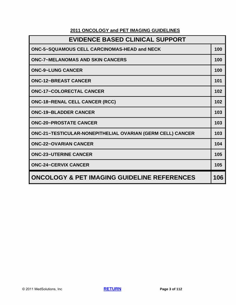
2011 ONCOLOGY and PET IMAGING GUIDELINES
EVIDENCE BASED CLINICAL SUPPORT
ONC-5~SQUAMOUS CELL CARCINOMAS-HEAD and NECK 100
ONC-7~MELANOMAS AND SKIN CANCERS 100
ONC-9~LUNG CANCER 100
ONC-12~BREAST CANCER 101
ONC-17~COLORECTAL CANCER 102
ONC-18~RENAL CELL CANCER (RCC) 102
ONC-19~BLADDER CANCER 103
ONC-20~PROSTATE CANCER 103
ONC-21~TESTICULAR-NONEPITHELIAL OVARIAN (GERM CELL) CANCER 103
ONC-22~OVARIAN CANCER 104
ONC-23~UTERINE CANCER 105
ONC-24~CERVIX CANCER 105
ONCOLOGY & PET IMAGING GUIDELINE REFERENCES 106

© 2011 MedSolutions, Inc RETURN Page 4 of 112
ABBREVIATIONS for ONCOLOGY GUIDELINES
ACTH adrenocorticotropic hormone
AFP alpha-fetoprotein AP Anteroposterior
beta-HCG beta human chorionic gonadotropin CA 19-9 cancer antigen 19-9
CA 125 cancer antigen 125 test CBC complete blood count
CEA carcinoembryonic antigen
CNS central nervous system
CTA computed tomography angiography
CR complete response DCIS ductal carcinoma in situ
DLBCL Diffuse Large B Cell Lymphomas DRE digital rectal exam
EGD esophagogastroduodensocopy ENT Ear, Nose, Throat
ERCP endoscopic retrograde cholangiopancreatography
ESR erythrocyte sedimentation rate EUA exam under anesthesia
EUS endoscopic ultrasound FDG Fluorodeoxyglucose
FNA fine needle aspiration FUO fever of unknown origin
GE gastroesophageal GI Gastrointestinal
GU Genitourinary
HIV human immunodeficiency disease
HRPC Hormone Refractory Prostate Cancer
LCIS lobular carcinoma in situ
LDH lactate dehydrogenase LFT liver function tests
MALT Mucosa Associated Lymphoid Tissue
MEN Multiple Endocrine Neoplasia MG myasthenia gravis
MGUS Monoclonal Gammopathy of Unknown Significance
MIBG I-123 metaiodobenzylguanidine scintigraphy
MRA magnetic resonance angiography
MRI magnetic resonance imaging
MUGA multiple gated acquisition scan
NCCN National Comprehensive Cancer Network
CONTINUED NEXT PAGE

© 2011 MedSolutions, Inc RETURN Page 5 of 112
ABBREVIATIONS for ONCOLOGY GUIDELINES
NHL Non-Hodgkin’s Lymphoma
NPC nasopharyngeal carcinoma
NSABP National Surgical Adjuvant Breast and Bowel Project
NSAIDS nonsteroidal anti-inflammatory drugs
NSCLC Non-Small Cell Lung Cancer
NSGCT Non-Seminomatous Germ Cell Tumor
PA Posteroanterior
PCI prophylactic cranial irradiation
PET positron emission tomography
POG Pediatric Oncology Group
PSA prostate specific antigen
RFA radiofrequency ablation
RPLND retroperitoneal lymph node dissection
SqCCa squamous cell carcinoma
SCLC Small cell lung cancer
SIADH syndrome of inappropriate secretion of antidiuretic hormone
TCC transitional cell carcinoma
TNM Tumor Node Metastasis staging system
TSH thyroid-stimulating hormone
TURBT trans-urethral resection of bladder tumor
VIPoma vasoactive intestinal polypeptide
WM Waldenstrom’s macroglobulinemia
© 2011 MedSolutions, Inc RETURN Page 6 of 112
2011 ONCOLOGY & PET IMAGING GUIDELINES
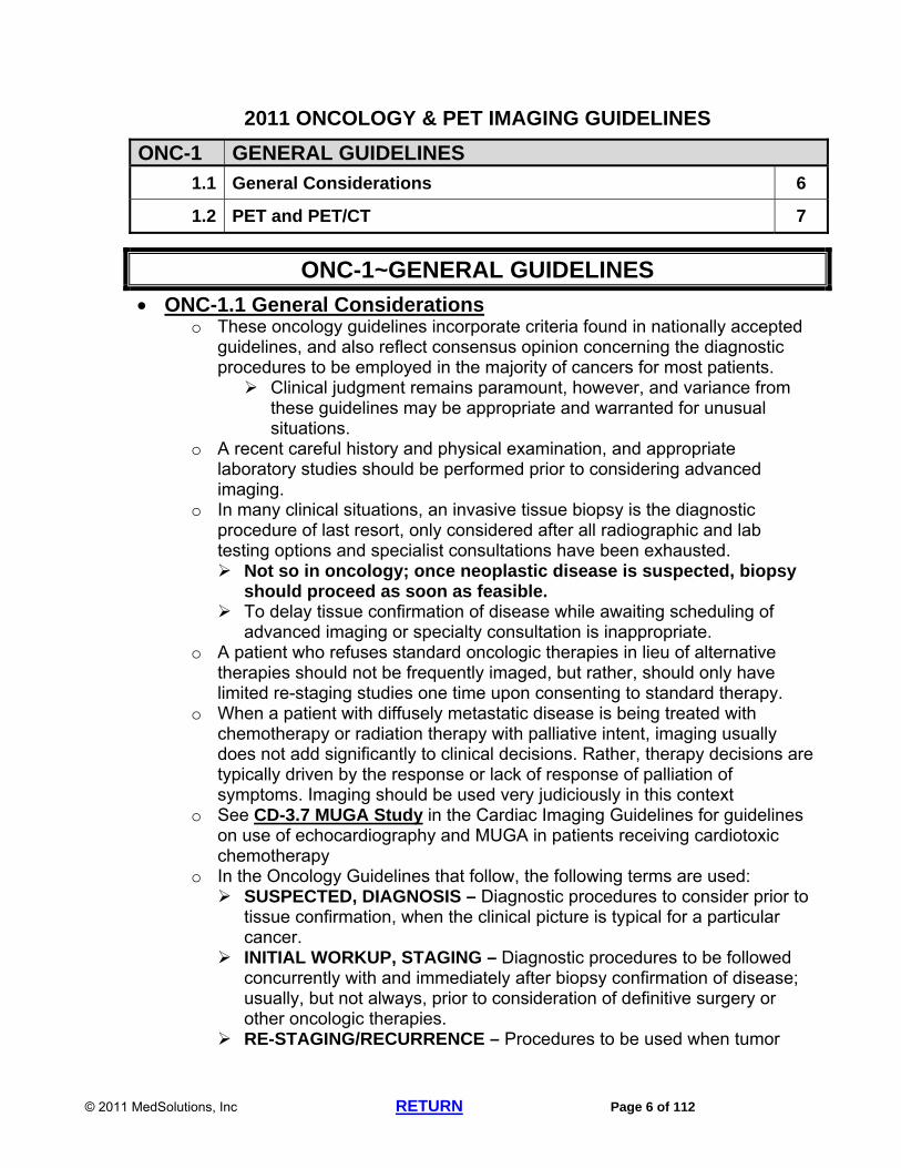
ONC-1 GENERAL GUIDELINES
1.1 General Considerations 6
1.2 PET and PET/CT 7
ONC-1~GENERAL GUIDELINES
ONC-1.1 General Considerations o These oncology guidelines incorporate criteria found in nationally accepted
guidelines, and also reflect consensus opinion concerning the diagnostic procedures to be employed in the majority of cancers for most patients. Clinical judgment remains paramount, however, and variance from
these guidelines may be appropriate and warranted for unusual situations.
o A recent careful history and physical examination, and appropriate laboratory studies should be performed prior to considering advanced imaging.
o In many clinical situations, an invasive tissue biopsy is the diagnostic procedure of last resort, only considered after all radiographic and lab testing options and specialist consultations have been exhausted. Not so in oncology; once neoplastic disease is suspected, biopsy
should proceed as soon as feasible. To delay tissue confirmation of disease while awaiting scheduling of
advanced imaging or specialty consultation is inappropriate. o A patient who refuses standard oncologic therapies in lieu of alternative
therapies should not be frequently imaged, but rather, should only have limited re-staging studies one time upon consenting to standard therapy.
o When a patient with diffusely metastatic disease is being treated with chemotherapy or radiation therapy with palliative intent, imaging usually does not add significantly to clinical decisions. Rather, therapy decisions are typically driven by the response or lack of response of palliation of symptoms. Imaging should be used very judiciously in this context
o See CD-3.7 MUGA Study in the Cardiac Imaging Guidelines for guidelines on use of echocardiography and MUGA in patients receiving cardiotoxic chemotherapy
o In the Oncology Guidelines that follow, the following terms are used: SUSPECTED, DIAGNOSIS – Diagnostic procedures to consider prior to
tissue confirmation, when the clinical picture is typical for a particular cancer.
INITIAL WORKUP, STAGING – Diagnostic procedures to be followed concurrently with and immediately after biopsy confirmation of disease; usually, but not always, prior to consideration of definitive surgery or other oncologic therapies.
RE-STAGING/RECURRENCE – Procedures to be used when tumor

© 2011 MedSolutions, Inc RETURN Page 7 of 112
progression is suspected. An exception to this is when high dose chemotherapy, with or
without radiation, has a high probability of “down-staging” a cancer, such as in lymphoma. Re-staging studies following such therapies are appropriate.
Re-staging a patient following adjuvant chemotherapy, whose primary modality of definitive local therapy was surgery, is inappropriate.*
*N Engl J Med 2006;354:496-507 SURVEILLANCE – Procedures to be used in the follow-up of patients
following standard therapies, when the patient is assumed to have either no known disease, or stable or clinically insignificant disease. Time frames listed under Surveillance sections in these Oncology
Imaging Guidelines mean from the end of active therapy for the primary disease, not including any “maintenance” therapies.
o MedSolutions does not routinely preauthorize requests for CT or MRI scans associated with image-directed biopsy or radiation therapy treatment planning. There is often no unique procedure code for a service performed only for
these indications. AMA instructions in CPT® state that if no specific code exists for a
particular service, the service is reported with an unlisted code. Imaging performed in support of radiation therapy treatment
planning should be reported with the corresponding therapeutic codes (CPT®77014 for CT scans, CPT®76498 for MRI scans, CPT®78999 for PET scans), not with diagnostic imaging codes. PET scans are being used for radiation therapy treatment planning,
but should be coded as CPT®78999 (unlisted procedure, diagnostic nuclear medicine) and NOT as diagnostic PET scans (CPT®78812, 78815, 78816) CPT®78999 does not require prior authorization by MedSolutions
Imaging associated with image-directed biopsy should be reported with the corresponding interventional codes (For specific CPT® codes, see Preface-4.2 CT-, MR-, or Ultrasound-Guided Procedures.
ONC-1.2.PET and PET/CT o All indications for PET also apply to PET/CT fusion scans In general, the anatomic detail acquired in PET/CT is reasonable for the
evaluation of many oncologic conditions; however, the diagnostic quality may be inconsistent.
For initial diagnosis or staging, a diagnostic CT may be appropriate in addition to a PET/CT.
For restaging, therapy monitoring, and evaluation of recurrence, either PET/CT or diagnostic CT, but not both, should be chosen by the clinician as the initial imaging modality.
o PET is a poor choice for imaging metastatic disease in the central nervous system (CNS).
o PET is unreliable for imaging lesions less than 7 mm in size.
© 2011 MedSolutions, Inc RETURN Page 8 of 112
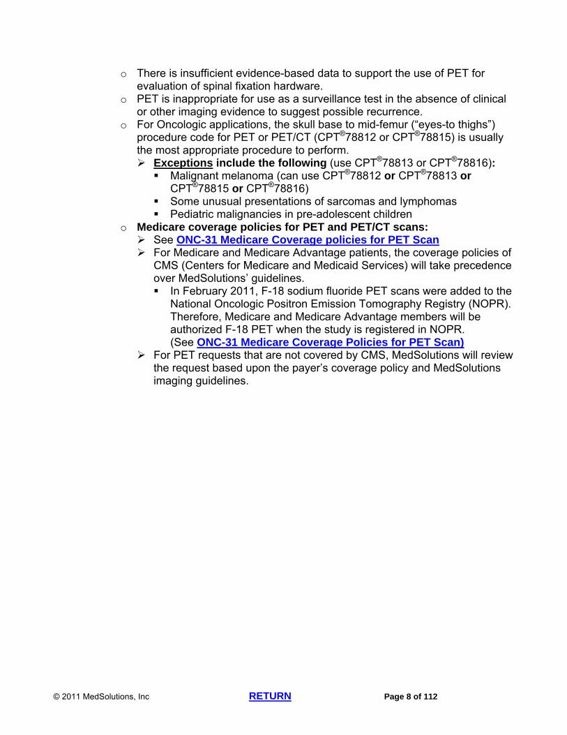
o There is insufficient evidence-based data to support the use of PET for evaluation of spinal fixation hardware.
o PET is inappropriate for use as a surveillance test in the absence of clinical or other imaging evidence to suggest possible recurrence.
o For Oncologic applications, the skull base to mid-femur (“eyes-to thighs”) procedure code for PET or PET/CT (CPT®78812 or CPT®78815) is usually the most appropriate procedure to perform. Exceptions include the following (use CPT®78813 or CPT®78816): Malignant melanoma (can use CPT®78812 or CPT®78813 or
CPT®78815 or CPT®78816) Some unusual presentations of sarcomas and lymphomas Pediatric malignancies in pre-adolescent children
o Medicare coverage policies for PET and PET/CT scans: See ONC-31 Medicare Coverage policies for PET Scan For Medicare and Medicare Advantage patients, the coverage policies of
CMS (Centers for Medicare and Medicaid Services) will take precedence over MedSolutions’ guidelines. In February 2011, F-18 sodium fluoride PET scans were added to the
National Oncologic Positron Emission Tomography Registry (NOPR). Therefore, Medicare and Medicare Advantage members will be authorized F-18 PET when the study is registered in NOPR. (See ONC-31 Medicare Coverage Policies for PET Scan)
For PET requests that are not covered by CMS, MedSolutions will review the request based upon the payer’s coverage policy and MedSolutions imaging guidelines.

© 2011 MedSolutions, Inc RETURN Page 9 of 112
2011 ONCOLOGY & PET IMAGING GUIDELINES
ONC-2~CENTRAL NERVOUS SYSTEM TUMORS and PET IN NEUROLOGY
See in the Head Imaging Guidelines: o HD-1 General Guidelines o HD-13 Dementia o HD-14 Adult Epilepsy/Seizure o HD-21 Movement Disorders o HD-23 Neuro-Oncology, Brain Tumors o HD-23.13 PET in Brain Tumor
See in the Pediatric and Congenital Head Imaging Guidelines: o PACHD-5 Pediatric Epilepsy/Seizure
Primary brain tumors presenting only with headache are very uncommon; most primary brain tumors present with a specific CNS finding, (e.g. seizures and/or symptoms of stroke).
Histologic confirmation is critical. Therapeutic decisions should not be made on radiographic findings alone. o Exceptions:
Medically fragile patients for whom attempted biopsy carries excess medical risk, as stated in writing by both the attending physician and surgeon.
Brain stem tumors or other sites where the risk of permanent neurological damage, even of a limited biopsy attempt, is excessive.
Brain MRI without and with contrast (CPT®70553) is generally all that is necessary prior to biopsy; however, some surgeons may appropriately desire both brain MRI and CT (contrast as requested). These may be approved when specifically requested by the responsible surgeon. o For posterior fossa tumors, tumors with evidence of leptomeningeal spread,
and multi-focal tumors, diagnosis by cytology via lumbar puncture should be considered, although this is not required.
Brain MRI without and with contrast (CPT®70553) every two to three months is indicated in the routine follow-up of aggressive tumor types such as anaplastic astrocytomas and glioblastomas, following definitive local therapy. o Such imaging is not indicated when supportive care only and/or hospice is
the primary care strategy.
ONC-3~CARDIAC PET SCAN See CD-7 Cardiac PET Scan in the Cardiac Imaging Guidelines

© 2011 MedSolutions, Inc RETURN Page 10 of 112
2011 ONCOLOGY & PET IMAGING GUIDELINES
ONC-4~USE OF PET FOR DISEASE THERAPY MONITORING in KNOWN METASTATIC DISEASE
Re-staging refers to studies performed at the end of a planned course of therapy and studies done at the time of a suspected or proven recurrence. Therapy monitoring refers to studies performed on an individual during a course of planned treatment when there is no significant change in the clinical status of the individual. o The use of PET in therapy monitoring is unproven and is considered
inappropriate. Exceptions: PET should only be considered for therapy monitoring in metastatic
disease when other modalities of assessment have been demonstrated to be of limited value and a change of therapy is indicated.
Therapy monitoring is appropriate in Lymphoma (See ONC-28 Lymphomas)
© 2011 MedSolutions, Inc RETURN Page 11 of 112
2011 ONCOLOGY & PET IMAGING GUIDELINES
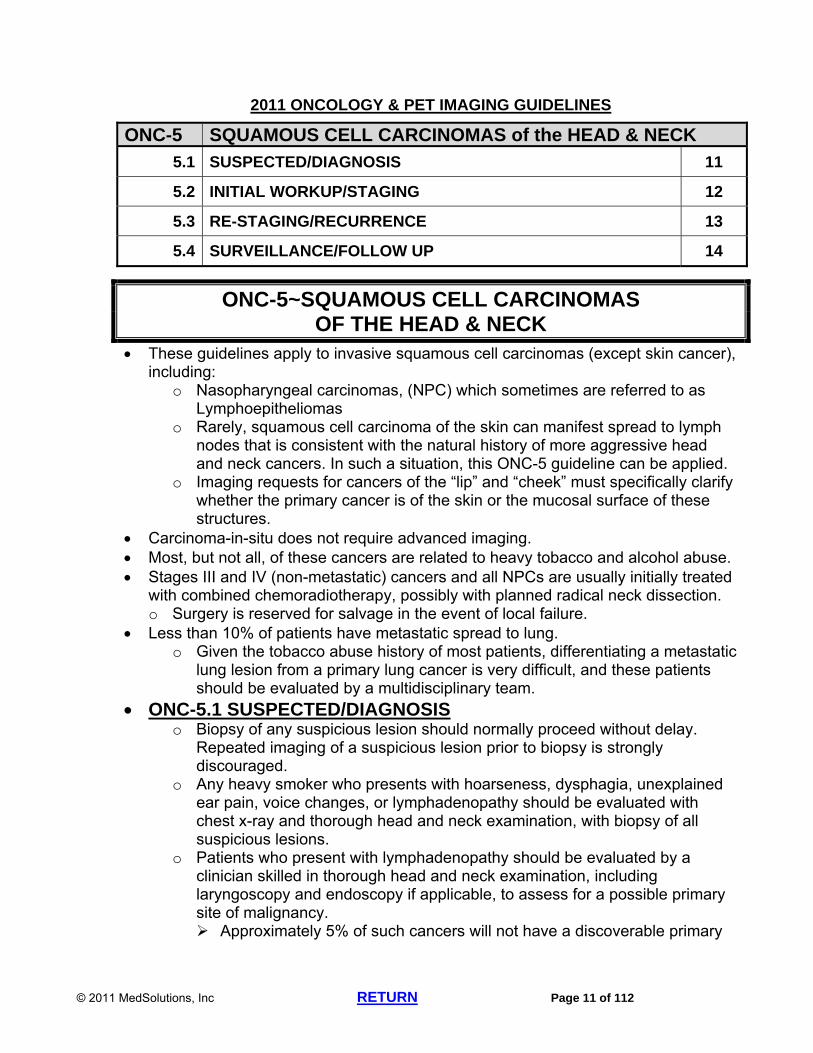
ONC-5 SQUAMOUS CELL CARCINOMAS of the HEAD & NECK
5.1 SUSPECTED/DIAGNOSIS 11
5.2 INITIAL WORKUP/STAGING 12
5.3 RE-STAGING/RECURRENCE 13
5.4 SURVEILLANCE/FOLLOW UP 14
ONC-5~SQUAMOUS CELL CARCINOMAS OF THE HEAD & NECK
These guidelines apply to invasive squamous cell carcinomas (except skin cancer), including:
o Nasopharyngeal carcinomas, (NPC) which sometimes are referred to as Lymphoepitheliomas
o Rarely, squamous cell carcinoma of the skin can manifest spread to lymph nodes that is consistent with the natural history of more aggressive head and neck cancers. In such a situation, this ONC-5 guideline can be applied.
o Imaging requests for cancers of the “lip” and “cheek” must specifically clarify whether the primary cancer is of the skin or the mucosal surface of these structures.
Carcinoma-in-situ does not require advanced imaging. Most, but not all, of these cancers are related to heavy tobacco and alcohol abuse. Stages III and IV (non-metastatic) cancers and all NPCs are usually initially treated
with combined chemoradiotherapy, possibly with planned radical neck dissection. o Surgery is reserved for salvage in the event of local failure.
Less than 10% of patients have metastatic spread to lung. o Given the tobacco abuse history of most patients, differentiating a metastatic
lung lesion from a primary lung cancer is very difficult, and these patients should be evaluated by a multidisciplinary team.
ONC-5.1 SUSPECTED/DIAGNOSIS o Biopsy of any suspicious lesion should normally proceed without delay.
Repeated imaging of a suspicious lesion prior to biopsy is strongly discouraged.
o Any heavy smoker who presents with hoarseness, dysphagia, unexplained ear pain, voice changes, or lymphadenopathy should be evaluated with chest x-ray and thorough head and neck examination, with biopsy of all suspicious lesions.
o Patients who present with lymphadenopathy should be evaluated by a clinician skilled in thorough head and neck examination, including laryngoscopy and endoscopy if applicable, to assess for a possible primary site of malignancy. Approximately 5% of such cancers will not have a discoverable primary

© 2011 MedSolutions, Inc RETURN Page 12 of 112
site. o CT neck with contrast (CPT®70491) is appropriate only after a thorough
head and neck examination, including laryngoscopy and endoscopy, or confirmation of a suspicious lesion on ultrasound.
o PET is not indicated until histologic diagnosis is confirmed, except in the following: Evaluation of pulmonary nodules: (See the following in the Chest
Imaging Guidelines: CH-12 Multiple Pulmonary Nodules CH-16 Solitary Pulmonary Nodule
In a patient who presents with a neck mass that requires direct laryngoscopy/exam under anesthesia for attempt at biopsy, PET may be helpful to direct efforts at biopsy.
ONC-5.2 INITIAL WORKUP/STAGING o All patients with these tumors should be evaluated by pan-endoscopy of
their upper aero-digestive system under general anesthesia and dental evaluation is immediately indicated unless the patient is edentulous.
o Staging is primarily by physical examination in these tumors. Imaging of the neck for possible nodal metastasis may be omitted at the clinician’s discretion in the presence of a clinically negative neck. “T” stage by physical exam must be stated in all imaging requests prior
to approval. o PET/CT (CPT®78815), neck CT with contrast (CPT®70491), and chest
imaging (chest x-ray or chest CT with contrast (CPT®71260) are indicated to complete staging. Neck MRI (contrast as requested) may also be required to further
evaluate unusual findings noted on CT. o PET/CT should NOT be used for imaging of a T1 lesion in sites with low
risk of nodal spread, such as the larynx, when there is no clinical evidence of adenopathy.
*J Oral Maxillofac Surg 2007;65:2524-2535 o PET/CT is indicated for the following: Occult primary To explain radiographic findings suggestive of disease outside the head
and neck area when a positive PET scan demonstrating metastatic disease will change management.
To provide information for directing biopsy for patients who present with a neck mass when initial attempts to find a primary source are difficult.
o Abdominal CT is only indicated when LFT’s are elevated, when signs and symptoms of metastatic disease are present, or in the setting of obviously abnormal intrathoracic findings. Liver lesions incidentally found during imaging do not require follow-up
advanced imaging unless LFT’s are elevated. o Pelvic CT is not indicated for the initial work-up of any head and neck
malignancy.

© 2011 MedSolutions, Inc RETURN Page 13 of 112
o Imaging of the CNS (head, spine) is only indicated to evaluate specific signs or symptoms suggesting such spread.
o Nasal cavity and paranasal sinuses may, in some cases, need both CT of maxillofacial area (CPT®70486) and facial MRI (CPT®70543) to assess extent of bony erosion, as well as skull base and intracranial involvement.
o For NPC’s initial staging is as above with the addition of the following: Neck MRI without and with contrast (CPT®70543) is preferred Head MRI without and with contrast (CPT®70553) is indicated for
neurological findings or if there is suspicion of base of skull invasion based on the above studies.
o PET/CT (CPT®78815) is indicated for all NPC’s. If not performed, neck CT with contrast (CPT®70491) and chest CT with contrast (CPT®71260) may be substituted in addition to the above MRI studies
o CT abdomen with contrast (CPT®74160) for lymph node positive NPC’s may be done, but this is optional.
ONC-5.3 RE-STAGING/RECURRENCE o Imaging studies for re-staging are inappropriate following complete resection
and/or radical neck dissection. The patient is considered to be in surveillance status after these procedures.
o Following radiation, patient should be followed clinically for 90 days, unless progression is suspected, as some tumors are slow to regress completely. Neck CT with contrast only (CPT®70491) (or Neck MRI, contrast as
requested, if a pre-treatment MRI will be used for comparison) can be performed at 30 days following completion of radiation.
o PET (CPT®78812) or PET/CT (CPT®78815) is optional after 90 days following completion of chemoradiotherapy, but only after documentation of a physical examination assessing the response to therapy. PET is particularly helpful in differentiating residual tumor from scar
tissue, which can be extensive. o PET is NOT indicated for the following: Less than 90 days following combined chemoradiotherapy, unless a new
lesion outside of the radiation portal is suspected. Exception: PET can be performed sooner in patients with
clinically apparent lymph nodes if the result of the PET will determine the need for radical neck dissection. In these patients, PET should be performed 8 to 12 weeks following therapy, to avoid excess radiation-induced fibrosis.*
* Head & Neck 2006;28:166-175 Laryngoscope 2005;115:2206-2208
For re-staging when surgery only was the primary treatment modality. When either complete response OR disease progression is clearly
obvious on physical examination. o Chest x-ray and LFT’s are usually adequate for re-staging. However, chest
CT with contrast (CPT®71260) can be performed if clinically indicated. o If recurrence is suspected, PET may be appropriate for apparent recurrent
lymphadenopathy, or for glottic tumors which cannot be adequately

© 2011 MedSolutions, Inc RETURN Page 14 of 112
visualized by a clinician capable of performing adequate examination with indirect laryngoscopy. PET scan requests in this context must be accompanied by
documentation of such an examination. Otherwise, recurrence must be confirmed by biopsy prior to
consideration of advanced imaging.
ONC-5.4 SURVEILLANCE/FOLLOW UP o Primarily clinical with repeated complete physical examination of all head
and neck structures. o Neck CT with contrast (CPT®70491), as well as any imaging found to be
abnormal during initial work-up, can be performed every 4 months for the first year, then every year after that for the next four years. Specific abnormalities may require more frequent follow-up.
o PET is not appropriate for surveillance. o Annual chest x-ray for life. o Chest CT with contrast (CPT®71260) is not routinely indicated for follow-up,
unless a suspicious abnormality arises on chest x-ray, or when suspicion of recurrence is detected elsewhere.

© 2011 MedSolutions, Inc RETURN Page 15 of 112
2011 ONCOLOGY & PET IMAGING GUIDELINES
ONC-6 SALIVARY GLAND CANCERS
6.1 SUSPECTED/DIAGNOSIS 15
6.2 INITIAL WORKUP/STAGING 16
6.3 RE-STAGING/RECURRENCE 16
6.4 SURVEILLANCE/FOLLOW UP 16
ONC-6~SALIVARY GLAND CANCERS Over a dozen histologic types of salivary gland tumors are described; most
common are adenoid cystic carcinomas, acinic cell carcinomas and mucoepidermoid carcinomas.
Parotid Tumors: o 83% are benign—the most common benign tumor is pleomorphic adenoma,
followed by Warthin’s tumor. o 17% are malignant—the most common malignant lesion is adenocystic
cancer. o A bilateral parotid mass is more likely to be a Warthin’s tumor. o Pleomorphic adenoma is a benign tumor but should be treated like a
malignant tumor if, and when, it recurs. o Parotid glands may also give rise to lymphomas;
Differentiating lymphomas from other histologies may be difficult on FNA.
o In parotid tumors requiring surgery, MRI is better than CT in assessing the position of the facial nerve in relationship to the parotid tumor. MRI gives a good assessment of the tumor mass and its anatomical
relationships in order to plan what operation will be required. Local recurrence and/or metastatic spread to lungs can occur. Lymph node spread is uncommon, therefore, repeated imaging of the neck and
elective therapy of neck (e.g. dissection, radiotherapy) are not indicated. Primary Squamous Cell Carcinoma of the Parotid Gland has been
described but is rare. If this histology is found in the parotid gland, metastatic spread from
another site should be aggressively ruled out. ONC-6.1 SUSPECTED/DIAGNOSIS
o Mass on palpation. o Palsies of cranial nerves VII, IX, or X. o Rarely, tumors in hypopharynx, larynx, or trachea may cause stridor. o Neck MRI without and with contrast (CPT®70543) is preferred. Alternatively, neck CT either with contrast or without contrast
(CPT®70491 or CPT®70490) can be performed, especially if requested by the ENT surgeon planning resection.

© 2011 MedSolutions, Inc RETURN Page 16 of 112
ONC-6.2 INITIAL WORKUP/STAGING o Biopsy by fine needle aspiration (FNA), open biopsy, or complete surgical
excision (as determined by an oral surgeon, or ENT physician, or other physician experienced in the management of salivary gland lesions) should proceed without delay.
o Brain imaging is usually not indicated unless abnormal neurologic signs or symptoms are present. Head CT without contrast (CPT®70450) is indicated if skull invasion is
suggested on MRI. o Chest x-ray o Chest CT (CPT®71260) is indicated only if there are abnormalities on chest
x-ray or if unusual lymphadenopathy is noted in the neck. o The role of PET in salivary gland tumors has yet to be established PET may be considered to evaluate suspicious abnormalities in the
lungs in accordance with the following in the Chest Imaging Guidelines: CH-12 Multiple Pulmonary Nodules CH-16 Solitary Pulmonary Nodule.
ONC-6.3 RE-STAGING/RECURRENCE o Imaging for re-staging is usually not indicated. o A single neck CT (CPT®70491) or neck MRI (CPT®70543) may be
performed 3 months after radiation therapy of an unresectable lesion. o If recurrence is suspected, neck MRI without and with contrast (CPT®70543)
is indicated. o Chest CT with contrast (CPT®72160) can be performed upon confirmation of
recurrence. ONC-6.4 SURVEILLANCE/FOLLOW UP
o Primarily by physical exam and chest x-ray only. o Neck CT with contrast (CPT®70491) every six months can be performed if
original tumor was unresectable. o The following imaging studies can be performed in the first year following
radiation therapy of an unresectable lesion: Two neck CT scans with contrast (CPT®70491), OR Two neck MRI scans without and with contrast (CPT®70543), OR One neck CT scan with contrast (CPT®70491) and one neck MRI without
and with contrast (CPT®70543) o PET is not indicated for routine surveillance.
© 2011 MedSolutions, Inc RETURN Page 17 of 112
2011 ONCOLOGY & PET IMAGING GUIDELINES
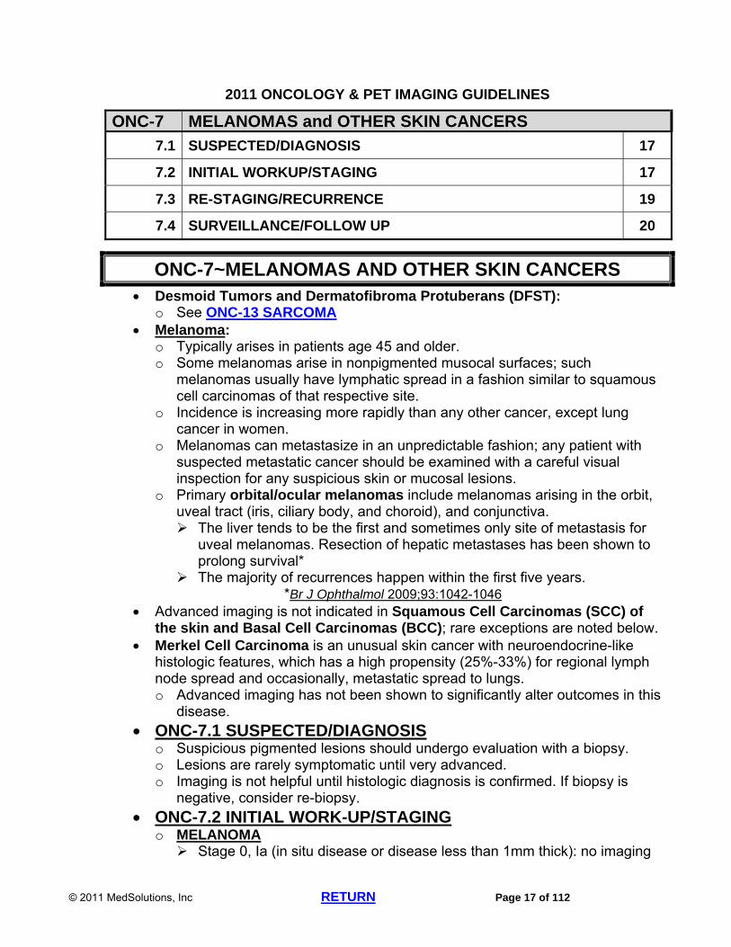
ONC-7 MELANOMAS and OTHER SKIN CANCERS
7.1 SUSPECTED/DIAGNOSIS 17
7.2 INITIAL WORKUP/STAGING 17
7.3 RE-STAGING/RECURRENCE 19
7.4 SURVEILLANCE/FOLLOW UP 20
ONC-7~MELANOMAS AND OTHER SKIN CANCERS Desmoid Tumors and Dermatofibroma Protuberans (DFST):
o See ONC-13 SARCOMA Melanoma:
o Typically arises in patients age 45 and older. o Some melanomas arise in nonpigmented musocal surfaces; such
melanomas usually have lymphatic spread in a fashion similar to squamous cell carcinomas of that respective site.
o Incidence is increasing more rapidly than any other cancer, except lung cancer in women.
o Melanomas can metastasize in an unpredictable fashion; any patient with suspected metastatic cancer should be examined with a careful visual inspection for any suspicious skin or mucosal lesions.
o Primary orbital/ocular melanomas include melanomas arising in the orbit, uveal tract (iris, ciliary body, and choroid), and conjunctiva. The liver tends to be the first and sometimes only site of metastasis for
uveal melanomas. Resection of hepatic metastases has been shown to prolong survival*
The majority of recurrences happen within the first five years. *Br J Ophthalmol 2009;93:1042-1046
Advanced imaging is not indicated in Squamous Cell Carcinomas (SCC) of the skin and Basal Cell Carcinomas (BCC); rare exceptions are noted below.
Merkel Cell Carcinoma is an unusual skin cancer with neuroendocrine-like histologic features, which has a high propensity (25%-33%) for regional lymph node spread and occasionally, metastatic spread to lungs. o Advanced imaging has not been shown to significantly alter outcomes in this
disease.
ONC-7.1 SUSPECTED/DIAGNOSIS o Suspicious pigmented lesions should undergo evaluation with a biopsy. o Lesions are rarely symptomatic until very advanced. o Imaging is not helpful until histologic diagnosis is confirmed. If biopsy is
negative, consider re-biopsy.
ONC-7.2 INITIAL WORK-UP/STAGING o MELANOMA Stage 0, Ia (in situ disease or disease less than 1mm thick): no imaging

© 2011 MedSolutions, Inc RETURN Page 18 of 112
Stage Ib, II (Lesions less than or equal to 1 mm with ulceration or high mitotic rate, or lesions >1 mm thick, but node negative):
Chest CT with contrast (CPT®71260) Additional CT or MRI of specific areas, only if signs or symptoms indicate
a need for further evaluation. Stage III or greater, node positive, including palpable or imaging-only
positive ) Chest CT with contrast (CPT®71260) for abnormalities noted on chest
x-ray. Abdominal CT with contrast (CPT®74160) if elevated LDH or
abdominal abnormalities noted on other imaging modalities. CT with contrast of body area containing regional lymphatics nearest
to the original site of disease. PET (CPT®78812 or 78813 or CPT®78815 or 78816) is indicated if
disease is initially found in a lymph node or distant organ. PET (CPT®78812 or 78813 or CPT®78815 or 78816) may be
considered to address specific signs and symptoms not explained by conventional imaging.
Brain MRI without and with contrast (CPT®70553), can be performed for any CNS symptoms, or preoperatively if the proposed procedure involves significant risk of morbidity.
All orbital/ocular melanomas should be considered to be locally advanced. Primary orbital melanomas include melanomas arising in the orbit,
uveal tract (iris, ciliary body, and choroid), and conjunctiva. Initial staging of any orbital/ocular melanoma can include PET/CT
(CPT®78815 or 78816) and CT Abdomen with contrast (CPT®74160).
References: N Engl J Med 2006;355:1307-1317 Arch Surg 2006;141:284-288 Melanoma Res 2007;17:147-154 Cancer 2005;104:570-579 Ann Surg Oncol 2006;13:525-532
Stage IV (metastatic) Brain MRI (CPT®70553) and CT of body areas not previously imaged,
one time, then only for specific sign or symptoms. PET (CPT®78812 or 78813 or CPT®78815 or 78816) and/or
abdominal MRI without and with contrast (CPT®74183) are helpful when the location of the primary melanoma site is in doubt or when the extent of organ involvement by melanoma is unclear based upon other studies.
o OTHER SKIN CANCERS Advanced imaging is not usually indicated, except to further define
abnormalities found on chest x-ray or physical examination. PET is not indicated for non-melanomatous skin cancers (Exception:
see Merkel Cell Carcinoma below).
© 2011 MedSolutions, Inc RETURN Page 19 of 112
Other exceptions where advanced imaging can be useful include the following: Any non-melanoma skin cancer of the head and neck area showing
significant evidence of perineural involvement should be evaluated with imaging, MRI or CT, (clinician’s preference), contrast as requested, of the base of skull to evaluate for neural ganglion involvement.
PET is beneficial if a pathology report suggests a skin lesion may be a dermal metastasis from a distant primary.
Squamous cell carcinoma of the skin of the head or neck presenting with regional lymphadenopathy should be evaluated with chest and neck CT with contrast (CPT®71260 and CPT®70491).
Merkel Cell Carcinoma PET for Merkel Cell carcinoma can be performed for staging of
apparently localized disease after standard imaging. PET is not indicated if other imaging studies show obvious metastatic disease.
Merkel Cell Carcinoma under consideration for adjuvant chemotherapy or radiotherapy: an additional CT or MRI of the affected region, at the oncologist’s discretion, may be performed to define appropriate therapy.
Merkel Cell Carcinoma, lymph node positive, may be evaluated with chest and abdomen CT with contrast (CPT®71260 and CPT®74160).
ONC-7.3 RE-STAGING/RECURRENCE o All recurrences should be confirmed histologically, except when excessive
morbidity from a biopsy may occur, such as a biopsy requiring craniotomy. o MELANOMA Re-staging in melanoma is not appropriate after adequate aggressive
local therapy, because all patients are then followed according to the surveillance strategy detailed under Surveillance/Follow-up section below.
For local recurrence only: Re-stage with chest x-ray, LDH, and CT scans of body areas judged
to be clinically relevant. MRI or CT head (CPT®70553 or CPT® 70470) only if there are new CNS signs or symptoms.
If melanoma recurs in regional lymph nodes or for metastatic recurrence: CT chest (CPT®71260), abdomen (CPT®74160 or CPT®74170),
pelvis (CPT®72193) or abdomen and pelvis (CPT®74178), head MRI without and with contrast (CPT®70553), and PET (CPT®78812 or 78813 or CPT®78815 or 78816) are appropriate.
Head Imaging (brain MRI or CT without and with contrast (CPT®70553 or CPT®70470) is indicated for the following: Presence of CNS related symptoms, Recurrence involving lymph nodes or in-transit disease

© 2011 MedSolutions, Inc RETURN Page 20 of 112
New discovery of metastatic disease. PET is not appropriate when CT scans demonstrate multi-organ
metastasis, but may be helpful when other imaging studies suggest either isolated metastatic disease amenable to resection, or recurrence only in the lymph node(s).
o OTHER SKIN CANCERS Recurrence of Merkel Cell Carcinoma: Chest CT (CPT® 71260), abdominal CT with contrast (CPT®74160),
and CT of the affected regional site. PET for Merkel Cell carcinoma can be performed for staging of
apparently localized disease after standard imaging. PET is not indicated if other imaging studies show obvious metastatic disease.
If non-melanomatous skin cancer recurs, and therapy is planned that is more extensive than simple wide local excision, then CT with contrast of the primary site is indicated.
ONC-7.4 SURVEILLANCE/FOLLOW UP o Primarily with physical exam. o Chest x-ray and LFT’s are performed in patients clinically judged to have
risk of metastatic spread. o The routine use of advanced imaging in the surveillance of melanoma has
not demonstrated any benefit, except in the setting of specific signs or symptoms suggesting the possibility of recurrence. EXCEPTION: For orbital/ocular melanomas, CT chest and abdomen with contrast
(CPT®71260 and CPT®74160) every 6 months for two years, then every year for three years can be performed.
o For melanomas, CT with contrast, (MRI only on a case-by-case basis) may be approved on an annual basis for five years for body areas with previously documented radiographic findings or pathologically involved lymph nodes.
o PET and head imaging for routine surveillance in all skin cancers, including melanoma, is not indicated.

© 2011 MedSolutions, Inc RETURN Page 21 of 112
2011 ONCOLOGY & PET IMAGING GUIDELINES
ONC-8 THYROID CANCER
8.1 SUSPECTED/DIAGNOSIS 21
8.2 INITIAL WORKUP/STAGING 22
8.3 RE-STAGING/RECURRENCE 22
8.4 SURVEILLANCE/FOLLOW UP 23
ONC-8~THYROID CANCER Thyroid cancer is three to four times more prevalent in women than men. One of the few cancers where a clear relationship between carcinogenesis and
radiation exposure is established. While most thyroid cancers occur randomly, a strong familial pre-disposition is
known to occur. Imaging decisions for the management of Hurthle Cell, Follicular, and Papillary
carcinomas are essentially identical. Medullary and Anaplastic Thyroid Carcinomas have significantly different
diagnostic and therapeutic algorithms. ONC-8.1 SUSPECTED/DIAGNOSIS
o Palpable nodules in patients with one or more of the following risk factors: Age <15 or >45 with new onset nodule Male sex Size greater than 4 cm or any size with rapid growth Vocal cord paralysis Regional lymphadenopathy Symptoms suggestive of tumor invasion into the neck structures Very firm or fixed nodule History of radiation exposure, particularly as a young person Family history of thyroid cancer Ultrasound appearance described by radiologist as suspicious
o Suspicious lesions having the above risk factors should be assessed by neck ultrasound (CPT®76536), TSH, and FNA of the nodule and/or any suspicious lymph nodes.* Neck MRI without contrast (CPT®70540) can be performed in situations
in which the utility of ultrasound is limited. CT with contrast is relatively contraindicated because of iodine load *
Nondiagnostic FNA is not an indication for advanced imaging. Consider repeat FNA under ultrasound guidance or surgery.
*Cancer Control 2006 April;13(2):89-98, 99-105 o Thyroid nodules with suspicious criteria but <1 cm without adenopathy or
suspicious findings on ultrasound, or nodules <4 cm with no suspicious criteria are followed clinically. No advanced imaging is indicated.

© 2011 MedSolutions, Inc RETURN Page 22 of 112
o An incidentally identified thyroid lesion that is positive on PET scan should be evaluated by ultrasound (CPT®76536), (not CT or MRI) per the above guidelines.
o There is insufficient evidence-based data to support the use of PET scan to distinguish indeterminate thyroid nodules that are benign from those that are malignant.*
*Am J Otolaryngol 2008;29:113-118
ONC-8.2 INITIAL WORK-UP/STAGING o Chest x-ray, laboratory studies o Surgery is the primary therapy for disease, may be required to confirm
diagnosis, and may proceed even in the setting of known metastatic disease.
o Noncontrast Neck CT (CPT®70490) or MRI (CPT®70540) is indicated if fixation is suggested by ultrasound or clinical exam, or if substernal disease precludes full ultrasound examination. Otherwise, pre-operative advanced imaging is not indicated.
o Chest CT without contrast (CPT®71250) can be performed if any suspicious lesions are found on chest x-ray or ultrasound only if results of the imaging will be used to change management decisions. Use of iodinated contrast is discouraged as its use complicates the utility
of post-operative radioactive iodine. MRI may be considered in selected cases if clinically warranted. Chest CT with contrast (CPT®71260), neck CT with contrast
(CPT®70491) or neck MRI without and with contrast (CPT®70543) can be performed preoperatively for medullary and anaplastic carcinomas.
CT scans of abdomen, pelvis, and head, contrast as requested, can be performed for Anaplastic Carcinomas.
o Skeletal pain should be assessed with bone scan initially. o Head imaging is not routinely indicated in the absence of neurological signs
or symptoms, except for initial staging of anaplastic carcinomas. o PET is not indicated in the initial diagnosis or staging of thyroid
cancer. ONC-8.3 RE-STAGING/RECURRENCE
o Most thyroid cancers of follicular cell origin are assessed postoperatively by serum thyroglobulin levels and radioiodine whole body scan with the patient off thyroid replacement medications.
o When recurrent or persistent disease is detected on radioiodine scan, proceed directly to considering radioiodine therapy. Advanced imaging is not relevant as it will not change therapeutic decisions. Imaging studies may take up to 6 months to normalize after radioiodine;
imaging during such time will not be expected to yield clinically significant information unless there is a change in the patient’s clinical status.
o Advanced imaging (CT, MRI) is usually unnecessary in the setting of low thyroglobulin levels and negative radioiodine scan, except for medullary and anaplastic carcinomas.
o PET is not indicated for routine use in re-staging.

© 2011 MedSolutions, Inc RETURN Page 23 of 112
o Recurrence suggested by elevated thyroglobulin level, radioiodine scan, neck ultrasound or physical exam: CT with contrast or MRI can be performed of any site of suspected
recurrence, if new symptoms develop. PET (CPT®78815) is indicated for any of the following: When radioiodine scan is negative but repeated serum thyroglobulin
tests demonstrate a rising thyroglobulin level. Anaplastic thyroid cancers.*
*Cancer Control 2005;12(4):254-260 When abnormalities are found on other imaging studies that cannot
be explained (especially true for Hurthle Cell carcinomas). If highly morbid surgery or radiotherapy is contemplated for salvage
or recurrence.* *Cancer Control 2006;13(2):89-105
o If elevated calcitonin or CEA levels in patients with medullary carcinoma: CT with contrast of Neck (CPT®70491), Chest (CPT®71260), and
Abdomen (CPT®74160) (but not pelvis) can be performed or PET/CT (CPT®78815)
ONC-8.4 SURVEILLANCE/FOLLOW UP o Predominantly by neck ultrasound (CPT®76536), chest x-ray, and laboratory
studies every three months for the first two years, then annually. Annual radioiodine scans if above are suspicious, or if patient had prior
requirement for radioiodine. CEA and calcitonin levels are required for monitoring Medullary
Carcinomas o Routine advanced imaging is not indicated. Exception: in anaplastic carcinomas, neck CT with contrast
(CPT®70491), and either chest x-ray or chest CT (CPT®71260) every 3 to 6 months are indicated.
o PET is NOT Indicated for routine use in surveillance.

© 2011 MedSolutions, Inc RETURN Page 24 of 112
2011 ONCOLOGY & PET IMAGING GUIDELINES
ONC-9 LUNG CANCER
9.1 SMALL CELL LUNG CARCINOMA (SCLC) 24
9.2 SUSPECTED/DIAGNOSIS 25
9.3 INITIAL WORKUP/STAGING 26
9.4 RE-STAGING/RECURRENCE 26
9.5 SURVEILLANCE/FOLLOW UP 28
ONC-9~LUNG CANCER Lung cancer is the leading cause of cancer death in both men and women in
the United States. o Approximately 90% of all lung cancers are felt to be linked to exposure to
tobacco products, including second hand smoke. A notable exception is adenocarcinoma in women, where the association with tobacco smoke exposure is just over 50%.
Non-small Cell Lung Cancers (NSCLC) are staged with a TNM staging system o Stage I, II, and some Stage III cancers can be primarily treated with surgery
and frequently with adjuvant chemotherapy. o Most stage III cancers and medically inoperable Stage I and II cancers are
treated with curative intent with combined chemoradiotherapy. o Certain patients with a single focus of metastatic disease can achieve a
significant survival advantage when the metastatic focus, along with the primary tumor, is aggressively treated with local modalities.
ONC-9.1 SMALL CELL LUNG CARCINOMA (SCLC) o Not staged by a TNM system. o Most small cell carcinomas present as “extensive stage” tumors, defined as
either metastatic disease or an extent of disease which cannot be encompassed by a single radiotherapy portal. These are treated with chemotherapy alone, except for palliative
radiotherapy directed at symptomatic sites. o Small cell carcinomas that are non-metastatic, and that also can be
encompassed by a single radiotherapy portal, are termed “limited-stage,” and are treated with combined chemoradiotherapy and sometimes with elective cranial irradiation.
o Surgical resection of a primary small cell carcinoma is appropriate in certain situations.
o PET is inappropriate for re-staging small cell carcinomas.* *J Thorac Oncol 2007;2(4):348-354 *Ruckdeschel J. Cancer of the Lung: NSCLC and SCLC. In Abeloff (Ed.) Clinical Oncology. 3rd Ed. Philadelphia, Elsevier, Inc, 2004,pp.1649-1743

© 2011 MedSolutions, Inc RETURN Page 25 of 112
ONC-9.2 SUSPECTED/DIAGNOSIS o Screening for Lung Cancer: There is currently insufficient data to recommend routine screening
for lung cancer. The American Cancer Society recommends that CT screening not be performed in asymptomatic at risk persons.*
*N Engl J Med 2005;352:2714-2720 *JNCCN 2006;4:591-594
o Suspected Lung Cancer and Workup of Solitary Pulmonary Nodule: Chest x-ray, (PA/Lateral), possibly with AP lordotic view, should be
performed whenever lung cancer is suspected. When clinical suspicion remains high in spite of a normal chest x-ray,
chest CT with contrast (CPT®71260) or without contrast (CPT®71250) can be approved.
PET is appropriate for the evaluation of one or more newly discovered, distinct pulmonary nodules, confirmed on CT and/or MRI and measuring greater than or equal to 7 mm (0.7 cm) on those scans.*
*Applied Radiology 2002;31(6):9-17 NOTE: Certain payers consider PET scan investigational for
evaluating pulmonary nodules ≤1 cm or lung masses >4 cm. Their coverage policies will take precedence over MedSolutions’ guidelines.
A PET scan used for evaluation of a pulmonary nodule or other suspicious imaging findings also qualifies as the initial staging PET scan in the event of a positive biopsy. Obtaining a second PET scan in this context is inappropriate.
PET is of limited value for lesions demonstrating a ground-glass or pneumonic (pneumonia-like) appearance.
Performing more than one PET scan is inappropriate PRIOR to biopsy or other histologic confirmation of cancer. Uncertain or nondiagnostic findings on PET scan should be followed by anatomic imaging or biopsy.
If PET scan is negative, chest CT should be performed at 3, 9 and 24 months.*
*Radiology 2005;237:395-400 See CH-16 Solitary Pulmonary Nodule in the Chest Imaging
Guidelines for additional guidelines on the use of PET for evaluation of a solitary pulmonary nodule.
PET may not be helpful in evaluating enlarged mediastinal lymph nodes and should not be considered unless biopsy has been performed, biopsy is medically contraindicated, or biopsy is technically not feasible.*
*Lung Cancer 2008 April;60:62-68 Lung cancers can cause paraneoplastic syndromes such as Lambert-
Eaton (proximal leg weakness), encephalomyelitis, and sensory neuropathy. Small cell lung cancer can produce ACTH (Cushing’s syndrome) or vasopressin.

© 2011 MedSolutions, Inc RETURN Page 26 of 112
Imaging to evaluate a paraneoplastic syndrome should still follow the guidelines listed in: ONC-30.3 Paraneoplastic Syndromes
ONC-9.3 INITIAL WORKUP/STAGING o Biopsy is performed by bronchoscopy or CT-directed biopsy. If these fail or
are not feasible, video-assisted thoracoscopy or thoracotomy should be considered.
o All patients should have chest x-ray, CT chest/abdomen with contrast (CPT®71260/74160), and PET/CT (CPT®78815) if histologic diagnosis is confirmed and these imaging studies have not already been performed prior to biopsy.
o A PET scan that was used for evaluation of a pulmonary nodule or other suspicious imaging findings also qualifies as the initial staging PET scan in the event of a positive biopsy. Obtaining a second PET scan for initial staging is inappropriate.
Bone scan is optional but recommended if any skeletal pain is present and to stage small cell lung carcinoma.*
*Lung Cancer 2008 April;60:62-68
o Carcinoid and other unusual neuroendocrine histologies should be evaluated according to ONC-16 Neuroendocrine Cancers and Adrenal Tumors
o PET for small cell lung carcinoma only for staging of apparently limited stage disease after standard imaging. PET is not indicated if other imaging studies show extensive stage disease, and PET is not indicated following chemotherapy.
o Brain Imaging: Brain MRI (contrast as requested) (preferred) or head CT (contrast as
requested) can be performed in patients with small cell lung carcinoma (any stage) or in patients with non-small cell lung cancer stage II (T1-2, N1) or above.
Brain MRI (CPT®70553) should be performed in patients with lung cancer of any type or stage who have neurologic signs or symptoms.
o Superior sulcus tumor (Pancoast tumor): MRI chest without and with contrast (CPT®71552) if surgery is a
therapeutic possibility. MRI of cervical (CPT®72156) and/or thoracic spine (CPT®72157), if other
imaging modalities or signs/symptoms suggest a possibility of neural foraminal encroachment by the tumor.
o CT of neck or pelvis, MRI of suspected bone metastases, MRI abdomen, and duplicative imaging of head (MR or CT) are usually not indicated unless the clinical rationale is fully explained.
ONC-9.4 RE-STAGING/RECURRENCE o Re-staging studies, including PET, are not indicated if definitive resection
was the initial treatment (applies to most Stage I and II cancers) and all known disease was completely resected if a definitive local procedure was performed, or for tumors initially treated with radiation therapy as the only treatment modality. Patients should go directly to surveillance status.

© 2011 MedSolutions, Inc RETURN Page 27 of 112
Radiofrequency ablation (RFA), Extra-cranial Stereotactic Radiosurgery (ESRS) and other types of radiosurgery are considered definitive local procedures.
o Re-staging studies are appropriate for the following: Any SCLC, but PET is not appropriate for re-staging SCLC Locally advanced NSCLC (Stage III, non-metastatic, unresectable, or
inoperable tumor if chemotherapy was the initial treatment modality, or for inadequately resected disease)
o Re-staging studies include CT chest and abdomen with contrast (CPT®71260 and CPT® 74160). CT or MRI scans of other areas can be performed if symptoms strongly
suggest possible metastatic disease. Chest MRI may be indicated to determine resectability following neo-
adjuvant therapy. o PET is not indicated for re-staging of lung cancer, except for the following: PET is useful for evaluation of abnormalities that are newly discovered
by CT or other imaging modalities used for re-staging. PET may be considered if LFT’s or tumor markers become elevated and
CT scans are negative or equivocal. PET can differentiate persistent or recurrent tumor from necrotic or
fibrotic tissue following chemotherapy or radiotherapy. PET may help differentiate persistent or recurrent tumor from radiation-
induced fibrosis or pleural thickening. NOTE: PET following radiotherapy should be delayed a minimum of
12 weeks, due to risk of false positive FDG uptake in lethally irradiated cells and in radiation pneumonitis, unless the clinical situation requires evaluation of disease outside the irradiated volume, or if needed to allow an imminent resection attempt.
o For metastatic disease: Re-staging with CT and clinical assessment of change of symptoms is
the preferable modality. PET scan can be considered on a case-by-case basis if symptom
response cannot be used to judge efficacy of therapy. There are no evidence-based data addressing the efficacy of imaging in
the setting of metastatic disease, since most therapy protocols are palliative in nature.
Imaging, if needed, should be performed no more than once every 60 days.
o Brain MRI (CPT®70553) (preferred) or head CT (CPT®70470): For limited stage small cell lung carcinomas, if prophylactic cranial
irradiation (PCI) is planned. Following pre-operative chemoradiation therapy, if an adequate
response to therapy is demonstrated, and if evidence of intracranial disease will preclude surgery
Upon evidence of recurrence of any lung cancer
© 2011 MedSolutions, Inc RETURN Page 28 of 112
If recurrence is suspected, the same imaging studies as initial work-up protocols plus head imaging (mentioned above) can be performed. Therapies for recurrence are rarely curative.
ONC-9.5 SURVEILLANCE/FOLLOW UP o PET is inappropriate for routine surveillance. o All patients: Chest x-ray or chest CT with contrast (CPT®71260) every 4 to 6
months for first two years, then annually for an additional eight years. o Bone scan is the preferred initial study for bone pain or suspicion of skeletal
disease, unless neurologic compromise is evident. See ONC-29.4 Metastatic Cancer Bone (and Spine)
o Imaging of CNS is only appropriate for any CNS related symptoms.

© 2011 MedSolutions, Inc RETURN Page 29 of 112
2011 ONCOLOGY & PET IMAGING GUIDELINES
ONC-10 ESOPHAGEAL CANCER
10.1 SUSPECTED/DIAGNOSIS 29
10.2 INITIAL WORKUP/STAGING 29
10.3 RE-STAGING 30
10.4 SURVEILLANCE/FOLLOW UP 30
ONC-10~ESOPHAGEAL CANCER Clinicians must describe esophageal cancer by cell type and in which third of
the esophagus they occur. o Cancers of the upper and middle third are usually squamous cell and are
highly associated with tobacco and alcohol abuse. o Cancers of the gastroesophageal (GE) junction are treated as lower third
cancers. Lower third cancers are usually adenocarcinomas; 62% of these arise in
the setting of Barrett’s esophagus, a condition associated with high body mass index.
ONC-10.1 SUSPECTED/DIAGNOSIS o Upper GI endoscopy (EGD) is the initial procedure of choice. o Barium swallow (esophagram) and endoscopic ultrasound can also be
useful. o CT chest with contrast (CPT®71260) can be performed o PET prior to histologic diagnosis is only indicated when the above studies
yield conflicting information or when an attempted biopsy is non-diagnostic or technically not feasible.
ONC-10.2 INITIAL WORKUP/STAGING o Transesophageal ultrasound is indicated if no metastases are suspected
and patient is considered a surgical candidate. o CT chest/abdomen with contrast (CPT®71260/74160) and PET/CT
(CPT®78815) for all patients with biopsy-proven esophageal cancer. Abdominal CT may be without and with contrast (CPT®74170) if clinically
justified. Pelvic imaging is not indicated Chest MRI is not routinely indicated but may be considered for specific
surgical indications. o CT neck with contrast (CPT®70491) may be appropriate for tumors of the
upper third and/or patients with possible neck mass. o Head imaging (MRI or CT) without and with contrast (CPT®70553 or
CPT®70470) should be performed only if neurological signs/symptoms are present.
© 2011 MedSolutions, Inc RETURN Page 30 of 112
o PET (CPT®78815) is indicated for initial staging, but is not appropriate if there is obvious metastatic disease found on other imaging or if performance status precludes consideration of aggressive local therapy.
ONC-10.3 RE-STAGING o PET/CT (CPT®78815) or CT chest (CPT®71260) and abdomen
(CPT®74160) following chemotherapy and/or radiation therapy can be performed for re-staging. PET should be delayed as much as feasible to allow time for tumor
response to be assessed, but not so late as to unduly delay surgery if surgery is feasible.
PET is not appropriate if there is clinically obvious progression of disease or patient deterioration.
o NOTE: PET/CT for interim re-staging (response assessment): PET/CT and other imaging studies following preoperative chemotherapy
are not required for patients who have adenocarcinoma of the distal esophagus or esophagogastric junction who are medically fit with resectable disease found on initial staging.
o Brain MRI (or CT), contrast as requested, should be performed only if there are new signs or symptoms of CNS involvement.
o Routine pelvic and neck imaging are not indicated unless there was previously documented involvement of these areas or there is new onset of signs or symptoms in these areas.
o If there is new or progressive elevation of LFT’s or tumor markers, or any other documented evidence of recurrence or progression of disease, CT chest (CPT®71260), CT abdomen (contrast as requested), and CT of any other symptomatic area can be performed.
o PET for re-staging when recurrence is found is only indicated if the patient is a candidate for aggressive local salvage therapy.
o PET is not indicated for patients being managed with best supportive care. ONC-10.4 SURVEILLANCE/FOLLOW UP
o Advanced imaging not routinely indicated. o Primarily with endoscopy, chest x-ray, clinical follow-up, laboratory studies o CT chest with contrast (CPT®71260) and/or abdomen with contrast
(CPT®74160) are indicated when patient has progression of symptoms, abnormalities of above tests, or continued weight loss.
o MRI is not routinely indicated unless specifically recommended by radiologist to address an abnormality not adequately described by CT.
o PET for surveillance is not indicated.

© 2011 MedSolutions, Inc RETURN Page 31 of 112
2011 ONCOLOGY & PET IMAGING GUIDELINES
ONC-11 OTHER THORACIC TUMORS
11.1 MALIGNANT PLEURAL MESOTHELIOMA 31
11.1.1 SUSPECTED/DIAGNOSIS 31
11.1.2 INITIAL WORKUP/STAGING 31
11.1.3 RE-STAGING 32
11.1.4 SURVEILLANCE 32
11.2 THYMOMA 32
11.2.1 SUSPECTED/DIAGNOSIS 32
11.2.2 INITIAL WORKUP/STAGING 32
11.2.3 RE-STAGING 32
11.2.4 SURVEILLANCE 32
ONC-11~OTHER THORACIC TUMORS
ONC-11.1 MALIGNANT PLEURAL MESOTHELIOMA o ONC-11.1.1 SUSPECTED/DIAGNOSIS Especially prevalent in patients with asbestos and smoking history. Presenting Signs/Symptoms: Dyspnea, chest pain, cough, night sweats, palpitations (from
arrhythmias secondary to pericardial involvement), fatigue, dysphasia, pleural effusions, ascites.
Frequently causes paraneoplastic syndromes, especially thrombocytosis. Chest x-ray, CT chest with contrast (CPT®71260), pleural cytology
analysis, and pleural biopsy (usually by thoracoscopy).* *Cancer Control 2006;13:255-263 *J Thorac Oncol 2011;6:602-605
*Cancer 2007;110:2248-2252
o ONC-11.1.2 INITIAL WORKUP/STAGING Tumors are usually locally extensive prior to becoming metastatic and
can extend locally or metastasize into the peritoneal cavity, contralateral pleura, and/or lung. Staging with CT chest and abdomen with contrast (CPT®71260 and
CPT®74160) and PET/CT (CPT®78815) is indicated. Imaging of other sites is indicated only for symptoms or clinical
suspicion. Chest MRI (CPT®71552) may be considered for surgical planning when
requested by the operating surgeon.

© 2011 MedSolutions, Inc RETURN Page 32 of 112
o ONC-11.1.3 RE-STAGING Suspected recurrence should be re-staged with the imaging studies
included in INITIAL WORKUP, STAGING section above. o ONC-11.1.4 SURVEILLANCE Chest CT (CPT®71260) every 3 months for 2 years, then every year for
life. Other imaging may be done of previously positive sites or of newly
symptomatic areas. Chest MRI (CPT®71552) may be done if chest CT is questionable.
ONC-11.2 THYMOMA o ONC-11.2.1 SUSPECTED/DIAGNOSIS Also see PN-6.1 Neuromuscular Disease in the Peripheral Nerve
Disorders Imaging Guidelines. Chest CT (CPT®71260) is indicated for any patient with suspicion of
myasthenia gravis, since thymus resection may cure or at least improve symptoms.
Thymomas cause anterior mediastinal densities seen on lateral chest x-rays. Chest CT with contrast (CPT®71260) can be performed for any
symptomatic patient with a mediastinal finding on plain chest x-ray. o ONC-11.2.2 INITIAL WORK-UP/STAGING Thymomas are difficult to categorize clearly as malignant or benign.
They are broadly characterized as encapsulated thymoma, invasive thymoma, and thymic carcinoma. Encapsulated thymomas do not need to be staged. Invasive thymomas usually are sufficiently staged with chest CT, as
mentioned above, but abdominal CT (CPT®74160) and/or neck CT (CPT®70491) may be indicated if mediastinal involvement is extensive.
PET is usually not indicated, but can be considered on a case-by-case basis to explain specific questions not well addressed by other modalities
Radiolabeled Octreotide Scan may be helpful in some situations Chest MRI without and with contrast (CPT®71552) may be indicated as a
preoperative study if requested by the operating surgeon. Thymic Carcinomas should be staged in a similar fashion to non-small
cell lung cancer (see ONC-9 Lung Cancer), except that there is no current literature supporting the use of PET for thymic carcinomas.
o ONC-11.2.3 RE-STAGING Suspected recurrence should be re-staged with the imaging studies
included in INITIAL WORKUP/STAGING section above. o ONC-11.2.4 SURVEILLANCE Chest CT with contrast (CPT®71260) or without contrast (CPT®71250)
should be performed twice a year for the first two years following treatment, and then annually for the next 20 years.
© 2011 MedSolutions, Inc RETURN Page 33 of 112
Thymomas respond to radiation therapy, but are slowly responding tumors. Therefore, CT scans to assess response are contraindicated for the
first 90 days after completion of radiation therapy unless there is a sudden change in the patient’s symptoms.

© 2011 MedSolutions, Inc RETURN Page 34 of 112
2011 ONCOLOGY & PET IMAGING GUIDELINES
ONC-12 BREAST CANCER
12.1 MALE BREAST CANCER 34
12.2 SUSPECTED/DIAGNOSIS 34
12.3 INITIAL WORKUP/STAGING 34
12.4 RE-STAGING/RECURRENCE 36
12.5 SURVEILLANCE/FOLLOW UP 38
ONC-12~BREAST CANCER Also see CH-27.2 Indications for MRI of the Breast in the Chest Imaging
Guidelines. While family history is relevant, clinicians should remember that the majority of
breast cancers diagnosed in the United States appear to be random events and are not associated with a familial disposition.
NSABP clinical trials have shown that in patients who were initially treated with breast conservation surgery and radiation therapy, then were found to have a local recurrence, a prompt salvage mastectomy confers the same survival advantage as an initial mastectomy would have originally. o This finding has led the National Cancer Institute (NCI) to recommend
breast conservation therapy as the first choice of therapy for breast cancer.
ONC-12.1 MALE BREAST CANCER o Although many forms of male breast cancer are considered to be more
virulent, on a stage-by-stage basis, the diagnostic and therapeutic decisions for breast cancer in males are identical to breast cancer in females.
o Therefore, the guidelines below apply equally to breast cancer in males and females unless specific clinical information justifies deviation from these guidelines.
ONC-12.2 SUSPECTED/DIAGNOSIS o Diagnostic mammography, supplemented with ultrasound if needed,
remains the mainstay test of choice for any suspected breast abnormality. o Breast MRI: See CH-27.2 Indications for MRI of the Breast in the Chest
Imaging Guidelines for MRI indications for screening and for initial evaluation of breast abnormalities prior to biopsy.
ONC-12.3 INITIAL WORKUP/STAGING o Prior negative screening mammograms do not obviate the need for
diagnostic mammography, as 5%-10% of mammographically detectable breast abnormalities are missed during screening mammograms.
o MRI Breast (CPT®77058 or CPT®77059 whichever is requested) is optional for the preoperative evaluation of newly diagnosed breast cancer. See CH-

© 2011 MedSolutions, Inc RETURN Page 35 of 112
27.2 Indications for MRI of the Breast in the Chest Imaging Guidelines for MRI indications Breast MRI may be useful prior to breast irradiation, but caution is
advised as there is no documentation of actual improvement in outcome after breast conservation with radiation in patients who had breast MRI at the time of initial diagnosis and evaluation versus those who did not have breast MRI.*
*AJR 2008;191:272-277 *J Clin Oncol 2008;(3):386-391
o Noninvasive Breast Cancer: Lobular or ductal carcinoma in situ (LCIS, DCIS): Bilateral breast MRI (CPT®77059) may be helpful in patients with
evidence of multifocal LCIS or DCIS, with the caution that there are currently no data demonstrating improvement in outcome with the use of this modality
No other advanced imaging for LCIS or DCIS is indicated o Invasive Breast Cancer: There must be a histologic diagnosis of invasive cancer prior to
advanced imaging. An initial bilateral breast MRI (CPT®77059) may be performed. Breast MRI is the procedure of choice for the following: Pre-chemotherapy evaluation of a patient requiring neo-adjuvant
therapy, when a post-chemotherapy MRI will be used to document response.
When there is no clinically evident breast disease, but a malignancy consistent with breast primary is found in an axillary lymph node.
Initial staging with PET is indicated for the following: Staging evaluation of unresectable clinical Stage III and Stage IV
disease (locally advanced or limited metastatic disease). This includes patients with inflammatory breast cancer or tumor that clearly involves the skin and/or pectoral muscle, and patients with breast cancers greater than 5 cm.
When lymph node disease is found in four or more axillary lymph nodes
When axillary lymph nodes are fixed to one another or to other structures
When disease is found in lymph node sites other than the axilla When needed to clarify positive findings on other studies Prior to neoadjuvant chemotherapy for locally advanced disease CT chest, abdomen, and pelvis with contrast (CPT® 71260, 74177)
can be performed in lieu or PET/CT if PET is not available or feasible. PET is NOT indicated for the following: Non-invasive breast cancers Prior to lymph node sampling in a patient with clinical Stage I, II, or
operable IIIa disease.*

© 2011 MedSolutions, Inc RETURN Page 36 of 112
Obvious multi-organ metastatic disease is present Preoperative assessment of response after neoadjuvant
chemotherapy References: J Clin Oncol 2006;24(Suppl 18):Abstract 530 J Clin Oncol 2007;25(Suppl 18):Abstract 558 Breast Cancer Res Treat 2006;98:267-274 J Natl Compr Canc Netw 2007;5(Suppl 1):S1-S22 Radiographics 2007;27(Suppl1):S215-229 J Clin Oncol 2004;22:277-285
Chest x-ray, mammograms, ultrasound if necessary and laboratory studies are all that is required for operable disease.
Bone scan should be the initial study for bone pain or suspicion of skeletal disease, unless neurologic compromise is evident. See ONC-29.4 Metastatic Cancer Bone (and Spine) In some circumstances, PET may be indicated if additional
information about organ systems other than skeletal is clinically relevant.*
*AJR 2005;184:1266-1273 Chest CT (CPT®71260) can be performed for: Suspicious findings on other studies. Patients with clinically palpable lymph nodes or histologically positive
lymph nodes that were not adequately removed during axillary resection.
Significant pulmonary symptoms CT abdomen (CPT®74160) or CT pelvis (CPT®72193) or CT abdomen
and pelvis (CPT®74177) can be performed for abnormal alkaline phosphatase, liver function studies, signs or symptoms suggesting abdominal and/or pelvic disease, or for patients undergoing neoadjuvant therapy for locally advanced disease.
Brain MRI (CPT®70553) (preferable) or head CT (CPT®70470) (if MRI is contraindicated) are only indicated in patients with neurological signs/symptoms. Routine CNS imaging in asymptomatic patients is not indicated.
Body or Spinal MRI can be considered to evaluate abnormalities noted on other imaging modalities.
See CD-3.7 MUGA Study in the Cardiac Imaging Guidelines for guidelines on use of echocardiography and MUGA in patients receiving cardiotoxic chemotherapy
ONC-12.4 RE-STAGING/RECURRENCE o PET is not indicated for re-staging if all known disease has been surgically
removed. o PET for the detection and evaluation of metastatic disease is generally
discouraged unless other imaging studies are suspicious or equivocal. o PET can be performed in Stage IV disease to document response to therapy
when identification of responders vs. non-responders will influence future therapy decisions.

© 2011 MedSolutions, Inc RETURN Page 37 of 112
Repeated use of PET in Stage IV disease is of unproven benefit. o Breast MRI can be performed for the following: Assess response to neoadjuvant chemotherapy for locally advanced
breast cancer. If no pre-chemotherapy breast MRI was performed, then a post-
chemotherapy MRI should not be performed, as the findings will be too confusing.*
*Huff JG. Clinical Applications of Breast MRI: Current Indications and Examples. Presented at: Identification and Management of Breast Cancer, October 6, 2007, Nashville, TN
Evaluate clinical suspicion of recurrence, following evaluations with mammography and/or ultrasound, if those evaluations are inconclusive or conflict with physical examination or other clinical indicators. This applies to intact breasts, reconstructed breasts, and possible chest wall recurrences following mastectomy.
o Re-staging is not indicated following cessation or change in hormonal therapy.
o If a palpable abnormality suggests a possible recurrence, diagnostic mammogram and ultrasound with possible biopsy should proceed at once. Breast MRI (CPT®77058 or CPT®77059) is indicated if mammography
with ultrasound findings is inconclusive. PET should not be considered a substitute for biopsy, especially when a
possible site of disease represents the first site of recurrence. o Bone scan and/or CT scan of chest (CPT®71260) or abdomen (CPT®74160
or CPT®74170) should be performed initially for the evaluation of symptoms or rising tumor markers.
o CT chest (CPT®71260) for any new findings on chest x-ray or significant pulmonary symptoms.
o CT chest (CPT®71260) or neck (CPT®70491) to evaluate lymphadenopathy of those sites.
o CT chest (CPT®71260) (not Neck) is indicated for suspicion of a supraclavicular node, unless other pathology of head and neck region is suspected.
o Bone scan should be the initial study for bone pain or suspicion of skeletal disease, unless neurologic compromise is evident. See ONC-29.4 Metastatic Cancer Bone (and Spine)
o Head Imaging (CT or MRI) for documented CNS findings or symptoms. o CT Abdomen (CPT®74160 or CPT®74170) is indicated following biopsy
confirmation of recurrence and/or in the presence of significant abdominal signs or symptoms.
o Elevated LFT’s or tumor markers should be verified with repeat laboratory measurements. If LFT abnormalities are persistent, abdominal CT without and with
contrast (CPT®74170) is preferred. o Routine use of pelvic imaging is discouraged, except when symptoms
indicate a need for that region, or in the setting of new abnormalities found

© 2011 MedSolutions, Inc RETURN Page 38 of 112
on abdominal CT, or for unexplained elevation of tumor markers, when other imaging is negative. Pelvic imaging should always be accompanied by a thorough
gynecologic examination. o Removal of vascular access port is NOT an indication for imaging.
ONC-12.5 SURVEILLANCE/FOLLOW UP o Advanced imaging is not indicated for asymptomatic patients, regardless of
prior stage or adverse histologic features. Routine CNS imaging without neurological signs or symptoms is not
indicated. PET for surveillance is not indicated.
o Physical examination and mammography, supplemented by ultrasound, are indicated in all patients following therapy for breast cancer.
o Breast MRI should not be used for routine surveillance in patients with a history of breast cancer, including DCIS and including premenopausal women with a history of breast cancer, unless the patient has dense breasts or extensive scar tissue that causes the mammogram to be uninterpretable, or the patient satisfies the criteria for annual screening with breast MRI outlined under CH-27.2 Indications for MRI of the Breast in the Chest Imaging Guidelines.
o Any suspicion of local recurrence should be evaluated according to “Re-staging/Recurrence” guidelines above.
o Patients on active Herceptin treatment can undergo echocardiogram (preferred) at 3, 6, and 9 months. See CD-3.7 MUGA Study in the Cardiac Imaging Guidelines for
guidelines on use of echocardiography and MUGA in patients receiving cardiotoxic chemotherapy
© 2011 MedSolutions, Inc RETURN Page 39 of 112
2011 ONCOLOGY & PET IMAGING GUIDELINES
ONC-13 SARCOMA 13.1 SOFT TISSUE SARCOMA 39
13.1.1 SUSPECTED/DIAGNOSIS 39
13.1.2 INITIAL WORKUP/STAGING 40
13.1.3 RE-STAGING/RECURRENCE 40
13.1.4 SURVEILLANCE/FOLLOW UP 41
13.2 GASTROINTESTINAL STROMAL TUMOR (GIST) 41
ONC-13~SARCOMA
ONC-13.1 SOFT TISSUE SARCOMA o Sarcomas are tumors of mesenchymal origin, usually presenting as an
asymptomatic mass. It may be difficult to distinguish sarcomas from lymphomas, metastases,
or lipomas and other benign masses. Histologic grade of the sarcoma is very important for determining type of
therapy needed Sarcomas are generally classified as high-, intermediate-, and low-
grade tumors. (Some centers only use high- and low-grade classifications).
o Bone Sarcoma (Osteosarcoma): Since most bone sarcomas are treated according to National Multi-
Institutional Study Protocols, imaging indicated by such protocols takes precedence over these guidelines.
In the absence of such protocols, imaging indicated in this guideline will be applicable.
o Ideally, sarcomas should be managed by a multidisciplinary team including an oncologic orthopedist or surgical oncologist.
ONC-13.1.2 SUSPECTED/DIAGNOSIS o Asymptomatic mass; frequently brought to medical attention after trauma,
although there is no association between development of this tumor and trauma.
o Pre-biopsy CT with contrast or MRI without and with contrast of area containing the suspicious mass. Both CT and MRI may be appropriate, prior to biopsy, if clinical
information justifies both studies. o Chest x-ray o Plain x-rays and/or ultrasound of the mass may be helpful but are not
required.

© 2011 MedSolutions, Inc RETURN Page 40 of 112
ONC-13.1.2 INITIAL WORKUP/STAGING o Biopsy should be by a carefully planned core-needle or open biopsy; FNA is
inappropriate. o CT with contrast of the next contiguous body region where first echelon
lymph nodes are found (e.g., pelvis if sarcoma involves lower extremity) may be indicated after histologic diagnosis is established for high-grade tumors, tumors greater than 5 cm, or certain unusual histologies known to have propensity for lymphatic spread.
o CT chest with contrast (CPT®71260) or without contrast (CPT®71250) can be performed for staging only after histologic diagnosis is established.
o Additional CT or MRI scans of the affected body part are appropriate if felt necessary for surgical and adjuvant therapeutic planning. In selected cases, CT angiogram (CTA) of the affected area may be
appropriate, especially if MRI is not feasible. Some national guidelines support performing CT Abdomen/pelvis for
myxoid round cell liposarcoma, epithelioid sarcoma, angiosarcoma, and leiomyosarcoma, as well as spine MRI for myxoid round cell liposarcoma.* Use of such imaging should be in highly selected cases.
*Ann Surg Oncol 2007;14:1507-1514 *Cancer 2007;110:1815-1822 *AJR 2004;182:725-731
o Abdominal CT and MRI scans of a region distant from the primary tumor are usually not indicated, except in cases of suspected unusual metastatic spread.
o PET should only be considered if the grade of a tumor remains in doubt after biopsy or when a solitary metastasis amenable to resection needs to be assessed.
o Desmoid Tumors and Dermatofibroma Protuberans (DFST) CT or MRI, contrast as requested, of the affected body part only is
appropriate. Imaging of lung and lymph node areas for these tumors is inappropriate.
ONC-13.1 3 RE-STAGING/RECURRENCE o Many centers use a plan of preoperative radiotherapy, followed by surgical
resection, followed by additional radiation. MRI (contrast as requested) of affected part can be performed following
each of these modalities. o Re-staging: MRI without and with contrast (preferable) or CT with contrast of primary
site following all local therapies can be performed as a baseline. Both CT and MRI may be indicated if prior studies or pathology
demonstrated bone involvement. PET may be considered if needed to differentiate tumor from radiation or
surgical fibrosis, or to determine response to therapy.* *Current Opinion in Oncology 2006 July;18(4):369-373
Chest CT is not indicated for re-staging if the pre-operative chest CT was negative and post-therapy chest x-ray is normal.

© 2011 MedSolutions, Inc RETURN Page 41 of 112
Body imaging to re-stage lymph nodes is not indicated unless abnormalities are suggested by physical exam or other imaging.
o If local recurrence is suspected, repeating all steps of initial work-up is appropriate.
o If lung metastasis is suspected, limited metastasectomy, with or without adjuvant chemotherapy and radiation therapy, may offer significant survival advantage. Imaging of a suspected metastasis may include PET, CT, and/or MRI, if
needed to determine eligibility for metastasectomy, extent of resection needed, and/or other therapies.
ONC-13.1.4 SURVEILLANCE/FOLLOW UP o Chest Imaging: Chest x-ray only for patients with completely resected low- and
intermediate- grade sarcomas every 6 to 12 months for 10 years. Chest CT (either with contrast [CPT®71260] or without [CPT® 71250]) or
chest x-ray every 3 to 4 months for 2 years, then every 6 months for next 2 years, then annually in patients with high-grade and/or unresectable sarcomas.
References: Cancer 2001:92:863-868 Cancer 2002;94:197-204
o Intra-abdominal and retroperitoneal sarcomas may undergo CT chest/abdomen/pelvis with contrast (CPT®71260, CPT®74177) every 3 to 4 months for 2 years, then every 6 months for next 2 years, then annually.
o Periodic imaging of primary site with MRI (preferable) or CT, contrast as requested, according to the clinician’s judgment of risk of local recurrence.
o The use of routine imaging of uninvolved body areas other than the chest is not appropriate.
o The use of PET for surveillance is not appropriate.
ONC-13.2 GASTROINTESTINAL STROMAL TUMOR (GIST) o GISTs are mesenchymal neoplasms of the gastrointestinal (GI) tract and are
most commonly found in the stomach and upper small bowel but can arise anywhere within the GI tract. Liver metastasis and dissemination within the abdominal cavity are
common sites of spread. Lung and/or lymph node spread are uncommon.
o Surgery is the primary treatment for GIST. o Use of the tyrosine kinase inhibitors (TKI) Imatinib mesylate (Gleevec®) and
Sunitinib malate (Sutent®) has substantially changed imaging and treatment paradigms for GIST.
o IMAGING STUDIES: Follow guidelines outlined in ONC-15 Upper GI Cancers, except for the
following: Chest CT (CPT®71260) and PET/CT (CPT®78815) can be performed
for initial staging and re-staging if there is documented recurrence, or

© 2011 MedSolutions, Inc RETURN Page 42 of 112
for short term follow-up of abnormalities described on previous imaging studies.
CT Abdomen with contrast (CPT®74160) can be performed as a baseline study of unresected gross disease prior to and during treatment with tyrosine kinase inhibitor (TKI) therapy.
PET may be useful to predict response to therapy 2 to 4 weeks after initiation or change of TKI therapy, or when needed to clarify ambiguous findings on CT or MRI. However, PET should not be considered to be a substitute for CT.
PET is not indicated following complete resection. Surveillance imaging studies: CT abdomen and pelvis with contrast (CPT®74177) every 6 months
for 5 years, then annually for life. PET is not indicated for surveillance.
o Reference: Oncologist 2008;13(Suppl 2):8-13

© 2011 MedSolutions, Inc RETURN Page 43 of 112
2011 ONCOLOGY & PET IMAGING GUIDELINES
ONC-14 PANCREATIC CANCER
14.1 Screening Studies for Pancreatic Cancer 43
14.2 SUSPECTED/DIAGNOSIS 44
14.3 INITIAL WORKUP/STAGING 44
14.4 RE-STAGING/RECURRENCE 45
14.5 SURVEILLANCE/FOLLOW UP 45
ONC-14~PANCREATIC CANCER This guideline refers only to adenocarcinoma of the exocrine pancreas, which
accounts for over 90% of pancreatic malignancies Endocrine and carcinoid tumors of the pancreas are not included in this
guideline; See ONC 16 Neuroendocrine Cancers and AdrenalTumors These guidelines or ONC-15 Upper GI Cancer guidelines may be used for
cancer of the Ampulla of Vater, duodenum, or common bile duct. Pancreatic cancer is an infrequently diagnosed but very virulent tumor. Long
term survival is rare. o Fifth leading cause of cancer death in adults; fourth leading cause of cancer
death in males. Peak Incidence in 7th and 8th decade of life. o Strongly associated with smoking and increased body mass index. o Less strongly associated with alcohol intake and family history.
ONC-14.1 Screening Studies for Pancreatic Cancer o “The U.S. Preventive Services Task Force (USPSTF) recommends against
routine screening for pancreatic cancer in asymptomatic adults using abdominal palpation, ultrasonography, or serologic markers.”*
*Screening for pancreatic cancer: recommendation statement. U.S. Preventive Services Task Force. Rockville, Maryland. Agency for Healthcare Research and Quality (AHRQ);2004 February. http://www.guideline.gov/summary/summary.aspx?doc_id=4790&nbr=003468&string=screening+AND+pancreatic+AND+cancer. Accessed December 18, 2009
o Individuals at increased risk for pancreatic cancer:* Family history of familial cancer syndromes including Peutz-Jeghers
Syndrome, Hereditary Breast and Ovarian Cancer Syndrome, Familial Atypical Multiple Mole-Melanoma Syndrome (FAMMM), Familial Adenomatous Polyposis
Hereditary pancreatitis Familial pancreatic cancer Hereditary pancreatic neuroendocrine tumors (Multiple Endocrine
Neoplasia Type I [MEN-1], von Hippel-Lindau disease, neurofibromatosis Type 1, tuberous sclerosis)

© 2011 MedSolutions, Inc RETURN Page 44 of 112
o Screening studies for individuals at increased risk:* Screening should start at age 40 or ten years earlier than the youngest
affected family member. Baseline endoscopic ultrasound (EUS) and abdominal CT with contrast
(CPT®74160) and tumor markers MRI of the abdomen without and with contrast (CPT®74183) can be
performed if CT is contraindicated EUS every year unless those with hereditary pancreatitis have so much
pancreatic damage that EUS is not interpretable. Abdominal CT with contrast (CPT®74160) every 3 years unless EUS
imaging results dictate the need for more frequent CT scans MRI of the abdomen without and with contrast (CPT®74183) can be
performed if CT is contraindicated *Neoptolemos JP. Does pancreatic cancer run in families? Pancreatic Cancer UK, February 22, 2009. http://www.pancreaticcancer.org.uk/pchereditary.htm. Accessed December 18, 2009
ONC-14.2 SUSPECTED/DIAGNOSIS o There is no “classic” symptom of pancreatic cancer. o Symptoms may include: jaundice, weight loss, floating stools, dyspepsia,
depression, pain, nausea, anorexia, inexplicable desire to quit smoking, and sudden exacerbation of previously well-controlled chronic disease.
o Should be considered in any adult over age 50 with sudden onset of adult type 2 diabetes.
o Liver enzymes, amylase, and CA 19-9 are usually, but not consistently, elevated.
o Ultrasound (CPT®76700 or CPT®76705) should be performed initially if patient presents with symptoms only.
o Abdominal CT with contrast (CPT®74160) can be performed when symptoms are accompanied by abnormal lab or physical exam findings, or if abnormality is noted on ultrasound or ERCP.
o Chest x-ray initially. o CT chest (CPT®71260) can be performed if chest x-ray or lung windows on
abdominal CT are abnormal. o Pelvic CT is not indicated. o If the above workup does not result in an attempt at resection or biopsy, the
patient can be followed by either transabdominal ultrasound (CPT®76705) or endoscopic ultrasound (preferred). PET is not appropriate in this situation.
o References: Ann Surg 2005;242(2):235-243 Radiology 2006;238;405-422
ONC-14.3 INITIAL WORKUP/STAGING o Full history and physical examination, liver enzymes, amylase, CA 19-9,
chest imaging (chest x-ray or chest CT with contrast [CPT®71260]), and abdominal CT (CPT®74160 or CPT®74170) are appropriate in all cases.
© 2011 MedSolutions, Inc RETURN Page 45 of 112
Endoscopic ultrasound should be performed if available and technically feasible.
Reference: Expert Rev Gastroenterol Hepatol 2009;3(1):89-96
o Because of high risk of false negative biopsies and risk of peritoneal seeding from biopsies, patients may proceed with all staging studies and initial attempt at surgical resection without histologic confirmation of cancer, as long as resectable disease is a possibility.
o Biopsy with histologic confirmation of cancer is required for unresectable disease, prior to preoperative chemotherapy or for any administration of radiation therapy.
o Abdominal MRI (CPT®74183) can be performed as a preoperative study or to further clarify clinical questions remaining from CT and ultrasound.
o Abdominal CTA (CPT®74175) to assess for vascular invasion by tumor can be performed if resection is being considered.
o Possible lymphadenopathy seen on CT may or may not contraindicate attempt at resection on a case-by-case basis. Surgical evaluation should not be delayed for additional imaging in the absence of metastasis. MRI has not been shown to add significantly to information from CT
concerning lymphatic involvement from pancreatic cancer. o The role of PET in the initial staging of pancreatic cancer is limited to
the following: Pancreatic cancers under consideration for resection by Whipple
procedure, or subtotal pancreatectomy of pancreatic tail tumors.* *Ann Surg 2005;242(2):235-243
PET can be performed if there is reasonable clinical suspicion that a pancreatic malignancy is a possible metastasis from an unknown primary.
o Pelvic CT is not indicated in the initial work up of pancreatic cancer, except for symptoms or other evidence of widespread disease.
ONC-14.4 RE-STAGING/RECURRENCE o CT of chest and abdomen (CPT®71260 and CPT®74160) and any other area
initially suspected of having disease can be performed for re-staging. o Recurrence may be assumed clinically if there is enlargement of the original
mass; biopsy is not required. o PET following therapy is not recommended; however, PET may be helpful
post-radiotherapy if follow-up CT scans suggest tumor growth or an unusual pattern of metastatic spread suggests a second primary cancer. PET following radiotherapy should be delayed a minimum of 12 weeks
due to risk of false positive FDG uptake in lethally irradiated cells, unless the clinical situation requires evaluation of disease outside the irradiated volume.
ONC-14.5 SURVEILLANCE/FOLLOW UP o History and physical exam, laboratory studies, abdominal CT with contrast
(CPT®74160), and chest x-ray every 3 to 6 months for 2 years, then annually.

© 2011 MedSolutions, Inc RETURN Page 46 of 112
o Chest CT with contrast (CPT®71260) should be performed only to address abnormalities noted on chest x-ray or other imaging, or if pulmonary symptoms appear.
o Abdominal MRI (CPT® 74183) for unexplained elevation of liver enzymes or if an abnormality is seen on CT.
o PET and Pelvic CT for routine surveillance are not indicated.

© 2011 MedSolutions, Inc RETURN Page 47 of 112
2011 ONCOLOGY & PET IMAGING GUIDELINES
ONC-15 UPPER GI CANCERS
15.1 SUSPECTED/DIAGNOSIS 47
15.2 INITIAL WORKUP/STAGING 47
15.3 RE-STAGING/RECURRENCE 48
15.4 SURVEILLANCE/FOLLOW UP 49
ONC-15~UPPER GI CANCERS This guideline applies to Gastric, Duodenal, Gall Bladder, Hepatocellular and
Hepatobiliary Tree Cancers. o This guideline does not apply to pancreas, neuroendocrine, or esophageal
cancers. Gastric cancer is a major health problem in other parts of the world; incidence
of these cancers within the U.S. has actually decreased over the last 50 years. Imaging studies in Liver Transplantation – See AB-48.1 Liver Transplant in
the Abdomen Imaging Guidelines. ONC-15.1 SUSPECTED/DIAGNOSIS
o Endoscopy, upper GI barium study, chest x-ray, and/or ultrasound (CPT®76700 or CPT®76705) are preferred as the initial imaging studies.
o Abdominal ultrasound (CPT®76705) at least annually, and possibly every six months, is the preferred imaging modality for screening individuals with cirrhosis of the liver judged to be at high risk for hepatocellular carcinoma.*
*Alimentary Pharmacology & Therapeutics 2009;30:37-47 o Abdominal CT without and with contrast (CPT®74170) is warranted if the
above studies are equivocal or abnormal, if multiple symptoms occur, or if the above symptoms are present and liver enzymes worsen on repeat evaluation. Liver lesions less than 1 cm found on ultrasound only require repeat
ultrasound (CPT®76705) every 3 to 6 months for two years. If no growth is documented in 2 years then ultrasound should be performed every 6 to 12 months.*
*Hepatology 2005 Nov;42(5):1208-1236 A lesion found to be unremarkable or stable on CT scan can be followed
using ultrasound (CPT®76705), if ultrasound detected the abnormality originally.
o CT chest (CPT®71260) can be performed if chest x-ray or lung windows on abdominal CT are abnormal.
o Pelvic CT is not routinely indicated. ONC-15.2 INITIAL WORKUP/STAGING
o Full history and physical exam, chest imaging (chest x-ray or chest CT with contrast [CPT®71260]), liver enzymes, and abdominal CT with contrast (CPT®74160) are appropriate in all cases.
© 2011 MedSolutions, Inc RETURN Page 48 of 112
Ultrasound (CPT®76700 or CPT®76705) is indicated if not already previously done to establish baseline for follow-up examinations.
Pelvic CT with contrast (CPT®72193) can be performed in females. Pelvic CT for males is not indicated unless abdominal metastatic disease
or unusual lymphadenopathy is noted on abdominal CT. Alpha fetoprotein (AFP), CEA, hepatic panel should be performed.
o PET is indicated for gastric cancer only.* *AJR 2004 Dec;183:1619-1628
PET is not indicated for T1 gastric cancers PET is not indicated if metastatic disease is already confirmed The role of PET for cancers of the Liver, Gallbladder, and Hepatobiliary
Tree (including Klatskin’s tumors) has not been established. o Abdominal MRI without and with contrast (CPT®74183) can be performed to
further evaluate liver lesions. o Abdominal CTA (CPT®74175) (preferred) or MRA (CPT®74185) can be
performed if requested by the surgeon considering resection. CTA/MRA is not indicated if the patient is not a candidate for resection
(e.g. patients with metastatic disease, cases where the lesion is clearly unresectable, or cases where the patient is medically inoperable).*
*Applied Radiology 2004 Dec (Suppl):pp.12-20
ONC-15.3 RE-STAGING/RECURRENCE o CT of abdomen (CPT®74160 or CPT®74170) and any other area involved
with disease can be performed for re-staging. Abdominal MRI without and with contrast (CPT®74183) can be used in
lieu of abdominal CT. o Pelvic CT is not indicated except for significant changes on abdominal
imaging, or for symptoms or other clinical information suggesting the possibility of pelvic disease or more extensive GI pathology (e.g. GI bleeding).
o Recurrence may be assumed clinically if the patient has an enlarging mass at the original site; Biopsy is desirable, if feasible, but not required.
o If the original local site seems to be under control and a new liver lesion appears, CT or MRI abdomen without and with contrast (CPT®74170 or CPT®74183) and chest CT with contrast (CPT®71260) can be performed, and biopsy confirmation should be considered.
o PET can be performed for gastric cancer or for other upper GI diseases only if needed to clarify other possible sites of metastatic disease that are equivocal on CT or chest x-ray. If radiation therapy is employed, PET is contraindicated up to 12 weeks
from completion of therapy. o Treatment of liver lesions with Radiofrequency Ablation (RFA) or other
interventional procedures is frequently done. Repeat CT (CPT®74160 or CPT®74170) NOT PET, is indicated after this
procedure.

© 2011 MedSolutions, Inc RETURN Page 49 of 112
ONC-15.4 SURVEILLANCE/FOLLOW UP o Hepatocelluar Carcinomas: abdominal CT with contrast (CPT®74160)
every 3 to 6 months for two years, then annually. o Biliary Tract and Gastric Cancers: abdominal CT with contrast
(CPT®74160) every six months for two years. o Chest x-ray and laboratory studies every 3 to 6 months per clinician’s
judgment. o Routine chest and pelvic CT are not indicated unless an abnormality arises
on chest x-ray or other studies. o Abdominal MRI (CPT®74183) can be performed only if CT scan is
contraindicated or if previous imaging findings suggest a certain abnormality is best followed by MRI in lieu of CT.
o PET is not indicated for surveillance

© 2011 MedSolutions, Inc RETURN Page 50 of 112
2011 ONCOLOGY & PET IMAGING GUIDELINES
ONC-16 NEUROENDOCRINE CANCERS & ADRENAL TUMORS
16.1 SUSPECTED/DIAGNOSIS 50
16.2 INITIAL WORKUP/STAGING 50
16.3 RE-STAGING/RECURRENCE 52
16.4 SURVEILLANCE 52
ONC-16~NEUROENDOCRINE CANCERS and ADRENAL TUMORS
Includes carcinoid, pheochromocytoma, and endocrine tumors of the pancreas such as insulinoma, glucagonoma, VIPoma, gastrinoma, and others o Also see AB-20 Zollinger-Ellison Syndrome in the Abdomen Imaging
Guidelines. Many are associated with Multiple Endocrine Neoplasia (MEN) familial
syndromes.
ONC-16.1 SUSPECTED/DIAGNOSIS o Ultrasound (CPT®76700 or CPT®76705), endoscopic ultrasound, complete
laboratory studies, and Octreotide scan should precede advanced imaging. If these tests are suggestive of neuroendocrine tumors, CT abdomen and pelvis with contrast (CPT®74177) can be performed. MRI may be substituted for CT if indicated.
o For adrenal lesions, see AB-21 Adrenal Cortical Lesions in the Abdomen Imaging Guidelines. Pelvic imaging is not indicated.
o Carcinoid is suspected with symptoms of episodic cutaneous flushing, abdominal cramps, cyanosis, and diarrhea.
o Pheochromocytoma is suspected in the setting of episodic, symptomatic hypertension in young people, especially when exacerbated by a maneuver that puts pressure on the tumor. Also see AB-21 Adrenal Cortical Lesions in the Abdomen Imaging
Guidelines. o Pancreatic endocrine tumors (islet cell tumors): Can be non-secreting and cause symptoms similar to typical pancreatic
cancer. Can be secretory, causing a wide variety of endocrine abnormalities
including atypical diabetes, hyperglycemia, carcinoid syndrome, diarrhea, and atypical peptic ulcers.
ONC-16.2 INITIAL WORKUP/STAGING o For Carcinoid: Octreoscan, chest x-ray, abdominal CT with contrast (CPT®74160) and
pelvic CT with contrast (CPT®72193).*
© 2011 MedSolutions, Inc RETURN Page 51 of 112
*Current Opin Gastroenterology 2007;23(1):74-78 MRI only when the above studies are negative or clinically
contraindicated, and clinical suspicion remains high. Chest CT (CPT®71260) if an abnormality is suspected on chest x-ray or
on the upper slices of the abdominal CT. If carcinoid symptoms are also associated with dysphagia, consider
upper GI exam prior to chest CT. Endoscopic ultrasound may also be useful.
o For Pheochromocytoma: MIBG (preferred) or Octreoscan is useful. CT of chest (CPT®71260) and abdomen with contrast (CPT®74160) are
indicated. In patients with elevated catecholamines/metanephrines, great care
should be exercised when considering IV contrast administration. These patients are known to have hypertensive crises with the bolus injection of IV contrast.
Abdominal MRI without and with contrast (CPT®74183) may be helpful to evaluate equivocal findings, especially in the adrenal glands.
Spine MRI without and with contrast of the affected area may be indicated when paraspinal sympathetic chain tumors are suspected.
PET may be useful for malignant and non-adrenal pheochromocytomas, and any symptomatic neuroendocrine tumor when an apparently complete resection fails to resolve secretion of pathologic levels of hormones or neurotransmitter compounds, if functional nuclear imaging (MIBG or Octreoscan) is negative.*
*Ann NY Acad Sci 2004;1018:495-504 Current Opinions Gastroenterology 2007;23(1):74-78
Uses of radiotracers other than FDG have been shown to be more sensitive but should be considered investigational at this time.
o For Pancreatic Endocrine Tumors: Abdominal CT (CPT®74160 or CPT®74170) or MRI (CPT®74183) and
chest x-ray for all suspected tumors. Octreoscan may be helpful. CT chest (CPT®71260) and/or pelvis (CPT®72193) if tumor is clearly
malignant, or when abnormality is suspected on chest x-ray. o For Adrenal Cortex Tumors: Following resection, CT chest (CPT®71260), abdomen and pelvis
(CPT®74177 or CPT®74178) Octreoscan may be helpful MRI abdomen (CPT®74183) may be substituted for CT abdomen
o For Tumors that are Judged to be Poorly Differentiated or Undifferentiated, or have Features of Small Cell Carcinoma: Brain MRI without and with contrast (CPT®70553) may be performed
even in the absence of symptoms. o For all other neuroendocrine tumors, imaging of the brain should be
reserved for patients with neurologic signs/symptoms. o For bone pain, nuclear bone scan is the imaging modality of choice.

© 2011 MedSolutions, Inc RETURN Page 52 of 112
o MRI of the relevant bony areas can be performed to evaluate lesions seen on nuclear medicine studies or on the above indicated CT scans.
ONC-16.3 RE-STAGING/RECURRENCE o Surgical resection is usually therapy of choice. o Following resection, proceed directly to surveillance. o Re-stage after aggressive chemotherapy of any unresectable or inoperable
disease using the imaging modality that was positive prior to therapy.
ONC-16.4 SURVEILLANCE o Repeat CT with contrast of any CT that was positive preoperatively at 3
months and 1 year postoperatively. If the initial surveillance studies are negative, no further imaging is indicated unless there is progression of symptoms, elevation of tumor markers, or abnormalities found on non-high tech imaging studies.
o Frequent assessment of tumor markers, as clinically indicated. o Repeat MIBG or Octreoscan studies that were previously positive annually if
risk of recurrence is high.

© 2011 MedSolutions, Inc RETURN Page 53 of 112
2011 ONCOLOGY & PET IMAGING GUIDELINES
ONC-17 COLORECTAL CANCER
17.1 SUSPECTED/DIAGNOSIS 53
17.2 INITIAL WORKUP/STAGING 53
17.3 RE-STAGING/RECURRENCE 54
17.4 SURVEILLANCE/FOLLOW UP 54
ONC-17~COLORECTAL CANCER Symptoms include obstructive symptoms, rectal pain, constipation, bleeding or
heme-postive stools, anemia, and change in stool caliber. Rectal cancer is defined as an adenocarcinoma <12 cm from the anal verge (as
determined by imaging or colonoscopy) OR tumor below peritoneal reflection (as stated by surgeon at time of laparotomy). o Distal tumors found to be squamous cell or basaloid carcinomas are
considered anal carcinomas and are managed according to: ONC-25 Anal Cancer, Vaginal Cancer, and Cancers of the External Genitalia A survival advantage has been demonstrated in carefully selected colorectal
cancer patients when limited metastatic disease involves a single organ and all known disease can be removed or treated with interventional techniques.
Pelvic recurrence from a colorectal cancer is an extremely painful and difficult condition; therefore, prevention of, prompt detection, and treatment of pelvic recurrence are high priorities.
ONC-17.1 SUSPECTED/DIAGNOSIS o Colorectal cancer is reasonably suspected in any adult over age 50 with
unexplained anemia, change in bowel habits, or elevated CEA or LFT’s. o Chest x-ray, routine labs, and CEA should be obtained in any patient with
suspected colorectal cancer. o Symptomatic patients should be evaluated with full colonoscopy and/or
barium enema. Abnormal findings on these studies should be further evaluated with biopsy and CT abdomen and pelvis with contrast (CPT®74177).
o Resection of symptomatic disease should never be delayed by a radiographic work-up of a colonic mass.
ONC-17.2 INITIAL WORK-UP/STAGING o No advanced imaging is indicated when a carcinoma within a polyp is
completely removed by endoscopy. Full panel of liver function studies and CEA should be obtained as a
baseline prior to resection. o CT chest/abdomen/pelvis with contrast (CPT® 71260 and CPT®74177)
should be performed in all patients with invasive cancers. MRI is acceptable if clinically justified.

© 2011 MedSolutions, Inc RETURN Page 54 of 112
Abdominal MRI without and with contrast (CPT®74183) can be performed to further evaluate a liver lesion seen on CT which requires further characterization prior to making a therapeutic decision.
o Head imaging should be performed only if there are neurologic signs/symptoms.
o PET is not routinely indicated but can be considered when an abnormality on standard imaging suggests a possible solitary metastatic lesion that might be amenable to resection and positive PET findings elsewhere will alter therapy decisions.*
*J Nucl Med 2008;49(9):1451-1457
ONC-17.3 RE-STAGING/RECURRENCE o Many rectal cancers and some colon cancers judged to be locally advanced
but potentially resectable are treated with preoperative chemotherapy and/or radiation therapy, followed by PLANNED resection. Because the resection is planned, resection may proceed without routine imaging. CT or MRI of primary cancer site, contrast as requested, can be
considered when requested by the operating surgeon to address a specific surgical issue.
Re-staging is NOT routinely appropriate following preoperative therapy, except if metastatic disease was suspected on initial staging.
o Imaging studies (CT chest/abdomen/pelvis with contrast and/or PET) for re-staging following resection are NOT routinely indicated, except in cases of unusual symptoms, unexpected intra-operative findings, unresectable disease, or persistently elevated LFT’s or CEA.
o PET may be considered in re-staging for the following: If postoperative CEA or LFT’s remain elevated and other attempts at
imaging are negative. To evaluate a potentially resectable metastatic lesion in order to confirm
that it is resectable and to confirm absence of other sites of disease. In differentiating local tumor recurrence from postoperative and/or post-
radiation scarring; PET should be delayed until at least 12 weeks following completion of
radiation therapy. o CT chest/abdomen/pelvis with contrast (CPT®71260 and CPT®74177) can
be performed upon histologic confirmation of any recurrence. o Abdominal MRI (CPT®74183) can be performed to evaluate a liver lesion
seen on CT that requires further characterization prior to making a therapeutic decision.
ONC-17.4 SURVEILLANCE/FOLLOW UP o PET is not indicated for routine surveillance. o All patients should undergo clinical examination, CEA, and LFT’s every 3
months for 2 years, then every 6 months until the 5 year point is reached, with annual colonoscopies.
o Lymph node positive colon cancer, locally advanced rectal cancer, and any other indication judged to be at high risk for recurrence: Annual CT scans, with contrast, of chest /abdomen/pelvis (CPT®71260
© 2011 MedSolutions, Inc RETURN Page 55 of 112
and CPT®74177) for five years. o Patients with metastatic disease, adequately treated, can undergo CT scans
of chest/abdomen/pelvis with contrast (CPT®71260 and CPT®74177) every 3 months for first year, then every six months until 5 years out.
o Head imaging is indicated only if neurological signs/symptoms are present. If indicated, brain MRI without and with contrast (CPT®70553) is
preferred. o Abdominal MRI (CPT®74183) can be performed to evaluate the liver for any
unexplained elevation of CEA. o MRI pelvis without and with contrast (CPT®72197) is indicated at any time
for unexplained onset or worsening pelvic pain for a colorectal patient who is judged by the clinician to be at risk for a pelvic failure.
© 2011 MedSolutions, Inc RETURN Page 56 of 112
2011 ONCOLOGY & PET IMAGING GUIDELINES
ONC-18 RENAL CELL CANCER (RCC)
18.1 SUSPECTED/DIAGNOSIS 56
18.2 INITIAL WORKUP/STAGING 56
18.3 RE-STAGING/RECURRENCE 57
18.4 SURVEILLANCE 57
ONC-18~RENAL CELL CANCER (RCC) This guideline pertains to Renal Cell Carcinoma (RCC) of the kidney. Transitional Cell Carcinomas of the renal pelvis are considered in:
ONC-19 Bladder Cancer Many renal cell carcinomas are asymptomatic and incidentally discovered on
CT or ultrasound while evaluating other conditions. Frequent presenting symptoms include hematuria, flank pain, mass, or
symptoms related to metastatic spread. Metastatic spread is highly variable and may involve any organ, but common
sites are brain, bone, liver, and lung.
ONC-18.1 SUSPECTED/DIAGNOSIS o CT abdomen and pelvis without and with contrast (CPT®74178) for flank
pain or hematuria without signs of infection. o Ultrasound (CPT®76770 or CPT®76775) and/or CT without contrast
(CPT®74176) are appropriate substitutes for initial imaging, if contrast is contraindicated.
o MRI is not appropriate as the initial imaging modality unless CT is contraindicated or findings on the above studies are equivocal.
ONC-18.2 INITIAL WORKUP/STAGING o 85% to 90% of renal masses that are radiographically solid are RCCs.
Work-up may proceed presumptively on this basis.* Chest imaging (chest x-ray or chest CT with contrast [CPT®71260]) Abdomen and pelvis CT with contrast (CPT®74177) CT Abdomen and Pelvis without and with contrast (CPT®74178) is
acceptable for initial staging. *Cancer Control 2006;13:199-210
o MRI abdomen and pelvis without and with contrast (CPT®74183 and CPT®72197) can be performed for lesions seen on ultrasound, if contrast is contraindicated (e.g. renal insufficiency), or if CT suggests extension of tumor into the vena cava.
o MRI abdomen (CPT®74183) as well as CTA (CPT®74175) or MRA (CPT®74185) abdomen, can be performed as preoperative studies (especially if partial nephrectomy is planned).
o PET is not routinely indicated for initial diagnosis or staging of RCC .
© 2011 MedSolutions, Inc RETURN Page 57 of 112
Exception: If there is suspicion of a metastatic lesion and biopsy of the lesion is considered potentially less invasive than biopsy of the kidney or nephrectomy, then PET can be performed.
o CNS imaging is not indicated except for specific signs or symptoms of neurological pathology
o Bone Scan should be the initial study for bone pain or suspicion of skeletal disease, unless neurologic compromise is evident. See ONC-29.4 Metastatic Cancer Bone (and Spine)
ONC-18.3 RE-STAGING/RECURRENCE o PET is appropriate, only when other imaging modalities are unable to clarify
suspicious findings in the following situations: Clarifying findings on other imaging modalities that are suspicious for,
but not diagnostic of, metastatic disease, bilateral disease, or recurrence, particularly in settings where a positive PET will avoid an invasive biopsy.
PET, in conjunction with conventional imaging, is helpful in differentiating possible osseous metastasis from benign bone lesions.
o Any suspicion of recurrence should undergo repeat initial work-up as outlined in Initial Work-up/Staging above.
o Bone Scan is the initial imaging test of choice for unexplained bone pain. MRI can also be performed for unexplained findings or if there is
neurological compromise. o Brain MRI (CPT®70553) can be performed for development of symptoms
suggestive of CNS metastasis. CT, contrast as requested, is acceptable if MRI is not feasible.
ONC-18.4 SURVEILLANCE o All patients who have undergone either partial or total nephrectomy should
be evaluated with baseline CT chest and abdomen with contrast (CPT®71260, CPT®74160) at 4 to 6 months, then as clinically indicated.
o Repeat Chest and Abdominal CT scans with contrast may be considered on an annual basis depending on specific risk factors which need to be documented prior to authorization. These requests should be sent for Medical Director review. High risk factors may include: T3 or T4 local tumor Positive surgical margin Positive nodal disease Very high grade histology (Grade IV on a scale of I to IV
Data is lacking on improvements in outcomes of renal cell cancer survivors based upon imaging schedules.
o Current national guidelines encourage liberal use of chest x-ray in place of chest CT in asymptomatic patients with clearly negative chest imaging.
o Pelvic imaging is not indicated after nephrectomy except for specific pelvic symptoms.
o Surveillance after Thermal Ablation:* Residual or recurrent disease after radiofrequency ablation or
© 2011 MedSolutions, Inc RETURN Page 58 of 112
cryoablation of renal masses is defined as evidence of enhancement or growth in a treated mass. A successfully ablated tumor shows no enhancement on follow-up imaging. Post-treatment granulation tissue can occasionally show a low
degree of enhancement Surveillance abdominal CT (CPT®74160 or CPT®74170) or MRI
(CPT®74183) can be performed every 6 months for 5 years. *AJR 2009;192:1571-1578 BJU Int 2008;101:467-471 J Vasc Interv Radiol 2009;20:S409-S416
o Patients with prior negative surveillance imaging who develop compromised renal function should have CT scans without contrast, reserving MRI to clarify findings that represent a change from prior imaging studies.
o Repeat imaging as indicated for any clinical abnormalities or change in symptoms.
o Reference: J Urol 2005;174:466-472
o PET is not indicated for routine surveillance.
© 2011 MedSolutions, Inc RETURN Page 59 of 112
2011 ONCOLOGY & PET IMAGING GUIDELINES
ONC-19 BLADDER CANCER
19.1 SUSPECTED/DIAGNOSIS 59
19.2 INITIAL WORKUP/STAGING 59
19.3 RE-STAGING/RECURRENCE 60
19.4 SURVEILLANCE 60
ONC-19~BLADDER CANCER This guideline includes the following:
o All urothelial (transitional cell) tumors of the bladder, renal pelvis, ureters, and urethra. Includes: Papillary urothelial carcinoma, Flat urothelial carcinoma Urothelial carcinoma with squamous metaplasia Urothelial carcinoma with glandular metaplasia Urothelial carcinoma with squamous and glandular metaplasia Superficially invasive urothelial carcinoma, Muscle invasive bladder cancer
o Transitional cell carcinoma of the prostate Strongly associated with tobacco use. Usually presents with painless hematuria and less commonly, increased urinary
frequency. Most superficial tumors do not require advanced imaging and are assessed by
cystoscopy with cytology. PET is not indicated in urothelial (transitional cell) tumors for any indication,
except for limited situations during staging, as noted below in INITIAL WORKUP/STAGING.
ONC-19.1 SUSPECTED/DIAGNOSIS o Urology consultation for cystoscopy is preferred; however, CT abdomen and
pelvis without and with contrast (CPT®74178) can be performed prior to Urology consultation for suspicious clinical situations in accordance with AB-44 Hematuria in the Abdomen Imaging Guidelines.
o Chest x-ray should be performed. ONC-19.2 INITIAL WORKUP/STAGING
o For many lesions, cystoscopy with transurethral resection (TURBT), along with bimanual exam under anesthesia (EUA), is both diagnostic and therapeutic. EUA should be documented in the clinical record.
o Chest imaging (chest x-ray or chest CT with contrast [CPT®71260]) and CT abdomen and pelvis (CPT®74178) for any muscle invasive bladder cancer,

© 2011 MedSolutions, Inc RETURN Page 60 of 112
for involvement of any site other than bladder, and/or any time all gross disease cannot be resected for technical reasons. MRI chest, abdomen, and pelvis (CPT®71551, 74183, and 72197) can
replace CT studies if CT is medically contraindicated or MRI can be performed in addition to CT to answer specific surgical questions, especially if all gross disease cannot be resected for technical reasons.
o Bone Scan should be the initial study for bone pain or suspicion of skeletal disease, unless neurologic compromise is evident. See ONC-29.4 Metastatic Cancer Bone (and Spine).
o PET is generally not indicated for staging, but may be appropriate on a case-by-case basis for the following (cases should be sent for Medical Director review): Very aggressive histology, or When other imaging is equivocal, or Tumors larger than 7 cm
ONC-19.3 RE-STAGING/RECURRENCE o Superficial and minimally invasive (Tis and Ti) transitional cell carcinoma of
the bladder do not require advanced imaging for re-staging. o Chest x-ray and CT abdomen and pelvis with contrast (CPT®74177) as
baseline and for re-staging, for the following: Following cystectomy for any indication Muscle invasive bladder cancer Ureter or renal pelvis tumor Histologies other than transitional cell carcinoma (TCC) Symptoms suggesting recurrence
o MRI abdomen and pelvis (CPT®74183 and CPT®72197) only if a clinical contraindication to CT is documented.
o CT chest, skeletal MRI, or CNS imaging is only indicated for symptoms or if other modalities suggest metastatic disease.
ONC-19.4 SURVEILLANCE o For superficial and minimally invasive (Tis and T1) transitional cell
carcinoma of the bladder with no additional risk factors: No advanced imaging is needed for surveillance Clinical follow up with cystoscopy only.
o For Muscle Invasive Bladder Cancer, for histologies other than transitional cell carcinoma, or for Upper GU tumors: CT abdomen and pelvis (CPT®74177) every 3 to 6 months for two years,
then only if new symptoms or hematuria occurs. o If cystectomy shows less than muscle invasive (t3) disease, then
surveillance CT scans are not indicated other than one set of baseline postoperative imaging studies.
o Chest x-ray annually, CT chest (CPT®71260) if abnormalities were noted on prior imaging.
o Bone Scan for evaluation of unexplained skeletal pain. o MRI of bone or spine for documented neurological compromise, for
unexplained bone scan or plain x-ray findings, or when clinical suspicion

© 2011 MedSolutions, Inc RETURN Page 61 of 112
remains high in spite of a negative bone scan. o PET is not indicated in urothelial (transitional cell) tumors for any indication.

© 2011 MedSolutions, Inc RETURN Page 62 of 112
2011 ONCOLOGY & PET IMAGING GUIDELINES
ONC-20 PROSTATE CANCER
20.1 SUSPECTED/DIAGNOSIS 62
20.2 INITIAL WORKUP/STAGING 63
20.3 RE-STAGING/RECURRENCE 63
20.4 SURVEILLANCE 65
ONC-20~PROSTATE CANCER A very common cancer of older men, yet the optimal therapy for this disease is
poorly defined due to the lack of prospective, randomized studies and because the natural history of prostate cancer is highly variable. o Recent updates of the American Urologic Association’s Prostate Cancer
guidelines continue to offer observation only as a reasonable therapeutic option, along with surgery and radiation therapy.
Metastatic spread is usually to the skeleton, and is almost always evaluated sufficiently by nuclear bone scan supplemented by plain x-rays. o NOTE: FDG-PET scan is an unreliable study to evaluate for bone
metastases in prostate cancer. o If skeletal metastatic disease is suspected, clinicians should have any prior
CT scans reviewed with bone windows, if available. Many studies have noted poorer outcomes if there is an adverse calculated
PSA-Velocity (PSAV) or Doubling Time (PSA-DT). o Although the way in which these calculations should impact imaging
decisions has not yet been fully determined, it seems prudent to give liberal considerations to patients with these adverse indicators.
Use of Imaging for Radiation Therapy Planning: The correct procedure code for CT scans used for radiation therapy
planning is CPT®77014. This includes post-implantation imaging following permanent
transperineal brachytherapy implantation. (so-called “seed” therapy)
ONC-20.1 SUSPECTED/DIAGNOSIS o Currently, most men diagnosed with prostate cancer are asymptomatic with
normal physical examinations. o All men over age 40 with new onset back pain, urinary symptoms, and/or
neurologic signs below the diaphragm should be evaluated by PSA and digital rectal exam (DRE).
o Trans-rectal ultrasound (TRUS), PSA, and Digital Rectal Examination (DRE) are critical evaluations for any man with suspected prostate cancer.
o CT and/or MRI are contraindicated prior to biopsy.

© 2011 MedSolutions, Inc RETURN Page 63 of 112
ONC-20.2 INITIAL WORKUP/STAGING o Chest x-ray, routine labs o Most decisions concerning therapy are driven by pre-diagnostic PSA,
Gleason score and DRE findings. o Pelvic CT (CPT®72193) (preferred) or pelvic MRI without and with contrast
(CPT®72197) can be performed to evaluate the following: Palpable disease possibly extending outside of the prostate(T3,4) Tumor with Gleason grade ≥ 7 Patients with PSA greater than 20 Other suspicious physical exam findings or specific factors indicating a
higher than 20% risk of nodal disease. References: J Urol 2004;171:2122-2127 Urology 1999;54:490-494 Radiology 2006 Feb;238(2):597-603
o MRI pelvis can be considered in addition to CT pelvis for high risk patients prior to prostatectomy, but only if ordered by the attending urologic surgeon.*
*J Urology 2005 Dec;174(6):2158-2163 o A decision to choose expectant management (watchful waiting) does not
indicate a need for any imaging.* *American Urological Association. Prostate cancer: Guideline for the management of clinically localized prostate cancer: 2007 update *J Clin Oncol 2005;23(32):8165-8169
o CT abdomen with contrast (CPT® 74160) can be performed for any suggestion of abnormal lymphadenopathy or GU pathology on pelvic CT, elevated creatinine, hematuria, or heme-occult positive stools.
o Bone scan should be the initial study for high risk patients, for bone pain, or suspicion of skeletal disease, unless neurologic compromise is evident. (See ONC-29.4 Metastatic Cancer Bone (and Spine) If skeletal metastatic disease is suspected, clinicians should have any
prior CT scans reviewed with bone windows. o Skeletal MRI is indicated after bone scan has been performed and clinical
suspicion remains high for metastatic disease, or for neurologic compromise.
o If cord compression is suspected, and neurologic findings are well documented, MRI of the entire spine without and with contrast (cervical—CPT®72156, thoracic—CPT®72157, lumbar—CPT®72158) is appropriate.
o PET for diagnosis and initial staging is not indicated.
ONC-20.3 RE-STAGING/RECURRENCE o Chest imaging is rarely helpful. Chest CT with contrast (CPT®71260) only if chest x-ray is abnormal
o Since decisions regarding adjuvant therapy for high risk features are usually based upon pathologic information such as margin or nodal status, rather than imaging findings, post-operative imaging is not generally indicated unless initial staging studies were inadequate or equivocal.
o Advanced imaging is not indicated following local therapy until the patient is

© 2011 MedSolutions, Inc RETURN Page 64 of 112
first evaluated by at least two PSAs and the following applies: If PSA is above 0.2 postoperatively, if there are three consecutive rising
PSAs above 2.0 following radiotherapy, or if symptoms or physical findings suggest recurrence, CT abdomen and pelvis with contrast (CPT®74177) can be performed. MRI pelvis, without and with contrast (CPT®72197) is appropriate only
for the following: In the setting of apparent local failure following radiation therapy,
and salvage local therapy is being considered Presence of palpable anastomotic recurrence Unusual CT scan findings are present and need further
clarification o For recurrent or advanced disease, decisions regarding therapy are usually
based upon clinical symptoms and calculation of PSA dynamics, such as PSA doubling time (PSA-DT) or PSA velocity (PSAV). Imaging is not indicated for symptomatic patients with PSA dynamics
that suggest low risk of progression, and imaging is not appropriate for assessment of response to therapy.
References: Radiother Oncol; 1997;44:213-221
o Some high risk patients may choose to receive adjuvant radiotherapy immediately following prostatectomy, or may choose to be observed. Imaging does not add to this clinical decision*
*International Journal of Radiation Oncology Biology Physics 2005 Oct;63(2)(Suppl1):S1 o PET is rarely indicated. PET may be helpful when there is a rising PSA and a suspicious finding
on other imaging modalities if a significant decision concerning salvage local therapy is being considered.
Uses of F-18 for evaluations of skeletal disease seem to offer exciting potential in clinical trials, but their use is considered investigational at this time. Medicare eligible patients on National Oncology Pet Registry (NOPR) trials will be authorized F-18 PET when the study is registered in NOPR.
o Hormone Refractory Prostate Cancer (HRPC): Advanced imaging can be performed to assess response to
chemotherapy CT with contrast of the body area previously found to have soft tissue
disease is appropriate. Bone scan is the study of choice for assessing response to therapy for
skeletal metastasis, since MRI will remain falsely positive. o Regarding ProstaScint scan, a nuclear medicine study (Indium-111 labeled
capromab pendetide imaging): ProstaScint (CPT®78803) does not require preauthorization by
MedSolutions at this time. The results of outcomes from ProstaScint are inconsistent; therefore, no
recommendations concerning use of this study can be made at this time.

© 2011 MedSolutions, Inc RETURN Page 65 of 112
Combined use of ProstaScint along with diagnostic CT (fused SPECT/CT) is considered investigational.
ONC-20.4 SURVEILLANCE/FOLLOW UP o Primarily done with physical exam, including DRE, and PSA every 6 months,
even in patients with metastatic disease. o Routine advanced imaging for surveillance is not indicated.

© 2011 MedSolutions, Inc RETURN Page 66 of 112
2011 ONCOLOGY & PET IMAGING GUIDELINES
ONC-21 TESTICULAR & Nonepithelial Ovarian (Germ Cell) Cancer
21.1 SUSPECTED/DIAGNOSIS 66
21.2 INITIAL WORKUP/STAGING 67
21.3 RE-STAGING/RECURRENCE 67
21.4 SURVEILLANCE 67
ONC-21~TESTICULAR and NONEPITHELIAL OVARIAN (GERM CELL) CANCER
These guidelines are for germ cell tumors of the testicle and any extragonadal site. o Ovarian germ cell tumors are managed similarly to testicular cancer, except
as noted below. Requests for imaging must state the histologic type of the cancer being
evaluated. Classified as pure seminomas (dysgerminomas) (40%) or Non-seminomatous
germ cell tumors (NSGCT) (60%) o Pure seminomas are defined as pure seminoma histology with a normal
serum concentration of alpha fetoprotein (AFP). Seminomas with elevated AFP are by definition Mixed.
o Mixed tumors are treated as NSGCTs, as they tend to be more aggressive. o The NSGCT histologies include: Yolk-Sac tumors Immature (malignant) teratomas Choriocarcinomas (<1%) Embryonal cell carcinomas (15%-20%) Endodermal Sinus Tumors (ovarian) Combinations of all of the above (Mixed)
ONC-21.1 SUSPECTED/DIAGNOSIS o Hard painless mass in the testicle or as stated in ONC-22 Ovarian Cancer. o Symptoms similar to “morning sickness” of pregnancy. o Anterior mediastinal mass on chest x-ray in a young adult male. o Prior to resection, the patient should be evaluated by thorough physical
exam, alpha fetoprotein (AFP), beta-HCG, chest x-ray, and chemistry profile including LDH, and CBC. Ultrasound (pelvic or testicular) (CPT®76856 or CPT®76857) is
suggested but is not required if physical exam is unequivocal. o Advanced imaging is not indicated prior to resection, unless an extragonadal
site is suspected from the above studies.

© 2011 MedSolutions, Inc RETURN Page 67 of 112
ONC-21.2 INITIAL WORKUP/STAGING o For males, orchiectomy is both diagnostic and therapeutic, and should be
accomplished promptly prior to advanced imaging. o Following orchiectomy/oophorectomy: Chest x-ray, CT Abdomen and Pelvis with contrast (CPT®74177). CT Chest (CPT®71260) only if positive findings on chest x-ray or
abdominal CT scan Brain MRI without and with contrast (CPT®70553) if neurologic
signs/symptoms are present. Rarely, head MRI without and with contrast (CPT®70553) is indicated
for NSGCT with very high tumor markers. Bone scan if suggested by clinical symptoms.
o PET is not indicated for initial work-up. o Body MRI is not considered a substitute for CT in testicular cancers and
should only be considered in unusual cases.
ONC-21.3 RE-STAGING/RECURRENCE o Re-staging is indicated for Stage II and higher disease if anatomic
abnormalities were described during initial staging. Otherwise, patients should proceed to surveillance.
o If extragonadal masses were confirmed during initial work-up: Repeat all laboratory studies, chest x-ray, and all previously positive CT
scans. PET for seminomas, NOT NSGCT, if residual mass is persistent post-
chemotherapy. PET is not appropriate for re-staging if CT is negative
CT abdomen and /pelvis with contrast (CPT®74177) for re-staging NSGCT following full course of planned chemotherapy or after retroperitoneal lymph node dissection (RPLND).
Chest CT with contrast (CPT®71260) if chest x-ray is positive, prior imaging studies suggests an abnormality, or there are unexplained symptoms despite a negative chest x-ray.
o Suspected recurrences should be worked-up according to initial work-up guidelines, including ultrasound (CPT®76856 or CPT®76857) of the remaining gonad. Re-biopsy should be considered but is not mandatory. Re-biopsy is strongly suggested if the clinical picture suggests
development of non-seminomatous histology in a patient previously diagnosed with pure seminoma.
PET can be performed to characterize residual pure seminoma mass following chemotherapy.
PET is not considered reliable for non-seminomatous histologies. CT chest/abdomen/pelvis with contrast (CPT®71260 and CPT®74177) if
there is elevation of tumor markers which had previously normalized. ONC-21.4 SURVEILLANCE
o Frequent clinical follow-up with laboratory work and chest x-ray are the hallmark of surveillance.
o PET scan is not appropriate for surveillance.

© 2011 MedSolutions, Inc RETURN Page 68 of 112
o For Stage I Ovarian Dysgerminomas or low grade immature Teratomas: No imaging necessary, assess with periodic physical examination and
laboratory studies o For Stage I seminoma treated with radiotherapy or chemotherapy: CT abdomen and pelvis with contrast (CPT®74177) annually for three
years. o For Stage I seminoma not treated with radiotherapy or chemotherapy: CT abdomen and pelvis with contrast (CPT®74177) every 3 to 4 months
for years 1 through 3, then every 6 months up to year seven, then annually thereafter.
o For higher stage seminomas or dysgerminomas, and for any recurrent seminomas: CT abdomen and pelvis with contrast (CPT®74177) at 4 months post-
therapy, then: annually for 3 years if previous nodal burden was less than 5 cm
(Stages IIA, IIB) or every 3 months until stable, then annually for metastatic or bulky
nodal (Stage IC or higher) disease. o For Non-seminomatous germ cell tumors, Stage I, not treated by
additional surgery or chemotherapy: CT abdomen and pelvis with contrast (CPT®74177) every 2 to 4 months
for first two years, then every 4 to 6 months for next two years, then annually until year 7, then every two years thereafter.
o For Non-seminomatous germ cell tumors, Stage II and higher, all other female germ cell tumors not listed above, treated with chemotherapy or RPLND, or for recurrent tumors: CT abdomen and pelvis with contrast (CPT®74177) every 6 months for
first two years, then annually until year 7, then every two years thereafter.
o Chest CT with contrast (CPT®71260) may be added to above surveillance plans if previous thoracic abnormalities have not normalized or for abnormal chest x-ray.
o Brain MRI without and with contrast (CPT®70553) or bone scan only if suggested by clinical symptoms.

© 2011 MedSolutions, Inc RETURN Page 69 of 112
2011 ONCOLOGY & PET IMAGING GUIDELINES
ONC-22 OVARIAN CANCER
22.1 Screening for Ovarian Cancer 69
22.2 SUSPECTED/DIAGNOSIS 70
22.3 INITIAL WORKUP/STAGING 70
22.4 RE-STAGING/RECURRENCE 70
22.5 SURVEILLANCE 71
ONC-22~OVARIAN CANCER Commonly occurs in advanced age (median age at time of diagnosis is 63
years old). Referral to and management by a gynecologic oncologist is helpful in
determining the appropriate imaging pathway for these patients. Most ovarian cancers are epithelial cancers.
o Less common histologies include borderline epithelial cancers (or epithelial ovarian cancer of low malignant potential), germ cell tumors, mixed Mullerian tumors, and stromal tumors.
o Germ Cell Tumors may be managed according to guideline: ONC-21 Testicular and Nonepithelial Ovarian (Germ Cell) Cancer
ONC-22.1 Screening for Ovarian Cancer o Women with BRCA 1 or BRCA 2 mutation or family history of these
mutations have a higher risk for ovarian, breast, peritoneal, and fallopian cancer. Screening ultrasound (CPT®76856 or CPT®76857 and/or CPT®76830
[transvaginal]) should be performed every 12 months if there is no mass on physical examination, beginning between age 30 and 35, or 5 to 10 years earlier than the earliest age of the first diagnosis of ovarian cancer in the family.
CA 125 level should be performed every 12 months in postmenopausal women
CA 125 is less helpful in premenopausal women, since more than 90% of CA 125 elevations are falsely positive for ovarian cancer in this age group.
o Other women at high risk (defined as low parity, decreased fertility, delayed childbearing, family history of ovarian cancer, hereditary ovarian cancer syndrome that includes ovarian, breast, and/or endometrial and gastrointestinal cancers [Lynch II syndrome] in multiple members of two to four generations) should undergo screening ultrasound (CPT®76856 or CPT®76857 and/or CPT®76830 [transvaginal]) every 12 months if there is no mass on physical examination.

© 2011 MedSolutions, Inc RETURN Page 70 of 112
CA 125 level should be performed every 12 months in postmenopausal women
CA 125 is less helpful in premenopausal women, since more than 90% of CA 125 elevations are falsely positive for ovarian cancer in this age group.
References: ACR Appropriateness Criteria, Ovarian Cancer Screening, 2009 ACOG Practice bulletin No. 103: Hereditary breast and ovarian cancer
syndrome. April 2009
ONC-22.2 SUSPECTED/DIAGNOSIS o Malignancy may be suggested by cytology from pelvic examination. o Ovarian cancer should be considered in any elderly woman with lower
extremity edema, abdominal distention, pelvic mass, or ascites. o Chest x-ray and ultrasound (CPT®76830 and/or CPT®76856) should be
performed as the initial imaging studies. Current ultrasound guidelines state that ultrasound (CPT®76830
and/or76856) is sufficient for determining the need for resection. The use of CT or MRI prior to histological confirmation of disease is not appropriate. Reference: ACR Appropriateness Criteria, Clinically suspected adnexal mass, 2009
ONC-22.3 INITIAL WORKUP/STAGING o Laboratory tests including LFT’s and CA-125 Serum beta-HCG, and alpha fetoprotein (AFP) levels should be obtained
if germ cell tumor is suspected. o Chest x-ray o CT abdomen and pelvis with contrast (CPT®74177) can be performed
unless findings from laparotomy and ultrasound clearly document Stage I disease.
o Timely histologic confirmation of neoplastic disease is extremely important. Most tumors are diagnosed and staged by laparotomy. Patients who are not surgical candidates or who are felt to need
neoadjuvant chemotherapy must have a histological diagnosis with FNA, core needle biopsy, open biopsy, or by paracentesis cytology.
o Chest CT with contrast (CPT®71260) if chest x-ray is abnormal or for symptoms suggestive of thoracic metastasis.
o PET scan is not indicated for the initial diagnosis or staging except for cases of primary peritoneal disease where the pelvic structures are normal.
ONC-22.4 RE-STAGING/RECURRENCE o For completely resected disease with elevated preoperative markers: If markers have normalized, no further imaging is indicated.
o For completely resected disease and unknown preoperative labs, or for unresected disease: CT abdomen and pelvis with contrast (CPT®74177) as baseline Following adjuvant chemotherapy, repeat of all labs which were elevated
preoperatively.

© 2011 MedSolutions, Inc RETURN Page 71 of 112
For gross disease treated primarily with chemotherapy, response should be assessed with physical examination and tumor marker levels. CT abdomen and pelvis with contrast (CPT®74177) can be performed
if tumor markers are not informative and examination is difficult. o For elevated CA-125 or other tumor markers (confirmed by repeat labs) or
for physical examination abnormalities: CT chest/abdomen/pelvis with contrast (CPT®71260 and CPT®74177). PET if CT scans are negative and CA-125 continues to rise If PET is not obtained, repeat CT scans (chest, abdomen, pelvis) can
be obtained after a minimum of 60 days o For elevation of LFT’s in the absence of other clinical abnormalities: CT abdomen and pelvis with contrast (CPT®74177). PET can be considered if abdominal imaging is negative
o MRI scans (contrast as requested) may be substituted for CT if CT scan is contraindicated or equivocal.
o Repeat PET scan to document response to therapy can be considered if prior conventional imaging failed to demonstrate tumor or if persistent radiographic mass is seen.
ONC-22.5 SURVEILLANCE o Clinical exams and CA-125, chemistry profiles, and other indicated tumor
markers every 2 to 4 months for first 2 years, then every six months for 3 years, then annually.
o Chest x-ray, CT abdomen and pelvis with contrast (CPT®74177) every six months for three years, for high risk patients, according to clinician’s judgment of risk for recurrence, or for elevated tumor markers or labs. CT chest (CPT®71260) if other studies suggest a possible thoracic
finding.
© 2011 MedSolutions, Inc RETURN Page 72 of 112
2011 ONCOLOGY & PET IMAGING GUIDELINES
ONC-23 UTERINE CANCER
23.1 SUSPECTED/DIAGNOSIS 72
23.2 INITIAL WORKUP/STAGING 72
23.3 RE-STAGING/RECURRENCE 72
23.4 SURVEILLANCE 73
ONC-23~UTERINE CANCER PET is not appropriate for any indication for uterine cancer. The most common female genital tract cancer. Although curable in most cases, this disease can occasionally be very
aggressive. Gestational trophoblastic neoplasia (GTN) – see PV-15 Molar Pregnancy in
the Pelvis Imaging Guidelines.
ONC-23.1 SUSPECTED/DIAGNOSIS o Must be suspected in any post-menopausal female with new onset
unexplained vaginal bleeding. o No imaging is required; patient should be evaluated with endometrial biopsy. o Transvaginal ultrasound (CPT®76830) can be helpful in most cases.
ONC-23.2 INITIAL WORKUP/WORK-UP o Chest x-ray, routine labs only. o Advanced imaging is NOT routinely required. Pelvic MRI without and with contrast (CPT®72197) or pelvic CT with
contrast (CPT®72193) is appropriate if one or more of the following is present: Cervical or extrauterine disease is suspected based upon laboratory
studies, physical examination, or ultrasound findings. Surgical staging was inadequate
CT Abdomen with contrast (CPT®74160) can be considered for specific signs and symptoms CT Abdomen without and with contrast (CPT®74170) can be
performed if LFT’s are elevated or if other imaging studies suggest liver involvement
Requests for CT Abdomen and Pelvis for aggressive histologies can be considered on a case-by-case basis
Reference: Expert Review of Obstetrics & Gynecology;2009 Nov;4(6):647-657
ONC-23.3 RE-STAGING/RECURRENCE o Re-staging is indicated in unresectable, medically inoperable, or
incompletely surgically staged patients. Abdomen/Pelvis CT with contrast (CPT®74177) or MRI without and with

© 2011 MedSolutions, Inc RETURN Page 73 of 112
contrast (CPT®74183 and CPT®72197). Chest x-ray CT chest (CPT®71260) if extensive intra-abdominal disease is found on
abdominal imaging, for abnormalities found on chest x-ray, or for patients with uterine sarcomas.
CT chest/abdomen/pelvis with contrast (CPT®71260 and 74177) for any suspected recurrence.
ONC-23.4 SURVEILLANCE o Physical examination every three months for two years, then annually. o Chest x-ray annually, more frequently for aggressive histologies. o Advanced imaging is not indicated routinely for asymptomatic patients with
unchanged physical exams. o CT abdomen and pelvis with contrast (CPT®74177) for change in symptoms
or examination, or as clinically indicated for at-risk sarcomas.

© 2011 MedSolutions, Inc RETURN Page 74 of 112
2011 ONCOLOGY & PET IMAGING GUIDELINES
ONC-24 CERVIX CANCER
24.1 SUSPECTED/DIAGNOSIS 74
24.2 INITIAL WORKUP/STAGING 74
24.3 RE-STAGING/RECURRENCE 75
24.4 SURVEILLANCE 75
ONC-24~CERVIX CANCER The third most common cancer in women worldwide, although the incidence in
developed countries is decreasing. Strongly associated with persistent Human Papilloma Virus (HPV) infection.
o Efforts at immunization against HPV are expected to decrease the incidence of this disease.
Other risk factors include smoking, multiple sexual partners, and early age of coitus.
ONC-24.1 SUSPECTED/DIAGNOSIS o Most cases in the United States are asymptomatic and found with screening
cytology (Pap smear). o Symptoms for early stage tumors include watery discharge and abnormal
vaginal bleeding, especially post-coital. o Symptoms for advanced tumors may be pelvic pain, edema, and/or bowel
and bladder habit changes. o Diagnosis must be confirmed by biopsy and full pelvic examination. Imaging for suspected cases prior to biopsy is inappropriate.
ONC-24.2 INITIAL WORKUP/STAGING o Staging is primarily by physical examination. o For any tumor less than 4 cm confined to the cervix (Stage IB1 or less),
no imaging other than chest x-ray is required CT abdomen and pelvis with contrast, (CPT®74177 may be useful, but
are optional. PET/CT (CPT®78815) should be reserved only to explain abnormal
findings on other imaging studies. o For Stage IB2 or higher stages: CT abdomen/pelvis with contrast (CPT® 74177) or PET/CT (CPT®78815)
scan. MRI abdomen and pelvis without and with contrast (CPT®74183 and
CPT®72197) may be substituted for CT, if a clinical reason is specifically stated.
o Abdomen/Pelvis CT (CPT®74177) or MRI (CPT®74183 and CPT®72197) and chest x-ray, or PET (CPT®78815) should be performed for incidental cervical cancer found in a hysterectomy specimen, regardless of size.

© 2011 MedSolutions, Inc RETURN Page 75 of 112
ONC-24.3 RE-STAGING/RECURRENCE o For residual gross disease, metastatic or advanced disease treated with
chemotherapy only: CT chest/abdomen/pelvis with contrast (CPT®71260 and 74177) PET is contraindicated for chemotherapy monitoring
o For localized disease following primary (not adjuvant) radiotherapy with or without chemotherapy: CT chest/abdomen/pelvis with contrast (CPT®71260 and 74177) or PET/CT (CPT®78815) If ordered for this restaging indication, only one PET/CT should be
performed is authorized, and only if surgical salvage is an option. After the one PET/CT scan, further follow-up imaging should be with
CT or MRI. PET is contraindicated until 12 weeks post-radiotherapy
o CT chest/abdomen/pelvis with contrast (CPT®71260 and 74177) or PET/CT (CPT®78815) can be performed to evaluate suspected recurrence. MRI abdomen and pelvis without and with contrast (CPT®74183 and
CPT®72197) may be substituted for CT, if a clinical reason is specifically stated.
PET is contraindicated until 12 weeks post-radiotherapy and is contra-indicated for chemotherapy monitoring. .
o Reference: JAMA 2007;298(19):2289-2295
ONC-24.4 SURVEILLANCE o Predominantly with physical examination, to include full pelvic examination
with cervical/vaginal cytology. Requests for imaging for surveillance will not be considered without documentation of a recent pelvic examination.
o For Stages less than 1B1, no advanced imaging is indicated. o For higher stages, CT abdomen and pelvis with contrast (CPT®74177) can
be performed if dictated by changes in symptoms or physical examination. o Routine use of PET for surveillance is not indicated.

© 2011 MedSolutions, Inc RETURN Page 76 of 112
2011 ONCOLOGY & PET IMAGING GUIDELINES
ONC-25 ANAL, VAGINAL CANCER & Cancers of the External Genitalia
25.1 SUSPECTED/DIAGNOSIS 76
25.2 INITIAL WORKUP/STAGING 76
25.3 RE-STAGING/RECURRENCE 77
25.4 SURVEILLANCE 77
ONC-25~ANAL CANCER, VAGINAL CANCER and CANCERS OF THE EXTERNAL GENITALIA
Most are squamous cell carcinomas, although some transitional and cloacogenic carcinomas are seen.
Tumors reported as adenocarcinomas of the anal canal should be treated as rectal cancers.
Tend to occur in the elderly and can be associated with exposure to Human Papilloma Virus and/or HIV.
Geographic location of the primary site should be carefully defined. Squamous cell carcinomas of the perianal and perigenital areas are essentially
skin cancers.
ONC-25.1 SUSPECTED/DIAGNOSIS o Sensation of mass, itching, or any bleeding lesion. o Tumors are usually visually seen and/or palpated. Biopsy should proceed
without delay, AND is important to differentiate squamous histology from melanomas and adenocarcinomas.
o Consider evaluation of HIV status. o Females should have full gynecologic evaluation, as some tumors may
actually represent local extension of cancers arising in other gynecologic organs.
o Advanced imaging prior to biopsy is not indicated. ONC-25.2 INITIAL WORKUP/STAGING
o Abdominal/Pelvic CT with contrast (CPT®74177) or MRI without and with contrast (CPT®74183 and CPT®72197).
o CBC and full chemistry panel, including LFT’s. o Anoscopy or flexible sigmoidoscopy, if indicated. o Careful clinical evaluation of inguinal lymph nodes. Consider FNA or biopsy of suspicious lymph nodes. Repeat advanced imaging of suspicious lymph nodes in lieu of biopsy is
not appropriate. o Chest x-ray for Stage I disease. o Chest CT with contrast (CPT®71260) for Stage II and higher disease or for
abnormal chest x-ray or if symptoms raise clinical suspicion. o Chest CT and PET are redundant for most situations and should not be
concurrently ordered.

© 2011 MedSolutions, Inc RETURN Page 77 of 112
o PET/CT (CPT®78815) may be approved for initial staging of a Squamous Cell Carcinoma of the Anal Canal (not Anal Margin such as Bowen’s disease or Paget’s disease).
ONC-25.3 RE-STAGING/RECURRENCE o Re-staging is not indicated if primary disease has been surgically managed. o Many of these cancers are treated by radiation therapy, with or without
chemotherapy. Response is assessed by direct palpation. Biopsy only after serial examinations.
o Advanced imaging for re-staging is not generally indicated. o Abdomen/Pelvis CT with contrast (CPT®74177) or MRI without and with
contrast (CPT®74183 and CPT®72197) can be performed for re-staging the following patients: Lymph nodes initially positive Recurrent disease Metastatic disease
o For any biopsy proven recurrence, CT chest/abdomen/pelvis with contrast (CPT®71260 and CPT®74177) can be obtained.
o Bone scan, brain MRI without and with contrast (CPT®70553) if symptoms indicate.
ONC-25.4 SURVEILLANCE o Every six months for five years: direct palpation, anoscopy, and careful
examination of the inguinal lymph node areas o Every year for five years: chest x-ray o For tumors initially 5 cm or greater, or for tumor previously found to have
lymphadenopathy, abdominal/pelvic CT with contrast (CPT®74177) or MRI without and with contrast (CPT®74183 and CPT®72197) can be performed annually for three years.
o PET is not indicated for routine surveillance of these cancers.

© 2011 MedSolutions, Inc RETURN Page 78 of 112
2011 ONCOLOGY & PET IMAGING GUIDELINES
ONC-26 Multiple Myeloma, Waldenstrom’s Macroglobulinemia & Plasmacytomas
26.1 SUSPECTED/DIAGNOSIS 78
26.2 INITIAL WORKUP/STAGING 78
26.3 RE-STAGING/RECURRENCE 79
26.4 SURVEILLANCE 80
ONC–26~MULTIPLE MYELOMA, WALDENSTROM’S MACROGLOBULINEMIA, AND PLASMACYTOMAS
Multiple myeloma is a plasma cell neoplasm and is essentially a disease of the bone marrow, manifesting either as diffuse involvement of the marrow throughout the skeleton, or as plasmacytomas in multiple sites.
Solitary plasmacytoma is a diagnosis, usually of bone, where malignant plasma cells are present in a single site, but the systemic blood tests to establish the diagnosis of multiple myeloma are negative or equivocal. Extramedulary plasmacytomas are rare.
Waldenstrom’s macroglobulinemia (WM) is a rare myeloma-like disease which can diffusely involve multiple visceral organs.
Use of CT is rarely needed, and use of contrast is usually NOT indicated. Use of contrast for either MRI or CT must be used with great caution, as renal compromise is common in this group of patients.
ONC-26.1 SUSPECTED/DIAGNOSIS o Bone pain, especially in an elderly patient, with proteinuria is a classic
presentation for myeloma. Many cases are minimally symptomatic when first discovered. Plain x-rays of any area having pain or tenderness should be obtained
without delay, along with urinalysis and CBC. o Advanced imaging is not indicated for Monoclonal Gammopathy of Unknown
Significance (MGUS) or for Systemic Light Chain Amyloidosis until previously stable labs begin to worsen.
ONC-26.2 INITIAL WORKUP/STAGING o The establishment of myeloma requires (with certain exceptions) all three of
the following: The presence of a monoclonal plasma protein (M-protein) in the urine or
serum. Monoclonal cells in the bone marrow and/or presence of a biopsy proven
plasmacytoma. Myeloma related organ dysfunction, including radiographic abnormalities,
as well as hypercalcemia, elevated creatinine, anemia, etc. Skeletal x-ray survey

© 2011 MedSolutions, Inc RETURN Page 79 of 112
o Advanced Imaging is not usually helpful, but may include the following if clinical justification for the imaging is documented: CT of a specific area if required for decisions concerning radiotherapy
and/or surgery, or if extraosseous plasmacytoma is suspected. MRI, without and with contrast, of the cervical, thoracic, and lumbar
spine and pelvis (CPT®72156, CPT®72157, CPT®72158, CPT®72197) or MRI Bone Marrow Survey (CPT®77084).
Bone scan is usually not contributory, but can be helpful in certain cases. PET Scan As a rule, PET is not indicated in patients who clearly have MGUS or
clearly have Full Stage (Stage III) Multiple Myeloma, when standard imaging and lab tests can define extent of disease and response to therapy
PET (CPT®78812 or CPT®78815) can be performed for the following: To confirm that an apparently solitary plasmacytoma is truly
solitary To evaluate for extraosseous plasmacytomas, if clinically
suspected To ensure that a patient with less than “full-blown” Multiple
Myeloma (Stage I or II, or so-called “smoldering” myeloma) may be safely observed
If lab studies suggest that an MGUS patient has possible progression to a more malignant form of disease, or for unusual signs, symptoms, or unexplained radiographic abnormalities.
In certain cases of refractory disease, to aid in determining additional therapies
o Initial evaluation of Waldenstrom’s Macroglobulinemia: CT chest/abdomen/pelvis with contrast (CPT®71260 and CPT®74177)
ONC-26.3 RE-STAGING/RECURRENCE o Re-staging is done primarily by repeating serum blood tests. o Imaging is not usually required for re-staging, except to define response to
therapy. CT of any area previously known to have extra-osseous plasmacytoma. CT chest, abdomen, pelvis (either with contrast or without contrast) to re-
stage Waldenstrom’s macroglobulinemia. CT or MRI of symptomatic areas, if laboratory tests obtained for re-
staging fail to normalize. o MRI without and with contrast of the cervical, thoracic, and lumbar spine
(CPT®72156, CPT®72157, CPT®72158) in patients with known lesions of the spine who develop new neurological signs/symptoms, or if response to therapy is in doubt because routine labs are equivocal.
o CT of any body area, if extra-osseous plasmacytoma is suspected. o PET (CPT®78812 or CPT®78815), is appropriate if a patient with previously
diagnosed early stage disease has symptoms suggesting progression.

© 2011 MedSolutions, Inc RETURN Page 80 of 112
o Neither PET nor skeletal MRI is appropriate for routine monitoring of therapy.
o PET can be approved if a negative PET will allow the oncologist to change a patient’s management plan from active treatment to surveillance.*
*Skeletal Radiology; 2007 Jan.36(1):5-16 o MRI, entire spine and pelvis, without and with contrast (CPT®72156,
CPT®72157, CPT®72158, and CPT® 72197) if patient is under consideration for bone marrow transplant. One series of MRI scans is obtained before and after transplant. Further follow-up imaging is not routinely indicated.
o Recurrence of Waldenstrom’s Macroglobulinemia: CT with contrast of relevant body area if new symptoms suggest
recurrence CT chest/abdomen/pelvis with contrast (CPT®71260 and CPT®74177) if
a previously responding case of Waldenstrom’s Macroglobulinemia demonstrates recurrence
ONC-26.4 SURVEILLANCE o Laboratory studies every month to every three months, as clinically indicated o Plain skeletal x-rays for symptoms or annually. o CT or MRI, without contrast for areas that are symptomatic and have
changes on plain films. o For plasmacytomas, CT or MRI, contrast as requested, every six months for
two years, then annually. o For Waldenstrom’s macroglobulinemia, CT every 3 to 6 months, only for
areas with previously documented abnormalities. o Routine PET scan for surveillance is not indicated.

© 2011 MedSolutions, Inc RETURN Page 81 of 112
2011 ONCOLOGY & PET IMAGING GUIDELINES
ONC-27~LEUKEMIA While most leukemia patients do not require advanced imaging, brain MRI
without and with contrast (CPT®70553) can be performed in high risk patients, patients exhibiting CNS symptoms, and in patients found to have obvious positive CNS cytology.
For Chronic Lymphocytic Leukemia (CLL)/Small Lymphocytic Lymphoma (SLL): o Routine imaging is not indicated for suspected cases or upon initial
diagnosis. o Prior to initiation of therapy, CT Chest/Abdomen/Pelvis with contrast
(CPT®71260 and CPT®74177) can be performed. o Additional CT scans to assess response to therapy are indicated only when
other indicators such as change in blood counts, symptoms, or physical examination fail to give adequate information regarding response to therapy.
o There is currently no routine indication for PET in the evaluation of leukemia. o Richter’s transformation is a rare and aggressive type of leukemia that
results from a transformation of CLL into diffuse large cell lymphoma. Diagnosis is made based on microscopic examination of blood cells and
by bone marrow biopsy. PET/CT may be considered in selected cases These requests should be sent for Medical Director Review
o Use of advanced imaging is inappropriate for patients who are not undergoing therapy or under consideration for therapy.
o References: Best Practice & Research Clinical Haematology 2007;20(3):469-477· Non-Hodgkin’s Lymphoma. NCCN Practice Guidelines in Oncology.v.1.2010.
pages CSLL-1 to CSLL-E

© 2011 MedSolutions, Inc RETURN Page 82 of 112
2011 ONCOLOGY & PET IMAGING GUIDELINES
ONC-28 LYMPHOMAS
28.1 SUSPECTED/DIAGNOSIS 82
28.2 INITIAL WORKUP/STAGING 83
28.3 RE-STAGING/RECURRENCE 83
28.4 SURVEILLANCE 85
ONC-28~LYMPHOMAS This guideline covers both Hodgkin’s and Non-Hodgkin’s (NHL) lymphomas.
While the natural history and treatment of these are significantly different, the decision rationale for advanced imaging is frequently similar. o See ONC-27 Leukemia for guidelines covering Chronic Lymphocytic
Leukemia (CLL)/Small Lymphocytic Lymphoma (SLL) Classic “B” symptoms (weight loss >10%, fever, drenching night sweats) were
first described for Hodgkin’s, but can occur with either disease. Both diseases can occur at any age: the greatest risk of developing either
Hodgkin’s or Non-Hodgkin’s lymphoma is in the 6th and greater decades of life. Non-Hodgkin’s lymphoma can present as a solid appearing tumor in organs
known to be at risk for carcinomas, such as the breast, pancreas, salivary glands, etc., and histologic differentiation of lymphoma versus a benign lymphocytosis can be difficult. o This is an important reason why biopsy and histologic confirmation of cell
type is emphasized early in the work-up of any such mass. CT scans are recommended to be performed with contrast only; however,
scans requested without and with contrast may be considered on a case-by-case basis if clinically justified.
MRI in lymphomas is usually inappropriate, except to confirm skeletal and/or neurological involvement.
ONC-28.1 SUSPECTED/DIAGNOSIS o Classic “B” symptoms, fatigue, elevated LDH. o Unexplained persistent painless lymphadenopathy after a trial of antibiotics
(7-10 days), should lead to consideration of biopsy. o Ultrasound (CPT®76700 or CPT®76705) is the preferred imaging study to
evaluate whether hepatic or splenic enlargement is present o Chest x-ray, CT of affected area. CT chest/abdomen/pelvis with contrast (CPT®71260 and CPT®74177)
can be performed for a patient with “B” symptoms and elevated labs, following detailed physical examination.
Usually CT scans of chest/abdomen/pelvis are inappropriate in the setting of palpable lymphadenopathy prior to biopsy.
o PET is usually inappropriate prior to attempt at histologic confirmation of disease.
© 2011 MedSolutions, Inc RETURN Page 83 of 112
Lymphadenopathy from neoplasms as well as benign sources of inflammation can result in a positive PET scan. Therefore, the use of PET may not be helpful prior to histologic diagnosis.
PET may be helpful when biopsy of a relatively inaccessible body region is contemplated, to confirm the likelihood of yielding a pathologic diagnosis and to determine if a more favorable site for biopsy exists.
o Delaying biopsy of an accessible pathologic lymph node while awaiting the results of imaging tests is usually ill-advised. Biopsy should proceed as quickly as feasible.
ONC-28.2 INITIAL WORKUP/STAGING o Following histologic confirmation, work-up includes standard lab tests,
including ESR, LDH, albumin, and bone marrow biopsy. o PET/CT [CPT® 78815] CT chest/abdomen/pelvis with contrast (CPT®71260 and CPT®74177)
can also be performed if requested and if these scans have not already been done.
CT neck with contrast (CPT®70491) may be indicated if neck disease is suspected, but should not be ordered routinely, especially if PET is being performed.
PET is not indicated for indolent Non-Hodgkin’s lymphomas, including Chronic Lymphocytic Leukemia/Small Lymphocytic Lymphoma (CLL/SLL), Gastric MALT lymphomas, and Low-grade Follicular.
PET is recommended for Hodgkin’s lymphoma and all intermediate and aggressive sub-types of Non-Hodgkin’s lymphoma, including (but not limited to) Follicular, Mantle Cell, Marginal Zone, Non-Gastric MALT, Mycosis Fungoides, Diffuse Large B-Cell (DLBCL), DLBCL mixed with other histologies, lymphoblastic anaplastic large cell, peripheral T-cell lymphomas, and AIDS-related lymphomas.
ONC-28.3 RE-STAGING/RECURRENCE o All imaging requests must clearly document the diagnosis with cell subtype
of lymphoma which is being evaluated. o Chest x-rays, labs, including ESR, LDH. o For known or suspected recurrence (based upon physical
examination, laboratory studies, or conventional imaging in a patient with history of Hodgkin’s or intermediate or aggressive sub-type of Non-Hodgkin’s lymphoma): Both PET/CT (CPT®78815) and CT chest/abdomen/pelvis with contrast
(CPT®71260 and CPT®74177) can be performed for evaluation of a recurrence of lymphoma that was previously in remission, especially if transformation to a more aggressive type is suspected. PET is not indicated for indolent Non-Hodgkin’s lymphomas, including
Chronic Lymphocytic Leukemia/Small Lymphocytic Lymphoma (CLL/SLL), Gastric MALT lymphomas, and Low-grade Follicular
o For interim re-staging (response assessment) (defined as advanced imaging used to monitor or assess the response to therapy in a patient undergoing active treatment):

© 2011 MedSolutions, Inc RETURN Page 84 of 112
Either CT scans, with contrast, of body areas previously positive or PET scan (but not both) can be performed. For indolent and intermediate grade lymphomas, including Follicular,
conventional imaging of previously involved areas is usually sufficient.
Use of CT for Chronic Lymphocytic Leukemia/Small Lymphocytic Lymphoma (CLL/SLL) is discouraged if previously performed imaging failed to demonstrate lymphadenopathy*
*J Clin Oncol 2007;25:5624-5629
Time Line for Interim Re-staging (Response Assessment): For NHL, Usually following 1 to 4 cycles of chemotherapy (minimum 8
weeks) Additional re-staging is warranted, after a minimum of 8 weeks, if less
than complete response was noted on first re-staging. For Hodgkin’s and aggressive non-Hodgkin’s lymphomas, interim re-
staging is appropriate after 2 to 4 cycles of chemotherapy. One additional repeat interim re-staging scan is indicated after an
additional 4 cycles of chemotherapy or following radiotherapy. PET should not be performed for at least 3 weeks following
conclusion of chemotherapy or 8 to 12 weeks after conclusion of radiation therapy.
Once PET becomes negative, additional PET scanning is discouraged.
o For Re-staging After Therapy is Completed: If CT scans only were used for interim re-staging, then CT scans with
contrast of areas previously involved are all that is required for final re-staging after therapy is completed.
If PET only was used for interim re-staging and the interim PET demonstrates adequate response, then PET only is all that is required for final re-staging after therapy is completed. PET should not be performed for at least 3 weeks following
conclusion of chemotherapy or 8 to 12 weeks after conclusion of radiation therapy
For other Non-Hodgkin’s lymphomas and for mycosis fungoides, a single PET for final re-staging can be performed for less than complete response, when pre-treatment PET demonstrates FDG-avid disease
Both PET and CT scans with contrast of areas previously involved can be performed for final re-staging after therapy is completed if one of the following circumstances applies: Radiation therapy is planned in a patient who initially presented with
bulky mediastinal adenopathy. PET only was used for interim re-staging and variable FDG uptake by
the tumor occurred, and/or FDG activity cleared but an enlarged abnormality remains.

© 2011 MedSolutions, Inc RETURN Page 85 of 112
o Reference: J Clin Oncol 2007;25:571-578
ONC-28.4 SURVEILLANCE o CT scans only should be used for surveillance once an acceptable response
to therapy has been documented. This includes patients under “maintenance” therapies such as Rituxan®. Ultrasound (CPT®76700 or CPT®76705) is the preferred imaging study to
evaluate whether hepatic or splenic enlargement is present o Annual CT scans with contrast for 5 years of areas not previously involved. o CT scans with contrast of areas previously involved, along with chest x-ray,
labs, clinical follow-up, every 3 to 6 months for first 2 to 3 years, then annually for additional 5 years.
o For Hodgkin’s disease: CT abdomen and pelvis with contrast (CPT®74177) every 6 to 12 months
for 3 years. Chest CT with contrast (CPT®71250) every 6 to 12 months for 5 years
o PET is not indicated for routine surveillance, with the following exceptions: One time, to confirm absence of lymphoma in a radiographically
persistent CT abnormality. When repeated lab tests such as LFT’s or ESR become elevated after
previously being normalized, and CT scans are negative.

© 2011 MedSolutions, Inc RETURN Page 86 of 112
2011 ONCOLOGY & PET IMAGING GUIDELINES
ONC-29 METASTATIC CANCER & CARCINOMAS of UNKNOWN PRIMARY
29.1 LUNG 86
29.2 LIVER 87
29.3 BRAIN 87
29.4 BONE (and SPINE) 88
29.5 ADRENAL GLAND 89
29.6 SPINAL CORD COMPRESSION 89
29.7 CARCINOMA of UNKNOWN PRIMARY SITE 90
29.8 EXTRATHORACIC SMALL CELL CARCINOMA 91
ONC-29~METASTATIC CANCER and CARCINOMAS OF UNKNOWN PRIMARY SITE
The following site-specific discussions concern patients who have a history of malignancy known to place the patient at risk for metastatic disease.
ONC-29.1 LUNG o Lung and/or mediastinal metastases can arise from nearly any site and
malignancy type and should be suspected in a patient with a history of cancer and worsening fatigue, cough, hemoptysis, dysphagia, pneumonia, vocal cord paralysis or pleural effusion. Vocal cord paralysis frequently indicates lymphadenopathy near the
“aorto-pulmonary window” of the mediastinum. Intrathoracic disease frequently causes dysphagia from growth of
paraesophageal and subcarinal lymph nodes. o Cytology from thoracentesis or sputum may be helpful. o CT chest with contrast (CPT®71260) if symptoms and history are highly
suggestive, or if chest x-ray shows abnormality. MRI is rarely indicated unless needed to define chest wall or brachial
plexus pathology. CT chest without contrast (CPT®71250) is appropriate for history of
sarcoma or if contrast is contraindicated. o PET scan is inappropriate if conventional chest imaging is normal. If chest
CT demonstrates pathology of greater than 7 mm, then PET may be considered. NOTE: Certain payers consider PET scan investigational for evaluating
pulmonary nodules ≤1 cm or lung masses >4 cm. Their coverage policies will take precedence over MedSolutions’ guidelines.

© 2011 MedSolutions, Inc RETURN Page 87 of 112
o PET is especially indicated to confirm that an isolated appearing metastatic lesion is, in fact, a solitary metastasis.
ONC-29.2 LIVER o Liver metastases tend to come from any GI malignancy, lung, breast,
gynecologic cancers, lymphoma, and melanoma. o Liver metastases should be suspected in any patient with the above high
risk malignancies who develops jaundice, unexplained weight loss, ascites, persistent (over 7 days) bowel changes, or increased abdominal girth.
o Liver metastases in the setting of completely normal LFT’s and other tumor markers are extremely rare.
o CT abdomen with contrast (CPT®74160) is indicated for elevated labs, or if clinically justified with above signs and symptoms.
o MRI abdomen without and with contrast (CPT®74183) can be performed for the following: If limited resection is assumed to confer a possible survival advantage. To confirm first site of metastatic failure. To clarify clinical questions remaining after CT.
o PET is indicated to confirm that an isolated appearing metastatic lesion, amenable to surgical resection is, in fact, a solitary metastasis. PET is also indicated if LFT’s and/or tumor markers continue to rise and
above studies are negative. PET is not indicated if CT scans and MRI scans demonstrate multi-
system disease. PET is not indicated for those histologies known to have poor uptake of
FDG, such as transitional cell carcinoma.
ONC-29.3 BRAIN o Also see HD-23 Neuro-Oncology/Brain Tumors in the Head Imaging
Guidelines. o Headache without other neurological signs is rarely caused by metastatic
disease. o Lung cancer, any small (oat) cell carcinoma, and melanoma very commonly
give rise to CNS disease, even if the primary tumors are in early stages. For these diseases, patients without neurologic signs/symptoms can
undergo either head CT without and with contrast (CPT®70470) or brain MRI without and with contrast (CPT®70553) for staging when aggressive local therapy is being contemplated.
Brain imaging is not appropriate if the patient is asymptomatic and Stage IV disease is demonstrated elsewhere.
o Esophaphageal, Breast, and Renal Cell Cancers, and less frequently, other GI malignancies can cause metastatic disease to brain. Advanced imaging is not indicated for these malignancies if the patient is
asymptomatic. Symptoms may be vague; frequently the earliest sign is a family member
reporting change in moods in the patient.

© 2011 MedSolutions, Inc RETURN Page 88 of 112
For symptomatic patients, brain MRI without and with contrast (CPT®70553) is the study of choice, although alternative head imaging studies and contrast options requests by oncologists should be honored.
When a solitary metastasis is detected and, if cancer elsewhere in the body appears to be stable or eliminated, then aggressive surgical resection or stereotactic radiosurgery can confer a survival advantage. Head CT and brain MRI, contrast as requested, can be considered
appropriate for this limited indication if requested by the neurosurgeon or radiation oncologist considering such aggressive local therapy.
o PET is a poor study to evaluate the CNS, but a skull base to mid-thigh PET (CPT®78812 or CPT®78815) is indicated in the setting of an apparently isolated brain lesion, to confirm the absence of other metastatic disease.
ONC-29.4 BONE (and SPINE) o Backache, osteoporosis, and arthritis are very common in the population at
large; therefore, they are also common in oncology patients. o Clinicians are strongly encouraged to perform careful clinical evaluations,
including focused history and physical examination, in any oncology patient presenting with bone pain, prior to considering advanced imaging modalities.
o Lung, breast, prostate, renal cell and other urogenital cancers give frequent rise to bone metastases. Patients without a prior cancer history who present with a pathologic
fracture should be evaluated according to: ONC-29.7 Carcinoma of Unknown Primary Site
o Bone scan, supplemented by plain x-rays, is the initial diagnostic imaging study of choice for suspicion of bone metastases, as geographic location of disease may not always correlate with subjective complaint of pain.*
*AJR 2005;184:1266-1273 ACR Appropriateness Criteria, Metastatic Bone Disease, 2009
Noncontrast CT can be useful to further clarify abnormalities seen on bone scan, plain x-ray, or other body imaging modalities.
Skeletal metastatic disease noted on bone scan in a weight bearing bone, or in the humerus, should periodically be evaluated by plain film to assess structural integrity.
Orthopedic or Radiation Oncology evaluation can be helpful for the prevention of pathologic fracture.
o MRI, without and with contrast, is useful for diagnosing skeletal metastases in the following situations: If bone scan is not feasible or readily available When clinical suspicion remains high in spite of negative bone scan or in
light of equivocal findings on other imaging modalities. When neurological compromise is evident. When soft tissue component is suggested on other imaging modalities or
physical examination. To help differentiate neoplastic disease from Paget’s disease of Bone.
© 2011 MedSolutions, Inc RETURN Page 89 of 112
o PET is inappropriate if the above modalities have confirmed skeletal disease.
ONC-29.5 ADRENAL GLAND o Adrenal metastases are frequently seen in lung and renal cell cancers. o See AB-21 Adrenal Cortical Lesions in the Abdomen Imaging Guidelines: CT abdomen with washout (CPT®74160 or CPT®74170) should be
performed to differentiate between benign adrenal adenoma and metastatic disease.
If more than two repeat CT scans are being considered to evaluate an adrenal lesion, then a more definitive method of diagnosis such as biopsy should be considered.
o Normally, no further work-up is necessary unless a solitary adrenal metastasis appears to be the only site of metastatic disease. If local control of the primary tumor site is achievable, definitive surgical
resection or radiotherapy of an adrenal metastasis is potentially curative. CT-directed needle biopsy (CPT®77012) is the diagnostic procedure of
choice. MRI abdomen (contrast as requested) can be performed if CT is
inadequate or contraindicated.
ONC-29.6 SPINAL CORD COMPRESSION o Spinal cord compression is suspected when there is clearly documented
neurological compromise in a patient who has, or is suspected to have, a malignancy at risk for skeletal metastases (e.g. lung, breast, prostate, renal cell, or other urogenital cancers). Complete spinal cord compression is considered to be a therapeutic
emergency since initiation of radiotherapy or surgical decompression within 24 hours of onset of symptoms has been shown to favorably restore neurological function in many cases.
If neoplastic disease has not been confirmed, immediate Neurosurgical, Orthopedic, or Interventional Radiology consultation for biopsy with immediate pathological evaluation is indicated.
Biopsy and initiation of therapy should never be delayed in order to perform additional work-up or radiographic search for a primary site.
o Classically, spinal cord compression presents with unexpected, sudden loss of bowel or bladder control, sudden loss of ability to ambulate, or complete loss of pinprick sensation corresponding to a specific vertebral level.
o Less often, a sign of spinal cord compression is loss of pain at a site that had previously been refractory to pain management.
o MRI of the entire spine (cervical, thoracic, and lumbar) without and with contrast (CPT®72156, CPT®72157, and CPT®72158) is indicated to evaluate for cord compression if the neurological signs described above are documented. If MRI is unavailable or contraindicated, CT myelogram of the entire
spine (CPT®72126, CPT®72129, and CPT®72132) can be performed. o Pain, unilateral weakness, extremity tremors, unilateral change in reflexes
and radicular symptoms may be signs of nerve root involvement, but

© 2011 MedSolutions, Inc RETURN Page 90 of 112
probably not cord compression. MRI without and with contrast of the appropriate spinal segment is
appropriate in this setting. ONC-29.7 CARCINOMA of UNKNOWN PRIMARY SITE
o Defined as carcinoma found in a lymph node or in an organ known not to be the primary for that cell type, (e.g. adenocarcinoma arising in the brain or in a neck lymph node). This guideline also applies to metastatic melanoma when a detailed skin
and mucosal surface examination has failed to find a primary site of disease.
This guideline also applies to a pathologic fracture that is clearly due to metastatic neoplastic disease in a patient without a previous cancer history.
o Detailed history and physical examination, to include pelvic and rectal exams, labs, and CT chest/abdomen/pelvis with contrast (CPT®71260 and CPT®74177) should be performed initially. Neck CT with contrast (CPT®70491) can be performed if palpable
cervical or supraclavicular lymphadenopathy is present. CT Abdomen without and with contrast (CPT®74170) can be performed if
LFT’s are elevated or there is other clinical evidence of liver pathology. o Clinicians should alert the pathologist that a search for an unknown primary
site is underway, to ensure that any possible clues from the pathology specimen can be obtained.
o Patients presenting with a thoracic squamous cell carcinoma described as metastatic appearing on chest imaging, or in lymph nodes above the clavicle, should undergo a detailed head and neck examination by a clinician skilled in laryngeal and pharyngeal examinations, especially in smokers.
o Females found to have adenocarcinoma should undergo diagnostic, (not screening) mammography, regardless of how recent the previous mammogram was obtained, and full pelvic examination. Breast MRI (CPT®77059) can be performed if immunohistochemical tests
done on the pathology specimen are consistent with a breast primary and mammography is not helpful.
o CT or MRI of additional sites only if symptomatic. o Bone scan is the initial imaging modality of choice for skeletal pain unless
PET/CT has already demonstrated skeletal disease. o PET/CT (CPT®78815 or CPT®78816 if for a melanoma) is appropriate if the
above studies have failed to clearly demonstrate site of primary and therapy decisions are clearly dependent upon information obtained from the PET/CT. If PET demonstrates primary site, then subsequent imaging decisions
should be made according to guidelines for that site. References: Radiology 2005;234:227-234 Q J Nucl Med Mol Imaging 2006;50:15-22 Eur J Nucl Med Mol Imaging 2005;32:589-592 J Natl Compr Canc Netw 2009;7(Suppl 2):S1-26

© 2011 MedSolutions, Inc RETURN Page 91 of 112
o Brain imaging (CT or MRI contrast as requested) may be obtained in patients without neurologic signs/symptoms if the results will change the therapeutic plan. If neurologic signs/symptoms exist, brain MRI without and with contrast
(CPT®70553) is appropriate and is the preferred imaging.
ONC 29.8 EXTRATHORACIC SMALL CELL CARCINOMAS o These can arise in virtually any site. The imaging decision pathways should
be similar to those of Small Cell Carcinoma of the Lung. (See ONC-9 Lung Cancer)

© 2011 MedSolutions, Inc RETURN Page 92 of 112
2011 ONCOLOGY & PET IMAGING GUIDELINES
ONC-30 Medical Conditions with Cancer in the Differential Diagnosis
30.1 FEVER of UNKNOWN ORIGIN (FUO) 92
30.2 UNEXPLAINED WEIGHT LOSS 92
30.3 PARANEOPLASTIC SYNDROMES 93
30.4 Monoclonal Gammopathy of Unknown Significance (MGUS) 94
30.5 SARCOIDOSIS 94
ONC-30~MEDICAL CONDITIONS WITH CANCER IN THE DIFFERENTIAL DIAGNOSIS
ONC-30.1 FEVER of UNKNOWN ORIGIN (FUO) o While fever is a classic “B” symptom of advanced lymphoma, a cancer-
related fever presenting in isolation without any other signs or symptoms of neoplastic disease is exceedingly rare.
o FUO is defined as a persistent body temperature of greater than or equal to 101oF (38.3 oC) for 3 weeks or longer without discovering the cause despite extensive investigation for at least one week.
o Careful head and neck and pelvic examination, to include digital rectal exam, must be performed, as these areas can harbor occult sources of fever and are frequently overlooked when multiple specialists become involved in a patient’s care.
o Chest x-ray and repeated battery of lab tests listed in most medical textbooks are the initial diagnostic procedures of choice. Any abnormalities found on these studies may focus appropriate imaging decisions.
o Echocardiogram may reveal cardiac valve vegetations. o Abdominal ultrasound (CPT®76700) should be performed to evaluate
pancreas, liver, spleen, and gallbladder. o Any CNS signs/symptoms can be evaluated by brain MRI without and with
contrast (CPT®70553). o If all tests listed above remain non-contributory, CT scans of
chest/abdomen/pelvis with contrast (CPT®71260 and CPT®74177) can be performed. No other imaging is appropriate for FUO. PET is not indicated in the work-up of patients with FUO.
ONC-30.2 UNEXPLAINED WEIGHT LOSS o Unexplained weight loss from neoplastic disease is very common in end-
stage cancer; however, cancer-related weight loss presenting as the sole symptom without any other signs or symptoms related to the cancer is exceedingly rare.
o Careful attention to symptoms related to dysphagia, early satiety, and food intake may indicate a problem with the upper GI system.
© 2011 MedSolutions, Inc RETURN Page 93 of 112
Endoscopy and/or barium swallow, and a detailed examination of the oral cavity, pharynx and upper esophagus should be performed.
o Panhypopituitarism or hyperthyroidism may give rise to weight loss. A thorough endocrine evaluation, including tests for TSH and ACTH, is
indicated. Any abnormality of pituitary hormones may indicate a need for MRI of the
sella turcica (CPT®70553). Elevated thyroglobulin level may indicate a need for nuclear thyroid scan
or thyroid ultrasound (CPT®76536). o Renal, hepatic, and cardiac pathologies must be carefully ruled out using lab
tests and imaging studies such as echocardiogram and abdominal ultrasound.
o Weight loss associated with anemia may suggest occult GI bleeding and/or hypogonadism. Serial tests for heme in stools and endocrine evaluations for gonadal
function may be helpful. o Depression and early dementia may be causes of weight loss. Detailed neurological examination should be performed. When considering such etiologies, care must be taken to consider that
the weight loss may be intentional but not disclosed for reasons of secondary gain.
o Unintentional weight loss may be an infrequent side effect of commonly prescribed medications and over-the-counter medications. Careful history taking is recommended.
o Chest x-ray should be performed. o If all of the above considerations fail to suggest an obvious abnormality, CT
of abdomen and pelvis with contrast (CPT®74177) can be performed. o PET is not appropriate in the work-up of patients with unexplained
weight loss. ONC-30.3 PARANEOPLASTIC SYNDROMES
o Also see PN-6 Muscle Disorders in the Peripheral Nerve Disorders Guidelines.
o Paraneoplastic syndromes are metabolic and neuromuscular disturbances not directly related to a tumor or to metastatic disease.
o Almost any tumor can give rise to these syndromes, but they are most commonly associated with lung cancer (especially Small Cell Lung Cancer).
o The following are the most common symptoms of paraneoplastic syndromes known to arise from various malignancies, but especially found in patients with lung cancer: Hypertrophic Pulmonary Osteoarthropathy Usually presents as a constellation of rheumatoid-like polyarthritis,
periostitis of long bones, and clubbing of fingers and toes. Amyloidosis Hypercalcemia Hypophosphatemia Cushing’s Syndrome

© 2011 MedSolutions, Inc RETURN Page 94 of 112
Somatostatinoma syndrome ( vomiting, abdominal pain, diarrhea, cholelithiasis)
Syndrome of inappropriate antidiuretic hormone secretion (SIADH) Polymyositis/dermatomyositis Opsoclonus Paraneoplastic sensory neuropathy Subacute cerebellar degeneration Eaton-Lambert syndrome (a myasthenia-like syndrome) Disseminated Intravascular Coagulation Migratory thrombophlebitis
o Patients with a paraneoplastic syndrome should be evaluated initially with chest x-ray and complete metabolic panel.
o Chest CT with contrast (CPT®71260) can be performed in a patient who is a past or present smoker if other causes of paraneoplastic syndrome have been ruled out. Specialist consultation may be helpful.*
*Chest 2003;123:97S-104S o In younger patients or non-smokers, abdominal ultrasound (CPT®76700),
labs with tumor markers, and careful urogenital physical examination is indicated.
o CT abdomen and pelvis with contrast (CPT®74177) can be performed if all other tests are negative.
o PET is not appropriate for this indication, except to characterize an abnormality seen on other imaging in a location difficult to biopsy.
ONC-30.4 Monoclonal Gammopathy of Unknown Significance (MGUS) o Defined as the presence of serum or urine M-protein in asymptomatic,
apparently healthy persons, with no other signs of multiple myeloma. o A condition that may precede multiple myeloma and may be occasionally
associated with other malignancies, but more frequently remains clinically insignificant.
o If the patient has progression of symptoms, abnormalities on CT scan, or if other labs become abnormal, see ONC-26 Multiple Myeloma, Waldenstrom’s Macroglobulinemia, and Plasmacytomas for evaluation of possible multiple myeloma.
o Bone pain is best evaluated with plain x-rays and a trial of conservative treatment with NSAIDs. CT or MRI of bone without and with contrast can be performed if clinical suspicion remains high in spite of normal plain x-rays.
o If bone marrow biopsy shows increasing plasmacytosis, routine labs worsen, or for equivocal x-ray findings, MRI Bone Marrow (CPT®77084) (or MRI spine and Pelvis) is appropriate if negative imaging will allow the patient to continue to be observed.
ONC30.5 SARCOIDOSIS o Also see CH-15 Sarcoid in the Chest Imaging Guidelines. o A multisystem granulomatous disorder of unknown etiology. o Occurs in African Americans and in Caucasians of Northern European
lineage.
© 2011 MedSolutions, Inc RETURN Page 95 of 112
o Chest x-ray findings include bilateral hilar and right para-tracheal adenopathy, frequently with small granulomas and/or ground glass infiltration.
o Bronchoscopy with biopsy is indicated; if positive for sarcoidosis, no further imaging is necessary.
o If the above findings are equivocal and clinical suspicion of adenopathy is concerning, chest CT with contrast, (CPT®71260) is appropriate.
o There is insufficient data to support the routine use of PET in this disease.

© 2011 MedSolutions, Inc RETURN Page 96 of 112
2011 ONCOLOGY & PET IMAGING GUIDELINES
ONC-31 MEDICARE COVERAGE POLICIES for PET SCAN
31.1 The NATIONAL ONCOLOGIC PET REGISTRY (NOPR) 96
31.2 Cancers and Indications Eligible for Entry in the NOPR 97
ONC-31~MEDICARE COVERAGE POLICIES for PET SCAN
For Medicare and Medicare Advantage patients, the coverage policies of CMS (Centers for Medicare and Medicaid Services) will take precedence over MedSolutions’ guidelines.
For PET requests that are not covered by CMS, MedSolutions will review the request based upon the payer’s coverage policy and MedSolutions imaging guidelines.
ONC-31.1 The NATIONAL ONCOLOGIC PET REGISTRY (NOPR) o In 2009, CMS expanded coverage for PET to many cancer types.
MedSolutions will authorize PET imaging for Medicare and Medicare Advantage patients per CMS’ coverage policy (see list below from the NOPR website: http://www.cancerpetregistry.org/index.htm ) In February 2011, F-18 sodium fluoride PET scans were added to the
National Oncologic Positron Emission Tomography Registry (NOPR). Therefore, Medicare and Medicare Advantage members will be authorized F-18 PET when the study is registered in NOPR
MedSolutions will grant prior authorizations as well as retrospective authorizations for PET scans performed as part of NOPR if the NOPR Registry case number and attestation of intent from the requesting physician are submitted to MedSolutions. The Case Completion Confirmation number does not need to be submitted to MedSolutions.
o MedSolutions may elect to authorize certain requests as medically necessary; however, most NOPR eligible studies will not meet MedSolutions’ appropriateness criteria.

© 2011 MedSolutions, Inc RETURN Page 97 of 112
ONC-31.2 Cancers & Indications Eligible for Entry in the NOPR Appendix III
Cancers and Indications Eligible for Entry in the NOPR Cancers and indications that are reimbursable by Medicare are NOT eligible for entry in the NOPR. Cancers and indications that are specifically excluded for Medicare reimbursement are also not eligible for entry in the NOPR. ** PET for surveillance of patients treated for cancer when there is NO clinical reason to
suspect recurrence is NOT covered by CMS and is NOT eligible for NOPR** ***A PET scan that was used for evaluation of a pulmonary nodule or other suspicious imaging findings also qualifies as the initial staging PET scan in the event of a positive biopsy. Obtaining a second PET scan for initial staging is inappropriate. C = covered – Not eligible for entry in the NOPR NC = non-covered nationally – Not eligible for entry in the NOPR NOPR = covered only with entry in the NOPR
Indications
Initial Treatment Strategy
(formerly Diagnosis and Initial Staging)
Subsequent Treatment Strategy
(includes Treatment Monitoring, Restaging and
Detection of Suspected Recurrence
Lip, Oral Cavity, & Pharynx (140-149) C C Esophagus (150) C C Stomach (151) C NOPR Small Intestine (152) C NOPR Colon (153) and Rectum (154) C C Anus (154) C NOPR1
Liver and intrahepatic bile ducts (156) C NOPR Gallbladder & extrahepatic bile ducts (156) C NOPR Pancreas (157) C NOPR Retroperitoneum and peritoneum (158) C NOPR Nasal Cavity, ear, and sinuses (160) C C Larynx (161) C C Lung, non-small cell (162) C C Lung, small cell (162) C NOPR Pleura (163) C NOPR Thymus, heart, mediastinum (164) C NOPR Bone/cartilage (170) C NOPR Connective/ other soft tissue (171) C NOPR Melanoma (172) C / NC2 C Non-melanoma skin (173) C NOPR Female breast (174) C / NC2,3 C Male breast (175) C / NC2,3 C Kaposi’s sarcoma (176) C NOPR
APPENDIX III CONTINUED ON NEXT PAGE

© 2011 MedSolutions, Inc RETURN Page 98 of 112
ONC-31.2 Cancers & Indications Eligible for Entry in the NOPR Appendix III Continued . . .
Cancers and Indications Eligible for Entry in the NOPR Cancers and indications that are reimbursable by Medicare are NOT eligible for entry in the NOPR. Cancers and indications that are specifically excluded for Medicare reimbursement are also not eligible for entry in the NOPR. ** PET for surveillance of patients treated for cancer when there is NO clinical reason to
suspect recurrence is NOT covered by CMS and is NOT eligible for NOPR** C = covered – Not eligible for entry in the NOPR NC = non-covered nationally – Not eligible for entry in the NOPR NOPR = covered only with entry in the NOPR
Indications
Initial Treatment Strategy
(formerly Diagnosis and Initial Staging)
Subsequent Treatment Strategy (includes Treatment
Monitoring, Restaging and Detection of Suspected
Recurrence Uterus, unspecified (179) C NOPR Cervix (180) C / NC4 C Uterus, body (182) C NOPR Placenta (181) C NOPR Ovary (183.0) C C Uterine adnexa (183.2 – 183.9) C NOPR Other & unspecified female genitalia (184) C NOPR Prostate (185) NC NOPR Testis (186) C NOPR Penis and other male genitalia (187) C NOPR Bladder (188) C NOPR Kidney and other urinary tract (189) C NOPR Eye (190) C NOPR Primary Brain (191) C NOPR Other and unspecified nervous system (192) C NOPR Thyroid (193) C C/NOPR5
Other endocrine glands/related structures (194) C NOPR Metastatic cancer/unknown primary (196-199) Lymphoma (200-202) C C Myeloma (203) C C Leukemia (204-208) NOPR NOPR Neuroendocrine tumor (209) C NOPR Other or not listed C NOPR
FOOTNOTES ARE ON NEXT PAGE

© 2011 MedSolutions, Inc RETURN Page 99 of 112
NOTES 1 Some Medicare contractors include anal cancer in their local coverage of “colorectal cancer”; for PET facilities served by those carriers, PET for subsequent treatment evaluation of anal cancer would be a covered indication. 2 PET is non-covered for initial staging of axillary lymph nodes in patients with breast cancer and of regional lymph nodes in patients with melanoma, but is covered for detection of distant metastatic disease in high-risk patients with breast cancer or melanoma. 3 PET is non-covered for “diagnosis” of breast cancer to evaluate a suspicious breast mass. However, PET is covered for initial treatment strategy evaluation of a patient with axillary nodal metastasis of unknown primary origin or in a patient with a paraneoplastic syndrome potentially caused by an occult breast cancer. 4 PET is non-covered for “diagnosis” of cervical cancer. PET is covered for initial staging of biopsy proven cervical cancer. 5 To qualify as a covered indication for subsequent treatment strategy evaluation, thyroid cancer must be of follicular cell origin and been previously treated by thyroidectomy and radioiodine ablation and the patient must have a serum thyroglobulin > 10 ng/mL and a negative whole-body I-131 scan. Patients who do not qualify for this covered indication (e.g., because the tumor is of other than follicular cell origin, the thyroglobulin is not elevated, or I-131 whole-body imaging was not performed or is positive) can be entered on NOPR.
© 2011 MedSolutions, Inc RETURN Page 100 of 112
Evidence Based Clinical Support
ONC-5~SQUAMOUS CELL CARCINOMAS OF THE HEAD AND NECK Surveillance in patients with history of head and neck cancer: Out of 3645 visits
over an 18-month period: recurrence or new primary tumor was documented in 180 (5%). 79% of patients in these 180 recurrences or new primaries had new symptoms or physical findings prior to the physician’s exam—usually a neck mass (38%), progressive pain (27%), or visible lesion or ulcer (14%). Recurrence was rare in the absence of reported symptoms or findings (1.2%).
Evidence Based Clinical Support ONC-7~MELANOMAS AND SKIN CANCERS
No investigations are necessary in patients with Stage I disease. Stage I and IIA disease should not be staged by imaging, as the true positive pick-up rate is low and false positive rate is high. Patients at intermediate or high risk of recurrence (stage IIB or over) should have chest x-ray, CT scans of chest, abdomen, pelvis.*
*British Association of Dermatologists Br.J Dermatol 2002;146(1):7-17
Most patients who are going to have recurrent disease will present in the first five years after treatment.
A review of the literature between Jan.1985 and Feb.2004 (72 articles): o Recurrences occurred in 6.6% of patients. 62% of recurrences were
detected by the patients themselves. o 2.6% of patients developed subsequent primary melanoma. o Of the various follow-up investigations requested by physicians, only
medical history and physical exam seem to be cost-effective.
Evidence Based Clinical Support ONC-9~LUNG CANCER
Screening for lung cancer: o Although the recent results of the International Early Lung Action Program
Investigators (I-ELCAP)* showed that 85% of patients diagnosed with lung cancer by chest CT screening had stage I lung cancer and the five-year survival rate was 92%, there are a number of caveats to consider regarding this study:
1) This was a case-controlled study, not a randomized clinical trial. 2) The true cost-effectiveness of chest CT screening is still an
unanswered question. The cost of low-dose CT in the study was below $200—this does not hold true in all centers across the country.
3) The longer survival shown in the study is exaggerated by lead time and length biases and does not necessarily mean that CT screening will reduce the number of lung cancer deaths (i.e. lung cancer mortality).

© 2011 MedSolutions, Inc RETURN Page 101 of 112
4) Even in the “at risk” population that was screened in the study, the number of cancers was low (484 cancers were diagnosed out of 31, 567 individuals scanned). a) If screening is started on a large-scale, the definition of who is
at high risk and who should be screened needs to be clarified. b) Inevitably, the general population, including low risk
individuals, will start being screened. This will result in an even lower prevalence of disease, so that the benefits of screening are at risk of being outweighed by the problems produced by false-positive findings.
c) There will be many, many lung nodules found on these screening tests, and the costs of following these lesions either with serial CT scans or with biopsy or PET scan, will potentially negate the benefits of detecting the early lung cancers. Individuals who are eager to be screened need to realize the spiral of further testing or procedures that this may generate.
*N Eng J Med 2006 Oct;355(17):1763-1771 o The National Lung Screening Trial (NLST) sponsored by the National
Cancer Institute has been ongoing since 2002 and is a randomized controlled trial of over 53,000 individuals receiving three sequential screenings with either chest x-ray or low-dose CT. This study has been designed to eliminate biases present in other
studies such as the I-ELCAP study and to definitively determine whether CT can improve on chest x-ray in decreasing the lung cancer mortality rate. Results from the NLST are expected in 2011 and 2012.
o Given the above weaknesses in the I-ELCAP study, MedSolutions supports awaiting the detailed results of the National Lung Screening Trial prior to changing its current guidelines on chest CT screening for lung cancer.
PET scan has a sensitivity of 84% and specificity of 89% in evaluating mediastinal lymph nodes in patients with non-small cell lung cancer.
A negative PET scan provides 90% certainty of the absence of mediastinal lymph node metastases.
Positive PET scan findings in the mediastinum need pathologic confirmation. Greater than 60% of patients have a change in their clinical stage when using
PET versus CT scan for staging.* *Chest 2003;123:137S-146S
Evidence Based Clinical Support ONC-12~BREAST CANCER
Next to obstetrical complications, the most commonly claimed allegation in malpractice suits is “failure to diagnose breast cancer.” Fear of cancer by patients and fear of lawsuits colors much of clinical practice for this disease.
Women with 4 positive axillary lymph nodes are at substantially increased risk for local recurrence.

© 2011 MedSolutions, Inc RETURN Page 102 of 112
Features of node negative tumors that predict a high rate of local recurrence include tumors >5 cm, positive pathologic margins, and close (<1 mm) margins.
For invasive breast cancer and DCIS post-resection: “The routine use of more sophisticated means to detect tumor recurrence such as tumor markers, imaging for metastases, and LFT’s has not been shown to be useful or cost-effective and is discouraged”*
*Singapore Ministry of Health, National Committee on Cancer Care. Breast Cancer. 2004 http://www.guideline.gov. Accessed November 27, 2006
The incidence of intrathoracic metastases in patients with Stage I breast cancer is <0.5% in asymptomatic women.
The yield of all tests (bone scan, liver ultrasound, chest x-ray) in Stage I patients is <1%.* *Breast Cancer Disease Site Group. Baseline staging tests in primary breast cancer. Toronto: Cancer Care Ontario. Updated April 2003 http://www.guideline.gov. Accessed November 27, 2006
Evidence Based Clinical Support ONC-17~COLORECTAL CANCER
A follow up study of 530 patients with resected Stage II or III colorectal cancer followed with CT scans at 12 and 24 months following commencement of adjuvant chemotherapy showed that 155 patients (29%) had relapse. Recurrence was detected first by CT scan in 49 patients and by increased serum CEA in 45 patients. Nearly eight times as many asymptomatic patients with relapses detected by CT (23.8%) underwent curative resection of liver or lung metastases as did patients with symptomatic recurrence (3.1%). The median overall survival from time of relapse was longer in patients with CT detection of recurrence (26.4 months) than in patients with symptomatic presentation (12.6 months).*
*J Clin Oncol 2004;22:1363-1365 and 1420-1429
Evidence Based Clinical Support ONC-18~RENAL CELL CANCER (RCC)
The most common sites for metastases are lung, bone, skin, liver, and brain. Following definitive resection, renal cell carcinoma will recur in up to 40% of
patients. Solitary metastases can occasionally be treated by resection. The incidence of recurrence in the resection site is similar or only slightly higher
in patients who had partial nephrectomy compared with those who had radical nephrectomy.
Most recurrences appear within 2 years after the initial resection and the lung is the most common site of recurrence.

© 2011 MedSolutions, Inc RETURN Page 103 of 112
Evidence Based Clinical Support ONC-19~BLADDER CANCER
The average age at diagnosis of bladder cancer is 65. Bladder cancer is unusual prior to age 50.
80% of patients present with hematuria, either gross or microscopic. The hematuria is usually painless and intermittent.
Over 90% of urothelial tumors originate in the bladder; 8% originate in the renal pelvis, and 2% originate in the ureter and urethra.
Most common sites of distant spread for transitional cell carcinoma of the bladder (TCCB) are lung, bone, liver, and brain.
70%-85% of TCCB is superficial at presentation, without muscle invasion. MRI may have an advantage over CT in differentiating between superficial and
deep invasion of the bladder wall. CT and MRI are considered similar in their ability to detect lymph node
enlargement. There are insufficient data to support the routine use of PET scan in patients
with transitional cell carcinoma.
Evidence Based Clinical Support ONC-20~PROSTATE CANCER
Multiple studies have indicated a poor accuracy for CT in staging prostate cancer. Overall accuracy in staging is 65%. For staging extra-capsular penetration, accuracy is as low as 24%.
Of 4,264 patients who underwent CT scan and pelvic dissection, 654 (15.8%) had pathologically proven lymph node metastases, while CT scan detected disease in only 105 (2.5%). CT detected lymph node metastases in 1.2% and 12.5% of prostate cancer cases with Gleason scores of 1-7 and 8, respectively.*
*J Urol 2004;171:2122-2127 Often patients in whom lymph nodes are identified on CT scan also have
clinical evidence of bone metastases.
Evidence Based Clinical Support ONC-21~TESTICULAR and NONEPITHELIAL OVARIAN
(GERM CELL) CANCER
Most testicular tumors spread along the regional lymphatic chain alongside the spermatic vessels. Left sided tumors spread to the renal hilar region and periaortic nodes. Right sided tumors spread to the paracaval region below the renal vessels and the nodes between the vena cava and aorta, precaval, and periaortic nodes.*
*CA Cancer J Clin 2005;55 (1):10-30

© 2011 MedSolutions, Inc RETURN Page 104 of 112
90% of patients with advanced nonseminomatous tumors will have elevated levels of alpha-fetoprotein (AFP), beta human chorionic gonadotropin (beta-HCG) or both.
10%-25% of patients with seminoma will have elevated levels of beta-HCG. Tumor markers can be used to follow patients to determine recurrence and
response to therapy. Patients with nonseminomatous germ cell tumors are at higher risk of relapse if
there is lymphatic or vascular invasion, or if there is embryonal cell cancer in the primary specimen. Risk of occult retroperitoneal disease can be >50%.*
*Can J Urol 2001;8(1):1184-1192
Evidence Based Clinical Support ONC-22~OVARIAN CANCER
70% of patients present with advanced disease. Patients with 2 first degree relatives with ovarian cancer are at risk for early
onset disease. Patients with ovarian cancer are more likely to have target symptoms,
especially abdominal swelling and pain, >6 months before diagnosis. The risk of recurrence is highest in the first 2 years following first line therapy. The majority of patients will suffer from intra-abdominal relapse within the first 5
years after surgery. CA-125 has high sensitivity in ovarian cancer patients. The median time to
clinical relapse in women with rising CA-125 level is 2 to 6 months. In a retrospective study of 58 patients with recurrent ovarian cancer, 45 out of
54 (83%) had elevated CA-125 at time of recurrence. Ultrasound showed tumor recurrence in 33 out of 47 patients (70%) and tumor was detected by physical exam in 45 out of 58 (78%) of patients. 98% of patients with recurrences were identified by physical exam and CA-125 alone. o In patients with pelvic recurrence, vaginal exam had the highest sensitivity
compared with vaginal ultrasound or CT scan. o The conclusion was that: “Imaging techniques did not add clinically relevant
information during follow up and should only be performed prior to surgical or therapeutic intervention.”*
*Anticancer Res 2005;25(3A):1551-1554 Brain metastases are rare in ovarian cancer. A retrospective study of all
patients diagnosed with brain metastases from epithelial ovarian cancer over the last 20 years showed a 1.17% incidence of brain mets.*
*Anticancer Res 2005;25(5):3553-3558 Lung metastases in ovarian cancer are rare. In a study of 127 women with
metastatic ovarian cancer, the rate of lung mets was 6%. In all of these patients, abdominal or pelvic disease had appeared on imaging studies before spreading to the chest, or pulmonary mets were preceded by a rise in tumor markers.
© 2011 MedSolutions, Inc RETURN Page 105 of 112
o CT chest can be eliminated in routine follow up but should be performed in patients with increased serum tumor markers and no evidence of abdomen or pelvic disease.*
*AJR 2001;177(4):857-859
Evidence Based Clinical Support ONC-23~UTERINE CANCER
Papillary serous endometrial cancer is an uncommon but particularly aggressive uterine malignancy with high propensity to spread. Treatment is surgery plus adjuvant chemoradiation.
Clear cell carcinoma is a more aggressive histology with higher incidence of extra-uterine disease at presentation.
In 75% of patients with adenocarcinoma of the endometrium, the tumor is confined to the uterus at diagnosis. Patients usually present with vaginal bleeding (90% of patients) in the postmenopausal period.
Evidence Based Clinical Support ONC-24~CERVIX CANCER
Cervical cancer spreads contiguously to adjacent organs via lymphatics. Common sites of distant metastases include lung, supraclavicular lymph nodes,
liver and bone. Metastases to brain are rare. MRI in early cervical cancer may overestimate the stage. Staging is more
accurate if vaginal opacification is used.* *Eur Radiol 2005;15(8):1727-1733
The accuracy of MRI in staging lymph node involvement is 67% versus 78% for PET. Therefore, PET is more useful in the evaluation of pelvic lymph nodes.*
*Jpn J Clin Oncol 2005;35(5):260-264 The positive predictive value of PET in the pelvis and para-aortic region is
sufficient to obviate lymph node sampling, but sampling is still required to exclude small volume disease cranial to the sites of abnormality on PET.*
*Int J Gynecol Cancer 2001;11(4):263-271 In the follow up of patients with recurrent cervical cancer treated with
intracavitary radiotherapy and brachytherapy, MRI is superior to CT.* *Rays 2004;29(2):201-208
In patients who have undergone curative radiation therapy for cervical cancer, the cumulative risk of developing a second primary cancer is 10.9% at 15 years and 19.8% at 25 years. o Outside of the irradiated field, the most common malignancies are uterine
cancer, leukemia, and lung cancer.

© 2011 MedSolutions, Inc RETURN Page 106 of 112
ONCOLOGY GUIDELINE REFERENCES ONC-1~General Guidelines
Juweid ME, Cheson BD. Positron-emission tomography and assessment of cancer therapy. N Engl J Med 2006;354:496-507.
ONC-5 ~Squamous Cell Carcinomas of the Head & Neck Nahmias C, Carlson ER, Duncan LD, et al. Positron emission
tomography/computerized tomography (PET/CT) scanning for preoperative staging of patients with oral/head and neck cancer. J Oral Maxillofac Surg 2007;65:2524-2535.
Pellitteri PK, Ferlito A, Rinaldo A, et al. Planned neck dissection following chemoradiotherapy for advanced head and neck cancer: is it necessary for all? Head & Neck 2006 Feb;28:166-175.
Canning CA, Gubbels S, Chinn C, et al. Positron emission tomography scan to determine the need for neck dissection after chemoradiation for head and neck cancer: Timing is everything. Laryngoscope 2005;115:2206-2208.
ONC-7 ~Melanomas and Skin Cancers Frenkel S, Nir I, Hendler K, et al. Long-term survival of uveal melanoma patients
after surgery for liver metastases. Br J Ophthalmol 2009;93:1042-1046. Morton DL, Thompson JF, Cochran AJ, et al. Sentinel-node biopsy or nodal
observation in melanoma. N Engl J Med 2006;355:1307-1317. Clark PB, Soo V, Kraas J, et al. Futility of fluorodeoxyglucose F18 positron
emission tomography in initial evaluation of patients with T2 to T4 melanoma. Arch Surg 2006;141:284-288.
Maubec E, Lumbroso J, Masson F, et al. F-18 fluorodeoxy-D-glucose positron emission tomography scan in the initial evaluation of patients with a primary melanoma thicker than 4 mm. Melanoma Res 2007;17:147-154.
Wagner JD, Schauwecker D, Davidson D, et al. Inefficacy of F-18 fluorodeoxy-D-glucose-positron emission tomography scans for initial evaluation in early-stage cutaneous melanoma. Cancer 2005;104:570-579.
Brady MS, Akhurst T, Spanknebel K, et al. Utility of preoperative [(18)]f fluorodeoxyglucose-positron emission tomography scanning in high-risk melanoma patients. Ann Surg Oncol 2006;13:525-532.
ONC-8 ~Thyroid Cancer Slough CM, Randolph GW. Workup of well-differentiated thyroid carcinoma. Cancer
Control 2006 April;13(2):99-105. Hales NW, Krempl GA, Medina JE. Is there a role for fluorodeoxyglucose positron
emission tomography/computed tomography in cytologically indeterminate thyroid nodules? Am J Otolaryngol 2008;29:113-118.
Lansford CD, Teknos TN. Evaluation of the thyroid nodule. Cancer Control 2006 April;13(2):89-98.
Khan N, Oriuchi N, Higuchi T, Endo K. Review of fluorine-18-2-fluoro-2-deoxy-D-glucose positron emission tomography (FDG-PET) in the follow-up of medullary and anaplastic thyroid carcinomas. Cancer Control 2005 Oct;12(4):254-260.
ONC-9 ~Lung Cancer Cheng S, Evans WK, Stys-Norman D, et al. Chemotherapy for relapsed small cell
lung cancer: a systematic review and practice guideline. J Thorac Oncol 2007 April;2(4):348-354.
Ruckdeschel J. Cancer of the Lung: NSCLC and SCLC. In Abeloff MD (Ed.) Clinical Oncology. 3rd Ed. Philadelphia, Elsevier, Inc, 2004,pp.1649-1743.

© 2011 MedSolutions, Inc RETURN Page 107 of 112
Mulshine JL, Sullivan DC. Lung cancer screening. N Engl J Med 2005;352:2714-2720.
Petersen, RP, Harpole DH. Computed tomography screening for the early detection of lung cancer. JNCCN 2006 July;4(6):591-594.
Line BR, Maragh MR, Ahamed T. Positron emission tomography imaging of lung and esophageal cancer. Applied Radiology 2002;31(6):9-17.
MacMahon H, Austin, JHM, Gamsu G, et al. Guidelines for management of small pulmonary nodules detected on CT scans: a statement from the Fleischner Society. Radiology 2005;237:395-400.
Al Sarraf N, Gately K, Lucey J, et al. Lymph node staging by means of positron emission tomography is less accurate in non-small cell lung cancer patients with enlarged lymph nodes: Analysis of 1145 lymph nodes. Lung Cancer 2008 April;60:62-68.
ONC-11 ~Other Thoracic Tumors Ismail-Khan R, Robinson LA, Williams CC, Jr., et al. Malignant pleural
mesothelioma: a comprehensive review. Cancer Control 2006 Oct;13(4):255-263. Kao SC, Yan TD, Lee K, et al. Accuracy of diagnostic biopsy for the histological
subtype of malignant pleural mesothelioma. J Thorac Oncol 2011 Mar;6(3):602-605.
Greillier L, Cavailles A, Fraticelli A, et al. Accuracy of pleural biopsy using thoracoscopy for the diagnosis of histologic subtype in patients with malignant pleural mesothelioma. Cancer 2007 Nov 15;110(10):2248-2252.
ONC-12 ~Breast Cancer Godinez J, Gombos EC, Chikarmane SA, et al. Breast MRI in the evaluation of
eligibility for accelerated partial breast irradiation. AJR 2008;191:272-277. Solin LJ, Orel SG, Hwang W-T, et al. Relationship of breast magnetic resonance
imaging to outcome after breast conservation treatment with radiation for women with early-stage invasive breast carcinoma or ductal carcinoma in situ. J Clin Oncol 2008;26(3):338-391.
Carr CE, Conant ER, Rosen MA, et al. The impact of FDG PET in the staging of breast cancer. J Clin Oncol 2006 June;24(Suppl 18):Abstract 530.
Khan QJ, O’Dea AP, Dusing R, et al. Integrated FDG-PET/CT for initial staging of breast cancer. J Clin Oncol 2007June;25(Suppl 18):Abstract 558.
Kumar R, Chauhan A, Zhuang H, et al. Clinicopathologic factors associated with false negative FDG-PET in primary breast cancer. Breast Cancer Res Treat 2006 Aug;98(3):267-274.
Podoloff DA, Advani RH, Allred C, et al. NCCN task force report: positron emission tomography (PET)/computed tomography (CT) scanning in cancer. J Natl Compr Canc Netw 2007May;5(Suppl 1):S1-S22.
Rosen EL, Eubank WB, Mankoff DA. FDG PET, PET/CT, and breast cancer imaging. Radiographics 2007 Oct;27(Suppl 1):S215-229.
Wahl RL, Siegel BA, Coleman RE, et al. Prospective multicenter study of axillary nodal staging by positron emission tomography in breast cancer: a report of the staging breast cancer with PET Study Group. J Clin Oncol 2004 Jan;22(2):277-285.
Uematsu T, Yuen S, Yukisawa S, et al. Comparison of FDG PET and SPECT for detection of bone metastases in breast cancer. AJR 2005;184:1266-1273.
Huff JG. Clinical Applications of Breast MRI: Current Indications and Examples. Presented at: Identification and Management of Breast Cancer, October 6, 2007; Nashville, TN.

© 2011 MedSolutions, Inc RETURN Page 108 of 112
ONC-13 ~Sarcoma Schwab JH, Boland P, Guo T, et al. Skeletal metastases in myxoid liposarcoma: an
unusual pattern of distant spread. Ann Surg Oncol 2007;14:1607-1514. Schwab JH, Boland PJ, Antonescu C, et al. Spinal metastases from myxoid
liposarcoma warrant screening with magnetic resonance imaging. Cancer 2007;110:1815-1822.
Tateishi U, Hasegawa T, Beppu Y, et al. Prognostic significance of MRI findings in patients with myxoid-round cell liposarcoma. AJR 2004;182:725-731.
Schuetze SM. Utility of positron emission tomography in sarcomas. Current Opinion in Oncology 2006 July;18(4):369-373.
Fleming JB, Cantor SB, Varma DG, et al. Utility of chest computed tomography for staging in patients with T1 extremity soft tissue sarcomas. Cancer 2001;92:863-868.
Porter GA, Cantor SB, Ahmad SA, et al. Cost-effectiveness of staging computed tomography of the chest in patients with T2 soft tissue sarcomas. Cancer 2002;94:197-204.
Van den Abbeele AD. The lessons of GIST—PET and PET/CT: a new paradigm for imaging. Oncologist 2008;13(Suppl 2):8-13.
ONC-14 ~Pancreatic Cancer Screening for pancreatic cancer: recommendation statement. U.S. Preventive
Services Task Force. Rockville, Maryland. Agency for Healthcare Research and Quality (AHRQ);2004 February. http://www.guideline.gov/summary/summary.aspx?doc_id=4790&nbr=003468&string=screening+AND+pancreatic+AND+cancer. Accessed December 18, 2009.
Neoptolemos JP. Does pancreatic cancer run in families? Pancreatic Cancer UK, February 22, 2009. http://www.pancreaticcancer.org.uk/pchereditary.htm. Accessed December 18, 2009.
Heinrich S, Goerres GW, Schafer M, et al. Positron emission tomography/computed tomography influences on the management of resectable pancreatic cancer and its cost-effectiveness. Ann Surg 2005;242(2):235-243.
Von Schulthess GK, Steinert HC, Hany TF. Integrated PET/CT: current applications and future directions. Radiology 2006 Feb;238(2):405-422.
Gemmel C, Eickhoff A, Helmstädter L, and Riemann JF. Pancreatic cancer screening:State of the art. Expert Rev Gastroenterol Hepatol 2009;3(1):89-96
ONC-15 ~Upper GI Cancers Singal A, Volk ML, Waljee A, et al. Meta analysis: Surveillance with ultrasound for
early stage hepatocellular carcinoma in patients with cirrhosis. Alimentary Pharmacology & Therapeutics 2009;30:37-47.
Bruix J and Sherman M. AASLD Practice Guideline: Management of hepatocellular carcinoma. Hepatology 2005 Nov;42(5):1208-1236.
Choi H, Charnsangavej C, de Castro Faria S, et al. CT evaluation of the response of gastrointestinal stromal tumors after Imatinib Mesylate treatment: a quantitative analysis correlated with FDG PET findings. AJR 2004 Dec;183:1619-1628.
Federle MP. CT: the key to the evaluation and management of hepatic malignancy. Applied Radiology 2004 Dec (Suppl): pp.12-20.
ONC-16 ~Other GI Neuroendocrine Cancers Northrop JA and Lee JH. Large bowel carcinoid tumors. Current Opin
Gastroenterology 2007;23(1):74-78.

© 2011 MedSolutions, Inc RETURN Page 109 of 112
Ilias I, Pacak K. Anatomical and functional imaging of metastatic pheochromocytoma. Ann NY Acad Sci 2004;1018:495-504.
ONC-17 ~Colorectal Cancer Scott AM, Gunawardana DH, Kelley B, et al. PET changes management and
improves prognostic stratification in patients with recurrent colorectal cancer. Results of a multicenter prospective study. J Nucl Med 2008;49(9):1451-1457.
ONC-18 ~Renal Cell Cancer (RCC) Rodriguez A, Sexton WJ. Management of locally advanced renal cell carcinoma.
Cancer Control 2006 July;13(3):199-210. Davenport MS, Caoili EM, Cohan RH, et al. MRI and CT characteristics of
successfully ablated renal masses: imaging surveillance after radiofrequency ablation. AJR 2009;192:1571-1578.
Bensalah K, Zeltser I, Tuncel A, et al. Evaluation of costs and morbidity associated with laparoscopic radiofrequency ablation and laparoscopic partial nephrectomy for treating small renal tumours. BJU Int 2008;101:467-471.
Clark TW, et al. Reporting standards for percutaneous thermal ablation of renal cell carcinoma. J Vasc Interv Radiol 2009;20:S409-S416.
Lam JS, Shvarts O, Leppert JT, et al. Postoperative surveillance protocol for patients with localized and locally advanced renal cell carcinoma based on a validated prognostic nomogram and risk group stratification system. J Urol 2005;174:466-472
ONC-20 ~Prostate Cancer Abuzallouf S, Dayes I, Lukka H. Baseline staging of newly diagnosed prostate
cancer: a summary of the literature. J Urol 2004 June;171:2122-2127.
Lee N, Newhouse JH, Olsson CA, et al. Which patients with newly diagnosed prostate cancer need a computed tomography scan of the abdomen and pelvis? An analysis based on 588 patients. Urology 1999;54:490-494.
Wang L, Hricak H, Kattan MW, et al. Prediction of organ-confined prostate cancer: incremental value of MR imaging and MR spectroscopic imaging to staging nomograms. Radiology 2006 Feb;238(2):597-603.
Mullerad M, Hricak H, Kuroiwa K, et al. Comparison of endorectal magnetic resonance imaging, guided prostate biopsy and digital rectal examination in the preoperative anatomical localization of prostate cancer. J Urol 2005 Dec;174(6):2158-2163.
American Urological Association. Prostate cancer: Guideline for the management of clinically localized prostate cancer: 2007 update.
Klotz L. Active surveillance for prostate cancer: for whom? J Clin Oncol 2005 Nov 10;23(32):8165-8169.
Zagars GK and Pollack A. Kinetics of serum prostate-specific antigen after external beam radiation for clinically localized prostate cancer. Radiother Oncol 1997;44:213-221.
Swanson GP, Thompson IM, Tangen C, et al. Phase III randomized study of adjuvant radiation therapy versus observation in patients with pathologic T3 prostate cancer (SWOG 8794). International Journal of Radiation Oncology Biology Physics 2005 Oct;63(2)(Suppl 1):S1.
ONC-22 ~Ovarian Cancer ACR Appropriateness Criteria, Ovarian Cancer Screening, 2009.

© 2011 MedSolutions, Inc RETURN Page 110 of 112
ACOG Practice bulletin No. 103: Hereditary breast and ovarian cancer syndrome. April 2009.
ACR Appropriateness Criteria, Clinically suspected adnexal mass, 2009. ONC-23 ~Uterine Cancer
o Fader AN, Boruta D, Olawaiye AB, and Gehrig PA. Updates on uterine papillary serous carcinoma. Expert Review of Obstetrics & Gynecology;2009 Nov;4(6):647-657.
ONC-24 ~Cervix Cancer Schwarz JK, Siegel BA, Dehdashti F, et al. Association of posttherapy positron
emission tomography with tumor response and survival in cervical carcinoma JAMA 2007;298(19):2289-2295.
ONC-26 ~Multiple Myeloma, Waldenstrom’s Macroglobulinemia and Plasmacytomas
Mulligan ME and Badros AZ. PET/CT and MR imaging in myeloma. Skeletal Radiology 2007 Jan;36(1):5-16.
ONC-27 ~Leukemia Eichorst B and Hallek M. Revision of the guidelines for diagnosis and therapy of
chronic lymphocytic leukemia (CLL). Best Practice & Research Clinical Haematology 2007;20(3):469-477.
Non-Hodgkin’s Lymphoma. NCCN Practice Guidelines in Oncology.v.1.2010. pages CSLL-1 to CSLL-E.
ONC-28 ~Lymphomas Blum KA, Young D, Broering S, et al. Computed tomography scans do not improve
predictive power of 1996 National Cancer Institute working group chronic lymphocytic leukemia response criteria. J Clin Oncol 2007;25:5624-5629.
Juwied ME, Stroobants S, Hoekstra OS et al. Use of positron emission tomography for response assessment of lymphoma. Consensus of the Imaging Subcommittee of International Harmonization Project in Lymphoma. J Clin Oncol 2007;25:571-578.
ONC-29 ~Metastatic Cancer and Carcinomas of Unknown Primary Site Uematsu T, Yuen S, Yukisawa S, et al. Comparison of FDG PET and SPECT for
detection of bone metastases in breast cancer. AJR 2005;184:1266-1273.
ACR Appropriateness Criteria, Metastatic Bone Disease, 2009. ACR Practice Guideline for Performing FDG-PET/CT in Oncology, 2007. Gutzeit A, Antoch G, Kühl H, et al. Unknown primary tumors: detection with dual-
modality PET/CT—initial experience. Radiology 2005 Jan;234(1):227-234. Pelosi E, Pennone M, Deandreis D, et al. Role of whole body positron emission
tomography/computed tomography scan with 18F-fluorodeoxyglucose in patients with biopsy proven tumor metastases from unknown primary site. Q J Nucl Med Mol Imaging 2006 Mar;50(1):15-22.
Nanni C, Rubello D, Castellucci P, et al. Role of 18F-FDG PET-CT imaging for the detection of an unknown primary tumour: preliminary results in 21 patients. Eur J Nucl Med Mol Imaging 2005 May;32(5):589-592.
Podoloff DA, Ball DW, Ben-Josef E. et al. NCCN task force: clinical utility of PET in a variety of tumor types. J Natl Compr Canc Netw 2009 Jun;7(Suppl 2):S1-26.
ONC-30 ~Medical Conditions with Cancer in the Differential Diagnosis Beckles MA, Spiro SG, Colice GL, Rudd RM. Initial evaluation of the patient with
lung cancer. Symptoms, signs, laboratory tests, and paraneoplastic syndromes. Chest 2003;123:97S-104S.

© 2011 MedSolutions, Inc RETURN Page 111 of 112
EVIDENCE BASED CLINICAL SUPPORT REFERENCES ONC-7 ~Melanomas and Skin Cancers, Evidence Based Clinical Support
Roberts DL, Anstey AV, Barlow RJ, et al. U.K. guidelines for the management of cutaneous melanoma. Br J Dermatol 2002 Jan;146(1):7-17.
ONC-9 ~Lung Cancer, Evidence Based Clinical Support The International Early Lung Cancer Action Program Investigators. Survival of
patients with stage I lung cancer detected on CT screening. N Engl J Med 2006 Oct;355(17):1763-1771.
Toloza EM, Harpole L, McCrory DC. Noninvasive staging of non-small cell lung cancer. A review of the current evidence. Chest 2003;123:137S-146S.
ONC-12 ~Breast Cancer, Evidence Based Clinical Support Singapore Ministry of Health, National Committee on Cancer Care. Breast Cancer.
2004. http://www.guideline.gov. Accessed November 27, 2006. Breast Cancer Disease Site Group. Baseline staging tests in primary breast cancer.
Toronto: Cancer Care Ontario. Updated April 2003. http://www.guideline.gov. Accessed November 27, 2006.
ONC-17 ~Colorectal Cancer, Evidence Based Clinical Support Johnson FE, Virgo KS, Fossati R. Follow-up for patients with colorectal cancer after
curative-intent primary treatment. J Clin Oncol 2004 April;22(8):1363-1365. Chau I, Allen MJ, Cunningham D, et al. The value of routine serum carcino-
embryonic antigen measurement and computed tomography in the surveillance of patients after adjuvant chemotherapy for colorectal cancer. J Clin Oncol 2004 April;22(8):1420-1429.
ONC-20 ~Prostate Cancer, Evidence Based Clinical Support Abuzallouf S, Dayes I, Lukka H. Baseline staging of newly diagnosed prostate
cancer: a summary of the literature. J Urol 2004 June;171:2122-2127. ONC-21 ~Testicular (Germ Cell) Cancer, Evidence Based Clinical Support
Jemal A, Murray T, Ward E, et al. Cancer Statistics, 2005. CA Cancer J Clin 2005;55(1):10-30.
Segal R, Lukka H, Klotz LH, et al. Cancer Care Ontario Practice Guidelines Initiative Genitourinary Cancer Disease Site Group. Surveillance programs for early stage non-seminomatous testicular cancer: a practice guideline. Can J Urol 2001;8:1184-1192.
ONC-22 ~Ovarian Cancer, Evidence Based Clinical Support Fehm T, Heller F, Kramer S, et al. Evaluation of CA125, physical and radiological
findings in follow-up of ovarian cancer patients. Anticancer Res 2005 May-June;25(3A):1551-1554.
Pectasides D, Aravantinos G, Fountzilas G, et al. Brain metastases from epithelial ovarian cancer. The Hellenic Cooperative Oncology Group (HeCOG) experience and review of the literature. Anticancer Res 2005 Sept-Oct;25(5):3553-3558.
Sella T, Rosenbaum E, Edelmann DZ, et al. Value of chest CT scans in routine ovarian carcinoma follow-up. AJR 2001;177:857-859.
ONC-24 ~Cervix Cancer, Evidence Based Clinical Support Akata D, Kerimoglu U, Hazirolan T, et al. Efficacy of transvaginal contrast-
enhanced MRI in the early staging of cervical carcinoma. Eur Radiol 2005 Aug;15(8):1727-1733.
Park W, Park YJ, Huh SJ, et al. The usefulness of MRI and PET imaging for the detection of parametrial involvement and lymph node metastasis in patients with cervical cancer. Jpn J Clin Oncol 2005;35(5):260-264.
© 2011 MedSolutions, Inc RETURN Page 112 of 112
Narayan K, Hicks RJ, Jobling T, et al. A comparison of MRI and PET scanning in surgically staged loco-regionally advanced cervical cancer: potential impact on treatment. Int J Gynecol Cancer 2001 July-Aug;11(4):263-271.
Misciasci T, Cozza G, Perrone L, et al. Diagnostic imaging in the follow-up of recurrent cervical carcinoma. Rays 2004 April-June;29(2):201-208.