immune cells and organs - roswell park … bredenkamp, svetlana ulyanchenko, kathy emma o’neill,...
TRANSCRIPT


Immune system
Purpose/function?
• First line of defense= epithelial integrity=
skin, mucosal surfaces
• Defense against pathogens
– Inside cells= kill the infected cell (Viruses)
– Systemic= kill- Bacteria, Fungi, Parasites
• Two phases of response
– Handle the acute infection, keep it from
spreading
– Prevent future infections
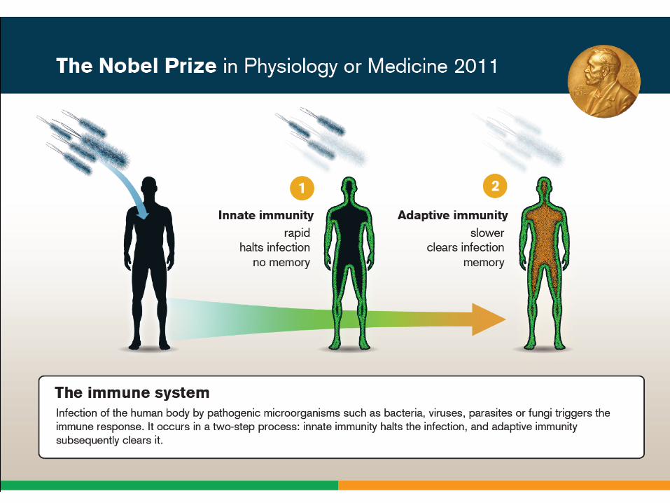


We didn’t know….
• What triggers innate immunity-
• What mediates communication between
innate and adaptive immunity-
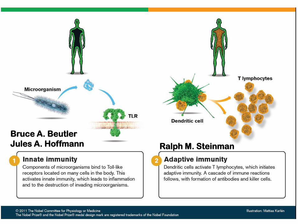
Bruce A. Beutler
Jules A. Hoffmann Ralph M. Steinman
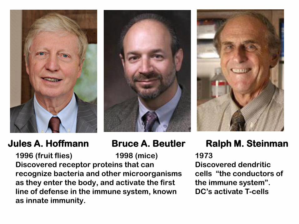
Jules A. Hoffmann Bruce A. Beutler Ralph M. Steinman
1996 (fruit flies) 1998 (mice)
Discovered receptor proteins that can
recognize bacteria and other microorganisms
as they enter the body, and activate the first
line of defense in the immune system, known
as innate immunity.
1973
Discovered dendritic
cells “the conductors of
the immune system”.
DC’s activate T-cells
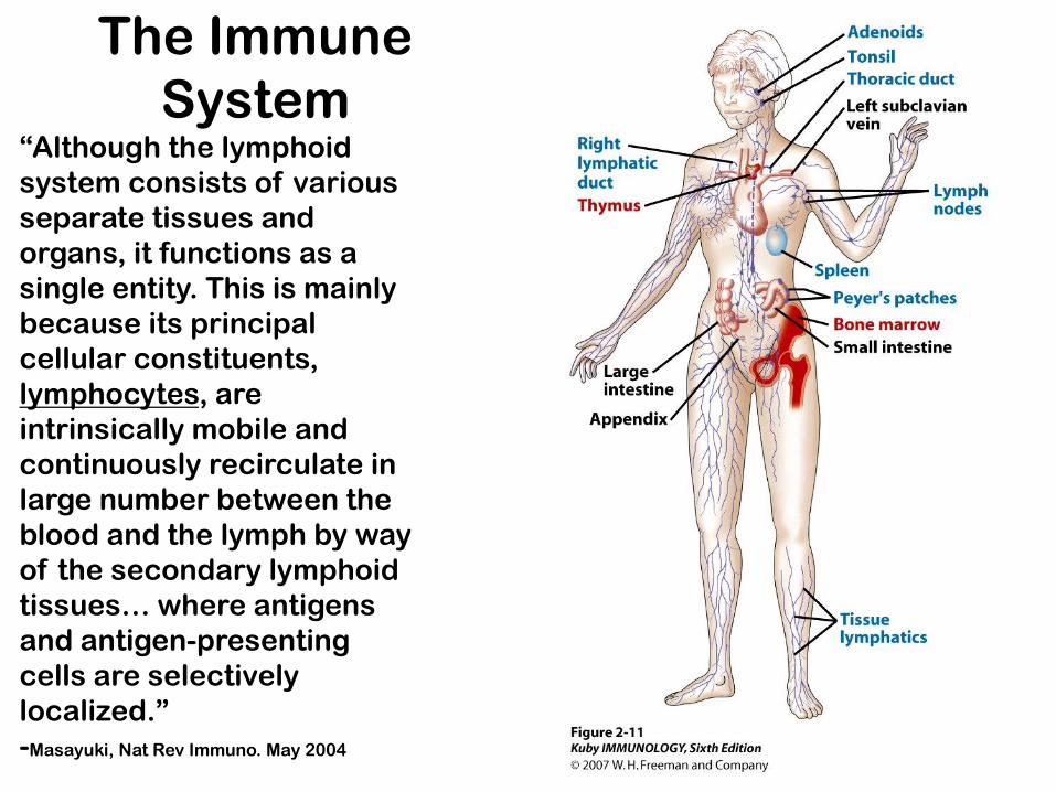
The Immune
System “Although the lymphoid
system consists of various
separate tissues and
organs, it functions as a
single entity. This is mainly
because its principal
cellular constituents,
lymphocytes, are
intrinsically mobile and
continuously recirculate in
large number between the
blood and the lymph by way
of the secondary lymphoid
tissues… where antigens
and antigen-presenting
cells are selectively
localized.”
-Masayuki, Nat Rev Immuno. May 2004
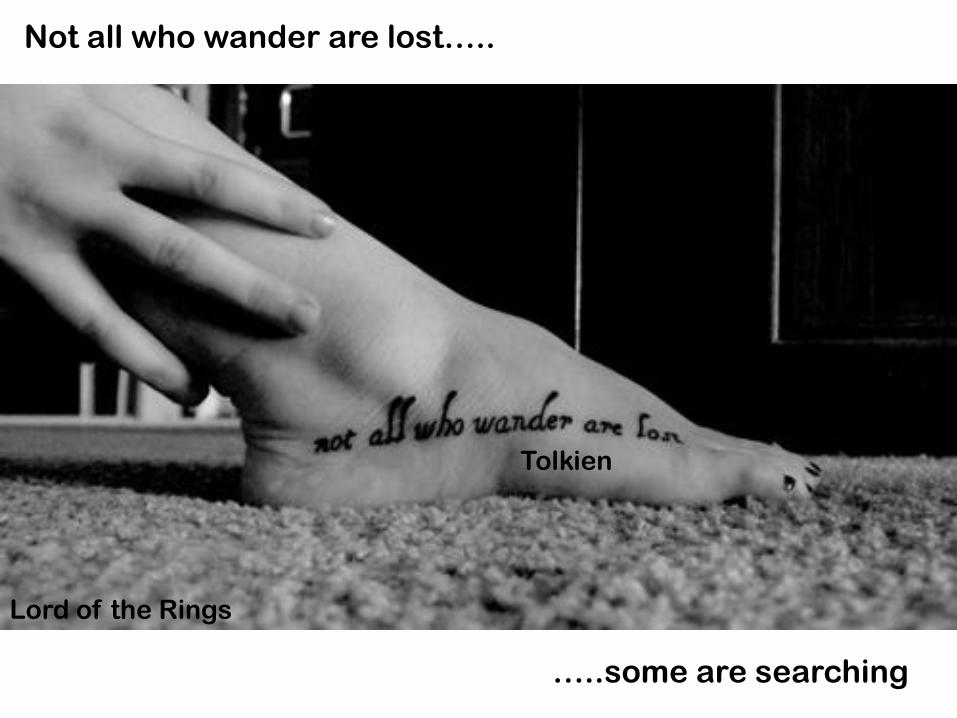
Tolkien
Not all who wander are lost…..
…..some are searching
Lord of the Rings
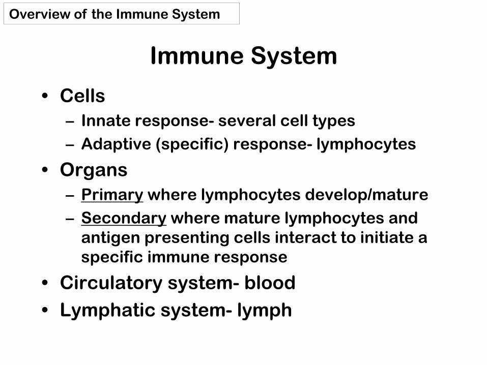
Immune System
• Cells
– Innate response- several cell types
– Adaptive (specific) response- lymphocytes
• Organs
– Primary where lymphocytes develop/mature
– Secondary where mature lymphocytes and
antigen presenting cells interact to initiate a
specific immune response
• Circulatory system- blood
• Lymphatic system- lymph
Overview of the Immune System

Cells= Leukocytes= white blood cells
Granulocytes
1. neutrophils
2. eosinophils
3. basophils
Non-granulocytes
4. monocytes
5. lymphocytes
Plasma (56%)
RBCs
After centrifugation in
Ficoll, leukocytes are
found in the “buffy
coat” 1%
Plasma- with anticoagulant
Serum- after coagulation
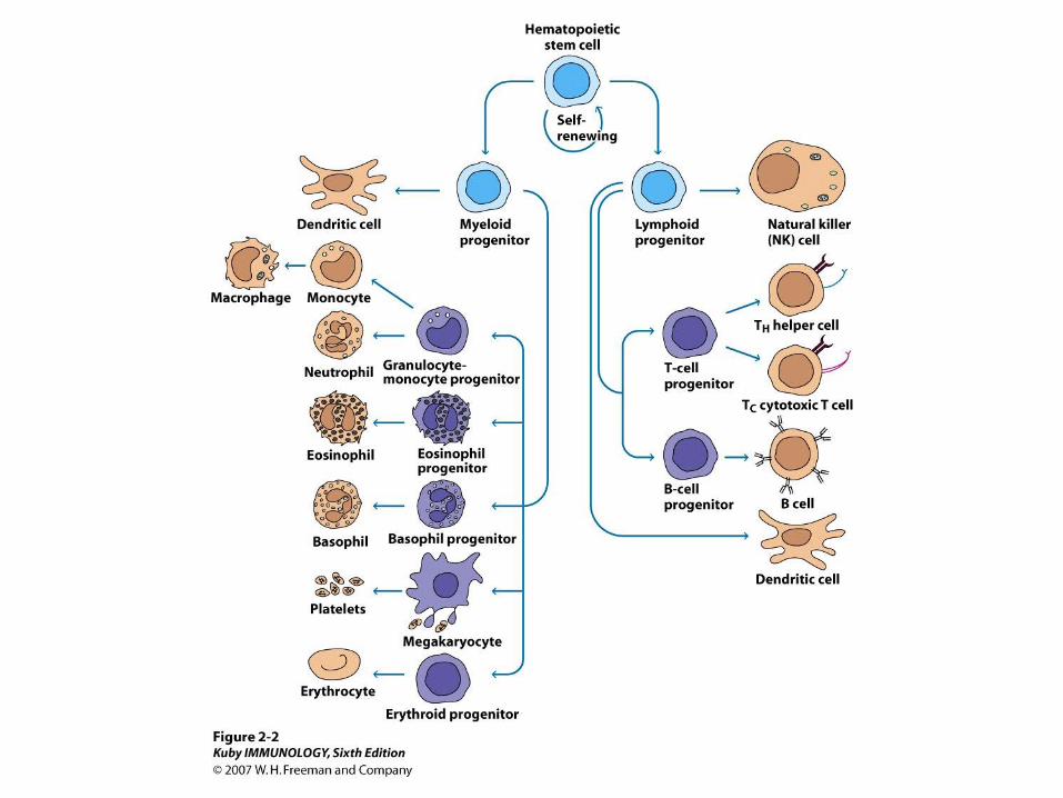
Where do all these cells come
from?

The cells of the immune system arise
from pluripotent hematopoeitic stem
cells (HSC) through two main lines of
differentiation
• Myeloid lineage produces phagocytes
(neutrophils..) and other cells
• Lymphoid lineage produces lymphocytes
Cells of the Immune System
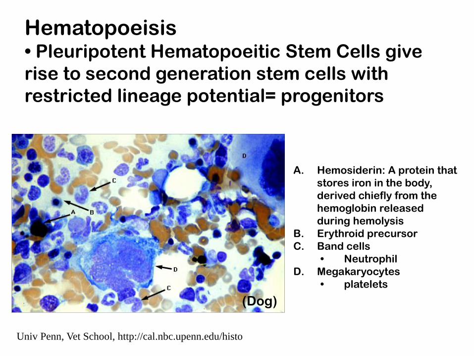
Univ Penn, Vet School, http://cal.nbc.upenn.edu/histo
A. Hemosiderin: A protein that
stores iron in the body,
derived chiefly from the
hemoglobin released
during hemolysis
B. Erythroid precursor
C. Band cells
• Neutrophil
D. Megakaryocytes
• platelets
(Dog)
Hematopoeisis • Pleuripotent Hematopoeitic Stem Cells give
rise to second generation stem cells with
restricted lineage potential= progenitors

Granulocyte
lineage
“First Responders” HSC- Pleuripotent
Common
Myeloid
Progenitor
Eosinophil
Neutrophil
Basophil
(Myeloid = of or
relating to the bone
marrow)
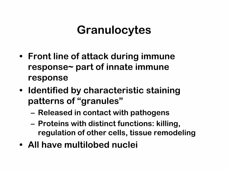
Granulocytes
• Front line of attack during immune
response~ part of innate immune
response
• Identified by characteristic staining
patterns of “granules”
– Released in contact with pathogens
– Proteins with distinct functions: killing,
regulation of other cells, tissue remodeling
• All have multilobed nuclei
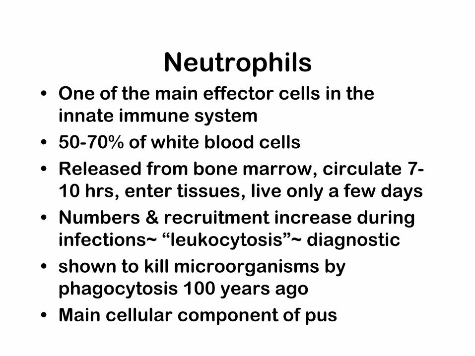
Neutrophils • One of the main effector cells in the
innate immune system
• 50-70% of white blood cells
• Released from bone marrow, circulate 7-
10 hrs, enter tissues, live only a few days
• Numbers & recruitment increase during
infections~ “leukocytosis”~ diagnostic
• shown to kill microorganisms by
phagocytosis 100 years ago
• Main cellular component of pus
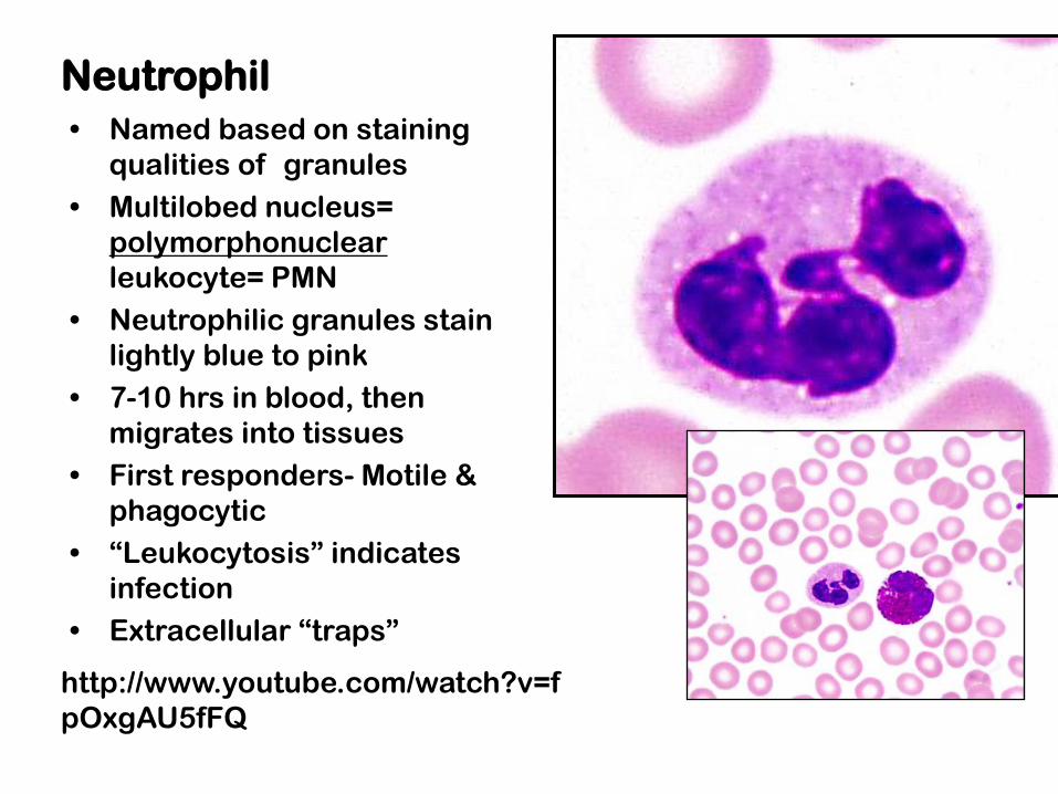
Neutrophil
• Named based on staining
qualities of granules
• Multilobed nucleus=
polymorphonuclear
leukocyte= PMN
• Neutrophilic granules stain
lightly blue to pink
• 7-10 hrs in blood, then
migrates into tissues
• First responders- Motile &
phagocytic
• “Leukocytosis” indicates
infection
• Extracellular “traps”
http://www.youtube.com/watch?v=f
pOxgAU5fFQ

https://www.youtube.com/watch?v=
VAhM9OxZDkU
Neutrophil movie

Soehniein, Trends in Immunol 2009
How neutrophils shape the immune response

COVER
Scanning electron micrograph of
Staphylococcus aureus bound to
neutrophil extracellular traps
(NETs). These novel structures
formed by activated neutrophils
can disarm and kill bacteria before
they reach host cells

neutrophils resting neutrophils activated
Brinkmann et al, Science 303, 2004
NETS
Brinkman/Zychlinsky Nat Rev Micro 5: 2007
“Beneficial suicide: why neutrophils die to make
NETS”

Stimulated neutrophil with NETs and some trapped Shigella (orange). Colored scanning electron micrograph.
Brinkmann: Max Planck Institute for Infection Biology
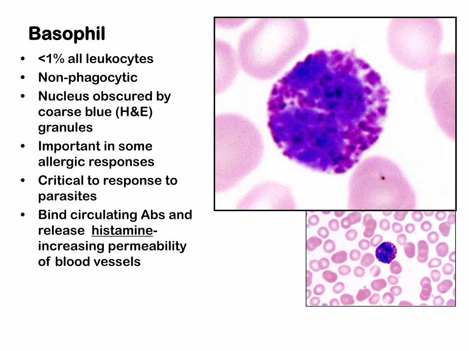
Basophil
• <1% all leukocytes
• Non-phagocytic
• Nucleus obscured by
coarse blue (H&E)
granules
• Important in some
allergic responses
• Critical to response to
parasites
• Bind circulating Abs and
release histamine-
increasing permeability
of blood vessels
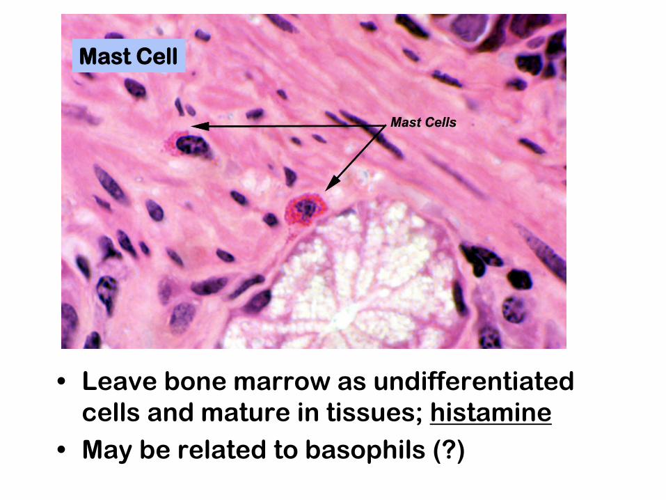
Mast Cell
• Leave bone marrow as undifferentiated
cells and mature in tissues; histamine
• May be related to basophils (?)
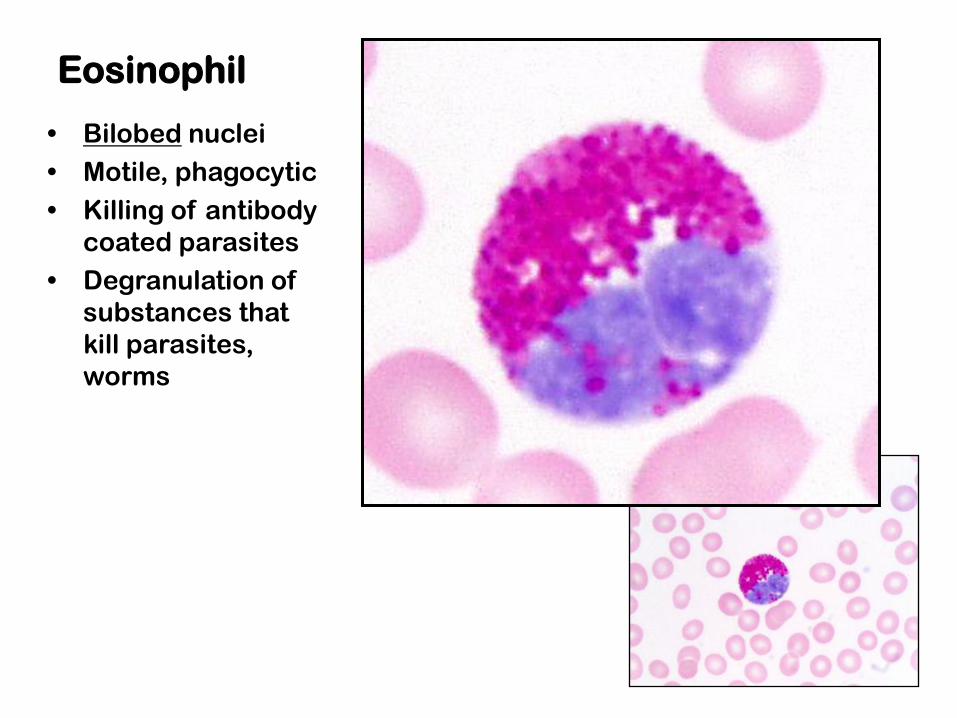
Eosinophil
• Bilobed nuclei
• Motile, phagocytic
• Killing of antibody
coated parasites
• Degranulation of
substances that
kill parasites,
worms

Myeloid antigen presenting cells: Monocytes, macrophages, dendritic cells
• Phagocytic
• Ingest, digest into peptides, present on
cell surface
• Bridge between innate and adaptive
immune responses
• Make contact with antigens in periphery
and then interact with lymphocytes in
lymph node
• Secrete proteins that attract and activate
other immune cells
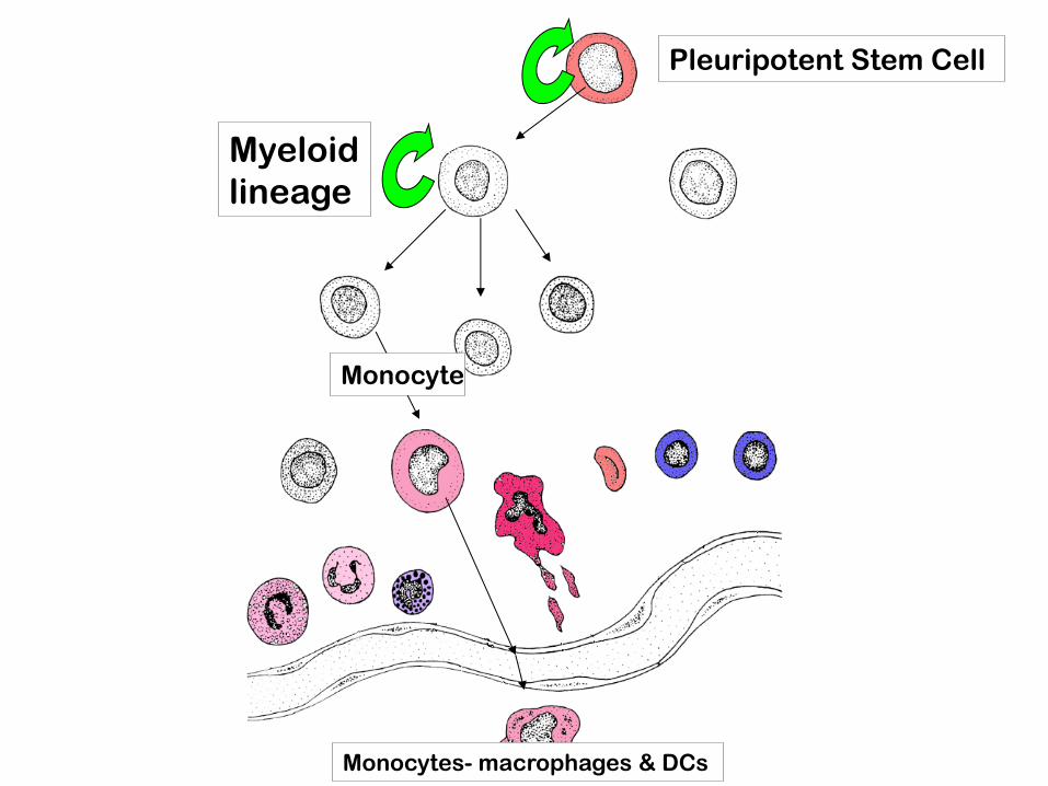
Pleuripotent Stem Cell
Myeloid
lineage
Monocytes- macrophages & DCs
Monocyte
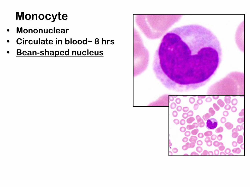
Monocyte
• Mononuclear
• Circulate in blood~ 8 hrs
• Bean-shaped nucleus
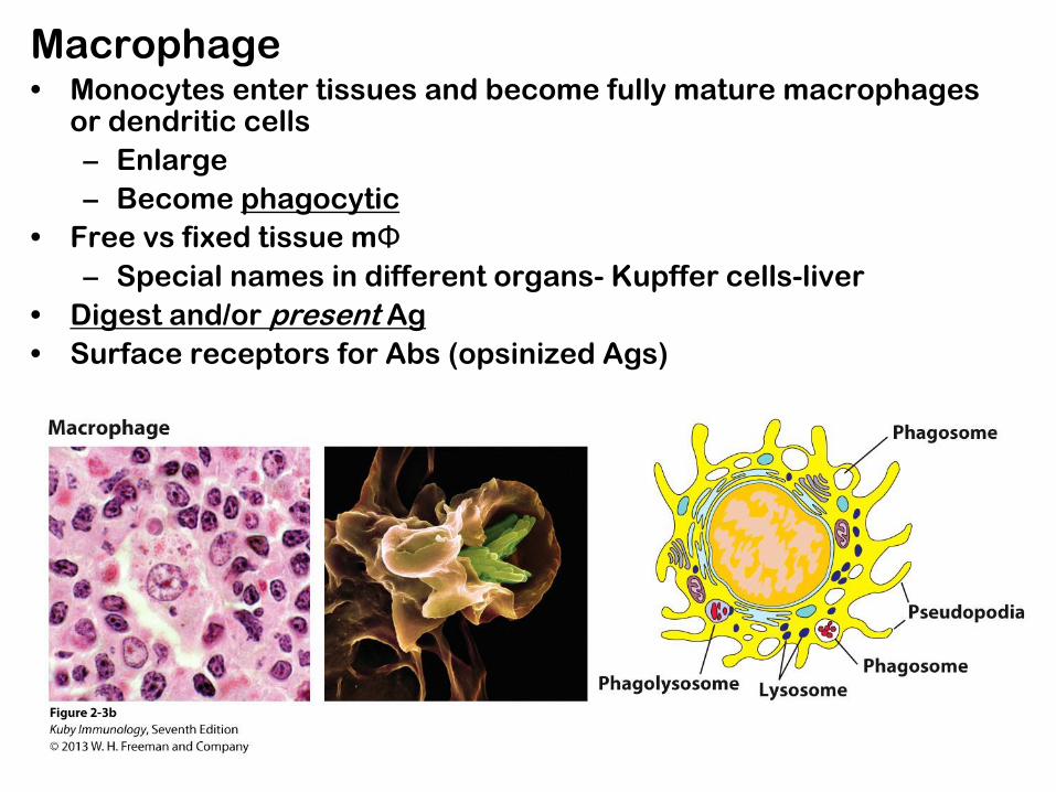
Macrophage • Monocytes enter tissues and become fully mature macrophages
or dendritic cells
– Enlarge
– Become phagocytic
• Free vs fixed tissue mΦ
– Special names in different organs- Kupffer cells-liver
• Digest and/or present Ag
• Surface receptors for Abs (opsinized Ags)

Dendritic cells: heterogeneous myeloid & lymphoid origins
• Best APC for presenting to naïve T-cells
• Ralph Steinman discovered them in mid
1970’s; just received Nobel Prize 2011
• Critical
• Named for long processes; actively extend
and retract sampling Ags & examining T cells
• Capture Ag in one place- then migrate-
present Ag in another place (eg. LN)
• Immature to mature; change in functionality
from Ag capture to Ag presentation


Dendritic cells
Source: National Cancer Institute (NCI) Sriram
Subramaniam
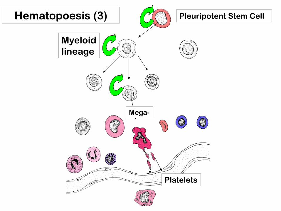
Hematopoesis (3) Pleuripotent Stem Cell
Myeloid
lineage
Platelets
Mega-
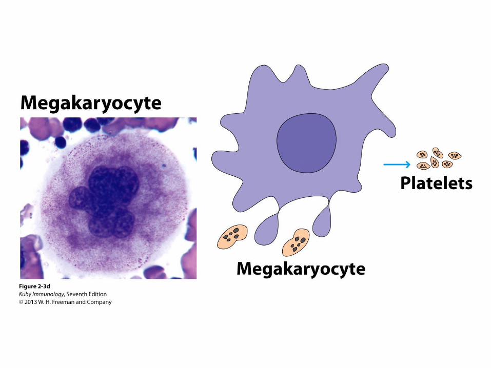

Platelets
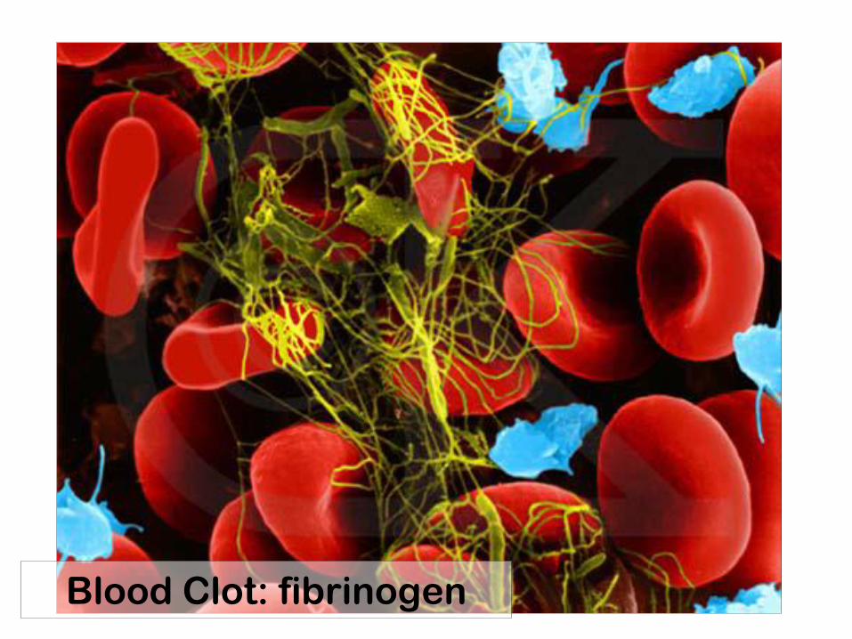
Blood Clot: fibrinogen

Hematopoesis (4) Pleuripotent Stem Cell
Myeloid
lineage
Erythroid
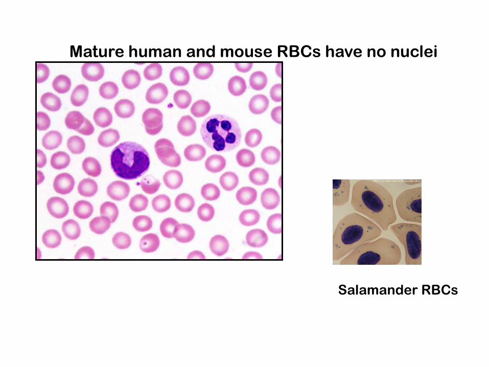
Mature human and mouse RBCs have no nuclei
Salamander RBCs
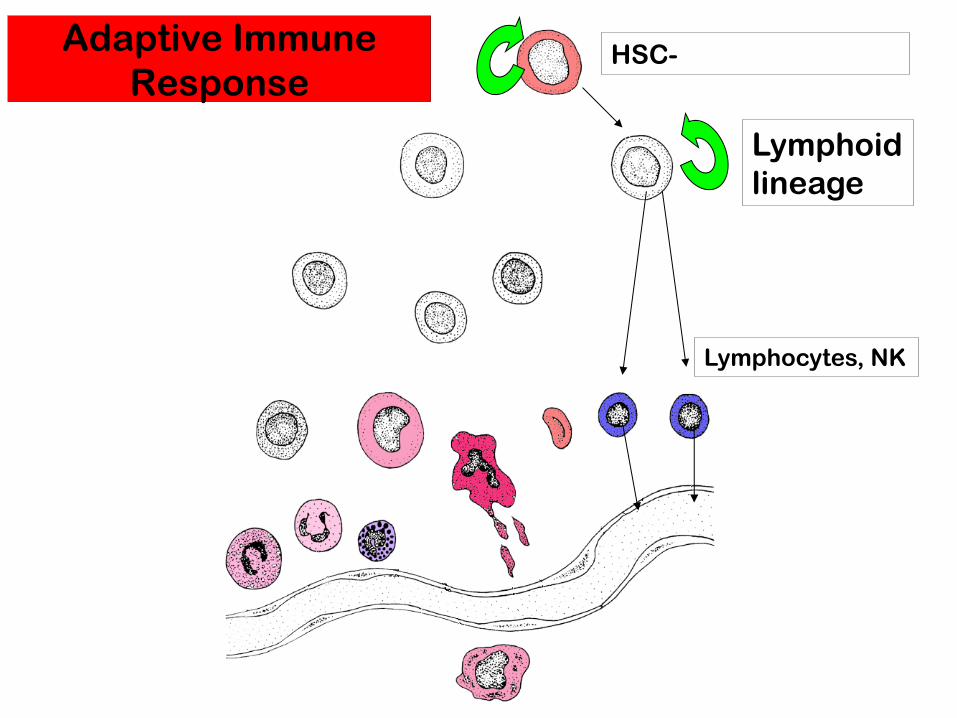
HSC-
Lymphoid
lineage
Lymphocytes, NK
Adaptive Immune
Response

Lymphocytes: 3 types
• 20-40% of WBC
• Cannot be distinguished morphologically
• T-cells
– helper CD4+ recognize Ag in context of MHCII
– cytotoxic CD8+ recognize Ab in MHCI
• B-cells
– become antibody producing plasma cells
• NK cells
– part of the innate immune response
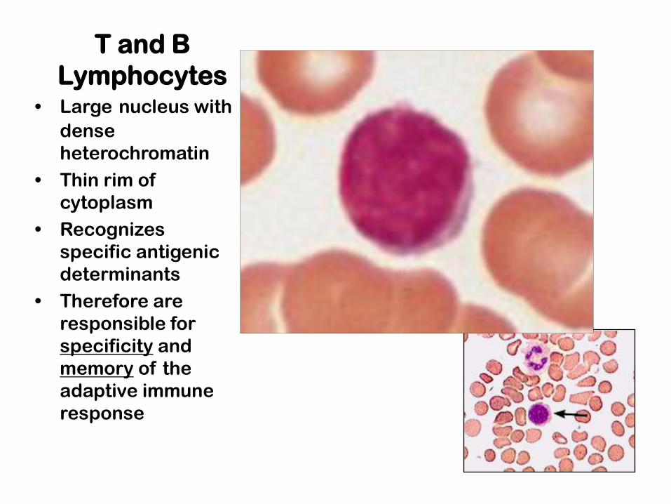
T and B
Lymphocytes • Large nucleus with
dense
heterochromatin
• Thin rim of
cytoplasm
• Recognizes
specific antigenic
determinants
• Therefore are
responsible for
specificity and
memory of the
adaptive immune
response
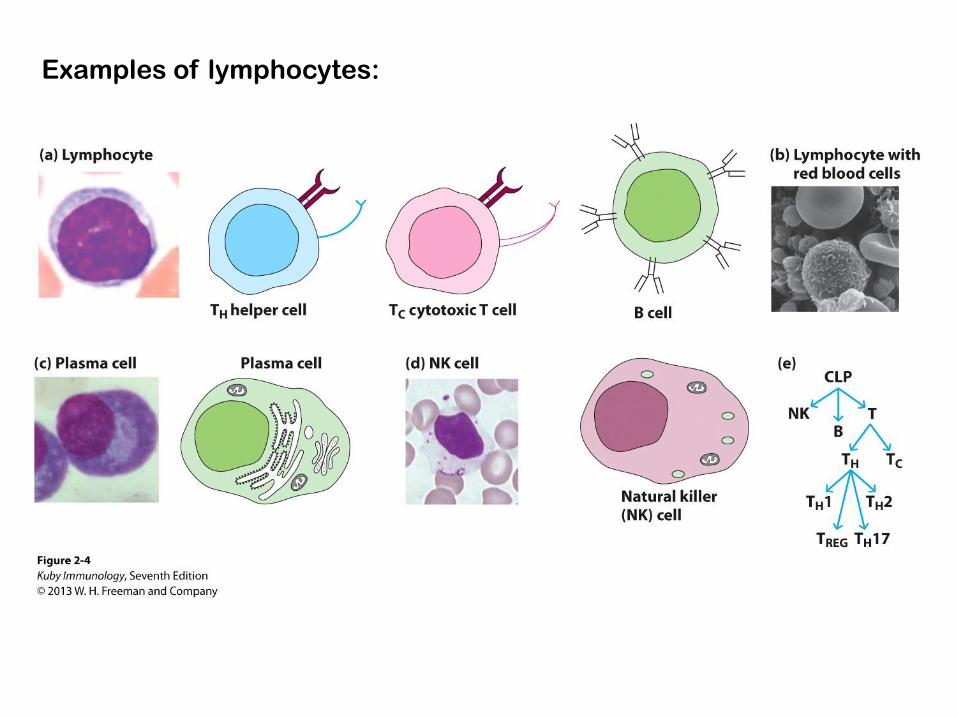
Examples of lymphocytes:


Condensed heterochromatin= resting
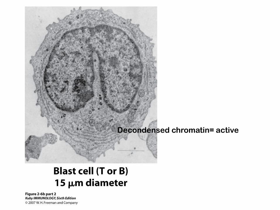
Decondensed chromatin= active

Usually lives 1-2 weeks
Secretes 100’s- 1000 Ab molecules/sec
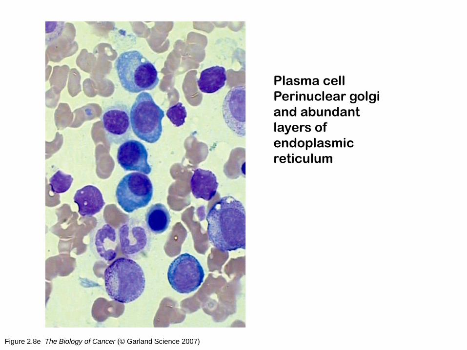
Figure 2.8e The Biology of Cancer (© Garland Science 2007)
Plasma cell
Perinuclear golgi
and abundant
layers of
endoplasmic
reticulum
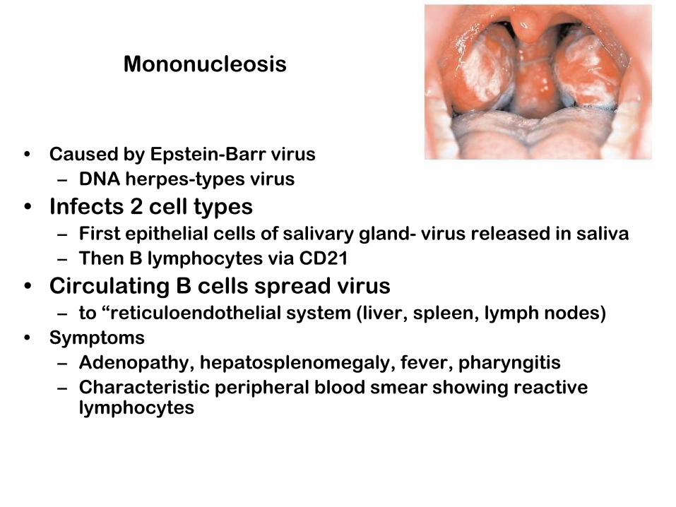
Mononucleosis
• Caused by Epstein-Barr virus
– DNA herpes-types virus
• Infects 2 cell types – First epithelial cells of salivary gland- virus released in saliva
– Then B lymphocytes via CD21
• Circulating B cells spread virus – to “reticuloendothelial system (liver, spleen, lymph nodes)
• Symptoms
– Adenopathy, hepatosplenomegaly, fever, pharyngitis
– Characteristic peripheral blood smear showing reactive lymphocytes

Dendritic cells Macrophage B-cell
Antigen
Presenting Cells
3 kinds of cells
present Ag to T-
cells
Dendritic cells:
Several types
Capture, process,
present Ag

Organs of Hematopoesis…
Yolk Sac
•3 weeks
•Blood islands
•Erythro-myeloid stem
cells
•RBC’s are large and
nucleated=primitive
•Cannot form lymphoid
progeny
Fetal Liver
•5-6 weeks
•Seeded from
both outside
sources
•Max 6 mos then
declines to
neonatal stage
Bone Marrow
•Source of all stem
cells in adult
•B-cell maturation
•T-cells to thymus
•spleen

Organs of the
Immune
System
Thymus Bone Marrow
Stem cell
(in bone marrow)
1. Development &
maturation in primary
lymphoid organs
2. Distribution to
Secondary lymphoid
organs for engagement
with antigens
•Tonsils •Lymph nodes •Spleen
•Peyer’s
Patches •Appendix
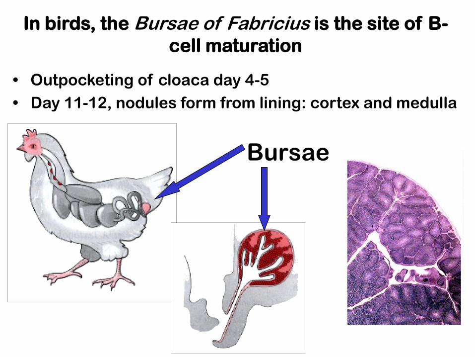
Bursae
• Outpocketing of cloaca day 4-5
• Day 11-12, nodules form from lining: cortex and medulla
In birds, the Bursae of Fabricius is the site of B-
cell maturation
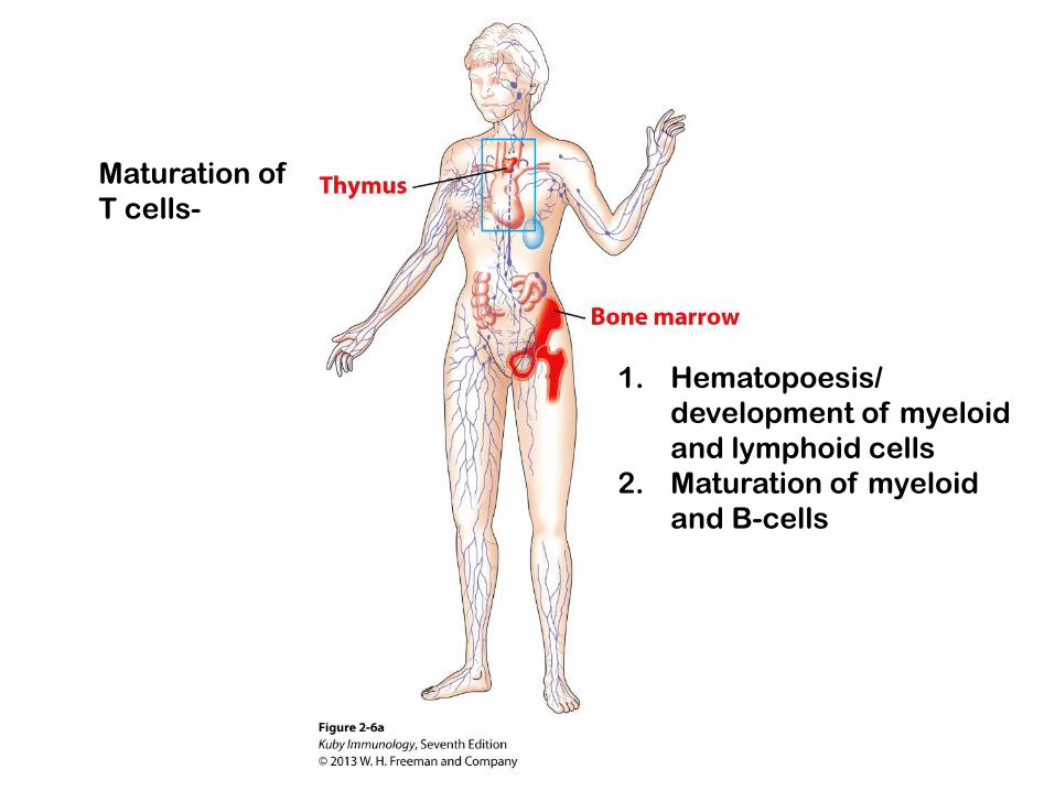
Maturation of
T cells-
1. Hematopoesis/
development of myeloid
and lymphoid cells
2. Maturation of myeloid
and B-cells

The thymus in the adult lies behind the
sternum, above the heart
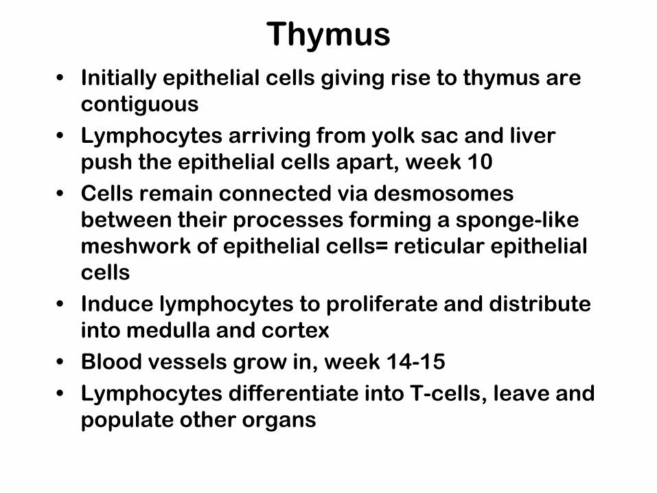
Thymus
• Initially epithelial cells giving rise to thymus are
contiguous
• Lymphocytes arriving from yolk sac and liver
push the epithelial cells apart, week 10
• Cells remain connected via desmosomes
between their processes forming a sponge-like
meshwork of epithelial cells= reticular epithelial
cells
• Induce lymphocytes to proliferate and distribute
into medulla and cortex
• Blood vessels grow in, week 14-15
• Lymphocytes differentiate into T-cells, leave and
populate other organs
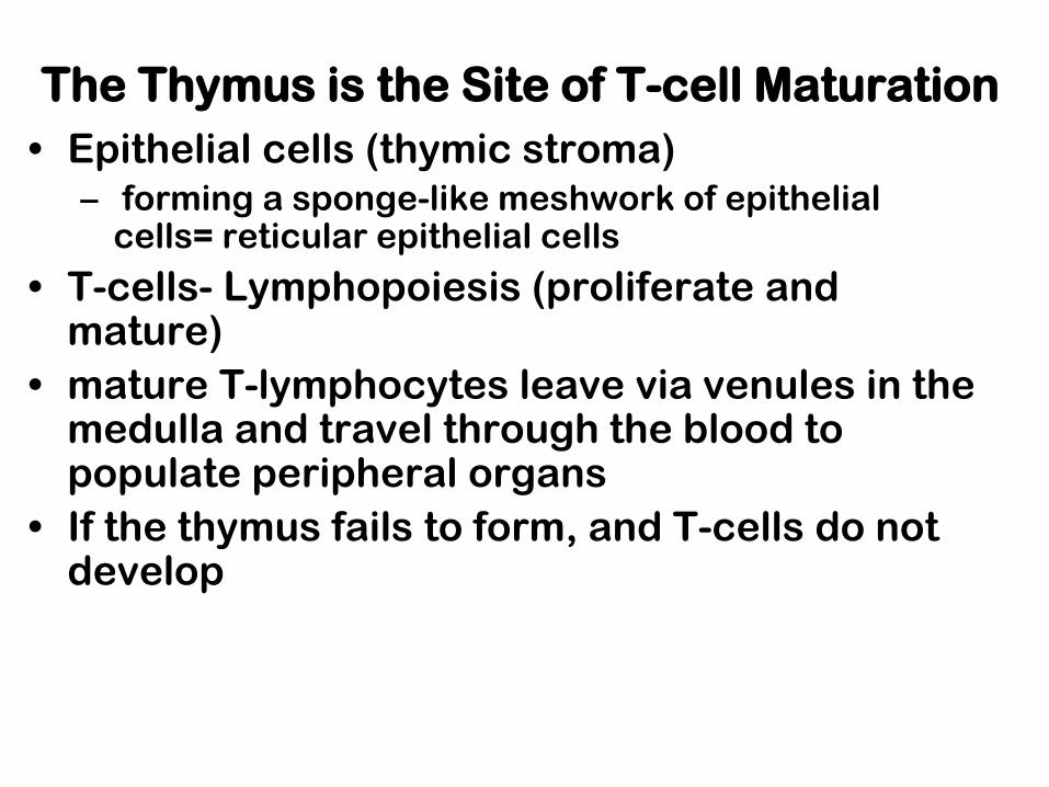
The Thymus is the Site of T-cell Maturation
• Epithelial cells (thymic stroma) – forming a sponge-like meshwork of epithelial
cells= reticular epithelial cells
• T-cells- Lymphopoiesis (proliferate and mature)
• mature T-lymphocytes leave via venules in the medulla and travel through the blood to populate peripheral organs
• If the thymus fails to form, and T-cells do not develop
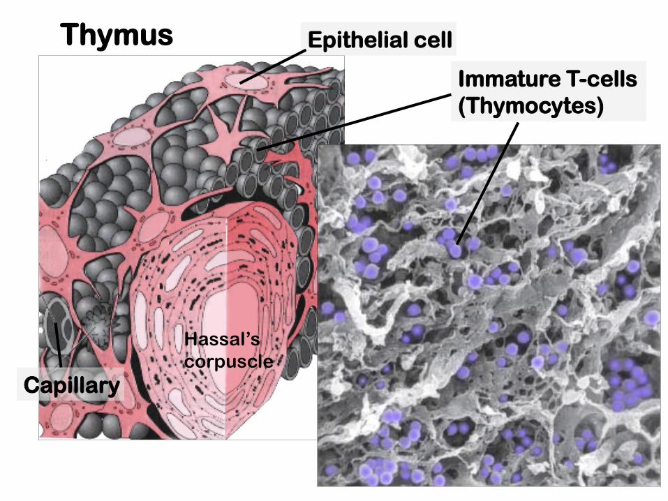
Thymus Epithelial cell
Immature T-cells
(Thymocytes)
Capillary
Hassal’s
corpuscle

Fetal Thymus: Lobes
Medulla- mature cells
Cortex- immature cells

The cortex contains immature thymocytes which move into the
medulla as they mature.
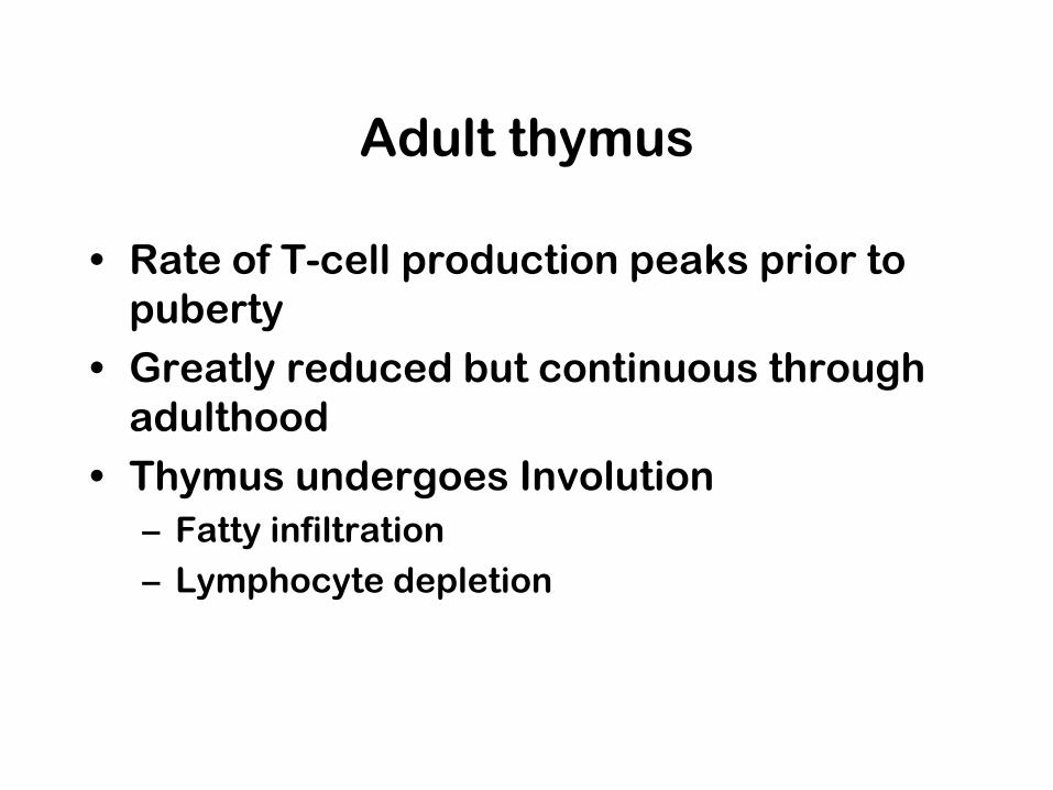
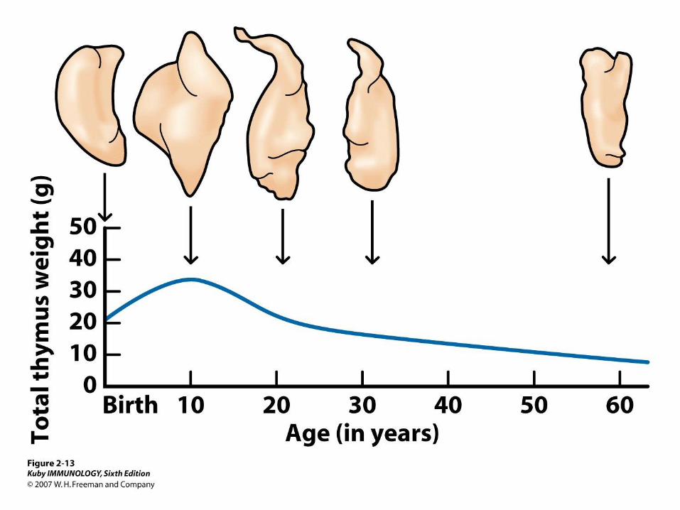
Adult thymus
• Rate of T-cell production peaks prior to
puberty
• Greatly reduced but continuous through
adulthood
• Thymus undergoes Involution
– Fatty infiltration
– Lymphocyte depletion
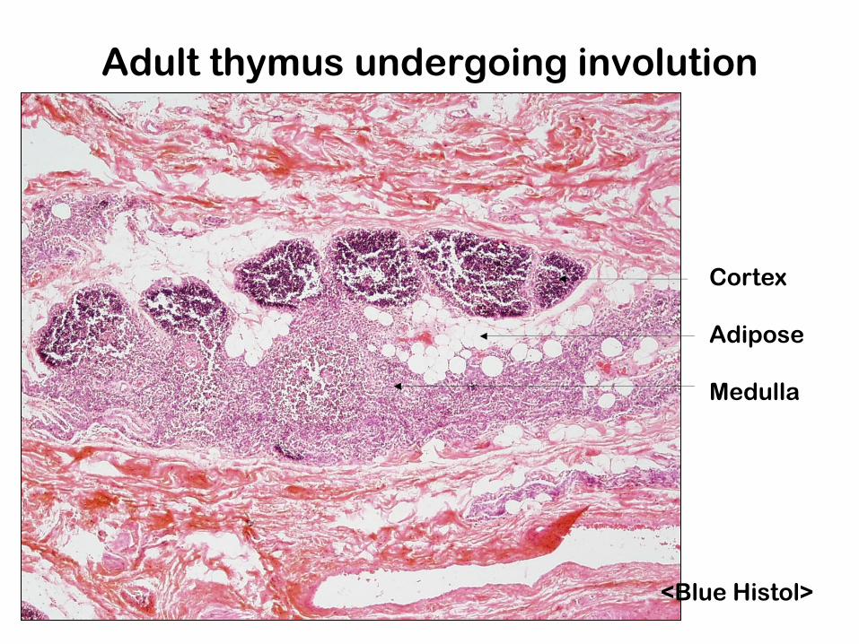
Adult thymus undergoing involution
<Blue Histol>
Cortex
Adipose
Medulla
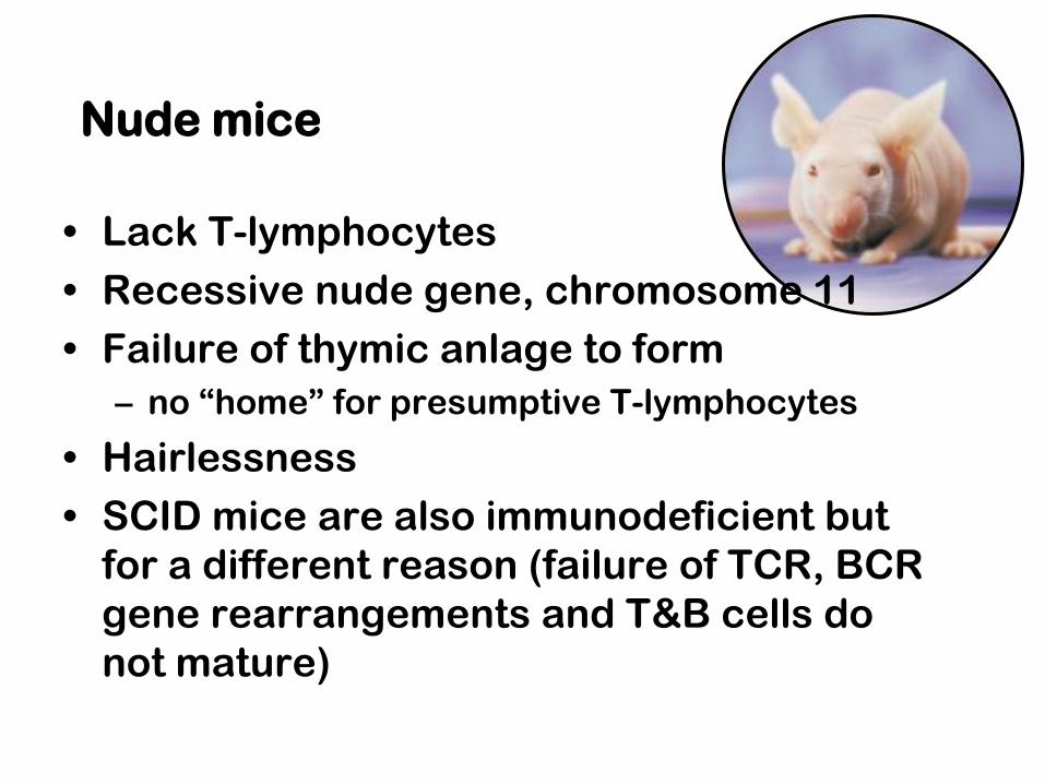
Nude mice
• Lack T-lymphocytes
• Recessive nude gene, chromosome 11
• Failure of thymic anlage to form
– no “home” for presumptive T-lymphocytes
• Hairlessness
• SCID mice are also immunodeficient but
for a different reason (failure of TCR, BCR
gene rearrangements and T&B cells do
not mature)

DiGeorge syndrome
• deletion on chromosome 22
• defect of cranial neural crest cell
migration into arches
• congenital thymic hypoplasia= anlage of the thymus does not form
• variety of other defects involving facial,
thyroid, parathyroid and cardiovascular
system “Anlage”- an organ in its earliest stage of development; the foundation for subsequent
development, primordium
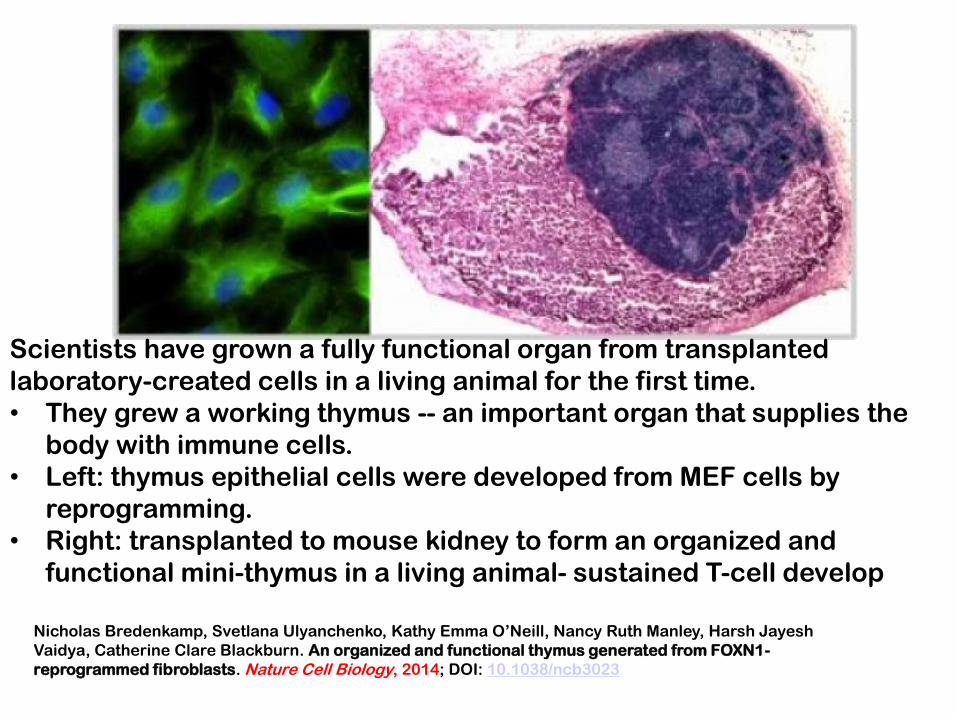
Scientists have grown a fully functional organ from transplanted
laboratory-created cells in a living animal for the first time.
• They grew a working thymus -- an important organ that supplies the
body with immune cells.
• Left: thymus epithelial cells were developed from MEF cells by
reprogramming.
• Right: transplanted to mouse kidney to form an organized and
functional mini-thymus in a living animal- sustained T-cell develop
Nicholas Bredenkamp, Svetlana Ulyanchenko, Kathy Emma O’Neill, Nancy Ruth Manley, Harsh Jayesh
Vaidya, Catherine Clare Blackburn. An organized and functional thymus generated from FOXN1-
reprogrammed fibroblasts. Nature Cell Biology, 2014; DOI: 10.1038/ncb3023
Secondary lymphoid organs
• Specialized for trapping antigen
facilitating presentation to lymphocytes
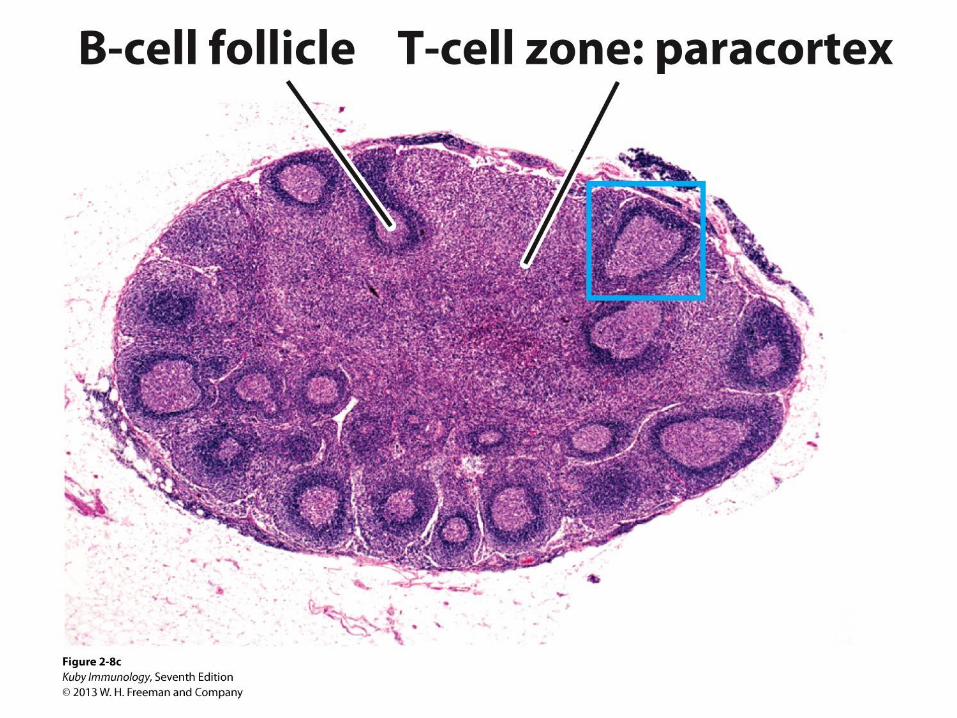
• Characterized by:
– Localized areas for T-cells and B-cells
– Follicles where B cells mature

Schematic
diagrams of
various types of
lymphoid tissue
Diffuse
Peyer’s patch Tonsil
Lymph Node
Spleen
Thymus
Lymphoid follicle
• Diffuse
• Solitary follicle
• Aggregated
follicle
• Lymph Node
• Spleen
• Thymus
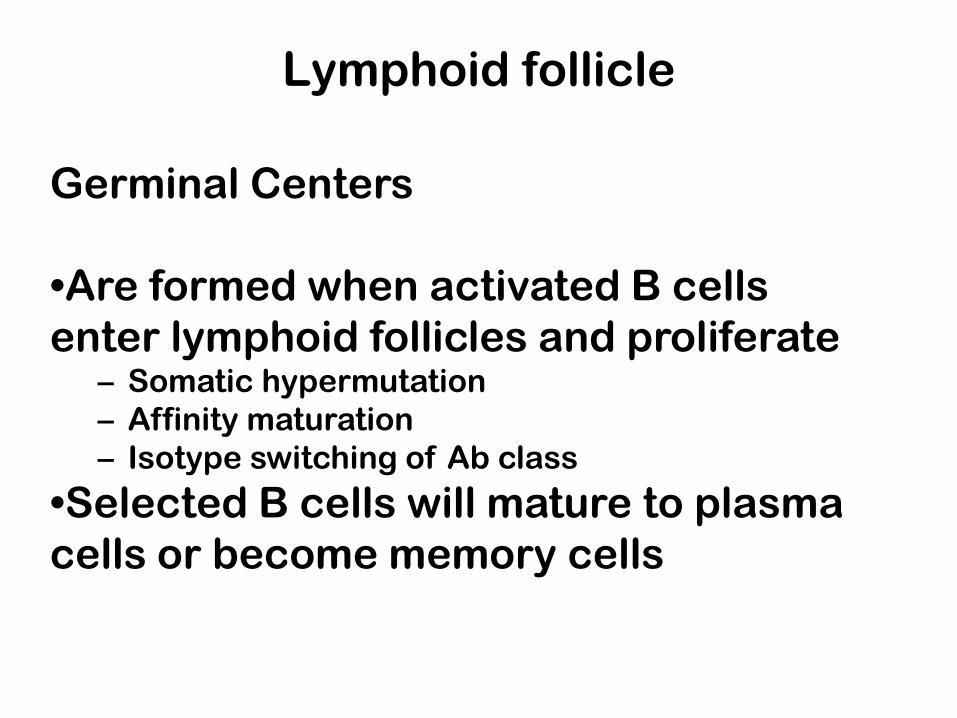
Lymphoid follicle
Germinal Centers
•Are formed when activated B cells
enter lymphoid follicles and proliferate – Somatic hypermutation
– Affinity maturation
– Isotype switching of Ab class
•Selected B cells will mature to plasma
cells or become memory cells
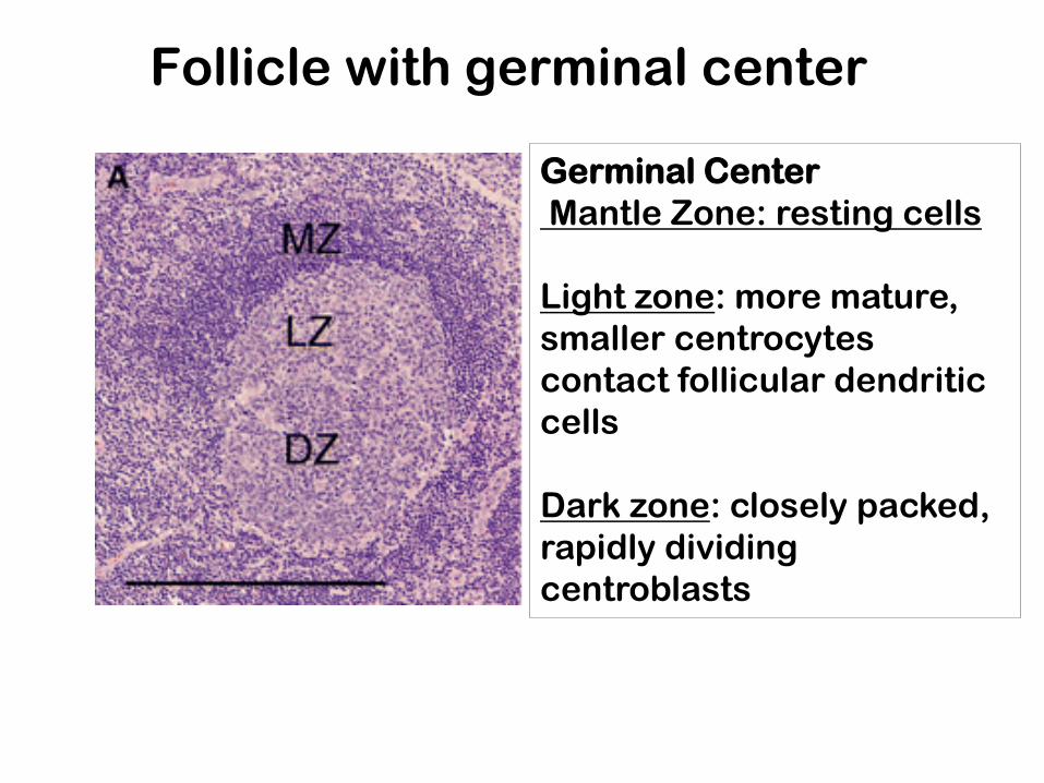
Follicle with germinal center
Germinal Center
Mantle Zone: resting cells
Light zone: more mature,
smaller centrocytes
contact follicular dendritic
cells
Dark zone: closely packed,
rapidly dividing
centroblasts
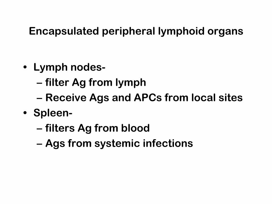
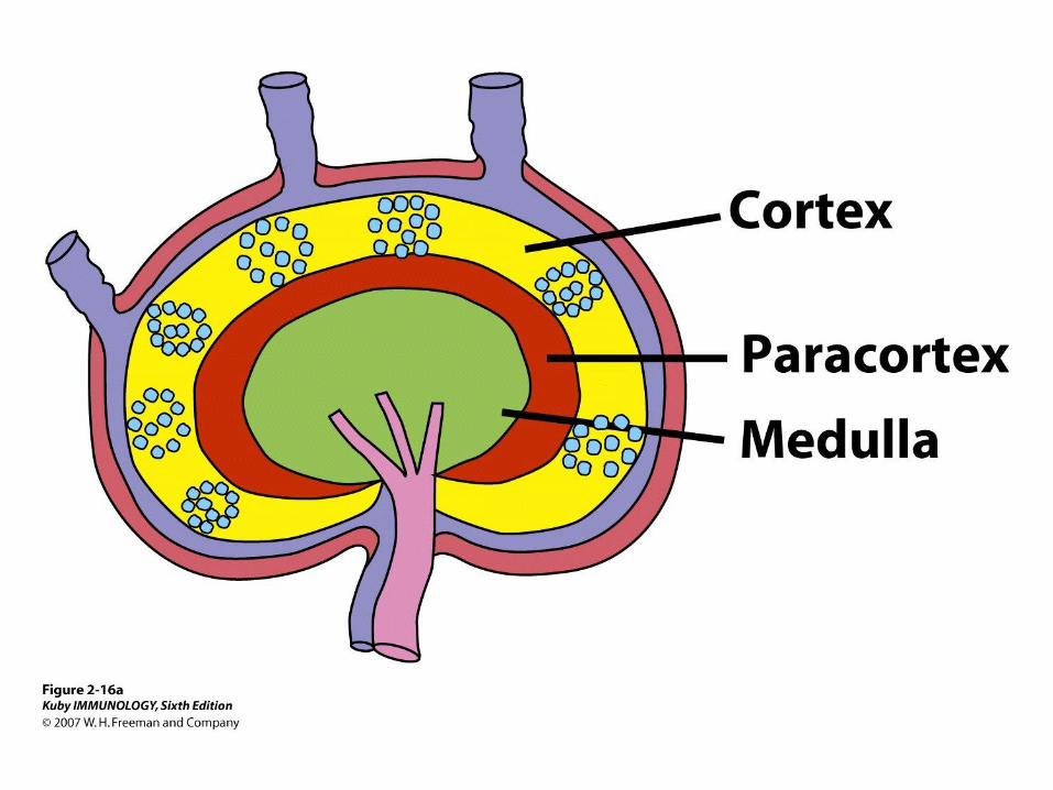
Encapsulated peripheral lymphoid organs
• Lymph nodes-
– filter Ag from lymph
– Receive Ags and APCs from local sites
• Spleen-
– filters Ag from blood
– Ags from systemic infections
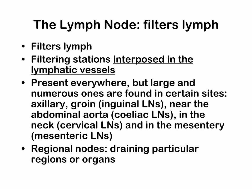
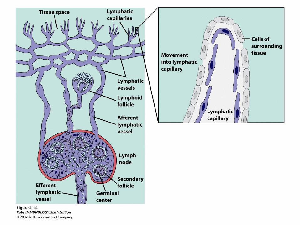
The Lymph Node: filters lymph
• Filters lymph
• Filtering stations interposed in the lymphatic vessels
• Present everywhere, but large and numerous ones are found in certain sites: axillary, groin (inguinal LNs), near the abdominal aorta (coeliac LNs), in the neck (cervical LNs) and in the mesentery (mesenteric LNs)
• Regional nodes: draining particular regions or organs

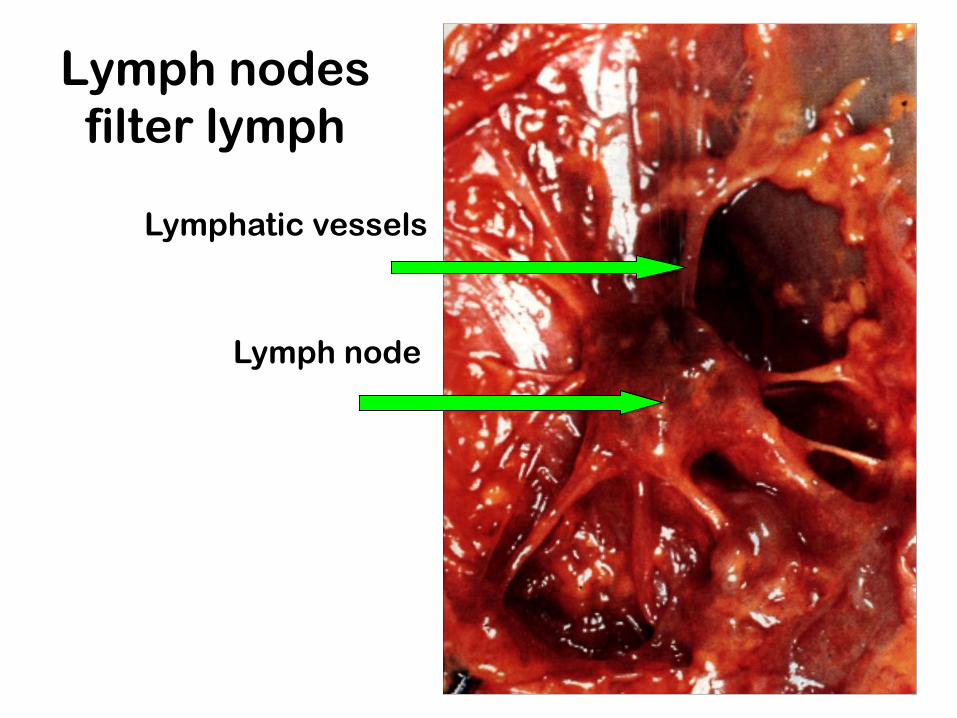
Lymph nodes
filter lymph
Lymph node
Lymphatic vessels
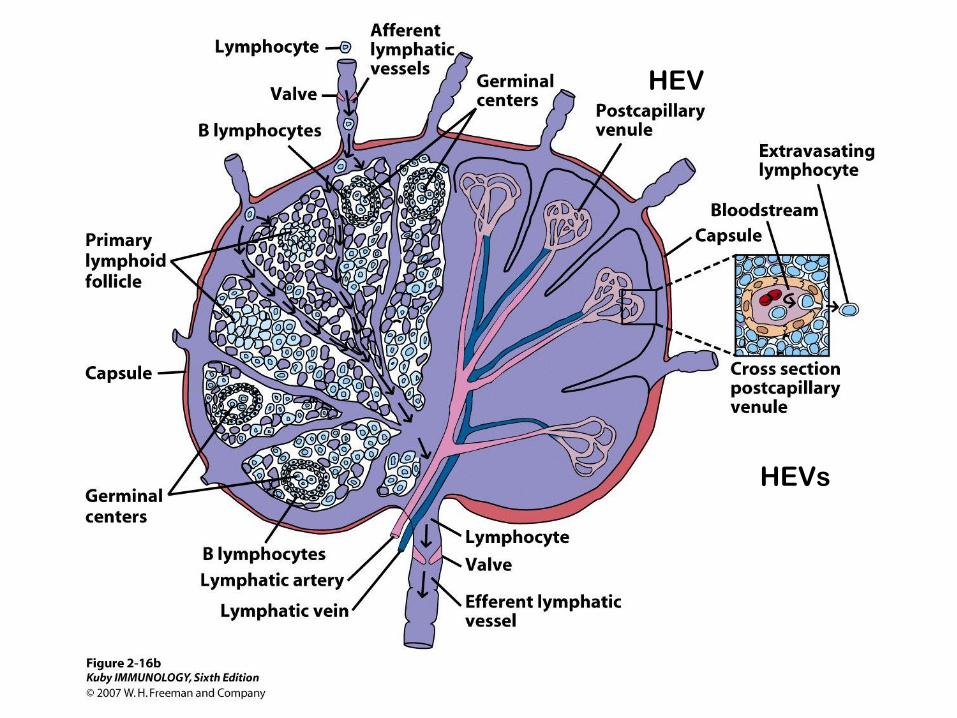
HEVs
HEV

The Spleen: filters blood
• In contrast to lymph nodes, which are
inserted in the lymph circulation, the
spleen is inserted in the blood circulation.
• Oblong, purplish body the size of a fist, on
the left side
• Smooth surfaced except for hilus, where
blood vessels enter and leave
* There are no lymphatics leading to the
spleen.

The Spleen has 2 major regions
• White Pulp: lymphatic
– Small arterioles surrounded by sheaths of
lymphocytes= Peri-Arteriole Lymphoid
Sheaths (PALS- human arrangement slightly
different)
– Surrounded by marginal zone
• Red Pulp: clears RBCs
– “Cords” of cells: Erythrocytes, macrophages,
dendritic cells, few lymphocytes and plasma
cells
– Also contains venous Sinusoids
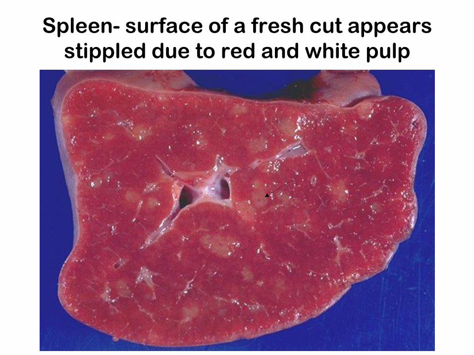
Spleen- surface of a fresh cut appears
stippled due to red and white pulp

White pulp: two components
1. PALS- T cells
• periarteriole lymphoid sheath
2. Lymphoid follicles- B cells
• spherical structures Scattered throughout PALS
• Visible to the naked eye on the surface of a freshly cut spleen as white spots.

Spleen- human
capsule
Red pulp
White pulp
trabeculae
Central artery
https://www.youtube.com/watch?v=
aEi_4Cyx4Uw
What does the spleen do?


Spleen: Red Pulp and Sinusoids
Reticular fibers and endothelial cells
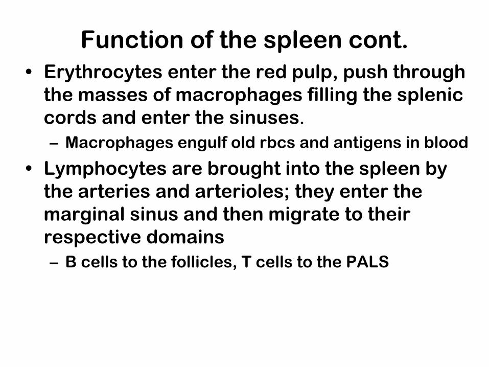
Function of the spleen cont.
• Erythrocytes enter the red pulp, push through
the masses of macrophages filling the splenic
cords and enter the sinuses.
– Macrophages engulf old rbcs and antigens in blood
• Lymphocytes are brought into the spleen by
the arteries and arterioles; they enter the
marginal sinus and then migrate to their
respective domains
– B cells to the follicles, T cells to the PALS
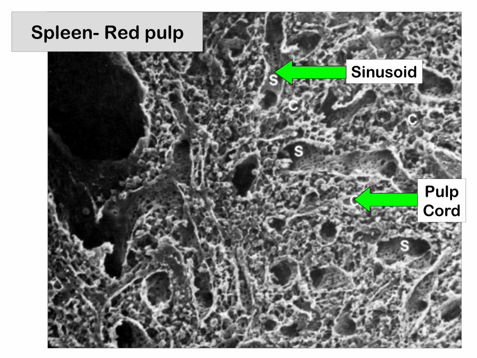
Spleen- Red pulp
Sinusoid
Pulp
Cord
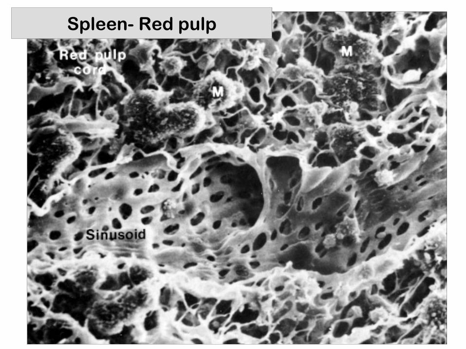
Spleen- Red pulp

Mucosal Immune System MALT- Mucosal Associated Lymphoid
Tissue
• Mucosal surfaces of mouth, respiratory and reproductive tracts are colonized by lymphocytes and accessory cells
• Respond to ingested, inhaled antigens
• BALT (bronchial) :
• GALT (gut) : – Tonsils
– Peyer’s Patches
– Appendix

Tonsils
• Latin tonsa (stake set up on the shore)
• At entrance to GI tract:
– 1 pharangeal= “adenoids”
– 2 tubal
– 2 palatine= “tonsils” (from pouch 2)
– 1 lingual
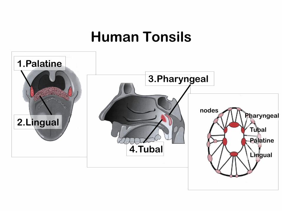
Human Tonsils
1.Palatine
2.Lingual
3.Pharyngeal
4.Tubal
Pharyngeal
Tubal
Palatine
Lingual
nodes
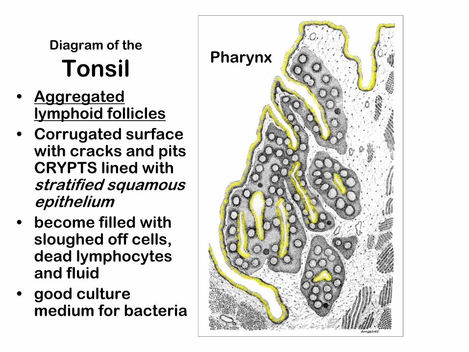
Pharynx Diagram of the
Tonsil • Aggregated
lymphoid follicles
• Corrugated surface with cracks and pits CRYPTS lined with stratified squamous epithelium
• become filled with sloughed off cells, dead lymphocytes and fluid
• good culture medium for bacteria
Pharynx

Peyer’s Patches (~30) are found in the ileum
(small intestine) in the wall opposite the
mesentary
Each is a collection of
many individual lymphoid
follicles (pink) scattered
between the microvilli
“like puffballs on a lawn”
X-section showing the
follicles in the submucosa

Plane of cross section shown in next slide

Identify the lymphoid organ indicated by the
green arrows.

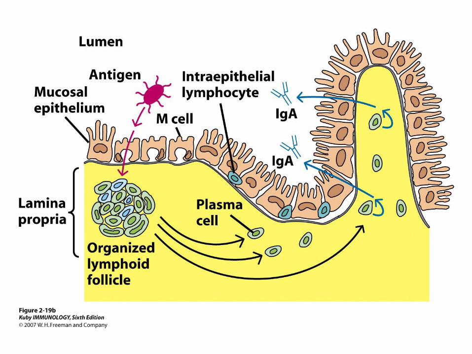
M cell (microfold cell) in the surface of
the Peyer’s patch is the cell
specialized to uptake Ag from the gut


Appendix
• Worm-like projection of the human large intestine, 10-15 cm long and up to 8mm in diameter.
• The lamina propria contains dense, diffuse lymphoid tissue packed with some 200 lymphoid follicles.

Appendix
There is a
network of
lymphatics
surrounding
each follicle
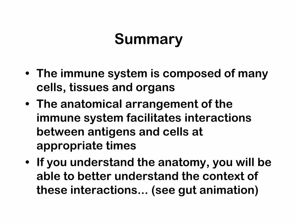
Summary
• The immune system is composed of many
cells, tissues and organs
• The anatomical arrangement of the
immune system facilitates interactions
between antigens and cells at
appropriate times
• If you understand the anatomy, you will be
able to better understand the context of
these interactions... (see gut animation)
http://www.nature.com/ni/multime
dia/mucosal/animation/index.html
• The gut mucosa is the largest and most dynamic immunological
environment of the body. It's often the first point of pathogen
exposure and many microbes use it as a beachhead into the rest
of the body. The gut immune system therefore needs to be ready
to respond to pathogens but at the same time it is constantly
exposed to innocuous environmental antigens, food particles and
commensal microflora which need to be tolerated. Misdirected
immune responses to harmless antigens are the underlying
cause of food allergies and debilitating conditions such as
inflammatory bowel disease.
• This animation introduces the key cells and molecular players
involved in gut immunohomeostasis and disease.
• Nature Immunology September 2013 - Vol 14 No 9