immune responses following bcg immunization of infants in
TRANSCRIPT

ORIGINAL RESEARCHpublished: 19 March 2021
doi: 10.3389/fimmu.2021.637114
Frontiers in Immunology | www.frontiersin.org 1 March 2021 | Volume 12 | Article 637114
Edited by:
Julia Kzhyshkowska,
Heidelberg University, Germany
Reviewed by:
Stephen M. Todryk,
Northumbria University,
United Kingdom
Paulo Bettencourt,
Independent Researcher,
Lisbon, Portugal
*Correspondence:
Patrice A. Mawa
†These authors have contributed
equally to this work
‡Present address:
Steven G. Smith,
Centre for Inflammation Research and
Translational Medicine, College of
Health, Medicine and Life Sciences,
Brunel University London, Uxbridge,
United Kingdom
Specialty section:
This article was submitted to
Vaccines and Molecular Therapeutics,
a section of the journal
Frontiers in Immunology
Received: 02 December 2020
Accepted: 22 February 2021
Published: 19 March 2021
Citation:
Mawa PA, Hasso-Agopsowicz M,
Lubyayi L, Nabakooza G,
Nakibuule M, Blitz R, Dun L, Govind A,
Kaleebu P, Webb EL, Elliott AM,
Dockrell HM, Cose S and Smith SG
(2021) Immune Responses Following
BCG Immunization of Infants in
Uganda and United Kingdom Are
Similar for Purified Protein Derivative
but Differ for Secretory Proteins of
Mycobacterium tuberculosis.
Front. Immunol. 12:637114.
doi: 10.3389/fimmu.2021.637114
Immune Responses Following BCGImmunization of Infants in Ugandaand United Kingdom Are Similar forPurified Protein Derivative but Differfor Secretory Proteins ofMycobacterium tuberculosis
Patrice A. Mawa 1,2,3*†, Mateusz Hasso-Agopsowicz 3†, Lawrence Lubyayi 1,4,
Grace Nabakooza 1, Marjorie Nakibuule 1, Rose Blitz 3, Li Dun 5, Abha Govind 5,
Pontiano Kaleebu 1,2, Emily L. Webb 6, Alison M. Elliott 1,7, Hazel M. Dockrell 3,
Stephen Cose 1,7† and Steven G. Smith 3†‡
1 Immunomodulation and Vaccines Programme, Medical Research Council-Uganda Virus Research Institute and London
School of Hygiene & Tropical Medicine Uganda Research Unit, Entebbe, Uganda, 2Department of Immunology, Uganda
Virus Research Institute, Entebbe, Uganda, 3Department of Infection Biology, London School of Hygiene and Tropical
Medicine, London, United Kingdom, 4Department of Epidemiology and Biostatistics, School of Public Health, University of
the Witwatersrand, Johannesburg, South Africa, 5 Fetal Medicine Unit, Gynaecology and Obstetrics Department, North
Middlesex University Hospital National Health Service Trust, London, United Kingdom, 6Medical Research Council Tropical
Epidemiology Group, Department of Infectious Disease Epidemiology, London School of Hygiene and Tropical Medicine,
London, United Kingdom, 7Department of Clinical Research, London School of Hygiene and Tropical Medicine, London,
United Kingdom
Introduction: The immunogenicity of BCG vaccination in infants differs between
populations. We hypothesized that prenatal exposure to mycobacterial antigens might
explain the differences in immune responses to BCG seen in other studies of infants in
Africa and the United Kingdom (UK) and we explored this in birth cohorts in Uganda and
the UK.
Materials and Methods: Blood samples were obtained from BCG-immunized infants
of mothers with (n = 110) and without (n = 121) latent Mycobacterium tuberculosis
infection (LTBI) in Uganda and BCG-immunized infants of mothers without LTBI (n = 25)
in the UK at 10 and 52weeks after birth. Cytokine and chemokine responses to PPDwere
measured to assess responses to BCG immunization, and to ESAT6/CFP10 to assess
exposure to or infection with M. tuberculosis or non-tuberculous mycobacteria (NTM) in
6-day whole blood culture supernatants by a 17-plex Luminex assay. Median responses
were compared between Ugandan infants (together, and separated by maternal LTBI
status) and UK infants.
Results: The IFN-γ response to BCG vaccination was similar between Ugandan
and UK infants at 10 and 52 weeks. At week 52, TNF production was marginally
higher in Ugandan infants, but after adjusting for multiple comparisons this difference
was not significant. At weeks 10 and 52, stimulation of blood with ESAT6/CFP10
produced significantly higher IFN-γ, TNF, IL-12p40, IL-1α, IL-1β, IL-1Ra, IP-10, MIP-1α,
MIP-1β, and GM-CSF in Ugandan compared to UK infants. Stimulation of blood with
Mawa et al. Infant Immune Response to BCG
ESAT6/CFP10 produced significantly higher amounts of IL-8 (p = 0.0001), IL-10
(p = 0.0022), and IL-13 (p = 0.0020) in the UK than in Ugandan infants of mothers
without LTBI at week 10, but not at week 52.
Conclusions: Immune responses to mycobacterial antigens following BCG
immunization are similar for PPD, but differ for ESAT6/CFP10, between infants in Uganda
and the UK. Neither maternal LTBI nor infant exposure to or infection with mycobacteria
impacts the response to BCG. The observed global differences in immune response to
BCG immunization are likely to be due to other causes.
Keywords: BCG vaccine, cytokine responses, chemokine responses, mycobacterial antigens, latent
Mycobacterium tuberculosis infection
INTRODUCTION
The protective efficacy of Bacille Calmette-Guerin againsttuberculosis (TB) varies geographically, ranging from 0 to 80%(1–3), with lower protection demonstrated in tropical countries(4). Factors including exposure to non-tuberculous mycobacteria(NTM) (3, 4) and other common infections in the tropics (5) mayalter the efficacy of BCG immunization.
BCG immunization of infants induces a measurable immuneresponse to mycobacterial antigens, such as purified proteinderivative (6) of Mycobacterium tuberculosis, with infantsshown to generate cytokine-expressing T cell responses ofthe same magnitude as adults, (7) characteristically with aT helper (Th) 1 bias (8). Various studies have demonstratedT-cell responses to mycobacterial antigens following BCGimmunization of infants (9–12). However, studies in Uganda,Malawi, The Gambia, Indonesia and the UK (9, 11, 13–16)have shown different patterns of response to BCG immunizationin infants, with Th1 responses predominant among infantsin the UK, contrasting with Th2 responses seen amonginfants in Malawi (13). Responses among infants in Malawiand The Gambia were partially explained by the season ofbirth (11, 14).
The profile of immune response to BCG immunization seenin infants from Malawi may be similar to other settings in sub-Saharan Africa where the protective efficacy of BCG is low. Weaimed to explore whether prenatal exposure to mycobacterialantigens explained the Africa-UK differences to any extent usingsamples obtained from a study that investigated the impactof maternal latent M. tuberculosis infection (LTBI) on theinfant response to BCG immunization (17). In murine studies,mycobacterial antigens have been shown to cross the placentafollowing gestational treatment with mycobacterial antigens (18).Our proposition is therefore that maternal LTBI might leadto exposure to mycobacterial antigens in utero resulting insensitization (19), or tolerance (19, 20) in the unborn babies.Passive transfer of maternal anti-mycobacterial antibodies ormaternal anti-idiotype antibodies (mimicking antigen) have beenproposed as an alternative mechanism (21). The maternal andplacental immunological environment could also be affectednon-specifically by maternal LTBI, with impact on neonatal andfetal immune responses (22).
We analyzed and compared responses between infants ofmothers with andwithout LTBI inUganda and infants of motherswithout LTBI in the UK and hypothesized that the responses toBCG immunization would differ between infants in Uganda andthe UK.We also anticipated that responses in Ugandan infants ofmothers without LTBI would be more similar to responses in UKinfants than responses in Ugandan infants of mothers with LTBI.
MATERIALS AND METHODS
Study Design and ParticipantsBetween June 2014 and October 2016, healthy Ugandan mothersand their infants were recruited at Entebbe General Hospital aspart of a larger study to investigate the impact of maternal LTBIon the infant response to BCG immunization (17).
To be included in the study, women had to give consentto participate, have a normal singleton pregnancy, have anuncomplicated delivery of a neonate >2,500 g, be residingin Entebbe municipality or Katabi sub-county, and be HIVnegative. Participants among whom cord blood was not obtained,delivery was not normal, the mother was unwilling to undergo arepeat HIV test or found to be HIV-positive on repeat testing,the mother had indeterminate LTBI status, the neonate wasunwell as judged by the midwife, or the neonate presentedwith significant congenital abnormalities likely to impair thechild’s general health and development were excluded. Allvaccines recommended by the Expanded Programme (EPI)on Immunization were administered to the enrolled Ugandaninfants and these included oral poliovirus vaccine (OPV) at birth,diphtheria-tetanus toxoids and pertussis (DTP), Haemophilusinfluenzae type B (HiB), hepatitis B (HBV) and OPV at 6, 10, and14 weeks of age, and measles at 9 months of age.
Between March 2015 and August 2017, infants in the UKwere recruited at North Middlesex Hospital, London if theirmothers were willing to participate, had a singleton pregnancy,had an uncomplicated delivery of a neonate >2,500 g andwere LTBI-negative as determined by the T-SPOT.TB assay(Oxford Immunotec). Infant vaccines administered in the UKwere the 6-in-1 vaccine (diphtheria, hepatitis B, HiB, polio,tetanus, pertussis, rotavirus vaccine and meningococcal (Men) Bat 8 weeks, 6-in-1 vaccine (2nd dose), pneumococcal conjugate
Frontiers in Immunology | www.frontiersin.org 2 March 2021 | Volume 12 | Article 637114

Mawa et al. Infant Immune Response to BCG
vaccine (PCV) (1st dose) vaccine and rotavirus vaccine (2nddose) at 12 weeks, 6-in-1 vaccine (3rd dose) andMenB (2nd dose)at 16 weeks and Hib/MenC (1st dose), MMR (measles, mumpsand rubella) (1st dose), PCV vaccine (2nd dose) and MenB (3rddose) at 52 weeks.
All Ugandan and UK infants were BCG-immunized at birthor within the first week of life with BCG Danish, Statens SerumInstitut (SSI). Infants in Uganda were randomly assigned in a1:1 ratio (stratified by maternal LTBI status) to two samplingstrategies to reduce the blood-sampling burden on individualinfants. This resulted in only half the Ugandan infants sampledat week 10. Peripheral venous blood samples were collected frominfants of mothers with and without LTBI in Uganda at 10 and 52weeks after BCG immunization. In the UK, maternal LTBI wasnot determined prior to collecting a sample from the infants at 10and 52 weeks. Therefore, all samples were collected regardless ofmaternal LTBI status, but only samples from infants of motherswithout LTBI were used for analysis.
Tests for Latent TB InfectionIn Uganda, women were examined for LTBI at ∼1 weekpost-delivery using the tuberculin skin test (TST) (PPD RT23SSI, Copenhagen, Denmark) and T-SPOT.TB assay (OxfordImmunotec, Abingdon, UK). The TST was performed in themothers after bleeding for the T-SPOT.TB assay and was read48–72 h later, and defined as positive if ≥10mm in diameter.Women positive on both tests were considered LTBI-positive;those negative on both tests were considered LTBI-negative;those with indeterminate and inconsistent results were excluded.LTBI-positive mothers were examined for active tuberculosis bysymptoms, sputum examination (if available), and chest x-rayand there were no cases of active TB disease detected in mothersand their infants during the study period, and data available onthe subjects has been reported (17).
In the UK, a 5ml blood sample was obtained from womenat the infant’s 10-week appointment and tested for LTBI usingthe T-SPOT.TB assay. If the mother tested positive, infantswere excluded from the study and the clinical team at NorthMiddlesex Hospital were informed of the result. TST testing wasnot available in the UK.
Whole Blood Assays for Cytokine andChemokine ResponsesIdentical standard operating procedures were used in Ugandaand the UK since the assays were performed in the differentlaboratories. Reagents from the same manufacturers were used.Venous blood was diluted 1/5 in RPMI 1640 (Invitrogen)supplemented with 2mM L-glutamine (Invitrogen) and culturedat 37◦C in 5% CO2 for 6 days in 96-well U-bottomedplates. Duplicate wells were incubated with medium alone(negative control), PPD (Statens Serum Institut, catalog # RT50;10µg/mL), and with a combination of M. tuberculosis-specificantigens [6 kDa Early Secreted Antigen ofM. tuberculosis (ESAT-6) and 10 kDa culture filtrate protein (CFP10)] (BEI Resources,catalog #s NR14868 and NR-49425; each at 5µg/mL).
After 6 days, plates were centrifuged at 400 g for 5min.Supernatants were removed from duplicate wells, pooled, and
stored at −80◦C prior to analysis. Thawed supernatants wererandomized across plates and analyzed using the multiplexbead array using the human cytokine/chemokine MilliplexTM
MAP 17-plex pre-mixed kit (Merck Millipore), followingthe manufacturer’s instructions. The pre-mixed bead setincluded interleukin (IL)-1α, IL-1β, IL-1Ra, IL-2, IL-5, IL-8, IL-10, IL-12p40, IL-13, IL-17A, interferon (IFN)-γ, IFN-γ-inducible protein (IP)-10, monocyte chemotactic protein (MCP)-1, macrophage inflammatory protein (MIP)-1α, MIP-1β, tumornecrosis factor (TNF), and granulocyte macrophage colony-stimulating factor (GM-CSF). Reference samples were includedat both sites to ensure data comparability. Data were acquiredusing the Biorad Luminex R© 200 system and Bioplex ManagerSoftware version 6.1 (Biorad).
Statistical MethodsWe conducted analyses to compare immune responses tomycobacterial antigens in Uganda and UK infants usingblood samples collected at 10 and 52 weeks following BCGimmunization. For the larger study (17), in Uganda, we aimedto recruit 150 women with LTBI and 150 without, to give 80%power to detect a difference of 0.35log10 (assuming a standarddeviation of 0.9log10) in infant cytokine response at 52 weeksbetween the two groups, and a difference of 0.5log10 at other timepoints (with 75 infants in each group). In the UK, we aimed torecruit a comparison group of 150 women.
The values of cytokine and chemokine response fromunstimulated samples were subtracted from values from antigen-stimulated samples. Values <3.2 pg/ml were assigned as 3.2pg/ml. Values above the upper detection limit were assigned11,000 pg/ml.
Baseline characteristics of participants were summarized byLTBI exposure status (for infants in Uganda) and by countryusing percentages, medians and ranges. Mann Whitney andchi-squared tests were used to compare baseline characteristicsbetween Ugandan and UK infants. Cytokine and chemokineresponses were compared by LTBI exposure status, country, andeach time point using the Mann-Whitney U test. Dot plotswere used to visualize distributions of log-transformed cytokineand chemokine responses at weeks 10 and 52; log-transformedresponses were preferred because they offered clearer visualcomparisons as compared to the raw values. These are presentedas Log10 analyte concentration measured in pg/ml. A significancelevel of 0.003 (0.05/17) was used, considering a Bonferronicorrection for multiple comparisons.
Data analysis and presentation were conducted using Stata15.1 (College Station, Texas, USA) and PRISM v8.11 (224)(GraphPad software, Inc., La Jolla, CA, USA).
Ethics Approval StatementEthics approval was given by the Uganda Virus Research InstituteResearch Ethics Committee (reference GC/127/13/09/16 andGC/127/16/03/434), Uganda National Council for Science andTechnology (reference HS 1526) and London School of Hygiene& Tropical Medicine (reference 7104 for the Uganda study, 8720-1 for the UK study) and 15/LO/0048 for the NHS in the UK study.
Frontiers in Immunology | www.frontiersin.org 3 March 2021 | Volume 12 | Article 637114

Mawa et al. Infant Immune Response to BCG
All mothers gave written informed consent for their own andtheir baby’s participation in the study.
RESULTS
Characteristics of Study ParticipantsBaseline characteristics of participants are shown in Table 1.Mothers from the UK were older on average than those fromUganda (p < 0.001), but median birth weight (p = 0.099)and sex (p = 0.776) distributions were similar across groups.Other details about the subjects in this study have been reportedas part of the larger study, including inclusion of those whowere HIV-negative and a lack of difference in the prevalence ofhelminth infections between LTBI positive and negative groupsin Uganda (17).
A general comparison of median cytokine responses betweenall infants in Uganda and the UK regardless of maternal LTBIstatus in Uganda was performed. These responses were furtheranalyzed to examine the impact of maternal LTBI. Due to lossof some study subjects to follow up, responses to PPD wereexamined at week 10 for 51 infants of mothers with LTBI (UG+)and 57 infants of mothers without LTBI (UG–) in Uganda and25 infants in the UK (UK); and at week 52 for 95 infants ofmothers with LTBI (UG+) and 110 infants of mothers withoutLTBI (UG–) in Uganda and 12 infants in the UK (UK).
For ESAT6/CFP10, responses were examined at week 10 for39 infants of mothers with (UG+) and 41 infants of motherswithout LTBI (UG–) in Uganda and 25 infants in the UK (UK);and at week 52 for 95 infants of mothers with LTBI (UG+) and109 infants of mothers without LTBI (UG–) in Uganda and 12infants in the UK (UK).
Immune Response to PPD StimulationCytokine and chemokine responses following stimulation ofinfant whole blood with PPD were measured to assess responsesinduced by BCG immunization.
At weeks 10 and 52, there were no differences in medianIFN-γ production in response to PPD stimulation of wholeblood between infants in Uganda and the UK (p = 0.2638,p = 0.8909, respectively). At week 52, median TNF productionwas marginally higher in Ugandan infants, but after correctionfor multiple testing this was not significant (p= 0.0078, Table 2).
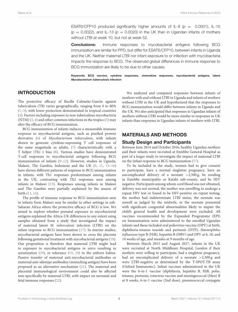
At week 10, the production of three of the 11 cytokinesmeasured differed significantly between Ugandan and UK infantsfollowing stimulation with PPD (Table 2 and Figure 1). MedianIL-1β responses were significantly higher in Ugandan infantsthan in UK infants (p < 0.0001; Table 2 and Figure 1A), whereasmedian IL-2 (p = 0.0001) and IL-10 (p < 0.0001) responseswere significantly higher in the UK compared to Ugandan infants(Table 2 and Figure 1). However, these differences in responseswere not sustained at week 52.
At weeks 10 and 52, the production of IP-10 (p < 0.0001,p= 0.0001, respectively) in response to PPD stimulation of infantblood was significantly higher in UK than in Ugandan infants(Table 2 and Figure 1B). At week 52, production of MIP-1βwas significantly higher in UK infants than in Ugandan infants(p= 0.0013, Table 2 and Figure 1B).
Immune Response to ESAT6/CFP10StimulationCytokine and chemokine responses following stimulation ofinfant whole blood with a combination of ESAT6 and CFP10proteins were measured to assess exposure to or infection withM. tuberculosis or NTM.
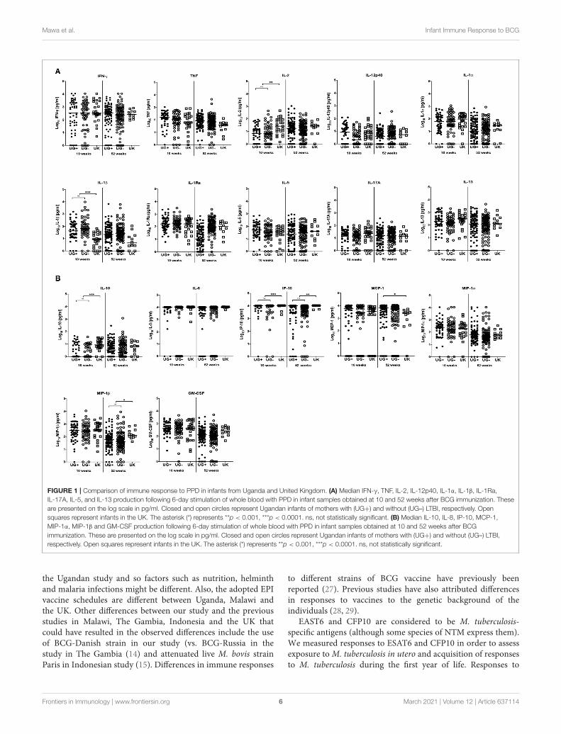
At weeks 10 and 52, the production of IFN-γ, TNF, IL-12p40,IL-1α, IL-1β, IL-1Ra, IP-10, MIP-1α, MIP-1β, and GM-CSF weresignificantly higher in Ugandan than in UK infants followingstimulation of blood with ESAT6/CFP10 (Table 3 and Figure 2).At week 10, blood samples from infants in the UK producedsignificantly higher IL-10 (p = 0.0038) and IL-8 (p = 0.0005)than Ugandan infants (Table 3 and Figure 2B), although theseresponses were not sustained at week 52. At week 52, bloodsamples from Ugandan infants produced significantly higheramounts of IL-17A than UK infants (p = 0.0009; Table 3 andFigure 2A).
Comparison of Responses Based onExposure to Maternal LTBI in UgandanInfantsWe expected that LTBI would be much more common inUgandan mothers compared to UK. We compared immuneresponses in infants of mothers with and without LTBI inUganda with those from UK infants of mothers without LTBI.As previously reported (17), responses to both PPD andESAT6/CFP10 were remarkably similar betweenUgandan infantsof mothers with and without LTBI. However, we expectedresponses in infants of Ugandan mothers without LTBI to becloser to responses from UK infants.
Immune Response to PPD StimulationThe responses to PPD stimulation of infant blood were generallysimilar between infants in Uganda and the UK regardless ofmaternal LTBI exposure in Ugandan infants, except for MCP-1 responses at week 52 where Ugandan infants of motherswith LTBI had significantly higher responses than UK infants(p= 0.0024; Figure 1B).
Immune Response to ESAT6/CFP10StimulationAt week 10, the production of IL-13 (p= 0.0020, Figure 2A), IL-10 (p = 0.0022, Figure 2B), and IL-8 (p = 0.0001, Figure 2B)following stimulation of infant blood with ESAT6/CFP10 wassignificantly higher in UK infants than in Ugandan infantsof mothers without LTBI, but comparable to responses fromUgandan infants of mothers with LTBI. However, these responseswere not sustained at week 52.
DISCUSSION
Based on previous results comparing infants in Malawi andthe UK we had expected differences in immune responses toBCG immunization between infants in Uganda and the UK.Immunization of infants in Malawi with BCG has been shownin previous studies to elicit lower IFN-γ responses to PPD than
Frontiers in Immunology | www.frontiersin.org 4 March 2021 | Volume 12 | Article 637114

Mawa et al. Infant Immune Response to BCG
TABLE 1 | Characteristics of study participants.
UG LTBI– (n = 121) UG LTBI+ (n = 110) UK (n = 25) p-value
Mother’s age (years), median (range) 23 (17–37) 25 (17–42) 34 (19–43) <0.001
Birth weight (kg), median (range) 3.2 (2.5–4.5) 3.2 (2.5–4.4) 3.35 (2.73–4.3) 0.099
Sex of the baby, male n (%) 63 (52%) 64 (58%) 13 (52%) 0.776
P-values are from the Mann Whitney-test for comparisons of mother’s age and birth weight and from the chi-squared-test for comparison of sex proportions between infants from
Uganda (overall) and those from the UK.
TABLE 2 | Median responses to PPD in infants from Uganda and United Kingdom at 10 and 52 weeks following BCG immunization.
Cytokine/
Chemokine
(pg/ml)
10 weeks after BCG 52 weeks after BCG
Uganda
(n = 108)
UnitedKingdom
(n = 25)
p-value Uganda
(n = 205)
UnitedKingdom
(n = 12)
p-value
IFN-γ 337.5 326.8 0.2638 264.7 265.1 0.8909
TNF 126 59.7 0.2636 69.26 36.04 0.0078
IL-2 5.51 25.5 0.0001 14.86 26.34 0.2908
IL-12p40 2.85 5.97 0.1256 2.395 4.370 0.5677
IL-1α 60.44 94.9 0.4134 10.21 27.25 0.2838
IL-1β 58.96 7.8 <0.0001 22.82 3.535 0.0090
IL-1Ra 180.30 164.6 0.4201 105.9 102.0 0.5018
IL-17A 16.87 22.2 0.1909 14.57 13.37 0.8108
IL-5 36.75 37.2 0.3394 27.92 39.21 0.2417
IL-13 163.8 319.1 0.0595 119.2 138.5 0.3788
IL-10 0.00 7.6 <0.0001 1.64 5.64 0.2087
IL-8 10,159 10605.3 0.0188 10,003 10,609 0.0115
IP-10 10,409 10,877 <0.0001 9,846 10,882 0.0001
MCP-1 4,294 3,525 0.3529 6,133 2,535 0.0104
MIP-1α 230.5 212.4 0.5594 47.49 57.10 0.2915
MIP-1β 268.9 366.6 0.2972 43.85 126.8 0.0013
GM-CSF 413.8 432.9 0.8493 122.5 274.5 0.0306
P-values are from the Mann Whitney-test for comparison of responses between infants from Uganda and those from the UK. Bold-faced values represent statistically significant p-values
at the 0.003 level (Bonferroni corrected).
infants in the UK (11, 23) and, in a more comprehensive analysiswhere more cytokines and chemokines were measured, sevenof 42 cytokines and chemokines tested were higher in the UKinfants (Th1 responses) while 20 were higher inMalawian infants(innate proinflammatory, regulatory, Th2, and Th17 cytokinesand growth factors) at 12 and 52 weeks (13).
We show that infant responses to PPD following BCGimmunization were much more similar between Ugandan andUK infants than expected, with no suggestion of suppressionof key Th1 responses in Uganda and no Th2 bias as previouslyobserved in Malawi (13). There were a few differences at week10 but these were not sustained at week 52 so arguably of nolong-term significance to protective immunity against TB in theinfants. Surprisingly, infants in the UKmounted similar immuneresponses to Ugandan infants of mothers with LTBI for some ofthe cytokines and chemokines analyzed.
There were important differences between our study and theMalawi study (13). First, BCG vaccination was administered atbirth or within the first week in our study vs. between 3 and13 weeks in Lalor’s study. Differences in immune responses
when BCG vaccination is given at birth vs. when delayedhave been previously reported (24, 25). Second, there weredifferences in the assays performed: we used 1/5 dilution ofwhole blood whereas the Malawi/UK study used 1/10 dilution;we used 10µg/mL of PPD vs. 5µg/mL in Lalor’s study. Third,the number of infants analyzed also differed (our Ugandan studysampled more infants than the Malawian study, but similarnumber of infants were sampled in the UK for both studies).These study-related differences could have contributed to theobserved differences between responses in infants in our studyand those in theMalawi/UK study. In previous studies inMalawi,infant responses to PPD at early time points were shown to beinfluenced by geographic location, season of birth and timingof vaccination (11, 14). It is possible that infants in Malawiwere vaccinated or blood samples collected after exposure toenvironmental mycobacteria that are common in the tropics,with the consequent low IFN-γ and high immunoregulatory andTh2 responses observed (26). Factors other than mycobacterialexposure may also explain the Malawi-UK differences. Of note,the Malawi study was conducted in a more rural setting than
Frontiers in Immunology | www.frontiersin.org 5 March 2021 | Volume 12 | Article 637114
Mawa et al. Infant Immune Response to BCG
FIGURE 1 | Comparison of immune response to PPD in infants from Uganda and United Kingdom. (A) Median IFN-γ, TNF, IL-2, IL-12p40, IL-1α, IL-1β, IL-1Ra,
IL-17A, IL-5, and IL-13 production following 6-day stimulation of whole blood with PPD in infant samples obtained at 10 and 52 weeks after BCG immunization. These
are presented on the log scale in pg/ml. Closed and open circles represent Ugandan infants of mothers with (UG+) and without (UG–) LTBI, respectively. Open
squares represent infants in the UK. The asterisk (*) represents **p < 0.001, ***p < 0.0001. ns, not statistically significant. (B) Median IL-10, IL-8, IP-10, MCP-1,
MIP-1α, MIP-1β and GM-CSF production following 6-day stimulation of whole blood with PPD in infant samples obtained at 10 and 52 weeks after BCG
immunization. These are presented on the log scale in pg/ml. Closed and open circles represent Ugandan infants of mothers with (UG+) and without (UG–) LTBI,
respectively. Open squares represent infants in the UK. The asterisk (*) represents **p < 0.001, ***p < 0.0001. ns, not statistically significant.
the Ugandan study and so factors such as nutrition, helminthand malaria infections might be different. Also, the adopted EPIvaccine schedules are different between Uganda, Malawi andthe UK. Other differences between our study and the previousstudies in Malawi, The Gambia, Indonesia and the UK thatcould have resulted in the observed differences include the useof BCG-Danish strain in our study (vs. BCG-Russia in thestudy in The Gambia (14) and attenuated live M. bovis strainParis in Indonesian study (15). Differences in immune responses
to different strains of BCG vaccine have previously beenreported (27). Previous studies have also attributed differencesin responses to vaccines to the genetic background of theindividuals (28, 29).
EAST6 and CFP10 are considered to be M. tuberculosis-specific antigens (although some species of NTM express them).We measured responses to ESAT6 and CFP10 in order to assessexposure toM. tuberculosis in utero and acquisition of responsesto M. tuberculosis during the first year of life. Responses to
Frontiers in Immunology | www.frontiersin.org 6 March 2021 | Volume 12 | Article 637114
Mawa et al. Infant Immune Response to BCG
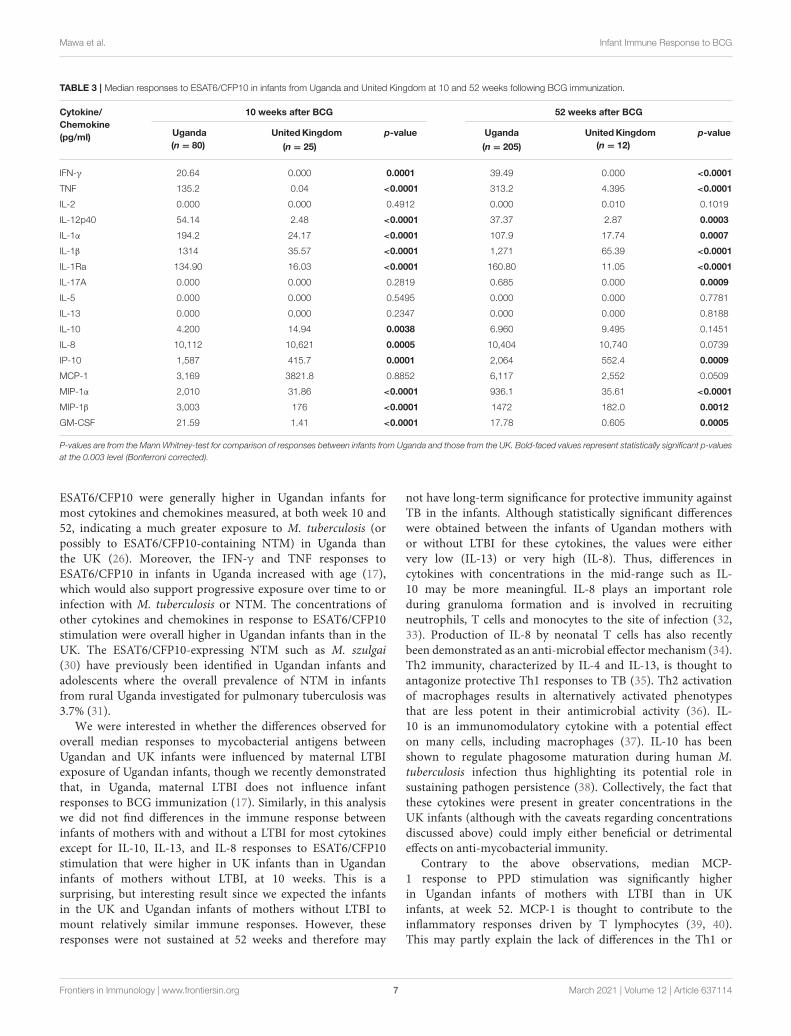
TABLE 3 | Median responses to ESAT6/CFP10 in infants from Uganda and United Kingdom at 10 and 52 weeks following BCG immunization.
Cytokine/
Chemokine
(pg/ml)
10 weeks after BCG 52 weeks after BCG
Uganda
(n = 80)
UnitedKingdom
(n = 25)
p-value Uganda
(n = 205)
UnitedKingdom
(n = 12)
p-value
IFN-γ 20.64 0.000 0.0001 39.49 0.000 <0.0001
TNF 135.2 0.04 <0.0001 313.2 4.395 <0.0001
IL-2 0.000 0.000 0.4912 0.000 0.010 0.1019
IL-12p40 54.14 2.48 <0.0001 37.37 2.87 0.0003
IL-1α 194.2 24.17 <0.0001 107.9 17.74 0.0007
IL-1β 1314 35.57 <0.0001 1,271 65.39 <0.0001
IL-1Ra 134.90 16.03 <0.0001 160.80 11.05 <0.0001
IL-17A 0.000 0.000 0.2819 0.685 0.000 0.0009
IL-5 0.000 0.000 0.5495 0.000 0.000 0.7781
IL-13 0.000 0.000 0.2347 0.000 0.000 0.8188
IL-10 4.200 14.94 0.0038 6.960 9.495 0.1451
IL-8 10,112 10,621 0.0005 10,404 10,740 0.0739
IP-10 1,587 415.7 0.0001 2,064 552.4 0.0009
MCP-1 3,169 3821.8 0.8852 6,117 2,552 0.0509
MIP-1α 2,010 31.86 <0.0001 936.1 35.61 <0.0001
MIP-1β 3,003 176 <0.0001 1472 182.0 0.0012
GM-CSF 21.59 1.41 <0.0001 17.78 0.605 0.0005
P-values are from the Mann Whitney-test for comparison of responses between infants from Uganda and those from the UK. Bold-faced values represent statistically significant p-values
at the 0.003 level (Bonferroni corrected).
ESAT6/CFP10 were generally higher in Ugandan infants formost cytokines and chemokines measured, at both week 10 and52, indicating a much greater exposure to M. tuberculosis (orpossibly to ESAT6/CFP10-containing NTM) in Uganda thanthe UK (26). Moreover, the IFN-γ and TNF responses toESAT6/CFP10 in infants in Uganda increased with age (17),which would also support progressive exposure over time to orinfection with M. tuberculosis or NTM. The concentrations ofother cytokines and chemokines in response to ESAT6/CFP10stimulation were overall higher in Ugandan infants than in theUK. The ESAT6/CFP10-expressing NTM such as M. szulgai(30) have previously been identified in Ugandan infants andadolescents where the overall prevalence of NTM in infantsfrom rural Uganda investigated for pulmonary tuberculosis was3.7% (31).
We were interested in whether the differences observed foroverall median responses to mycobacterial antigens betweenUgandan and UK infants were influenced by maternal LTBIexposure of Ugandan infants, though we recently demonstratedthat, in Uganda, maternal LTBI does not influence infantresponses to BCG immunization (17). Similarly, in this analysiswe did not find differences in the immune response betweeninfants of mothers with and without a LTBI for most cytokinesexcept for IL-10, IL-13, and IL-8 responses to ESAT6/CFP10stimulation that were higher in UK infants than in Ugandaninfants of mothers without LTBI, at 10 weeks. This is asurprising, but interesting result since we expected the infantsin the UK and Ugandan infants of mothers without LTBI tomount relatively similar immune responses. However, theseresponses were not sustained at 52 weeks and therefore may
not have long-term significance for protective immunity againstTB in the infants. Although statistically significant differenceswere obtained between the infants of Ugandan mothers withor without LTBI for these cytokines, the values were eithervery low (IL-13) or very high (IL-8). Thus, differences incytokines with concentrations in the mid-range such as IL-10 may be more meaningful. IL-8 plays an important roleduring granuloma formation and is involved in recruitingneutrophils, T cells and monocytes to the site of infection (32,33). Production of IL-8 by neonatal T cells has also recentlybeen demonstrated as an anti-microbial effector mechanism (34).Th2 immunity, characterized by IL-4 and IL-13, is thought toantagonize protective Th1 responses to TB (35). Th2 activationof macrophages results in alternatively activated phenotypesthat are less potent in their antimicrobial activity (36). IL-10 is an immunomodulatory cytokine with a potential effecton many cells, including macrophages (37). IL-10 has beenshown to regulate phagosome maturation during human M.tuberculosis infection thus highlighting its potential role insustaining pathogen persistence (38). Collectively, the fact thatthese cytokines were present in greater concentrations in theUK infants (although with the caveats regarding concentrationsdiscussed above) could imply either beneficial or detrimentaleffects on anti-mycobacterial immunity.
Contrary to the above observations, median MCP-1 response to PPD stimulation was significantly higherin Ugandan infants of mothers with LTBI than in UKinfants, at week 52. MCP-1 is thought to contribute to theinflammatory responses driven by T lymphocytes (39, 40).This may partly explain the lack of differences in the Th1 or
Frontiers in Immunology | www.frontiersin.org 7 March 2021 | Volume 12 | Article 637114

Mawa et al. Infant Immune Response to BCG
FIGURE 2 | Comparison of immune response to ESAT6/CFP10 in infants from Uganda and United Kingdom. (A) Median IFN-γ, TNF, IL-2, IL-12p40, IL-1α, IL-1β,
IL-1Ra, IL-17A, IL-5, and IL-13 production following 6-day stimulation of whole blood with ESAT6/CFP10 in infant samples obtained at 10 and 52 weeks after BCG
immunization. These are presented on the log scale in pg/ml. Closed and open circles represent Ugandan infants of mothers with (UG+) and without (UG–) LTBI,
respectively. Open squares represent infants in the UK. The asterisk (*) represents **p < 0.001, ***p < 0.0001. ns, not statistically significant. (B) Median IL-10, IL-8,
IP-10, MCP-1, MIP-1α, MIP-1β, and GM-CSF production following 6-day stimulation of whole blood with ESAT6/CFP10 in infant samples obtained at 10 and 52
weeks after BCG immunization. These are presented on the log scale in pg/ml. Closed and open circles represent Ugandan infants of mothers with (UG+) and without
(UG–) LTBI, respectively. Open squares represent infants in the UK. The asterisk (*) represents **p < 0.001, ***p < 0.0001. ns, not statistically significant.
proinflammatory responses observed between infants in Ugandaand the UK.
Additional assays such as intracellular cytokine staining andflow cytometry would be needed to identify the cellular sourcesof these cytokines and chemokines. We chose the Luminex assayinstead as our primary read out, so that we could assess a rangeof cytokine/chemokine responses. Moreover, cell phenotype and
function following BCG immunization of infants have previouslybeen assessed by our team and others (9, 41–43).
The finding that there is no difference in Th1 responses to PPDbetween infants in Uganda and the UK calls for a re-evaluationof our understanding of the variability in protection affordedby BCG. In the UK, only infants of mothers without LTBIwere included. The remarkable similarity in IFN-γ responses to
Frontiers in Immunology | www.frontiersin.org 8 March 2021 | Volume 12 | Article 637114

Mawa et al. Infant Immune Response to BCG
PPD between Ugandan and UK infants post-BCG may suggestthat the stimulus of BCG was able to override effects of prior(prenatal), or even concurrent, mycobacterial exposures (or lackof them).
Previous studies in Uganda and the UK assessed innateimmune responses in cord blood and in BCG vaccinatedinfants, measuring cytokine responses after 24 h of stimulation(16, 44), although the design of the Ugandan study did notallow assessment of post-vaccination immune responses. Ourrecent longitudinal study (17) showed very high proportionsof infants who produced high cord blood cytokine levels inresponse to stimulation with mycobacterial antigens, regardlessof mother’s TB exposure status. However, there was no evidenceof differences in the subsequent evolution of responses to PPDbased on cord blood cytokine profiles. In the present study alonger 6-day culture period was used to enable restimulationof antigen-specific memory T cell responses, so although wecannot rule out that these may have been influenced by earlierrelease of innate cytokines, or that some cytokines derivedfrom innate immune cells might still be present [as a resultof influence of innate immunity on adaptive immunity (45)and BCG-induced increases in function of innate cells (46,47)], we predict that most of the cytokines are derived fromT cells.
One limitation of recruiting volunteers in the UK was thatthe study period overlapped with a global BCG shortage thatalso affected the UK (48), thus restricting the number of BCGvaccinated infants that could be recruited within the studyperiod. Moreover, the smaller than planned sample size didnot impact the findings, since we see differences for ESAT6alongside no differences for PPD. Another limitation of thestudy is the small amount of blood available from infantsand this limited the number of assays possible. This is acommon occurrence with exploratory immunological studies.Moreover, we used an assay (Luminex) that enabled us to assessa range of cytokines/chemokines using the available amountof sample (which was sufficient for this purpose). We weretherefore able to address the objectives of the study. In theUK, by study design, infant samples were only collected atweeks 10 and 52 and from infants of mothers without LTBI.Thus, we did not have pre-vaccination or cord blood samplescollected from UK infants for a pre-vaccination comparisonwith the infants in Uganda. As TB incidence is relatively lowin the UK, recruitment of mothers with LTBI would requirea targeted recruitment drive (for example, identifying contactsof TB cases) which this study was not designed to do. Also,we did not adjust for confounders since the data available wasquite minimal.
In conclusion, immune responses to mycobacterial antigensfollowing BCG immunization are similar for PPD, butdiffer for ESAT6/CFP10, between infants in Uganda andthe UK. We show that neither maternal LTBI nor infantexposure to or infection with M. tuberculosis (or NTM)impacts response to PPD following BCG immunization.The observed differences in immune response to, andefficacy of, BCG immunization are likely to be due toother causes.
DATA AVAILABILITY STATEMENT
The raw data supporting the conclusions of this article will bemade available by the authors, without undue reservation.
ETHICS STATEMENT
The studies involving human participants were reviewedand approved by Uganda Virus Research Institute ResearchEthics Committee (Reference GC/127/13/09/16 andGC/127/16/03/434), Uganda National Council for Scienceand Technology (Reference HS 1526) and London School ofHygiene and Tropical Medicine (Reference 7104 for the Ugandastudy, 8720-1 for the UK study) and 15/LO/0048 for the NHSin the UK study. Written informed consent to participate inthis study was provided by the participants’ legal guardian/nextof kin.
AUTHOR CONTRIBUTIONS
AE, SC, PM, PK, EW, SS, and HD conceived the study andsecured funding. PM, GN, MN, and MH-A performed alllaboratory assays. RB, LD, and AG were members of the clinicalteam involved in recruitment, follow up, and clinical reporting inthe UK. AE, SC, SS, HD, EW, LL, and PM reviewed the data andwrote the initial drafts of the manuscript. LL and EW providedstatistical expertise and support. All authors read and approvedthe final version of the manuscript.
FUNDING
This work was supported by a project grant from the UKMedical Research Council, Grant Number MR/K019708. PMwas supported by a Commonwealth Scholarships CommissionPhD scholarship (UGCS-2012-602). LL was supported by a PhDfellowship through the DELTAS Africa Initiative SSACAB (GrantNo. 107754). SC was supported by funds from the DELTASInitiative MUII-plus (Grant No. 107743). The DELTAS AfricaInitiative is an independent funding scheme of the AfricanAcademy of Sciences (AAS) Alliance for Accelerating Excellencein Science in Africa (AESA) and was supported by the NewPartnership for Africa’s Development Planning and CoordinatingAgency (NEPAD Agency) with funding from the WellcomeTrust (Grant No. 107754/Z/15/Z) and the UK Government. Theviews expressed in this study are those of the authors and notnecessarily those of the AAS, NEPAD Agency, Wellcome Trustor the UK Government. SS, MH-A, and HD received supportfrom the EUHorizon 2020 programme (TBVAC2020, AgreementNumber 643381) and MH-A was supported by an MRC LondonIntercollegiate Doctoral Training Partnership studentship. EWreceived funding from MRC Grant Reference MR/K012126/1;this grant and the MRC/UVRI and LSHTM Uganda ResearchUnit are jointly funded by the UK Medical Research Council(MRC) and the UK Department for International Development(DFID) under the MRC/DFID Concordat agreement andare also part of the EDCTP2 programme supported by theEuropean Union.
Frontiers in Immunology | www.frontiersin.org 9 March 2021 | Volume 12 | Article 637114
Mawa et al. Infant Immune Response to BCG
ACKNOWLEDGMENTS
We thank the study participants, the staff of the
Immunomodulation and Vaccines Programme at the
MRC/UVRI and LSHTM Uganda Research Unit and the
midwives of the Entebbe General Hospital. The followingreagents were obtained through BEI Resources, NIAID,NIH: CFP10, Recombinant Protein Reference Standard,NR49425 and ESAT6, Recombinant Protein ReferenceStandard, NR49424.
REFERENCES
1. Brewer TF. Preventing tuberculosis with bacillus Calmette-Guerin vaccine:
a meta-analysis of the literature. Clin Infect Dis. (2000) 31(Suppl. 3):S64–7.
doi: 10.1086/314072
2. Colditz GA, Berkey CS, Mosteller F, Brewer TF, Wilson ME, Burdick E, et al.
The efficacy of bacillus Calmette-Guerin vaccination of newborns and infants
in the prevention of tuberculosis: meta-analyses of the published literature.
Pediatrics. (1995) 96:29–35.
3. Fine PE, Variation in protection by BCG: implications of and for heterologous
immunity. Lancet. (1995) 346:1339–45. doi: 10.1016/S0140-6736(95)92348-9
4. Wilson ME, Fineberg HV, Colditz GA. Geographic latitude and the efficacy
of bacillus Calmette-Guerin vaccine. Clin Infect Dis. (1995) 20:982–91.
doi: 10.1093/clinids/20.4.982
5. Elliott AM, Mawa PA, Webb EL, Nampijja M, Lyadda N, Bukusuba J, et al.
Effects of maternal and infant co-infections, and of maternal immunisation,
on the infant response to BCG and tetanus immunisation. Vaccine. (2010)
29:247–55. doi: 10.1016/j.vaccine.2010.10.047
6. Aaby P, Knudsen K, Jensen TG, Tharup J, Poulsen A, Sodemann M, et al.
Measles incidence, vaccine efficacy, and mortality in two urban African
areas with high vaccination coverage. J Infect Dis. (1990) 162:1043–8.
doi: 10.1093/infdis/162.5.1043
7. Ritz N, Strach M, Yau C, Dutta B, Tebruegge M, Connell TG, et al. A
comparative analysis of polyfunctional T cells and secreted cytokines induced
by Bacille Calmette-Guerin immunisation in children and adults. PLoS ONE.
(2012) 7:e37535. doi: 10.1371/journal.pone.0037535
8. Marchant A, Goetghebuer T, Ota MO, Wolfe I, Ceesay SJ, De Groote D,
et al. Newborns develop a Th1-type immune response toMycobacterium bovis
bacillus Calmette-Guerin vaccination. J Immunol. (1999) 163:2249–55.
9. Mawa PA, Nkurunungi G, Egesa M, Webb EL, Smith SG, Kizindo R,
et al. The impact of maternal infection with Mycobacterium tuberculosis
on the infant response to bacille Calmette-Guerin immunization.
Philos Trans R Soc Lond B Biol Sci. (2015) 370:137. doi: 10.1098/rstb.
2014.0137
10. Tena-Coki NG, Scriba TJ, Peteni N, Eley B, Wilkinson RJ, Andersen
P, et al. CD4 and CD8 T-cell responses to mycobacterial antigens
in African children. Am J Respir Crit Care Med. (2010) 182:120–9.
doi: 10.1164/rccm.200912-1862OC
11. Lalor MK, Ben-Smith A, Gorak-Stolinska P, Weir RE, Floyd S, Blitz R,
et al. Population differences in immune responses to Bacille Calmette-Guerin
vaccination in infancy. J Infect Dis. (2009) 199:795–800. doi: 10.1086/597069
12. Lalor MK, Smith SG, Floyd S, Gorak-Stolinska P, Weir RE, Blitz R, et al.
Complex cytokine profiles induced by BCG vaccination in UK infants.
Vaccine. (2010) 28:1635–41. doi: 10.1016/j.vaccine.2009.11.004
13. Lalor MK, Floyd S, Gorak-Stolinska P, Ben-Smith A, Weir RE, Smith SG,
et al. BCG vaccination induces different cytokine profiles following infant
BCG vaccination in the UK and Malawi. J Infect Dis. (2011) 204:1075–85.
doi: 10.1093/infdis/jir515
14. Hur YG, Gorak-Stolinska P, Lalor MK, Mvula H, Floyd S, Raynes J,
et al. Factors affecting immunogenicity of BCG in infants, a study
in Malawi, The Gambia and the UK. BMC Infect Dis. (2014) 14:184.
doi: 10.1186/1471-2334-14-184
15. Djuardi Y, Sartono E, Wibowo H, Supali T, Yazdanbakhsh M. A
longitudinal study of BCG vaccination in early childhood: the development
of innate and adaptive immune responses. PLoS ONE. (2010) 5:e14066.
doi: 10.1371/journal.pone.0014066
16. Mawa PA, Webb EL, Filali-Mouhim A, Nkurunungi G, Sekaly RP,
Lule SA, et al. Maternal BCG scar is associated with increased infant
proinflammatory immune responses. Vaccine. (2017) 35: 273–82.
doi: 10.1016/j.vaccine.2016.11.079
17. Lubyayi L, Mawa PA, Nabakooza G, Nakibuule M, Tushabe JV, Serubanja J,
et al. Maternal latent Mycobacterium tuberculosis does not affect the infant
immune response following BCG at birth: an observational longitudinal study
in Uganda. Front Immunol. (2020) 11:e00929. doi: 10.3389/fimmu.2020.00929
18. Rahman MJ, Degano IR, Singh M, Fernandez C. Influence of maternal
gestational treatment with mycobacterial antigens on postnatal
immunity in an experimental murine model. PLoS ONE. (2010) 5:e9699.
doi: 10.1371/journal.pone.0009699
19. Potian JA, Rafi W, Bhatt K, McBride A, Gause WC, Salgame P. Preexisting
helminth infection induces inhibition of innate pulmonary anti-tuberculosis
defense by engaging the IL-4 receptor pathway. J Exp Med. (2011) 208:1863–
74. doi: 10.1084/jem.20091473
20. Gebreegziabiher D, Desta K, Desalegn G, Howe R, Abebe M. The
effect of maternal helminth infection on maternal and neonatal immune
function and immunity to tuberculosis. PLoS ONE. (2014) 9:e93429.
doi: 10.1371/journal.pone.0093429
21. Novato-Silva E, Gazzinelli G, Colley DG. Immune responses during
human schistosomiasis mansoni. XVIII. Immunologic status of pregnant
women and their neonates. Scand J Immunol. (1992) 35:429–37.
doi: 10.1111/j.1365-3083.1992.tb02878.x
22. Holt PG, Strickland DH, Soothing signals: transplacental transmission
of resistance to asthma and allergy. J Exp Med. (2009) 206:2861–4.
doi: 10.1084/jem.20092469
23. Black GF, Weir RE, Floyd S, Bliss L, Warndorff DK, Crampin AC, et al.
BCG-induced increase in interferon-gamma response to mycobacterial
antigens and efficacy of BCG vaccination in Malawi and the UK:
two randomised controlled studies. Lancet. (2002) 359:1393–401.
doi: 10.1016/S0140-6736(02)08353-8
24. Lutwama F, Kagina BM, Wajja A, Waiswa F, Mansoor N, Kirimunda S,
et al. Distinct T-cell responses when BCG vaccination is delayed from birth
to 6 weeks of age in Ugandan infants. J Infect Dis. (2014) 209:887–97.
doi: 10.1093/infdis/jit570
25. Burl S, Adetifa UJ, Cox M, Touray E, Ota MO, Marchant A, et al. Delaying
bacillus Calmette-Guerin vaccination from birth to 4 1/2 months of age
reduces postvaccination Th1 and IL-17 responses but leads to comparable
mycobacterial responses at 9 months of age. J Immunol. (2010) 185:2620–8.
doi: 10.4049/jimmunol.1000552
26. Black GF, Dockrell HM, Crampin AC, Floyd S, Weir RE, Bliss L, et al. Patterns
and implications of naturally acquired immune responses to environmental
and tuberculous mycobacterial antigens in northern Malawi. J Infect Dis.
(2001) 184:322–9. doi: 10.1086/322042
27. Anderson EJ, Webb EL, Mawa PA, Kizza M, Lyadda N, Nampijja M, et al. The
influence of BCG vaccine strain on mycobacteria-specific and non-specific
immune responses in a prospective cohort of infants in Uganda. Vaccine.
(2012) 30:2083–9. doi: 10.1016/j.vaccine.2012.01.053
28. Quach H, Rotival M, Pothlichet J, Loh YE, Dannemann M, Zidane
N, et al. Genetic adaptation and neandertal admixture shaped the
immune system of human populations. Cell. (2016) 167:643–56.e17.
doi: 10.1016/j.cell.2016.09.024
29. Zimmermann P, Curtis N. Factors that influence the immune
response to vaccination. Clin Microbiol Rev. (2019) 32:e00084-18.
doi: 10.1128/CMR.00084-18
30. van Ingen J, de Zwaan R, Dekhuijzen R, Boeree M, van Soolingen D.
Region of difference 1 in nontuberculous Mycobacterium species adds a
phylogenetic and taxonomical character. J Bacteriol. (2009) 191:5865–7.
doi: 10.1128/JB.00683-09
Frontiers in Immunology | www.frontiersin.org 10 March 2021 | Volume 12 | Article 637114

Mawa et al. Infant Immune Response to BCG
31. Asiimwe BB, Bagyenzi GB, Ssengooba W, Mumbowa F, Mboowa G,
Wajja A, et al. Species and genotypic diversity of non-tuberculous
mycobacteria isolated from children investigated for pulmonary tuberculosis
in rural Uganda. BMC Infect Dis. (2013) 13:88. doi: 10.1186/1471-233
4-13-88
32. Gerszten RE, Garcia-Zepeda EA, Lim YC, Yoshida M, Ding HA, Gimbrone
MA, Jr., et al. MCP-1 and IL-8 trigger firm adhesion of monocytes to
vascular endothelium under flow conditions. Nature. (1999) 398:718–23.
doi: 10.1038/19546
33. Mukaida N, Harada A, Matsushima K. Interleukin-8 (IL-8) and monocyte
chemotactic and activating factor (MCAF/MCP-1), chemokines essentially
involved in inflammatory and immune reactions.Cytokine Growth Factor Rev.
(1998) 9:9–23. doi: 10.1016/S1359-6101(97)00022-1
34. Gibbons D, Fleming P, Virasami A, Michel ML, Sebire NJ, Costeloe K, et al.
Interleukin-8 (CXCL8) production is a signatory T cell effector function of
human newborn infants. Nat Med. (2014) 20:1206–10. doi: 10.1038/nm.3670
35. Rook GA. Th2 cytokines in susceptibility to tuberculosis. Curr Mol Med.
(2007) 7:327–37. doi: 10.2174/156652407780598557
36. Gordon S, Martinez FO. Alternative activation of macrophages:
mechanism and functions. Immunity. (2010) 32:593–604.
doi: 10.1016/j.immuni.2010.05.007
37. Bogdan C, Vodovotz Y, Nathan C.Macrophage deactivation by interleukin 10.
J Exp Med. (1991) 174:1549–55. doi: 10.1084/jem.174.6.1549
38. O’Leary S, O’Sullivan MP, Keane J. IL-10 blocks phagosome maturation
in mycobacterium tuberculosis-infected human macrophages. Am
J Respir Cell Mol Biol. (2011) 45:172–80. doi: 10.1165/rcmb.2010-
0319OC
39. Luo Y, Chen X, O’Donnell MA. Mycobacterium bovis bacillus Calmette-
Guerin (BCG) induces human CC- and CXC-chemokines in vitro and in vivo.
Clin Exp Immunol. (2007) 147:370–8. doi: 10.1111/j.1365-2249.2006.03288.x
40. Taub DD, Proost P, Murphy WJ, Anver M, Longo DL, van Damme
J, et al. Monocyte chemotactic protein-1 (MCP-1),−2, and−3 are
chemotactic for human T lymphocytes. J Clin Invest. (1995) 95:1370–6.
doi: 10.1172/JCI117788
41. Smith SG, Zelmer A, Blitz R, Fletcher HA, Dockrell HM. Polyfunctional
CD4 T-cells correlate with in vitro mycobacterial growth inhibition following
Mycobacterium bovis BCG-vaccination of infants. Vaccine. (2016) 34:5298–
305. doi: 10.1016/j.vaccine.2016.09.002
42. Kagina BM, Abel B, Bowmaker M, Scriba TJ, Gelderbloem S, Smit E, et al.
Delaying BCG vaccination from birth to 10 weeks of age may result in
an enhanced memory CD4T cell response. Vaccine. (2009) 27:5488–95.
doi: 10.1016/j.vaccine.2009.06.103
43. Fletcher HA, Filali-Mouhim A, Nemes E, Hawkridge A, Keyser A, Njikan
S, et al. Human newborn bacille Calmette-Guerin vaccination and risk
of tuberculosis disease: a case-control study. BMC Med. (2016) 14:76.
doi: 10.1186/s12916-016-0617-3
44. Smith SG, Kleinnijenhuis J, Netea MG, Dockrell HM. Whole blood profiling
of bacillus calmette-guerin-induced trained innate immunity in infants
identifies epidermal growth factor, IL-6, platelet-derived growth factor-
AB/BB, and natural killer cell activation. Front Immunol. (2017) 8:644.
doi: 10.3389/fimmu.2017.00644
45. Jain A, Pasare C. Innate control of adaptive immunity: beyond the three-signal
paradigm. J Immunol. (2017) 198:3791–800. doi: 10.4049/jimmunol.1602000
46. Netea MG, Quintin J, van der Meer JW. Trained immunity: a
memory for innate host defense. Cell Host Microbe. (2011) 9:355–61.
doi: 10.1016/j.chom.2011.04.006
47. Kleinnijenhuis J, Quintin J, Preijers F, Joosten LA, Ifrim DC, Saeed S, et al.
Bacille Calmette-Guerin induces NOD2-dependent nonspecific protection
from reinfection via epigenetic reprogramming of monocytes. Proc Natl Acad
Sci USA, (2012) 109:17537–42. doi: 10.1073/pnas.1202870109
48. Harris RC, Dodd PJ, White RG. The potential impact of BCG vaccine supply
shortages on global paediatric tuberculosis mortality. BMC Med. (2016)
14:138. doi: 10.1186/s12916-016-0685-4
Conflict of Interest: The authors declare that the research was conducted in the
absence of any commercial or financial relationships that could be construed as a
potential conflict of interest.
Copyright © 2021 Mawa, Hasso-Agopsowicz, Lubyayi, Nabakooza, Nakibuule, Blitz,
Dun, Govind, Kaleebu, Webb, Elliott, Dockrell, Cose and Smith. This is an open-
access article distributed under the terms of the Creative Commons Attribution
License (CC BY). The use, distribution or reproduction in other forums is permitted,
provided the original author(s) and the copyright owner(s) are credited and that the
original publication in this journal is cited, in accordance with accepted academic
practice. No use, distribution or reproduction is permitted which does not comply
with these terms.
Frontiers in Immunology | www.frontiersin.org 11 March 2021 | Volume 12 | Article 637114