immunodeficiency virus type 1 (hiv-1) primer trnalys3...
TRANSCRIPT
ANTIMICROBIAL AGENTS AND CHEMOTHERAPY,0066-4804/97/$04.0010
Oct. 1997, p. 2141–2148 Vol. 41, No. 10
Copyright © 1997, American Society for Microbiology
Phosphorothioate Oligonucleotides Derived from HumanImmunodeficiency Virus Type 1 (HIV-1) Primer tRNALys3 AreStrong Inhibitors of HIV-1 Reverse Transcriptase and Arrest
Viral Replication in Infected CellsRIM EL DIRANI-DIAB,1,2 LEILA SARIH-COTTIN,1,2 BRIGITTE DELORD,1,3 BEATRICE DUMON,1,3
SERGE MOREAU,1,4 JEAN-JACQUES TOULME,1,4 HERVE FLEURY,1,3 AND SIMON LITVAK1,2*
Institut Federatif de Recherches “Pathologies Infectieuses” (IFR 66),1 and IBGC-CNRS, 33077 Bordeaux Cedex,2 andLaboratoire de Virologie3 and INSERM, Unite 386,4 Universite Victor Segalen Bordeaux II,
33076 Bordeaux Cedex, France
Received 31 December 1996/Returned for modification 14 April 1997/Accepted 15 July 1997
Retroviral reverse transcriptase (RT) is involved in the selection of a specific tRNA primer which initiatesproviral DNA minus-strand synthesis. Studies of the interactions between human immunodeficiency virus type1 (HIV-1) RT and primer tRNALys3 have shown that the dihydrouridine (diHU), anticodon, and pseudouridineregions of tRNA are highly protected in the RT-tRNA complex. The CCA 3* end of tRNA is also in close contactwith the enzyme during the cDNA initiation step. Using synthetic oligoribonucleotides corresponding to theanticodon and diHU regions, we have previously shown a low but significant inhibition of HIV-1 RT activity(37). We extend this observation and show that primer tRNA-derived oligodeoxynucleotides (ODNs) carryinga phosphorothioate (PS) modification are strong inhibitors of HIV-1 RT. The affinity of PS-ODNs for theenzyme was monitored by gel mobility shift electrophoresis. Experiments with HIV-1-infected human cells(MT-2 cells) were performed with the latter ODNs. A PS-ODN corresponding to the 3* end of tRNALys3
(acceptor stem [AS]) was able to inhibit HIV-1 replication. No effect of the other modified ODNs was observedin infected cells. The analysis of HIV-1 RNase H activity in a cell-free system strongly suggests that theinhibitory effect of the PS-AS may be mediated via both a sense and an antisense mechanism.
All retroviral reverse transcriptases (RTs) initiate proviralfirst-strand DNA synthesis from a tRNA hybridized to theprimer binding site (PBS), a retroviral genomic region near the59 end which is complementary to the first 18 nucleotides of the39 end of primer tRNA. (For recent reviews on RT, see refer-ences 7, 25, 39, and 43). Determination of the genomic se-quences of human immunodeficiency virus type 1 (HIV-1) andHIV-2 and further experimental approaches showed thattRNALys3 is their specific primer (45). A stable and specificcomplex between HIV-1 RT and tRNALys3 has been shown bydifferent approaches in the absence of the PBS region. How-ever, HIV-1 RT is also able to form a complex with othertRNAs (6, 35, 44). Using the footprinting technique, we havefound that the anticodon and dihydrouridine (diHU) regionsof tRNALys3 interact strongly with HIV-1 RT (37). This resultagrees with those of Barat et al. (9), who showed that theanticodon region of primer tRNA was found to be preferen-tially linked to RT after UV irradiation. More recent resultshave shown that the TCC region of tRNALys3 also interactswith HIV-1 RT (46). Using a 4-thiouridine UV-induced cross-linking strategy, Mishima and Steitz (29) localized the 59 halfof the primer tRNA at the interface between the two HIV-1RT subunits, close to the RNase H domain of the viral poly-merase. Mutations of the TCC stem partially interfered withRT binding, while mutations in the D arm abolished the abilityof tRNALys3 to bind HIV-1 RT. These results suggest thatHIV-1 RT is able to recognize the central domain of theprimer tRNA tertiary structure (30). Moreover, the same in-vestigators (30) showed that HIV-1 RT facilitates the
tRNALys3-PBS annealing, confirming our previous results (37).RT is not the only factor involved in the annealing between theprimer and the template since the gag-derived basic nucleo-capsid protein has been reported to stimulate tRNALys3 an-nealing to the PBS region (see reference 18 for a review).Recently, it has been shown that the U-rich anticodon regionof tRNALys3 facilitates the formation of the RNA template-tRNA primer duplex by interacting with an A-rich region lo-calized downstream, near the PBS sequence (20).
Oligonucleotides have successfully been used to interferewith retrovirus replication. The most common approach is theantisense strategy which has been proposed for the potentialtreatment of AIDS, cancer, and other diseases (1). The anti-sense strategy has been used to arrest reverse transcription bymurine and avian RTs in cell-free systems (12, 14) and byHIV-1 RT in vitro (13, 15), as well as HIV-1 replication ininfected cells (16).
The decoy (or sense) strategy rests on the selective bindingof an oligodeoxynucleotide (ODN) to a nucleic acid bindingprotein. The ODN mimics the natural ligand of the protein andtherefore competes with it for complex formation with theprotein. This association traps the protein in a nonfunctionalcomplex. An example of the use of ODNs as decoys is thetargetting of the NF-kB transcription factor involved in theinitiation of eucaryotic transcription (42). Complex formationbetween primer tRNA and RT during the selection of thespecific tRNALys3 primer and further positioning of the prim-er-template duplex on the PBS site are crucial steps in theinitiation of retroviral replication and may be useful targets asdecoy agents that can be used to block retroviral proliferation.The latter idea is supported by previous results showing theinhibition of HIV-1 RT DNA polymerase activity observed
* Corresponding author. Mailing address: IBGC-CNRS, 1 rue Ca-mille Saint Saens, 33077 Bordeaux Cedex, France.
2141
on Novem
ber 6, 2018 by guesthttp://aac.asm
.org/D
ownloaded from
with oligoribonucleotides derived from the anticodon anddiHU regions of tRNALys3. Under the same conditions a ran-dom sequence did not affect the enzyme activity (37). Thelatter results showed that HIV RT was inhibited by phosphodi-ester (PO) oligoribonucleotides, but the in vivo application ofunmodified ODNs is limited due to their sensitivities to nucle-ases and their relatively short half-lives. Therefore, novelODNs with modifications to their phosphate backbones havebeen developed. Compared with other modified ODNs, thephosphorothioate (PS) derivatives (PS-ODNs) have severaladvantages: increased lifetime due to improved nuclease resis-tance, ability to elicit RNase H activity, and ease of synthesis.Many reports on the use of these compounds as potentialanti-AIDS agents have been published (2, 8, 23).
In this article we describe the strong inhibition of HIV-1 RTactivity by PS-ODNs derived from the tRNALys3 sequence.The best inhibitory effect on DNA synthesis in a cell-freesystem was obtained with a PS-ODN corresponding to the 39end of the primer tRNALys3 acceptor stem (AS) (PS-ASLys3),although other PS-ODNs also strongly inhibited cDNA syn-thesis. Interestingly, the PS-ASLys3 was able to inhibit the pro-liferation of HIV-1 in infected human cells. In contrast, theother tRNALys3-derived PS-ODNs did not show any ex vivoeffect. Long-term treatment (28 days) of the infected cells withthe PS-AS agent showed that the inhibitory effect was main-tained.
MATERIALS AND METHODS
Materials. Unlabeled nucleotides, unmodified oligonucleotides, and poly-nucleotides were purchased from Boehringer Mannheim, Sigma, and Pharmacia.Radioisotopes were purchased from Amersham. Calf intestinal alkaline phos-phatase and polynucleotide kinase were from Boehringer Mannheim. T7 RNApolymerase, human placental RNase inhibitor, the SphI restriction enzyme, andterminal deoxynucleotidyl transferase were from Bethesda Research Laborato-ries. Proteinase K was from Boehringer Mannheim, and pancreatic RNase A wasfrom Sigma. 39-Azido-39-deoxythymidine (AZT) was purchased from Sigma.Activated calf thymus DNA was prepared as described before (5).
Enzyme purification. Recombinant HIV RT was purified from transformedyeast cells as described previously (11, 36).
Oligonucleotide synthesis. Oligonucleotides were synthesized on an AppliedBiosystems automatic synthesizer by following the manufacturer’s instructions.PS-ODN synthesis was performed on a Millipore Expedite synthesizer as de-scribed previously (33).
HIV-1 RNA synthesis in vitro. The plasmid pmCG6 containing the fragmentcorresponding to nucleotides 1 to 4005 of HIV-1 (Mal isolate) in pSP64, underthe control of the bacteriophage T7 promoter, was a kind gift from J. L. Darlix(Institut National de la Sante et de la Recherche Medicale, Lyon, France).Escherichia coli HB101 (recA mutant) was used for plasmid amplification. Afterdigestion of this clone with pSt 1 and in vitro transcription with T7 RNApolymerase, we obtained RNAs starting at position 150 of the mal sequence.The nucleotide sequence was determined and was shown to be the same as thatof the original clone (3).
Viral RNA, corresponding to nucleotides 50 to 997 of the HIV-1 RNA (Malisolate), was synthesized by in vitro transcription with T7 RNA polymerase.Three micrograms of linearized plasmid DNA was incubated for 2 h at 37°C ina total volume of 0.1 ml containing 40 mM Tris-HCl (pH 8.0), 8 mM MgCl2, 10mM spermidine, 25 mM NaCl, 10 mM dithiothreitol, 0.5 mM (each) ribonucle-oside triphosphate, 100 U of T7 RNA polymerase, and 20 U of the placentalRNase inhibitor RNasin. After treatment with 12 U of RNase-free DNase I (for10 min at 37°C), RNA transcripts were extracted with 1 volume of a solutioncontaining phenol-chloroform-isoamyl alcohol (24:24:1) and then with chloro-form and were precipitated with 2.5 volumes of ethanol and 0.3 M ammoniumacetate.
Purified enzyme assays in a cell-free system. (i) Reverse transcription withHIV-1 RNA template. Reverse transcription was performed with HIV-1 RNAtemplate in a final volume of 50 ml. Hybridization was performed in the presenceof 50 mM Tris-HCl (pH 8.0), 6 mM MgCl2, 2 mM dithiothreitol, 30 mM NaCl,150 nM HIV-1 RNA, and 1.6 mM synthetic deoxyoligonucleotide primer com-plementary to the PBS of HIV RNA for 30 min at 37°C. Then, 0.1 mM (each)deoxynucleoside triphosphate, 10 mCi of [a-32P]dGTP (3,000 Ci/mmol), and 150nM HIV-1 RT were added. The incubation was continued for 30 min at 37°C.The samples were extracted with phenol-chloroform and collected by ethanolprecipitation at 220°C. Reaction products were analyzed by electrophoresis in10% polyacrylamide–TBE (Tris-borate-EDTA)–7 M urea (urea-polyacrylamide
gel electrophoresis [PAGE]) denaturing gels. The gels were autoradiographedand the films were analyzed by using the NIH Image Program (Macintosh).
(ii) Reverse transcription with activated DNA. For reverse transcription withactivated DNA, the reaction mixture contained, in a final volume of 0.05 ml, 50mM Tris-HCl (pH 8.0), 5 mM MgCl2, 10 mM dithiothreitol, 50 mM KCl, 0.4 to10 mg of activated DNA per ml, 50 mM (each) dATP, dCTP, and dGTP, 2 mMdTTP, 0.5 to 1 mCi of [3H]dTTP (56 Ci/mmol), and recombinant RT. Thereaction mixture was incubated for 15 min at 37°C, and the reaction wasstopped by the addition of 1 ml of cold 10% trichloroacetic acid (TCA) plus0.1 M sodium pyrophosphate. The precipitate was filtered through nitrocellulosemembranes, washed with ice-cold 2% TCA, dried, and counted in a 2,5-diphe-nyloxazole–1,4-bis(5-phenyloxazolyl)benzene–toluene scintillation mixture.
Urea-PAGE analysis. Samples were extracted with phenol, precipitated withethanol, dried, and resuspended in sample buffer (50% sucrose, 0.2% bromo-phenol blue, 0.2% xylene cyanol, and 8 M urea) and migration buffer 13 TBE(100 mM Tris-borate, 2 mM EDTA [pH 8.0]). The 8 M urea–10% polyacryl-amide gels were prerun at 1,000 V for 1 h. Migration was for 3 to 4 h at 1,500 V.After electrophoresis, the gels were autoradiographed.
Gel mobility shift assays. HIV-1 RT and oligonucleotides were incubated at37°C in 10-ml reaction mixtures containing 50 mM Tris-acetate buffer (pH 8.5)and 10 mM MgCl2 for 10 min. Electrophoresis of the RT-oligonucleotide com-plexes was performed through a 6% polyacrylamide gel in 13 TBE buffer at 100V for 3 to 4 h. The gels were dried and autoradiographed.
Cells, virus strains, and cell culture assays. MT-2 cells were chosen for theirhigh degree of susceptibility to most laboratory strains of HIV-1. These cellswere grown at 37°C in RPMI 1640 medium (Gibco-BRL) supplemented with10% heat-inactivated fetal calf serum (Boehringer Mannheim), 2 mM L-glu-tamine, penicillin (100 IU/ml), streptomycin (0.1 ng/ml), and anti-interferonalpha (700 IU/ml; Boehringer Mannheim).
An HIV-1 stock (strain HTLV-IIIB) was obtained from the cell-free super-natant of HIV-1-infected H9 cells (H9/HTLV-IIIB cells). The 50% tissue cultureinfective dose (TCID50) of cell-free virus stock was determined on day 4 byendpoint titration with MT-2 cells in 96-well microculture plates by the methodof Reed and Muench (34). The titer of virus stock was about 106 TCID50s/ml. Insome experiments, viruses were pelleted by ultracentrifugation from clarifiedculture supernatants of H9/HTLV-IIIB cells, and the titer was calculated as1.6 3 108 TCID50s/ml, in duplicate, to allow for the simultaneous evaluation oftheir effects on HIV-infected cells. Uninfected or untreated samples (negativecontrol) and infected and untreated samples (positive control) were included ineach experiment. The inhibitory effect of AZT was tested in parallel.
Infection of MT-2 cells. (i) Posttreatment inhibitor experiments. A total of6 3 104 MT-2 cells per well were exposed to HIV-1 for 2 h at 37°C. Infection wasmade at a multiplicity of infection of 1. After the unadsorbed viruses werewashed off, cells were resuspended in fresh medium and were mixed with dif-ferent concentrations of compounds in 96-well plates. The cultures were incu-bated at 37°C for 4 days.
(ii) Long-term effects of PS-ODNs on human cells infected with HIV-1. Un-treated MT-2 cells were infected as described above and were incubated for 2 hat 37°C. The addition of different concentrations of ODNs was made as de-scribed above for the posttreatment inhibitor experiments, and the cells wereincubated for 4 days. Fresh medium containing the same ODN concentrationswas then added every 4 days for 28 days.
(iii) Virus adsorption inhibitor experiments. Compounds were added simul-taneously to the mixture of HIV-1 and cells and were then washed off after 2 hof incubation at 37°C. Thereafter, the cultures were kept in drug-free medium for4 days.
Antiviral assays in cell systems. The activities of the oligonucleotides againstHIV-1/HTLV-IIIB strain replication were determined by following several cri-teria: appearance of cytopathic effect, protection of virus-induced cytopathoge-nicity in MT-2 cells, inhibition of RT activity, and specific antigen expression.
(i) Protection of virus-induced cytopathogenicity (MTT assay). The protectiveeffects of the inhibitory agents were tested as follows. The viabilities of cells thathad been infected or not infected with HIV-1 and then exposed to variouscompounds were measured spectrophotometrically via the reduction of 3-(4,5-dimethylthiazol-2-yl)2,5-diphenyl tetrazolium bromide (MTT) as described be-fore (31).
(ii) Virion RT activity assay. The assay for RT activity was performed asdescribed previously (38). Supernatants (50 ml) from each culture were incubatedwith 10 ml of virus-disrupting buffer (500 mM KCl, 50 mM dithiothreitol, 0.5%Triton X-100) for 15 min at 4°C. Thereafter, a solution (40 ml) containing 25 mMTris-HCl (pH 7.8), 1.25 mM EGTA, 12.5 mM MgCl2, 0.5 A260 units of poly(rA)-oligo(dT) per ml, and 3 mCi of [3H]dTTP at 30 Ci/mmol was added, and themixture was incubated for 1 h at 37°C. The reaction was stopped by the additionof 20 ml of 60% TCA containing 120 mM sodium pyrophosphate. The acid-insoluble nucleic acids were precipitated for 15 min at 4°C and were collected onglass fiber filters. The filters were washed with 5% TCA, dried, and placed inscintillation vials containing 1 ml of scintillation fluid, and the radioactivity wascounted.
2142 EL DIRANI-DIAB ET AL. ANTIMICROB. AGENTS CHEMOTHER.
on Novem
ber 6, 2018 by guesthttp://aac.asm
.org/D
ownloaded from

RESULTS AND DISCUSSION
We have focused our investigations on the search for agentsable to interfere with the formation of the initiation complex ofretroviral replication. RT and primer tRNA are key elementsof the reverse transcription initiation complex. Our aim was toblock the interaction between the retroviral HIV-1 RT and itsprimer tRNA to prevent initiation of cDNA synthesis by as-suming that this enzyme or its in vivo precursor proteinp160gag-pol is involved in tRNA selection (27). One way toinhibit the initiation of reverse transcription is via interferencewith the formation of the RT-tRNA complex by using tRNA-derived ODNs, which have previously been shown by foot-printing, UV cross-linking analysis, etc., to be in close contactwith the enzyme (10, 37, 46). These ODNs include anticodon,diHU, and TCC. Another potential inhibitory ODN is the 39end of the CCA acceptor stem, a region of the tRNA primerinvolved in the RT-primer-template complex during initiationof cDNA synthesis. The 39 AS region of primer tRNA interactswith the active site of the enzyme. Moreover, it is involved inthe annealing to the retroviral PBS region. The scheme in Fig.
1 illustrates the cloverleaf configuration of rabbit livertRNALys3, whose sequence is assumed to be the same as thatin its human counterpart (32).
Inhibition of HIV-1 RT by primer tRNA-derived PS-ODNs.The effect of the tRNALys3-derived ODNs on DNA synthesisby HIV-1 RT was first studied with activated DNA as thetemplate-primer. No inhibition was observed when PO-ODNsderived from the sequence of tRNALys3 were used, even atconcentrations as high as 40 mM. However, with the PS-ODNsthe situation was drastically different. The PS-ODNs werestrong inhibitors of HIV-1 RT: Ki values for the inhibition ofDNA synthesis are presented in Table 1. All these PS deriva-tives interfered with DNA synthesis, with the most powerfulinhibitors being the ODNs corresponding to the ASLys3 andthe anticodon (AC) stem-loop, with Kis of 14 and 22 nM,respectively. On the basis of the results showing a strong inhi-bition by the PS-ASLys3, we changed the sequence of the ac-ceptor stem sequence (ODNs 101 and 103), mainly the G1Ccontent. These bases may play an important role in the sec-ondary structure of the AS-ODN since they are able to interactpreferentially with the enzyme (19). Results of studies in whichdiHU, pseudouridine (pseudo-U), or different variants of theAS sequence of tRNALys3 were used as inhibitors of the DNApolymerase activity of HIV-1 RT indicated that of all thePS-ODNs used, the best inhibitor of the retroviral DNA poly-merase giving the lowest Ki value was the acceptor stem (Table1).
The deletion of the CCA end, compound PS-ODN 104, ledto a decrease in the level of inhibition which may be partlyrelated to the shorter size of this ODN. Oligonucleotides 101,103, and 106 (mismatched and scrambled sequences) weregood inhibitors of HIV-1 RT but gave higher Ki values thanthose obtained with the ASLys3 sequence. These results are inagreement with the strong interaction between HIV RT andthe anticodon region, as well as the inhibitory effect observedwith a dithioate derivative of the 39 end of primer RNA (28).
In order to study the effects of several PS-ODN derivativeson the RNA-dependent DNA polymerase activity of RT, weused an HIV-1 RNA fragment corresponding to the 59 longterminal repeat of the retroviral genome as the template and asynthetic 18-mer DNA primer complementary to the PBS re-gion. As shown in Fig. 2, the expected full-length product (147nucleotides) was obtained after in vitro cDNA synthesis, al-though some intermediate-size bands, which may correspondto pauses during reverse transcription, were also present. The
FIG. 1. Cloverleaf structure of tRNALys3. Bold lines indicate the sequence ofthe different synthetic ODNs used in this work.
TABLE 1. Inhibition of HIV-1 reverse transcription by PS-ODNsa
PS-ODN Length (no. ofnucleotides) Sequence (59-39) Ki (nM) IC50 (nM)
ASLys 16 CCC TGT TCG GGC GCC A 14 6 1 40Anticodon 15 CAG ACT TTT AAT CTG 22 6 6 140diHU 14 CTC AGT CGG TAG AG 37 6 12 .280Pseudo-U 15 AGG GTT CAA GTC CCT 35 6 13 140Mismatched ASLys (101) 16 CCC TGT TCA AACGCC A 42 6 2 75Mismatched ASLys (103) 16 TTT TGT TCG GGC GCC A 32 6 6 100DCCA (104) 13 CCC TGT TCG GGC G 87 6 13 150INV-ASLys 16 A CCG CGG GCT TGT CCC 58 6 6 NDb
Scrambled ASLys (106) 16 CTC GCT GCG ACC GTG C 45 6 4 NDASVal 16 CCG GGC GGA AAC ACC A 26 6 3 140
a Ki values were obtained with PS-ODNs corresponding to different domains of primer tRNALys3 and different sequences derived from the AS of tRNALys3:oligonucleotides 101 and 103 are mismatched PS-ODNs, oligonucleotide 104 is a PS-ODN lacking the CCA at the 39 end, INV-ASLys corresponds to the ASLys3 invertedsequence, oligonucleotide 106 is a scrambled ODN, and ASVal corresponds to the AS domain of bovine tRNAVal. HIV-1 RT was assayed with activated DNA asdescribed in the Materials and Methods. IC50s were determined from densitometer readings from Fig. 2, 3, and 4. The nucleotides in boldface type are changes fromthe nucleotide sequence of ASLys3.
b ND, not determined.
VOL. 41, 1997 HIV-1 REVERSE TRANSCRIPTASE INHIBITORS 2143
on Novem
ber 6, 2018 by guesthttp://aac.asm
.org/D
ownloaded from

PS-AS ODN derived from tRNALys3 strongly inhibited thereaction, with a 50% inhibitory concentration (IC50) of 40 nM,while inhibition by the PS-AS corresponding to tRNAVal wasobserved at significantly higher concentrations (IC50, 140 nM;Table 1). Similar results were obtained when an RNA tem-plate-tRNALys3 primer duplex was used (data not shown).
Figure 3 indicates the inhibitory effects of PS-ODNs corre-sponding to different regions of primer tRNA on cDNA syn-thesis. The IC50s are presented in Table 1. The most importantinhibitory effect was observed with PS-ASLys3. PS-ODNs cor-responding to the anticodon and pseudo-U stem-loop regionsgave similar levels of inhibition with IC50s of 140 nM, while thediHU ODN barely affected DNA synthesis.
Using the same system, we evaluated the inhibitory effect ofPS-ODN derivatives of PS-ASLys3 (Fig. 4). A lower degree ofinhibition was obtained with mismatched oligonucleotides 101(IC50, 75 nM) and 103 (IC50, 100 nM). Oligonucleotide 104,from which the CCA end is deleted, was not inhibitory at theseconcentrations. Scrambled oligonucleotide 106 and the in-verted ODN of the acceptor stem (INV-AS) gave weaker in-hibitory values for cDNA synthesis (data not shown).
Binding of HIV-1 RT to PS and PO oligonucleotides moni-tored by gel mobility shift assay. An efficient approach toshowing the formation of a complex and to measuring theaffinity between nucleic acids and proteins or other ligands isthe gel mobility shift or gel retardation technique. As describedin the previous paragraph, the AS-PS corresponding totRNALys3 is a very strong inhibitor of cDNA synthesis byHIV-1 RT. The direct or decoy effect of ODNs implies the
formation of a stable complex between the inhibitory agentand the target protein. To check this point we performed gelmobility shift assays to follow complex formation between aradioactively labeled PS-ODN derived from the acceptor stemand HIV-1 RT. As indicated in Fig. 5 (lanes 1 to 4), withPS-ODNs, two retarded bands were observed. Two differenthypotheses can explain this observation. (i) We may speculatethat two kinds of complexes are evidenced by this method, oneinvolving the heterodimeric form p66/p51 and the other involv-ing a complex between the labeled ODN and other ho-modimeric recombinant forms of HIV-1 RT; or (ii) in thecomplex formed by HIV-1 RT and PS-ODN, the enzyme maybind to two molecules of ODNs with different affinities, leading
FIG. 2. Inhibition of reverse transcription by HIV-1 RT in the presence ofthe PS-AS ODNs derived from primer tRNALys3 and from tRNAVal. cDNAsynthesis was performed as described in Materials and Methods in the absenceof PS-ODN (lane 1) or in the presence of 50 nM (lanes 2 and 6), 100 nM (lanes3 and 7), 150 nM (lanes 4 and 8), and 200 nM (lanes 5 and 9) concentrations ofthe corresponding PS-ODNs. NT, nucleotides.
FIG. 3. Inhibition of HIV-1 reverse transcription by PS-ODNs correspond-ing to different regions of the tRNALys3 sequence: AC, diHU (DHU), AS, andpseudo-U. Experiments were performed in the presence of 50 nM (lanes 1, 5, 9,and 13), 100 nM (lanes 2, 6, 10, and 14), 150 nM (lanes 3, 7, 11, and 15), and 200nM (lanes 4, 8, 12, and 16) concentrations of each PS-ODN. Lane 17, control inthe absence of PS-ODN.
FIG. 4. Inhibition of HIV-1 reverse transcription by PS-ODNs derived fromthe AS domain of primer tRNALys3. cDNA synthesis was performed as describedin Materials and Methods in the presence of 50 nM (lanes 2, 5, 8, and 11), 100nM (lanes 3, 6, 9, and 12), and 150 nM (lanes 4, 7, 10, and 13) concentrations ofthe PS-ODNs whose sequences are presented in Table 1. Lane 1, a control in theabsence of PS-ODN.
FIG. 5. Gel mobility shift of the complex between HIV-1 RT and the ODN(PS or PO) corresponding to the AS domain of tRNALys3. Experiments wereperformed as described in Materials and Methods with 50 nM 59[g-32P]ODNsand increasing concentrations of HIV-1 RT: no RT (lanes 1 and 5) and 75 nM(lanes 4 and 8), 150 nM (lanes 3 and 7), and 300 nM (lanes 2 and 6) RT.
2144 EL DIRANI-DIAB ET AL. ANTIMICROB. AGENTS CHEMOTHER.
on Novem
ber 6, 2018 by guesthttp://aac.asm
.org/D
ownloaded from
to the observation of two retarded bands. Supporting this hy-pothesis, we have already shown by spectrofluorometric mea-surements that dimeric HIV-1 RT binds to two molecules ofprimer tRNALys3 with different Kd values (4, 26).
When a PO-ODN with the same sequence was used, therewas no complex formation (Fig. 5, lanes 5 to 8), which coin-cides with the lack of inhibition by these ODNs. These resultsindicate the importance of the thioate group on the stability ofthe enzyme-ODN complex. They are in agreement with thelower affinity of PO agents toward a protein compared with theaffinity of PS-ODN. This has been evidenced by the lack ofcompetition in gel retardation assays by unmodified PO-ODNsagainst PS-ODNs used as decoy agents against the NFkB tran-scription factor (17).
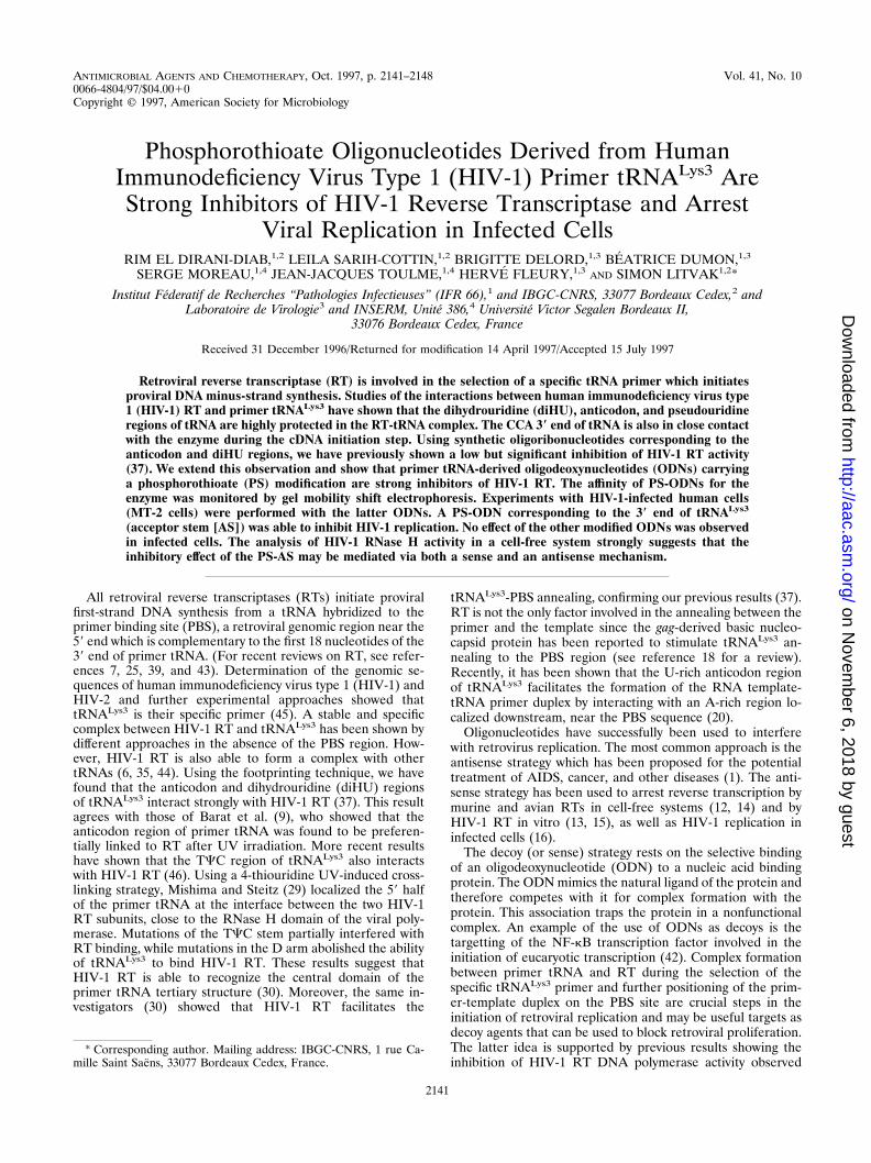
PS-ASVal may act as a good inhibitor of RT, even though itssequence is not related to that of the natural HIV-1 primer(Table 1). The interaction between HIV-1 RT and this PS-ODN was compared with that of HIV-1 RT and PS-ASLys3.Figure 6 shows that binding of PS-ASVal to HIV-1 RT isweaker compared with that of PS-ASLys3. A similar decreasedinteraction was observed with the PS-ODN derived from theacceptor stem lacking the terminal CCA (data not shown).These results are in agreement with the lower level of inhibi-tion of in vitro DNA synthesis by HIV-1 RT in the presence ofthese PS-ODNs. To demonstrate that the retarded bands arethe result of binding of the PS-ASLys3 to the tRNA bindingregion of HIV-1 RT, we performed competition experimentswith tRNALys3. A gel mobility shift assay was performed withlabeled tRNALys3 and RT in the presence of increasingamounts of either unlabeled tRNALys3 or PS-ASLys3. The re-tarded labeled tRNA was displaced by tRNALys3 as well asbeing displaced by PS-ASLys3, showing that it is able to com-pete for the tRNA site of HIV-1 RT.
The high affinity between PS-ASLys3 and HIV-1 RT was alsoobserved by protein fluorescence quenching. Excitation wasperformed at 295 nm and emission was measured from 300 to
420 nm. By increasing the concentrations of PS-ASLys3, weshowed an important quenching effect of the enzyme fluores-cence, while the PO-ODN (at between 0 and 5 mM) with thesame sequence gave no quenching effect under the same con-ditions (data not shown).
Effect of phosphorothioate oligonucleotides on cells chron-ically infected with HIV-1. The effects of PS-ODNs on theinfection of MT-2 cells by HIV-1 were followed by using dif-ferent criteria. The cytopathogenic effect was shown by theemergence of giant cells. The antiviral effect of PS agents wasdetermined by their ability to inhibit the cytopathogenic effectin the MT-2 cells infected with the HIV-1/HTLV-IIIB strain aswell as by the detection of RT activity. MT-2 cells were incu-bated with different amounts of the PS-ODN either after orduring viral adsorption.
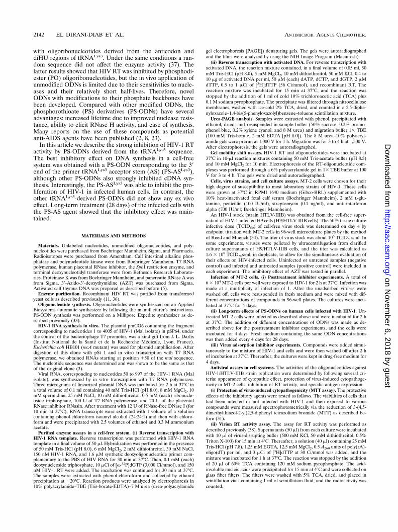
Antiviral activity and cytotoxicity with MT-2 cells. The ef-fects of PS-ODNs on HIV-1 replication were determined bymeasuring the RT activity in the supernatants of infected cells.The only PS-ODN that showed an antiviral effect was thePS-ASLys3, with a 50% inhibitory dose of 400 to 750 nM indifferent experiments (Fig. 7A). PS oligonucleotides 106 andINV-AS gave 50% inhibitory doses four to five times higher,while PS-ODNs corresponding to other regions of primertRNA, like AC, diHU, and pseudo-U stem-loops, did not showany effect under the same experimental conditions. AZT wasused as a reference compound. No cytotoxic effect, determinedby the MTT method, was observed with PS-ODN concentra-tions up to 10 mM (data not shown).
The latter results were obtained when ODNs were addedafter viral adsorption. Preliminary experiments performed byadding the inhibitory agents during viral adsorption led to astronger inhibition by PS-ASLys3, since a 90% inhibition of RTactivity was obtained when a concentration of about 100 nMwas used (data not shown).
The viabilities of HIV-1-infected or -noninfected cells incu-bated with different concentrations of PS-ODN were deter-mined spectrophotometrically as described by Pauwels et al.(31). Control experiments were done in parallel with AZT. Asindicated in Fig. 7B, when compared with the value obtainedwith uninfected cells, PS-ASLys3 was able to inhibit HIV-1proliferation, and this was followed by an increase in cell via-bility. At 0.5 mM there was a significant inhibition and at 1 mMthe cytopathogenic effect was abolished to the same extent asit was with AZT. A very slight inhibitory effect was obtainedwith PS-AC at 1 mM. Other PS-ODNs (diHU, pseudo-U)showed no inhibitory effect under these conditions.
Although PS-ODNs are very effective in vitro inhibitors ofreverse transcription, these compounds may exert their cyto-protective effects in part by interfering with the binding andadsorption of HIV-1 to the target cells (40, 41). Stein et al. (40)have shown that PS-ODNs may bind to the third variable loopdomain (V3) of HIV gp120, competing with the binding of thisdomain to the CD4 receptor. It is important to point out thatthe latter results were obtained with an IC50 of approximately30 mM, a concentration much higher than those used in thiswork (41). Although we cannot exclude the possibility that thestrong inhibitory effect described in this report may be relatedin part to decreased viral adsorption, the lack of effect ofseveral control PS-ODNs on HIV-1-infected human cell cul-tures and the specific effect of PS-ASLys3 point to an effect ofthe latter modified ODN on cDNA synthesis.
Long-term inhibition of HIV proliferation in infected hu-man cells. Several anti-HIV drugs including some PS-ODNshave been shown to be effective in short-term infection assaysbut were not able to suppress virus replication in long-termcultures (23). However, a 25-mer antisense PS-ODN targeted
FIG. 6. Gel mobility shift of PS-AS corresponding to tRNAVal and tRNALys3
in the presence of HIV-1 RT. Assays were done as described in the legend to Fig.5 for labeled ODNs ASVal (lanes 1 to 5) and ASLys3 (lanes 6 to 10) withincreasing concentrations of RT: no RT (lanes 1 and 6) and 50 nM (lanes 2 and7), 100 nM (lanes 3 and 8), 200 nM (lanes 4 and 9), and 400 nM (lanes 5 and 10)RT.
VOL. 41, 1997 HIV-1 REVERSE TRANSCRIPTASE INHIBITORS 2145
on Novem
ber 6, 2018 by guesthttp://aac.asm
.org/D
ownloaded from
to the gag mRNA region of HIV-1 (GEM-91) inhibited HIV-1replication in a sequence- and dose-dependent manner duringa 28-day treatment of an HIV-1-infected T-cell line (24).
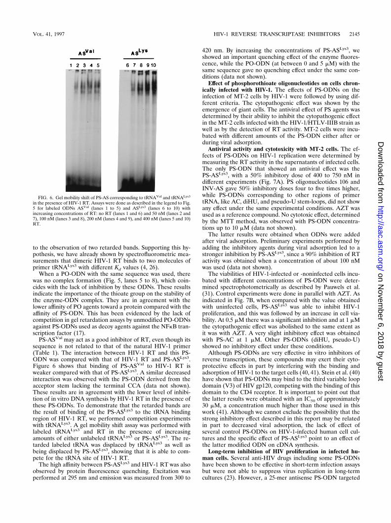
To evaluate the long-term efficacy of the PS-ASLys, infectedMT-2 cells were maintained in the presence of either PS-ASLys, two control PS-ODNs (mismatched oligonucleotide 101and scrambled oligonucleotide 106), or AZT for 28 days (Fig.8). In the presence of PS-ODNs 101 and 106, a transient
suppression of HIV replication was observed. Treatment with1 mM AZT or 2.5 mM PS-AS showed similar efficiencies: bothcompounds completely blocked HIV-1 replication for up to 28days. In contrast, lower doses (1.25 mM PS-AS or 0.2 mMAZT) failed to prevent virus replication after 7 and 24 days,respectively.
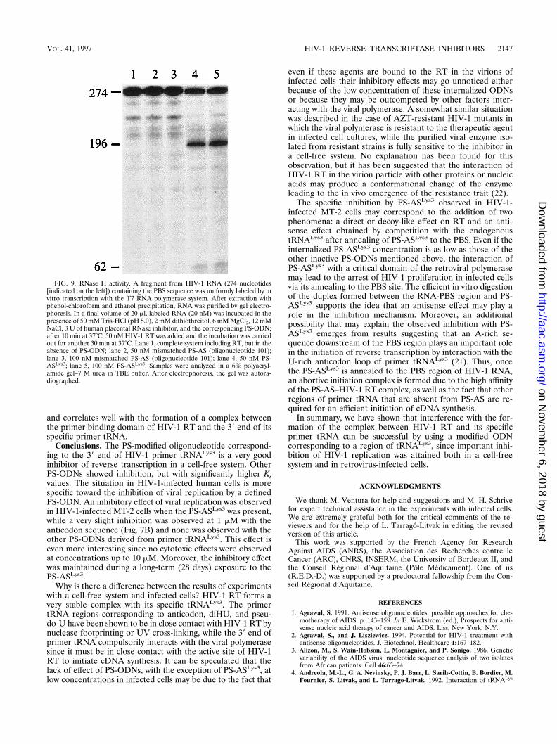
Inhibition by AS in infected cells may be explained by ad-dition of a decoy and an antisense effect. The fact that thePS-ASLys3 was the only primer tRNA-derived fragment to in-hibit HIV-1 proliferation in infected cells, while the othertRNA-derived PS-ODNs were strong inhibitors of cDNA syn-thesis in a cell-free system, was a surprising result. A possibleexplanation is that the PS-ASLys3 may act both directly on theviral polymerase (like other PS-ODNs) and by annealing to thePBS sequence to compete with the natural tRNA primer forthe initiation of DNA synthesis. Moreover, the annealing ofthe PS-ASLys3 to the PBS region would provide a substrate forthe retroviral RNase H activity. To address the question ofwhether a duplex formed by PS-ODN and HIV-1 RNA canelicit the RNase H activity associated with HIV-1 RT, differentPS-ODNs were incubated with retroviral RNase H. Figure 9presents the results obtained with two duplexes: PS-ASLys3 andmismatched PS-AS oligonucleotide 101. Only in the case ofPS-ASLys3 did we observe RNA hydrolysis due to RNase Hactivity. After incubation of the duplex with the enzyme, twofragments with the expected lengths were obtained (Fig. 9,lanes 4 and 5). The other PS-ODNs tested, like PS-AS oligo-nucleotide 101, gave no digestion product, as indicated in Fig.9, lanes 2 and 3. A control incubation was done in the presenceof the labeled RNA and RT to show the absence of contami-nating RNases that could degrade the RNA (Fig. 9, lane 1).
These results strongly suggest that at low ODN concentra-tions, viral proliferation may be selectively inhibited by thePS-AS, at least in part, by specific degradation of the viralRNA by the retroviral RNase H-associated activity. In additionto this antisense effect, the strong inhibition of cDNA synthesisby PS-ASLys ODN when using templates devoid of the PBSsequence [poly(rA)-oligo(dT) or activated DNA] supports adirect inhibitory effect of the PS-ODN on the viral polymerase
FIG. 7. Antiviral activities of PS-ODNs in MT-2-infected cells. Cells were infected with HIV-1/HTLV-IIIB strain and after 2 h of adsorption were incubated in thepresence of serial dilutions of ODNs and AZT. (A) Virus production was determined by measuring RT activity in culture supernatants as a function of increasingconcentrations of ODNs. ■, AC, pseudo-U, and diHU (DHU); 3, ASLys3; h, AZT. (B) The protective effect (or cell viability) produced by PS-ODNs was tested bythe MTT assay as described in Materials and Methods. Increasing concentrations of each compound, including AZT, were used, as follows: 62.5, 125, 250, 500, and1,000 nM. OD530 nm, optical density at 530 nm. For the bars labeled 0, results are for uninfected (■) and infected (1) cells.
FIG. 8. Long-term inhibition of HIV-1/HTLV-IIIB strain replication (28days) by PS-ODNs and AZT in MT-2 cells. Experimental conditions are de-scribed in Materials and Methods. ‚, AS, 2.5 mM; Œ, AS 1.25 mM; E, oligonu-cleotide 101, 2.5 mM; F, oligonucleotide 101, 1.25 mM; �, oligonucleotide 106,2.5 mM; ƒ, oligonucleotide 106, 1.25 mM; ■, AZT, 1 mM; h, AZT, 0.2 mM.
2146 EL DIRANI-DIAB ET AL. ANTIMICROB. AGENTS CHEMOTHER.
on Novem
ber 6, 2018 by guesthttp://aac.asm
.org/D
ownloaded from
and correlates well with the formation of a complex betweenthe primer binding domain of HIV-1 RT and the 39 end of itsspecific primer tRNA.
Conclusions. The PS-modified oligonucleotide correspond-ing to the 39 end of HIV-1 primer tRNALys3 is a very goodinhibitor of reverse transcription in a cell-free system. OtherPS-ODNs showed inhibition, but with significantly higher Kivalues. The situation in HIV-1-infected human cells is morespecific toward the inhibition of viral replication by a definedPS-ODN. An inhibitory effect of viral replication was observedin HIV-1-infected MT-2 cells when the PS-ASLys3 was present,while a very slight inhibition was observed at 1 mM with theanticodon sequence (Fig. 7B) and none was observed with theother PS-ODNs derived from primer tRNALys3. This effect iseven more interesting since no cytotoxic effects were observedat concentrations up to 10 mM. Moreover, the inhibitory effectwas maintained during a long-term (28 days) exposure to thePS-ASLys3.
Why is there a difference between the results of experimentswith a cell-free system and infected cells? HIV-1 RT forms avery stable complex with its specific tRNALys3. The primertRNA regions corresponding to anticodon, diHU, and pseu-do-U have been shown to be in close contact with HIV-1 RT bynuclease footprinting or UV cross-linking, while the 39 end ofprimer tRNA compulsorily interacts with the viral polymerasesince it must be in close contact with the active site of HIV-1RT to initiate cDNA synthesis. It can be speculated that thelack of effect of PS-ODNs, with the exception of PS-ASLys3, atlow concentrations in infected cells may be due to the fact that
even if these agents are bound to the RT in the virions ofinfected cells their inhibitory effects may go unnoticed eitherbecause of the low concentration of these internalized ODNsor because they may be outcompeted by other factors inter-acting with the viral polymerase. A somewhat similar situationwas described in the case of AZT-resistant HIV-1 mutants inwhich the viral polymerase is resistant to the therapeutic agentin infected cell cultures, while the purified viral enzyme iso-lated from resistant strains is fully sensitive to the inhibitor ina cell-free system. No explanation has been found for thisobservation, but it has been suggested that the interaction ofHIV-1 RT in the virion particle with other proteins or nucleicacids may produce a conformational change of the enzymeleading to the in vivo emergence of the resistance trait (22).
The specific inhibition by PS-ASLys3 observed in HIV-1-infected MT-2 cells may correspond to the addition of twophenomena: a direct or decoy-like effect on RT and an anti-sense effect obtained by competition with the endogenoustRNALys3 after annealing of PS-ASLys3 to the PBS. Even if theinternalized PS-ASLys3 concentration is as low as those of theother inactive PS-ODNs mentioned above, the interaction ofPS-ASLys3 with a critical domain of the retroviral polymerasemay lead to the arrest of HIV-1 proliferation in infected cellsvia its annealing to the PBS site. The efficient in vitro digestionof the duplex formed between the RNA-PBS region and PS-ASLys3 supports the idea that an antisense effect may play arole in the inhibition mechanism. Moreover, an additionalpossibility that may explain the observed inhibition with PS-ASLys3 emerges from results suggesting that an A-rich se-quence downstream of the PBS region plays an important rolein the initiation of reverse transcription by interaction with theU-rich anticodon loop of primer tRNALys3 (21). Thus, oncethe PS-ASLys3 is annealed to the PBS region of HIV-1 RNA,an abortive initiation complex is formed due to the high affinityof the PS-AS–HIV-1 RT complex, as well as the fact that otherregions of primer tRNA that are absent from PS-AS are re-quired for an efficient initiation of cDNA synthesis.
In summary, we have shown that interference with the for-mation of the complex between HIV-1 RT and its specificprimer tRNA can be successful by using a modified ODNcorresponding to a region of tRNALys3, since important inhi-bition of HIV-1 replication was attained both in a cell-freesystem and in retrovirus-infected cells.
ACKNOWLEDGMENTS
We thank M. Ventura for help and suggestions and M. H. Schrivefor expert technical assistance in the experiments with infected cells.We are extremely grateful both for the critical comments of the re-viewers and for the help of L. Tarrago-Litvak in editing the revisedversion of this article.
This work was supported by the French Agency for ResearchAgainst AIDS (ANRS), the Association des Recherches contre leCancer (ARC), CNRS, INSERM, the University of Bordeaux II, andthe Conseil Regional d’Aquitaine (Pole Medicament). One of us(R.E.D.-D.) was supported by a predoctoral fellowship from the Con-seil Regional d’Aquitaine.
REFERENCES
1. Agrawal, S. 1991. Antisense oligonucleotides: possible approaches for che-motherapy of AIDS, p. 143–159. In E. Wickstrom (ed.), Prospects for anti-sense nucleic acid therapy of cancer and AIDS. Liss, New York, N.Y.
2. Agrawal, S., and J. Lisziewicz. 1994. Potential for HIV-1 treatment withantisense oligonucleotides. J. Biotechnol. Healthcare 1:167–182.
3. Alizon, M., S. Wain-Hobson, L. Montagnier, and P. Sonigo. 1986. Geneticvariability of the AIDS virus: nucleotide sequence analysis of two isolatesfrom African patients. Cell 46:63–74.
4. Andreola, M.-L., G. A. Nevinsky, P. J. Barr, L. Sarih-Cottin, B. Bordier, M.Fournier, S. Litvak, and L. Tarrago-Litvak. 1992. Interaction of tRNALys
FIG. 9. RNase H activity. A fragment from HIV-1 RNA (274 nucleotides[indicated on the left]) containing the PBS sequence was uniformly labeled by invitro transcription with the T7 RNA polymerase system. After extraction withphenol-chloroform and ethanol precipitation, RNA was purified by gel electro-phoresis. In a final volume of 20 ml, labeled RNA (20 nM) was incubated in thepresence of 50 mM Tris-HCl (pH 8.0), 2 mM dithiothreitol, 6 mM MgCl2, 12 mMNaCl, 3 U of human placental RNase inhibitor, and the corresponding PS-ODN;after 10 min at 37°C, 50 nM HIV-1 RT was added and the incubation was carriedout for another 30 min at 37°C. Lane 1, complete system including RT, but in theabsence of PS-ODN; lane 2, 50 nM mismatched PS-AS (oligonucleotide 101);lane 3, 100 nM mismatched PS-AS (oligonucleotide 101); lane 4, 50 nM PS-ASLys3; lane 5, 100 nM PS-ASLys3. Samples were analyzed in a 6% polyacryl-amide gel–7 M urea in TBE buffer. After electrophoresis, the gel was autora-diographed.
VOL. 41, 1997 HIV-1 REVERSE TRANSCRIPTASE INHIBITORS 2147
on Novem
ber 6, 2018 by guesthttp://aac.asm
.org/D
ownloaded from

with the p66/p66 form of HIV-1 reverse transcriptase stimulates DNA poly-merase and ribonuclease H activities. J. Biol. Chem. 267:19356–19362.
5. Aposhian, H. V., and A. Kornberg. 1962. Enzymatic synthesis of DNA. Thepolymerase formed after T2 bacteriophage infection of E. coli: a new en-zyme. J. Biol. Chem. 237:519–526.
6. Arion, D., R. Harada, X. Li, M. A. Wainberg, and M. A. Parniak. 1996.HIV-1 reverse transcriptase shows no specificity for the binding of primertRNALys3. Biochem. Biophys. Res. Commun. 225:839–843.
7. Arts, E. A., and M. A. Wainberg. 1996. Human immunodeficiency virus type1 reverse transcriptase and early events in reverse transcription. Adv. VirusRes. 46:97–163.
8. Azad, R. F., V. Brown-Driver, R. W. Buckheit, and K. P. Anderson. 1995.Antiviral activity of phosphorothioate oligonucleotides complementary tohuman cytomegalovirus RNA when used in combination with antiviral nu-cleoside analogs. Antivir. Res. 28:101–111.
9. Barat, C., V. Lullien, O. Schatz, G. Keith, M. T. Nugeyre, F. Gruninger-Leitch, F. Barre-Sinoussi, S. F. J. Le Grice, and J. L. Darlix. 1989. HIV-1reverse transcriptase specifically interacts with the anticodon domain of itscognate primer tRNA. EMBO J. 8:3279–3285.
10. Barat, C., O. Schatz, S. Le Grice, and J.-L. Darlix. 1993. Analysis of theinteractions of HIV-1 replication primer tRNALys3 with nucleocapsid pro-tein and reverse transcriptase. J. Mol. Biol. 231:185–190.
11. Barr, P. J., C. T. Power, H. L. Gibson, and P. A. Luciw. 1987. Expression ofactive HIV reverse transcriptase in Saccharomyces cerevisiae. Bio/Technol-ogy 5:486–489.
12. Boiziau, C., F. Debart, B. Rayner, J.-L. Imbach, and J.-J. Toulme. 1995.Chimeric a-b oligonucleotides as antisense inhibitors of reverse transcrip-tion. FEBS Lett. 361:41–45.
13. Boiziau, C., L. Tarrago-Litvak, N. D. Sinha, S. Moreau, S. Litvak, and J.-J.Toulme. 1996. Antisense oligonucleotides inhibit in vitro cDNA synthesis byHIV-1 reverse transcriptase. Antisense Nucleic Acid Drug Dev. 6:103–109.
14. Boiziau, C., N. T. Thuong, and J.-J. Toulme. 1992. Mechanism of inhibitionof reverse transcription by antisense oligonucleotides. Proc. Natl. Acad. Sci.USA 89:768–772.
15. Bordier, B., C. Helene, P. J. Barr, S. Litvak, and L. Sarih-Cottin. 1992. Invitro effect of antisense oligonucleotides on HIV-1 reverse transcription.Nucleic Acids Res. 20:5999–6006.
16. Bordier, B., M. Perala-Heape, G. Degols, B. Lebleu, S. Litvak, L. Sarih-Cottin, and C. Helene. 1995. Sequence-specific inhibition of HIV reversetranscription by antisense oligonucleotides: comparative study in cell-freeassays and in HIV-infected cells. Proc. Natl. Acad. Sci. USA 92:9383–9387.
17. Brown, D. A., S.-H. Khang, S. M. Gryaznov, L. DeDionisio, O. Heideinreich,S. Sullivan, X. Xu, and M. L. Nerenberg. 1994. Effect of phosphorothioatemodification of oligodeoxynucleotides on specific protein binding. J. Biol.Chem. 269:26801–26805.
18. Darlix, J. L., M. Lapadat-Tapolsky, H. De Rocquigny, and B. P. Roques.1995. First glimpses at structure-function relationships of the nucleocapsidprotein of retroviruses. J. Mol. Biol. 254:523–537.
19. Idriss, H., and A. K. Stammers. 1994. Inhibition of HIV-1 RT by definedtemplate-primer DNA oligonucleotides: effect of template length and bind-ing characteristics. J. Enzyme Inhibition 8:97–112.
20. Isel, C., C. Ehresmann, G. Keith, B. Ehresmann, and R. Marquet. 1995.Initiation of reverse transcriptase of HIV-1: secondary structure of theHIV-1 RNA:tRNALys3 (template/primer) complex. J. Mol. Biol. 247:236–250.
21. Isel, C., R. Marquet, G. Keith, C. Ehresmann, and B. Ehresmann. 1993.Modified nucleotides of tRNALys3 modulate primer/template loop-loop in-teraction in the initiation complex of HIV-1 reverse transcription. J. Biol.Chem. 268:25269–25272.
22. Larder, B. A., and S. D. Kemp. 1989. Multiple mutations in HIV-1 reversetranscriptase confer high level resistance to zidovudine (AZT). Science 246:1155–1158.
23. Lisziewicz, J., D. Sun, V. Metelev, P. Zamecnik, R. C. Gallo, and S. Agrawal.1993. Long-term treatment of HIV-infected cells with antisense oligonucle-otide phosphorothioates. Proc. Natl. Acad. Sci. USA 90:3860–3864.
24. Lisziewicz, J., D. Sun, F. F. Weichold, A. R. Thierry, P. Lusso, J. Tang, R. C.Gallo, and S. Agrawal. 1994. Antisense oligodeoxynucleotide phosphoro-thioate complementary to Gag mRNA blocks replication of HIV-1 in human
peripheral blood cells. Proc. Natl. Acad. Sci. USA 91:7942–7946.25. Litvak, S. 1996. Retroviral reverse transcriptases. RG Landes/Chapman &
Hall, Austin, Tex.26. Litvak, S., L. Sarih-Cottin, M. Fournier, M. Andreola, and L. Tarrago-
Litvak. 1994. Priming of HIV replication by tRNALys3: role of reverse tran-scriptase. Trends Biochem. Sci. 19:114–118.
27. Mak, J., M. Jiang, M. A. Wainberg, M.-L. Hammarskjold, D. Rekosh, and L.Kleiman. 1994. Role of Pr160gag-pol in mediating the selective incorporationof tRNALys into HIV-1 particles. J. Virol. 68:2065–2072.
28. Marshall, M. S., and M. H. Caruthers. 1993. Phosphorodithioate DNA as apotential therapeutic drug. Science 259:1564–1570.
29. Mishima, Y., and J. A. Steitz. 1995. Site-specific cross-linking of 4-thiouri-dine-modified human tRNALys3 to reverse transcriptase from HIV-1.EMBO J. 14:2679–2687.
30. Oude Essink, B. B., A. T. Das, and B. Berkhout. 1995. Structural require-ments for the binding of tRNALys3 to reverse transcriptase of the HIV-1.J. Biol. Chem. 270:23867–23874.
31. Pauwels, R., J. Balzarini, and M. Baba. 1988. Rapid and automated tetra-zolium-based colorimetric assay for the detection of anti-HIV-compounds.J. Virol. Methods 20:309–321.
32. Raba, M., K. Limbourg, M. Burghagen, J. R. Katz, M. Simsek, J. E. Heck-mann, U. L. RajBhandary, and H. J. Gross. 1979. Nucleotide sequence ofthree isoaccepting lysine tRNALys from rabbit liver and SV40-transformedmouse fibroblasts. Eur. J. Biochem. 97:305–318.
33. Ramazeilles, C., R. K. Mishra, S. Moreau, E. Pascolo, and J.-J. Toulme.1994. Antisense phosphorothioate oligonucleotides: selective killing of theintracellular parasite Leishmania amazonensis. Proc. Natl. Acad. Sci. USA91:7859–7863.
34. Reed, L. J., and H. A. Muench. 1938. A simple method of estimating fiftypercent of end points. Am. J. Hyg. 27:493–497.
35. Robert, D., M. Sallafranque-Andreola, B. Bordier, L. Sarih-Cottin, L. Tar-rago-Litvak, P. V. Graves, P. J. Barr, M. Fournier, and S. Litvak. 1990.Interactions with tRNALys induce important structural changes in HIV re-verse transcriptase. FEBS Lett. 277:239–242.
36. Sallafranque-Andreola, M., D. Robert, P. J. Barr, M. Fournier, S. Litvak, L.Sarih-Cottin, and L. Tarrago-Litvak. 1989. HIV-1 reverse transcriptase ex-pressed in transformed yeast cells. Eur. J. Biochem. 184:367–374.
37. Sarih-Cottin, L., B. Bordier, K. Musier-Forsyth, M.-L. Andreola, J.-P. Barr,and S. Litvak. 1992. Preferential interaction of HIV reverse transcriptasewith two regions of primer tRNALys as evidenced by footprinting studies andinhibition with synthetic oligoribonucleotides. J. Mol. Biol. 226:1–6.
38. Schwartz, O., Y. Henin, V. Marechal, and L. Montagnier. 1988. A rapid andsimple colorimetric test for the study of anti-HIV agents. AIDS Res. Hum.Retroviruses 4:441–448.
39. Skalka, A. M., and S. P. Goff. 1993. Reverse transcriptase. Cold SpringHarbor Laboratory Press, Cold Spring Harbor, N.Y.
40. Stein, C. A., A. M. Cleary, L. Yakubov, and S. Lederman. 1993. Phosphoro-thioate oligodeoxynucleotides bind to the third variable loop domain (v3) ofHIV-1 gp 120. Antisense Res. Dev. 3:19–31.
41. Stein, C. A., L. M. Neckers, B. C. Nair, S. Mumbauer, G. Hoke, and R. Pal.1991. Phosphorothioate oligodeoxycytidine interferes with binding of HIV-1gp120 to CD4. J. Acquired Immune Defic. Syndr. 4:686–693.
42. Tanaka, H., P. Vickart, J.-R. Bertrand, B. Rayner, F. Morvan, J.-L. Imbach,D. Paulin, and C. Malvy. 1994. Sequence-specific interaction of a b-ano-meric double-stranded DNA with the p50 subunit of NFkB: application tothe decoy approach. Nucleic Acids Res. 22:3069–3074.
43. Tarrago-Litvak, L., M.-L. Andreola, G. A. Nevinsky, L. Sarith-Cottin, and S.Litvak. 1994. The reverse transcriptase of HIV-1: from enzymology to ther-apeutic intervention. FASEB J. 8:497–503.
44. Thrall, S. H., J. Reinstein, B. M. Worhl, and R. S. Goody. 1996. Evaluationof HIV-1 reverse transcriptase primer binding tRNA binding by fluorescencespectroscopy: specificity and comparison to primer/template binding. Bio-chemistry 35:4609–4618.
45. Wain-Hobson, S., J. P. Vartanian, M. Henry, O. Danos, S. Cole, and M.Alizon. 1985. Nucleotide sequence of the AIDS virus, LAV. Cell 40:9–17.
46. Wohrl, B. M., B. Ehresmann, G. Keith, and S. F. J. Le Grice. 1993. Nucleasefoot-printing of HIV RT/tRNALys3 complexes. J. Biol. Chem. 268:13617–13624.
2148 EL DIRANI-DIAB ET AL. ANTIMICROB. AGENTS CHEMOTHER.
on Novem
ber 6, 2018 by guesthttp://aac.asm
.org/D
ownloaded from